REVIEW Open Access Alternative polyadenylation: methods, mechanism, function, and role in cancer Yi Zhang 1 , Lian Liu 1 , Qiongzi Qiu 2 , Qing Zhou 2 , Jinwang Ding 3* , Yan Lu 2,5* and Pengyuan Liu 1,4,5* Abstract Occurring in over 60% of human genes, alternative polyadenylation (APA) results in numerous transcripts with differing 3’ends, thus greatly expanding the diversity of mRNAs and of proteins derived from a single gene. As a key molecular mechanism, APA is involved in various gene regulation steps including mRNA maturation, mRNA stability, cellular RNA decay, and protein diversification. APA is frequently dysregulated in cancers leading to changes in oncogenes and tumor suppressor gene expressions. Recent studies have revealed various APA regulatory mechanisms that promote the development and progression of a number of human diseases, including cancer. Here, we provide an overview of four types of APA and their impacts on gene regulation. We focus particularly on the interaction of APA with microRNAs, RNA binding proteins and other related factors, the core pre- mRNA 3’end processing complex, and 3’UTR length change. We also describe next-generation sequencing methods and computational tools for use in poly(A) signal detection and APA repositories and databases. Finally, we summarize the current understanding of APA in cancer and provide our vision for future APA related research. Keywords: Alternative polyadenylation (APA), 3’UTR, Poly(A) sites usage, Cancer Background The maturation of nascent RNAs is a key step in tran- scription. For mRNA, the maturation of messenger RNA precursors (pre-mRNAs), involving the processing of 3’termini, is critical for mRNA function and stability [1]. In the processing of the 3’termini, the 3’end of nascent mRNA is cleaved, followed by addition of a poly(A) tail (i.e., polyadenylation). Polyadenylation protects the pre- mRNA from enzymatic degradation and facilitates nuclear export and translation [2]. The processing of poly(A) tail addition and length control of the poly(A) tail is modulated by polyadenylation polymerase and polyadenylation specificity factors [3]. Both cleavage and polyadenylation occur at polyadenylation sites (PASs) which are located within the 3’untranslated regions (3’UTRs), introns, or internal exons [4, 5]. Most eukaryotic genes contain multiple PASs. A conserved hexameric sequence AAUAAA [6], occurring upstream of the PASs, contains the most important signal (i.e., poly(A) signal) of pre-mRNA cleavage and polyadenyla- tion. Both this canonical poly(A) signal and the PASs are widespread in eukaryotic mRNA. Cleavage or polyadeny- lation can generate transcript isoforms which differ in their coding regions or 3’UTRs [7]. This phenomenon, which gives rise to various transcript isoforms, is termed as alternative polyadenylation (APA). Recent studies have shown that the global regulation of APA and the resulting distinct transcripts are in- volved in various aspects of tumorigenesis and cancer © The Author(s). 2021 Open Access This article is licensed under a Creative Commons Attribution 4.0 International License, which permits use, sharing, adaptation, distribution and reproduction in any medium or format, as long as you give appropriate credit to the original author(s) and the source, provide a link to the Creative Commons licence, and indicate if changes were made. The images or other third party material in this article are included in the article's Creative Commons licence, unless indicated otherwise in a credit line to the material. If material is not included in the article's Creative Commons licence and your intended use is not permitted by statutory regulation or exceeds the permitted use, you will need to obtain permission directly from the copyright holder. To view a copy of this licence, visit http://creativecommons.org/licenses/by/4.0/. The Creative Commons Public Domain Dedication waiver (http://creativecommons.org/publicdomain/zero/1.0/) applies to the data made available in this article, unless otherwise stated in a credit line to the data. * Correspondence: [email protected]; [email protected]; [email protected] 3 Department of Head and Neck Surgery, Cancer Hospital of the University of Chinese Academy of Sciences, Zhejiang Cancer Hospital, Key Laboratory of Head & Neck Cancer Translational Research of Zhejiang Province, Hangzhou 310022, Zhejiang, China 2 Center for Uterine Cancer Diagnosis & Therapy Research of Zhejiang Province, Women’s Reproductive Health Key Laboratory of Zhejiang Province, Department of Gynecologic Oncology, Women’s Hospital and Institute of Translational Medicine, Zhejiang University School of Medicine, Hangzhou 310006, Zhejiang, China 1 Department of Respiratory Medicine, Sir Run Run Shaw Hospital and Institute of Translational Medicine, Zhejiang University School of Medicine, Hangzhou 310016, Zhejiang, China Full list of author information is available at the end of the article Zhang et al. Journal of Experimental & Clinical Cancer Research (2021) 40:51 https://doi.org/10.1186/s13046-021-01852-7

Welcome message from author
This document is posted to help you gain knowledge. Please leave a comment to let me know what you think about it! Share it to your friends and learn new things together.
Transcript

REVIEW Open Access
Alternative polyadenylation: methods,mechanism, function, and role in cancerYi Zhang1, Lian Liu1, Qiongzi Qiu2, Qing Zhou2, Jinwang Ding3*, Yan Lu2,5* and Pengyuan Liu1,4,5*
Abstract
Occurring in over 60% of human genes, alternative polyadenylation (APA) results in numerous transcripts withdiffering 3’ends, thus greatly expanding the diversity of mRNAs and of proteins derived from a single gene. As akey molecular mechanism, APA is involved in various gene regulation steps including mRNA maturation, mRNAstability, cellular RNA decay, and protein diversification. APA is frequently dysregulated in cancers leading tochanges in oncogenes and tumor suppressor gene expressions. Recent studies have revealed various APAregulatory mechanisms that promote the development and progression of a number of human diseases, includingcancer. Here, we provide an overview of four types of APA and their impacts on gene regulation. We focusparticularly on the interaction of APA with microRNAs, RNA binding proteins and other related factors, the core pre-mRNA 3’end processing complex, and 3’UTR length change. We also describe next-generation sequencing methodsand computational tools for use in poly(A) signal detection and APA repositories and databases. Finally, wesummarize the current understanding of APA in cancer and provide our vision for future APA related research.
Keywords: Alternative polyadenylation (APA), 3’UTR, Poly(A) sites usage, Cancer
BackgroundThe maturation of nascent RNAs is a key step in tran-scription. For mRNA, the maturation of messenger RNAprecursors (pre-mRNAs), involving the processing of3’termini, is critical for mRNA function and stability [1].In the processing of the 3’termini, the 3’end of nascentmRNA is cleaved, followed by addition of a poly(A) tail(i.e., polyadenylation). Polyadenylation protects the pre-mRNA from enzymatic degradation and facilitates
nuclear export and translation [2]. The processing ofpoly(A) tail addition and length control of the poly(A)tail is modulated by polyadenylation polymerase andpolyadenylation specificity factors [3]. Both cleavage andpolyadenylation occur at polyadenylation sites (PASs)which are located within the 3’untranslated regions(3’UTRs), introns, or internal exons [4, 5]. Mosteukaryotic genes contain multiple PASs. A conservedhexameric sequence AAUAAA [6], occurring upstreamof the PASs, contains the most important signal (i.e.,poly(A) signal) of pre-mRNA cleavage and polyadenyla-tion. Both this canonical poly(A) signal and the PASs arewidespread in eukaryotic mRNA. Cleavage or polyadeny-lation can generate transcript isoforms which differ intheir coding regions or 3’UTRs [7]. This phenomenon,which gives rise to various transcript isoforms, is termedas alternative polyadenylation (APA).Recent studies have shown that the global regulation
of APA and the resulting distinct transcripts are in-volved in various aspects of tumorigenesis and cancer
© The Author(s). 2021 Open Access This article is licensed under a Creative Commons Attribution 4.0 International License,which permits use, sharing, adaptation, distribution and reproduction in any medium or format, as long as you giveappropriate credit to the original author(s) and the source, provide a link to the Creative Commons licence, and indicate ifchanges were made. The images or other third party material in this article are included in the article's Creative Commonslicence, unless indicated otherwise in a credit line to the material. If material is not included in the article's Creative Commonslicence and your intended use is not permitted by statutory regulation or exceeds the permitted use, you will need to obtainpermission directly from the copyright holder. To view a copy of this licence, visit http://creativecommons.org/licenses/by/4.0/.The Creative Commons Public Domain Dedication waiver (http://creativecommons.org/publicdomain/zero/1.0/) applies to thedata made available in this article, unless otherwise stated in a credit line to the data.
* Correspondence: [email protected]; [email protected]; [email protected] of Head and Neck Surgery, Cancer Hospital of the University ofChinese Academy of Sciences, Zhejiang Cancer Hospital, Key Laboratory ofHead & Neck Cancer Translational Research of Zhejiang Province, Hangzhou310022, Zhejiang, China2Center for Uterine Cancer Diagnosis & Therapy Research of ZhejiangProvince, Women’s Reproductive Health Key Laboratory of Zhejiang Province,Department of Gynecologic Oncology, Women’s Hospital and Institute ofTranslational Medicine, Zhejiang University School of Medicine, Hangzhou310006, Zhejiang, China1Department of Respiratory Medicine, Sir Run Run Shaw Hospital andInstitute of Translational Medicine, Zhejiang University School of Medicine,Hangzhou 310016, Zhejiang, ChinaFull list of author information is available at the end of the article
Zhang et al. Journal of Experimental & Clinical Cancer Research (2021) 40:51 https://doi.org/10.1186/s13046-021-01852-7

progression [8]. Differential PAS usage plays a key rolein cell proliferation and gene versatility [9, 10]. For ex-ample, cell division cycle 6 (CDC6) is a critical gene inDNA replication. CDC6 can limit the rate of S-phaseentry and regulate the initiation of DNA replication inmammalian cells [11]. CDC6 is upregulated in multiplehuman cancers and can inhibit the tumor suppressorsp15INK4b, p16INK4a, and ARF [12]. Estrogen can inducethe shortening of the 3’UTR of CDC6, and it has beenobserved that the resultant truncated isoforms can leadto aberrant expression of CDC6 via its avoidance ofmiRNA-mediated repression [13]. Such a 3’UTR lengthchange does not simply occur in isolation on a certaingene but can be part of more global events in tumors orin certain other physiological conditions and contexts.Compared with normal cells, transcript isoforms in pro-liferated cancer cells are noted as having a tendency tobe shortened [14], while transcript isoforms in senescentcells tend to be lengthened [15].This review provides a general summary of four types
of APA and their effects on gene regulation. We focuson APA regulatory mechanisms, including the inter-action of APA with microRNAs, RNA binding proteinsand other related factors, the core pre-mRNA 3’end pro-cessing complex, and 3’UTR length change. We alsointroduce high-throughput sequencing methods andcomputational tools for poly(A) signal detection and re-lated corresponding additions to APA databases. Finally,we summarize recent research on APA in cancer andprovide our vision for future APA related research.
APA categoriesAPA is a phenomenon that generates various transcriptisoforms with different 3’termini from the same gene. Itis observed in all eukaryotes species as an importantmechanism of gene regulation. APA was first discoveredin 1980 in the genes encoding immunoglobulin M (IgM)and dihydrofolate reductase (DHFR) [16, 17]. Over thenext two decades, about 95 genes were identified as hav-ing APAs [18]. With the advent of next-generation se-quencing (NGS) things accelerated greatly and by nowmore than two-thirds of human genes and one-third ofmouse genes have been reported with more than onePAS containing a hexameric consensus motif AAUAAA,i.e., the canonical poly(A) signal [7, 19–22]. It is worthnoting that the sequence AAUAAA (termed as poly(A)signal or pA signal) is different from the polyadenylationsite (termed as poly(A) site or PAS). The poly(A) signallocates in upstream of the PAS. Undergoing diversemodifications, precursor RNAs with multiple PASs forminto distinct isoforms. These can be divided into twosubtypes according to the locations of the PASs (Fig. 1).One class of APAs are tandem 3’UTR-APAs, also knownas 3’UTR-APAs, which contain two or more cleavage
PASs in the 3’UTR and which generate various tran-scripts with different 3’UTR lengths. Tandem 3’UTR-APAs have a high number of incidences and have im-portant impacts on mRNA stability, translation effi-ciency, nuclear export, cellular localization andlocalization of encoded protein. The other class of APAfurther changes the potential for protein-coding. Thisclass occurs upstream of the last exon and thus istermed as upstream region APA (UR-APAs) [5, 23, 24].It contains three subclasses, specifically, “alternative ter-minal exon APA” or “splicing APA” which generatestranscripts with distinct 3’UTR sequences and encodesproteins with altered C-terminal amino acids; “In-tronic APA” that occurs in an intron; and “Internalexon APA”, being the small fraction that appears ininternal exons. These subtypes are involved in thecell-cycle and cell differentiation in many ways, suchas in aspects of protein diversification and the inhib-ition of gene expression [25, 26].
Tandem 3’UTR-APAsTandem 3’UTR-APA occurs in the 3’UTR and canchange the structure of 3’UTRs or generate various iso-forms of RNAs with different 3’UTR lengths (Fig. 1a).The longer the length of the 3’UTR, the more bindingloci occur for microRNAs (miRNAs) and RNA-bindingproteins (RBPs), and the more alternative RNA second-ary structures are exhibited [4, 25, 27–29]. Like othercis-elements, these binding loci or RNA secondary struc-tures can be specifically recognized by post-transcriptional factors and play important roles in generegulation. Multiple mechanisms of gene regulation by3’UTR-APA have been revealed. One major example ismiRNA-mediated gene regulation at the 3’UTR of RNAs.Since 3’UTR-APA generates various 3’UTRs of differentlengths, the number of miRNA binding sites in theseisoforms is also different. The ability of miRNAs todown-regulate target genes varies with the number ofbinding sites, thereby affecting the stability and thetranslation of mRNAs [30].Among these mechanisms, some are relevant to the
progression and invasion of tumors. For example, GALNT5 uaRNA (a UTR-associated RNA) is a lncRNA derivedfrom the 3’UTR of GALNT5. It promotes the prolifera-tion of gastric cancer by interacting with the molecularchaperone HSP90 [31]. miRNA-200a reduces the level ofPTEN expression by directly binding the 3’UTR ofPTEN, thereby promoting the invasion of ovarian cancercells [32]. These studies indicate that 3’UTR plays an im-portant role in post-transcriptional gene regulation.
UR-APAUR-APA occurs upstream of the last exon, a location farremoved from the 3’UTR. It can be further divided into
Zhang et al. Journal of Experimental & Clinical Cancer Research (2021) 40:51 Page 2 of 19

Fig. 1 (See legend on next page.)
Zhang et al. Journal of Experimental & Clinical Cancer Research (2021) 40:51 Page 3 of 19

three subclasses, namely alternative terminal exon APA, in-tronic APA, and internal exon APA. Alternative terminalexon APA occurs as a consequence of alternative splicing(Fig. 1b) [23, 24]. Both intronic and internal exon APA arecomponents of mRNA decay pathways, including the non-stop decay pathway and nonsense-mediated decay pathway(Fig. 1c and d) [33–36]. Similar to 3’UTR-APA, UR-APA isalso involved in many aspects of gene regulation.
APA functionsInteraction with miRNAmiRNAs are a type of trans-acting element that can bindto the 3’UTR of mRNA and regulate gene expression at apost-transcriptional level [37–40]. They regulate the trans-lation and stability of their binding mRNAs through trans-lation inhibition and degradation of mRNA [41, 42]. Dueto the existence of APAs in the 3’UTR, various isoformswith different 3′ termini are generated [43]. This mechan-ism can change which miRNA binding sites the 3’UTRcontains (Fig. 2a and b). Distinct miRNAs targeting3’UTR-APA were first discovered in cancer cells and acti-vated T cells. Compared with the non-activated T cellsand non-transformed cells, the length of 3’UTR in acti-vated T cells and cancer cells becomes significantly short-ened [44, 45]. Shorter 3’UTRs only possess proximalmiRNA binding sites in male mouse germ cells, whilethose with longer 3’UTRs tend to contain distal miRNAbinding sites [46]. Similarly, in-depth analysis of the3’UTR isoforms of IGF2BP1 found nine functional PASsin human HLF cancer cell lines. Many of them have alsobeen revealed to lack miRNA binding sites in these short-ened isoforms [45]. This demonstrates that different num-bers of miRNA binding sites occur among these 3’UTRisoforms and shows that differential PAS usage can be aclinical indicator for human disease. In addition, the re-duction of miRNA binding sites is not the only conse-quence of 3’UTR shortening. Conserved miRNA bindingsites are also seen to be preferentially enriched upstreamof APA sites. 3’UTR shortening was found to be able toenhance the targeting efficiency of miRNAs that bind up-stream of the proximal PAS [47]. Hence, 3’UTR shorten-ing, resulting from APA, affects not only the number ofmiRNA binding sites within the 3’UTR, but also the tar-geting efficiency of miRNAs.
Interaction with RNA-binding proteinThe interaction between RNA and protein is essentialfor regulating gene expression at the post-transcriptional
level (Fig. 2c and d). As a class of highly evolutionarilyconserved proteins, RBP plays a key role in post-transcriptional gene regulation (PTGR) including aspectsof maturation, stability, transport, and degradation ofcellular RNAs. Most RBPs bind with mRNA and non-coding RNA, of which only ~ 2% are tissue-specific.RBPs are widely expressed and usually show higher ex-pression levels than the average levels of cellular pro-teins [48–50]. The complex formed by RBP and RNA,ribonucleoprotein (RNP), is the major regulator in thePTGR. Defects in RBP function and RNP assembly areimportant causal factors leading to various human dis-eases including cancers. The types of RNA (e.g., mRNA,ribosomal RNA, and tRNA) that are predominantlybound by the RBPs lead to the characteristic phenotypesof these RBP related diseases [51–53].RBPs contain specific RNA-binding domains (RBDs).
These provide preferential selection of binding sites andtargets and interact with RNA through these recognitionregions. These RBDs include the RNA recognition motif(RRM), the K homology domain (KH), DEAD motif,double-stranded RNA-binding motif (DSRM), CCCH tan-dem zinc-finger domain, and Pumilio p-homology andFem-3 mRNA binding factor (PUF) domains [48, 54–56].Through their RRM, KH, and the zinc finger domains, theRBPs recognize Adenylate-undylate-rich elements (AREs),which are embedded in the 3’UTR and are present in 5–8% of human genes. These RBPs are called ARE-RBPs[57]. As in the miRNA binding sites, the altered number ofthe RBP binding motifs (such as AREs or GU-rich elements)caused by 3’UTR-APA can mediate mRNA stability. For ex-ample, the mRNA regulatory protein tristetraprolin (TTP,also known as ZFP36) can recruit the CCR4-NOT complexto the AREs in the 3’UTR of the target gene and then deade-nylate mRNA, thereby destabilizing it. A lack of these AREswill result in an exceptional increase in mRNA expression[58–60]. As for TTP, the K homology splicing regulatoryprotein (KSRP) is another protein involved in mRNA deg-radation. Gherzi et al. showed that KSRP is an essential fac-tor for ARE-directed mRNA decay. The depletion of KSRPresults in the stabilization of several ARE-containing mRNAssuch as TNFα and c-Fos. This stabilization is observedin KSRP-depleted S100 from several cell types, includ-ing Jurkat, HeLa, and HT1080 cells [61]. Furthermore,due to APA, human IFN-regulatory factor 5 (IRF5) hastwo isoforms with different 3’UTRs. The alternative ex-pression levels of these two isoforms can cause systemiclupus erythematosus [62].
(See figure on previous page.)Fig. 1 Categories of APA. a Tandem 3’UTR-APA containing two or more poly(A) sites in the 3’untranslated region. b, c, d UR-APAs occurringupstream of the last exon, therefore termed as an upstream region APA. b Splicing APA (alternative terminal exon APA) possessing a proximalPAS in the last exon and resulting in internal exon skipping. c Intronic APA occurring in the introns. d Internal exon APA generating a 3’UTR-lacking isoform via the PAS usage in the upstream exon
Zhang et al. Journal of Experimental & Clinical Cancer Research (2021) 40:51 Page 4 of 19

As can be seen from the above studies, the interactionbetween RBPs and the 3’UTR is deeply involved inPTGR and mRNA stability. It is often difficult to disas-sociate disease from transcription and translation. Theregulation of RBP-RNA binding is a very importantpathogenic mechanism of disease. For example, cold-inducible RNA binding protein (CIRP, also known asCIRBP or A18 hnRNP) is a stress-induced protein in-volved in cancer. CIRP can bind to the transcripts ofpro-survival genes, which contain RNA signature motifsin their 3’UTRs, and stabilize them. In ectopic mousexenograft models of human breast cancer and melano-mas, CIRP promotes tumor growth by increasing the ex-pression level of HIF-1α. Immunohistochemical analysisshows that CIRP is over expressed in the stroma andhypoxic areas of human tumors [63]. Furthermore, CIRP
can also be transferred from the nucleus to the cytoplasmand bind to the 3’UTR of cyclin E1 mRNA and hTERTmRNA, thereby stabilizing and upregulating them [64].Musashi (MSI) is another RNA binding protein, a medi-ator of a number of critical biological processes relevantto tumor initiation and progression. MSI was observed tobe upregulated in many human cancer types, includingcolorectal, lung, and pancreatic cancers and glioblastomas.MSI regulates cancer invasion and metastasis through theregulation of mRNA stability and translation of proteinsin several essential oncogenic signaling pathways, includ-ing those of NUMB/Notch, PTEN/mTOR, TGFβ/SMAD3, MYC, cMET, and others [65].RBPs and RNAs assemble into a dynamic RNP com-
plex. This plays an important role in RNA maturation,regulation, and transportation. Mutations in the
Fig. 2 APA functions. A schematic diagram illustrating RNA-RBP interaction and RNA-miRNA interaction. a Multiple RBP binding sites and miRNAbinding sites are located in the 3’UTR of RNA. As for the interaction between miRNA and 3’UTR, miRNA usually inhibits and silences the targetRNA. b The scheme of RNA-miRNA interaction. MiRNAs can be firstly transcribed as long primary miRNA (pri-miRNA) transcripts with 5′ cap and3’poly(A) tail by Pol II. Then pri-miRNA is cut by Drosha RNase III and turns into pre-miRNA in the nucleus. Pre-miRNA is delivered out the nucleiand processes into 21-nucleotide-long double-stranded RNAs. One strand combines with AGO proteins to form miRNA-containing RNPs (miRNPs).The miRNP complex binds to the complementary target mRNA and recruits deadenylase to repress translation. c, d RNA-RBP interactions. c ELAVleads to the expression of long 3’UTR isoforms during neurogenesis by inhibiting proximal PAS usage. d TTP recruits the CCR4-NOT complex intothe ARE in the 3’UTR of the target gene and deadenylates the mRNA that causes its instability
Zhang et al. Journal of Experimental & Clinical Cancer Research (2021) 40:51 Page 5 of 19

heterogeneous nuclear RNPs (hnRNPs) cause amyo-trophic lateral sclerosis (ALS) [66, 67]. Survival motorneuron 1 (SMN1) is one component of the small nuclearRNPs (snRNPs) assembly complex. Its loss of functiondirectly affects the spliceosome and leads to spinal mus-cular atrophy [68]. The cyclin-dependent kinase inhibi-tor 1B (CDKN1B) mRNA is destabilized by the synergyof miR-221 and/or miR-222 and Pumilio homolog pro-teins (PUM) [69]. In Drosophila melanogaster,embryonic-lethal abnormal visual protein (ELAV) can berecruited to RNA polymerase II (Pol II) at promoter re-gions with GAGA sequences and then suspend Pol II.ELAV increases the expression of long 3’UTR isoformsduring neurogenesis by inhibiting proximal PAS usage[70, 71]. All these studies indicate that not only that RBPexpression, but also the type of RNA bound by the RBP,are involved in disease pathogenesis. These characteristicphenotypes and RBP factors could be investigated as po-tential novel markers for use in disease diagnosis andprognosis.
Impacts on gene repression and versatilityUR-APA plays an important role in generating truncatedtranscripts. For example, Singh et al. showed that in-tronic APA isoforms, as widely expressed in immunecells and as participants in the development of B cells,lead to the production of truncated proteins lackingfunctional C-terminal domains. Furthermore, the num-ber of intronic APA isoforms is decreased in multiplemyeloma cells. This may contribute to the progressionof multiple myelomas and is a factor associated withshorter progression-free survival [72]. A terminal exoncharacterization (TEC) tool has been developed for theanalysis of RNA-sequencing data in order to identify iso-forms ending at intronic poly(A) sites and to discoverthe prevalence of these APA isoforms [73]. A cleavagestimulation factor subunit named CSTF3 was seen withhighly conserved intronic PASs which could lead to theproduction of severely truncated, probably nonfunc-tional, proteins [74]. This also involved a negative feed-back regulation to reduce the expression of CSTF3 as ahigh expression level could induce the production of thisUR-APA isoform. Similarly, retinoblastoma-binding pro-tein 6 (RBBP6) has an isoform called Iso3, which is pro-duced by the intronic APA of RBBP6. Iso3 isdownregulated in several human cancers and can com-pete with normal RBBP6 for binding to core machinery,thereby inhibiting polyadenylation and regulating APA[75]. The truncated isoforms of Dicer and Forkhead boxN3 (two tumor suppressor proteins), also lack tumorsuppressive ability in tumors [76]. These studies suggestthat truncated protein generation by UR-APA mightrepresent a wide-spread gene inhibition mechanism.
On the other hand, the diversification of protein canalso be a key part of gene versatility. For example, thereare two isoforms of immunoglobulin M (IgM) heavychain mRNA. The longer one, with the distal PAS usagein the 3’end of the third exon, is appropriate formembrane-binding, while the shorter one, with theproximal PAS in a composite terminal exon usage, is in-volved in secretion. Different mRNAs also predominateat different stages of immunocyte development, the lon-ger ones at the lymphocyte stages and the shorter one atthe secretion stages [10]. Another classic case is thecalcitonin-related polypeptide-α gene (CALCA). CALCAhas two transcript isoforms. The one with proximal PASusage contains a skipped terminal exon and encodes theprotein calcitonin. The other one, with distal PAS usage,generates an mRNA encoding calcitonin gene-relatedpeptide 1 (CGRP). The expression of these two isoformsis tissue specific. Calcitonin mRNA is enriched in thethyroid and the other is enriched in the hypothalamus[77]. All these studies showed that UR-APA is a crucialingredient of gene versatility and that, in many cases,each of these many isoforms of transcripts and proteinscan perform unique functions.
The core pre-mRNA 3’end processing complexThe core pre-mRNA 3’end processing complex containsfour subcomplexes, namely cleavage and polyadenylationfactor (CPSF), cleavage stimulation factor (CSTF), andcleavage factors I and II (CFI and CFII). These play acritical roles in APA formation and regulation (Fig. 3).Each of these will be introduced in detail in the follow-ing sections.
CPSFCPSF covers a class of regulators of PAS usage and aseries of key proteins in pre-mRNA processing. TheCPSF group contains CPSF1 (also known as CPSF160),CPSF2 (also known as CPSF100), CPSF3 (also known asCPSF73), CPSF4 (also known as CPSF30), FIP1 (alsoknown as FIP1L1), and WDR33. It has been found thatCPSF1 plays a key role in pre-mRNA 3’end formation.Recent studies have shown that the depletion of CPSF1can induce cell cycle arrest at the G0/G1 phase and pro-mote cell apoptosis in ovarian cancer cells [78]. Anotherstudy also indicated that the early-onset high myopiaand retinal ganglion cell exon projection are related toCPSF1 [79]. In Arabidopsis, CPSF2 has been found toanchor poly(A) sites and mediate transcription termin-ation [80]. CPSF2 can also be a prognostic marker forpapillary thyroid carcinomas (PTC). In PTC patients, alower expression of CPSF2 correlates with a worse prog-nosis [81]. As a pre-mRNA 3′-end-processing endo-nuclease, CPSF3 is involved in the termination of thetranscript cycle, including RNA cleavage [82, 83]. CPSF4,
Zhang et al. Journal of Experimental & Clinical Cancer Research (2021) 40:51 Page 6 of 19

a crucial subunit in this group, is closely related to tumorprogression. For instance, CPSF4 can promote the growthand progression of lung cancer by targeting NF-κB/cyclooxy-genase-2 signaling. In addition, CPSF4 is expressed aber-rantly in colon cancer cells and then transcriptionallyactivates hTERT which facilitates colorectal tumorigenesisand development [84, 85]. FIP1 is a factor interacting withpoly(A) polymerase (PAP). Via its C-terminal domain it canbind to the U-rich elements located upstream of theAAUAAA hexamer to modulate PAS recognition. FIP1 canalso regulate APA in embryonic stem cells (ESCs) which isvery important for ESC self-renewal [86, 87]. WDR33 is oneof the main subunits of the AAUAAA hexamer binding fac-tors in the mRNA 3’end processing in mammals, the otherhexamer binding factor being CPSF4 [88, 89].
CSTFCSTF contains three subunits, CSTF1 (also known asCSTF50), CSTF2 (also known as CSTF64), and CSTF3(also known as CSTF77). The CSTF complex can en-hance CPSF’s recognition of upstream PASs. Specifically,
CSTF1 plays a key role in the regulation of 3’end processingsignal recognition. Studies have also shown that CSTF1 is in-volved in chromatin remodeling during DNA damage re-sponses [90, 91]. CSTF2 has a paralogue named CSTF2t(also known as CstF64τ). Both forms are important in thepromotion of the usage of non-canonical poly(A) sites.Knockdown of CSTF2 or CSTF2t will induce significantAPA changes [92]. CSTF2 directly interacts with RNA via itsRNA recognition motif, while the function of CSTF2t par-tially overlaps with CSTF2 [93]. CSTF3 is another crucialcomponent of nuclear localization and polyadenylation [94].In most cases, these three subunits are involved in the pro-cessing of mRNA 3’ends. For instance, CSTF1 is recruited tothe CSTF to mediate the ability of PAS recognition by inter-acting with CSTF3, thereby increasing the affinity of CSTF2for target RNAs. The Hinge domain of CSTF2 is essentialfor CSTF3 interaction [94, 95].
CFI and CFIICFI and CFII (also known as CFIm, CFIIm) are two corecomponents of cleavage machinery and regulators of
Fig. 3 Core pre-mRNA 3’end processing factors. a The CPSF complex can recognize the AAUAAA hexamer and directly bind to the poly(A)site through CPSF4 and WDR33. CPSF3 is an endonuclease which preferentially targets cleavage sites containing CA elements. FIP1 binds to U-rich elements located upstream of the hexamer through its C-terminal domain, thereby modulating PAS recognition. It can also interact with PAPthat is involved in cleavage. The CSTF complex is composed of dimers which can recognize and interact with U- and GU- rich elementsdownstream. CSTF can also interact with RBBP6, another important APA regulator. The CFI complex which contains CFIm68/59 and CFIm25, bindsto the UGUA sequence as dimers in a similar manner to CSTF. As a part of the CFII complex it is responsible for the cleavage process. Both PAPand CFII are weakly or transiently involved in the pre-mRNA 3’end processing. Symplekin and RNA Pol II carboxy-terminal domain (CTD) have animpact on this interaction as scaffolds. b WDR33 recognizes the poly(A) signal and interacts with the AAUAAA hexamer directly. CPSF4 binds tothe AAUAAA hexamer via its two zinc finger domains ZF2 and ZF3. c CLP1 and PCF11 interact via key residues of PCF11 which are highlyconserved across eukaryotes. The mRNA binding is mediated by the two zinc finger domains of PCF11. The PCF11-CLP1 complex (CFII) targetsthe cleavage site which is located preferentially after a cytosine. d CPSF2, CPSF3 and symplekin can form a functional complex and interact withdifferent accessory proteins to complete the maturation of pre-mRNAs
Zhang et al. Journal of Experimental & Clinical Cancer Research (2021) 40:51 Page 7 of 19

APA in mammals. CFI contains two small subunits ofCFIm25 and two alternative large subunits of CFIm68and/or CFIm59 [96]. CFI is a crucial regulator of 3’UTRlength. CFI preferentially interacts with distal poly(A)sites in terminal exons to enhance distal PAS usage. Ithas been found that the CFI complex can help CPSF tointeract with PASs more stably [97]. Furthermore, theloss-of-function of CFI, especially CFIm25 and CFIm68,leads to a transcriptome-wide increase in proximal PASusage in HEK293 cells [98, 99]. CFII is the least charac-terized component of the 3’end processing machinery.CFII contains only two subunits, namely polyadenylationfactor CLP1 (also known as hClp1) and PCF11. CLP1controls the cleavage ability of CFII, whilst PCF11 affectsthe binding affinity of CFII with RNAs [100, 101].
Other related factorsOther related factors can regulate APA and participatein the processing of its precursors, including poly(A)polymerase (PAP) complex (composed of PAPα andPAPγ), retinoblastoma-binding protein 6 (RBBP6), andothers. For example, PAP is responsible for the efficientcleavage of PAS sites via the recruitment of FIP1 andCPSF1. PAP can also bind to an RBP-RNA complexcalled U1 small nuclear ribonucleoprotein (U1 snRNP)and inhibit polyadenylation [86, 102]. As a binding pro-tein of p53 and Rb, the N-terminal of RBBP6 can inter-act with the CSTF complex and regulate APAprocessing [103, 104]. Di Giammartino found that theabsence of RBBP6 in mammalian cells could lead to ex-tensive 3’UTR lengthening and preferential inhibition ofthe usage of PASs containing AU-rich elements withintheir 3’UTRs [75]. Furthermore, scaffold symplekin andRNA Pol II carboxy-terminal domain (CTD) are notedas involved in the recruitment of polyadenylation regula-tors and seen to play a crucial role in the interaction be-tween these core factors.
3’UTR length change3’UTR shortening3’UTR shortening is a significant consequence of APAregulation (Fig. 4a). On account of APA there are vari-ous transcripts with different 3’UTRs. The expressionlevel of shorter transcripts can be increased via escapingmiRNAs targeting their 3’UTRs [4]. In general, mRNAswith short 3’UTRs degrade more slowly than those ofnormal or lengthened subtypes. This may provide cluesfor identifying disease-related genes and uncovering keyaspects of disease pathogenesis [105–107].With the advent of NGS technologies, genome-wide
profiling of APA sites has been performed in a variety ofspecies, tissues, and disease states [105–107]. Thesestudies have revealed that APA is a crucial regulatorymechanism for oncogene activation. Genes related to
cell growth will be upregulated in proliferating cells byevading miRNA-mediated gene repression via theirshortened 3’UTRs [25, 100]. Mayr and Bartel discovereda global enrichment of truncated transcript isoformswith shortened 3’UTRs in tumor tissues, in contrast totheir adjacent normal tissues. These discoveries demon-strate that the truncation of mRNAs and the aberrantproteins caused by APA play crucial roles in tumor pro-gression and invasion [30, 45]. Lembo et al. also found astrong correlation between 3’UTR shortening and theprognosis of breast cancer and lung cancer [30]. In alarge sample analysis, Xia et al. identified 1346 genesfrom 358 pairs of tumor tissues and matched normal tis-sues in 7 tumor types of TCGA. The transcripts of thesegenes were generated by tumor-specific and recurrentAPA. Most of these transcripts (~ 61–98%) displayed3’UTR shortening in tumors [8]. In gastric cancer, Laiobserved widespread 3’UTR shortening in more than500 genes. Using a novel sequencing approach, this teamidentified ~ 28,000 poly(A) sites and revealed the poten-tial connection between APA events and tumor metasta-sis. These shortened genes were mostly significantlyenriched in the Rho GTPase pathway. The Rho GTPasepathway controls cytoskeletal regulation and representsimportant roles in the invasion of gastric cancer. Theirstudy further demonstrated that NET1, a regulator ofthe Rho GTPase pathway, prefers proximal PAS usage inthe MKN28 gastric cancer cell line with a high meta-static ability. Using a luciferase reporter assay, theshorter isoforms of NET1 were seen to exhibit a strongrole in promoting transcriptional activity of the reportergene in gastric cancer cell lines. Moreover, MKN28 cellstransfected with short isoforms of NET1 had strongercapabilities of wound healing than those transfected withthe longer isoforms [108]. These data provide strong evi-dence of the relevance of APA in cancer metastasis. An-other recent study also found that 3’UTR-APA isenriched in triple-negative breast cancer (TNBC) andthe shortening of 3’UTRs is more common in tumor tis-sues compared with normal breast tissues. This indicatesthat 3’UTR shortening can be a potential biomarker ofTNBC recurrence and prognosis [109, 110]. Most ofthese genes with shortened 3’UTRs in tumor tissues areproliferation-related transcripts and are related to theclinical outcome of cancer patients, supporting the con-cept of APA-based proto-oncogene activation.
3’UTR lengtheningA wide-spread shortening of 3’UTRs in mRNAs by APAhas recently been discovered in cancer cells. However,the post-transcription regulation of 3’UTR lengtheninghas not been fully illustrated (Fig. 4b). In 2018, Chenfound global lengthening of 3’UTR in senescent cellsdue to APA. Genes that preferentially select distal PA
Zhang et al. Journal of Experimental & Clinical Cancer Research (2021) 40:51 Page 8 of 19
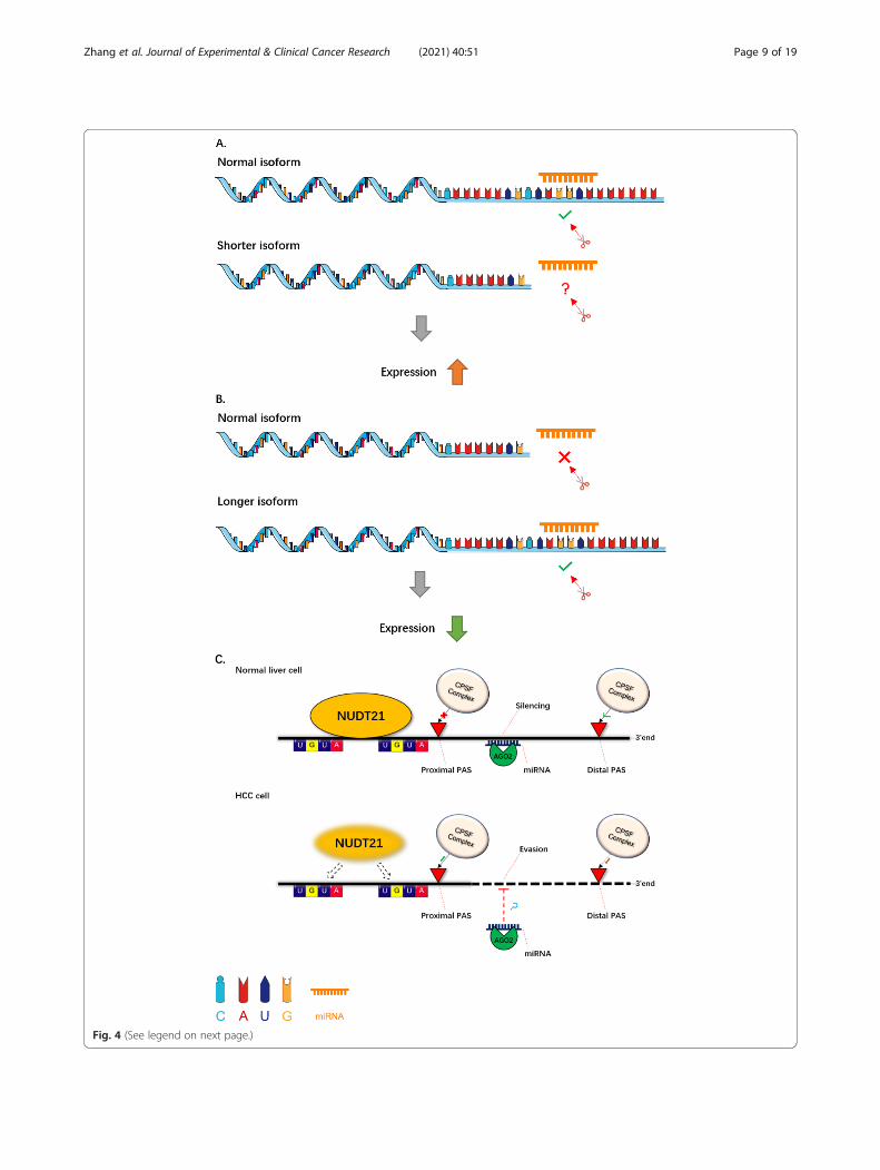
Fig. 4 (See legend on next page.)
Zhang et al. Journal of Experimental & Clinical Cancer Research (2021) 40:51 Page 9 of 19

sites in senescent cells are enriched in senescence-associated pathways [15]. The HNRNPA1-mediated3’UTR lengthening of HN1 contributes to cancer- andsenescence-associated phenotypes [111]. In a like man-ner, 3’UTR lengthening of Mdm2 can mediate the ex-pression of p53, thereby contributing to cellularsenescence in aged rat testis [112]. In addition to cellularsenescence, 3’UTR lengthening also affects cell differen-tiation. 3’UTRs are reprogrammed by APAs during thegeneration of induced pluripotent stem (iPS) cells andthe genes involved in this iPS cell generation were foundto be more likely to exhibit 3′UTR lengthening [113]. Asembryonic development progresses, mouse genes tendto express mRNAs with a longer 3’UTRs. This mechan-istic regulation of 3’UTR-APA is coordinated with theonset of organogenesis and various aspects of embryonicdevelopment (including morphogenesis, differentiation,and proliferation) [114]. However, upstream factors con-trolling 3’UTR lengthening during cellular senescenceand differentiation require further exploration.
Global regulation of APAGlobal 3’UTR regulation has been observed in variousbiological systems and processes including those of em-bryonic development, differentiation of myoblasts, andembryonic stem cells [114, 115]. For example, duringthe activation of primary murine CD4+ T lymphocytes aglobal decrease in the relative expression of distal 3′UTRs was observed. This indicated that the 3’UTR wasglobally shortened [44]. This is consistent with the factthat transcripts with shorter 3’UTRs escape frommiRNA targeting and thus increase their protein levels[116, 117]. Isoforms with proximal PAS usage that havegreater translational potentials than others are generallyupregulated when the membrane depolarization agentsactivate neurocytes [25, 118]. Another novel mechanismfor global 3’UTR shortening is the activation of themTOR pathway [119].Global programs of APA-dependent isoform expres-
sion have been discovered in human cancers. SpecificAPA events have been implicated in various pathologicalconditions such as malignancies and autoimmune
disorders. It has been hypothesized that the global regu-lation of polyadenylation activity might underlie the glo-bal APA profile changes. The usage of PASs is oftenaltered in human hematological, immunological, andneurological diseases, as well as in cancers [8, 120].There are various specific extracellular signals that canglobally regulate APA. For instance, a poly(C)-bindingprotein named αCP was discovered as a global regulatorof APA and a mediator of mRNA stability and transla-tion [121, 122]. CSTF2 and CSTF2t are also essentialglobal regulators of APA. CSTF2-RNA interactions arehighly specific at PASs. Such interactions differ greatlyin affinity and may be differentially required for PASrecognition. Furthermore, the co-depletion of the CSTF2and CSTF2t can lead to striking APA changes, most ofwhich are characterized by increased usages of distalPAS [123].
Poly(A) signal detectionSince pre-mRNA isoforms with differing lengths of3’UTRs are widely present in cells, many studies on thepost-transcription regulation of pre-mRNA highlight theregulation of 3’UTR’s APA and poly(A) tail lengthchanges. These studies not only reveal the mechanismsand factors that regulate cytoplasmic and nuclearchanges in the poly(A) domain, but also clarify the rela-tionship between these mechanisms. The relationshipbetween 3’UTR-APA and miRNA targeting has beenparticularly illuminated. Similarly, relationships betweendeadenylation and the change of PAS usage in inflam-mation, or between cytoplasmic polyadenylation and the3’UTR shortening in neurons, or relating to the alterna-tive lengths of poly(A) tails in germ cells and tumors,have all been elucidated. As a novel mechanism forregulating various gene functions, APA has been in-volved in various biological processes including mamma-lian development, immune system function, diseasepathogenesis, etc. [44, 124–128]. Hence, the detection ofpoly(A) signalling is very important for studying APAregulation and can be used as a powerful method to re-veal disease pathogenesis and related aspects of diagno-sis and treatment.
(See figure on previous page.)Fig. 4 3’UTR length change. Dynamic mRNA isoforms with differential 3’UTR are generated by APA events. This is a schematic diagramillustrating two types of 3’UTR length change. a 3’UTR shortening. Various genes possess a tendency to generate shorter mRNA isoforms intumors than in normal tissues. With the loss of miRNA target sites, the shorter isoform will escape miRNA-mediated decay, resulting in itsaberrant up-regulation. b 3’UTR lengthening. In senescent cells, many genes possess a tendency to generate longer mRNA isoforms than innormal cells. With the use of distal PASs, the longer isoforms contain more miRNA binding sites and so are more likely to be silenced. This is asuppression mechanism to reduce the expression of genes. c An example of the APA regulation mechanism. In normal liver cells, an APAregulator NUDT21, which recognizes the 2 UGUA sequences upstream of the PAS, can protect the proximal poly(A) sites from cleavage of theCPSF complex. Therefore, the expression of the target gene can be regulated by AGO2-mediated miRNA. Conversely, the expression level ofNUDT21 is downregulated in HCC cells. Lacking the protection of NUDT21, the proximal PAS is more likely to be recognized and cleaved by theCPSF complex than the distal PAS. Thus, the target gene can escape from the miRNA silencing due to lack of miRNA binding sites and thusexpress aberrantly [9]
Zhang et al. Journal of Experimental & Clinical Cancer Research (2021) 40:51 Page 10 of 19

Experimental methods for detecting APAIn 2014, two different high-throughput sequencing ap-proaches were developed to sequence the 3′-terminome.Using the first of these methods, TAIL-Seq, researchersmeasured the length of the poly(A) tail and found themedian poly(A) length to be 50-100 nt in HeLa and NIH313 cells [129]. The second technique, Poly(A)-taillength profiling by sequencing (PAL-Seq), was first usedto measure poly(A) tails of millions of individual RNAsin mouse livers, and zebrafish and frog embryos. It re-vealed an embryonic switch in translational control viaAPA regulation [130]. Soon after, an improved TAIL-Seq (mRNA-TAIL-seq, mTAIL-Seq) technique was de-veloped, combining the strengths of TAIL-Seq and PAL-Seq. This was used to analyze poly(A) tails in C. elegans.The study revealed short poly(A) tails as a conservedfeature of highly expressed genes [131]. Subsequentstudies using these poly(A) sequencing methods revealedthat the poly(A)-tail G-content and terminal uridylyl-transferase regulate translational efficiency and the tran-scriptome [132, 133]. In 2015, another deep sequencingof mRNA 3′ termini (termed 3 T-Seq) was developed toidentify APA events in gastric cancer cell lines. Using 3T-Seq, researchers identified > 28,000 novel poly(A)
sites, of which 513 genes had been observed to expressshortened isoforms. They further characterized one ofthese 3′ UTR shortening genes, NET1, and found thatthe NET1 isoform with a short 3’UTR had strongerin vitro cell migration and invasion capabilities than thatwith a long 3’UTR, suggesting that APA plays a role intumor metastasis [108]. More recently, two new APAdetection methods based on single-cell RNA-seq, namelyFull-length poly(A) and mRNA sequencing (FLAM-seq)[134] and Poly(A) inclusive RNA isoform sequencing(PAIso−seq) [135], have been developed. Using theirnew algorithm “tailfindr” [136], these new sequencingmethods can detect poly(A) sites at a single-cell sensitiv-ity and estimate poly(A) tail length from long-read se-quencing data.
Computational tools for detecting APAIn parallel with the advancement of experimentalmethods, computational tools to detect APA have beenactively developed. These are summarized in Table 1.We will now introduce several of these popular toolsthat can complete the process from the sequence align-ment to APA detection result.
Table 1 Computational tools for detecting APA
Name Description Environment Year Website Ref.
InPAS A package that can detect the dynamics of APA events from RNA-seq data by removing false sites due to internal-priming.
R 2013 http://www.bioconductor.org/packages/release/bioc/html/InPAS.html
[137]
ChangePoint A change-point model based on a likelihood ratio test for detect-ing 3’UTR switching.
Java 2014 http://utr.sourceforge.net/ [13]
DaPars A bioinformatics algorithm for the de novo identification ofdynamic APAs from standard RNA-seq.
Python 2014 https://github.com/ZhengXia/dapars [8]
Roar A strategy for detecting alternative PAS usage and comparingthese between two biological conditions.
R 2016 https://github.com/vodkatad/roar/ [138]
QAPA An approach to infer and quantify APA from RNA-seq data. Python & R 2018 https://www.github.com/morrislab/qapa
[139]
PAQR_KAPAC
A combined method that can quantify PAS usage from RNA-seqdata and infer regulatory sequence motifs on PAS usage.
Python & R 2018 https://github.com/zavolanlab/PAQR_KAPAC.git
[120]
APAtrap An approach to identify and quantify APA sites from RNA-seq databased on the mean squared error model.
R 2018 https://apatrap.sourceforge.io. [140]
IntMap An integrated method for detecting novel APA events from RNA-seq and PAS-seq data.
Matlab 2018 http://compbio.cs.umn.edu/IntMAP/ [141]
TAPAS A tool that can detect more than two APA sites in a gene andAPA sites before the last exon from RNA-seq data.
R 2018 https://github.com/arefeen/TAPAS [142]
APARENT A deep learning approach to predict APA from DNA sequences. Python 2019 https://github.com/johli/aparent [143]
DeepPASTA A deep learning method to predict APA from DNA sequences andRNA secondary structure data.
Python 2019 https://github.com/arefeen/DeepPASTA
[144]
scDAPA A tool to detect and visualize APA events from single-cell RNA-seqdata.
R 2019 https://scdapa.sourceforge.io/ [145]
APAlyzer A bioinformatics package which can examine 3’UTR-APA, intronicAPA, and gene expression changes using RNA-seq data.
R 2020 https://bioconductor.org/packages/release/bioc/html/APAlyzer.html
[146]
APA-Scan A robust program that infers 3’UTR-APA events and visualizes theRNA-seq short-read coverage with gene annotations.
Python 2020 https://github.com/compbiolabucf/APA-Scan
[147]
Zhang et al. Journal of Experimental & Clinical Cancer Research (2021) 40:51 Page 11 of 19

DaPars is a powerful tool to identify numerous APA eventsfrom standard RNA-Seq data. It employs a piecewise linearregression to model read count data of RNA-seq to identifythe location of the de novo proximal poly(A) sites. UsingDaPars, Xia et al. identified 1346 genes with tumor-specificAPAs from 358 pairs of tumor/normal samples across sevencancer types. Compared with normal tissue samples, morethan 90% of these APA genes had shorter-length isoforms inthe tumor samples. This tool has been widely used to detectAPA events from RNA-seq data and has also been adoptedby many databases [8].APAtrap is capable of APA identification and quantifi-
cation. Based on the mean squared error model, APA-trap can identify differential PAS usage and predict allpotential poly(A) sites. When APAtrap was applied tothe simulation data and real RNA-Seq data from humanand Arabidopsis tissues, it showed higher accuracy thanother tools in identifying APA events [140].DeepPASTA is a deep neural network method to detect
APA events. It was the first tool to predict poly(A) sites fromboth sequence and RNA secondary structure data. Inaddition, this tool can predict the most dominant poly(A)site of a gene in a specific tissue and predict the relativeabundance of two polyA sites of the same gene [144].Finally, scDAPA is a software package that can be used
to detect APA profiles from single-cell RNA-seq data. Itincludes three main modules, namely 3’end annotation,APA event identification, and APA event visualization.scDAPA has a high degree of confidence for APA detec-tion. This tool facilitates the portrait of dynamic APA pro-files in different cell types from scRNA-seq data [145].
APA databasesA large quantity of APA data has been produced usingNGS techniques. Using these data, several databases
have been established to facilitate the research commu-nity to obtain APA information from various samples.These are summarized in Table 2. In the following sec-tion, we introduce several major APA databases.The PolyA_DB is a database for analyzing pre-mRNA
cleavage and polyA sites. It contains a large amount ofdata on polyA sites in humans, mice, rats, and chickens.In 2018, this database had been updated to version 3.0(renamed as PolyA_DB 3). Based on deep sequencingdata, using the 3’READS method, this version containslarge volumes of data from multiple samples to supple-ment PAS information. The database can also be visual-ized by the UCSC genome browser [150].TC3A focuses on human cancers with large-scale
RNA-Seq datasets from TCGA which contains 10,537tumor samples across 32 cancer types and provides APAusage analysis and visualization. This atlas is based on abioinformatics algorithm called DaPars and its updatedversion, DaPars2. Users can compare the PAS usage ofgenes between tumor and normal samples [151].PolyASite is a resource of PAS information generated
using 3’end sequencing in humans and mice. In 2019, itwas updated to version 2.0 containing new PAS datasetsfrom worm genomes. PolyASite 2.0 integrates sequen-cing data generated by multiple sequencing methods(such as 3’READS, SAPAS, PolyA-Seq, etc) [152].The APAatlas contains 1,125,143 APA events from
9475 samples across a total of 53 human tissue types. Itfocuses on the APA events located in 3’UTR regions andprovides a view of the APA landscape across tissues.APA events in the APAatlas were inferred using DaParsand SAAP-RS. Since the APAatlas includes a largeamount of normal human tissue samples, compared withother databases, it contains more APA events from nor-mal samples and provides a good opportunity for
Table 2 APA databases
Name Description Year Species Website Ref.
PolyA-SeqAtlas
A quantitative atlas of poly(A) sites using the PolyA-Seq protocol. Filtered sites areavailable via the UCSC Genome Browser.
2012 human, rhesus,dog, mouse, andrat
http://genome.ucsc.edu/
[19]
APADB Database of APA sites and miRNA regulation events. 2014 human, chicken,and mouse
http://tools.genxpro.net/apadb/
[148]
APASdb Database of APA sites and heterogeneous cleavage sites downstream of poly(A)signals.
2015 human, mouse andzebrafish
http://mosas.sysu.edu.cn/utr
[149]
PolyA_DB3 Database of cleavage and Poly(A) sites identified by the 3ʹREADS protocol. 2018 human, mouse, rat,and chicken
http://www.polya-db.org/v3
[150]
TC3A Database of robust APA data from 10,537 tumors across 32 cancer types. It isfocused on human cancers and utilizes routinely available large-scale RNA-Seqdatasets from TCGA.
2018 human http://tc3a.org [151]
PolyAsite2.0 Web portal of poly(A) sites identified by all 3’end sequencing datasets. 2020 human, mouse andworm
https://polyasite.unibas.ch
[152]
APAatlas Atlas of APA across a large number of normal human tissues from the Genotype-Tissue Expression project.
2020 human https://hanlab.uth.edu/apa/
[153]
Zhang et al. Journal of Experimental & Clinical Cancer Research (2021) 40:51 Page 12 of 19
investigation of the correlation between PAS usage andgene expression [153].
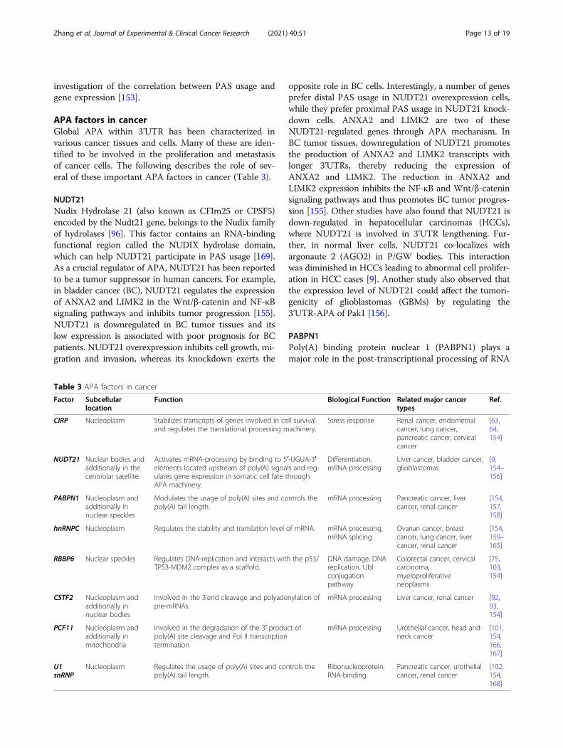
APA factors in cancerGlobal APA within 3’UTR has been characterized invarious cancer tissues and cells. Many of these are iden-tified to be involved in the proliferation and metastasisof cancer cells. The following describes the role of sev-eral of these important APA factors in cancer (Table 3).
NUDT21Nudix Hydrolase 21 (also known as CFIm25 or CPSF5)encoded by the Nudt21 gene, belongs to the Nudix familyof hydrolases [96]. This factor contains an RNA-bindingfunctional region called the NUDIX hydrolase domain,which can help NUDT21 participate in PAS usage [169].As a crucial regulator of APA, NUDT21 has been reportedto be a tumor suppressor in human cancers. For example,in bladder cancer (BC), NUDT21 regulates the expressionof ANXA2 and LIMK2 in the Wnt/β-catenin and NF-κBsignaling pathways and inhibits tumor progression [155].NUDT21 is downregulated in BC tumor tissues and itslow expression is associated with poor prognosis for BCpatients. NUDT21 overexpression inhibits cell growth, mi-gration and invasion, whereas its knockdown exerts the
opposite role in BC cells. Interestingly, a number of genesprefer distal PAS usage in NUDT21 overexpression cells,while they prefer proximal PAS usage in NUDT21 knock-down cells. ANXA2 and LIMK2 are two of theseNUDT21-regulated genes through APA mechanism. InBC tumor tissues, downregulation of NUDT21 promotesthe production of ANXA2 and LIMK2 transcripts withlonger 3’UTRs, thereby reducing the expression ofANXA2 and LIMK2. The reduction in ANXA2 andLIMK2 expression inhibits the NF-κB and Wnt/β-cateninsignaling pathways and thus promotes BC tumor progres-sion [155]. Other studies have also found that NUDT21 isdown-regulated in hepatocellular carcinomas (HCCs),where NUDT21 is involved in 3’UTR lengthening. Fur-ther, in normal liver cells, NUDT21 co-localizes withargonaute 2 (AGO2) in P/GW bodies. This interactionwas diminished in HCCs leading to abnormal cell prolifer-ation in HCC cases [9]. Another study also observed thatthe expression level of NUDT21 could affect the tumori-genicity of glioblastomas (GBMs) by regulating the3’UTR-APA of Pak1 [156].
PABPN1Poly(A) binding protein nuclear 1 (PABPN1) plays amajor role in the post-transcriptional processing of RNA
Table 3 APA factors in cancer
Factor Subcellularlocation
Function Biological Function Related major cancertypes
Ref.
CIRP Nucleoplasm Stabilizes transcripts of genes involved in cell survivaland regulates the translational processing machinery.
Stress response Renal cancer, endometrialcancer, lung cancer,pancreatic cancer, cervicalcancer
[63,64,154]
NUDT21 Nuclear bodies andadditionally in thecentriolar satellite
Activates mRNA-processing by binding to 5′-UGUA-3′elements located upstream of poly(A) signals and reg-ulates gene expression in somatic cell fate throughAPA machinery.
Differentiation,mRNA processing
Liver cancer, bladder cancer,glioblastomas
[9,154–156]
PABPN1 Nucleoplasm andadditionally innuclear speckles
Modulates the usage of poly(A) sites and controls thepoly(A) tail length.
mRNA processing Pancreatic cancer, livercancer, renal cancer
[154,157,158]
hnRNPC Nucleoplasm Regulates the stability and translation level of mRNA. mRNA processing,mRNA splicing
Ovarian cancer, breastcancer, lung cancer, livercancer, renal cancer
[154,159–165]
RBBP6 Nuclear speckles Regulates DNA-replication and interacts with the p53/TP53-MDM2 complex as a scaffold.
DNA damage, DNAreplication, Ublconjugationpathway
Colorectal cancer, cervicalcarcinoma,myeloproliferativeneoplasms
[75,103,154]
CSTF2 Nucleoplasm andadditionally innuclear bodies
Involved in the 3’end cleavage and polyadenylation ofpre-mRNAs.
mRNA processing Liver cancer, renal cancer [92,93,154]
PCF11 Nucleoplasm andadditionally inmitochondria
Involved in the degradation of the 3′ product ofpoly(A) site cleavage and Pol II transcriptiontermination
mRNA processing Urothelial cancer, head andneck cancer
[101,154,166,167]
U1snRNP
Nucleoplasm Regulates the usage of poly(A) sites and controls thepoly(A) tail length.
Ribonucleoprotein,RNA-binding
Pancreatic cancer, urothelialcancer, renal cancer
[102,154,168]
Zhang et al. Journal of Experimental & Clinical Cancer Research (2021) 40:51 Page 13 of 19
and in controlling the poly(A) tail length of RNA tran-scripts. PABPN1 binds at proximal poly(A) sites to blocktheir cleavage. Yu et al. characterized the APA profilesof 6398 patient samples across 17 cancer types from TheCancer Genome Atlas (TCGA) and of 739 cancer celllines from the Cancer Cell Line Encyclopedia (CCLE).They identified 1971 clinically relevant APA events andtheir analysis further illustrated PABPN1 as a mastermodulator of 3’UTR shortening. PABPN1 possess thecapacity of proximal PAS binding and then alters theAPA site selection [170]. In triple-negative breast cancer(TNBC), Wang et al. identified 1631 significant APAevents in 165 TNBC tissues and 33 matched adjacentnormal tissues. Among these significant APA events, ap-proximately 69% exhibited a preference for proximalPAS usage. This team identified CPSF1 and PABPN1 astwo major regulators of APA events in TNBC using apooled shRNA library screening. They then demon-strated that the tandem 3’UTR length of various genes iscorrelated with the expression level of CPSF1 andPABPN1. Knockdown of PABPN1 interferes with APAregulation, resulting in an extensive 3’UTR shortening incell cycle related genes. Consequently, this inhibits cellproliferation and causes apoptosis and S phase arrest inTNBC cell lines [171]. In muscle cells, PABPN1 interactswith Matrin 3 (MATR3) and regulates RNA processing.Mutations in PABPN1 can also cause oculopharyngealmuscular dystrophy (OPMD) [157, 158].
hnRNPCHeterogeneous nuclear ribonucleoproteins C (hnRNPC)is an RNA-binding protein encoded by the HNRNPCgene in humans. hnRNPC regulates genome-wide PASusage selection. By generating a pre-mRNA 3’end se-quencing library from hnRNPC-knockdown cell lines,Gruber et al. observed that nearly 54% of PASs in exonshad altered their usage from that of the control group.Mechanistically, hnRNPC binds the poly(U) motifs thatare frequently located near distal poly(A) sites. HNRNPC’s binding in close proximity of distal poly(A) sitesprevents them from cleavage and polyadenylation,thereby increasing genome-wide proximal PAS usage[172]. Aberrant up-regulation of hnRNPC has been ob-served in a variety of cancers or cancer cell lines includ-ing breast cancers, glioblastomas, hepatocellularcarcinomas, ovarian cancers, and lung cancers [159–163,173]. One recent study revealed that the up-regulationof hnRNPC plays a crucial role in establishing APA pro-files that are characteristic for metastatic colon cancercells. hnRNPC is responsible for the regulation of UTR-APA of a group of genes including MTHFD1L, which isclosely related to cancer progression [164]. The level ofhnRNPC expression is also related to clinical outcomes.Patients with a high levels of hnRNPC transcripts have
poor overall survival and disease-free survival in humangastric cancers [165]. These studies suggest the potentialof hnRNPC as a valuable prognostic biomarker andtherapeutic target for cancer treatment.
PCF11As a part of CFII, PCF11 contains an N-terminal RNA-PII C-terminal domain (CTD)-interacting domain (CID)and plays a role in transcription termination and mRNAnuclear export control [174, 175]. Li et al. showed thatthe depletion of PCF11 in mouse C2C12 cells led to glo-bal 3’UTR lengthening by APA [24]. PCF11, as a keyAPA regulator, has also been recognized as responsiblefor the extensive 3’end alterations observed in neuro-blastomas. Postnatal down-regulation of PCF11 inducesneurodifferentiation and a low expression of PCF11 isassociated with a favorable outcome and spontaneoustumor regression in such neuroblastomas. Mechanistic-ally, GNB1, a subunit of the Gβγ-complex, is an import-ant modulator of Wnt signalling. It is mediated byPCF11 through APA regulation. In the presence ofPCF11, the GNB1 transcript with short 3’UTR is pre-dominant in neuroblastoma differentiation. The shortisoform of GNB1 has higher translation efficiency andthis corresponds to the higher expression level of theGNB1 protein, thereby leading to the suppression ofWnt signalling. The expression level of GNB1 becomessignificantly reduced upon PCF11 depletion. All-transretinoic acid (ATRA) is the first-line therapeutic drugfor treating neuroblastomas. After neuroblastomas weretreated with ATRA, the expression level of PCF11 wassignificantly reduced, confirming its anti-cancer effect[166]. These studies suggest that PCF11 is a major regu-lator of the APA process and an important modulator ofWnt signalling during the neuronal differentiation ofneuroblastomas.
Conclusions and perspectiveMounting evidence is now demonstrating APA as a newlayer of regulation for gene expression. The four types ofAPA work synergistically with miRNAs, RBPs, and otherfactors, to regulate gene expression and functional versa-tility. Due to the differential usage of PASs, various tran-script isoforms can be generated in cells. Thesetranscript isoforms are involved in multiple cellular pro-cesses including control of the cell cycle, mRNA transla-tion efficiency, and cell proliferation and differentiation.APA is frequently dysregulated in cancer and this pro-motes tumorigenesis and progression by increasing theexpression of oncogenes and reducing the expression oftumor suppressor genes [45, 176–178]. It is worth not-ing that not all APA events have biological significanceand the secondary poly(A) site can be important in de-velopment, differentiation and transformation processes.
Zhang et al. Journal of Experimental & Clinical Cancer Research (2021) 40:51 Page 14 of 19

Some APA events may lead to cryptic unstable tran-scripts, many of which are rapidly degraded in cells [19].Generally, the identification of biologically significantAPA events involves computational prediction and stat-istical testing, followed by tailored in vitro and/or in vivoassays.Many computational tools and databases have also
been developed to detect APA events (Tables 1 and 2).Most of these infer information of PAS usage fromstandard RNA-seq data. Using deep learning models,some of these can predict novel APA events under dif-ferent biological conditions. These tools make a greatcontribution to the analysis of genome-wide APA pro-files, thereby greatly improving our understanding of theAPA regulation of gene expression and functional versa-tility. However, these tools are mainly focused on tan-dem 3’UTR-APA. The potential impact of UR-APA,such as effects of internal exon APA on gene regulation,requires further exploration. It will be interesting toknow whether 3’UTR-APA and UR-APA are mutuallyexclusive or co-occurr in genes, and to what extent theycoordinate their respective regulation of genes to pro-mote tumorigenesis and cancer progression. There is anurgent need to develop new computational tools tailoredtowards identifying UR-APA. Additionally, the direct se-quencing of natural poly(A) RNAs by long-read sequen-cing technologies (such as Oxford Nanopore and PacificBiosciences) [179–182] provides broad prospects for thefurther detection and quantification of these UR-APAs.Extensive APA occurs during the pathophysiology of
many diseases including cancers. In these, APA eventsare emerging as clinical biomarkers of high potential.Most of the differentially regulated APA events result intranscript isoforms with different lengths of 3’UTRs.These are often related to a variety of clinical character-istics. These APA events are independent of commonlyused molecular data (e.g., gene expression and somaticmutations) [8], and have been found to associate withprognosis, recurrence, tumor subtypes, and staging inmultiple cancer types [30, 45, 109, 110, 170]. Addition-ally, APA events are potential therapeutic targets forcancer treatment and clinical biomarkers for drug resist-ance. APA events are commonly observed in clinicallyactionable genes such as CTNNB1, PI3KR1, and FGFR2.PABPN1, an APA master regulator, regulates large num-bers of clinically actionable genes. Associations betweenAPA events and the sensitivities of FDA-approved anti-cancer drugs tested in cancer cells are also readily ob-servable [170].Although recent studies have greatly enriched our
knowledge of APA, we still know little about certainfunctions such as the differential affinity of PASs, the re-cruitment of the 3’end processing complex and other de-tails on the regulation of APA factors. Continuing in-
depth research on the modulation of APA regulation,the impact of APA on biological processes, and the pos-sibility of manipulating APA in disease treatment, re-mains of high priority.
Abbreviations3’UTR: 3’Untranslated region; AGO2: Argonaute 2; ALS: Amyotrophic lateralsclerosis; ANXA2: Annexin A2; APA: Alternative polyadenylation;ARE: Adenylate-uridylate-rich element; BC: Bladder cancer; CALCA: Calcitonin-related polypeptide-α gene; CCLE: Cancer Cell Line Encyclopedia; CCR4-NOT: Carbon catabolite repression 4–negative on TATA-less; CDC6: Celldivision cycle 6; CDKN1B: Cyclin-dependent kinase inhibitor 1B; CFI: Cleavagefactors I; CFII: Cleavage factors II; c-Fos: Fos proto-oncogene; CGRP: Calcitoningene-related peptide 1; CID: C-terminal domain (CTD)-interacting domain;CIRP: Cold-inducible RNA binding protein; CLP1: Cleavage factorpolyribonucleotide kinase subunit 1; cMET: MET proto-oncogene;CPSF: Cleavage and polyadenylation factor; CSTF: Cleavage stimulation factor;CTD: Carboxy-terminal domain; CTNNB1: Catenin beta 1; DHFR: Dihydrofolatereductase; DSRM: Double-stranded RNA-binding motif; ELAV: Embryonic-lethal abnormal visual protein; ESC: Embryonic stem cell; FGFR2: Fibroblastgrowth factor receptor 2; FIP1: Cleavage polyadenylation factor subunit FIP1;GALNT5: Polypeptide N-acetylgalactosaminyltransferase 5;HCC: Hepatocellular carcinoma; HIF-1α: Hypoxia inducible factor 1 subunitalpha; HLF: Human Lung Fibroblasts; HN1: Hematological and neurologicalexpressed 1; hnRNP: Heterogeneous nuclear RNP; HNRNPA1: Heterogeneousnuclear ribonucleoprotein A1; hnRNPC: Heterogeneous nuclearribonucleoproteins C; hTERT: Telomerase reverse transcriptase in humans;IGF2BP1: Insulin like growth factor 2 mRNA binding protein 1;IgM: Immunoglobulin M; iPS cell: Induced pluripotent stem cell; IRF5: IFN-regulatory factor 5; KHdomain: K homology domain; KSRP: K homologysplicing regulatory protein; LIMK2: LIM-domain kinase-2; MATR3: Matrin 3;Mdm2: MDM2 proto-oncogene; miRNA: MicroRNA; MSI: RNA-binding proteinMusashi; MTHFD1L: Methylenetetrahydrofolate dehydrogenase (NADP+dependent) 1 like; MYC: MYC proto-oncogene; NGS: Next-generationsequencing; NUDT21: Nudix Hydrolase 21; NUMB: NUMB endocytic adaptorprotein; OPMD: Oculopharyngeal muscular dystrophy; PABPN1: Poly(A)binding protein nuclear 1; PAP: Poly(A) polymerase; PAS: Poly(A) site; pAsignal: Poly(A) signal; PCF11: Cleavage and polyadenylation factor subunitPCF11; PIK3R1: Phosphoinositide-3-kinase regulatory subunit 1; Pol II: RNApolymerase II; pre-mRNA: Messenger RNA precursor; PTC: Papillary thyroidcarcinoma; PTEN: Phosphatase and tensin homolog; PTGR: Post-transcriptional gene regulation; PUF: Pumilio p-homology and Fem-3 mRNAbinding factor; PUM: Pumilio homologue proteins; RBBP6: Retinoblastoma-binding protein 6; RBD: RNA-binding domain; RBP: RNA-binding protein;RNP: Ribonucleoprotein; RRM: RNA recognition motif; SMAD: SMAD family;SMN1: Survival motor neuron 1; snRNP: Small nuclear RNP; TCGA: The CancerGenome Atlas; TEC: Terminal exon characterization; TGFβ: Transforminggrowth factor beta; TNBC: Triple-negative breast cancer; TNFα: Tumornecrosis factor α; TTP: Tristetraprolin; U1 snRNP: U1 small nuclearribonucleoprotein; uaRNA: UTR-associated RNA; UR-APA: Upstream regionAPA; WDR33: Cleavage polyadenylation factor subunit WDR33
AcknowledgmentsWe thank anonymous reviewers and Christopher R. Wood for reading andcommenting on the manuscript.
Authors’ contributionsPL, YL and JD designed the study. YZ drafted the manuscript. PL, YL, JD, LL,QQ and QZ revised the manuscript. All of the authors have read andapproved the paper.
FundingThis work has been supported in part by the National Natural ScienceFoundation of China (81871864, 81772766 and 82072857), Key Research andDevelopment Program of Zhejiang Province (2021C03126C), Key Program ofZhejiang Provincial Natural Science Foundation of China (LZ20H160001),Medical Health Science and Technology Key Project of Zhejiang ProvincialHealth Commission (WKJ-ZJ-2007 and 2017211914), and National KeyResearch and Development Program of China (2019YFC1315700 and2016YFA0501800).
Zhang et al. Journal of Experimental & Clinical Cancer Research (2021) 40:51 Page 15 of 19

Availability of data and materialsAll data generated or analyzed during this study are included in thispublished article.
Ethics approval and consent to participateNot Applicable.
Consent for publicationAll authors have agreed to publish this manuscript.
Competing interestsNo potential conflicts of interest are disclosed.
Author details1Department of Respiratory Medicine, Sir Run Run Shaw Hospital andInstitute of Translational Medicine, Zhejiang University School of Medicine,Hangzhou 310016, Zhejiang, China. 2Center for Uterine Cancer Diagnosis &Therapy Research of Zhejiang Province, Women’s Reproductive Health KeyLaboratory of Zhejiang Province, Department of Gynecologic Oncology,Women’s Hospital and Institute of Translational Medicine, Zhejiang UniversitySchool of Medicine, Hangzhou 310006, Zhejiang, China. 3Department ofHead and Neck Surgery, Cancer Hospital of the University of ChineseAcademy of Sciences, Zhejiang Cancer Hospital, Key Laboratory of Head &Neck Cancer Translational Research of Zhejiang Province, Hangzhou 310022,Zhejiang, China. 4Department of Physiology, Center of Systems MolecularMedicine, Medical College of Wisconsin, Milwaukee, WI 53226, USA. 5CancerCenter, Zhejiang University, Hangzhou 310029, Zhejiang, China.
Received: 17 October 2020 Accepted: 20 January 2021
References1. Mandel CR, Bai Y, Tong L. Protein factors in pre-mRNA 3′-end processing.
Cell Mol Life Sci. 2008;65:1099–122.2. Guhaniyogi J, Brewer G. Regulation of mRNA stability in mammalian cells.
Gene. 2001;265:11–23.3. Balbo PB, Bohm A. Mechanism of poly(A) polymerase: structure of the
enzyme-MgATP-RNA ternary complex and kinetic analysis. Structure. 2007;15:1117–31.
4. Millevoi S, Vagner S. Molecular mechanisms of eukaryotic pre-mRNA 3′ endprocessing regulation. Nucleic Acids Res. 2009;38:2757–74.
5. Turner RE, Pattison AD, Beilharz TH. Alternative polyadenylation in theregulation and dysregulation of gene expression. Semin Cell Dev Biol. 2018;75:61–69.
6. Proudfoot NJ, Brownlee GG. 3′ non-coding region sequences in eukaryoticmessenger RNA. Nature. 1976;263:211–4.
7. Tian B, Hu J, Zhang H, Lutz CS. A large-scale analysis of mRNApolyadenylation of human and mouse genes. Nucleic Acids Res. 2005;33:201–12.
8. Xia Z, Donehower LA, Cooper TA, Neilson JR, Wheeler DA, Wagner EJ, et al.Dynamic analyses of alternative polyadenylation from RNA-seq reveal a 3′2-UTR landscape across seven tumour types. Nat Commun. 2014;5:5274.
9. Sun M, Ding J, Li D, Yang G, Cheng Z, Zhu Q. NUDT21 regulates 3′-UTRlength and microRNA-mediated gene silencing in hepatocellular carcinoma.Cancer Lett. 2017;410:158–68.
10. Alt FW, Bothwell ALM, Knapp M, Siden E, Mather E, Koshland M, et al.Synthesis of secreted and membrane-bound immunoglobulin mu heavychains is directed by mRNAs that differ at their 3′ ends. Cell. 1980;20:293–301.
11. Yan Z, Degregori J, Shohet R, Leone G, Stillman B, Nevins JR, et al. Cdc6 isregulated by E2F and is essential for DNA replication in mammalian cells.Proc Natl Acad Sci U S A. 1998;95:3603–8.
12. Aguilo F, Zhou MM, Walsh MJ. Long noncoding RNA, polycomb, and theghosts haunting INK4b-ARF-INK4a expression. Cancer Res. 2011;71:5365–9.
13. Wang W, Wei Z, Li H. A change-point model for identifying 3’UTR switchingby next-generation RNA sequencing. Bioinformatics. 2014;30:2162–70.
14. López De Silanes I, Paz Quesada M, Esteller M. Aberrant regulation ofmessenger RNA 3′-untranslated region in human cancer. Cell Oncol. 2007;29:1–17.
15. Chen M, Lyu G, Han M, Nie H, Shen T, Chen W, et al. 3′ UTR lengthening asa novel mechanism in regulating cellular senescence. Genome Res. 2018;28:285–94.
16. Rogers J, Early P, Carter C, Calame K, Bond M, Hood L, et al. Two mRNAswith different 3′ ends encode membrane-bound and secreted forms ofimmunoglobulin μ chain. Cell. 1980;20:303–12.
17. Setzer DR, McGrogan M, Nunberg JH, Schimke RT. Size heterogeneity in the3′ end of dihydrofolate reductase messenger RNAs in mouse cells. Cell.1980;22:361–70.
18. Edwalds-Gilbert G, Veraldi KL, Milcarek C. Alternative poly(A) site selection incomplex transcription units: means to an end? Nucleic Acids Res. 1997;25:2547–61.
19. Derti A, Garrett-Engele P, MacIsaac KD, Stevens RC, Sriram S, Chen R, et al. Aquantitative atlas of polyadenylation in five mammals. Genome Res. 2012;22:1173–83.
20. Shi Y. Alternative polyadenylation: new insights from global analyses. RNA.2012;18:2105–17.
21. Wang R, Zheng D, Yehia G, Tian B. A compendium of conserved cleavageand polyadenylation events in mammalian genes. Genome res. 2018;28:1427–41.
22. Reyes A, Huber W. Alternative start and termination sites of transcriptiondrive most transcript isoform differences across human tissues. NucleicAcids Res. 2018;46:582–92.
23. Tian B, Pan Z, Ju YL. Widespread mRNA polyadenylation events in intronsindicate dynamic interplay between polyadenylation and splicing. GenomeRes. 2007;17:156–65.
24. Li W, You B, Hoque M, Zheng D, Luo W, Ji Z, et al. Systematic profiling ofpoly(A)+ transcripts modulated by Core 3′ end processing and splicingfactors reveals regulatory rules of alternative cleavage and Polyadenylation.PLoS Genet. 2015;11:e1005166.
25. Tian B, Manley JL. Alternative polyadenylation of mRNA precursors. Nat RevMol Cell Biol. 2016;18:18–30.
26. Yuan F, Hankey W, Wagner EJ, Li W, Wang Q. Alternative polyadenylation ofmRNA and its role in cancer. Genes Dis. 2019.
27. Elkon R, Ugalde AP, Agami R. Alternative cleavage and polyadenylation:extent, regulation and function. Nat Rev Genet. 2013;14:496–506.
28. Berkovits BD, Mayr C. Alternative 3′ UTRs act as scaffolds to regulatemembrane protein localization. Nature. 2015;522:363–7.
29. Mayr C. Evolution and biological roles of alternative 3’UTRs. Trends Cell Biol.2016;26:227–37.
30. Lembo A, Di Cunto F, Provero P. Shortening of 3′UTRs correlates with poorprognosis in breast and lung cancer. PLoS One. 2012;7:e31129.
31. Guo H, Zhao L, Shi B, Bao J, Zheng D, Zhou B, et al. GALNT5 uaRNApromotes gastric cancer progression through its interaction with HSP90.Oncogene. 2018;37:4505–17.
32. Jiang JH, Lv QY, Yi YX, Liao J, Wang XW, Zhang W. MicroRNA-200apromotes proliferation and invasion of ovarian cancer cells by targetingPTEN. Eur Rev Med Pharmacol Sci. 2018;22:6260–7.
33. Li QQ, Liu Z, Lu W, Liu M. Interplay between alternative splicing andalternative Polyadenylation defines the expression outcome of the plantunique OXIDATIVE TOLERANT-6 gene. Sci Rep. 2017;7:2052.
34. Vasudevan S, Peltz SW, Wilusz CJ. Non-stop decay - A new mRNAsurveillance pathway. BioEssays. 2002;24:785–8.
35. Lareau LF, Brooks AN, Soergel DAW, Meng Q, Brenner SE. The coupling ofalternative splicing and nonsense-mediated mRNA decay. Adv Exp MedBiol. 2007;623:190–211.
36. Lykke-Andersen S, Jensen TH. Nonsense-mediated mRNA decay: an intricatemachinery that shapes transcriptomes. Nat Rev Mol Cell Biol. 2015;16:665–77.
37. Karnati HK, Panigrahi MK, Gutti RK, Greig NH, Tamargo IA. MiRNAs: keyplayers in neurodegenerative disorders and epilepsy. J Alzheimer’s Dis. 2015;48:563–80.
38. Rocci A, Hofmeister CC, Pichiorri F. The potential of miRNAs as biomarkersfor multiple myeloma. Expert rev Mol Diagn. 2014;14:947–59.
39. Bushati N, Cohen SM. MicroRNA functions. Annu Rev Cell Dev Biol. 2007;23:175–205.
40. Croce CM. Causes and consequences of microRNA dysregulation in cancer.Nat Rev Genet. 2009;10:704–14.
41. Bartel DP. MicroRNAs: target recognition and regulatory functions. Cell.2009;136:215–33.
42. Syeda ZA, Langden SSS, Munkhzul C, Lee M, Song SJ. Regulatorymechanism of microrna expression in cancer. Int J Mol Sci. 2020;21:1732.
Zhang et al. Journal of Experimental & Clinical Cancer Research (2021) 40:51 Page 16 of 19

43. Ogorodnikov A, Kargapolova Y, Danckwardt S. Processing and transcriptomeexpansion at the mRNA 3′ end in health and disease: finding the right end.Pflugers Arch - Eur J Physiol. 2016;468:993–1012.
44. Sandberg R, Neilson JR, Sarma A, Sharp PA, Burge CB. Proliferating cellsexpress mRNAs with shortened 3′ untranslated regions and fewer microRNAtarget sites. Science. 2008;320:1643–7.
45. Mayr C, Bartel DP. Widespread shortening of 3′UTRs by alternative cleavageand Polyadenylation activates oncogenes in Cancer cells. Cell. 2009;138:673–84.
46. Zhang Y, Tang C, Yu T, Zhang R, Zheng H, Yan W. MicroRNAs control mRNAfate by compartmentalization based on 3′ UTR length in male germ cells.Genome Biol. 2017;18:105.
47. Hoffman Y, Bublik DR, P. Ugalde A, Elkon R, Biniashvili T, Agami R, et al.3’UTR shortening potentiates MicroRNA-based repression of pro-differentiation genes in proliferating human cells. PLoS Genet. 2016;12:e1005879.
48. Gerstberger S, Hafner M, Tuschl T. A census of human RNA-bindingproteins. Nat Rev Genet. 2014;15:829–45.
49. Vaquerizas JM, Kummerfeld SK, Teichmann SA. Luscombe NM. A census ofhuman transcription factors: Function, expression and evolution. Nat RevGenet. 2009;10:252–63.
50. Kechavarzi B, Janga SC. Dissecting the expression landscape of RNA-bindingproteins in human cancers. Genome Biol. 2014;15:R14.
51. Castello A, Fischer B, Hentze MW, Preiss T. RNA-binding proteins inMendelian disease. Trends Genet. 2013;29:318–27.
52. Shukla S, Parker R. Hypo- and hyper-assembly diseases of RNA–proteincomplexes. Trends Mol Med. 2016;22:615–28.
53. Brinegar AE, Cooper TA. Roles for RNA-binding proteins in developmentand disease. Brain Res. 1647;2016:1–8.
54. Maris C, Dominguez C, FHT A. The RNA Recognition motif, a plastic RNA-binding platform to regulate post-transcriptional gene expression. FEBS J.2005;272:2118–31.
55. Schmitz-Linneweber C, Small I. Pentatricopeptide repeat proteins: a socketset for organelle gene expression. Trends Plant Sci. 2008;13:663–70.
56. Abbasi N. Park Y il, Choi SB. Pumilio puf domain RNA-binding proteins inArabidopsis. Plant Signal Behav. 2011;6:364–8.
57. García-Mauriño SM, Rivero-Rodríguez F, Velázquez-Cruz A, Hernández-VelliscaM, Díaz-Quintana A, De la Rosa MA, et al. RNA binding protein regulation andcross-talk in the control of AU-rich mRNA fate. Front Mol Biosci. 2017;4:71.
58. Mukherjee N, Jacobs NC, Hafner M, Kennington EA, Nusbaum JD, Tuschl T,et al. Global target mRNA specification and regulation by the RNA-bindingprotein ZFP36. Genome biol. 2014;15:R12.
59. Fabian MR, Cieplak MK, Frank F, Morita M, Green J, Srikumar T, et al. MiRNA-mediated deadenylation is orchestrated by GW182 through two conservedmotifs that interact with CCR4-NOT. Nat Struct Mol Biol. 2011;18:1211–7.
60. Brooks SA, Blackshear PJ. Tristetraprolin (TTP): Interactions with mRNA andproteins, and current thoughts on mechanisms of action. Biochim BiophysActa - Gene Regul Mech. 2013;1829:666–79.
61. Gherzi R, Lee KY, Briata P, Wegmüller D, Moroni C, Karin M, et al. A KHdomain RNA binding protein, KSRP, promotes ARE-directed mRNA turnoverby recruiting the degradation machinery. Mol Cell. 2004;14:571–83.
62. Graham RR, Kyogoku C, Sigurdsson S, Vlasova IA, Davies LRL, Baechler EC,et al. Three functional variants of IFN regulatory factor 5 (IRF5) define riskand protective haplotypes for human lupus. Proc Natl Acad Sci U S A. 2007;104:6758–63.
63. Chang ET, Parekh PR, Yang Q, Nguyen DM, Carrier F. Heterogenousribonucleoprotein A18 (hnRNP A18) promotes tumor growth by increasingprotein translation of selected transcripts in cancer cells. Oncotarget. 2016;7:10578–93.
64. Lujan DA, Ochoa JL, Hartley RS. Cold-inducible RNA Binding protein incancer and inflammation. RNA. 2018;9:e1462.
65. Kudinov AE, Karanicolas J, Golemis EA, Boumber Y. Musashi RNA-bindingproteins as cancer drivers and novel therapeutic targets. Clin Cancer Res.2017;23:2143–53.
66. Lagier-Tourenne C, Polymenidou M, Cleveland DW. TDP-43 and FUS/TLS:emerging roles in RNA processing and neurodegeneration. Hum Mol Genet.2010;19:R46–64.
67. Kim HJ, Kim NC, Wang YD, Scarborough EA, Moore J, Diaz Z, et al.Mutations in prion-like domains in hnRNPA2B1 and hnRNPA1 causemultisystem proteinopathy and ALS. Nature. 2013;495:467–73.
68. Cooper TA, Wan L, Dreyfuss G. RNA and disease. Cell. 2009;136:777–93.
69. Kedde M, Van Kouwenhove M, Zwart W, Oude Vrielink JAF, Elkon R, AgamiR. A Pumilio-induced RNA structure switch in p27-3′2 UTR controls miR-221and miR-222 accessibility. Nat Cell Biol. 2010;12:1014–20.
70. Hilgers V, Lemke SB, Levine M. ELAV mediates 3′ UTR extension in theDrosophila nervous system. Genes Dev. 2012;26:2259–64.
71. Oktaba K, Zhang W, Lotz TS, Jun DJ, Lemke SB, Ng SP, et al. ELAV linkspaused pol II to alternative polyadenylation in the drosophila nervoussystem. Mol Cell. 2015;57:341–8.
72. Singh I, Lee SH, Sperling AS, Samur MK, Tai YT, Fulciniti M, et al. Widespreadintronic polyadenylation diversifies immune cell transcriptomes. NatCommun. 2018;9:1716.
73. Gruber AJ, Gypas F, Riba A, Schmidt R, Zavolan M. Terminal exoncharacterization with TECtool reveals an abundance of cell-specific isoforms.Nat Methods. 2018;15:832–6.
74. Pan Z, Zhang H, Hague LK, Lee JY, Lutz CS, Tian B. An intronicpolyadenylation site in human and mouse CstF-77 genes suggests anevolutionarily conserved regulatory mechanism. Gene. 2006;366:325–34.
75. Di Giammartino DC, Li W, Ogami K, Yashinskie JJ, Hoque M, Tian B, et al. RBBP6isoforms regulate the human polyadenylation machinery and modulateexpression of mRNAs with AU-rich 39 UTRs. Genes Dev. 2014;28:2248–60.
76. Lee SH, Singh I, Tisdale S, Abdel-Wahab O, Leslie CS, Mayr C. Widespreadintronic polyadenylation inactivates tumour suppressor genes in leukaemia.Nature. 2018;561:127–31.
77. Amara SG, Jonas V, Rosenfeld MG, Ong ES, Evans RM. Alternative RNAprocessing in calcitonin gene expression generates mRNAs encodingdifferent polypeptide products. Nature. 1982;298:240–4.
78. Zhang B, Liu Y, Liu D, Yang L. Targeting cleavage and polyadenylationspecific factor 1 via shRNA inhibits cell proliferation in human ovariancancer. J Biosci. 2017;42:417–25.
79. Ouyang J, Sun W, Xiao X, Li S, Jia X, Zhou L, et al. CPSF1 mutations areassociated with early-onset high myopia and involved in retinal ganglioncell axon projection. Hum Mol genet. 2019;28:1959–70.
80. Lin J, Xu R, Wu X, Shen Y, Li QQ. Role of cleavage and polyadenylationspecificity factor 100: anchoring poly(A) sites and modulating transcriptiontermination. Plant J. 2017;91:829–39.
81. Sung TY, Kim M, Kim TY, Kim WG, Park Y, Song DE, et al. Negativeexpression of CPSF2 predicts a poorer clinical outcome in patients withpapillary thyroid carcinoma. Thyroid. 2015;25:1020–5.
82. Mandel CR, Kaneko S, Zhang H, Gebauer D, Vethantham V, Manley JL, et al.Polyadenylation factor CPSF-73 is the pre-mRNA 3′-end-processingendonuclease. Nature. 2006;444:953–6.
83. Eaton JD, Davidson L, Bauer DLV, Natsume T, Kanemaki MT, West S. Xrn2accelerates termination by RNA polymerase II, which is underpinned byCPSF73 activity. Genes Dev. 2018;32:127–39.
84. Yi C, Wang Y, Zhang C, Xuan Y, Zhao S, Liu T, et al. Cleavage andpolyadenylation specific factor 4 targets NF-κB/cyclooxygenase-2 signalingto promote lung cancer growth and progression. Cancer Lett. 2016;381:1–13.
85. Yang Q, Fan W, Zheng Z, Lin S, Liu C, Wang R, et al. Cleavage andpolyadenylation specific factor 4 promotes colon cancer progression bytranscriptionally activating hTERT. Biochim Biophys Acta - Mol cell res. 2019;1866:1533–43.
86. Kaufmann I, Martin G, Friedlein A, Langen H, Keller W. Human Fip1 is asubunit of CPSF that binds to U-rich RNA elements and stimulates poly(A)polymerase. EMBO J. 2004;23:616–26.
87. Lackford B, Yao C, Charles GM, Weng L, Zheng X, Choi EA, et al. Fip1regulates mRNA alternative polyadenylation to promote stem cell self-renewal. EMBO J. 2014;33:878–89.
88. Chan SL, Huppertz I, Yao C, Weng L, Moresco JJ, Yates JR, et al. CPSF30 andWdr33 directly bind to AAUAAA in mammalian mRNA 3′ processing. GenesDev. 2014;28:2370–80.
89. Schönemann L, Kühn U, Martin G, Schäfer P, Gruber AR, Keller W, et al.Reconstitution of CPSF active in polyadenylation: recognition of thepolyadenylation signal by WDR33. Genes Dev. 2014;28:2381–93.
90. Yang W, Hsu PL, Yang F, Song JE, Varani G. Reconstitution of the CstFcomplex unveils a regulatory rolefor CstF-50 in recognition of 3-endprocessing signals. Nucleic Acids Res. 2018;46:493–503.
91. Fonseca D, Baquero J, Murphy MR, Aruggoda G, Varriano S, Sapienza C,et al. mRNA Processing Factor CstF-50 and Ubiquitin Escort Factor p97 AreBRCA1/BARD1 Cofactors Involved in Chromatin Remodeling during theDNA Damage Response. Mol Cell Biol. 2017;38:e00364–17.
Zhang et al. Journal of Experimental & Clinical Cancer Research (2021) 40:51 Page 17 of 19
92. Hwang HW, Park CY, Goodarzi H, Fak JJ, Mele A, Moore MJ, et al. PAPERCLIPidentifies MicroRNA targets and a role of CstF64/64tau in promoting non-canonical poly(A) site usage. Cell Rep. 2016;15:423–35.
93. Takagaki Y, Seipelt RL, Peterson ML, Manley JL. The polyadenylation factorCstF-64 regulates alternative processing of IgM heavy chain pre-mRNAduring B cell differentiation. Cell. 1996;87:941–52.
94. Hockert JA, Yeh HJ, MacDonald CC. The hinge domain of the cleavagestimulation factor protein CstF-64 is essential for CstF-77 interaction, nuclearlocalization, and polyadenylation. J Biol Chem. 2010;285:695–704.
95. Grozdanov PN, Masoumzadeh E, Latham MP, MacDonald CC. The structuralbasis of CstF-77 modulation of cleavage and polyadenylation throughstimulation of CstF-64 activity. Nucleic Acids Res. 2018;46:12022–39.
96. Rüegsegger U, Blank D, Keller W. Human pre-mRNA cleavage factor Im isrelated to spliceosomal SR proteins and can be reconstituted in vitro fromrecombinant subunits. Mol Cell. 1998;1:243–53.
97. Rüegsegger U, Beyer K, Keller W. Purification and characterization of humancleavage factor Im involved in the 3′ end processing of messenger RNAprecursors. J Biol Chem. 1996;271:6107–13.
98. Martin G, Gruber AR, Keller W, Zavolan M. Genome-wide analysis of pre-mRNA 3′ end processing reveals a decisive role of human cleavage factor Iin the regulation of 3′ UTR length. Cell Rep. 2012;1:753–63.
99. Gruber AR, Martin G, Keller W, Zavolan M. Cleavage factor Im is a keyregulator of 3′ UTR length. RNA Biol. 2012;9:1405–12.
100. Gruber AJ, Zavolan M. Alternative cleavage and polyadenylation in healthand disease. Nat Rev Genet. 2019;20:599–614.
101. Schäfer P, Tüting C, Schönemann L, Kühn U, Treiber T, Treiber N, et al.Reconstitution of mammalian cleavage factor II involved in 3′ processing ofmRNA precursors. RNA. 2018;24:1721–37.
102. Gunderson SI, Polycarpou-Schwarz M, Mattaj IW. U1 snRNP inhibits pre-mRNA polyadenylation through a direct interaction between U1 70K andpoly(A) polymerase. Mol Cell. 1998;1:255–64.
103. Sakai Y, Saijo M, Coelho K, Kishino T, Niikawa N, Taya Y. cDNA sequence andchromosomal localization of a novel human protein, RBQ-1 (RBBP6), thatbinds to the retinoblastoma gene product. Genomics. 1995;30:98–101.
104. Simons A, Melamed-Bessudo C, Wolkowicz R, Sperling J, Sperling R,Eisenbach L, et al. PACT: cloning and characterization of a cellular p53binding protein that interacts with Rb. Oncogene. 1997;14:145–55.
105. Curinha A, Braz SO, Pereira-Castro I, Cruz A, Moreira A. Implications ofpolyadenylation in health and disease. Nucleus. 2014;5:508–19.
106. Chang JW, Yeh HS, Yong J. Alternative polyadenylation in human diseases.Endocrinol Metab. 2017;32:413–21.
107. Lin Y, Li Z, Ozsolak F, Kim SW, Arango-Argoty G, Liu TT, et al. An in-depthmap of polyadenylation sites in cancer. Nucleic Acids Res. 2012;40:8460–71.
108. Lai D-P, Tan S, Kang Y-N, Wu J, Ooi H-S, Chen J, et al. Genome-wideprofiling of polyadenylation sites reveals a link between selectivepolyadenylation and cancer metastasis. Hum Mol Genet. 2015;24:3410–7.
109. Wang L, Hu X, Wang P, Shao ZM. The 3’UTR signature defines a highlymetastatic subgroup of triple-negative breast cancer. Oncotarget. 2016;7:59834–44.
110. Wang L, Hu X, Wang P, Shao Z. Integrative 3′ Untranslated region-basedmodel to identify patients with low risk of axillary lymph node metastasis inoperable triple-negative breast Cancer. Oncologist. 2019;24:22–30.
111. Jia Q, Nie H, Yu P, Xie B, Wang C, Yang F, et al. HNRNPA1-mediated 3′ UTRlength changes of HN1 contributes to cancer- and senescence-associatedphenotypes. Aging. 2019;11:4407–37.
112. Wang L, Chen M, Fu H, Ni T, Wei G. Tempo-spatial alternativepolyadenylation analysis reveals that 3′ UTR lengthening of Mdm2 regulatesp53 expression and cellular senescence in aged rat testis. Biochem BiophysRes Commun. 2020;523:1046–52.
113. Ji Z, Tian B. Reprogramming of 3′ Untranslated regions of mRANs byalternative Polyadenylation in generation of pluripotent stem cells fromdifferent cell types. PLoS One. 2009;4:e8419.
114. Ji Z, Lee JY, Pan Z, Jiang B, Tian B. Progressive lengthening of 3′untranslated regions of mRNAs by alternative polyadenylation duringmouse embryonic development. Proc Natl Acad Sci U S A. 2009;106:7028–33.
115. Shepard PJ, Choi EA, Lu J, Flanagan LA, Hertel KJ, Shi Y. Complex anddynamic landscape of RNA polyadenylation revealed by PAS-Seq. RNA.2011;17:761–72.
116. Lu J, Getz G, Miska EA, Alvarez-Saavedra E, Lamb J, Peck D, et al. MicroRNAexpression profiles classify human cancers. Nature. 2005;435:834–8.
117. Kumar MS, Lu J, Mercer KL, Golub TR, Jacks T. Impaired microRNAprocessing enhances cellular transformation and tumorigenesis. Nat Genet.2007;39:673–7.
118. Flavell SW, Kim TK, Gray JM, Harmin DA, Hemberg M, Hong EJ, et al.Genome-wide analysis of MEF2 transcriptional program reveals synaptictarget genes and neuronal activity-dependent Polyadenylation siteselection. Neuron. 2008;60:1022–38.
119. Chang JW, Zhang W, Yeh HS, De Jong EP, Jun S, Kim KH, et al. MRNA 3′-UTRshortening is a molecular signature of mTORC1 activation. Nat Commun.2015;6:7218.
120. Gruber AJ, Schmidt R, Ghosh S, Martin G, Gruber AR, van Nimwegen E, et al.Discovery of physiological and cancer-related regulators of 3′ UTRprocessing with KAPAC. Genome Biol. 2018;19:44.
121. Ji X, Wan J, Vishnu M, Xing Y, Liebhaber SA. αCP poly(C) binding proteins act asglobal regulators of alternative Polyadenylation. Mol Cell Biol. 2013;33:2560–73.
122. Makeyev AV, Liebhaber SA. The poly(C)-binding proteins: A multiplicity offunctions and a search for mechanisms. RNA. 2002;8:265–78.
123. Yao C, Biesinger J, Wan J, Weng L, Xing Y, Xie X, et al. Transcriptome-wideanalyses of CstF64-RNA interactions in global regulation of mRNAalternative polyadenylation. Proc Natl Acad Sci U S A. 2012;109:18773–8.
124. Montero L, Nagamine Y. Regulation by p38 mitogen-activated proteinkinase of adenylate- and uridylate-rich element-mediated urokinase-typeplasminogen activator (uPA) messenger RNA stability and uPA-dependentin vitro cell invasion. Cancer Res. 1999;59:5286–93.
125. Braun JE, Huntzinger E, Fauser M, Izaurralde E. GW182 proteins directlyrecruit cytoplasmic deadenylase complexes to miRNA targets. Mol Cell.2011;44:120–33.
126. Ashraf SI, McLoon AL, Sclarsic SM, Kunes S. Synaptic protein synthesisassociated with memory is regulated by the RISC pathway in Drosophila.Cell. 2006;124:191–205.
127. Weill L, Belloc E, Bava FA, Méndez R. Translational control by changes inpoly(A) tail length: recycling mRNAs. Nat Struct Mol Biol. 2012;19:577–85.
128. Carpenter S, Ricci EP, Mercier BC, Moore MJ, Fitzgerald KA. Post-transcriptional regulation of gene expression in innate immunity. Nat RevImmunol. 2014;14:361–76.
129. Chang H, Lim J, Ha M, Kim VN. TAIL-seq: genome-wide determination ofpoly(A) tail length and 3′ end modifications. Mol Cell. 2014;53:1044–52.
130. Subtelny AO, Eichhorn SW, Chen GR, Sive H, Bartel DP. Poly(A)-tail profilingreveals an embryonic switch in translational control. Nature. 2014;508:66–71.
131. Lima SA, Chipman LB, Nicholson AL, Chen YH, Yee BA, Yeo GW, et al. Shortpoly(A) tails are a conserved feature of highly expressed genes. Nat StructMol Biol. 2017;24:1057–63.
132. Chang H, Yeo J, Kim JG, Kim H, Lim J, Lee M, et al. TerminalUridylyltransferases Execute Programmed Clearance of MaternalTranscriptome in Vertebrate Embryos. Mol Cell. 2018;70:72–82.e7.
133. Zhao T, Huan Q, Sun J, Liu C, Hou X, Yu X, et al. Impact of poly(A)-tail G-content on Arabidopsis PAB binding and their role in enhancingtranslational efficiency. Genome Biol. 2019;20:189.
134. Legnini I, Alles J, Karaiskos N, Ayoub S, Rajewsky N. FLAM-seq: full-lengthmRNA sequencing reveals principles of poly(A) tail length control. NatMethods. 2019;16:879–86.
135. Liu Y, Nie H, Liu H, Lu F. Poly(A) inclusive RNA isoform sequencing (PAIso−seq) reveals wide-spread non-adenosine residues within RNA poly(A) tails.Nat Commun. 2019;10:5292.
136. Krause M, Niazi AM, Labun K, Torres Cleuren YN, Müller FS, Valen E.TailFindR: alignment-free poly(A) length measurement for Oxford NanoporeRNA and DNA sequencing. RNA. 2019;25:1229–41.
137. Sheppard S, Lawson ND, Zhu LJ. Accurate identification of polyadenylationsites from 30 end deep sequencing using a naive Bayes classifier.Bioinformatics. 2013;29:2564–71.
138. Grassi E, Mariella E, Lembo A, Molineris I, Provero P. Roar: detectingalternative polyadenylation with standard mRNA sequencing libraries.Bioinformatics. 2016;17:423.
139. Ha KCH, Blencowe BJ, Morris Q. QAPA: A new method for the systematicanalysis of alternative polyadenylation from RNA-seq data. Genome Biol.2018;19:45.
140. Ye C, Long Y, Ji G, Li QQ, Wu X. APAtrap: identification and quantification ofalternative polyadenylation sites from RNA-seq data. Bioinformatics. 2018;34:1841–9.
141. Chang JW, Zhang W, Yeh HS, Park M, Yao C, Shi Y, et al. An integrativemodel for alternative polyadenylation, IntMAP, delineates mTOR-modulated
Zhang et al. Journal of Experimental & Clinical Cancer Research (2021) 40:51 Page 18 of 19

endoplasmic reticulum stress response. Nucleic Acids Res. 2018;46:5996–6008.
142. Arefeen A, Liu J, Xiao X, Jiang T. TAPAS: tool for alternative polyadenylationsite analysis. Bioinformatics. 2018;34:2521–9.
143. Bogard N, Linder J, Rosenberg AB, Seelig G. A Deep Neural Network forPredicting and Engineering Alternative Polyadenylation. Cell. 2019;178:91–106.e23.
144. Arefeen A, Xiao X, Jiang T, Birol I. DeepPASTA: deep neural network basedpolyadenylation site analysis. Bioinformatics. 2019;35:4577–85.
145. Ye C, Zhou Q, Wu X, Yu C, Ji G, Saban DR, et al. ScDAPA: detection andvisualization of dynamic alternative polyadenylation from single cell RNA-seq data. Bioinformatics. 2020;36:1262–4.
146. Wang R, Tian B. APAlyzer: a bioinformatics package for analysis of alternativepolyadenylation isoforms. Bioinformatics. 2020;36:3907–9.
147. Fahmi NA, Chang J-W, Nassereddeen H, Ahmed KT, Fan D, Yong J, et al.APA-Scan: Detection and Visualization of 3′-UTR APA with RNA-seq and 3′-end-seq Data. bioRxiv. 2020:2020.02.16.951657.
148. Müller S, Rycak L, Afonso-Grunz F, Winter P, Zawada AM, Damrath E, et al.APADB: a database for alternative polyadenylation and microRNA regulationevents. Database. 2014;2014:bau076.
149. You L, Wu J, Feng Y, Fu Y, Guo Y, Long L, et al. APASdb: A databasedescribing alternative poly(A) sites and selection of heterogeneous cleavagesites downstream of poly(A) signals. Nucleic Acids Res. 2015;43:D59–67.
150. Wang R, Nambiar R, Zheng D, Tian B. PolyA-DB 3 catalogs cleavage andpolyadenylation sites identified by deep sequencing in multiple genomes.Nucleic Acids Res. 2018;46:D315–9.
151. Feng X, Li L, Wagner EJ, Li W. TC3A: the Cancer 3′ UTR atlas. Nucleic AcidsRes. 2018;46:D1027–30.
152. Herrmann CJ, Schmidt R, Kanitz A, Artimo P, Gruber AJ, Zavolan M.PolyASite 2.0: A consolidated atlas of polyadenylation sites from 3′ endsequencing. Nucleic Acids Res. 2020;48:D174–9.
153. Hong W, Ruan H, Zhang Z, Ye Y, Liu Y, Li S, et al. APAatlas: decodingalternative polyadenylation across human tissues. Nucleic Acids Res. 2020;48:D34–9.
154. Uhlen M, Zhang C, Lee S, Sjöstedt E, Fagerberg L, Bidkhori G, et al. Apathology atlas of the human cancer transcriptome. Science. 2017;357:eaan2507.
155. Xiong M, Chen L, Zhou L, Ding Y, Kazobinka G, Chen Z, et al. NUDT21promotes bladder cancer progression through ANXA2 and LIMK2 byalternative polyadenylation. Theranostics. 2019;9:7156–67.
156. Chu Y, Elrod N, Wang C, Li L, Chen T, Routh A, et al. Nudt21 regulates thealternative polyadenylation of Pak1 and is predictive in the prognosis ofglioblastoma patients. Oncogene. 2019;38:4154–68.
157. Banerjee A, Vest KE, Pavlath GK, Corbett AH. Nuclear poly(A) binding protein1 (PABPN1) and matrin3 interact in muscle cells and regulate RNAprocessing. Nucleic Acids Res. 2017;45:10706–25.
158. Banerjee A, Apponi LH, Pavlath GK, Corbett AH. PABPN1: molecular functionand muscle disease. FEBS J. 2013;280:4230–50.
159. Wu Y, Zhao W, Liu Y, Tan X, Li X, Zou Q, et al. Function of HNRNPC inbreast cancer cells by controlling the dsRNA-induced interferon response.EMBO J. 2018;37:e99017.
160. Park YM, Hwang SJ, Masuda K, Choi K-M, Jeong M-R, Nam D-H, et al.Heterogeneous Nuclear Ribonucleoprotein C1/C2 Controls the MetastaticPotential of Glioblastoma by Regulating PDCD4. Mol Cell Biol. 2012;32:4237–44.
161. Sun DQ, Wang Y, Liu DG. Overexpression of hnRNPC2 inducesmultinucleation by repression of Aurora B in hepatocellular carcinoma cells.Oncol Lett. 2013;5:1243–9.
162. Kleemann M, Schneider H, Unger K, Sander P, Schneider EM, Fischer-Posovszky P, et al. MiR-744-5p inducing cell death by directly targetingHNRNPC and NFIX in ovarian cancer cells. Sci Rep. 2018;8:9020.
163. Yan M, Sun L, Li J, Yu H, Lin H, Yu T, et al. RNA-binding protein KHSRPpromotes tumor growth and metastasis in non-small cell lung cancer. J ExpClin Cancer Res. 2019;38:478.
164. Fischl H, Neve J, Wang Z, Patel R, Louey A, Tian B, et al. hnRNPC regulatescancer-specific alternative cleavage and polyadenylation profiles. NucleicAcids Res. 2019;47:7580–91.
165. Huang H, Han Y, Zhang C, Wu J, Feng J, Qu L, et al. HNRNPC as a candidatebiomarker for chemoresistance in gastric cancer. Tumor Biol. 2016;37:3527–34.
166. Ogorodnikov A, Levin M, Tattikota S, Tokalov S, Hoque M, Scherzinger D,et al. Transcriptome 3′end organization by PCF11 links alternativepolyadenylation to formation and neuronal differentiation ofneuroblastoma. Nat Commun. 2018;9:5331.
167. Wang R, Zheng D, Wei L, Ding Q, Tian B. Regulation of IntronicPolyadenylation by PCF11 Impacts mRNA Expression of Long Genes. CellRep. 2019;26:2766–2778.e6.
168. Luo W, Ji Z, Pan Z, You B, Hoque M, Li W, et al. The conserved Introniccleavage and Polyadenylation site of CstF-77 gene imparts control of 3′ endprocessing activity through feedback autoregulation and by U1 snRNP.PLoS Genet. 2013;9:e1003613.
169. McLennan AG. The Nudix hydrolase superfamily. Cell Mol Life Sci. 2006;63:123–43.
170. Xiang Y, Ye Y, Lou Y, Yang Y, Cai C, Zhang Z, et al. Comprehensivecharacterization of alternative polyadenylation in human cancer. J NatlCancer Inst. 2018;110:379–89.
171. Wang L, Lang G-T, Xue M-Z, Yang L, Chen L, Yao L, et al. Dissecting theheterogeneity of the alternative polyadenylation profiles in triple-negativebreast cancers. Theranostics. 2020;10:10531–47.
172. Gruber AJ, Schmidt R, Gruber AR, Martin G, Ghosh S, Belmadani M, et al. Acomprehensive analysis of 3′ end sequencing data sets reveals novelpolyadenylation signals and the repressive role of heterogeneousribonucleoprotein C on cleavage and polyadenylation. Genome Res. 2016;26:1145–59.
173. Sarbanes SL, Le Pen J, Rice CM. Friend and foe, HNRNPC takes onimmunostimulatory RNAs in breast cancer cells. EMBO J. 2018;37:e100923.
174. Larochelle M, Hunyadkürti J, Bachand F. Polyadenylation site selection:linking transcription and RNA processing via a conserved carboxy-terminaldomain (CTD)-interacting protein. Curr Genet. 2017;63:195–9.
175. Volanakis A, Kamieniarz-Gdula K, Schlackow M, Proudfoot NJ. Wnk1 kinaseand the termination factor PCF11 connect nuclear mRNA export withtranscription. Genes Dev. 2017;31:2175–85.
176. Nagaike T, Logan C, Hotta I, Rozenblatt-Rosen O, Meyerson M, Manley JL.Transcriptional activators enhance Polyadenylation of mRNA precursors. MolCell. 2011;41:409–18.
177. Ji Z, Luo W, Li W, Hoque M, Pan Z, Zhao Y, et al. Transcriptional activityregulates alternative cleavage and polyadenylation. Mol Syst Biol. 2011;7:534.
178. oki IH, Tomari Y. The Functions of MicroRNAs: mRNA Decay andTranslational Repression. Trends Cell Biol. 2015;25:651–65.
179. Feng Y, Zhang Y, Ying C, Wang D, Du C. Nanopore-based fourth-generationDNA sequencing technology. Genomics, Proteomics Bioinformatics. 2015;13:4–16.
180. Jain M, Koren S, Miga KH, Quick J, Rand AC, Sasani TA, et al. Nanoporesequencing and assembly of a human genome with ultra-long reads. NatBiotechnol. 2018;36:338–45.
181. Jain M, Olsen HE, Paten B, Akeson M. The Oxford Nanopore MinION:delivery of nanopore sequencing to the genomics community. GenomeBiol. 2016;17:239.
182. Flusberg BA, Webster DR, Lee JH, Travers KJ, Olivares EC, Clark TA, et al.Direct detection of DNA methylation during single-molecule, real-timesequencing. Nat Methods. 2010;7:461–5.
Publisher’s NoteSpringer Nature remains neutral with regard to jurisdictional claims inpublished maps and institutional affiliations.
Zhang et al. Journal of Experimental & Clinical Cancer Research (2021) 40:51 Page 19 of 19
Related Documents


