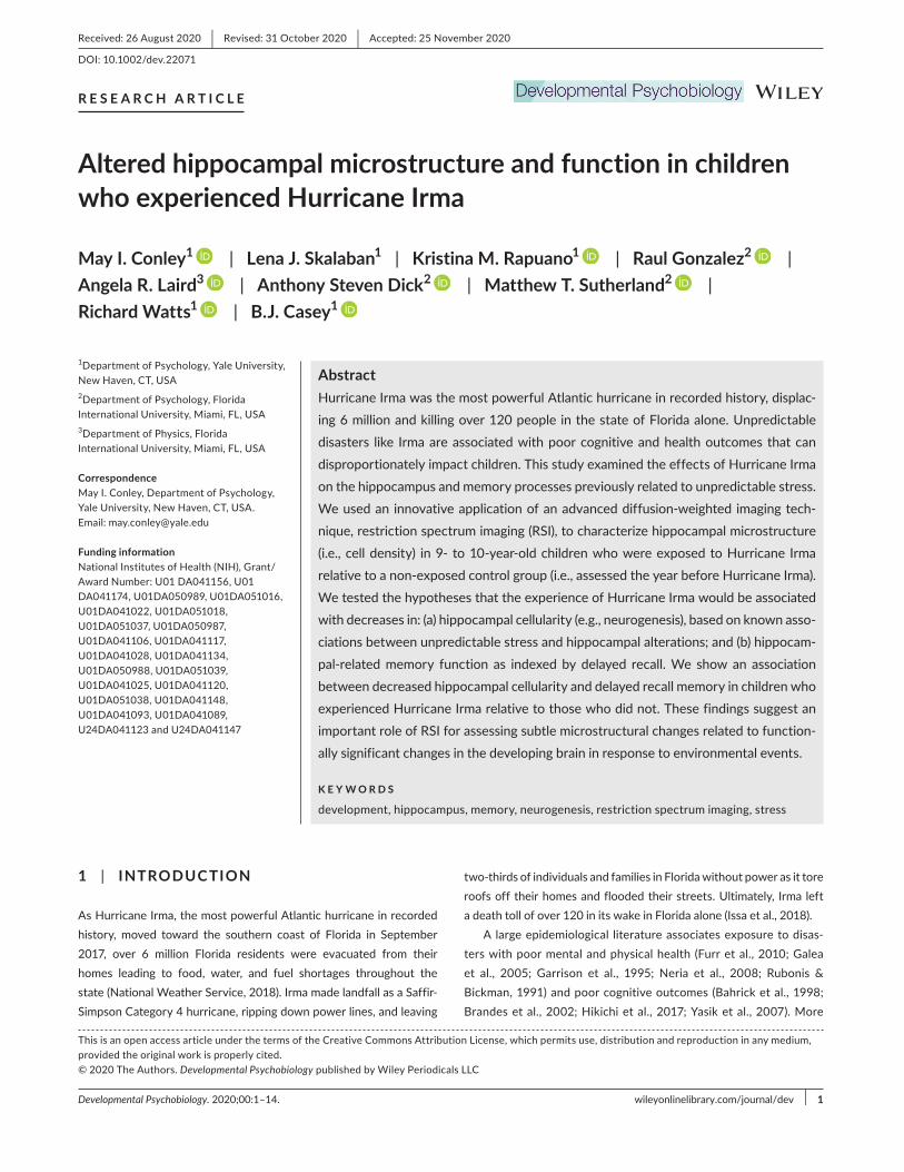
Developmental Psychobiology. 2020;00:1–14. wileyonlinelibrary.com/journal/dev | 1 1 | INTRODUCTION As Hurricane Irma, the most powerful Atlantic hurricane in recorded history, moved toward the southern coast of Florida in September 2017, over 6 million Florida residents were evacuated from their homes leading to food, water, and fuel shortages throughout the state (National Weather Service, 2018). Irma made landfall as a Saffir- Simpson Category 4 hurricane, ripping down power lines, and leaving two-thirds of individuals and families in Florida without power as it tore roofs off their homes and flooded their streets. Ultimately, Irma left a death toll of over 120 in its wake in Florida alone (Issa et al., 2018). A large epidemiological literature associates exposure to disas- ters with poor mental and physical health (Furr et al., 2010; Galea et al., 2005; Garrison et al., 1995; Neria et al., 2008; Rubonis & Bickman, 1991) and poor cognitive outcomes (Bahrick et al., 1998; Brandes et al., 2002; Hikichi et al., 2017; Yasik et al., 2007). More Received: 26 August 2020 | Revised: 31 October 2020 | Accepted: 25 November 2020 DOI: 10.1002/dev.22071 RESEARCH ARTICLE Altered hippocampal microstructure and function in children who experienced Hurricane Irma May I. Conley 1 | Lena J. Skalaban 1 | Kristina M. Rapuano 1 | Raul Gonzalez 2 | Angela R. Laird 3 | Anthony Steven Dick 2 | Matthew T. Sutherland 2 | Richard Watts 1 | B.J. Casey 1 1 Department of Psychology, Yale University, New Haven, CT, USA 2 Department of Psychology, Florida International University, Miami, FL, USA 3 Department of Physics, Florida International University, Miami, FL, USA Correspondence May I. Conley, Department of Psychology, Yale University, New Haven, CT, USA. Email: [email protected] Funding information National Institutes of Health (NIH), Grant/ Award Number: U01 DA041156, U01 DA041174, U01DA050989, U01DA051016, U01DA041022, U01DA051018, U01DA051037, U01DA050987, U01DA041106, U01DA041117, U01DA041028, U01DA041134, U01DA050988, U01DA051039, U01DA041025, U01DA041120, U01DA051038, U01DA041148, U01DA041093, U01DA041089, U24DA041123 and U24DA041147 Abstract Hurricane Irma was the most powerful Atlantic hurricane in recorded history, displac- ing 6 million and killing over 120 people in the state of Florida alone. Unpredictable disasters like Irma are associated with poor cognitive and health outcomes that can disproportionately impact children. This study examined the effects of Hurricane Irma on the hippocampus and memory processes previously related to unpredictable stress. We used an innovative application of an advanced diffusion-weighted imaging tech- nique, restriction spectrum imaging (RSI), to characterize hippocampal microstructure (i.e., cell density) in 9- to 10-year-old children who were exposed to Hurricane Irma relative to a non-exposed control group (i.e., assessed the year before Hurricane Irma). We tested the hypotheses that the experience of Hurricane Irma would be associated with decreases in: (a) hippocampal cellularity (e.g., neurogenesis), based on known asso- ciations between unpredictable stress and hippocampal alterations; and (b) hippocam- pal-related memory function as indexed by delayed recall. We show an association between decreased hippocampal cellularity and delayed recall memory in children who experienced Hurricane Irma relative to those who did not. These findings suggest an important role of RSI for assessing subtle microstructural changes related to function- ally significant changes in the developing brain in response to environmental events. KEYWORDS development, hippocampus, memory, neurogenesis, restriction spectrum imaging, stress This is an open access article under the terms of the Creative Commons Attribution License, which permits use, distribution and reproduction in any medium, provided the original work is properly cited. © 2020 The Authors. Developmental Psychobiology published by Wiley Periodicals LLC

Welcome message from author
This document is posted to help you gain knowledge. Please leave a comment to let me know what you think about it! Share it to your friends and learn new things together.
Transcript
Developmental Psychobiology. 2020;00:1–14. wileyonlinelibrary.com/journal/dev | 1
1 | INTRODUC TION
As Hurricane Irma, the most powerful Atlantic hurricane in recorded
history, moved toward the southern coast of Florida in September
2017, over 6 million Florida residents were evacuated from their
homes leading to food, water, and fuel shortages throughout the
state (National Weather Service, 2018). Irma made landfall as a Saffir-
Simpson Category 4 hurricane, ripping down power lines, and leaving
two-thirds of individuals and families in Florida without power as it tore
roofs off their homes and flooded their streets. Ultimately, Irma left
a death toll of over 120 in its wake in Florida alone (Issa et al., 2018).
A large epidemiological literature associates exposure to disas-
ters with poor mental and physical health (Furr et al., 2010; Galea
et al., 2005; Garrison et al., 1995; Neria et al., 2008; Rubonis &
Bickman, 1991) and poor cognitive outcomes (Bahrick et al., 1998;
Brandes et al., 2002; Hikichi et al., 2017; Yasik et al., 2007). More
Received: 26 August 2020 | Revised: 31 October 2020 | Accepted: 25 November 2020
DOI: 10.1002/dev.22071
R E S E A R C H A R T I C L E
Altered hippocampal microstructure and function in children who experienced Hurricane Irma
May I. Conley1 | Lena J. Skalaban1 | Kristina M. Rapuano1 | Raul Gonzalez2 | Angela R. Laird3 | Anthony Steven Dick2 | Matthew T. Sutherland2 | Richard Watts1 | B.J. Casey1
1Department of Psychology, Yale University,
New Haven, CT, USA
2Department of Psychology, Florida
International University, Miami, FL, USA
3Department of Physics, Florida
International University, Miami, FL, USA
CorrespondenceMay I. Conley, Department of Psychology,
Yale University, New Haven, CT, USA.
Email: [email protected]
Funding informationNational Institutes of Health (NIH), Grant/
Award Number: U01 DA041156, U01
DA041174, U01DA050989, U01DA051016,
U01DA041022, U01DA051018,
U01DA051037, U01DA050987, U01DA041106, U01DA041117,
U01DA041028, U01DA041134, U01DA050988, U01DA051039, U01DA041025, U01DA041120,
U01DA051038, U01DA041148, U01DA041093, U01DA041089, U24DA041123 and U24DA041147
AbstractHurricane Irma was the most powerful Atlantic hurricane in recorded history, displac-
ing 6 million and killing over 120 people in the state of Florida alone. Unpredictable
disasters like Irma are associated with poor cognitive and health outcomes that can
disproportionately impact children. This study examined the effects of Hurricane Irma
on the hippocampus and memory processes previously related to unpredictable stress.
We used an innovative application of an advanced diffusion-weighted imaging tech-
nique, restriction spectrum imaging (RSI), to characterize hippocampal microstructure
(i.e., cell density) in 9- to 10-year-old children who were exposed to Hurricane Irma
relative to a non-exposed control group (i.e., assessed the year before Hurricane Irma).
We tested the hypotheses that the experience of Hurricane Irma would be associated
with decreases in: (a) hippocampal cellularity (e.g., neurogenesis), based on known asso-
ciations between unpredictable stress and hippocampal alterations; and (b) hippocam-
pal-related memory function as indexed by delayed recall. We show an association
between decreased hippocampal cellularity and delayed recall memory in children who
experienced Hurricane Irma relative to those who did not. These findings suggest an
important role of RSI for assessing subtle microstructural changes related to function-
ally significant changes in the developing brain in response to environmental events.
K E Y W O R D S
development, hippocampus, memory, neurogenesis, restriction spectrum imaging, stress
This is an open access article under the terms of the Creative Commons Attribution License, which permits use, distribution and reproduction in any medium,
provided the original work is properly cited.
© 2020 The Authors. Developmental Psychobiology published by Wiley Periodicals LLC
2 | CONLEY Et aL.
specifically, time-marked, unpredictable events such as hurricanes
and other natural disasters have been linked to alterations in brain
and behavior (Chen et al., 2019; Ke et al., 2018; Kessel et al., 2018;
Kopala-Sibley et al., 2016), with some evidence suggesting that chil-
dren are disproportionately affected by natural disasters relative to
adults (Satcher et al., 2007). Notably, prior investigations of neural
mechanisms impacted by other forms of unpredictable stress pro-
vide important insight into how unpredictable events can lead to
lasting or transitory alterations in brain and behavior.
The experience and expression of emotions related to environ-
mental events is associated with the limbic system (Blum et al., 2000).
One limbic region known to be impacted by unpredictable stress in the hippocampus (Cameron & Schoenfeld, 2018; McEwen et al., 2015;
Redish, 2016), which plays a key role in learning and memory (Cohen
& Eichenbaum, 1993; O’Keefe & Nadel, 1978; Scoville & Milner, 1957; Squire, 2009) about emotional events and places (Girardeau et al., 2017;
Kensinger & Corkin, 2004; LeDoux, 1993; Phelps, 2004). The duration, intensity, and predictability of stressful events (Joëls & Baram, 2009;
Tottenham & Sheridan, 2010) can differentially impact hippocampal
structure and function at microstructural (i.e., cellular) and macrostruc-
tural levels (Chen et al., 2010; Liston & Gan, 2011; Lupien et al., 2009;
McEwen, 1999, 2007). Hurricanes like Irma vary in their trajectory, du-
ration, and intensity of destruction, often bringing about unpredictable
events including evacuation, floods, power outages, and food and water
shortages, which can increase hurricane-related stress and vulnera-
bility for long-term mental health problems (McLaughlin et al., 2010).
Time-limited stressors can inhibit the induction of long-term potentia-
tion (LTP) in the hippocampus (Diamond et al., 1990; Foy et al., 1987),
with predictability modulating the magnitude of LTP (Kavushansky
et al., 2006; Shors et al., 1989, 1990). LTP is a reflection of synaptic
plasticity and associated with dendritic arborization and the forma-
tion of new synapses (Bliss & Gardner-Medwin, 1973; Bliss & Lømo, 1973). Hippocampal neurogenesis is also linked to neural plasticity and together these processes are thought to support optimal exploration
of novel events and environments (Glasper et al., 2012). Unpredictable
stressful events are associated with hippocampal microstructure
changes in adult animal models including reductions in hippocampal
neurogenesis (Gould et al., 1997, 1998; Tanapat et al., 2001), spine den-
sity in basal dendrites of CA1 (Diamond et al., 2006), and apical den-
drites of CA3 (Chen et al., 2008, 2010; Magariños & McEwen, 1995; Stewart et al., 2005). Importantly, for the purposes of this study, al-
terations in the hippocampus to unpredictable events are observed in
younger non-human animals (Hollis et al., 2013; Romeo, 2017; Simon et al., 2005; Tanapat et al., 1998). Specifically, pre-pubertal and adoles-
cent non-human animals exhibit protracted stress-responses relative
to adults (Lupien et al., 2009; Romeo, 2017). While these long lasting
changes may be associated with elevated risk for disease (Kim et al.,
2015), dendritic atrophy and decreases in hippocampal neurogenesis
may facilitate adaptive responding and focus on a stressor by mitigat-
ing structural changes and increasing temporary vigilance in the face
of uncertain environmental conditions (Cameron & Schoenfeld, 2018).
Together this work highlights the responsivity of the hippocampus to
unpredictable experiences. However, less is known about microstruc-
tural changes that may underlie these associations in the developing
human brain underscoring the importance of elucidating behaviorally
significant changes in the hippocampus following natural disasters like
Irma.
Recent advances in diffusion-weighted imaging (DWI) allow for
non-invasively examining microstructural detail of the hippocampus.
For example, changes in hippocampal microstructure (e.g., neurite
density and dispersion) measured with advanced DWI techniques are
associated with age-related changes in memory performance in adults
(Radhakrishnan et al., 2020; Zhang et al., 2012), demonstrating that
probing the microstructure of gray matter may be important for char-
acterizing functionally relevant changes. Other recent methodological advances using diffusion magnetic resonance imaging provide further
opportunity to examine the microstructure of deep gray matter by
providing estimates of cell and neurite density. Restriction spectrum
imaging (RSI) provides an estimate of the cellularity of gray-matter tis-
sue in specific areas of the brain by separating the diffusion-weighted
MRI signal into restricted and hindered diffusion components, which
represent intracellular and extracellular water signal, respectively
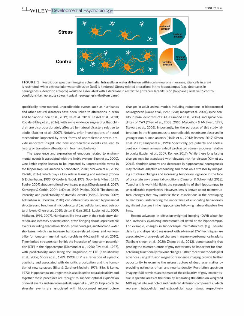
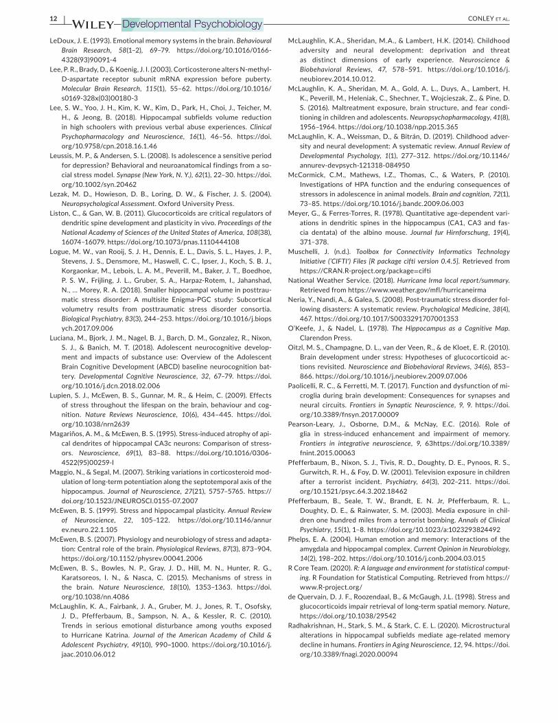
F I G U R E 1 Restriction spectrum imaging schematic. Intracellular water diffusion within cells (neurons in orange; glial cells in gray)
is restricted, while extracellular water diffusion (teal) is hindered. Stress-related alterations in the hippocampus (e.g., decreases in
neurogenesis, dendritic atrophy) would be associated with a decrease in restricted (intracellular) diffusion (top panel) relative to control
conditions (i.e., no acute stress; typical neurogenesis) (bottom panel)
| 3CONLEY Et aL.
(White, Leergaard, et al., 2013; White, McDonald, et al., 2013; White et al., 2014; Figure 1). Accordingly, a greater fraction of restricted com-
ponent (i.e., a greater proportion of restricted motion of water mole-
cules) is believed to indicate greater tissue cellularity, an interpretation
which has been histologically confirmed (White, Leergaard, et al.,
2013). This methodology provides a novel opportunity to investigate microstructural changes in the developing human brain following ex-
posure to unpredictable and/or stressful events.
Here, we utilized RSI to test for microstructural differences in the hip-
pocampus of children exposed to Hurricane Irma, a naturalistic, unpre-
dictable event. More specifically, we evaluated whether Irma-exposed
children, tested in the year prior to Hurricane Irma, show decreased
hippocampal cellularity relative to a non-exposed control group, tested
during the year before Hurricane Irma. To test for regional specificity of
any hippocampal effects, we examined all subcortical brain regions. To
evaluate functional consequences of Irma and changes in hippocampal
cellularity between Irma-exposed and non-exposed groups, we tested
whether Irma-exposure was associated with group differences in hippo-
campal-related memory (i.e., delayed recall). We hypothesized that the
experience of Hurricane Irma would be associated with decreases in (a)
hippocampal cell density and (b) delayed recall memory.
2 | METHODS
2.1 | Participants
Participants were 9- and 10-year-old children from the South
Florida site of the Adolescent Brain Cognitive Development (ABCD)
Study®
(https://ABCDS tudy.org, https://nda.nih.gov/abcd; Volkow
et al., 2018) included in the 2.0.1 ABCD Data Release (https://doi.
org/10.15154/ 1504041). Detailed descriptions of ABCD Study®
sampling procedures and design are detailed elsewhere (Compton
et al., 2019; Garavan et al., 2018). To assess whether Hurricane Irma
exposure was related to hippocampal microstructure and memory,
participants were divided into non-exposed and Irma-exposed
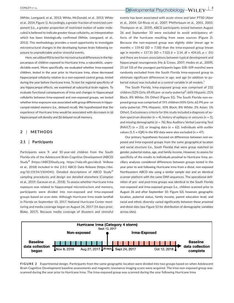
groups based on scan date. Although Hurricane Irma made landfall
in Florida on September 10, 2017, National Hurricane Center moni-
toring and media coverage began on August 26, 2017 (14 days prior;
Blake, 2017). Because media coverage of disasters and stressful
events has been associated with acute stress and later PTSD (Aber
et al., 2004; Gil-Rivas et al., 2007; Pfefferbaum et al., 2001, 2003; Thompson et al., 2019), ABCD participants tested between August
26 and September 10 were excluded to avoid anticipatory ef-
fects of the hurricane resulting from news sources (Figure 2).
Because the non-exposed group was slightly older (mean age in
months = 119.42 (SD = 7.18)) than the Irma-exposed group (mean
age in months = 117.31 (SD = 7.52)) (t = 3.14, df = 424.65, p < .01)
and there are known associations between typical development and
hippocampal neurogenesis (He & Crews, 2007; Hodes et al., 2009),
25 (of 53) of the youngest participants (ages 108–109 months) were randomly excluded from the South Florida Irma-exposed group to
eliminate significant differences in age, and age (in addition to pu-
bertal status) was included as a control variable in all models.1
The South Florida, Irma-exposed group was comprised of 232 children (52% Girls; 69.4% pre- or early-pubertal
2; 66% Hispanic; 21%
Black; 8% White; 5% Other) (Figure S1). The South Florida non-ex-
posed group was comprised of 191 children (45% Girls; 62.9% pre- or
early-pubertal; 79% Hispanic; 10% Black; 8% White; 2% Asian; 1%
Other). Exclusionary criteria for this study included a diagnosis of au-
tism spectrum disorder (n = 4), history of epilepsy or seizures (n = 5),
and missing demographic (n = 76), Rey Auditory Verbal Learning Test
(RAVLT) (n = 23), or imaging data (n = 62). Individuals with outlier
values (1.5 × IQR) in the RSI data were also excluded (n = 47).
Our primary hypotheses focused on differences between non-ex-
posed and Irma-exposed groups from the same geographical location
and social structure (i.e., South Florida) that were group matched on
gender, pubertal status, age, and family income. However, to assess for
specificity of the results to individuals proximal to Hurricane Irma, an-
cillary analyses considered differences between groups tested in the
year prior to and following Hurricane Irma from a distal, non-exposed
Northeastern ABCD site using a similar sample size and an identical
scanner platform with the same DWI sequences. The operational defi-
nition of pre- and post-Irma groups was identical to the South Florida
non-exposed and Irma-exposed groups (i.e., children scanned prior to
August 26 and after September 10; Figure S2), however, geographic
location, pubertal status, family income, parent education level, and
racial and ethnic diversity varied significantly between these proximal
and distal sites (see Figure S3 for distribution of demographic variables across sites).
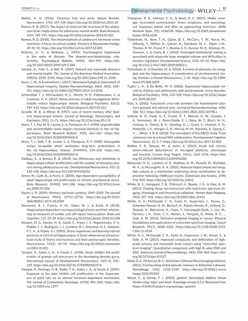
F I G U R E 2 Experimental design. Participants from the same geographic location were divided into two groups based on when Adolescent
Brain Cognitive Development baseline assessments and magnetic resonance imaging scans were acquired. The Irma non-exposed group was
scanned during the year prior to Hurricane Irma. The Irma-exposed group was scanned during the year following Hurricane Irma
4 | CONLEY Et aL.
The distal, Northeastern pre-Irma group was comprised of 181
children (44.1% Girls; 80.9% pre- or early-pubertal; 20.0% Hispanic;
9.2% Black; 61.0% White; 3.6% Asian; 6.2% Other). The distal, Northeastern post-Irma group was comprised of 195 children (48.1%
Girls; 68.0% pre- or early-pubertal; 24.9% Hispanic; 20.4% Black;
45.3% White; 9.4% Other).
2.2 | Neuroimaging data collection
The ABCD scanning protocol includes 3D T1- and T2-weighted im-
ages, diffusion-weighted images, and resting-state and task-based
function MRI measures previously detailed in Casey et al. (2018). Data
were collected on a 3 Tesla Siemens MAGNETOM Prisma scanner with a 32-channel head coil. Diffusion images were collected using a spin-echo EPI acquisition with the following parameters: TR = 88 ms,
TE = 4,100 ms, flip angle = 90°, 81 slices, voxel size = 1.7 mm3, multi-
band slice acceleration factor = 3, 7 b = 0 s/mm2 frames, and 6 direc-
tions at b = 500 s/mm2, 15 directions at b = 1,000 s/mm
2, 15 directions
at b = 2,000 s/mm2, and 60 directions at b = 3,000 s/mm2
.
2.3 | Image preprocessing, RSI, and volumetric data
Diffusion magnetic resonance imaging and structural magnetic
resonance imaging (sMRI) data were processed by the ABCD Study
Data Analysis, Informatics and Resources Center using methods
previously detailed in Hagler et al. (2019) . Restricted normalized
isotropic (N0) metrics were calculated for subcortical gray matter
using a linear estimation approach (White, Leergaard, et al., 2013; White, McDonald, et al., 2013; White et al., 2014) with atlas-based segmentation (Fischl et al., 2002). Hippocampal volume differences
were tested using data computed from the pre-processed T1 images
using FreeSurfer v5.3 and labeled using an atlas-based volumetric segmentation procedure (Fischl et al., 2002; Hagler et al., 2019).
2.4 | Hippocampal-related behavioral function
The RAVLT is a widely used and robust measure of auditory learning,
memory, and recall (Lezak et al., 2004; Luciana et al., 2018). The test
involves five learning trials of 15 unrelated words (list A). After each
trial, participants are asked to recall as many words as possible. After
the initial five learning trials, participants are presented with a distrac-
tor list of 15 new words (list B) and are then asked to recall as many
words as possible from the new list (list B). Next, an immediate recall
trial is assessed for words from the initial list (list A). After a 30-min delay (where participants complete other non-verbal tasks or rest), a
final delayed recall trial is assessed for words from the initial list (list
A). Previous work has established the RAVLT as a reliable measure of
hippocampal integrity (Saury & Emanuelson, 2017) and hippocampal-
dependent memory (Stevenson et al., 2018), linking the delayed recall
trial in particular to hippocampal function (Wolk et al., 2011). Here, we
assessed a behavioral correlate of hippocampal function using perfor-
mance (total correct) on the delayed recall trial (i.e., RAVLT Trial VII).
2.5 | Analytic approach
Analyses were performed in R version 3.6.3 (R Core Team, 2020) using the gamm4 package (Wood & Scheip, 2020). Mixed-effect
models were used to evaluate RSI measures in subcortical regions
as well as verbal memory between non-exposed and Irma-exposed
groups. For all models, covariates included fixed effects for gender,
interview age, race/ethnicity, parental education, and household in-
come, and a random effect for family ID. In addition, RSI models also
included intracranial volume, motion, pubertal development, and the
interaction of gender and pubertal development as fixed covariates.
Supplemental analyses restricting the sample to only pre- and early-
pubertal participants were conducted (Supplemental Analyses 1) in
addition to supplemental analyses including trauma history, threat
exposure, and history of anxiety disorders and PTSD as other fixed
covariates (Supplemental Analyses 2). All analyses were Bonferroni
corrected for multiple comparisons. Non-parametric significance
was assessed using permutation testing by randomly shuffling data
10,000 times. Non-parametric p-values were computed by dividing
the number of times the randomly permuted t-statistic was greater
than the observed t-statistic by the number of tests performed (i.e.,
# observations > |t|/10,000 + 1). Additionally, because there were
no a priori hypotheses that effects would be lateralized, RSI was av-
eraged between hemispheres for each subcortical region. Ancillary
analyses applied identical models to the distal, Northeastern sam-
ple to evaluate if any of the findings detected in the South Florida
sample could be attributed to other background cohort characteris-
tics (e.g., sampling protocols, other overlooked events occurring in
September 2017).
Following the initial region of interest (ROI) analyses, a post hoc
analysis of voxel-wise data was performed to further examine the
spatial specificity of our imaging results. Post hoc analyses were per-
formed by applying the same model from the ROI analyses to every voxel within the subcortex utilizing the cifti (Muschelli, n.d.) and lme4
(Bates et al., 2015) packages. Given previous work showing that the
effects of stress can impact different subfields of the hippocampus
(Hawley & Leasure, 2012; McEwen et al., 2015), the distribution
of voxel-wise RSI (i.e., restricted diffusion) values was plotted as a
function of the anterior/posterior coordinate axis (i.e., y-axis coordi-
nates) to further evaluate spatial specificity within the hippocampus.
3 | RESULTS
3.1 | Decreased hippocampal cellularity in Irma-exposed group
Although a substantial literature across the fields of neuroscience
and psychology describes relationships between unpredictable

| 5CONLEY Et aL.
events and macroscale changes in hippocampal structure and func-
tion, less is known about how these associations emerge in the de-
veloping human brain. Thus, the primary aim of the current study
was to determine if RSI could be used to detect subtle microstruc-
tural differences in the hippocampus of children exposed to a natu-
ralistic unpredictable event, Hurricane Irma (hypothesis 1).
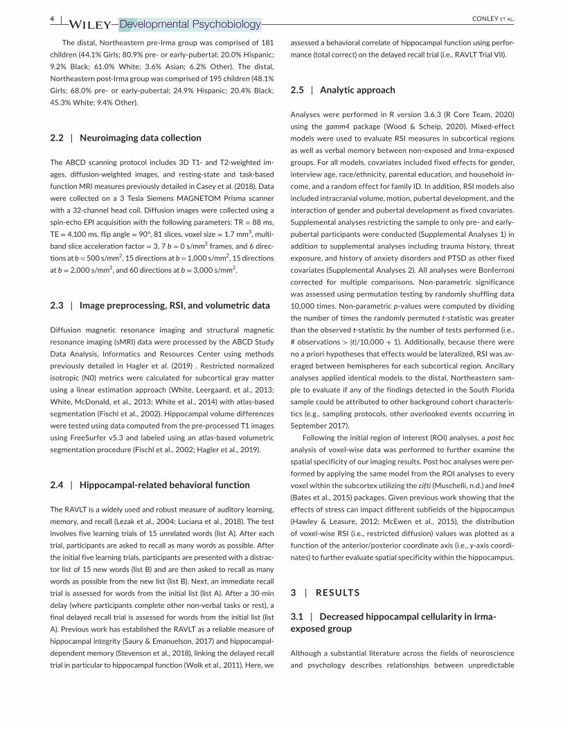
Consistent with hypothesis 1, and with previous literature linking
unpredictable stress to dendritic atrophy and decreased neurogen-
esis in the hippocampus, lower restricted diffusion signal, consistent
with lower cellularity, was observed in the hippocampus of the South
Florida Irma-exposed group relative to the South Florida non-ex-
posed control group (β = −3.90 × 10⁻³
(SE = 1.35 × 10⁻³
), t = −2.875, p = .004, r2
(adj) = .05, Δr2 (adj) = .02; Figure 3a).3,4
Control analyses
demonstrated that these findings are robust to the impact of pu-
berty and various adverse life events or experiences on hippocampal
microstructure and are described in supplemental materials. Next,
we used permutation testing to evaluate the specificity of this ef-
fect to the hippocampus by testing all subcortical regions. Across
F I G U R E 3 Differences in restriction spectrum imaging-based hippocampal cellularity among South Florida Irma-exposed relative to
non-exposed children. (a) Irma exposure was related to decreased hippocampal cell density; (b) post hoc voxel-wise analysis demonstrated
spatial specificity of findings to the hippocampus in right and left hemispheres; (c) results of permutation testing (10,000 + 1 iterations)
demonstrated specificity of Irma exposure on hippocampal cellularity and not to other subcortical regions. The histograms show the null
distribution of t-statistics with observed effects indicated with vertical lines (solid lines represent non-parametric significance and dashed
lines represent non-parametric non-significance)
6 | CONLEY Et aL.
all subcortical areas, only the hippocampus showed significant dif-
ferences in cellularity between the Irma-exposed and non-exposed
control group (Figure 3c).A post hoc voxel-wise analysis of deep gray matter further re-
vealed the spatial specificity of these findings. Consistent with the
ROI results above, voxel-wise models revealed a robust relationship between cellularity measured with RSI and Irma exposure in the hip-
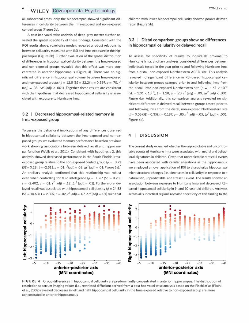
pocampus (Figure 3b). Further evaluation of the spatial distribution of differences in hippocampal cellularity between the Irma-exposed
and non-exposed groups revealed that this effect was more con-
centrated in anterior hippocampus (Figure 4). There was no sig-
nificant difference in hippocampal volume between Irma-exposed
and non-exposed groups (β = 12.5 (SE = 32.2), t = 0.389, p = .70, r2
(adj) = .38, Δr2 (adj) < .001). Together these results are consistent
with the hypothesis that decreased hippocampal cellularity is asso-
ciated with exposure to Hurricane Irma.
3.2 | Decreased hippocampal-related memory in Irma-exposed group
To assess the behavioral implications of any differences observed
in hippocampal cellularity between the Irma-exposed and non-ex-
posed groups, we evaluated memory performance based on previous
work showing associations between delayed recall and hippocam-
pal function (Wolk et al., 2011). Consistent with hypothesis 2, this
analysis showed decreased performance in the South Florida Irma-
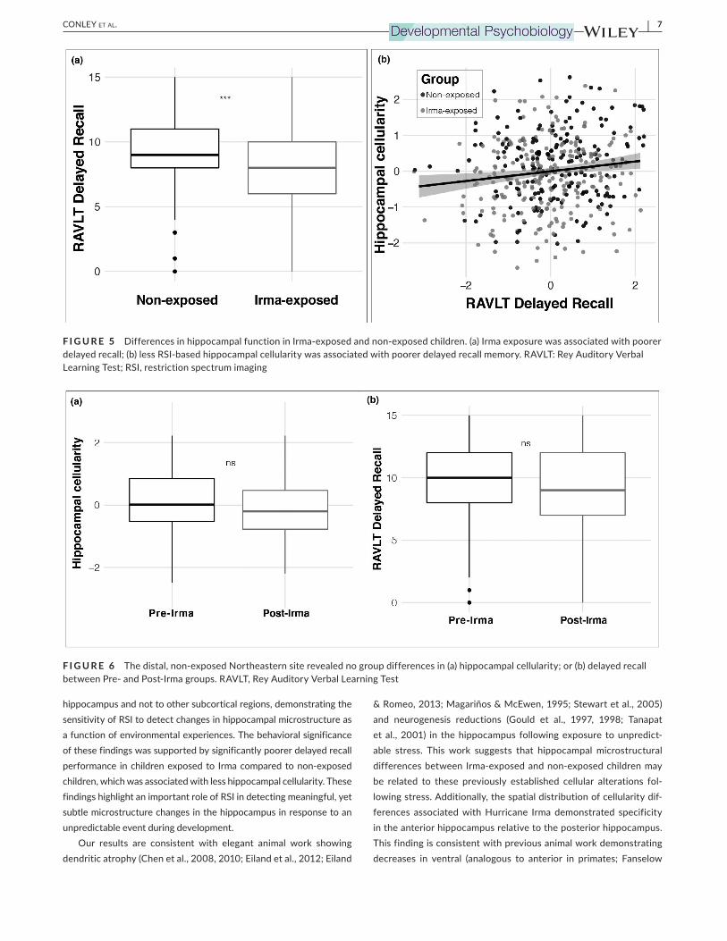
exposed group relative to the non-exposed control group (β = −0.71 (SE = 0.28), t = −2.511, p = .01, r2
(adj) = .08, Δr2 (adj) = .01; Figure 5a).
5
An ancillary analysis confirmed that this relationship was robust
even when controlling for fluid intelligence (β = −0.67 (SE = 0.28),
t = −2.402, p = .01, r2 (adj) = .12, Δr2
(adj) = .01). Furthermore, de-
layed recall was associated with hippocampal cell density (β = 24.53 (SE = 10.63), t = 2.307, p = .02, r2
(adj) = .07, Δr2 (adj) = .01) such that
children with lower hippocampal cellularity showed poorer delayed
recall (Figure 5b).
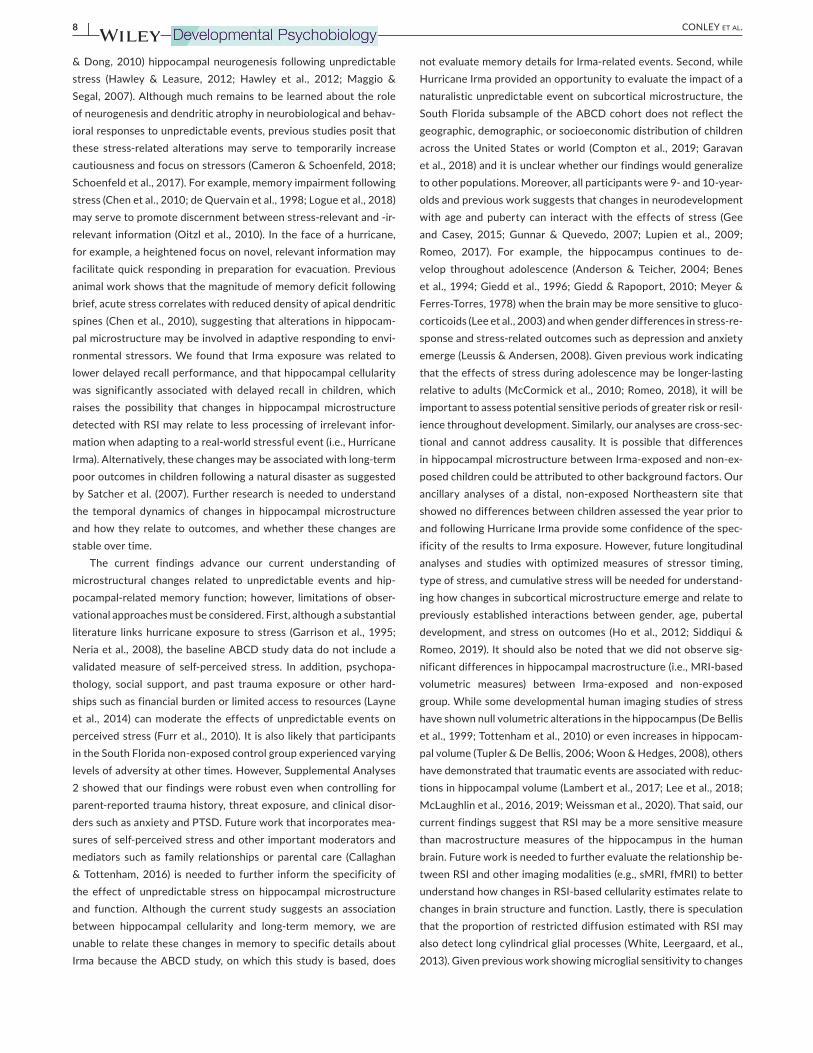
3.3 | Distal comparison groups show no differences in hippocampal cellularity or delayed recall
To assess for specificity of results to individuals proximal to
Hurricane Irma, ancillary analyses considered differences between
individuals tested in the year prior to and following Hurricane Irma
from a distal, non-exposed Northeastern ABCD site. This analysis
revealed no significant difference in RSI-based hippocampal cel-
lularity between groups scanned prior to and following Irma from
the distal, Irma non-exposed Northeastern site (β = −1.67 × 10⁻³
(SE = 1.31 × 10⁻³
), t = 1.28, p = .20, r2 (adj) = .03, Δr2
(adj) < .001;
Figure 6a). Additionally, this comparison analysis revealed no sig-
nificant difference in delayed recall between groups tested prior to
and following Irma from the distal, non-exposed Northeastern site
(β = 0.06 (SE = 0.35), t = 0.187, p = .85, r2 (adj) = .05, Δr2
(adj) < .001;
Figure 6b).
4 | DISCUSSION
The current study examined whether the unpredictable and uncontrol-
lable events of Hurricane Irma were associated with neural and behav-
ioral signatures in children. Given that unpredictable stressful events
have been associated with cellular alterations in the hippocampus,
we employed a novel application of RSI to characterize hippocampal
microstructural changes (i.e., decreases in cellularity) in response to a
naturalistic, unpredictable, and stressful event. The results showed an
association between exposure to Hurricane Irma and decreased RSI-
based hippocampal cellularity in 9- and 10-year-old children. Analyses
across all subcortical regions revealed specificity of this finding to the
F I G U R E 4 Group differences in hippocampal cellularity are predominantly concentrated in anterior hippocampus. The distribution of
restriction spectrum imaging values (i.e., restricted diffusion) derived from a post hoc voxel-wise analysis based on the Fischl atlas (Fischl
et al., 2002) revealed decreases in left and right hippocampal cellularity in the Irma-exposed relative to non-exposed group are more
concentrated in anterior hippocampus
| 7CONLEY Et aL.
hippocampus and not to other subcortical regions, demonstrating the
sensitivity of RSI to detect changes in hippocampal microstructure as
a function of environmental experiences. The behavioral significance
of these findings was supported by significantly poorer delayed recall
performance in children exposed to Irma compared to non-exposed
children, which was associated with less hippocampal cellularity. These
findings highlight an important role of RSI in detecting meaningful, yet
subtle microstructure changes in the hippocampus in response to an
unpredictable event during development.
Our results are consistent with elegant animal work showing dendritic atrophy (Chen et al., 2008, 2010; Eiland et al., 2012; Eiland
& Romeo, 2013; Magariños & McEwen, 1995; Stewart et al., 2005) and neurogenesis reductions (Gould et al., 1997, 1998; Tanapat
et al., 2001) in the hippocampus following exposure to unpredict-
able stress. This work suggests that hippocampal microstructural
differences between Irma-exposed and non-exposed children may
be related to these previously established cellular alterations fol-
lowing stress. Additionally, the spatial distribution of cellularity dif-
ferences associated with Hurricane Irma demonstrated specificity
in the anterior hippocampus relative to the posterior hippocampus.
This finding is consistent with previous animal work demonstrating
decreases in ventral (analogous to anterior in primates; Fanselow
F I G U R E 5 Differences in hippocampal function in Irma-exposed and non-exposed children. (a) Irma exposure was associated with poorer
delayed recall; (b) less RSI-based hippocampal cellularity was associated with poorer delayed recall memory. RAVLT: Rey Auditory Verbal
Learning Test; RSI, restriction spectrum imaging
F I G U R E 6 The distal, non-exposed Northeastern site revealed no group differences in (a) hippocampal cellularity; or (b) delayed recall
between Pre- and Post-Irma groups. RAVLT, Rey Auditory Verbal Learning Test
8 | CONLEY Et aL.
& Dong, 2010) hippocampal neurogenesis following unpredictable
stress (Hawley & Leasure, 2012; Hawley et al., 2012; Maggio &
Segal, 2007). Although much remains to be learned about the role
of neurogenesis and dendritic atrophy in neurobiological and behav-
ioral responses to unpredictable events, previous studies posit that
these stress-related alterations may serve to temporarily increase
cautiousness and focus on stressors (Cameron & Schoenfeld, 2018;
Schoenfeld et al., 2017). For example, memory impairment following
stress (Chen et al., 2010; de Quervain et al., 1998; Logue et al., 2018)
may serve to promote discernment between stress-relevant and -ir-
relevant information (Oitzl et al., 2010). In the face of a hurricane, for example, a heightened focus on novel, relevant information may
facilitate quick responding in preparation for evacuation. Previous
animal work shows that the magnitude of memory deficit following
brief, acute stress correlates with reduced density of apical dendritic
spines (Chen et al., 2010), suggesting that alterations in hippocam-
pal microstructure may be involved in adaptive responding to envi-
ronmental stressors. We found that Irma exposure was related to
lower delayed recall performance, and that hippocampal cellularity
was significantly associated with delayed recall in children, which
raises the possibility that changes in hippocampal microstructure
detected with RSI may relate to less processing of irrelevant infor-
mation when adapting to a real-world stressful event (i.e., Hurricane
Irma). Alternatively, these changes may be associated with long-term
poor outcomes in children following a natural disaster as suggested
by Satcher et al. (2007). Further research is needed to understand
the temporal dynamics of changes in hippocampal microstructure
and how they relate to outcomes, and whether these changes are
stable over time.
The current findings advance our current understanding of
microstructural changes related to unpredictable events and hip-
pocampal-related memory function; however, limitations of obser-
vational approaches must be considered. First, although a substantial
literature links hurricane exposure to stress (Garrison et al., 1995;
Neria et al., 2008), the baseline ABCD study data do not include a
validated measure of self-perceived stress. In addition, psychopa-
thology, social support, and past trauma exposure or other hard-
ships such as financial burden or limited access to resources (Layne
et al., 2014) can moderate the effects of unpredictable events on
perceived stress (Furr et al., 2010). It is also likely that participants
in the South Florida non-exposed control group experienced varying
levels of adversity at other times. However, Supplemental Analyses
2 showed that our findings were robust even when controlling for
parent-reported trauma history, threat exposure, and clinical disor-
ders such as anxiety and PTSD. Future work that incorporates mea-
sures of self-perceived stress and other important moderators and
mediators such as family relationships or parental care (Callaghan
& Tottenham, 2016) is needed to further inform the specificity of
the effect of unpredictable stress on hippocampal microstructure
and function. Although the current study suggests an association
between hippocampal cellularity and long-term memory, we are
unable to relate these changes in memory to specific details about
Irma because the ABCD study, on which this study is based, does
not evaluate memory details for Irma-related events. Second, while
Hurricane Irma provided an opportunity to evaluate the impact of a
naturalistic unpredictable event on subcortical microstructure, the
South Florida subsample of the ABCD cohort does not reflect the
geographic, demographic, or socioeconomic distribution of children
across the United States or world (Compton et al., 2019; Garavan
et al., 2018) and it is unclear whether our findings would generalize
to other populations. Moreover, all participants were 9- and 10-year-
olds and previous work suggests that changes in neurodevelopment
with age and puberty can interact with the effects of stress (Gee
and Casey, 2015; Gunnar & Quevedo, 2007; Lupien et al., 2009;
Romeo, 2017). For example, the hippocampus continues to de-
velop throughout adolescence (Anderson & Teicher, 2004; Benes
et al., 1994; Giedd et al., 1996; Giedd & Rapoport, 2010; Meyer &
Ferres-Torres, 1978) when the brain may be more sensitive to gluco-
corticoids (Lee et al., 2003) and when gender differences in stress-re-
sponse and stress-related outcomes such as depression and anxiety
emerge (Leussis & Andersen, 2008). Given previous work indicating
that the effects of stress during adolescence may be longer-lasting
relative to adults (McCormick et al., 2010; Romeo, 2018), it will be
important to assess potential sensitive periods of greater risk or resil-
ience throughout development. Similarly, our analyses are cross-sec-
tional and cannot address causality. It is possible that differences
in hippocampal microstructure between Irma-exposed and non-ex-
posed children could be attributed to other background factors. Our ancillary analyses of a distal, non-exposed Northeastern site that
showed no differences between children assessed the year prior to
and following Hurricane Irma provide some confidence of the spec-
ificity of the results to Irma exposure. However, future longitudinal
analyses and studies with optimized measures of stressor timing,
type of stress, and cumulative stress will be needed for understand-
ing how changes in subcortical microstructure emerge and relate to
previously established interactions between gender, age, pubertal
development, and stress on outcomes (Ho et al., 2012; Siddiqui &
Romeo, 2019). It should also be noted that we did not observe sig-
nificant differences in hippocampal macrostructure (i.e., MRI-based
volumetric measures) between Irma-exposed and non-exposed
group. While some developmental human imaging studies of stress
have shown null volumetric alterations in the hippocampus (De Bellis
et al., 1999; Tottenham et al., 2010) or even increases in hippocam-
pal volume (Tupler & De Bellis, 2006; Woon & Hedges, 2008), others
have demonstrated that traumatic events are associated with reduc-
tions in hippocampal volume (Lambert et al., 2017; Lee et al., 2018;
McLaughlin et al., 2016, 2019; Weissman et al., 2020). That said, our
current findings suggest that RSI may be a more sensitive measure
than macrostructure measures of the hippocampus in the human
brain. Future work is needed to further evaluate the relationship be-
tween RSI and other imaging modalities (e.g., sMRI, fMRI) to better
understand how changes in RSI-based cellularity estimates relate to
changes in brain structure and function. Lastly, there is speculation
that the proportion of restricted diffusion estimated with RSI may
also detect long cylindrical glial processes (White, Leergaard, et al.,
2013). Given previous work showing microglial sensitivity to changes

| 9CONLEY Et aL.
in environment (Walker et al., 2013), it is possible that the changes in hippocampal microstructure observed in the current study may also
relate to changes in glial processes. However, current research on
the response of glial cells to unpredictable stress is mixed (Delpech
et al., 2016; Jauregui-Huerta et al., 2010; Paolicelli & Ferretti, 2017;
Pearson-Leary et al., 2016) and more research is needed.
Overall, the present study provides novel evidence suggesting the potential utility of RSI in detecting behaviorally significant, yet subtle
microstructure changes in the hippocampus in response to naturalis-
tic stressful events. Although further research is needed, our results
suggest that advanced diffusion MRI methodology may provide novel
opportunities to elucidate how cumulative stressors and dimensions
of stressors (e.g., threat vs. deprivation; McLaughlin et al., 2014), such
as those experienced during the coronavirus disease 19 pandemic,
impact youth. Given changing weather patterns and predicted in-
creases in hurricanes and other natural disasters (Bender et al., 2010),
our results provide important information that has relevance for re-
searchers, clinicians, families, and policy makers for identifying and
ultimately minimizing the impact of these events on young people.
ACKNOWLEDG EMENTSThis work was inspired by the work of the late Bruce McEwen and
his novel insights on the impact of stress on brain structure and
function.
CONFLIC T OF INTERE STThe authors declare no competing financial or other interests.
AUTHOR ' S CONTRIBUTIONMIC and BJC developed the study concept. MIC, KMR, RW, and BJC
contributed to the study design. MIC performed data analysis and
interpretation under the supervision of BJC. MIC drafted the manu-
script and LJS, KMR, RG, ARL, ASD, MTS, RW, and BJC provided
critical edits and revisions. All authors read and approved the final
manuscript.
DATA AVAIL ABILIT Y STATEMENTData used in the preparation of this article were obtained from
the ABCD Study (https://abcds tudy.org), held in the NIMH Data
Archive (NDA). This is a multisite, longitudinal study designed to
recruit more than 10,000 children ages 9–10 and follow them over
10 years into early adulthood. The ABCD Study is supported by the
National Institutes of Health and additional federal partners under
award numbers U01DA041048, U01DA050989, U01DA051016,
U01DA041022, U01DA051018, U01DA051037, U01DA050987, U01DA041174, U01DA041106, U01DA041117, U01DA041028,
U01DA041134, U01DA050988, U01DA051039, U01DA041156, U01DA041025, U01DA041120, U01DA051038, U01DA041148, U01DA041093, U01DA041089, U24DA041123, and U24DA041147. A full list of supporters is available at https://abcds tudy.org/feder al-
partn ers.html. A listing of participating sites and a complete listing
of the study investigators can be found at https://abcds tudy.org/
conso rtium_membe rs/. ABCD consortium investigators designed
and implemented the study and/or provided data but did not neces-
sarily participate in analysis or writing of this report. This manuscript
reflects the views of the authors and may not reflect the opinions or
views of the NIH or ABCD consortium investigators. The ABCD data
repository grows and changes over time. The ABCD data used in this
report came from https://doi.org/10.15154/ 1506087. DOIs can be found at nda.nih.gov/study.html?id=817.
ORCIDMay I. Conley https://orcid.org/0000-0002-0961-387X
Kristina M. Rapuano https://orcid.org/0000-0003-4682-098X
Raul Gonzalez https://orcid.org/0000-0002-5431-4828
Angela R. Laird https://orcid.org/0000-0003-3379-8744
Anthony Steven Dick https://orcid.org/0000-0001-9907-8155
Matthew T. Sutherland https://orcid.org/0000-0002-6091-4037
Richard Watts https://orcid.org/0000-0003-1026-2293
B.J. Casey https://orcid.org/0000-0003-2905-9834
ENDNOTE S 1 All main effects hold when including or excluding these participants.
However, we focus on the sample with 25 participants randomly ex-
cluded in our main text to eliminate significant differences in age be-
tween the Irma-exposed and non-exposed groups.
2 Given previous work showing that development and hormonal
changes during puberty can affect neuronal proliferation within the
hippocampus (Allen et al., 2014; Hueston et al., 2017) and stress re-
sponse (Viau, 2002), supplementary analyses using data from only
pre- or early-pubertal participants was conducted and is provided in
supplementary materials (Supplemental Analyses 1).
3 This result was consistent when including the 25 participants that
were randomly excluded to reduce significant differences in age be-
tween the Irma-exposed and non-exposed groups, (β = −4.20 × 10⁻³
(SE = 1.34 × 10⁻³
), t = −3.29, p = .001, r2 (adj) = .05, Δr2
(adj) = .02).
4 Ancillary analyses revealed no significant difference in hippocampal
cellularity between groups scanned prior to and following Irma from
the distal, Irma-non-exposed Northeastern site (β = −1.67 × 10⁻³
(SE = 1.31 × 10⁻³
), t = 1.28, p = .20, r2 (adj) = .03, Δr2
(adj) < .001;
Figure 6a).
5 Ancillary analysis revealed no significant difference in delayed re-
call between groups tested prior to and following Irma from the dis-
tal, non-exposed Northeastern site (β = 0.06 (SE = 0.35), t = 0.187,
p = .85, r2 (adj) = .05, Δr2
(adj) < .001; Figure 6b).
R E FE R E N CE SAber, J. L., Gershoff, E. T., Ware, A., & Kotler, J. A. (2004). Estimating the
effects of September 11th and other forms of violence on the Mental
Health and Social Development of New York City’s Youth: A matter of context. Applied Developmental Science, 8(3), 111–129. https://doi.org/10.1207/s1532 480xa ds0803_2
Allen, K.M., Fung, S.J., Rothmond, D.A., Noble, P.L., & Shannon Weickert,
C. (2014). Gonadectomy increases neurogenesis in the male adoles-
cent rhesus macaque hippocampus. Hippocampus, 24(2), 225–238. https://doi.org/10.1002/hipo.22217
Andersen, S.L., & Teicher, M.H. (2004). Delayed effects of early stress
on hippocampal development. Neuropsychopharmacology, 29(11),
1988–1993. https://doi.org/10.1038/sj.npp.1300528Bahrick, L. E., Parker, J. F., Fivush, R., & Levitt, M. (1998). The ef-
fects of stress on young children's memory for a natural disaster.
10 | CONLEY Et aL.
Journal of Experimental Psychology: Applied, 4(4), 308. https://doi.org/10.1037/1076-898X.4.4.308
Bates, D., Mächler, M., Bolker, B., & Walker, S. (2015). Fitting linear
mixed-effects models using lme4. Journal of Statistical Software, Articles, 67(1), 1–48. https://doi.org/10.18637/ jss.v067.i01
Bender, M. A., Knutson, T. R., Tuleya, R. E., Sirutis, J. J., Vecchi, G. A.,
Garner, S. T., & Held, I. M. (2010). Modeled impact of anthropogenic
warming on the frequency of intense Atlantic hurricanes. Science,
327(5964), 454–458. https://doi.org/10.1126/scien ce.1180568
Benes, F. M., Turtle, M., Khan, Y., & Farol, P. (1994). Myelination of a
key relay zone in the hippocampal formation occurs in the human
brain during childhood, adolescence, and adulthood. Archives of General Psychiatry, 51(6), 477–484. https://doi.org/10.1001/archp
syc.1994.03950 06004 1004Blake, F. (2017, August 26). NHC graphical outlook archive. National
Oceanic and Atmospheric Administration. Retrieved from https://www.nhc.noaa.gov/archi ve/xgtwo/ gtwo_archi ve.php?curre nt_issua
nce=20170 82623 39&basin =atl&fdays =5
Bliss, T. V., & Gardner-Medwin, A. R. (1973). Long-lasting potentiation of synaptic transmission in the dentate area of the unanaesthetized
rabbit following stimulation of the perforant path. The Journal of Physiology, 232(2), 357–374. https://doi.org/10.1113/jphys iol.1973.sp010274
Bliss, T. V. P., & Lømo, T. (1973). Long-lasting potentiation of synaptic transmission in the dentate area of the anaesthetized rabbit fol-
lowing stimulation of the perforant path. The Journal of Physiology,
232(2), 331–356. https://doi.org/10.1113/jphys iol.1973.sp010273Blum, K., Braverman, E. R., Holder, J. M., Lubar, J. F., Monastra, V. J.,
Miller, D., Lubar, J. O., Chen, T. J., & Comings, D. E. (2000). The reward deficiency syndrome: A biogenetic model for the diagnosis and treat-
ment of impulsive, addictive and compulsive behaviors. Journal of Psychoactive Drugs, 32(sup1), 1–112. https://doi.org/10.1080/02791
072.2000.10736099Brandes, D., Ben-Schachar, G., Gilboa, A., Bonne, O., Freedman, S., &
Shalev, A. Y. (2002). PTSD symptoms and cognitive performance
in recent trauma survivors. Psychiatry Research, 110(3), 231–238. https://doi.org/10.1016/S0165 -1781(02)00125 -7
Callaghan, B. L., & Tottenham, N. (2016). The neuro-environmental loop
of plasticity: A cross-species analysis of parental effects on emo-
tion circuitry development following typical and adverse caregiving.
Neuropsychopharmacology, 41(1), 163–176. https://doi.org/10.1038/npp.2015.204
Cameron, H. A., & Schoenfeld, T. J. (2018). Behavioral and structural
adaptations to stress. Frontiers in Neuroendocrinology, 49, 106–113. https://doi.org/10.1016/j.yfrne.2018.02.002
Casey, B. J., Cannonier, T., Conley, M. I., Cohen, A. O., Barch, D. M., Heitzeg, M. M., Soules, M. E., Teslovich, T., Dellarco, D. V., Garavan,
H., Orr, C. A., Wager, T. D., Banich, M. T., Speer, N. K., Sutherland, M. T., Riedel, M. C., Dick, A. S., Bjork, J. M., Thomas, K. M., …
Dale, A. M. (2018). The Adolescent Brain Cognitive Development
(ABCD) study: Imaging acquisition across 21 sites. Developmental Cognitive Neuroscience, 32, 43–54. https://doi.org/10.1016/j.dcn.2018.03.001
Chen, H. J., Zhang, L. I., Ke, J., Qi, R., Xu, Q., Zhong, Y., Pan, M., Li, J., Lu, G. M., & Chen, F. (2019). Altered resting-state dorsal anterior cingu-
late cortex functional connectivity in patients with post-traumatic
stress disorder. Australian & New Zealand Journal of Psychiatry, 53(1),
68–79. https://doi.org/10.1177/00048 67418 812674
Chen, Y., Dubé, C. M., Rice, C. J., & Baram, T. Z. (2008). Rapid loss
of dendritic spines after stress involves derangement of spine
dynamics by corticotropin-releasing hormone. The Journal of Neuroscience, 28(11), 2903–2911. https://doi.org/10.1523/JNEUR OSCI.0225-08.2008
Chen, Y., Rex, C. S., Rice, C. J., Dubé, C. M., Gall, C. M., Lynch, G., &
Baram, T. Z. (2010). Correlated memory defects and hippocampal
dendritic spine loss after acute stress involve corticotropin-releasing
hormone signaling. Proceedings of the National Academy of Sciences of the United States of America, 107(29), 13123–13128. https://doi.org/10.1073/pnas.10038 25107
Cohen, N. J., & Eichenbaum, H. (1993). Memory, amnesia, and the hippo-campal system. MIT Press.
Compton, W. M., Dowling, G. J., & Garavan, H. (2019). Ensuring the best
use of data: The adolescent brain cognitive development study.
JAMA Pediatrics, 173(9), 809. https://doi.org/10.1001/jamap ediat
rics.2019.2081
De Bellis, M. D., Keshavan, M. S., Clark, D. B., Casey, B. J., Giedd, J. N.,
Boring, A. M., Frustaci, K., & Ryan, N. D. (1999). Developmental trau-
matology part II: Brain development. Biological Psychiatry, 45(10),
1271–1284. https://doi.org/10.1016/S0006 -3223(99)00045 -1Delpech, J. C., Wei, L., Hao, J., Yu, X., Madore, C., Butovsky, O., &
Kaffman, A. (2016). Early life stress perturbs the maturation of mi-
croglia in the developing hippocampus. Brain, Behavior, and Immunity,
57, 79–93. https://doi.org/10.1016/j.bbi.2016.06.006Diamond, D. M., Campbell, A. M., Park, C. R., Woodson, J. C., Conrad, C.
D., Bachstetter, A. D., & Mervis, R. F. (2006). Influence of predator
stress on the consolidation versus retrieval of long-term spatial mem-
ory and hippocampal spinogenesis. Hippocampus, 16(7), 571–576.
https://doi.org/10.1002/hipo.20188
Diamond, D. M., Catherine Bennett, M., Stevens, K. E., Wilson, R. L., & Rose,
G. M. (1990). Exposure to a novel environment interferes with the in-
duction of hippocampal primed burst potentiation in the behaving rat.
Psychobiology, 18(3), 273–281. https://doi.org/10.3758/BF033 27243Eiland, L., Ramroop, J., Hill, M. N., Manley, J., & McEwen, B. S. (2012).
Chronic juvenile stress produces corticolimbic dendritic architec-
tural remodeling and modulates emotional behavior in male and
female rats. Psychoneuroendocrinology, 37(1), 39–47. https://doi.org/10.1016/j.psyne uen.2011.04.015
Eiland, L., & Romeo, R. D. (2013). Stress and the developing adolescent brain. Neuroscience, 249, 162–171. https://doi.org/10.1016/j.neuro
scien ce.2012.10.048
Fanselow, M. S., & Dong, H. W. (2010). Are the dorsal and ventral hippo-
campus functionally distinct structures? Neuron, 65(1), 7–19. https://
doi.org/10.1016/j.neuron.2009.11.031Fischl, B., Salat, D. H., Busa, E., Albert, M., Dieterich, M., Haselgrove, C.,
van der Kouwe, A., Killiany, R., Kennedy, D., Klaveness, S., Montillo,
A., Makris, N., Rosen, B., & Dale, A. M. (2002). Whole brain seg-
mentation: Automated labeling of neuroanatomical structures in
the human brain. Neuron, 33(3), 341–355. https://doi.org/10.1016/s0896 -6273(02)00569 -x
Foy, M. R., Stanton, M. E., Levine, S., & Thompson, R. F. (1987).
Behavioral stress impairs long-term potentiation in rodent hippo-
campus. Behavioral and Neural Biology, 48(1), 138–149. https://doi.org/10.1016/S0163 -1047(87)90664 -9
Furr, J. M., Comer, J. S., Edmunds, J. M., & Kendall, P. C. (2010). Disasters
and youth: A meta-analytic examination of posttraumatic stress.
Journal of Consulting and Clinical Psychology, 78(6), 765–780. https://
doi.org/10.1037/a0021482Galea, S., Nandi, A., & Vlahov, D. (2005). The epidemiology of post-trau-
matic stress disorder after disasters. Epidemiologic Reviews, 27(1),
78–91. https://doi.org/10.1093/epire v/mxi003Garavan, H., Bartsch, H., Conway, K., Decastro, A., Goldstein, R. Z.,
Heeringa, S., Jernigan, T., Potter, A., Thompson, W., & Zahs, D.
(2018). Recruiting the ABCD sample: Design considerations and pro-
cedures. Developmental Cognitive Neuroscience, 32, 16–22. https://
doi.org/10.1016/j.dcn.2018.04.004
Garrison, C. Z., Bryant, E. S., Addy, C. L., Spurrier, P. G., Freedy, J. R.,
& Kilpatrick, D. G. (1995). Posttraumatic stress disorder in adoles-
cents after Hurricane Andrew. Journal of the American Academy of Child & Adolescent Psychiatry, 34(9), 1193–1201. https://doi.org/10.1097/00004 583-19950 9000-00017
| 11CONLEY Et aL.
Gee, D.G., & Casey, B.J. (2015). The impact of developmental timing for
stress and recovery. Neurobiology of stress, 1, 184–194. https://doi.
org/10.1016/j.ynstr.2015.02.001
Giedd, J.N., & Rapoport, J.L. (2010). Structural MRI of pediatric brain de-
velopment: what have we learned and where are we going?. Neuron,
67(5), 728–734. https://doi.org/10.1016/j.neuron.2010.08.040Giedd, J.N., Vaituzis, A.C., Hamburger, S.D., Lange, N., Rajapakse, J.C.,
Kaysen, D., Vauss, Y.C., & Rapoport, J.L. (1996). Quantitative MRI
of the temporal lobe, amygdala, and hippocampus in normal human
development: ages 4–18 years. Journal of Comparative Neurology,
366(2), 223–230. https://doi.org/10.1002/(SICI)1096-9861(19960 304)366:2<223::AID-CNE3>3.0.CO;2-7
Gil-Rivas, V., Silver, R. C., Holman, E. A., McIntosh, D. N., & Poulin,
M. (2007). Parental response and adolescent adjustment to the
September 11, 2001 terrorist attacks. Journal of Traumatic Stress,
20(6), 1063–1068. https://doi.org/10.1002/jts.20277Girardeau, G., Inema, I., & Buzsáki, G. (2017). Reactivations of emo-
tional memory in the hippocampus–amygdala system during
sleep. Nature Neuroscience, 20(11), 1634. https://doi.org/10.1038/nn.4637
Glasper, E. R., Schoenfeld, T. J., & Gould, E. (2012). Adult neuro-
genesis: Optimizing hippocampal function to suit the environ-
ment. Behavioural Brain Research, 227(2), 380–383. https://doi.org/10.1016/j.bbr.2011.05.013
Gould, E., McEwen, B. S., Tanapat, P., Galea, L. A., & Fuchs, E. (1997).
Neurogenesis in the dentate gyrus of the adult tree shrew is reg-
ulated by psychosocial stress and NMDA receptor activation. The Journal of Neuroscience, 17(7), 2492–2498. https://doi.org/10.1523/JNEUR OSCI.17-07-02492.1997
Gould, E., Tanapat, P., McEwen, B. S., Flügge, G., & Fuchs, E. (1998).
Proliferation of granule cell precursors in the dentate gyrus of adult
monkeys is diminished by stress. Proceedings of the National Academy of Sciences of the United States of America, 95(6), 3168–3171. https://doi.org/10.1073/pnas.95.6.3168
Gunnar, M., & Quevedo, K. (2007). The neurobiology of stress and de-
velopment. Annual Review of Psychology, 58, 145–173. https://doi.org/10.1146/annur ev.psych.58.110405.085605
Hagler Jr, D.J., Hatton, S., Cornejo, M.D., Makowski, C., Fair, D.A., Dick,
A.S., Sutherland, M.T., Casey, B.J., Barch, D.M., Harms, M.P., & Watts,
R. (2019). Image processing and analysis methods for the Adolescent
Brain Cognitive Development Study. Neuroimage, 202, 116091.
https://doi.org/10.1016/j.neuro image.2019.116091
Hawley, D. F., & Leasure, J. L. (2012). Region-specific response of the
hippocampus to chronic unpredictable stress. Hippocampus, 22(6),
1338–1349. https://doi.org/10.1002/hipo.20970Hawley, D. F., Morch, K., Christie, B. R., & Leasure, J. L. (2012).
Differential response of hippocampal subregions to stress and
learning. PLoS One, 7(12), e53126. https://doi.org/10.1371/journ al.pone.0053126
He, J., & Crews, F. T. (2007). Neurogenesis decreases during brain
maturation from adolescence to adulthood. Pharmacology Biochemistry and Behavior, 86(2), 327–333. https://doi.org/10.1016/j.pbb.2006.11.003
Hikichi, H., Tsuboya, T., Aida, J., Matsuyama, Y., Kondo, K., Subramanian,
S.V., & Kawachi, I. (2017). Social capital and cognitive decline in
the aftermath of a natural disaster: a natural experiment from
the 2011 Great East Japan Earthquake and Tsunami. The Lancet Planetary Health, 1(3), e105–e113. https://doi.org/10.1016/S2542 - 5196(17)30041 -4
Ho, A., Villacis, A. J., Svirsky, S. E., Foilb, A. R., & Romeo, R. D. (2012). The
pubertal-related decline in cellular proliferation and neurogenesis in
the dentate gyrus of male rats is independent of the pubertal rise
in gonadal hormones. Developmental Neurobiology, 72(5), 743–752. https://doi.org/10.1002/dneu.20987
Hodes, G. E., Yang, L., Van Kooy, J., Santollo, J., & Shors, T. J. (2009).
Prozac during puberty: Distinctive effects on neurogenesis as a
function of age and sex. Neuroscience, 163(2), 609–617. https://doi.
org/10.1016/j.neuro scien ce.2009.06.057
Hollis, F., Isgor, C., & Kabbaj, M. (2013). The consequences of adoles-
cent chronic unpredictable stress exposure on brain and behavior.
Neuroscience, 249, 232–241. https://doi.org/10.1016/j.neuro scien ce.2012.09.018
Hueston, C. M., Cryan, J. F., & Nolan, Y. M. (2017). Stress and adolescent
hippocampal neurogenesis: Diet and exercise as cognitive modula-
tors. Translational Psychiatry, 7(4), e1081. https://doi.org/10.1038/tp.2017.48
Issa, A., Ramadugu, K., Mulay, P., Hamilton, J., Siegel, V., Harrison, C.,
Campbell, C. M., Blackmore, C., Bayleyegn, T., & Boehmer, T. (2018).
Deaths related to Hurricane Irma—Florida, Georgia, and North Carolina,
September 4–October 10, 2017. Morbidity and Mortality Weekly Report, 67(30), 829. https://doi.org/10.15585/ mmwr.mm6730a5
Jauregui-Huerta, F., Ruvalcaba-Delgadillo, Y., Gonzalez-Perez, O., Gonzalez-Castañeda, R., Garcia-Estrada, J., & Luquin, S. (2010). Responses of glial cells to stress and glucocorticoids. Current Immunology Reviews, 6(3), 195–204. https://doi.org/10.2174/15733 95107 91823790
Joëls, M., & Baram, T. Z. (2009). The neuro-symphony of stress. Nature Reviews Neuroscience, 10(6), 459–466. https://doi.org/10.1038/nrn2632
Kavushansky, A., Vouimba, R.-M., Cohen, H., & Richter-Levin, G.
(2006). Activity and plasticity in the CA1, the dentate gyrus, and
the amygdala following controllable vs. uncontrollable water stress.
Hippocampus, 16(1), 35–42. https://doi.org/10.1002/hipo.20130Ke, J., Zhang, L. I., Qi, R., Xu, Q., Zhong, Y., Liu, T., Li, J., Lu, G., & Chen, F.
(2018). Typhoon-related post-traumatic stress disorder and trauma
might lead to functional integration abnormalities in intra-and in-
ter-resting state networks: A resting-state fMRI independent com-
ponent analysis. Cellular Physiology and Biochemistry, 48(1), 99–110.
https://doi.org/10.1159/00049 1666
Kensinger, E. A., & Corkin, S. (2004). Two routes to emotional memory:
Distinct neural processes for valence and arousal. Proceedings of the National Academy of Sciences of the United States of America, 101(9),
3310–3315. https://doi.org/10.1073/pnas.03064 08101Kessel, E. M., Nelson, B. D., Kujawa, A., Hajcak, G., Kotov, R., Bromet, E.
J., Carlson, G. A., & Klein, D. N. (2018). Hurricane Sandy exposure
alters the development of neural reactivity to negative stimuli in chil-
dren. Child Development, 89(2), 339–348. https://doi.org/10.1111/cdev.12691
Kim, E. J., Pellman, B., & Kim, J. J. (2015). Stress effects on the hippocam-
pus: a critical review. Learning & memory, 22(9), 411–416. https://doi.
org/10.1101/lm.037291.114Kopala-Sibley, D. C., Danzig, A. P., Kotov, R., Bromet, E. J., Carlson, G. A.,
Olino, T. M., Bhatia, V., Black, S. R., & Klein, D. N. (2016). Negative emotionality and its facets moderate the effects of exposure to
Hurricane Sandy on children’s postdisaster depression and anxiety symptoms. Journal of Abnormal Psychology, 125(4), 471. https://doi.
org/10.1037/abn00 00152Lambert, H. K., Sheridan, M. A., Sambrook, K., Rosen, M., Askren, M.
K., & McLaughlin, K. A. (2017). Hippocampal contribution to con-
text encoding across development is disrupted following early-life
adversity. Journal of Neuroscience, 37(7), 1925–1934. https://doi.org/10.1523/JNEUR OSCI.2618-16.2017
Layne, C. M., Greeson, J. K., Ostrowski, S. A., Kim, S., Reading, S., Vivrette, R. L., Briggs, E. C., Fairbank, J. A., & Pynoos, R. S. (2014).
Cumulative trauma exposure and high risk behavior in adolescence:
Findings from the National Child Traumatic Stress Network Core
Data Set. Psychological Trauma: Theory, Research, Practice, and Policy,
6(Suppl. 1), S40.
12 | CONLEY Et aL.
LeDoux, J. E. (1993). Emotional memory systems in the brain. Behavioural Brain Research, 58(1–2), 69–79. https://doi.org/10.1016/0166-
4328(93)90091 -4Lee, P. R., Brady, D., & Koenig, J. I. (2003). Corticosterone alters N-methyl-
D-aspartate receptor subunit mRNA expression before puberty.
Molecular Brain Research, 115(1), 55–62. https://doi.org/10.1016/
s0169 -328x(03)00180 -3Lee, S. W., Yoo, J. H., Kim, K. W., Kim, D., Park, H., Choi, J., Teicher, M.
H., & Jeong, B. (2018). Hippocampal subfields volume reduction
in high schoolers with previous verbal abuse experiences. Clinical Psychopharmacology and Neuroscience, 16(1), 46–56. https://doi.
org/10.9758/cpn.2018.16.1.46
Leussis, M. P., & Andersen, S. L. (2008). Is adolescence a sensitive period
for depression? Behavioral and neuroanatomical findings from a so-
cial stress model. Synapse (New York, N. Y.), 62(1), 22–30. https://doi.org/10.1002/syn.20462
Lezak, M. D., Howieson, D. B., Loring, D. W., & Fischer, J. S. (2004).
Neuropsychological Assessment. Oxford University Press.Liston, C., & Gan, W. B. (2011). Glucocorticoids are critical regulators of
dendritic spine development and plasticity in vivo. Proceedings of the National Academy of Sciences of the United States of America, 108(38), 16074–16079. https://doi.org/10.1073/pnas.11104 44108
Logue, M. W., van Rooij, S. J. H., Dennis, E. L., Davis, S. L., Hayes, J. P.,
Stevens, J. S., Densmore, M., Haswell, C. C., Ipser, J., Koch, S. B. J.,
Korgaonkar, M., Lebois, L. A. M., Peverill, M., Baker, J. T., Boedhoe,
P. S. W., Frijling, J. L., Gruber, S. A., Harpaz-Rotem, I., Jahanshad,
N., … Morey, R. A. (2018). Smaller hippocampal volume in posttrau-
matic stress disorder: A multisite Enigma-PGC study: Subcortical
volumetry results from posttraumatic stress disorder consortia.
Biological Psychiatry, 83(3), 244–253. https://doi.org/10.1016/j.biops ych.2017.09.006
Luciana, M., Bjork, J. M., Nagel, B. J., Barch, D. M., Gonzalez, R., Nixon,
S. J., & Banich, M. T. (2018). Adolescent neurocognitive develop-
ment and impacts of substance use: Overview of the Adolescent Brain Cognitive Development (ABCD) baseline neurocognition bat-
tery. Developmental Cognitive Neuroscience, 32, 67–79. https://doi.
org/10.1016/j.dcn.2018.02.006
Lupien, S. J., McEwen, B. S., Gunnar, M. R., & Heim, C. (2009). Effects
of stress throughout the lifespan on the brain, behaviour and cog-
nition. Nature Reviews Neuroscience, 10(6), 434–445. https://doi.org/10.1038/nrn2639
Magariños, A. M., & McEwen, B. S. (1995). Stress-induced atrophy of api-cal dendrites of hippocampal CA3c neurons: Comparison of stress-
ors. Neuroscience, 69(1), 83–88. https://doi.org/10.1016/0306-4522(95)00259 -l
Maggio, N., & Segal, M. (2007). Striking variations in corticosteroid mod-
ulation of long-term potentiation along the septotemporal axis of the
hippocampus. Journal of Neuroscience, 27(21), 5757–5765. https://
doi.org/10.1523/JNEUR OSCI.0155-07.2007McEwen, B. S. (1999). Stress and hippocampal plasticity. Annual Review
of Neuroscience, 22, 105–122. https://doi.org/10.1146/annur
ev.neuro.22.1.105
McEwen, B. S. (2007). Physiology and neurobiology of stress and adapta-
tion: Central role of the brain. Physiological Reviews, 87(3), 873–904. https://doi.org/10.1152/physr ev.00041.2006
McEwen, B. S., Bowles, N. P., Gray, J. D., Hill, M. N., Hunter, R. G.,
Karatsoreos, I. N., & Nasca, C. (2015). Mechanisms of stress in
the brain. Nature Neuroscience, 18(10), 1353–1363. https://doi.org/10.1038/nn.4086
McLaughlin, K. A., Fairbank, J. A., Gruber, M. J., Jones, R. T., Osofsky, J. D., Pfefferbaum, B., Sampson, N. A., & Kessler, R. C. (2010).
Trends in serious emotional disturbance among youths exposed
to Hurricane Katrina. Journal of the American Academy of Child & Adolescent Psychiatry, 49(10), 990–1000. https://doi.org/10.1016/j.
jaac.2010.06.012
McLaughlin, K.A., Sheridan, M.A., & Lambert, H.K. (2014). Childhood
adversity and neural development: deprivation and threat
as distinct dimensions of early experience. Neuroscience & Biobehavioral Reviews, 47, 578–591. https://doi.org/10.1016/j.
neubi orev.2014.10.012.
McLaughlin, K. A., Sheridan, M. A., Gold, A. L., Duys, A., Lambert, H.
K., Peverill, M., Heleniak, C., Shechner, T., Wojcieszak, Z., & Pine, D.
S. (2016). Maltreatment exposure, brain structure, and fear condi-
tioning in children and adolescents. Neuropsychopharmacology, 41(8),
1956–1964. https://doi.org/10.1038/npp.2015.365McLaughlin, K. A., Weissman, D., & Bitrán, D. (2019). Childhood adver-
sity and neural development: A systematic review. Annual Review of Developmental Psychology, 1(1), 277–312. https://doi.org/10.1146/annur ev-devps ych-12131 8-084950
McCormick, C.M., Mathews, I.Z., Thomas, C., & Waters, P. (2010).
Investigations of HPA function and the enduring consequences of
stressors in adolescence in animal models. Brain and cognition, 72(1),
73–85. https://doi.org/10.1016/j.bandc.2009.06.003Meyer, G., & Ferres-Torres, R. (1978). Quantitative age-dependent vari-
ations in dendritic spines in the hippocampus (CA1, CA3 and fas-
cia dentata) of the albino mouse. Journal fur Hirnforschung, 19(4),
371–378.Muschelli, J. (n.d.). Toolbox for Connectivity Informatics Technology
Initiative (’CIFTI') Files [R package cifti version 0.4.5]. Retrieved from
https://CRAN.R-proje ct.org/packa ge=cifti
National Weather Service. (2018). Hurricane Irma local report/summary.
Retrieved from https://www.weath er.gov/mfl/hurri caneirma
Neria, Y., Nandi, A., & Galea, S. (2008). Post-traumatic stress disorder fol-
lowing disasters: A systematic review. Psychological Medicine, 38(4),
467. https://doi.org/10.1017/S0033 29170 7001353O’Keefe, J., & Nadel, L. (1978). The Hippocampus as a Cognitive Map.
Clarendon Press.
Oitzl, M. S., Champagne, D. L., van der Veen, R., & de Kloet, E. R. (2010). Brain development under stress: Hypotheses of glucocorticoid ac-
tions revisited. Neuroscience and Biobehavioral Reviews, 34(6), 853–866. https://doi.org/10.1016/j.neubi orev.2009.07.006
Paolicelli, R. C., & Ferretti, M. T. (2017). Function and dysfunction of mi-
croglia during brain development: Consequences for synapses and
neural circuits. Frontiers in Synaptic Neuroscience, 9, 9. https://doi.
org/10.3389/fnsyn.2017.00009Pearson-Leary, J., Osborne, D.M., & McNay, E.C. (2016). Role of
glia in stress-induced enhancement and impairment of memory.
Frontiers in integrative neuroscience, 9, 63https://doi.org/10.3389/fnint.2015.00063
Pfefferbaum, B., Nixon, S. J., Tivis, R. D., Doughty, D. E., Pynoos, R. S.,
Gurwitch, R. H., & Foy, D. W. (2001). Television exposure in children
after a terrorist incident. Psychiatry, 64(3), 202–211. https://doi.org/10.1521/psyc.64.3.202.18462
Pfefferbaum, B., Seale, T. W., Brandt, E. N. Jr, Pfefferbaum, R. L.,
Doughty, D. E., & Rainwater, S. M. (2003). Media exposure in chil-dren one hundred miles from a terrorist bombing. Annals of Clinical Psychiatry, 15(1), 1–8. https://doi.org/10.1023/a:10232 93824492
Phelps, E. A. (2004). Human emotion and memory: Interactions of the
amygdala and hippocampal complex. Current Opinion in Neurobiology,
14(2), 198–202. https://doi.org/10.1016/j.conb.2004.03.015R Core Team. (2020). R: A language and environment for statistical comput-
ing. R Foundation for Statistical Computing. Retrieved from https://
www.R-proje ct.org/
de Quervain, D. J. F., Roozendaal, B., & McGaugh, J.L. (1998). Stress and
glucocorticoids impair retrieval of long-term spatial memory. Nature,
https://doi.org/10.1038/29542Radhakrishnan, H., Stark, S. M., & Stark, C. E. L. (2020). Microstructural
alterations in hippocampal subfields mediate age-related memory
decline in humans. Frontiers in Aging Neuroscience, 12, 94. https://doi.
org/10.3389/fnagi.2020.00094
| 13CONLEY Et aL.
Redish, A. D. (2016). Vicarious trial and error. Nature Reviews. Neuroscience, 17(3), 147–159. https://doi.org/10.1038/nrn.2015.30
Romeo, R. D. (2017). The impact of stress on the structure of the adoles-
cent brain: Implications for adolescent mental health. Brain Research,
1654, 185–191. https://doi.org/10.1016/j.brain res.2016.03.021Romeo, R. D. (2018). The metamorphosis of adolescent hormonal stress
reactivity: A focus on animal models. Frontiers in Neuroendocrinology,
49, 43–51. https://doi.org/10.1016/j.yfrne.2017.12.003Rubonis, A. V., & Bickman, L. (1991). Psychological impairment
in the wake of disaster: The disaster-psychopathology rela-
tionship. Psychological Bulletin, 109(3), 384–399. https://doi.org/10.1037/0033-2909.109.3.384
Satcher, D., Friel, S., & Bell, R. (2007). Natural and manmade disasters
and mental health. The Journal of the American Medical Association,
298(21), 2540–2542. https://doi.org/10.1001/jama.298.21.2540
Saury, J.-M., & Emanuelson, I. (2017). Neuropsychological assessment of
hippocampal integrity. Applied Neuropsychology: Adult, 24(2), 140–
151. https://doi.org/10.1080/23279 095.2015.1113536Schoenfeld, T. J., McCausland, H. C., Morris, H. D., Padmanaban, V., &
Cameron, H. A. (2017). Stress and loss of adult neurogenesis differ-
entially reduce hippocampal volume. Biological Psychiatry, 82(12),
914–923. https://doi.org/10.1016/j.biops ych.2017.05.013Scoville, W. B., & Milner, B. (1957). Loss of recent memory after bilat-
eral hippocampal lesions. Journal of Neurology, Neurosurgery, and Psychiatry, 20(1), 11–21. https://doi.org/10.1136/jnnp.20.1.11
Shors, T. J., Foy, M. R., Levine, S., & Thompson, R. F. (1990). Unpredictable
and uncontrollable stress impairs neuronal plasticity in the rat hip-
pocampus. Brain Research Bulletin, 24(5), 663–667. https://doi.org/10.1016/0361-9230(90)90005 -K
Shors, T. J., Seib, T. B., Levine, S., & Thompson, R. F. (1989). Inescapable
versus escapable shock modulates long-term potentiation in
the rat hippocampus. Science, 244(4901), 224–226. https://doi.
org/10.1016/0361-9230(90)90005 -KSiddiqui, A., & Romeo, R. D. (2019). Sex differences and similarities in
hippocampal cellular proliferation and the number of immature neu-
rons during adolescence in rats. Developmental Neuroscience, 41(1–2),
132–138. https://doi.org/10.1159/00050 2056Simon, M., Czéh, B., & Fuchs, E. (2005). Age-dependent susceptibility of
adult hippocampal cell proliferation to chronic psychosocial stress.
Brain Research, 1049(2), 244–248. https://doi.org/10.1016/j.brain
res.2005.05.006
Squire, L. R. (2009). Memory and brain systems: 1969–2009. The Journal of Neuroscience, 29(41), 12711–12716. https://doi.org/10.1523/JNEUR OSCI.3575-09.2009
Stevenson, R. J., Francis, H. M., Oaten, M. J., & Schilt, R. (2018). Hippocampal dependent neuropsychological tests and their relation-
ship to measures of cardiac and self-report interoception. Brain and Cognition, 123, 23–29. https://doi.org/10.1016/j.bandc.2018.02.008
Stewart, M. G., Davies, H. A., Sandi, C., Kraev, I. V., Rogachevsky, V. V.,
Peddie, C. J., Rodriguez, J. J., Cordero, M. I., Donohue, H. S., Gabbott,
P. L. A., & Popov, V. I. (2005). Stress suppresses and learning induces
plasticity in CA3 of rat hippocampus: A three-dimensional ultrastruc-
tural study of thorny excrescences and their postsynaptic densities.
Neuroscience, 131(1), 43–54. https://doi.org/10.1016/j.neuro scien ce.2004.10.031
Tanapat, P., Galea, L. A., & Gould, E. (1998). Stress inhibits the prolif-
eration of granule cell precursors in the developing dentate gyrus.
International Journal of Developmental Neuroscience, 16(3–4), 235–239. https://doi.org/10.1016/S0736 -5748(98)00029 -X
Tanapat, P., Hastings, N. B., Rydel, T. A., Galea, L. A., & Gould, E. (2001).
Exposure to fox odor inhibits cell proliferation in the hippocam-
pus of adult rats via an adrenal hormone-dependent mechanism.
The Journal of Comparative Neurology, 437(4), 496–504. https://doi.
org/10.1002/cne.1297
Thompson, R. R., Holman, E. A., & Silver, R. C. (2019). Media cover-
age, forecasted posttraumatic stress symptoms, and psycholog-
ical responses before and after an approaching hurricane. JAMA Network Open, 2(1), e186228. https://doi.org/10.1001/jaman etwor
kopen.2018.6228
Tottenham, N., Hare, T. A., Quinn, B. T., McCarry, T. W., Nurse, M.,
Gilhooly, T., Millner, A., Galvan, A., Davidson, M. C., Eigsti, I.-M.,
Thomas, K. M., Freed, P. J., Booma, E. S., Gunnar, M. R., Altemus, M.,
Aronson, J., & Casey, B. J. (2010). Prolonged institutional rearing is
associated with atypically large amygdala volume and difficulties in
emotion regulation. Developmental Science, 13(1), 46–61. https://doi.
org/10.1111/j.1467-7687.2009.00852.x
Tottenham, N., & Sheridan, M. A. (2010). A review of adversity, the amyg-
dala and the hippocampus: A consideration of developmental tim-
ing. Frontiers in Human Neuroscience, 3, 68. https://doi.org/10.3389/neuro.09.068.2009
Tupler, L. A., & De Bellis, M. D. (2006). Segmented hippocampal vol-
ume in children and adolescents with posttraumatic stress disorder.
Biological Psychiatry, 59(6), 523–529. https://doi.org/10.1016/j.biops ych.2005.08.007
Viau, V. (2002). Functional cross-talk between the hypothalamic-pitu-
itary-gonadal and-adrenal axes. Journal of Neuroendocrinology, 14(6),
506–513. https://doi.org/10.1046/j.1365-2826.2002.00798.xVolkow, N. D., Koob, G. F., Croyle, R. T., Bianchi, D. W., Gordon, J.
A., Koroshetz, W. J., Pérez-Stable, E. J., Riley, W. T., Bloch, M. H.,
Conway, K., Deeds, B. G., Dowling, G. J., Grant, S., Howlett, K. D.,
Matochik, J. A., Morgan, G. D., Murray, M. M., Noronha, A., Spong, C.
Y., … Weiss, S. R. B. (2018). The conception of the ABCD study: From
substance use to a broad NIH collaboration. Developmental Cognitive Neuroscience, 32, 4–7. https://doi.org/10.1016/j.dcn.2017.10.002
Walker, F. R., Nilsson, M., & Jones, K. (2013). Acute and chronic stress-induced disturbances of microglial plasticity, phenotype
and function. Current Drug Targets, 14(11), 1262–1276. https://doi.
org/10.2174/13894 50111 31499 90208Weissman, D. G., Lambert, H. K., Rodman, A. M., Peverill, M., Sheridan,
M. A., & McLaughlin, K. A. (2020). Reduced hippocampal and amyg-
dala volume as a mechanism underlying stress sensitization to de-
pression following childhood trauma. Depression and Anxiety, 37(9),
916–925. https://doi.org/10.1002/da.23062White, N. S., Leergaard, T. B., D’Arceuil, H., Bjaalie, J. G., & Dale, A. M.
(2013). Probing tissue microstructure with restriction spectrum im-
aging: Histological and theoretical validation. Human Brain Mapping,
34(2), 327–346. https://doi.org/10.1002/hbm.21454White, N. S., McDonald, C. R., Farid, N., Kuperman, J., Karow, D.,
Schenker-Ahmed, N. M., Bartsch, H., Rakow-Penner, R., Holland, D.,
Shabaik, A., Bjørnerud, A., Hope, T., Hattangadi-Gluth, J., Liss, M., Parsons, J. K., Chen, C. C., Raman, S., Margolis, D., Reiter, R. E., …
Dale, A. M. (2014). Diffusion-weighted imaging in cancer: Physical
foundations and applications of restriction spectrum imaging. Cancer Research, 74(17), 4638–4652. https://doi.org/10.1158/0008-5472.CAN-13-3534
White, N. S., McDonald, C. R., Farid, N., Kuperman, J. M., Kesari, S., &
Dale, A. M. (2013). Improved conspicuity and delineation of high-grade primary and metastatic brain tumors using “restriction spec-
trum imaging”: Quantitative comparison with high B-value DWI and
ADC. American Journal of Neuroradiology, 34(5), 958–964. https://doi.
org/10.3174/ajnr.A3327Wolk, D. A., Dickerson, B. C.; Alzheimer’s Disease Neuroimaging Initiative.
(2011). Fractionating verbal episodic memory in Alzheimer’s disease. NeuroImage, 54(2), 1530–1539. https://doi.org/10.1016/j.neuro image.2010.09.005
Wood, S., & Scheip, F. I. (2020). gamm4: Generalized Additive Mixed Models using 'mgcv' and 'lme4'. R package version 0.2-6. Retrieved from
https://CRAN.R-proje ct.org/packa ge=gamm4

14 | CONLEY Et aL.
Woon, F. L., & Hedges, D. W. (2008). Hippocampal and amygdala vol-
umes in children and adults with childhood maltreatment-related
posttraumatic stress disorder: A meta-analysis. Hippocampus, 18(8),
729–736. https://doi.org/10.1002/hipo.20437Yasik, A. E., Saigh, P. A., Oberfield, R. A., & Halamandaris, P. V. (2007).
Posttraumatic stress disorder: Memory and learning performance
in children and adolescents. Biological Psychiatry, 61(3), 382–388. https://doi.org/10.1016/j.biops ych.2006.06.005
Zhang, H., Schneider, T., Wheeler-Kingshott, C. A., & Alexander, D. C.
(2012). NODDI: Practical in vivo neurite orientation dispersion and density imaging of the human brain. NeuroImage, 61(4), 1000–1101.
https://doi.org/10.1016/j.neuro image.2012.03.072
SUPPORTING INFORMATIONAdditional supporting information may be found online in the
Supporting Information section.
How to cite this article: Conley MI, Skalaban LJ, Rapuano
KM, et al. Altered hippocampal microstructure and function
in children who experienced Hurricane Irma. Dev Psychobiol. 2020;00:1–14. https://doi.org/10.1002/dev.22071
Related Documents