Altered expression of neuronal tryptophan hydroxylase-2 mRNA in the dorsal and median raphe nuclei of three genetically modified mouse models relevant to depression and anxiety Ali Jahanshahi a,b,c,d, *, Erwan Le Maitre a , Yasin Temel b,c,d , Laurence Lanfumey e , Michel Hamon e , Klaus-Peter Lesch f , Rosa M. Tordera g , Joaquı ´n Del Rı ´o g , Ester Aso h , Rafael Maldonado h , Tomas Ho ¨ kfelt a , Harry W.M. Steinbusch b,d a Department of Neuroscience, Karolinska Institutet, Stockholm, Sweden b Department of Neuroscience, Faculty of Health, Medicine and Life Sciences, School for Mental Health and Neuroscience, Maastricht University, Maastricht, The Netherlands c Department of Neurosurgery, Maastricht University Medical Center, Maastricht, The Netherlands d European Graduate School of Neuroscience (EURON), The Netherlands e UMR 894 INSERM-UPMC, Faculty of Medicine Pierre & Marie Curie, Paris, France f Molecular Psychiatry, Department of Psychiatry, Psychosomatics and Psychotherapy, University of Wu ¨rzburg, Wu ¨rzburg, Germany g Department of Pharmacology, University of Navarra, Pamplona, Spain h Laboratori de Neurofarmacologia, Departament de Cie `ncies Experimentals i de la Salut, Universitat pompeu Fabra, PRBB, Barcelona, Spain H I G H L I G H T S Our finding shows decreases in TPH2 mRNA expression in the DRN of 5-HTT/ mice. Increases were observed in TPH2 mRNA levels in the DRN of VGLUT+/ mice. We also found substantial increase in TPH2 mRNA levels in the DRN of CB1/ mice. TPH2 mRNA expression in different raphe subregions are differentially regulated. 1. Introduction Depression is a leading cause of disability worldwide and a serious health problem (Greenberg et al., 2003), but the exact mechanisms underlying its pathophysiology are still not well Journal of Chemical Neuroanatomy 41 (2011) 227–233 A R T I C L E I N F O Article history: Received 18 February 2011 Received in revised form 22 May 2011 Accepted 25 May 2011 Available online 15 June 2011 Keywords: In situ hybridization Serotonin transporter Cannabinoid receptor 1 Glutamate vesicular transporter 1 Dorsal raphe nucleus Median raphe nucleus A B S T R A C T Depression and anxiety are among the leading causes of societal burden. Abnormalities in 5- hydroxytryptamine (5-HT; serotonin) neurotransmission are known to be associated with depressive and anxiety symptoms. The rostral projections of brainstem dorsal (DRN) and median (MRN) raphe nuclei are the main sources of forebrain 5-HT. The expression, turnover and distribution of tryptophan hydroxylase 2 (TPH2), the rate-limiting enzyme in 5-HT biosynthesis in the DRN and MRN are complex, in keeping with the existence of different subpopulations of 5-HT neurons in this area. In the present study, we measured the expression of TPH2 mRNA in the DRN and MRN using in situ hybridization in three genetically modified mouse models, all relevant to depression and anxiety, and matched wild-type controls. Our results show quantitative modifications in TPH2 mRNA expression in the three main subregions of the DRN as well as the MRN in relation to changes in serotonergic, glutamatergic and endocannabinoid neurotransmission systems. Thus, there were significant decreases in TPH2 transcript levels in 5-HT transporter (5-HTT)/ mutant mice, whereas increases were observed in the vesicular glutamate transporter 1 hemi knock out (VGLUT1+/) and cannabinoid receptor 1 mutant (CB1R/) mice. Based on these findings, we suggest that TPH2 mRNA expression is under the influence of multiple messenger systems in relation to presynaptic and/or postsynaptic feedback control of serotonin synthesis that, 5-HTT, VGLUT1 and CB1R seem to be involved in these feedback mechanisms. Finally, our data are in line with previous reports suggesting that TPH2 activity within different raphe subregions is differentially regulated under specific conditions. ß 2011 Published by Elsevier B.V. * Corresponding author at: Department of Neuroscience, FHML, Maastricht University, UNS 50, Box 38, 6229 ER Maastricht, The Netherlands. Tel.: +31 43 3881174; fax: +31 43 3671096. E-mail addresses: [email protected], [email protected] (A. Jahanshahi). Contents lists available at ScienceDirect Journal of Chemical Neuroanatomy jo ur n al ho mep ag e: www .elsevier .c om /lo cate/jc h emn eu 0891-0618/$ – see front matter ß 2011 Published by Elsevier B.V. doi:10.1016/j.jchemneu.2011.05.015

Welcome message from author
This document is posted to help you gain knowledge. Please leave a comment to let me know what you think about it! Share it to your friends and learn new things together.
Transcript

Journal of Chemical Neuroanatomy 41 (2011) 227–233
Altered expression of neuronal tryptophan hydroxylase-2 mRNA in the dorsaland median raphe nuclei of three genetically modified mouse models relevantto depression and anxiety
Ali Jahanshahi a,b,c,d,*, Erwan Le Maitre a, Yasin Temel b,c,d, Laurence Lanfumey e, Michel Hamon e,Klaus-Peter Lesch f, Rosa M. Tordera g, Joaquın Del Rıo g, Ester Aso h, Rafael Maldonado h, Tomas Hokfelt a,Harry W.M. Steinbusch b,d
a Department of Neuroscience, Karolinska Institutet, Stockholm, Swedenb Department of Neuroscience, Faculty of Health, Medicine and Life Sciences, School for Mental Health and Neuroscience, Maastricht University, Maastricht, The Netherlandsc Department of Neurosurgery, Maastricht University Medical Center, Maastricht, The Netherlandsd European Graduate School of Neuroscience (EURON), The Netherlandse UMR 894 INSERM-UPMC, Faculty of Medicine Pierre & Marie Curie, Paris, Francef Molecular Psychiatry, Department of Psychiatry, Psychosomatics and Psychotherapy, University of Wurzburg, Wurzburg, Germanyg Department of Pharmacology, University of Navarra, Pamplona, Spainh Laboratori de Neurofarmacologia, Departament de Ciencies Experimentals i de la Salut, Universitat pompeu Fabra, PRBB, Barcelona, Spain
H I G H L I G H T S
� Our finding shows decreases in TPH2 mRNA expression in the DRN of 5-HTT�/� mice.� Increases were observed in TPH2 mRNA levels in the DRN of VGLUT+/� mice.� We also found substantial increase in TPH2 mRNA levels in the DRN of CB1�/� mice.� TPH2 mRNA expression in different raphe subregions are differentially regulated.
A R T I C L E I N F O
Article history:
Received 18 February 2011
Received in revised form 22 May 2011
Accepted 25 May 2011
Available online 15 June 2011
Keywords:
In situ hybridization
Serotonin transporter
Cannabinoid receptor 1
Glutamate vesicular transporter 1
Dorsal raphe nucleus
Median raphe nucleus
A B S T R A C T
Depression and anxiety are among the leading causes of societal burden. Abnormalities in 5-
hydroxytryptamine (5-HT; serotonin) neurotransmission are known to be associated with depressive
and anxiety symptoms. The rostral projections of brainstem dorsal (DRN) and median (MRN) raphe nuclei
are the main sources of forebrain 5-HT. The expression, turnover and distribution of tryptophan hydroxylase
2 (TPH2), the rate-limiting enzyme in 5-HT biosynthesis in the DRN and MRN are complex, in keeping with
the existence of different subpopulations of 5-HT neurons in this area. In the present study, we measured the
expression of TPH2 mRNA in the DRN and MRN using in situ hybridization in three genetically modified
mouse models, all relevant to depression and anxiety, and matched wild-type controls. Our results show
quantitative modifications in TPH2 mRNA expression in the three main subregions of the DRN as well as the
MRN in relation to changes in serotonergic, glutamatergic and endocannabinoid neurotransmission
systems. Thus, there were significant decreases in TPH2 transcript levels in 5-HT transporter (5-HTT)�/�mutant mice, whereas increases were observed in the vesicular glutamate transporter 1 hemi knock out
(VGLUT1+/�) and cannabinoid receptor 1 mutant (CB1R�/�) mice.
Based on these findings, we suggest that TPH2 mRNA expression is under the influence of multiple
messenger systems in relation to presynaptic and/or postsynaptic feedback control of serotonin
synthesis that, 5-HTT, VGLUT1 and CB1R seem to be involved in these feedback mechanisms. Finally, our
data are in line with previous reports suggesting that TPH2 activity within different raphe subregions is
differentially regulated under specific conditions.
� 2011 Published by Elsevier B.V.
Contents lists available at ScienceDirect
Journal of Chemical Neuroanatomy
jo ur n al ho mep ag e: www .e lsev ier . c om / lo cate / jc h emn eu
* Corresponding author at: Department of Neuroscience, FHML, Maastricht
University, UNS 50, Box 38, 6229 ER Maastricht, The Netherlands.
Tel.: +31 43 3881174; fax: +31 43 3671096.
E-mail addresses: [email protected],
[email protected] (A. Jahanshahi).
0891-0618/$ – see front matter � 2011 Published by Elsevier B.V.
doi:10.1016/j.jchemneu.2011.05.015
1. Introduction
Depression is a leading cause of disability worldwide and aserious health problem (Greenberg et al., 2003), but the exactmechanisms underlying its pathophysiology are still not well

A. Jahanshahi et al. / Journal of Chemical Neuroanatomy 41 (2011) 227–233228
understood. One line of evidence suggests that abnormalities in 5-hydroxytryptamine (5-HT; serotonin) neurotransmission areassociated with depressive and anxiety symptoms (Cowen, 2008).
The brainstem dorsal (DRN) and median (MRN) raphe nuclei,cell groups B7 and B8, respectively, according to Dahlstrom andFuxe (1964), are the main source of forebrain serotonergicinnervation and subsequently 5-HT release (Steinbusch, 1981,1984; Steinbusch and Nieuwenhuys, 1983; Michelsen et al., 2007,2008). The DRN and MRN project heavily to several forebrain areasthat modulate emotional and cognitive processes (Kosofsky andMolliver, 1987). The DRN can be subdivided anatomically intoventromedial (DRV) and dorsomedial (DRD) subregions over muchof its length, and dorsolateral wings (DRL) in the mid-regions(Steinbusch et al., 1981). Each of these subregions has been shownto project to specific parts of the cerebral cortex and subcorticalregions, and consequently, may differentially regulate 5-HTneurotransmission (O’Hearn and Molliver, 1984; Steinbusch andde Vente, 1997). Moreover, these brainstem nuclei receivedescending projections from forebrain regions such as prefrontalcortex (Hajos et al., 1998; Celada et al., 2001; Peyron et al., 1998).
Tryptophan hydroxylase (TPH) is the rate-limiting enzyme in 5-HT biosynthesis. The discovery of a neuronal isoform, TPH2, byWalther and Bader (2003) opened up a new way to reliably mapbrain 5-HT neurons with immunohistochemistry and in situ
hybridization. Results confirm a wide expression, including cellbodies in DRN and MRN (Zhang et al., 2004; Clark et al., 2008). Infact, it is now established that TPH2 is the predominant isoform inthe rodent brain (Gutknecht et al., 2008, 2009). However, earlystudies have previously reported that TPH expression, turnoverand distribution in DRN are complex, in line with the existence ofdifferent subpopulations of 5-HT neurons in this area (Weissmannet al., 1990).
Changes in TPH2 gene and/or protein expression in the brainhave been reported in various mood disorders and have beenvalidated in animal models (Hiroi et al., 2006; Bach-Mizrachi et al.,2008; Bonkale and Austin, 2008). TPH2 genetic variants have alsobeen extensively reported to be associated with major depression(Zill et al., 2004; Van Den Bogaert et al., 2006; Haghighi et al.,2008). However, whether these changes might reflect alterationsin possible modulatory effects of other neurotransmitter systemson 5-HT systems is still largely unknown. To address this questiondirectly, we measured TPH2 mRNA expression in three differenttransgenic mouse models, all related to pathological states ofdepression and anxiety, but caused by mutations affectingdifferent neurotransmitter systems.
One of the models is a 5-HT transporter-deficient mouse (5-HTT�/�) (Bengel et al., 1998; for review see Murphy and Lesch,2008). By mediating the 5-HT reuptake in the nerve terminal (andother parts of the 5-HT neuron), 5-HTT fine-tunes the magnitudeand duration of serotonergic signaling (Lesch and Mossner, 2006;Canli and Lesch, 2007) which makes it the target for manyantidepressant drugs, including the selective serotonin reuptakeinhibitors (SSRIs) (Owens and Nemeroff, 1998). They exhibit majoradaptive changes in 5-HT neurotransmission, when comparedwith their wild-type controls. It has been shown that lack of 5-HTTdepletes 5-HT and its metabolite 5-hydroxyindoleacetic acid by60–80% in several brain areas such as the brainstem, striatum,hippocampus and frontal cortex (Bengel et al., 1998; Fabre et al.,2000). Functional desensitization of 5-HT1A, 5-HT1B autorecep-tors has been reported in the DRN of the 5-HTT�/� mutants as aconsequence of high extracellular 5-HT levels in the vicinity of theserotonergic cells in the DRN. However, in terms of their targetareas such as hippocampus and forebrain increased or no changeswere observed in expression of these receptors (Fabre et al., 2000;Mannoury la Cour et al., 2001). Autoradiographic labeling of 5-HT2A receptors has also revealed a 30–40% reduction in the density
of these receptors in the cerebral cortex and lateral striatum of 5-HTT�/� in comparison to the wild type mice (Rioux et al., 1999). Inaddition, anxiolytic- and antidepressant-like responses have beenobserved in 5HTT�/� mice in behavioral test paradigms such asthe elevated plus maze, tail suspension and forced swim test(Holmes et al., 2003a,b; Renoir et al., 2008).
The second model consists of mice heterozygous for thevesicular glutamate transporter 1 (VGLUT1+/�) (Wojcik et al.,2004). As the first of three vesicular glutamate transporters(VGLUT1, VGLUT2 and VGLUT3) (Takamori et al., 2000), VGLUT1has been shown to have a high level of expression in glutamatergicneurons in the cerebral cortex (Hisano, 2003). By concentratingglutamate in synaptic vesicles, VGLUT1 mediates glutamaterelease from synaptic terminals and facilitates efficient glutama-tergic transmission (Fremeau et al., 2004; Wilson et al., 2005).Recent studies have demonstrated that VGLUT1+/� mice exhibitdeficient glutamate transmission (Balschun et al., 2009), depres-sive-like behavior and neurochemical changes, which are relatedto depression and anxiety (Tordera et al., 2007; Garcia-Garcia et al.,2009). Aberrations in glutamate synthesis and its dysregulationalso appear to play a relevant role in major depression (Krystalet al., 2002). In keeping with this, recent post-mortem studiesshowing decreased cortical VGLUT1 in depressed subjects (Uezatoet al., 2009) together with clinical findings of an excitatoryinhibitory imbalance in the cortex of depressed patients (Sanacoraet al., 2004; Bhagwagar et al., 2007) suggest that decreasedVGLUT1 levels may have clinical implications.
Finally, mice with targeted disruption of the gene encodingthe cannabinoid 1 receptor (CB1R�/�) (Ledent et al., 1999) wereused as an additional model of mood disorders. The endocan-nabinoid system is a major neuromodulatory system thatcontributes to the control of emotional behavior (Maldonadoet al., 2006). The pharmacological and genetic blockade of theCB1R induces a behavioral state analogous to depression inexperimental animals (Hill and Gorzalka, 2005). Thus, CB1R�/�mice exhibit depressive-like symptoms, such as reducedresponsiveness to reward stimuli (Sanchis-Segura et al., 2004;Maldonado et al., 2006) and enhanced anxiety levels andsensitivity to stress (Martin et al., 2002; Aso et al., 2008).Moreover, the chronic absence of CB1R activity inducesalterations in 5-HT-dependent negative feedback. In particular,enhanced extracellular 5-HT levels in the prefrontal cortexdecreased 5-HTT binding site density and caused functionaldesensitization of the 5-HT1A autoreceptors. As well reduced5-HT2C receptor expression in different brain regions haspreviously been described in these mutants (Aso et al., 2009).In addition, according to Mato et al. (2007) mice lackingCB1R exert impaired post-synaptic serotonergic signaling,suggesting that CB1R�/� mice are useful models to revealmore regarding the nature of cannabinoid-5-HT interactions inmood disorders.
2. Experimental procedures
2.1. Animals
The experiments were carried out on 8–12-week-old male 5-HTT�/�,
VGLUT1+/� and CB1R�/� mice. Corresponding wild-type littermates were used
as controls for each genetic model. All animals used in a given experiment were
matched for age and weight. Mice were housed five per cage in a temperature
(21 � 1 8C)- and humidity-controlled (55 � 10%) room with a 12:12-h light/dark
cycle (light on 08:00) with food and water ad libitum. Animal procedures were
conducted according to European ethical guidelines (European Communities Council
Directive 86/609/EEC) and approved by the respective local Ethical Committees
(PRBB).
Homozygous male 5-HTT�/� mice and wild-type (WT) littermates (obtained
from Pierre & Marie Curie, Paris, France) born from heterozygous mutants at the
tenth generation (F10) of backcrossing with C57BL/6J mice were used (Renoir
et al., 2008). Genotyping was performed as described by Bengel et al. (1998).
A. Jahanshahi et al. / Journal of Chemical Neuroanatomy 41 (2011) 227–233 229
Heterozygous VGLUT1 mice (VGLUT1+/�; C57BL/6N) were obtained from S.
Wojcik (Gottingen, Germany). The targeted knockout allele was generated by
truncation of the coding region of the VGLUT1 gene between the start codon and a
BglII site in the fifth coding exon through homologous recombination in embryonic
stem cells (129/ola background). A colony of WT and VGLUT1+/� mice was bred in
the animal house of the University of Navarra from heterozygous fathers and WT
mothers (Harlan, France). In this laboratory, mice were weaned and genotyped at
the age of 3 weeks. The first heterozygous generation has already been bred for
more than 20 generations in the C57BL/6N background. VGLUT1+/� mice were
studied and compared to their WT littermates. Heterozygous mice exhibited no
apparent phenotypic abnormalities during development and adulthood.
Mice lacking CB1R (obtained from PRBB, Barcelona, Spain) were generated as
previously described by Ledent et al. (1999). In order to obtain homogeneous
genetic background, the first heterozygous generation was bred for 30 generations
on a CD1 background, with selection for the mutant CB1R gene at each generation.
After the 30th generation of backcross, heterozygote–heterozygote mating of CB1R
knockout mice produced WT and CB1R�/� littermates for subsequent experiments.
All mice were sacrificed between 9:00 and 10:00 am, alternating between WT and
KO mice excluding all bias linked to the circadian cycle. Frozen brains from all three
mouse models were sent on dry ice from the respective collaborating university to
the Karolinska Institutet (Stockholm).
2.2. In situ hybridization
2.2.1. Tissue preparation
Mice were sacrificed by decapitation, and the brains were rapidly removed,
frozen in isopentane and stored at �80 8C until use. Mice brainstem were cut into
20 mm thick sections through the rostro-caudal extent of the DRN and MRN using
cryostat, thaw-mounted on Superfrost slides (Fisher Scientific) and stored at
�20 8C. From the series of 50 sections through the rostro-caudal extent of DRN and
MRN, first of every four sections (12 in total) were subjected to in situ hybridization
(ISH). Therefore, optical density measurements for each subregion were carried out
using 12 sections taken from each brain and average value used for statistics.
2.2.2. Oligoprobes
Antisense oligoprobes complementary to mouse TPH2 mRNA (50-TCC GTC CAA
ATG TTG TCA GGT GGA TCC AGC CTC ACA ATG GTG GTC-30, position 505; accession
# NM_173391) were synthesized by CyberGene AB (Huddinge, Sweden). The
oligonucleotides were labeled at the 30 end using terminal deoxynucleotidyl-
transferase (Amersham, Buckinghamshire, UK) with [a-33P]dATP (NEN, Boston,
MA, USA) to a specific activity of 1–4 � 106 cpm/ng oligonucleotide. The labeled
oligoprobes were purified using ProbeQuant G-50 Micro Columns (Amersham).
Sections were hybridized as described previously (Schalling et al., 1988; Dagerlind
et al., 1992). Briefly, air dried sections were incubated in a hybridization cocktail
(50% formamide, 4xSSC, 1� Denhardt’s solution 1% sarcosyl, 0.02 M phosphate
buffer, PH 7.6, 10% dextran sulfate, 500 mg/ml heat-denatured salmon sperm DNA,
1 � 107 cpm/ml of the labeled probe) in a humidified chamber for 16–18 h at 42 8C.
After hybridization, the sections were washed in 1� SSC for 4� 15 min at 55 8C and
for 30 min at RT, then air-dried and dipped into Kodak NTB 2 emulsion (Kodak,
Rochester, NY) diluted 1:1 with water. After exposure at 4 8C for 72 h, the slides
were developed in Kodak D19, fixed in Kodak Unifix and mounted in glycerol–
phosphate buffer. For specificity control, adjacent sections were incubated with an
excess (100�) of unlabelled probe.
Fig. 1. (A) Optical densitometry of TPH2 mRNA levels in the dorsomedial (DRD), ventrom
median raphe nucleus (MRN) of wild-type (WT) controls and 5-HTT knock-out (5-HTT�/�the DRN and the MRN from a WT and a 5-HTT�/� mouse. Note that 5-HTT�/� mice show
each genotype. Scale bar: 500 mm, *p < 0.05 and **p < 0.01.
2.3. Quantitative analysis of the TPH2 mRNA expression levels in the dorsal and median
raphe nuclei
The expression levels of TPH2 mRNA in the DRD, DRV, DRL and MRN were
quantified using an image analysis system (Nikon Microphot-MX microscope
equipped with a dark-field condenser) from digital photos taken with a digital
camera, DXM1200 (Nikon). Densitometric measurements (Image J software version
1.38�; NIH, Bethesda, USA) were obtained from the DRN after delineation of DRD,
DRV and DRL and from the MRN based on a mouse brain atlas (Paxinos and Franklin,
2001). The data are expressed as optical density ratios.
2.4. Statistical analyses
For all experiments, samples from transgenic and wild type mice were treated in
parallel. Data are presented as means and standard errors of means (S.E.M.). The
quantitative data of the TPH2 mRNA expression levels in the DRD, DRV, DRL and the
MRN were analyzed using two-way ANOVA and statistically significant differences
were evaluated further by a Tukey’s post hoc test. All statistical analyses were
performed with SPSS 15.0 version for Windows. p-values lower than 0.05 were
considered significant.
In all of the experiments, there were no significant differences between the right
and left sides of the DRL in any of the groups. Therefore, the data were pooled for
these subregions. Note that the ISH for each strain of mice and corresponding
controls was carried out independently at separate times. Therefore, the baseline of
the optical density signal differs in different sets of experiments.
3. Results
3.1. Decreased TPH2 mRNA expression in the dorsal and median raphe
nuclei of mice deficient in 5-HT transporter
Quantitative optical density measurements of in situ hybridiza-tion labeling showed that TPH2 mRNA expression was significantlydecreased in the DRD and DRV subregions of the DRN as well as in theMRN (p < 0.01, p < 0.05 and p < 0.05, respectively) of homozygous5-HTT�/� mice (n = 4) compared with WT counterparts (n = 4).Although a decrease was also noted in the DRL, the differencebetween 5-HTT�/� and WT genotypes did not reach the critical levelof statistical significance in this DRN subregion (Fig. 1).
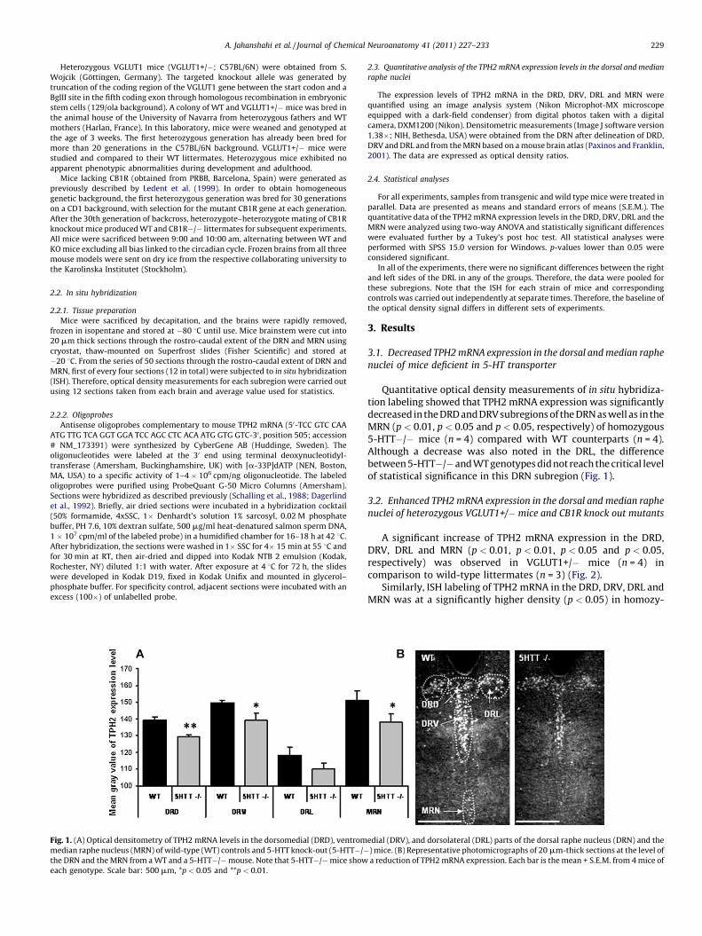
3.2. Enhanced TPH2 mRNA expression in the dorsal and median raphe
nuclei of heterozygous VGLUT1+/� mice and CB1R knock out mutants
A significant increase of TPH2 mRNA expression in the DRD,DRV, DRL and MRN (p < 0.01, p < 0.01, p < 0.05 and p < 0.05,respectively) was observed in VGLUT1+/� mice (n = 4) incomparison to wild-type littermates (n = 3) (Fig. 2).
Similarly, ISH labeling of TPH2 mRNA in the DRD, DRV, DRL andMRN was at a significantly higher density (p < 0.05) in homozy-
edial (DRV), and dorsolateral (DRL) parts of the dorsal raphe nucleus (DRN) and the
) mice. (B) Representative photomicrographs of 20 mm-thick sections at the level of
a reduction of TPH2 mRNA expression. Each bar is the mean + S.E.M. from 4 mice of

Fig. 2. (A) Optical densitometry of TPH2 mRNA levels in the dorsomedial (DRD), ventromedial (DRV), and dorsolateral (DRL) parts of the dorsal raphe nucleus (DRN) and the
median raphe nucleus (MRN) of wild-type (WT) mice and paired heterozygous VGLUT1+/� mice. (B) Representative photomicrographs of the DRN and the MRN from a WT
and a VGLUT1+/� mouse. VGLUT1+/� mice show increased TPH2 mRNA expression compared to WT. Each bar is the mean + S.E.M. of independent data obtained in 3–4 mice.
Scale bar: 500 mm, *p < 0.05 and **p < 0.01.
Fig. 3. (A) Optical densitometry of TPH2 mRNA levels in the dorsomedial (DRD), ventromedial (DRV), and dorsolateral (DRL) parts of the dorsal raphe nucleus (DRN) and the
median raphe nucleus (MRN) in wild-type (WT) mice and paired CB1R knock-out (CB1R�/�). (B) Representative photomicrographs at the level of DRN and MRN from a WT
and a CB1R�/� mouse. A significant increase of TPH2 mRNA expression is observed in CB1R�/� compared to WT mice. Each bar is the mean + S.E.M. from 5 mice of each
genotype. Scale bar: 500 mm. *p < 0.05.
A. Jahanshahi et al. / Journal of Chemical Neuroanatomy 41 (2011) 227–233230
gous CB1R�/� mutants (n = 5) than in paired wild-type controls(n = 5) (Fig. 3).
4. Discussion
Using three genetically modified mouse models, all character-ized by altered behaviors in depression-related paradigms, ourresults show quantitative differences in TPH2 mRNA expression inthe three main subregions of the DRN as well as in the MRN. Thesechanges are related to serotonergic or glutamatergic neurotrans-mission and to endocannabinoid systems. Thus, there weresignificant decreases in TPH2 mRNA expression in the 5-HTT�/� mutant mice, whereas increases in TPH2 mRNA expression wereobserved in the VGLUT1+/� and CB1R�/� mutant mice.
4.1. The 5-HTT�/� mouse
It has previously been shown that TPH mRNA levels and activitycan be regulated by the end-product 5-HT. For example, lower 5-HT levels result in higher TPH mRNA expression in vivo (Park et al.,1994). There is evidence that 5-HTT�/� mice exhibit markedchanges in 5-HT synthesis and turn-over. In particular, lack of 5-HTT is associated with a 60–80% depletion of 5-HT and itsmetabolite 5-hydroxyindoleacetic acid in several brain areas suchas the brain stem, striatum, hippocampus and frontal cortex ascompared with wild-type controls (Bengel et al., 1998; Fabre et al.,
2000). Desensitization of 5-HT1A, 5-HT1B autoreceptors has beenreported in 5-HTT�/� mutants as a consequence of highextracellular 5-HT levels in the DRN (Fabre et al., 2000; Mannouryla Cour et al., 2001). Interestingly, such changes were also observedin mice after long-term SSRI administration (Landgrebe et al.,2002), which are accompanied by decreased expression of TPH2mRNA in DRN and MRN in rodents (Abumaria et al., 2007). In viewof behavioral and biochemical evidence of similar alterations ofserotonergic neurotransmission in mice lacking 5-HTT and in micereceiving long-term SSRI therapy, it can be proposed that highconcentrations of 5-HT in the extracellular space of 5-HTT�/�mice are also causally related to the reduced TPH2 mRNAexpression in these mutants. Conversely, high TPH2 protein andmRNA levels have been reported in the DRN of depressed patientscompared to control subjects, possibly due to a compensatoryresponse to counteract deficits in extracellular 5-HT concentra-tions (Bach-Mizrachi et al., 2006). Increased TPH2 mRNA levels inthe DRN have also been observed in rats after adverse experiencesduring early life and adulthood (Gardner et al., 2009). However,further investigations are needed in order to elucidate the preciserelationships between extracellular levels of 5-HT and TPH2 mRNAexpression because changes opposite to those noted above in SSRI-treated rodents (i.e., increased TPH2 mRNA expression) have alsobeen reported (Shishkina et al., 2007).
The lack of a significant decrease of TPH2 mRNA level in the DRLin 5HTT�/� mice in comparison with the other subregions of the

A. Jahanshahi et al. / Journal of Chemical Neuroanatomy 41 (2011) 227–233 231
DRN, known to contain fewer serotonergic neurons (Steinbusch,1981), suggests that the DRL may be less involved in this model. Inagreement with our data, previous studies already reported aheterogeneous distribution of TPH2 mRNA in the DRN with thegreatest number of TPH2 mRNA-positive neurons in the DRD andDRV subnuclei (Austin and O’Donnell, 1999; Clark et al., 2006).Perhaps, the DRL exhibits a lower sensitivity to extracellular 5-HTlevels and consequently does not display any alteration in TPH2expression. Accordingly, changes in TPH2 expression in theventromedial and dorsomedial DRN might play a more predomi-nant role in modulating serotonergic neurotransmission anddepressive behavior (Bonkale et al., 2006; Lowry et al., 2008).Nevertheless, it must be taken in account that due to the samplesizes (n = 4) the statistical analysis might not have sufficient powerto detect a decrease in TPH2 expression in the DRL of 5-HTT�/�mice.
4.2. The VGLUT1+/� mouse
Alterations in glutamate and GABA neurotransmission appearto play a key role in depression (Krystal et al., 2002). Interestingly,beside high TPH2 mRNA levels in the raphe nuclei (Bach-Mizrachiet al., 2008), marked changes in glutamatergic and GABA-ergicneurotransmission have been observed in depressed patients, e.g.,increased glutamate and decreased GABA at cortical and hippo-campal levels (Bhagwagar et al., 2007). However, the underlyingmolecular mechanisms responsible for the abnormal glutamateneurotransmission in the brain of depressed patients remain to beelucidated.
Interestingly, the VGLUT1+/� mice also exhibit changes incortical and hippocampal release of glutamate and GABA togetherwith increased anxiogenic/depressive-like responses (Torderaet al., 2007; Garcia-Garcia et al., 2009). VGLUT1 plays a criticalrole in refilling synaptic glutamate vesicles (Wilson et al., 2005;Takamori, 2006) and indeed, a recent study has demonstrated thatVGLUT1+/� mice exhibit deficient glutamate transmission(Balschun et al., 2009). Moreover, the lack of VGLUT1 in glial cellsalso increases the probability of excitotoxic effects of excessiveextracellular glutamate in brain, which in turn can lead to moredisturbed glutamatergic neurotransmission (Valentine and Sana-cora, 2009).
The present study demonstrates that the glutamatergic systemthrough VGLUT1 affects TPH2 mRNA expression. In particular,VGLUT1+/� mice exhibited increased TPH2 mRNA levels in theDRN and MRN that may be related to alterations in theglutamatergic/GABAergic-5-HT negative feedback system. Specifi-cally, it has been established that forebrain glutamatergic neuronssend projections to the ventral periaqueductal grey, stimulatinglocal GABA neurons, which in turn inhibit 5-HT neurons (Arnstenand Goldman-Rakic, 1984; Sharp et al., 2007). Compromisedglutamatergic signaling due to the reduced VGLUT1s in corticalprefrontal descending projections to the brainstem, as expected inVGLUT1+/� mice, would lead to attenuated stimulation of GABAneurons, and decreased inhibition of the 5-HT neurons andpresumably, increased TPH2 mRNA levels. Moreover, the altered 5-HT function could contribute to explain the increased vulnerabilityof these mice to depressive like-behavior after exposure to chronicmild stress (Garcia-Garcia et al., 2009). With regard to theascending component of the circuitry, 5-HT modulates themagnitude of responses in many cortical glutamatergic andGABA-ergic neurons (Araneda and Andrade, 1991; Aghajanianand Marek, 1999; Zhou and Hablitz, 1999). In addition, it has beenreported that 5-HT controls, in turn, the activity of descendingexcitatory inputs through the activation of pyramidal corticalneurons (Amargos-Bosch et al., 2004). Since the neurochemicalpathways regulating glutamate and GABA signaling are closely
related (Choudary et al., 2005), changes in the levels and theactivity of these neurotransmitters can lead to altered excitation–inhibition ratios in the cortex (Bhagwagar et al., 2007).
4.3. The CB1R�/� mouse
Previous evidence demonstrates that the endocannabinoidsystem through CB1R regulates the activity of the serotonergicsystem by modulating different components of serotonergicfeedback (Hill et al., 2006; Bambico et al., 2007; Aso et al.,2009). In the present study, our data reveal additional alterationsin the serotonergic activity at the levels of 5-HT synthesis in theabsence of the CB1R. Mice lacking CB1R exhibit increased TPH2mRNA expression in the MRN and all subregions of the DRNcompared to wild-type littermates. Considering the role of TPH2 in5-HT synthesis and the reciprocal innervation between theprefrontal cortex and DRN (Jankowski and Sesack, 2004), theincreased expression of TPH2 in the DRN of CB1R�/� mice couldcontribute to the enhanced extracellular 5-HT levels in theprefrontal cortex of these mutants (Aso et al., 2009). Similarly,the alterations of different components involved in 5-HT depen-dent negative feedback, such as enhanced extracellular 5-HT levelsin the prefrontal cortex, decreased 5-HTT binding site density,functional desensitization of the 5-HT1A receptors and reduced 5-HT2C receptors have been described in CB1R�/� mice (Aso et al.,2009), as well as the enhancement of the 5-HT2A receptorexcitatory effect on DRN and MRN produced by the lack of CB1Ractivity (Gorzalka et al., 2005), could also contribute to theincreased 5-HT extracellular levels in the prefrontal cortex inCB1R�/� mutants. The synergistic alteration of these multipleserotonergic components modulated by CB1R on 5-HT signalingmight be a reason for the robust up-regulation of TPH2 mRNA in allsubregions of the DRN/MRN complex in CB1R�/� mice.
In contrast to the expected consequence of the enhancedserotonergic release, the genetic and pharmacological blockade ofthe CB1R induces a depressive-like phenotype (Hill and Gorzalka,2005). This could be explained by the impairment of othermechanisms controlling emotional homeostasis beyond 5-HTneurotransmission in the absence of CB1R, such as the effects ofstress on glucocorticoids and neurotrophic factor levels (Aso et al.,2008). The alterations observed on 5-HT neurotransmission havebeen proposed to be a substrate for counteracting the stress-induced emotional impairment in mice lacking CB1R since similarchanges have been described after antidepressant treatments(Lanfumey et al., 2000; Le Poul et al., 2000; Gould et al., 2006).Nevertheless, an impaired post-synaptic serotonergic signaling hasbeen reported in CB1R�/� mice (Mato et al., 2007), which couldlead to a reduction in the efficacy of 5-HT for ameliorating theirdepressive-like phenotype.
Taken together, these data suggest that the CB1R gene knockout should lead to a reduction of the inhibitory effect exertedthrough 5-HT1A and 5-HT2C receptors and enhancement of theexcitatory effect through 5-HT2A receptors on DRN and MRN 5-HTneurons, possibly leading to increased expression of TPH2 mRNA.Additionally, CB1R�/� mice are characterized by impairedserotonergic negative feedback (Aso et al., 2009), and CB1Rstimulation in the cortex directly inhibits 5-HT release (Nakaziet al., 2000), whereas a CB1R antagonist exerts the opposite effect(Tzavara et al., 2003). Therefore, changes in negative feedbackinput to the raphe nuclei from medial prefrontal cortex (Hajoset al., 1999) due to the absence of CB1R in DRN could compromisethe feedback mechanisms in the raphe nuclei. The synergisticattenuation of the multiple actions mediated by CB1R on 5-HTsignaling might be a reason why CB1R�/� mice show such arobust up regulation of TPH2 mRNA in all parts of the DRN/MRNcomplex.

A. Jahanshahi et al. / Journal of Chemical Neuroanatomy 41 (2011) 227–233232
5. Conclusion
In conclusion, based on the findings of the present study, wesuggest that TPH2 mRNA expression is under the influence ofmultiple messenger systems in relation with presynaptic and/orpostsynaptic feedback control of serotonin synthesis. 5-HTT,VGLUT1 and CB1R seem to be involved in the execution of thesefeedback mechanisms. Finally, our data are consistent withprevious reports suggesting that TPH2 activity within differentraphe subregions are differentially regulated under specificconditions.
Acknowledgements
This study was supported by an EC grant (Newmood; LHSM-CT-2003-503474), the Swedish Research Council (2887), The Mar-ianne and Marcus Wallenberg Foundation, a FP6 Marie Curie EarlyStage Training Fellwoship (MEST-CT-2005-020589), the FrenchInstitut National de la Sante et de la Recherche Medicale (INSERM),the University Pierre & Marie Curie, and the German ResearchFoundation (SFB 581, SFB TRR 58).
We are grateful to Professor S. Wojcik, University of Gottingen,Germany for supplying the original VGLUT1 heterozygous mice.We are grateful to Prof. D. A. Hopkins, Dalhousie University, Halifaxand Visiting Professor Maastricht University for his commends.
References
Abumaria, N., Rygula, R., Hiemke, C., Fuchs, E., Havemann-Reinecke, U., Ruther, E.,Flugge, G., 2007. Effect of chronic citalopram on serotonin-related and stress-regulated genes in the dorsal raphe nucleus of the rat. Eur. Neuropsychophar-macol. 17, 417–429.
Aghajanian, G.K., Marek, G.J., 1999. Serotonin, via 5-HT2A receptors, increases EPSCsin layer V pyramidal cells of prefrontal cortex by an asynchronous mode ofglutamate release. Brain Res. 825, 161–171.
Amargos-Bosch, M., Bortolozzi, A., Puig, M.V., Serrats, J., Adell, A., Celada, P., Toth, M.,Mengod, G., Artigas, F., 2004. Co-expression and in vivo interaction of seroto-nin1A and serotonin2A receptors in pyramidal neurons of prefrontal cortex.Cereb. Cortex 14, 281–299.
Araneda, R., Andrade, R., 1991. 5-Hydroxytryptamine2 and 5-hydroxytryptamine1A receptors mediate opposing responses on membrane excitability in ratassociation cortex. Neuroscience 40, 399–412.
Arnsten, A.F., Goldman-Rakic, P.S., 1984. Selective prefrontal cortical projections tothe region of the locus coeruleus and raphe nuclei in the rhesus monkey. BrainRes. 306, 9–18.
Aso, E., Ozaita, A., Valdizan, E.M., Ledent, C., Pazos, A., Maldonado, R., Valverde, O.,2008. BDNF impairment in the hippocampus is related to enhanced despairbehavior in CB1 knockout mice. J. Neurochem. 105, 565–572.
Aso, E., Renoir, T., Mengod, G., Ledent, C., Hamon, M., Maldonado, R., Lanfumey, L.,Valverde, O., 2009. Lack of CB1 receptor activity impairs serotonergic negativefeedback. J. Neurochem. 109, 935–944.
Austin, M.C., O’Donnell, S.M., 1999. Regional distribution and cellular expression oftryptophan hydroxylase messenger RNA in postmortem human brainstem andpineal gland. J. Neurochem. 72, 2065–2073.
Bach-Mizrachi, H., Underwood, M.D., Kassir, S.A., Bakalian, M.J., Sibille, E., Tamir, H.,Mann, J.J., Arango, V., 2006. Neuronal tryptophan hydroxylase mRNA expressionin the human dorsal and median raphe nuclei: major depression and suicide.Neuropsychopharmacology 31, 814–824.
Bach-Mizrachi, H., Underwood, M.D., Tin, A., Ellis, S.P., Mann, J.J., Arango, V., 2008.Elevated expression of tryptophan hydroxylase-2 mRNA at the neuronal level inthe dorsal and median raphe nuclei of depressed suicides. Mol. Psychiatry 13,507–513.
Balschun, D., Moechars, D., Callaerts-Vegh, Z., Vermaercke, B., Van Acker, N.,Andries, L., D’Hooge, R., 2009. Vesicular glutamate transporter VGLUT1 has arole in hippocampal long-term potentiation and spatial reversal learning. Cereb.Cortex 20, 684–693.
Bambico, F.R., Katz, N., Debonnel, G., Gobbi, G., 2007. Cannabinoids elicit antide-pressant-like behavior and activate serotonergic neurons through the medialprefrontal cortex. J. Neurosci. 27, 11700–11711.
Bengel, D., Murphy, D.L., Andrews, A.M., Wichems, C.H., Feltner, D., Heils, A.,Mossner, R., Westphal, H., Lesch, K.P., 1998. Altered brain serotonin homeosta-sis and locomotor insensitivity to 3, 4-methylenedioxymethamphetamine(‘‘Ecstasy’’) in serotonin transporter-deficient mice. Mol. Pharmacol. 53, 649–655.
Bhagwagar, Z., Wylezinska, M., Jezzard, P., Evans, J., Ashworth, F., Sule, A., Matthews,P.M., Cowen, P.J., 2007. Reduction in occipital cortex gamma-aminobutyric acidconcentrations in medication-free recovered unipolar depressed and bipolarsubjects. Biol. Psychiatry 61, 806–812.
Bonkale, W.L., Turecki, G., Austin, M.C., 2006. Increased tryptophan hydroxylaseimmunoreactivity in the dorsal raphe nucleus of alcohol-dependent, depressedsuicide subjects is restricted to the dorsal subnucleus. Synapse 60, 81–85.
Bonkale, W.L., Austin, M.C., 2008. 3,4-Methylenedioxymethamphetamine inducesdifferential regulation of tryptophan hydroxylase 2 protein and mRNA levels inthe rat dorsal raphe nucleus. Neuroscience 155, 270–276.
Canli, T., Lesch, K.P., 2007. Long story short: the serotonin transporter in emotionregulation and social cognition. Nat. Neurosci. 10, 1103–1109.
Celada, P., Puig, M.V., Casanovas, J.M., Guillazo, G., Artigas, F., 2001. Control of dorsalraphe serotonergic neurons by the medial prefrontal cortex: involvement ofserotonin-1A, GABA(A), and glutamate receptors. J. Neurosci. 21, 9917–9929.
Choudary, P.V., Molnar, M., Evans, S.J., Tomita, H., Li, J.Z., Vawter, M.P., Myers, R.M.,Bunney Jr., W.E., Akil, H., Watson, S.J., Jones, E.G., 2005. Altered cortical gluta-matergic and GABAergic signal transmission with glial involvement in depres-sion. Proc. Natl. Acad. Sci. U.S.A. 102, 15653–15658.
Clark, J.A., Flick, R.B., Pai, L.Y., Szalayova, I., Key, S., Conley, R.K., Deutch, A.Y., Hutson,P.H., Mezey, E., 2008. Glucocorticoid modulation of tryptophan hydroxylase-2protein in raphe nuclei and 5-hydroxytryptophan concentrations in frontalcortex of C57/Bl6 mice. Mol. Psychiatry 13, 498–506.
Clark, M.S., McDevitt, R.A., Neumaier, J.F., 2006. Quantitative mapping of tryptophanhydroxylase-2, 5-HT1A, 5-HT1B, and serotonin transporter expression acrossthe anteroposterior axis of the rat dorsal and median raphe nuclei. J. Comp.Neurol. 498, 611–623.
Cowen, P.J., 2008. Serotonin and depression: pathophysiological mechanism ormarketing myth? Trends Pharmacol. Sci. 29, 433–436.
Dagerlind, A., Friberg, K., Bean, A.J., Hokfelt, T., 1992. Sensitive mRNA detectionusing unfixed tissue: combined radioactive and non-radioactive in situ hybrid-ization histochemistry. Histochemistry 98, 39–49.
Dahlstrom, A., Fuxe, K., 1964. Localization of monoamines in the lower brain stem.Experientia 20, 398–399.
Fabre, V., Beaufour, C., Evrard, A., Rioux, A., Hanoun, N., Lesch, K.P., Murphy, D.L.,Lanfumey, L., Hamon, M., Martres, M.P., 2000. Altered expression and functionsof serotonin 5-HT1A and 5-HT1B receptors in knock-out mice lacking the 5-HTtransporter. Eur. J. Neurosci. 12, 2299–2310.
Fremeau Jr., R.T., Kam, K., Qureshi, T., Johnson, J., Copenhagen, D.R., Storm-Mathisen,J., Chaudhry, F.A., Nicoll, R.A., Edwards, R.H., 2004. Vesicular glutamate trans-porters 1 and 2 target to functionally distinct synaptic release sites. Science 304,1815–1819.
Garcia-Garcia, A.L., Elizalde, N., Matrov, D., Harro, J., Wojcik, S.M., Venzala, E.,Ramirez, M.J., Del Rio, J., Tordera, R.M., 2009. Increased vulnerability to depres-sive-like behavior of mice with decreased expression of VGLUT1. Biol. Psychia-try 66, 275–282.
Gardner, K.L., Hale, M.W., Oldfield, S., Lightman, S.L., Plotsky, P.M., Lowry, C.A., 2009.Adverse experience during early life and adulthood interact to elevate tph2mRNA expression in serotonergic neurons within the dorsal raphe nucleus.Neuroscience 163, 991–1001.
Gorzalka, B.B., Hill, M.N., Sun, J.C., 2005. Functional role of the endocannabinoidsystem and AMPA/kainate receptors in 5-HT2A receptor-mediated wet dogshakes. Eur. J. Pharmacol. 516, 28–33.
Gould, G.G., Altamirano, A.V., Javors, M.A., Frazer, A., 2006. A comparison of thechronic treatment effects of venlafaxine and other antidepressants on serotoninand norepinephrine transporters. Biol. Psychiatry 59, 408–414.
Greenberg, P.E., Kessler, R.C., Birnbaum, H.G., Leong, S.A., Lowe, S.W., Berglund, P.A.,Corey-Lisle, P.K., 2003. The economic burden of depression in the United States:how did it change between 1990 and 2000? J. Clin. Psychiatry 64, 1465–1475.
Gutknecht, L., Waider, J., Kraft, S., Kriegebaum, C., Holtmann, B., Reif, A., Schmitt, A.,Lesch, K.P., 2008. Deficiency of brain 5-HT synthesis but serotonergic neuronformation in Tph2 knockout mice. J. Neural Transm. 115, 1127–1132.
Gutknecht, L., Kriegebaum, C., Waider, J., Schmitt, A., Lesch, K.P., 2009. Spatio-temporal expression of tryptophan hydroxylase isoforms in murine and humanbrain: convergent data from Tph2 knockout mice. Eur. Neuropsychopharmacol.19, 266–282.
Haghighi, F., Bach-Mizrachi, H., Huang, Y.Y., Arango, V., Shi, S., Dwork, A.J., Rosoklija,G., Sheng, H.T., Morozova, I., Ju, J., Russo, J.J., Mann, J.J., 2008. Genetic architec-ture of the human tryptophan hydroxylase 2 gene: existence of neural isoformsand relevance for major depression. Mol. Psychiatry 13, 813–820.
Hajos, M., Richards, C.D., Szekely, A.D., Sharp, T., 1998. An electrophysiological andneuroanatomical study of the medial prefrontal cortical projection to themidbrain raphe nuclei in the rat. Neuroscience 87, 95–108.
Hajos, M., Hajos-Korcsok, E., Sharp, T., 1999. Role of the medial prefrontal cortex in5-HT1A receptor-induced inhibition of 5-HT neuronal activity in the rat. Br. J.Pharmacol. 126, 1741–1750.
Hill, M.N., Gorzalka, B.B., 2005. Is there a role for the endocannabinoid system in theetiology and treatment of melancholic depression? Behav. Pharmacol. 16, 333–352.
Hill, M.N., Sun, J.C., Tse, M.T., Gorzalka, B.B., 2006. Altered responsiveness ofserotonin receptor subtypes following long-term cannabinoid treatment. Int.J. Neuropsychopharmacol. 9, 277–286.
Hiroi, R., McDevitt, R.A., Neumaier, J.F., 2006. Estrogen selectively increases trypto-phan hydroxylase-2 mRNA expression in distinct subregions of rat midbrainraphe nucleus: association between gene expression and anxiety behavior inthe open field. Biol. Psychiatry 60, 288–295.
Hisano, S., 2003. Vesicular glutamate transporters in the brain. Anat. Sci. Int. 78,191–204.
Holmes, A., Yang, R.J., Lesch, K.P., Crawley, J.N., Murphy, D.L., 2003a. Mice lacking theserotonin transporter exhibit 5-H T1A receptor-mediated abnormalities in testsfor anxiety-like behavior. Neuropsychopharmacology 28, 2077–2088.

A. Jahanshahi et al. / Journal of Chemical Neuroanatomy 41 (2011) 227–233 233
Holmes, A., Murphy, D.L., Crawley, J.N., 2003b. Abnormal behavioral phenotypes ofserotonin transporter knockout mice: parallels with human anxiety and de-pression. Biol. Psychiatry 54, 953–959.
Jankowski, M.P., Sesack, S.R., 2004. Prefrontal cortical projections to the rat dorsalraphe nucleus: ultrastructural features and associations with serotonin andgamma-aminobutyric acid neurons. J. Comp. Neurol. 468, 518–529.
Kosofsky, B.E., Molliver, M.E., 1987. The serotoninergic innervation of cerebralcortex: different classes of axon terminals arise from dorsal and median raphenuclei. Synapse 1, 153–168.
Krystal, J.H., Sanacora, G., Blumberg, H., Anand, A., Charney, D.S., Marek, G., Epper-son, C.N., Goddard, A., Mason, G.F., 2002. Glutamate and GABA systems astargets for novel antidepressant and mood-stabilizing treatments. Mol. Psychi-atry 7 (Suppl. 1), S71–80.
Landgrebe, J., Welzl, G., Metz, T., van Gaalen, M.M., Ropers, H., Wurst, W., Holsboer,F., 2002. Molecular characterisation of antidepressant effects in the mouse brainusing gene expression profiling. J. Psychiatr. Res. 36, 119–129.
Lanfumey, L., Mannoury La Cour, C., Froger, N., Hamon, M., 2000. 5-HT-HPAinteractions in two models of transgenic mice relevant to major depression.Neurochem. Res. 25, 1199–1206.
Le Poul, E., Boni, C., Hanoun, N., Laporte, A.M., Laaris, N., Chauveau, J., Hamon, M.,Lanfumey, L., 2000. Differential adaptation of brain 5-HT1A and 5-HT1B recep-tors and 5-HT transporter in rats treated chronically with fluoxetine. Neuro-pharmacology 39, 110–122.
Ledent, C., Valverde, O., Cossu, G., Petitet, F., Aubert, J.F., Beslot, F., Bohme, G.A.,Imperato, A., Pedrazzini, T., Roques, B.P., Vassart, G., Fratta, W., Parmentier, M.,1999. Unresponsiveness to cannabinoids and reduced addictive effects ofopiates in CB1 receptor knockout mice. Science 283, 401–404.
Lesch, K.P., Mossner, R., 2006. Inactivation of 5HT transport in mice: modelingaltered 5HT homeostasis implicated in emotional dysfunction, affective dis-orders, and somatic syndromes. Handb. Exp. Pharmacol. 417–456.
Lowry, C.A., Hale, M.W., Evans, A.K., Heerkens, J., Staub, D.R., Gasser, P.J., Shekhar, A.,2008. Serotonergic systems, anxiety, and affective disorder: focus on thedorsomedial part of the dorsal raphe nucleus. Ann. N. Y. Acad. Sci. 1148, 86–94.
Maldonado, R., Valverde, O., Berrendero, F., 2006. Involvement of the endocanna-binoid system in drug addiction. Trends Neurosci. 29, 225–232.
Mannoury la Cour, C., Boni, C., Hanoun, N., Lesch, K.P., Hamon, M., Lanfumey, L.,2001. Functional consequences of 5-HT transporter gene disruption on 5-HT(1a) receptor-mediated regulation of dorsal raphe and hippocampal cellactivity. J. Neurosci. 21, 2178–2185.
Martin, M., Ledent, C., Parmentier, M., Maldonado, R., Valverde, O., 2002. Involve-ment of CB1 cannabinoid receptors in emotional behaviour. Psychopharmacol-ogy (Berl.) 159, 379–387.
Mato, S., Aso, E., Castro, E., Martin, M., Valverde, O., Maldonado, R., Pazos, A., 2007.CB1 knockout mice display impaired functionality of 5-HT1A and 5-HT2A/Creceptors. J. Neurochem. 103, 2111–2120.
Michelsen, K.A., Schmitz, C., Steinbusch, H.W.M., 2007. The dorsal raphe nucleus—from silver stainings to a role in depression. Brain Res. Rev. 55, 329–342.
Michelsen, K.A., Prickaerts, J., Steinbusch, H.W.M., 2008. The dorsal raphe nucleusand serotonin: implications for neuroplasticity linked to major depression andAlzheimer’s disease. Prog. Brain Res. 172, 233–264.
Murphy, D.L., Lesch, K.P., 2008. Targeting the murine serotonin transporter: insightsinto human neurobiology. Nat. Rev. Neurosci. 9 (2), 85–96.
Nakazi, M., Bauer, U., Nickel, T., Kathmann, M., Schlicker, E., 2000. Inhibition ofserotonin release in the mouse brain via presynaptic cannabinoid CB1 recep-tors. Naunyn Schmiedebergs Arch. Pharmacol. 361, 19–24.
O’Hearn, E., Molliver, M.E., 1984. Organization of raphe-cortical projections in rat: aquantitative retrograde study. Brain Res. Bull. 13, 709–726.
Owens, M.J., Nemeroff, C.B., 1998. The serotonin transporter and depression.Depress. Anxiety 8 (Suppl. 1), 5–12.
Park, D.H., Stone, D.M., Baker, H., Kim, K.S., Joh, T.H., 1994. Early induction of ratbrain tryptophan hydroxylase (TPH) mRNA following parachlorophenylalanine(PCPA) treatment. Brain Res. Mol. Brain Res. 22, 20–28.
Paxinos, G., Franklin, K.B.J., 2001. The Mouse Brain Atlas in Stereotaxic Coordinates.Academic Press, San Diego.
Peyron, C., Petit, J.M., Rampon, C., Jouvet, M., Luppi, P.H., 1998. Forebrain afferents tothe rat dorsal raphe nucleus demonstrated by retrograde and anterogradetracing methods. Neuroscience 82, 443–468.
Renoir, T., Paizanis, E., El Yacoubi, M., Saurini, F., Hanoun, N., Melfort, M., Lesch, K.P.,Hamon, M., Lanfumey, L., 2008. Differential long-term effects of MDMA on theserotoninergic system and hippocampal cell proliferation in 5-HTT knock-outvs. wild-type mice. Int. J. Neuropsychopharmacol. 11, 1149–1162.
Rioux, A., Fabre, V., Lesch, K.P., Moessner, R., Murphy, D.L., Lanfumey, L., Hamon, M.,Martres, M.P., 1999. Adaptive changes of serotonin 5-HT2A receptors in micelacking the serotonin transporter. Neurosci. Lett. 262, 113–116.
Sanacora, G., Gueorguieva, R., Epperson, C.N., Wu, Y.T., Appel, M., Rothman, D.L.,Krystal, J.H., Mason, G.F., 2004. Subtype-specific alterations of gamma-amino-butyric acid and glutamate in patients with major depression. Arch. Gen.Psychiatry 61, 705–713.
Sanchis-Segura, C., Cline, B.H., Marsicano, G., Lutz, B., Spanagel, R., 2004. Reducedsensitivity to reward in CB1 knockout mice. Psychopharmacology (Berl.) 176,223–232.
Schalling, M., Seroogy, K., Hokfelt, T., Chai, S.Y., Hallman, H., Persson, H., Larhammar,D., Ericsson, A., Terenius, L., Graffi, J., et al., 1988. Neuropeptide tyrosine in therat adrenal gland—immunohistochemical and in situ hybridization studies.Neuroscience 24, 337–349.
Sharp, T., Boothman, L., Raley, J., Queree, P., 2007. Important messages in the ‘post’:recent discoveries in 5-HT neurone feedback control. Trends Pharmacol. Sci. 28,629–636.
Shishkina, G.T., Kalinina, T.S., Dygalo, N.N., 2007. Up-regulation of tryptophanhydroxylase-2 mRNA in the rat brain by chronic fluoxetine treatment correlateswith its antidepressant effect. Neuroscience 150, 404–412.
Steinbusch, H.W.M., 1984. Distribution of serotonin-immunoreactive neuronsand their projections in the central nervous system of the rat. In: Bjorklund,A., Hokfelt, T., Kuhar, M.J. (Eds.), Classical Transmitters and TransmitterReceptors in the CNS: Part II. The Handbook of Chemical Neuroanatomy,Vol. 3. Elsevier, Amsterdam, pp. 68–125.
Steinbusch, H.W., 1981. Distribution of serotonin-immunoreactivity in thecentral nervous system of the rat-cell bodies and terminals. Neuroscience6, 557–618.
Steinbusch, H.W.M., de Vente, J., 1997. New vistas on the neurobiology ofdepression: colocalization of serotonin-, dopamine- and nitric oxidesynthase-containing neurons in the dorsal raphe nucleus. In: Honig, A.,Praag, H.M. (Eds.), Depression: Neurobiological, Psychopathological andTherapeutic Advances. John Wiley and Sons, pp. 179–196.
Steinbusch, H.W.M., Nieuwenhuys, R., 1983. The raphe nuclei of the rat brain stem:a cytoarchitectonic and immunohistochemical study using antibodies to sero-tonin. In: Emson, P. (Ed.), Chemical Neuroanatomy. Raven Press, New York, pp.131–207.
Steinbusch, H.W., Nieuwenhuys, R., Verhofstad, A.A., Van der Kooy, D., 1981. Thenucleus raphe dorsalis of the rat and its projection upon the caudatoputamen. Acombined cytoarchitectonic, immunohistochemical and retrograde transportstudy. J. Physiol. (Paris) 77, 157–174.
Takamori, S., Rhee, J.S., Rosenmund, C., Jahn, R., 2000. Identification of a vesicularglutamate transporter that defines a glutamatergic phenotype in neurons.Nature 407, 189–194.
Takamori, S., 2006. VGLUTs: ‘exciting’ times for glutamatergic research? Neurosci.Res. 55, 343–351.
Tordera, R.M., Totterdell, S., Wojcik, S.M., Brose, N., Elizalde, N., Lasheras, B., Del Rio,J., 2007. Enhanced anxiety, depressive-like behaviour and impaired recognitionmemory in mice with reduced expression of the vesicular glutamate transport-er 1 (VGLUT1). Eur. J. Neurosci. 25, 281–290.
Tzavara, E.T., Davis, R.J., Perry, K.W., Li, X., Salhoff, C., Bymaster, F.P., Witkin, J.M.,Nomikos, G.G., 2003. The CB1 receptor antagonist SR141716A selectivelyincreases monoaminergic neurotransmission in the medial prefrontal cortex:implications for therapeutic actions. Br. J. Pharmacol. 138, 544–553.
Uezato, A., Meador-Woodruff, J.H., McCullumsmith, R.E., 2009. Vesicular glutamatetransporter mRNA expression in the medial temporal lobe in major depressivedisorder, bipolar disorder, and schizophrenia. Bipolar Disord. 11, 711–725.
Valentine, G.W., Sanacora, G., 2009. Targeting glial physiology and glutamatecycling in the treatment of depression. Biochem. Pharmacol. 78, 431–439.
Van Den Bogaert, A., Sleegers, K., De Zutter, S., Heyrman, L., Norrback, K.F., Adolfs-son, R., Van Broeckhoven, C., Del-Favero, J., 2006. Association of brain-specifictryptophan hydroxylase TPH2, with unipolar and bipolar disorder in a NorthernSwedish, isolated population. Arch. Gen. Psychiatry 63, 1103–1110.
Walther, D.J., Bader, M., 2003. A unique central tryptophan hydroxylase isoform.Biochem. Pharmacol. 66, 1673–1680.
Weissmann, D., Chamba, G., Debure, L., Rousset, C., Richard, F., Maitre, M., Pujol, J.F.,1990. Variation of tryptophan-5-hydroxylase concentration in the rat raphedorsalis nucleus after p-chlorophenylalanine administration: II. Anatomicaldistribution of the tryptophan-5-hydroxylase protein and regional variationof its turnover rate. Brain Res. 536, 46–55.
Wilson, N.R., Kang, J., Hueske, E.V., Leung, T., Varoqui, H., Murnick, J.G., Erickson, J.D.,Liu, G., 2005. Presynaptic regulation of quantal size by the vesicular glutamatetransporter VGLUT1. J. Neurosci. 25, 6221–6234.
Wojcik, S.M., Rhee, J.S., Herzog, E., Sigler, A., Jahn, R., Takamori, S., Brose, N.,Rosenmund, C., 2004. An essential role for vesicular glutamate transporter 1(VGLUT1) in postnatal development and control of quantal size. Proc. Natl.Acad. Sci. U.S.A. 101, 7158–7163.
Zhang, X., Beaulieu, J.M., Sotnikova, T.D., Gainetdinov, R.R., Caron, M.G., 2004.Tryptophan hydroxylase-2 controls brain serotonin synthesis. Science 305, 217.
Zhou, F.M., Hablitz, J.J., 1999. Activation of serotonin receptors modulates synaptictransmission in rat cerebral cortex. J. Neurophysiol. 82, 2989–2999.
Zill, P., Baghai, T.C., Zwanzger, P., Schule, C., Eser, D., Rupprecht, R., Moller, H.J.,Bondy, B., Ackenheil, M., 2004. SNP and haplotype analysis of a novel trypto-phan hydroxylase isoform (TPH2) gene provide evidence for association withmajor depression. Mol. Psychiatry 9, 1030–1036.
Related Documents