Alterations of Alterations of Musculoskeletal Function in Musculoskeletal Function in Children Children Chapter 43 Chapter 43 Mosby items and derived items © 2010, 2006 by Mosby, Inc., an affiliate of Mosby items and derived items © 2010, 2006 by Mosby, Inc., an affiliate of Elsevier Inc. Elsevier Inc.

Welcome message from author
This document is posted to help you gain knowledge. Please leave a comment to let me know what you think about it! Share it to your friends and learn new things together.
Transcript

Alterations of Musculoskeletal Alterations of Musculoskeletal Function in ChildrenFunction in Children
Chapter 43Chapter 43
Mosby items and derived items © 2010, 2006 by Mosby, Inc., an affiliate of Elsevier Inc.Mosby items and derived items © 2010, 2006 by Mosby, Inc., an affiliate of Elsevier Inc.

22Mosby items and derived items © 2010, 2006 by Mosby, Inc., an affiliate of Elsevier Inc.Mosby items and derived items © 2010, 2006 by Mosby, Inc., an affiliate of Elsevier Inc.
Musculoskeletal AlterationsMusculoskeletal Alterations OverviewOverview
CongenitalCongenital• ClubfootClubfoot
HereditaryHereditary• Muscular dystrophyMuscular dystrophy
AcquiredAcquired• Legg-Calvé-Perthes Legg-Calvé-Perthes
May be acute, chronic, or terminalMay be acute, chronic, or terminal

33Mosby items and derived items © 2010, 2006 by Mosby, Inc., an affiliate of Elsevier Inc.Mosby items and derived items © 2010, 2006 by Mosby, Inc., an affiliate of Elsevier Inc.
Bone FormationBone Formation Bone formation begins in two phases at about Bone formation begins in two phases at about
the eighth week of gestationthe eighth week of gestation Delivery of bone cell precursors to sites of bone Delivery of bone cell precursors to sites of bone
formationformation Aggregation of the bone cell precursors at primary Aggregation of the bone cell precursors at primary
centers of ossificationcenters of ossification
44Mosby items and derived items © 2010, 2006 by Mosby, Inc., an affiliate of Elsevier Inc.Mosby items and derived items © 2010, 2006 by Mosby, Inc., an affiliate of Elsevier Inc.
Bone FormationBone Formation Intramembranous formationIntramembranous formation
On or within the mesenchymeOn or within the mesenchyme Endochondral formationEndochondral formation
Cartilage anlageCartilage anlage PerichondriumPerichondrium Periosteal collarPeriosteal collar Secondary centers of ossificationSecondary centers of ossification

55Mosby items and derived items © 2010, 2006 by Mosby, Inc., an affiliate of Elsevier Inc.Mosby items and derived items © 2010, 2006 by Mosby, Inc., an affiliate of Elsevier Inc.
Bone FormationBone Formation

66Mosby items and derived items © 2010, 2006 by Mosby, Inc., an affiliate of Elsevier Inc.Mosby items and derived items © 2010, 2006 by Mosby, Inc., an affiliate of Elsevier Inc.
Bone GrowthBone Growth Until adult stature achieved, bone growth Until adult stature achieved, bone growth
occurs at the epiphyseal plate through occurs at the epiphyseal plate through endochondral ossificationendochondral ossification
Epiphyseal closureEpiphyseal closure Unites the metaphysis and the epiphysisUnites the metaphysis and the epiphysis Occurs earlier in females than males because of Occurs earlier in females than males because of
earlier puberty in femalesearlier puberty in females

77Mosby items and derived items © 2010, 2006 by Mosby, Inc., an affiliate of Elsevier Inc.Mosby items and derived items © 2010, 2006 by Mosby, Inc., an affiliate of Elsevier Inc.
Bone GrowthBone Growth Factors affecting bone growthFactors affecting bone growth
Growth hormone (secreted by pituitary)Growth hormone (secreted by pituitary) NutritionNutrition General healthGeneral health Many growth factors and regulators (fibroblast Many growth factors and regulators (fibroblast
growth factor)growth factor)

88Mosby items and derived items © 2010, 2006 by Mosby, Inc., an affiliate of Elsevier Inc.Mosby items and derived items © 2010, 2006 by Mosby, Inc., an affiliate of Elsevier Inc.
Skeletal DevelopmentSkeletal Development
In the newborn, the entire spine is concave In the newborn, the entire spine is concave anteriorly (kyphosed)anteriorly (kyphosed)
In the first 3 months of life, the cervical spine In the first 3 months of life, the cervical spine begins to arch (lordotic)begins to arch (lordotic)
Curve of lumbar spine develops with sittingCurve of lumbar spine develops with sitting Compared to adult, a newborn has a large head, Compared to adult, a newborn has a large head,
long spine, and short extremitieslong spine, and short extremities

99Mosby items and derived items © 2010, 2006 by Mosby, Inc., an affiliate of Elsevier Inc.Mosby items and derived items © 2010, 2006 by Mosby, Inc., an affiliate of Elsevier Inc.
Skeletal DevelopmentSkeletal Development Genu varum (peaks by 2½ years)Genu varum (peaks by 2½ years)
Occurs in all newborns due to intrauterine stressOccurs in all newborns due to intrauterine stress BowlegBowleg
Genu valgum (peaks by 5-6 years)Genu valgum (peaks by 5-6 years) Knock-kneesKnock-knees
Persistence past peak times is pathologicPersistence past peak times is pathologic

1010Mosby items and derived items © 2010, 2006 by Mosby, Inc., an affiliate of Elsevier Inc.Mosby items and derived items © 2010, 2006 by Mosby, Inc., an affiliate of Elsevier Inc.
Muscle DevelopmentMuscle Development Between birth and maturity, muscle nuclei in Between birth and maturity, muscle nuclei in
the body increase 14 times in boys and 10 the body increase 14 times in boys and 10 times in girlstimes in girls
The composition and size of muscles vary The composition and size of muscles vary with agewith age

1111Mosby items and derived items © 2010, 2006 by Mosby, Inc., an affiliate of Elsevier Inc.Mosby items and derived items © 2010, 2006 by Mosby, Inc., an affiliate of Elsevier Inc.
Congenital DefectsCongenital Defects SyndactylySyndactyly
Webbing of the fingersWebbing of the fingers Fusion of the soft tissues of the fingersFusion of the soft tissues of the fingers True syndactyly also includes fusion of the bones and True syndactyly also includes fusion of the bones and
nailsnails Vestigial tabsVestigial tabs
Extra digitExtra digit

1212Mosby items and derived items © 2010, 2006 by Mosby, Inc., an affiliate of Elsevier Inc.Mosby items and derived items © 2010, 2006 by Mosby, Inc., an affiliate of Elsevier Inc.
Congenital DefectsCongenital Defects Anomalies on the medial or radial aspect of Anomalies on the medial or radial aspect of
the arm often associated with abnormalities the arm often associated with abnormalities of blood, heart, or kidneys. of blood, heart, or kidneys.
Lateral or ulnar-sided defects are less often Lateral or ulnar-sided defects are less often associated with systemic anomalies and are associated with systemic anomalies and are far more rarefar more rare

1313Mosby items and derived items © 2010, 2006 by Mosby, Inc., an affiliate of Elsevier Inc.Mosby items and derived items © 2010, 2006 by Mosby, Inc., an affiliate of Elsevier Inc.
SyndactylySyndactyly

1414Mosby items and derived items © 2010, 2006 by Mosby, Inc., an affiliate of Elsevier Inc.Mosby items and derived items © 2010, 2006 by Mosby, Inc., an affiliate of Elsevier Inc.
Congenital DefectsCongenital Defects Developmental dysplasia of the hipDevelopmental dysplasia of the hip
Formerly: congenital dislocation of the hipFormerly: congenital dislocation of the hip Abnormality of the proximal femur, acetabulum, or Abnormality of the proximal femur, acetabulum, or
bothboth Risk factorsRisk factors
• Female, metatarsus adductus, torticollis, Female, metatarsus adductus, torticollis, oligohydramnios, first pregnancy, and breech oligohydramnios, first pregnancy, and breech presentationpresentation
The hip can present as subluxated, dislocatable, The hip can present as subluxated, dislocatable, or dislocatedor dislocated

1515Mosby items and derived items © 2010, 2006 by Mosby, Inc., an affiliate of Elsevier Inc.Mosby items and derived items © 2010, 2006 by Mosby, Inc., an affiliate of Elsevier Inc.
Congenital DefectsCongenital Defects Developmental dysplasia of the hipDevelopmental dysplasia of the hip
ManifestationsManifestations• Asymmetry of gluteal or thigh foldsAsymmetry of gluteal or thigh folds
• Limb length discrepancyLimb length discrepancy
• Limitation of hip abductionLimitation of hip abduction
• Positive Ortolani signPositive Ortolani sign
• Positive Barlow testPositive Barlow test
• Positive Trendelenburg gaitPositive Trendelenburg gait
• PainPain

1616Mosby items and derived items © 2010, 2006 by Mosby, Inc., an affiliate of Elsevier Inc.Mosby items and derived items © 2010, 2006 by Mosby, Inc., an affiliate of Elsevier Inc.
Developmental Dysplasia of the HipDevelopmental Dysplasia of the Hip

1717Mosby items and derived items © 2010, 2006 by Mosby, Inc., an affiliate of Elsevier Inc.Mosby items and derived items © 2010, 2006 by Mosby, Inc., an affiliate of Elsevier Inc.
Congenital DefectsCongenital Defects Deformities of the footDeformities of the foot
Metatarsus adductus (forefoot adduction)Metatarsus adductus (forefoot adduction)• Mild, moderate, or severe (degree of deformity and Mild, moderate, or severe (degree of deformity and
flexibility)flexibility)
Equinovarus deformity (clubfoot)Equinovarus deformity (clubfoot)• Positional equinovarusPositional equinovarus
• Idiopathic congenital equinovarusIdiopathic congenital equinovarus
• Tetratologic equinovarusTetratologic equinovarus
• Pes planus (flat foot)Pes planus (flat foot)

1818Mosby items and derived items © 2010, 2006 by Mosby, Inc., an affiliate of Elsevier Inc.Mosby items and derived items © 2010, 2006 by Mosby, Inc., an affiliate of Elsevier Inc.
Congenital DefectsCongenital Defects TreatmentTreatment
BracesBraces Sequential castsSequential casts SurgerySurgery

1919Mosby items and derived items © 2010, 2006 by Mosby, Inc., an affiliate of Elsevier Inc.Mosby items and derived items © 2010, 2006 by Mosby, Inc., an affiliate of Elsevier Inc.
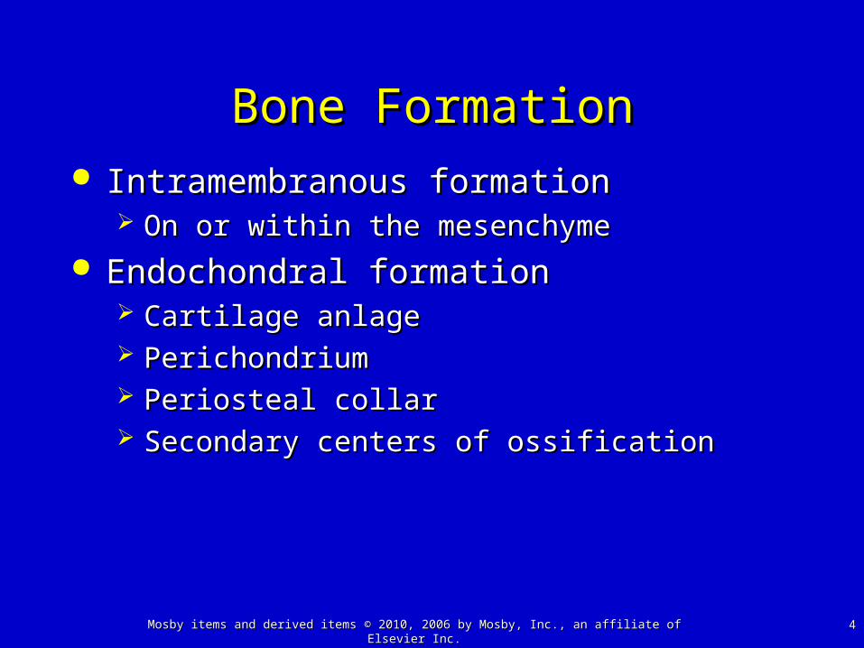
Osteogenesis ImperfectaOsteogenesis Imperfecta
““Brittle bone disease”Brittle bone disease” Defect in collagen productionDefect in collagen production
Bone and vessel collagenBone and vessel collagen Sillence classificationSillence classification Results in osteoporosis, bowed and deformed Results in osteoporosis, bowed and deformed
limbs, short stature, spine curvature, and bluish limbs, short stature, spine curvature, and bluish sclerasclera
Can be evident before birth (in utero fractures)Can be evident before birth (in utero fractures)

2020Mosby items and derived items © 2010, 2006 by Mosby, Inc., an affiliate of Elsevier Inc.Mosby items and derived items © 2010, 2006 by Mosby, Inc., an affiliate of Elsevier Inc.
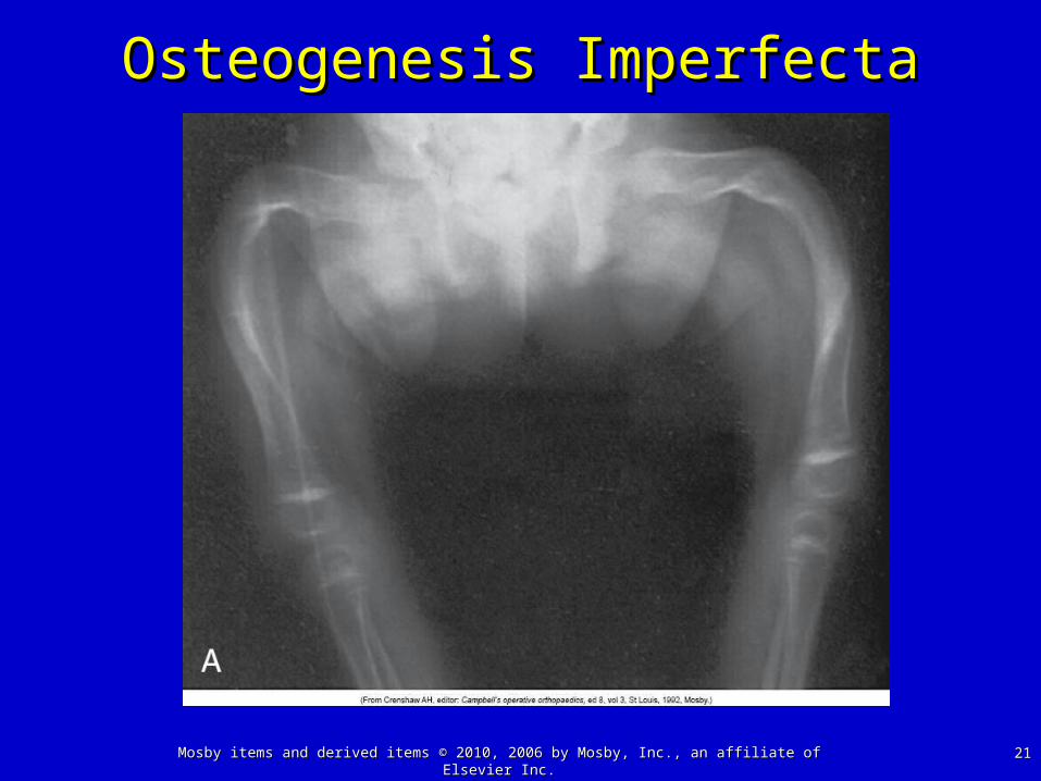
Osteogenesis ImperfectaOsteogenesis Imperfecta
SevereSevere Child may be stillborn or die soon after birth; Child may be stillborn or die soon after birth;
intrauterine fracturesintrauterine fractures MildMild
May not be diagnosed until child begins to walkMay not be diagnosed until child begins to walk May be mistaken for child abuseMay be mistaken for child abuse
2121Mosby items and derived items © 2010, 2006 by Mosby, Inc., an affiliate of Elsevier Inc.Mosby items and derived items © 2010, 2006 by Mosby, Inc., an affiliate of Elsevier Inc.
Osteogenesis ImperfectaOsteogenesis Imperfecta
2222Mosby items and derived items © 2010, 2006 by Mosby, Inc., an affiliate of Elsevier Inc.Mosby items and derived items © 2010, 2006 by Mosby, Inc., an affiliate of Elsevier Inc.
RicketsRickets
Disorder causing mineralization failure, “soft” Disorder causing mineralization failure, “soft” bones, and skeletal deformitybones, and skeletal deformity
CausesCauses Insufficient vitamin DInsufficient vitamin D Insensitivity to vitamin DInsensitivity to vitamin D Renal wasting of vitamin DRenal wasting of vitamin D Inability to absorb calcium or vitamin D in the gutInability to absorb calcium or vitamin D in the gut

2323Mosby items and derived items © 2010, 2006 by Mosby, Inc., an affiliate of Elsevier Inc.Mosby items and derived items © 2010, 2006 by Mosby, Inc., an affiliate of Elsevier Inc.
RicketsRickets

2424Mosby items and derived items © 2010, 2006 by Mosby, Inc., an affiliate of Elsevier Inc.Mosby items and derived items © 2010, 2006 by Mosby, Inc., an affiliate of Elsevier Inc.
ScoliosisScoliosis Rotational curvature of the spineRotational curvature of the spine
NonstructuralNonstructural• Curvature is from a cause other than the spineCurvature is from a cause other than the spine
Structural Structural • Curvature associated with vertebral rotationCurvature associated with vertebral rotation
• Skeletal abnormalities, neuromuscular disease, trauma, Skeletal abnormalities, neuromuscular disease, trauma, extraspinal contractures, bone infections of the extraspinal contractures, bone infections of the vertebrae, metabolic bone disorders, joint disease, and vertebrae, metabolic bone disorders, joint disease, and tumorstumors

2525Mosby items and derived items © 2010, 2006 by Mosby, Inc., an affiliate of Elsevier Inc.Mosby items and derived items © 2010, 2006 by Mosby, Inc., an affiliate of Elsevier Inc.
ScoliosisScoliosis

2626Mosby items and derived items © 2010, 2006 by Mosby, Inc., an affiliate of Elsevier Inc.Mosby items and derived items © 2010, 2006 by Mosby, Inc., an affiliate of Elsevier Inc.
OsteomyelitisOsteomyelitis
Often associated with septic arthritis because Often associated with septic arthritis because infant’s bone has blood vessels that perforate infant’s bone has blood vessels that perforate the growth platethe growth plate
In children frequently begins as a blood abscess In children frequently begins as a blood abscess in the metaphysis of the bonein the metaphysis of the bone
In adolescents and adults may involve the In adolescents and adults may involve the vertebraevertebrae Back pain for several weeks may be only complaintBack pain for several weeks may be only complaint This age group is less often affected than younger This age group is less often affected than younger
populationspopulations

2727Mosby items and derived items © 2010, 2006 by Mosby, Inc., an affiliate of Elsevier Inc.Mosby items and derived items © 2010, 2006 by Mosby, Inc., an affiliate of Elsevier Inc.
OsteomyelitisOsteomyelitis Much less common after the epiphyseal Much less common after the epiphyseal
plates are closed, except in the vertebral plates are closed, except in the vertebral bodybody Infection may develop in any part of a bone, and Infection may develop in any part of a bone, and
abscesses spread slowlyabscesses spread slowly Destruction of the cortex in a localized area may Destruction of the cortex in a localized area may
result in a pathologic fracture result in a pathologic fracture

2828Mosby items and derived items © 2010, 2006 by Mosby, Inc., an affiliate of Elsevier Inc.Mosby items and derived items © 2010, 2006 by Mosby, Inc., an affiliate of Elsevier Inc.
OsteomyelitisOsteomyelitis Infection spreads under the periosteum and Infection spreads under the periosteum and
along the bone shaft or into the bone marrow along the bone shaft or into the bone marrow SequestraSequestra
• Sections of dead bone from periosteal separationSections of dead bone from periosteal separation
InvolucrumInvolucrum• Periosteal new bonePeriosteal new bone

2929Mosby items and derived items © 2010, 2006 by Mosby, Inc., an affiliate of Elsevier Inc.Mosby items and derived items © 2010, 2006 by Mosby, Inc., an affiliate of Elsevier Inc.
OsteomyelitisOsteomyelitis

3030Mosby items and derived items © 2010, 2006 by Mosby, Inc., an affiliate of Elsevier Inc.Mosby items and derived items © 2010, 2006 by Mosby, Inc., an affiliate of Elsevier Inc.
Juvenile Rheumatoid Arthritis Juvenile Rheumatoid Arthritis (JRA)(JRA)
Childhood form of rheumatoid arthritisChildhood form of rheumatoid arthritis The basic pathophysiology of JRA is the The basic pathophysiology of JRA is the
same as the adult formsame as the adult form One difference is the mode of onsetOne difference is the mode of onset
Arthritis in fewer than five jointsArthritis in fewer than five joints Arthritis in more than five jointsArthritis in more than five joints Systemic diseaseSystemic disease

3131Mosby items and derived items © 2010, 2006 by Mosby, Inc., an affiliate of Elsevier Inc.Mosby items and derived items © 2010, 2006 by Mosby, Inc., an affiliate of Elsevier Inc.
Juvenile Rheumatoid Arthritis Juvenile Rheumatoid Arthritis (JRA)(JRA)
Differences in JRA and adult RADifferences in JRA and adult RA Large joints are affectedLarge joints are affected Subluxation, ankylosis of the cervical spineSubluxation, ankylosis of the cervical spine Joint pain is not as severeJoint pain is not as severe Positive antinuclear antibody testPositive antinuclear antibody test Chronic uveitisChronic uveitis Low detection of rheumatoid factorLow detection of rheumatoid factor Limited subcutaneous rheumatoid nodulesLimited subcutaneous rheumatoid nodules
• Common in heart, lungs, eyes, and other organsCommon in heart, lungs, eyes, and other organs

3232Mosby items and derived items © 2010, 2006 by Mosby, Inc., an affiliate of Elsevier Inc.Mosby items and derived items © 2010, 2006 by Mosby, Inc., an affiliate of Elsevier Inc.
OsteochondrosisOsteochondrosis Avascular diseases of the boneAvascular diseases of the bone Legg-CalvLegg-Calvéé-Perthes disease-Perthes disease
Interrupted blood supply to the femoral headInterrupted blood supply to the femoral head Self-limiting diseaseSelf-limiting disease Deformation due to ischemia is permanentDeformation due to ischemia is permanent
Osgood-Schlatter diseaseOsgood-Schlatter disease Tendinitis of the anterior patellar tendon and Tendinitis of the anterior patellar tendon and
osteochondrosis of the tubercle of the tibiaosteochondrosis of the tubercle of the tibia

3333Mosby items and derived items © 2010, 2006 by Mosby, Inc., an affiliate of Elsevier Inc.Mosby items and derived items © 2010, 2006 by Mosby, Inc., an affiliate of Elsevier Inc.
Legg-CalvLegg-Calvéé-Perthes Disease-Perthes Disease

3434Mosby items and derived items © 2010, 2006 by Mosby, Inc., an affiliate of Elsevier Inc.Mosby items and derived items © 2010, 2006 by Mosby, Inc., an affiliate of Elsevier Inc.
Cerebral PalsyCerebral Palsy A static disorder of muscle tone and balance A static disorder of muscle tone and balance
caused by an ischemic insult to the braincaused by an ischemic insult to the brain Perinatal disorderPerinatal disorder Disease patternsDisease patterns
Hemiplegia, diplegia, quadriplegia Hemiplegia, diplegia, quadriplegia

3535Mosby items and derived items © 2010, 2006 by Mosby, Inc., an affiliate of Elsevier Inc.Mosby items and derived items © 2010, 2006 by Mosby, Inc., an affiliate of Elsevier Inc.
Muscular DystrophiesMuscular Dystrophies Group of disorders that cause degeneration Group of disorders that cause degeneration
of skeletal muscle fibersof skeletal muscle fibers The muscular dystrophies cause progressive, The muscular dystrophies cause progressive,
symmetric weakness, and wasting of skeletal symmetric weakness, and wasting of skeletal muscle groupsmuscle groups

3636Mosby items and derived items © 2010, 2006 by Mosby, Inc., an affiliate of Elsevier Inc.Mosby items and derived items © 2010, 2006 by Mosby, Inc., an affiliate of Elsevier Inc.
Duchenne Muscular DystrophyDuchenne Muscular Dystrophy Most common muscular dystrophyMost common muscular dystrophy X-linked recessive inheritanceX-linked recessive inheritance
Deletion of segment of DNA or single gene defect Deletion of segment of DNA or single gene defect on short arm of the X chromosomeon short arm of the X chromosome
Duchenne muscular dystrophy geneDuchenne muscular dystrophy gene Encodes for the dystrophin proteinEncodes for the dystrophin protein Dystrophin mediates the anchorage of the actin Dystrophin mediates the anchorage of the actin
cytoskeleton of the skeletal muscle fiber to the cytoskeleton of the skeletal muscle fiber to the basement membranebasement membrane

3737Mosby items and derived items © 2010, 2006 by Mosby, Inc., an affiliate of Elsevier Inc.Mosby items and derived items © 2010, 2006 by Mosby, Inc., an affiliate of Elsevier Inc.
Duchenne Muscular DystrophyDuchenne Muscular Dystrophy Manifestations appear by 3 years of ageManifestations appear by 3 years of age
Slow motor developmentSlow motor development Progressive weaknessProgressive weakness Muscle wastingMuscle wasting Sitting and standing are delayedSitting and standing are delayed The child is clumsy, falls frequently, and has The child is clumsy, falls frequently, and has
difficulty climbing stairsdifficulty climbing stairs

3838Mosby items and derived items © 2010, 2006 by Mosby, Inc., an affiliate of Elsevier Inc.Mosby items and derived items © 2010, 2006 by Mosby, Inc., an affiliate of Elsevier Inc.
Muscular DystrophyMuscular Dystrophy

3939Mosby items and derived items © 2010, 2006 by Mosby, Inc., an affiliate of Elsevier Inc.Mosby items and derived items © 2010, 2006 by Mosby, Inc., an affiliate of Elsevier Inc.
Muscular DystrophiesMuscular Dystrophies Becker muscular dystrophyBecker muscular dystrophy Fascioscapulohumeral muscular dystrophyFascioscapulohumeral muscular dystrophy Scapuloperoneal muscular dystrophyScapuloperoneal muscular dystrophy Limb girdle muscular dystrophyLimb girdle muscular dystrophy

4040Mosby items and derived items © 2010, 2006 by Mosby, Inc., an affiliate of Elsevier Inc.Mosby items and derived items © 2010, 2006 by Mosby, Inc., an affiliate of Elsevier Inc.
Bone and Muscle TumorsBone and Muscle Tumors Nonossifying fibromaNonossifying fibroma Simple bone cystsSimple bone cysts Aneurysmal bone cystsAneurysmal bone cysts Osteoid osteomaOsteoid osteoma Fibrous dysplasiaFibrous dysplasia OsteosarcomaOsteosarcoma Ewing sarcomaEwing sarcoma RhabdomyosarcomaRhabdomyosarcoma

4141Mosby items and derived items © 2010, 2006 by Mosby, Inc., an affiliate of Elsevier Inc.Mosby items and derived items © 2010, 2006 by Mosby, Inc., an affiliate of Elsevier Inc.
Nonaccidental TraumaNonaccidental Trauma ““Corner” metaphyseal fracturesCorner” metaphyseal fractures
Long bone fractures caused by a twisting forceLong bone fractures caused by a twisting force Transverse tibial fractures are the most commonTransverse tibial fractures are the most common Associated with child abuse, but osteogenesis Associated with child abuse, but osteogenesis
imperfecta must be ruled outimperfecta must be ruled out

4242Mosby items and derived items © 2010, 2006 by Mosby, Inc., an affiliate of Elsevier Inc.Mosby items and derived items © 2010, 2006 by Mosby, Inc., an affiliate of Elsevier Inc.
Nonaccidental TraumaNonaccidental Trauma
Related Documents