Alterations in lung arginine metabolism in lambs with pulmonary hypertension associated with increased pulmonary blood flow Shruti Sharma a , Sanjiv Kumar a , Neetu Sud a , Dean A. Wiseman a , Jing Tian a , Imran Rehmani a , Sanjeev Datar b , Peter Oishi b , Sohrab Fratz d , Richard C. Venema a , Jeffrey R. Fineman b,c , and Stephen M. Black a,* a Vascular Biology Center, 1459 Laney Walker Boulevard, CB3201B, Medical College of Georgia, Augusta, GA 30912, USA b Department of Pediatrics, University of California, San Francisco, CA, 94143, USA c Department of Cardiovascular Research Institute, University of California, San Francisco, CA, 94143, USA d Department of Pediatric Cardiology and Congenital Heart Disease, Deutsches Herzzentrum München, Klinik an der Technischen Universität München, Lazarettstrasse 36, 80636 Munich, Germany Abstract Previous studies demonstrate impaired nitric oxide (NO) signaling in children and animal models with congenital heart defects and increased pulmonary blood flow. However, the molecular mechanisms underlying these alterations remain incompletely understood. The purpose of this study was to determine if early changes in arginine metabolic pathways could play a role in the reduced NO signaling demonstrated in our lamb model of congenital heart disease with increased pulmonary blood flow (Shunt lambs). The activities of the arginine recycling enzymes, argininosuccinate synthetase (ASS) and argininosuccinate lyase (ASL) were both decreased in lung tissues of Shunt lambs while arginase activity was increased. Associated with these alterations, lung L-arginine levels were decreased. These changes correlated with an increase in NO synthase-derived reactive oxygen species (ROS) generation. This study provides further insights into the molecular mechanisms leading to decreased NO signaling in Shunt lambs and suggests that altered arginine metabolism may play a role in the development of the endothelial dysfunction associated with pulmonary hypertension secondary to increased pulmonary blood flow. Keywords Pulmonary hypertension; Argininosuccinate lyase; Argininosuccinate synthetase; Cationic amino acid transporter-1; Arginase 1. Introduction Children with congenital heart disease and increased pulmonary blood flow suffer significant morbidity and mortality due to altered pulmonary vascular reactivity and remodeling. Endothelial dysfunction may contribute to the pathogenesis of pulmonary hypertension through impaired production and bioavailability of, and responsiveness to, NO. Utilizing in utero placement of an aorto-pulmonary vascular graft, we have previously generated a lamb * Corresponding author. [email protected] (S.M. Black). NIH Public Access Author Manuscript Vascul Pharmacol. Author manuscript; available in PMC 2010 November 1. Published in final edited form as: Vascul Pharmacol. 2009 ; 51(5-6): 359–364. doi:10.1016/j.vph.2009.09.005. NIH-PA Author Manuscript NIH-PA Author Manuscript NIH-PA Author Manuscript

Welcome message from author
This document is posted to help you gain knowledge. Please leave a comment to let me know what you think about it! Share it to your friends and learn new things together.
Transcript
Alterations in lung arginine metabolism in lambs with pulmonaryhypertension associated with increased pulmonary blood flow
Shruti Sharmaa, Sanjiv Kumara, Neetu Suda, Dean A. Wisemana, Jing Tiana, ImranRehmania, Sanjeev Datarb, Peter Oishib, Sohrab Fratzd, Richard C. Venemaa, Jeffrey R.Finemanb,c, and Stephen M. Blacka,*a Vascular Biology Center, 1459 Laney Walker Boulevard, CB3201B, Medical College of Georgia,Augusta, GA 30912, USAb Department of Pediatrics, University of California, San Francisco, CA, 94143, USAc Department of Cardiovascular Research Institute, University of California, San Francisco, CA,94143, USAd Department of Pediatric Cardiology and Congenital Heart Disease, Deutsches HerzzentrumMünchen, Klinik an der Technischen Universität München, Lazarettstrasse 36, 80636 Munich,Germany
AbstractPrevious studies demonstrate impaired nitric oxide (NO) signaling in children and animal modelswith congenital heart defects and increased pulmonary blood flow. However, the molecularmechanisms underlying these alterations remain incompletely understood. The purpose of this studywas to determine if early changes in arginine metabolic pathways could play a role in the reducedNO signaling demonstrated in our lamb model of congenital heart disease with increased pulmonaryblood flow (Shunt lambs). The activities of the arginine recycling enzymes, argininosuccinatesynthetase (ASS) and argininosuccinate lyase (ASL) were both decreased in lung tissues of Shuntlambs while arginase activity was increased. Associated with these alterations, lung L-arginine levelswere decreased. These changes correlated with an increase in NO synthase-derived reactive oxygenspecies (ROS) generation. This study provides further insights into the molecular mechanismsleading to decreased NO signaling in Shunt lambs and suggests that altered arginine metabolism mayplay a role in the development of the endothelial dysfunction associated with pulmonary hypertensionsecondary to increased pulmonary blood flow.
KeywordsPulmonary hypertension; Argininosuccinate lyase; Argininosuccinate synthetase; Cationic aminoacid transporter-1; Arginase
1. IntroductionChildren with congenital heart disease and increased pulmonary blood flow suffer significantmorbidity and mortality due to altered pulmonary vascular reactivity and remodeling.Endothelial dysfunction may contribute to the pathogenesis of pulmonary hypertensionthrough impaired production and bioavailability of, and responsiveness to, NO. Utilizing inutero placement of an aorto-pulmonary vascular graft, we have previously generated a lamb
*Corresponding author. [email protected] (S.M. Black).
NIH Public AccessAuthor ManuscriptVascul Pharmacol. Author manuscript; available in PMC 2010 November 1.
Published in final edited form as:Vascul Pharmacol. 2009 ; 51(5-6): 359–364. doi:10.1016/j.vph.2009.09.005.
NIH
-PA Author Manuscript
NIH
-PA Author Manuscript
NIH
-PA Author Manuscript
model (Reddy et al., 1995) that mimics a congenital heart defect with increased pulmonaryblood flow. In these Shunt lambs, we have previously identified the development of progressiveendothelial dysfunction (Steinhorn et al., 2001), decreased NO signaling (Sud et al., 2007),increased oxidative stress (Sud et al., 2007), and loss of mitochondrial function (Sud et al.,2007). However, the molecular mechanisms of impaired NO signaling in pulmonaryhypertension secondary to increased pulmonary blood flow remain unclear.
One key regulator of NO generation is arginine bioavailability. L-arginine in the endothelialcells can be metabolized by arginase to form urea and ornithine or bind to eNOS to form NOand citrulline (Fig. 1). Ornithine and citrulline can be recycled to synthesize L-arginine by theenzymes ornithine transcarbamylase, ASS, and ASL (Fig. 1). However, an early, detailedevaluation of the balance of arginine and its catabolic byproducts, its mechanisms of alterations,and its potential association with decreased NO signaling, has not been performed. Thus, inthis study we tested the overall hypothesis that early changes in arginine metabolic pathwaysare involved in the reduced NO signaling observed in Shunt lambs. Therefore, in the presentstudy we investigated whether there were alterations in the arginine recycling and metabolicpathways, that were associated with altered NO signaling in our Shunt model of increasedpulmonary blood flow.
2. Materials and methods2.1. Surgical preparations and care
Six mixed-breed Western pregnant ewes (137–141 days gestation, term=145 days) wereoperated on as previously described in detail (Reddy et al., 1995). The twin gestation lambserved as controls; they were exposed to hysterotomy but did not undergo sham thoracotomy.Thus, for each study a total of twelve lambs were analyzed: 6 control and 6 shunts. We havepreviously demonstrated that these sham-operated twin control lambs do not differ from sham-operated controls (Gutierrez et al., 2001). Two weeks after spontaneous delivery just prior tosacrifice, the lambs were instrumented to measure vascular pressure and pulmonary blood flow,and the patency of the vascular graft was confirmed by inspection of a thrill, and an increasein oxygen saturation between the right ventricle and the distal pulmonary artery. Fourperipheral lung biopsies (~300 mg) were then collected and blood was obtained from thefemoral artery. At the end of the protocol, all lambs were killed with a lethal injection of sodiumpentobarbital followed by bilateral thoracotomy as described in the NIH Guidelines for theCare and Use of Laboratory Animals. All animal surgery was performed at UCSF. All tissueanalysis was performed at MCG. All protocols and procedures were approved by theCommittees on Animal Research at UCSF and MCG.
2.2. Hemodynamic measurementsPulmonary arterial, and right and left atrial pressures were measured using Sorenson NeonatalTransducers (Abbott Critical Care Systems). Mean pressures were obtained by electricalintegration. Heart rate was measured by a cardiotachometer triggered from the phasic systemicarterial pressure pulse wave. Left pulmonary blood flow was measured on an ultrasonic flowmeter (Transonic Systems). All hemodynamic variables were measured continuously utilizingthe Gould Ponemah Physiology Platform (Version 4.2) and Acquisition Interface (ModelACG-16, Gould Inc). Shunt fraction (Qp/Qs) was determined utilizing the Fick principle.
2.3. Generation of ASS and ASL-specific antibodiesAs there are no commercially available antibodies for ASS and ASL, polyclonal antisera foreach protein were prepared by injecting rabbits with protein fragments corresponding toNH2-GCEDFEEARKKALKL-GAKKV-COOH for ASS and NH2-GCNDEDIHTANERRLKELI-COOH for ASL. These sequences were determined based on
Sharma et al. Page 2
Vascul Pharmacol. Author manuscript; available in PMC 2010 November 1.
NIH
-PA Author Manuscript
NIH
-PA Author Manuscript
NIH
-PA Author Manuscript
sequence alignments of all available mammalian species for ASS and ASL. Rabbits were bledat 6-, 8-and 10-weeks, and the plasma from the 8-week bleed was immuno-purified(BioSynthesis Inc.) and used for the Western blot analyses.
2.4. Western blot analysisLung protein extracts were prepared as previously described (Sud et al., 2007; Sharma et al.,2008) and used for Western blot analyses. Protein extracts (25 μg) were separated on 4–20%Tris–SDS–Hepes gels and electrophoretically transferred to Immuno-Blot™ PVDF membrane(Bio-Rad Laboratories), then blocked with 5% nonfat dry milk in Tris-buffered saline. Thesemembranes were probed with antibodies to arginase 1 and 2 (Santa Cruz Biotechnology, Inc.),eNOS (Transduction Laboratories), ASS, ASL (rabbit polyclonal antibody, Biosynthesis Inc)and the CAT-1 transporter (rabbit polyclonal antibody raised against intracellular domain 6 ofmouse CAT-1). Reactive bands were visualized using chemiluminescence (PierceLaboratories) on a Kodak 440CF image station. Band intensity was quantified using Kodak1D image processing software. Protein expression was normalized by re-probing with a mouseanti β-actin with the exception of the mitochondrial assays evaluating eNOS translocationwhich were normalized by re-probing with voltage-dependent anion channel (anti-VDAC,1:1000, Cell Signaling) antibody. Western blot analyses using antiserum raised against eitherASS or ASL protein on whole cell lysates from COS-7 cells transfected with either a controlplasmid (containing a cDNA for GFP) or plasmids containing the complete coding sequencesfor ASS or ASL was also carried out to verify specificity.
2.5. Quantification of arginase 1 and 2 mRNA by real-time RT-PCRQuantitative RT-PCR by SYBR green I dye for specific detection of double-stranded DNAwas employed to determine arginase 1 and arginase 2 mRNA levels by our previously describedmethod (Kumar et al., 2008). The sequences were arginase 1 Forward, 5′-CACACGGACATCAACACTCC-3′, Reverse, 5′-AGGGAGCCACCCAGTAGAAT-3′; arginase 2 Forward,5′-AGACCTTGGTGTGATCTGGG-3′, Reverse, 5′-AGGAAAATC CTGGGAGCTGT-3′; β-actin Forward, 5′-CTC TTC CAG CCT TCC TTC CT-3′, Reverse, 5′-GGG CAG TGA TCTCTT TCT GC-3′. Each sample was normalized to β-actin mRNA levels.
2.6. Measurement of arginine and citrulline levelsL-arginine and L-citrulline levels were analyzed by high-performance liquid chromatography(HPLC) as we have previously described (Sud et al., 2008). L-arginine and L-citrullineconcentrations were calculated using standards and homoarginine as an internal standard.
2.7. Measurement of ornithine and lysine levelsOrnithine and lysine levels were measured in the 2-week old control and Shunt lunghomogenates and plasma using methods described by Erbas et al (2004) and Hsieh et al(1995) respectively. Tissue ornithine and lysine levels were expressed as nmols/gww andplasma levels were expressed as μmols/L.
2.8. Measurement of activities of arginine metabolizing enzymesThe ASS and ASL enzyme activities were measured in lung homogenates as described byDhanakoti et al (1992) except, for the measurement of argininosuccinate and arginine levels,a sensitive fluorimetric HPLC method was used (Wu et al., 1994).
Sharma et al. Page 3
Vascul Pharmacol. Author manuscript; available in PMC 2010 November 1.
NIH
-PA Author Manuscript
NIH
-PA Author Manuscript
NIH
-PA Author Manuscript
2.9. Measurement of arginase activityArginase activity was measured by the conversion of L-arginine to urea, as previouslydescribed (Liu et al., 2005). The amount of urea formed was determined spectrophotometricallyat 540 nm and the final results expressed as microgram urea/milligram protein.
2.10. Measurement of NOS activityNO and its metabolites were determined in the peripheral lung tissue from Shunt and controllambs as we have previously described (Black et al., 1999). In addition, total NOS activity(Vmax) was determined using the conversion of 3H–L-arginine to 3H–L-citrulline (Black et al.,1999).
2.11. Measurement of superoxide levels in peripheral lung tissueEPR measurements were performed as described previously (Sud et al., 2007; Sharma et al.,2008). NOS-derived superoxide levels were determined by incubating samples with the NOS-inhibitor, ethylisothiourea (ETU, 100 μM).
2.12. Statistical analysisStatistical analysis was performed using GraphPad Prism version 4.01 for Windows (GraphPadSoftware). The mean±SEM were calculated for all samples and significance was determinedeither by the unpaired t-test (for 2 groups) or ANOVA (for ≥ 3 groups) with Newman–Keulspost-hoc testing. A value of p<0.05 was considered significant.
3. Results3.1. Hemodynamics
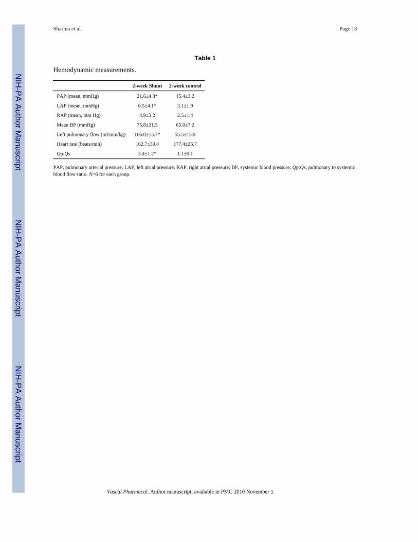
The hemodynamic data for the 2-week old control and Shunt lambs are shown in Table 1.Pulmonary arterial pressure (PAP), left atrial pressure (LAP) and left pulmonary blood flow(LPF) were significantly greater in Shunt lambs than twin controls (Table 1). In addition, therewas a significant difference in pulmonary to systemic blood flow ratio in Shunt lambs (Table1). There were no significant differences in the mean systemic blood pressure, heart rate andright atrial pressure (Table 1).
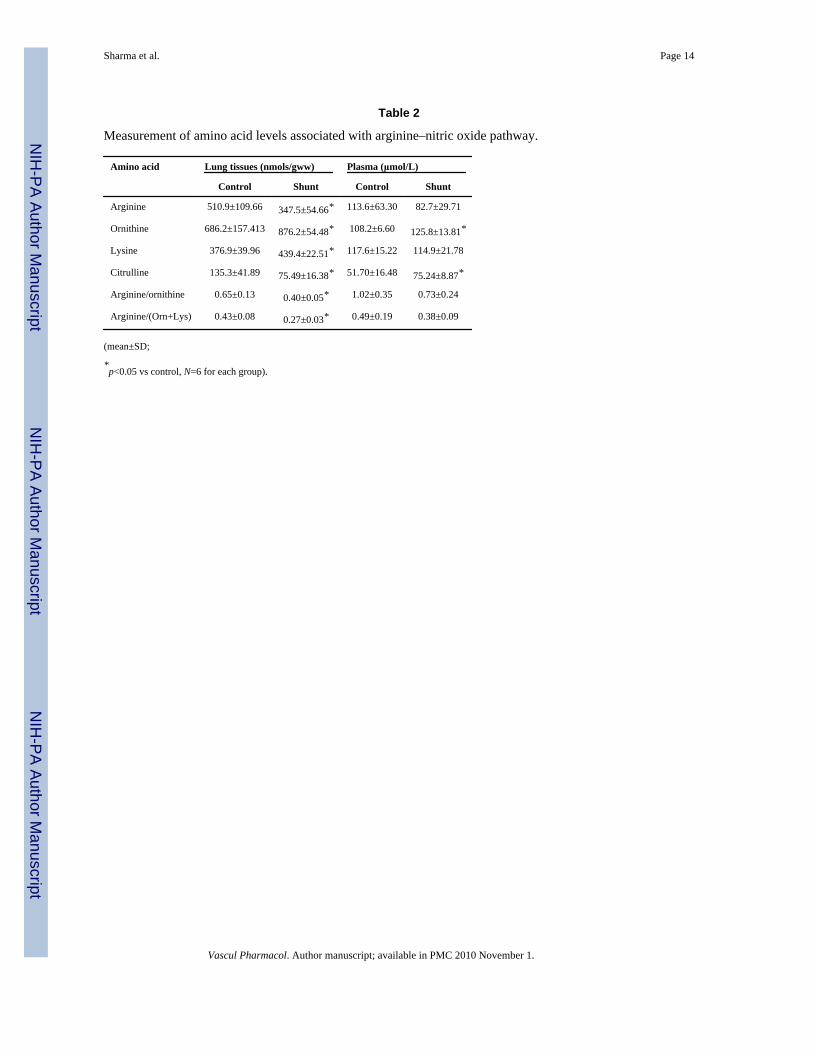
3.2. Lung and plasma amino acid levelsOur data indicate that although plasma L-arginine concentrations trended lower they did notreach statistical significance, whereas, tissue L-arginine concentrations were significantlylower in Shunt lambs (Table 2). Lung L-citrulline levels were also significantly decreased whileplasma levels were significantly higher in the Shunt lambs (Table 2). We also foundsignificantly increased ornithine and lysine levels in lung tissue in Shunt lambs, whereas, inplasma, only ornithine levels were significantly increased (Table 2). We also determined thebiomarkers of arginine bioavailability (arginine/ornithine; arginine/[ornithine+lysine]) inplasma and peripheral lung. These ratios were significantly lower in peripheral lung tissue ofthe Shunt lambs but were not altered in the plasma (Table 2).
3.3. Expression and activity of arginine biosynthetic enzymesWe next examined the expression and activity of the arginine recycling enzymes,argininosuccinate synthase (ASS) and argininosuccinate lyase (ASL). We initially verified ourASS (Fig. 2A) and ASL (Fig. 2D) antibodies by over-expressing each protein in COS-7 cells.Our results indicated that ASS expression was significantly increased in Shunt lambs (Fig. 2B).However, although ASS expression was increased there was a significant decrease in ASS
Sharma et al. Page 4
Vascul Pharmacol. Author manuscript; available in PMC 2010 November 1.
NIH
-PA Author Manuscript
NIH
-PA Author Manuscript
NIH
-PA Author Manuscript
activity in Shunt lambs (Fig. 2C). We found that ASL was significantly decreased in Shuntlambs (Fig. 2E) and this was associated with a significant decrease in ASL activity (Fig. 2F).
3.4. Arginase expression and activityUsing Western blot analysis we found no significant changes in protein levels of arginase 1(Fig. 3A) or arginase 2 (Fig. 3B) nor were the mRNA levels of each isoform altered (Fig. 3C).However, there was a significant increase in arginase activity (Fig. 3D) and CAT-1 transporterprotein (Fig. 3E) in Shunt lambs.
3.5. NO signalingFinally, we determined if the alterations in arginine metabolism altered NO signaling in theShunt lambs. Although we found no change in total eNOS protein levels (Fig. 4A), relativeNOS activity (determined by tissue NOx levels as a fraction of calcium-dependent [3H]–L-arginine to [3H]–L-citrulline conversion) was significantly decreased in Shunt lambs (Fig. 4B).Further this decrease in relative NOS activity correlated with a significant increase in NOS-dependent superoxide generation, indicative of eNOS uncoupling (Fig. 4C).
4. DiscussionIn this study we investigated the role of the arginine metabolism in the progressive loss of NOsignaling and the development of endothelial dysfunction and pulmonary hypertension in 2-week old Shunt lambs. The important findings of this study are as follows: 1) Shunt lambshave decreased activities of the arginine recycling enzymes (ASS, ASL) while arginase activityis increased. 2) L-arginine levels are significantly decreased whereas, there is increased cationicamino acid transporter (CAT-1) expression. 3) These changes correlate with an increase inNOS-derived superoxide and a decrease in relative NO signaling.
Alterations in arginine bioavailability have been implicated in a variety of vascular disorders.It can be impacted by increased consumption by arginase and/or decreased recycling by ASSand ASL. Alterations in the normal balance of arginine and other amino acids involved inarginine–NO pathway (citrulline, ornithine, and lysine) can also significantly affect the cellulararginine uptake and its bioavailability. In this study we identified a decrease in biomarkers ofarginine bioavailability in the lungs, but not the plasma of Shunt lambs. This discrepancy maybe due to the fact that plasma levels reflect levels from sources in addition to the lung. Indeed,our findings are consistent with a recent study in sickle cell disease patients where lowerarginine bioavailability (arginine–ornithine ratio) secondary to increased arginase activity wasassociated with greater severity of pulmonary hypertension and mortality (Morris et al.,2005). Similarly, arginine levels are decreased in children with congenital heart disease andincreased pulmonary blood flow (Gorenflo et al., 2001), and abnormalities in argininemetabolism are associated with increased mortality in patients with cardiogenic shock afteracute myocardial infarction (Nicholls et al., 2007). Arginine bioavailability can be impactedby increased consumption by arginase and we found that arginase activity was increased in thelung tissues of Shunt lambs. However, this increase in activity was independent of changes ineither arginase-1 or -2 protein levels suggesting that a yet unresolved post-translationalmodification is involved. One possibility is that arginase activity can be regulated by its sub-cellular localization with the redistribution of arginase from the cytoskeleton to the cytosolassociated with increased activity (Ryoo et al., 2006). However, further studies will be requiredto test this possibility. It is possible that the increased arginase activity we have identified isinvolved in the development of pulmonary hypertension in this model by shifting argininemetabolism towards ornithine. Indeed, increased arginase-2 has been shown to be involved inreducing NO synthesis in PAH (Xu et al., 2004). While arginase activity is elevated in theserum of sickle cell patients who had associated secondary pulmonary hypertension (Morris
Sharma et al. Page 5
Vascul Pharmacol. Author manuscript; available in PMC 2010 November 1.
NIH
-PA Author Manuscript
NIH
-PA Author Manuscript
NIH
-PA Author Manuscript
et al., 2003). In addition, a recent study has shown decreased plasma arginine levels in sicklecell patients (Morris et al., 2005). Further, although no significant differences in plasmaarginine levels was observed within the sickle cell patients with or without PAH, there werealterations in “arginine bioavailability” between sickle cell patients with severe PAH vs. thosewith mild or no PAH, when ornithine and citrulline levels were taken into consideration.Another study reported decreased plasma L-arginine levels in human infants with pulmonaryhypertension (Pearson et al., 2001). In addition, although we found only a trend towards adecrease in plasma L-arginine levels in this study using 2-week old lambs, our previous studieshave found that plasma levels of L-arginine are significantly decreased at one month of age inthe Shunt lamb (Reddy et al., 1996) suggesting L-arginine levels in the plasma may decreasewith advancing disease. Further developmental studies will be required to test this possibility.
The involvement of the arginine homeostasis pathways in pulmonary hypertension is likelyvery complex and may be altered in several ways. One of the main sources of the L-arginineutilized by eNOS is the cationic amino acid (CAT-1) transporter which is responsible for 60–80% of total carrier mediated arginine transport into endothelial cells (McDonald et al.,1997). Thus, the increase in CAT-1 expression we have identified in Shunt lambs may be amechanism to maintain normal levels of L-arginine in the face of increased L-argininedegradation. In addition, there is another arginine metabolic pathway termed the citrulline–NOcycle, involved in regulating NO signaling. Citrulline produced in the conversion of arginineto NO is efficiently recycled to arginine by two urea cycle enzymes: ASS and ASL. In thisstudy we have observed a significant decrease in the activity of both ASS and ASL. However,the mechanism by which this occurs appears to be complex. Although decreases in ASL proteinlevels correlated with decreased activity, ASS protein levels were significantly increased inShunt lambs but activity decreased. Again, suggesting that a post-translational mechanism caninhibit ASS activity. However, this mechanism is unknown and will require further studies tounravel. This failure of the Shunt lambs to adequately recycle L-citrulline back to L-arginineis also of interest for ongoing clinical studies that are evaluating the therapeutic potential of L-citrulline to decrease the development of pulmonary hypertension after surgical repair ofvarious CHD (Smith et al., 2006; Barr et al., 2007). Indeed, studies have shown the cardio-pulmonary bypass during the surgical correction of both ventricular septal defects andatrioventricular septal defects leads to a decrease in urea cycle repair intermediates includingarginine (Smith et al., 2006) suggesting that there may be alterations in enzyme activity thatcompromises the ability of arginine recycling and it is possible this could limit the efficacy ofL-citrulline therapy.
In conclusion our data indicate that lambs with increased pulmonary blood flow havealterations in the enzyme activities of the arginine biosynthetic and metabolic pathways withinthe pulmonary system. Further, these alterations lead to the disruption of the arginine poolutilized by the eNOS reaction. Thus, we speculate that the arginine metabolic pathway couldbe a potential therapeutic target to maintain NO signaling and endothelial function.
AcknowledgmentsThis research was supported in part by grants HL60190 (to SMB), HL67841 (to SMB), HL72123 (to SMB), HL70061(to SMB), HL084739 (to SMB), R21HD057406 (to SMB), and HL61284 (to JRF) all from the National Institutes ofHealth, by a Transatlantic Network Development Grant from the Fondation Leducq (to SMB, JRF & SF), and by09BGIA2310050 from the Southeast Affiliates of the American Heart Association (to SS). This work was alsosupported by a Programmatic Development award (to SMB) and Seed Awards (to SS and SJ) from the CardiovascularDiscovery Institute of the Medical College of Georgia. Neetu Sud was supported in part by an AHA postdoctoralfellowship from the AHA Southeast affiliates and by 1K99HL097153 from the National Institutes of Health.
Sharma et al. Page 6
Vascul Pharmacol. Author manuscript; available in PMC 2010 November 1.
NIH
-PA Author Manuscript
NIH
-PA Author Manuscript
NIH
-PA Author Manuscript
Abbreviations
ASL Argininosuccinate lyase
ASS Argininosuccinate synthetase
CAT-1 Cationic amino acid transporter-1
ReferencesBarr F, Tirona R, Taylor M, Rice G, Arnold J, Cunningham G, Smith H, Campbell A, Canter JA, Christian
K, Drinkwater D, Scholl F, Kavanaugh-McHugh A, Summar M. Pharmacokinetics and safety ofintravenously administered citrulline in children undergoing congenital heart surgery: potentialtherapy for postoperative pulmonary hypertension. J Thorac Cardiovasc Surg 2007;134:319–326.[PubMed: 17662768]
Black SM, Heidersbach RS, McMullan DM, Bekker JM, Johengen MJ, Fineman JR. Inhaled nitric oxideinhibits NOS activity in lambs: a potential mechanism for rebound pulmonary hypertension. Am JPhysiol 1999;277:H1849–H1856. [PubMed: 10564139]
Dhanakoti SN, Brosnan ME, Herzberg GR, Brosnan JT. Cellular and subcellular localization of enzymesof arginine metabolism in rat kidney. Biochem J 1992;282 (Pt 2):369–375. [PubMed: 1312326]
Erbas H, Aydogdu N, Kaymak K. Effects of N-acetylcysteine on arginase, ornithine and nitric oxide inrenal ischemia-reperfusion injury. Pharmacol Res 2004;50:523–527. [PubMed: 15458774]
Gorenflo M, Zheng C, Pöge A, Bettendorf M, Werle E, Fiehn W, Ulmer H. Metabolites of the L-arginine-NO pathway in patients with left-to-right shunt. Clin Lab 2001;47:441–447. [PubMed: 11596905]
Gutierrez JA, Parry AJ, McMullan DM, Chapin CJ, Fineman JR. Decreased surfactant proteins in lambswith pulmonary hypertension secondary to increased blood flow. Am J Physiol Lung Cell Mol Physiol2001;281:L1264–1270. [PubMed: 11597919]
Hsieh CL, Hsiung KP, Su JC. Determination of lysine with ninhydrin-ferric reagent. Anal Biochem1995;224:187–189. [PubMed: 7710069]
Kumar S, Sun X, Wedgwood S, Black SM. Hydrogen peroxide decreases endothelial nitric oxide synthasepromoter activity through the inhibition of AP-1 activity. Am J Physiol Lung Cell Mol Physiol2008;295:L370–377. [PubMed: 18556800]
Liu H, Drew P, Gaugler AC, Cheng Y, Visner GA. Pirfenidone inhibits lung allograft fibrosis throughL-arginine-arginase pathway. Am J Transplant 2005;5:1256–1263. [PubMed: 15888029]
McDonald KK, Zharikov S, Block ER, Kilberg MS. A caveolar complex between the cationic aminoacid transporter 1 and endothelial nitric-oxide synthase may explain the “arginine paradox”. J BiolChem 1997;272:31213–31216. [PubMed: 9395443]
Morris CR, Morris SM Jr, Hagar W, Van Warmerdam J, Claster S, Kepka-Lenhart D, Machado L,Kuypers FA, Vichinsky EP. Arginine therapy: a new treatment for pulmonary hypertension in sicklecell disease? Am J Respir Crit Care Med 2003;168:63–69. [PubMed: 12626350]
Morris CR, Kato GJ, Poljakovic M, Wang X, Blackwelder WC, Sachdev V, Hazen SL, Vichinsky EP,Morris SM Jr, Gladwin MT. Dysregulated arginine metabolism, hemolysis-associated pulmonaryhypertension, and mortality in sickle cell disease. Jama 2005;294:81–90. [PubMed: 15998894]
Nicholls SJ, Wang Z, Koeth R, Levison B, DelFraino B, Dzavik V, Griffith OW, Hathaway D, PanzaJA, Nissen SE, Hochman JS, Hazen SL. Metabolic profiling of arginine and nitric oxide pathwayspredicts hemodynamic abnormalities and mortality in patients with cardiogenic shock after acutemyocardial infarction. Circulation 2007;116:2315–2324. [PubMed: 17967979]
Pearson DL, Dawling S, Walsh WF, Haines JL, Christman BW, Bazyk A, Scott N, Summar ML. Neonatalpulmonary hypertension–urea-cycle intermediates, nitric oxide production, and carbamoyl-phosphate synthetase function. N Engl J Med 2001;344:1832–1838. [PubMed: 11407344]
Reddy VM, Meyrick B, Wong J, Khoor A, Liddicoat JR, Hanley FL, Fineman JR. In utero placement ofaortopulmonary shunts: a model of postnatal pulmonary hypertension with increased pulmonaryblood flow in lambs. Circulation 1995;92:1–8. [PubMed: 7788902]
Sharma et al. Page 7
Vascul Pharmacol. Author manuscript; available in PMC 2010 November 1.
NIH
-PA Author Manuscript
NIH
-PA Author Manuscript
NIH
-PA Author Manuscript
Reddy VM, Wong J, Liddicoat JR, Johengen M, Chang R, Fineman JR. Altered endothelium-dependentresponses in lambs with pulmonary hypertension and increased pulmonary blood flow. Am J Physiol1996;271:H562–570. [PubMed: 8770097]
Ryoo S, Lemmon CA, Soucy KG, Gupta G, White AR, Nyhan D, Shoukas A, Romer LH, Berkowitz DE.Oxidized low-density lipoprotein-dependent endothelial arginase II activation contributes toimpaired nitric oxide signaling. Circ Res 2006;99:951–960. [PubMed: 17008605]
Sharma S, Sud N, Wiseman DA, Carter AL, Kumar S, Hou Y, Rau T, Wilham J, Harmon C, Oishi P,Fineman JR, Black SM. Altered carnitine homeostasis is associated with decreased mitochondrialfunction and altered nitric oxide signaling in lambs with pulmonary hypertension. Am J Physiol LungCell Mol Physiol 2008;294:L46–56. [PubMed: 18024721]
Smith H, Canter J, Christian K, Drinkwater D, Scholl F, Christman B, Rice G, Barr F, Summar M. Nitricoxide precursors and congenital heart surgery: a randomized controlled trial of oral citrulline. JThorac Cardiovasc Surg 2006;132:58–65. [PubMed: 16798303]
Steinhorn R, Russell J, Lakshminrusimha S, Gugino S, Black S, Fineman J. Altered endothelium-dependent relaxations in lambs with high pulmonary blood flow and pulmonary hypertension. AmJ Physiol Heart Circ Physiol 2001;280:H311–H317. [PubMed: 11123246]
Sud N, Sharma S, Wiseman DA, Harmon C, Kumar S, Venema RC, Fineman JR, Black SM. Nitric oxideand superoxide generation from endothelial NOS: modulation by HSP90. Am J Physiol Lung CellMol Physiol 2007;293:L1444–1453. [PubMed: 17827253]
Sud N, Wells SM, Sharma S, Wiseman DA, Wilham J, Black SM. Asymmetric dimethylarginine inhibitsHSP90 activity in pulmonary arterial endothelial cells: role of mitochondrial dysfunction. Am JPhysiol Cell Physiol 2008;294:C1407–1418. [PubMed: 18385287]
Wu G, Knabe DA, Flynn NE. Synthesis of citrulline from glutamine in pig enterocytes. Biochem J1994;299 (Pt 1):115–121. [PubMed: 8166628]
Xu W, Kaneko FT, Zheng S, Comhair SA, Janocha AJ, Goggans T, Thunnissen FB, Farver C, Hazen SL,Jennings C, Dweik RA, Arroliga AC, Erzurum SC. Increased arginase II and decreased NO synthesisin endothelial cells of patients with pulmonary arterial hypertension. Faseb J 2004;18:1746–1748.[PubMed: 15364894]
Sharma et al. Page 8
Vascul Pharmacol. Author manuscript; available in PMC 2010 November 1.
NIH
-PA Author Manuscript
NIH
-PA Author Manuscript
NIH
-PA Author Manuscript
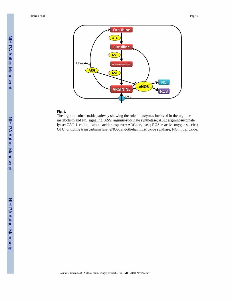
Fig. 1.The arginine–nitric oxide pathway showing the role of enzymes involved in the argininemetabolism and NO signaling. ASS: argininosuccinate synthetase; ASL: argininosuccinatelyase; CAT-1: cationic amino acid transporter; ARG: arginase; ROS: reactive oxygen species;OTC: ornithine transcarbamylase; eNOS: endothelial nitric oxide synthase; NO: nitric oxide.
Sharma et al. Page 9
Vascul Pharmacol. Author manuscript; available in PMC 2010 November 1.
NIH
-PA Author Manuscript
NIH
-PA Author Manuscript
NIH
-PA Author Manuscript
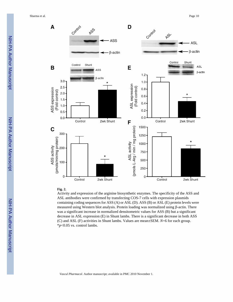
Fig. 2.Activity and expression of the arginine biosynthetic enzymes. The specificity of the ASS andASL antibodies were confirmed by transfecting COS-7 cells with expression plasmidscontaining coding sequences for ASS (A) or ASL (D). ASS (B) or ASL (E) protein levels weremeasured using Western blot analysis. Protein loading was normalized using β-actin. Therewas a significant increase in normalized densitometric values for ASS (B) but a significantdecrease in ASL expression (E) in Shunt lambs. There is a significant decrease in both ASS(C) and ASL (F) activities in Shunt lambs. Values are mean±SEM. N=6 for each group.*p<0.05 vs. control lambs.
Sharma et al. Page 10
Vascul Pharmacol. Author manuscript; available in PMC 2010 November 1.
NIH
-PA Author Manuscript
NIH
-PA Author Manuscript
NIH
-PA Author Manuscript
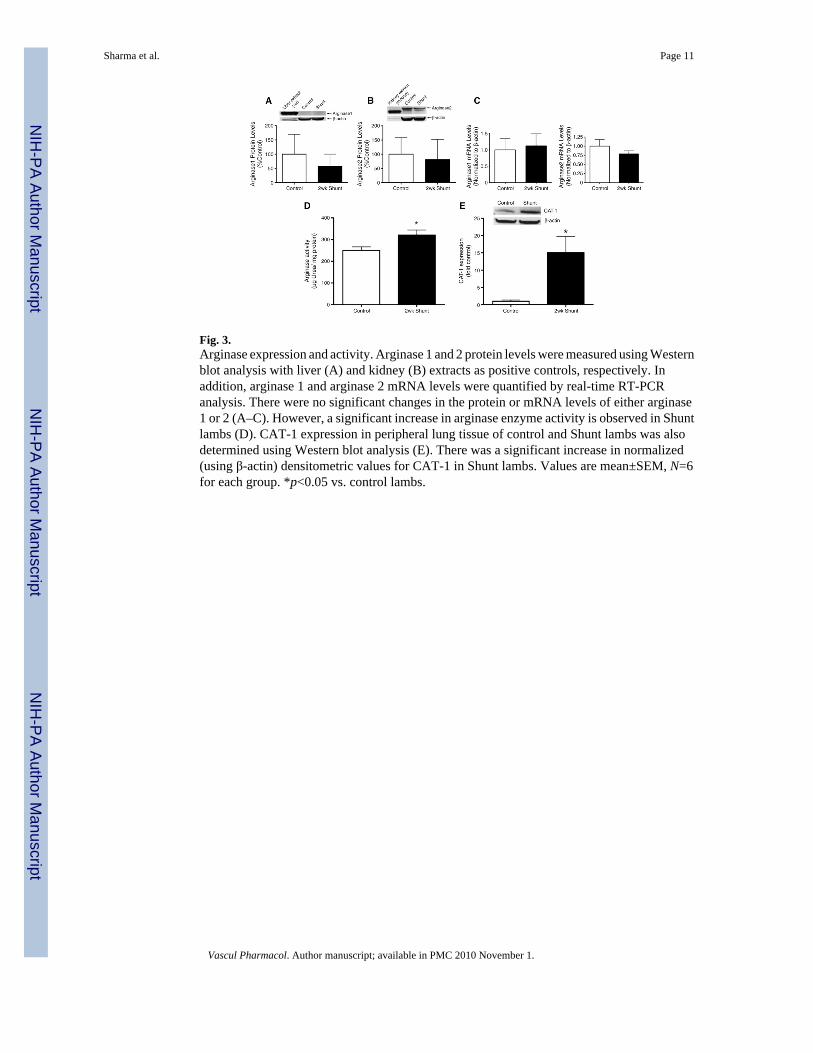
Fig. 3.Arginase expression and activity. Arginase 1 and 2 protein levels were measured using Westernblot analysis with liver (A) and kidney (B) extracts as positive controls, respectively. Inaddition, arginase 1 and arginase 2 mRNA levels were quantified by real-time RT-PCRanalysis. There were no significant changes in the protein or mRNA levels of either arginase1 or 2 (A–C). However, a significant increase in arginase enzyme activity is observed in Shuntlambs (D). CAT-1 expression in peripheral lung tissue of control and Shunt lambs was alsodetermined using Western blot analysis (E). There was a significant increase in normalized(using β-actin) densitometric values for CAT-1 in Shunt lambs. Values are mean±SEM, N=6for each group. *p<0.05 vs. control lambs.
Sharma et al. Page 11
Vascul Pharmacol. Author manuscript; available in PMC 2010 November 1.
NIH
-PA Author Manuscript
NIH
-PA Author Manuscript
NIH
-PA Author Manuscript
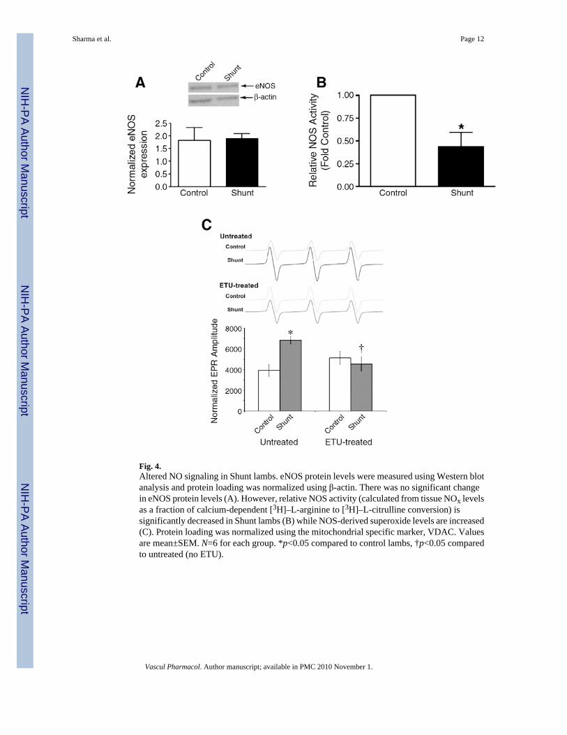
Fig. 4.Altered NO signaling in Shunt lambs. eNOS protein levels were measured using Western blotanalysis and protein loading was normalized using β-actin. There was no significant changein eNOS protein levels (A). However, relative NOS activity (calculated from tissue NOx levelsas a fraction of calcium-dependent [3H]–L-arginine to [3H]–L-citrulline conversion) issignificantly decreased in Shunt lambs (B) while NOS-derived superoxide levels are increased(C). Protein loading was normalized using the mitochondrial specific marker, VDAC. Valuesare mean±SEM. N=6 for each group. *p<0.05 compared to control lambs, †p<0.05 comparedto untreated (no ETU).
Sharma et al. Page 12
Vascul Pharmacol. Author manuscript; available in PMC 2010 November 1.
NIH
-PA Author Manuscript
NIH
-PA Author Manuscript
NIH
-PA Author Manuscript
NIH
-PA Author Manuscript
NIH
-PA Author Manuscript
NIH
-PA Author Manuscript
Sharma et al. Page 13
Table 1
Hemodynamic measurements.
2-week Shunt 2-week control
PAP (mean, mmHg) 21.6±4.3* 15.4±3.2
LAP (mean, mmHg) 6.5±4.1* 3.1±1.9
RAP (mean, mm Hg) 4.9±3.2 2.5±1.4
Mean BP (mmHg) 75.8±31.5 65.0±7.2
Left pulmonary flow (ml/min/kg) 166.0±15.7* 55.5±15.9
Heart rate (beats/min) 162.7±30.4 177.4±26.7
Qp:Qs 3.4±1.2* 1.1±0.1
PAP, pulmonary arterial pressure; LAP, left atrial pressure; RAP, right atrial pressure; BP, systemic blood pressure; Qp:Qs, pulmonary to systemicblood flow ratio. N=6 for each group.
Vascul Pharmacol. Author manuscript; available in PMC 2010 November 1.
NIH
-PA Author Manuscript
NIH
-PA Author Manuscript
NIH
-PA Author Manuscript
Sharma et al. Page 14
Table 2
Measurement of amino acid levels associated with arginine–nitric oxide pathway.
Amino acid Lung tissues (nmols/gww) Plasma (μmol/L)
Control Shunt Control Shunt
Arginine 510.9±109.66 347.5±54.66* 113.6±63.30 82.7±29.71
Ornithine 686.2±157.413 876.2±54.48* 108.2±6.60 125.8±13.81*
Lysine 376.9±39.96 439.4±22.51* 117.6±15.22 114.9±21.78
Citrulline 135.3±41.89 75.49±16.38* 51.70±16.48 75.24±8.87*
Arginine/ornithine 0.65±0.13 0.40±0.05* 1.02±0.35 0.73±0.24
Arginine/(Orn+Lys) 0.43±0.08 0.27±0.03* 0.49±0.19 0.38±0.09
(mean±SD;
*p<0.05 vs control, N=6 for each group).
Vascul Pharmacol. Author manuscript; available in PMC 2010 November 1.
Related Documents