Alterations in Left Ventricular Structure and Function in Type-1 Diabetics: A Focus on Left Atrial Contribution to Function Linda R. Peterson, MD, FACC, Alan D. Waggoner, MHS, Lisa de las Fuentes, MD, Kenneth B. Schechtman, PhD, Janet B. McGill, MD, Robert J. Gropler, MD, FACC, Victor G. Dávila-Román, MD, FACC, St. Louis, Missouri This study was designed to determine the effects of type 1 diabetes mellitus (T1DM) on left ventricular (LV) and particularly left atrial (LA) structure and function. We evaluated 88 non-obese subjects: 44 with T1DM, 44 age- and gender-matched normal controls (age 39 11 years). LV and LA structure and function were quantified using two-dimensional echocardiography, pulse-wave Doppler, and tissue Doppler imaging, including early and late diastolic myocardial velocities (Em global and Am global, respectively). The T1DM subjects averaged higher heart rate, relative wall thickness, and ejection frac- tion, and lower indexed end-systolic volume than normal controls (P < .001, P < .05, P .01, and P < .05, respectively). T1DM was related to A wave veloc- ity, Am global, A wave integral, LA ejection fraction, and LA systolic ejection fraction (P < .01, P < .05, P < .0005, P < .001, and P < .0005, respectively). In multivariate analyses, T1DM was an independent predictor of the A wave integral, LA ejection frac- tion, and LA systolic ejection fraction (P < .01, P < .01, and P < .005, respectively). Thus, despite in- creased relative wall thickness, LV systolic function is increased and early diastolic filling is normal in T1DM subjects; however, they possess changes in LA transport function suggesting increased reliance on LA contribution to LV filling. (J Am Soc Echocardiogr 2006;19:749-755.) The effects of type 1 diabetes mellitus (T1DM) on left ventricular (LV) structure and function are con- troversial. Some studies have shown that there is no change in LV structure, whereas others demonstrate an increase in LV mass index or concentric remod- eling. 1,2 There are also conflicting reports regarding the effect of T1DM on LV diastolic function with some studies showing decreased peak early filling velocities, increased atrial filling velocities, and pro- longation of deceleration and isovolumic relaxation time intervals, 1,3-11 but others showing either no abnormalities or isolated increases in late diastolic filling velocities. 12-14 In addition, few studies have focused specifically on the effect of T1DM on the left atrial (LA) function and contribution to LV filling. 13 Relatively new methods for measuring LA trans- port function include LA ejection fraction measures and tissue Doppler imaging (TDI). Specifically, LA transport function can be described by the two- dimensional–derived LA ejection fraction and the LA systolic ejection fraction (the amount of blood ejected from the LA into the LV during LA contrac- tion.) 15 TDI-derived late diastolic (Am) velocities at the mitral annulus are relatively load-independent measures of LA function. 16 Thus, the purpose of the current study was to evaluate the effect of moderately controlled T1DM uncomplicated by hypertension or other clinically evident cardiovascular disease on LV structure and function, with particular attention to the effect of T1DM on LA structure and its contribution to LV filling. METHODS Subjects Forty-four patients (ages 18-56 years, with T1DM or insu- lin-dependent diabetes, were evaluated at the Cardiovas- cular Imaging and Clinical Research Core Laboratory and met the following entry criteria for this study: a body mass index (BMI) greater than 18 and less than 30 kg/m 2 , systolic blood pressure (SBP) less than 140 mm Hg, diastolic blood pressure (DBP) less than 90 mm Hg, and no From the Cardiovascular Division, Division of Geriatrics and Nu- tritional Sciences, Department of Medicine; Division of Biostatis- tics; Division of Endocrinology and Metabolism; Department of Radiology, Washington University School of Medicine. This work was supported by NIH grants R01HL58878, S10RR14778, K24HL67002, HL13581, AG154666, HL69100, RR00036 (General Clinical Research Center), and by a grant from the Barnes-Jewish Hospital Foundation to the Cardiovascular Imaging and Clinical Research Core Laboratory. Reprint requests: Linda R. Peterson, MD, Washington University School of Medicine, 660 S Euclid Avenue, Campus Box 8086, St. Louis, MO 63110 (E-mail: [email protected]). 0894-7317/$32.00 Copyright 2006 by the American Society of Echocardiography. doi:10.1016/j.echo.2006.01.009 749

Welcome message from author
This document is posted to help you gain knowledge. Please leave a comment to let me know what you think about it! Share it to your friends and learn new things together.
Transcript
Alterations in Left Ventricular Structure andFunction in Type-1 Diabetics: A Focus on Left
Atrial Contribution to FunctionLinda R. Peterson, MD, FACC, Alan D. Waggoner, MHS, Lisa de las Fuentes, MD,Kenneth B. Schechtman, PhD, Janet B. McGill, MD, Robert J. Gropler, MD, FACC,
Victor G. Dávila-Román, MD, FACC, St. Louis, Missouri
This study was designed to determine the effects oftype 1 diabetes mellitus (T1DM) on left ventricular(LV) and particularly left atrial (LA) structure andfunction. We evaluated 88 non-obese subjects: 44with T1DM, 44 age- and gender-matched normalcontrols (age 39 � 11 years). LV and LA structure andfunction were quantified using two-dimensionalechocardiography, pulse-wave Doppler, and tissueDoppler imaging, including early and late diastolicmyocardial velocities (Em global and Am global,respectively). The T1DM subjects averaged higherheart rate, relative wall thickness, and ejection frac-tion, and lower indexed end-systolic volume than
normal controls (P < .001, P < .05, P � .01, and P <doi:10.1016/j.echo.2006.01.009
.05, respectively). T1DM was related to A wave veloc-ity, Am global, A wave integral, LA ejection fraction,and LA systolic ejection fraction (P < .01, P < .05,P < .0005, P < .001, and P < .0005, respectively). Inmultivariate analyses, T1DM was an independentpredictor of the A wave integral, LA ejection frac-tion, and LA systolic ejection fraction (P < .01, P <.01, and P < .005, respectively). Thus, despite in-creased relative wall thickness, LV systolic functionis increased and early diastolic filling is normal inT1DM subjects; however, they possess changes in LAtransport function suggesting increased reliance onLA contribution to LV filling. (J Am Soc Echocardiogr
2006;19:749-755.)The effects of type 1 diabetes mellitus (T1DM) onleft ventricular (LV) structure and function are con-troversial. Some studies have shown that there is nochange in LV structure, whereas others demonstratean increase in LV mass index or concentric remod-eling.1,2 There are also conflicting reports regardingthe effect of T1DM on LV diastolic function withsome studies showing decreased peak early fillingvelocities, increased atrial filling velocities, and pro-longation of deceleration and isovolumic relaxationtime intervals,1,3-11 but others showing either noabnormalities or isolated increases in late diastolicfilling velocities.12-14 In addition, few studies havefocused specifically on the effect of T1DM on theleft atrial (LA) function and contribution to LVfilling.13
From the Cardiovascular Division, Division of Geriatrics and Nu-tritional Sciences, Department of Medicine; Division of Biostatis-tics; Division of Endocrinology and Metabolism; Department ofRadiology, Washington University School of Medicine.This work was supported by NIH grants R01HL58878,S10RR14778, K24HL67002, HL13581, AG154666, HL69100,RR00036 (General Clinical Research Center), and by a grant fromthe Barnes-Jewish Hospital Foundation to the CardiovascularImaging and Clinical Research Core Laboratory.Reprint requests: Linda R. Peterson, MD, Washington UniversitySchool of Medicine, 660 S Euclid Avenue, Campus Box 8086, St.Louis, MO 63110 (E-mail: [email protected]).0894-7317/$32.00Copyright 2006 by the American Society of Echocardiography.
Relatively new methods for measuring LA trans-port function include LA ejection fraction measuresand tissue Doppler imaging (TDI). Specifically, LAtransport function can be described by the two-dimensional–derived LA ejection fraction and the LAsystolic ejection fraction (the amount of bloodejected from the LA into the LV during LA contrac-tion.)15 TDI-derived late diastolic (Am) velocities atthe mitral annulus are relatively load-independentmeasures of LA function.16
Thus, the purpose of the current study was toevaluate the effect of moderately controlled T1DMuncomplicated by hypertension or other clinicallyevident cardiovascular disease on LV structure andfunction, with particular attention to the effect ofT1DM on LA structure and its contribution to LVfilling.
METHODS
Subjects
Forty-four patients (ages 18-56 years, with T1DM or insu-lin-dependent diabetes, were evaluated at the Cardiovas-cular Imaging and Clinical Research Core Laboratory andmet the following entry criteria for this study: a body massindex (BMI) greater than 18 and less than 30 kg/m2,systolic blood pressure (SBP) less than 140 mm Hg,
diastolic blood pressure (DBP) less than 90 mm Hg, and no749
Journal of the American Society of Echocardiography750 Peterson et al June 2006
history of major systemic disease (e.g., cancer, lupus)other than T1DM or its related complications, a stressechocardiogram negative for inducible ischemia or sys-tolic dysfunction (defined as an LV ejection fraction�55%), pericardial or valvular disease, and no subject waspregnant or lactating. Five T1DM subjects were takingangiotensin-converting enzyme inhibitors; no other sub-jects were taking any other vasoactive medications. TheT1DM patients had measurements of serum glycatedhemoglobin (%HbA1c) and creatinine levels obtainedwithin 1 month of study. Heart rate was measured after 10minutes in the supine position. The resting 12-lead elec-trocardiogram (lead II or III) was used to measure the QTand RR intervals, and corrected for the cardiac cyclelength (QTc) according to Bazett’s formula (QTc � QT/[RR]1/2). Values were averaged over three consecutivebeats. Peripheral complications were determined by apatient’s self-reported history of and/or evidence of reti-nopathy, nephropathy, or peripheral neuropathy. Echo-cardiographic results from a group of 44 age- and gender-matched normal controls (NC) who also had normal stressechocardiograms and met the above-listed entry criteriawere used for comparison. All subjects signed an informedconsent, which was approved by the Institutional ReviewBoard at Washington University School of Medicine.
Two-dimensional and Pulsed-wave DopplerEchocardiography
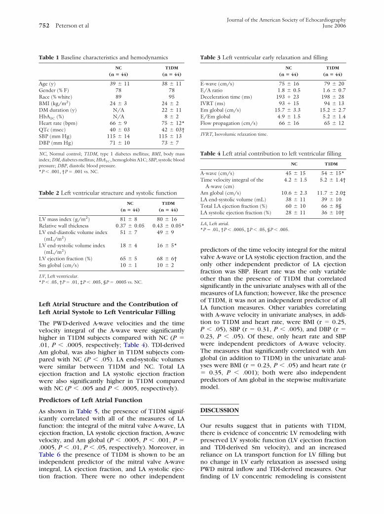
All subjects underwent a complete two-dimensionaland pulsed-wave Doppler (PWD)-echocardiographic ex-amination using a commercially available ultrasoundsystem (Sequoia-C256, Acuson-Siemens, Mountain View,CA) equipped with second harmonic imaging software.Quantitative measurements at rest included LV volumes(method of discs) at end-diastole and end-systole indexedfor body surface area; the LV ejection fraction was calcu-lated as (LV end-diastolic volume – LV end-systolic vol-ume)/LV end-diastolic volume. The LV mass was deter-mined using the area-length method and was indexed forbody surface area.17 LA volumes were measured by themethod of discs in the apical four-chamber view atend-systole, end-diastole, and late diastole (before atrialcontraction, determined from the onset of the P wave onthe surface electrocardiogram).15 Total LA ejection frac-tion was determined by the following equation: LA ejec-tion fraction � (LA end-systolic volume – LA end-diastolicvolume)/LA end-systolic volume (Fig. 1 A and C). The LAsystolic ejection fraction (i.e., the fractional volumeejected during atrial systole) was determined as the ratioof (LA late-diastolic volume – LA end-diastolic volume)/LAlate-diastolic volume (Fig. 1 B and C).
PWD-derived transmitral velocities were obtained at themitral leaflet tips per the American Society of Echocardi-ography guidelines.18 Measurements included the earlydiastolic E-wave and atrial A-wave velocities, E/A ratio,isovolumic relaxation and deceleration time intervals, andintegral of the area under the A-wave (Fig. 1, D). Bothtwo-dimensional echocardiographic and PWD-derived
transmitral indices were measured by one observer andaveraged over three to five consecutive cardiac cycles.Interobserver variability in measurements of PWD, LVmass, and TDI-derived Em global has been reported.19
Tissue Doppler Imaging and Color M-Mode FlowPropagation
TDI was performed in the apical four- and two-chamberviews by placement of a 3-mm sample gate at the lateral,septal, anterior, and inferior mitral annulus. Measurementsof Em and Am velocities were obtained at each annularsite, as described in a previous study from our laboratory(Fig. 1E).19 The Em global and Am global velocities werederived by averaging the velocities from the four annularsites. The ratios of the PWD-derived E wave to theTDI-derived Em global velocity (E/Em) were calculated toprovide an estimation of LV filling pressures.20
The early diastolic flow propagation velocities wereobtained in the apical four-chamber view by positioningan M-Mode cursor within the color flow velocity profile ofmitral inflow.18 The slope (cm/s) of the early diastolicvelocities was measured at the mitral leaflets to a distanceof 4 cm into the LV chamber.
Statistical Analysis
Statistical analyses were performed using the SAS softwareversion 8.2 (SAS Institute, Cary, NC). All continuousvariables are expressed as mean � standard deviation.Comparisons of echocardiographic variables of T1DMsubjects and NC were performed using Student t test forunpaired data. Chi-square tests were used for comparisonof categoric variables. Pearson’s correlation coefficientswere used to assess the strength of the relationshipbetween continuous variables. All predetermined inde-pendent variables that correlated with the dependentmeasures of diastolic function (Am, A, the time velocityintegral of the mitral valve A-wave, the LA ejection frac-tion, and the LA systolic ejection fraction) with a P valueof less than .1 in the Pearson’s correlations were insertedinto a stepwise, multiple regression analysis. To determinethe covariate-adjusted effect of T1DM on the dependentvariables, all multivariate analyses required that diabetesbe included in the model. Statistical significance wasdetermined as a P value of less than .05; this was notcorrected for the performance of multiple analyses.
RESULTS
The demographic and hemodynamic variables ofT1DM subjects and NC are shown in Table 1. Themajority of patients in both groups were women.The average duration of T1DM was 22 � 11 years.There was no significant difference between theracial compositions of the two groups, but theabsolute numbers of non-white subjects was small(five in the NC and two in the T1DM group,respectively). The resting heart rate was significantly
higher and QTc was longer in T1DM compared with
Journal of the American Society of EchocardiographyVolume 19 Number 6 Peterson et al 751
NC. Of the T1DM patients, the mean %HbA1c of theT1DM patients was 8.2 � 2.0%; 19% of T1DMsubjects had peripheral vascular complications, in-cluding seven with retinopathy, three with neurop-athy, and one with nephropathy, defined as a serumcreatinine level greater than 0.115 mmol/L (1.3mg/dL).
Left Ventricular Structure and SystolicFunction
Table 2 shows the LV end-systolic volume index waslower, and the relative wall thickness was higher(P � .05 for both) in T1DM subjects compared withNC. The increased relative wall thickness with nor-mal LV mass index is consistent with a pattern ofconcentric LV remodeling.21 The LV ejection frac-tion was increased in the T1DM subjects compared
Figure 1 The following illustrations are from a repwithout T1DM: A, Left atrial (LA) volume at endmL). C, LA volume at end-diastole (12 mL). Tsystolic ejection fraction is 29%. D, Pulsed-wave Dintegral (5.1 cm). E, Tissue Doppler mitral annul(11.3 cm/s) at the septal annulus.
with NC (P � .01). However, the TDI-derived
measure of LV systolic longitudinal shortening, Smglobal, was not different between the two groups.
Left Ventricular Early Relaxation and Filling
As shown in Table 3, mean mitral E-wave velocity,E/A ratio, deceleration time, isovolumic relaxationtime, color M-Mode flow propagation velocity, andEm velocity were similar in T1DM and NC. Themitral E/Em ratios were also not different betweenthe two groups. T1DM did not correlate with therelatively load-independent measure of early LV re-laxation, Em global. Em was negatively correlatedwith BMI (r � �0.25, P � .05), SBP (r � �0.22, P �.05), DBP (r � �0.35, P � .001), and diabetesduration (r � �0.31, P � .05); of these only DBPwas an independent predictor in the multivariate
tative echocardiogram from a 40-year-old womane (38 mL). B, LA volume before atrial systole (17culated LA ejection fraction is 68% and the LAr mitral inflow with measurement of the A-wave
y diastolic (Em) and late diastolic (Am) velocities
resen-systolhe calopple
ar earl
model (P � .001).
Journal of the American Society of Echocardiography752 Peterson et al June 2006
Left Atrial Structure and the Contribution ofLeft Atrial Systole to Left Ventricular Filling
The PWD-derived A-wave velocities and the timevelocity integral of the A-wave were significantlyhigher in T1DM subjects compared with NC (P �.01, P � .0005, respectively; Table 4). TDI-derivedAm global, was also higher in T1DM subjects com-pared with NC (P � .05). LA end-systolic volumeswere similar between T1DM and NC. Total LAejection fraction and LA systolic ejection fractionwere also significantly higher in T1DM comparedwith NC (P � .005 and P � .0005, respectively).
Predictors of Left Atrial Function
As shown in Table 5, the presence of T1DM signif-icantly correlated with all of the measures of LAfunction: the integral of the mitral valve A-wave, LAejection fraction, LA systolic ejection fraction, A-wavevelocity, and Am global (P � .0005, P � .001, P �.0005, P � .01, P � .05, respectively). Moreover, inTable 6 the presence of T1DM is shown to be anindependent predictor of the mitral valve A-waveintegral, LA ejection fraction, and LA systolic ejec-
Table 1 Baseline characteristics and hemodynamics
NC
(n � 44)
T1DM
(n � 44)
Age (y) 39 � 11 38 � 11Gender (% F) 78 78Race (% white) 89 95BMI (kg/m2) 24 � 3 24 � 2DM duration (y) N/A 22 � 11HbA1C (%) N/A 8 � 2Heart rate (bpm) 66 � 9 75 � 12*QTc (msec) 40 � 03 42 � 03†SBP (mm Hg) 115 � 14 115 � 13DBP (mm Hg) 71 � 10 73 � 7
NC, Normal control; T1DM, type 1 diabetes mellitus; BMI, body massindex; DM, diabetes mellitus; HbA1C, hemoglobin A1C; SBP, systolic bloodpressure; DBP, diastolic blood pressure.*P � .001, †P � .001 vs. NC.
Table 2 Left ventricular structure and systolic function
NC
(n � 44)
T1DM
(n � 44)
LV mass index (g/m2) 81 � 8 80 � 16Relative wall thickness 0.37 � 0.05 0.43 � 0.05*LV end-diastolic volume index
(mL/m2)51 � 7 49 � 9
LV end-systolic volume index(mL/m2)
18 � 4 16 � 5*
LV ejection fraction (%) 65 � 5 68 � 6†Sm global (cm/s) 10 � 1 10 � 2
LV, Left ventricular.*P � .05, †P � .01, ‡P � .005, §P � .0005 vs. NC.
tion fraction. There were no other independent
predictors of the time velocity integral for the mitralvalve A-wave or LA systolic ejection fraction, and theonly other independent predictor of LA ejectionfraction was SBP. Heart rate was the only variableother than the presence of T1DM that correlatedsignificantly in the univariate analyses with all of themeasures of LA function; however, like the presenceof T1DM, it was not an independent predictor of allLA function measures. Other variables correlatingwith A-wave velocity in univariate analyses, in addi-tion to T1DM and heart rate, were BMI (r � 0.25,P � .05), SBP (r � 0.31, P � .005), and DBP (r �0.23, P � .05). Of these, only heart rate and SBPwere independent predictors of A-wave velocity.The measures that significantly correlated with Amglobal (in addition to T1DM) in the univariate anal-yses were BMI (r � 0.23, P � .05) and heart rate (r� 0.35, P � .001); both were also independentpredictors of Am global in the stepwise multivariatemodel.
DISCUSSION
Our results suggest that in patients with T1DM,there is evidence of concentric LV remodeling withpreserved LV systolic function (LV ejection fractionand TDI-derived Sm velocity), and an increasedreliance on LA transport function for LV filling butno change in LV early relaxation as assessed usingPWD mitral inflow and TDI-derived measures. Our
Table 3 Left ventricular early relaxation and filling
NC
(n � 44)
T1DM
(n � 44)
E-wave (cm/s) 75 � 16 79 � 20E/A ratio 1.8 � 0.5 1.6 � 0.7Deceleration time (ms) 193 � 23 198 � 28IVRT (ms) 93 � 15 94 � 13Em global (cm/s) 15.7 � 3.3 15.2 � 2.7E/Em global 4.9 � 1.5 5.2 � 1.4Flow propagation (cm/s) 66 � 16 65 � 12
IVRT, Isovolumic relaxation time.
Table 4 Left atrial contribution to left ventricular filling
NC T1DM
A-wave (cm/s) 45 � 15 54 � 15*Time velocity integral of the
A-wave (cm)4.2 � 1.5 5.2 � 1.4†
Am global (cm/s) 10.6 � 2.3 11.7 � 2.0‡LA end-systolic volume (mL) 38 � 11 39 � 10Total LA ejection fraction (%) 60 � 10 66 � 8§LA systolic ejection fraction (%) 28 � 11 36 � 10†
LA, Left atrial.*P � .01, †P � .0005, ‡P � .05, §P � .005.
finding of LV concentric remodeling is consistent
of LA fu
Journal of the American Society of EchocardiographyVolume 19 Number 6 Peterson et al 753
with some previous studies in which non-hyperten-sive T1DM patients had an increase in wall thick-ness, relative wall thickness, and/or LV mass.1-3 Incontrast, a study in children and adolescents (meanage 11 years) did not show a difference betweenT1DM patients and NC, suggesting that the LVremodeling in T1DM may be a progressive processrequiring years to evolve.22 The current study alsoshows that in T1DM patients free of clinically evi-dent coronary disease, LV ejection fraction andlongitudinal systolic function (i.e., TDI-derived Smvelocity) are either increased or similar to NC,suggesting no adverse effect of T1DM on systolicperformance.
The finding that the primary abnormality of LVdiastolic function in T1DM patients without optimalglycemic control was the increased requirement foratrial contribution to LV filling was supported by allof our TDI, PWD, and two dimensional measures ofLA function. T1DM and heart rate were the only twovariables that were significantly related to all of themeasures of LA function studied. However, theincreased heart rate in T1DM group did not accountfor all of the effect of T1DM on LA function; T1DMwas an independent predictor of the time velocityintegral of the A-wave, LA ejection fraction, and LAsystolic ejection fraction. Thus, our data suggest thatthe LV of patients with T1DM is dependent on anincreased atrial contribution to LV filling. This isconsistent with a study in T1DM patients in whichLA transport function, determined using acousticquantification, is increased during late diastolic LVfilling, but peak early diastolic filling (indexed for LV
Table 5 Univariate correlations with left atrial function
A-wave integral LA ejection fractio
T1DM P < .0005 P < .001HbA1c r � 0.05, P � .73 r � 0.32, P � .05T1DM duration r � 0.04, P � .78 r � �0.28, P � .BMI r � 0.16, P � 14 r � �0.18, P � .HR r � 0.30, P < .005 r � 0.23, P < .0SBP r � 0.20, P � .07 r � �0.19, P � .DBP r � 0.10, P � .36 r � �0.07, P � .Triglycerides r � 0.05, P � .76 r � 0.36, P < .0Relative wall thickness r � 0.15, P � .16 r � 0.01, P � .96
Table 6 Independent predictors of left atrial function
A-wave integral LA ejection fraction
T1DM (P < .01) T1DM (P < .01)SBP (P < .05)
R2 for the model 0.17 0.19
LA, Left atrial; T1DM, type 1 diabetes mellitus; HBA1c, hemoglobin A1C;blood pressure.Variables in bold type are statistically significantly related to the measures
size) was normal.9
The current investigation found that measures ofearly LV relaxation, both load-dependent PWD-de-rived indices (E-wave velocity, E/A ratio, decelera-tion time, isovolumic relaxation time, and colorM-Mode flow propagation velocities) and a relativelyload-independent measure (TDI-derived Em veloc-ity), remain normal in moderately controlled T1DMpatients; these findings suggest that active relaxationby the LV is preserved. This is in contrast with asmaller study that also used TDI to investigate theeffects of T1DM on LV diastolic function.23 In thatstudy, Em was lower in the T1DM group than in theNC group.23 Differences in TDI methods, patientpopulations, screening procedures, and numbers ofpatients may explain some of the differences be-tween the results of our TDI-derived measures andthose of the previous study.23 To our knowledge,these are the only studies to use TDI to investigatethe load-independent effects of T1DM on LV dia-stolic function. Interestingly, in that study and in theCardiovascular Health Study, atrial function definedby PWD-derived measures of A-wave velocity wasaltered in the T1DM patients, which is concordantwith our findings.23,24
Proposed Mechanisms of Increased Left AtrialFunction in Type 1 Diabetes Mellitus
Abnormalities of the balance between sympatheticinput and parasympathetic tone, which are oftenpresent in T1DM subjects, may contribute to theobserved alterations in the LA contribution to LVfilling. The increased resting heart rate and prolon-
systolic ejection
fraction A-wave velocity Am global
P � .0005 P < .01 P < .050.05, P � .75 r � 0.09, P � .55 r � �0.03, P � .850.30, P < .05 r � 0.21, P � .18 r � 0.24, P � .120.06, P � .59 r � 0.25, P < .05 r � 0.23, P < .050.23, P < .05 r � 0.39, P < .0005 r � 0.35, P < .0010.12, P � .28 r � 0.31, P < .005 r � 0.18, P � .090.03, P � .78 r � 0.23, P < .05 r � 0.11, P � .300.03, P � .84 r � �0.03, P � .87 r � 0.12, P � .460.14, P � .19 r � 0.15, P � .16 r � 0.03, P � 0.82
stolic ejection fraction A-wave velocity Am global
T1DM (P < .005) HR (P < .005) HR (P < .005)SBP (P < .05) BMI (P < .05)
0.28 0.28 0.18
dy mass index; HR, heart rate; SBP, systolic blood pressure; DBP, diastolic
nction.
n
LA
r �07 r �10 r �5 r �07 r �54 r �5 r �
r �
LA sy
BMI, bo
gation of the QTc interval in the T1DM patients
Journal of the American Society of Echocardiography754 Peterson et al June 2006
suggest that the balance between parasympatheticand sympathetic systems is not the same as innondiabetic subjects.25-28 T1DM may cause a relativeincrease in sympathetic stimulation and decrease inparasympathetic tone, which may contribute tosmaller LV end-systolic volumes and shorter LVfilling time (resulting from the increased restingheart rate), thus resulting in increased reliance onthe LA contribution to LV filling because less spaceand time are available for early LV filling. However,because T1DM is an independent predictor of manyof the measures of LA contractility, the associationbetween T1DM and LA contractility does not seemto be exclusively mediated by an increased heartrate.
The increases in TDI Am velocities and othermeasures of late LV filling may also, in part, reflectincreases in LV end-diastolic pressures because ofincreased passive stiffness that would require aug-mented LA transport function.29 Alternations in pas-sive LV stiffness may result from increased myocar-dial collagen formation, which has been suggestedby studies using ultrasonic integrated backscatter tocharacterize the myocardium of T1DM patients.8
Limitations of the Study
This study is not longitudinal and, thus, cannotdetermine causation but rather demonstrates thestrength and independence of the associations be-tween T1DM and measures of LA contractility. Thisstudy assessed LV systolic and diastolic function in arelatively young, healthy population of T1DM pa-tients with few peripheral complications. The datafrom this study cannot necessarily be used to predictoutcomes in patients whose profiles do not fit theentry criteria used in this study. Also, given therelatively small numbers of non-white subjects in thisstudy, the possible effect of race on the outcomevariables cannot be assessed in this study. More inva-sive and/or expensive measures for quantifying eitherLV wall compliance (from catheterization-derivedLV pressure-volume loops) or the balance betweensympathetic outflow and parasympathetic tone werenot used in this study. The study by Stevens et al.30
highlights the complexity of studying the effects ofthe sympathetic nervous system in patients withT1DM, that is, there seems to be heterogeneity inthe effects of this system on different regions of themyocardium.
CONCLUSIONS
Relatively healthy T1DM patients without evidenceof coronary artery disease or heart failure have LVconcentric remodeling, normal LV systolic function,normal early diastolic LV relaxation, but increased
LA transport function. This reliance on increased LAtransport cannot be completely accounted for byincreases in resting heart rate.
The authors thank Joann Reagan, RN, Donna Lesniak, RN,and Jeffrey Baumstark for their assistance with the perfor-mance of this study, and Ava Ysaguirre for her assistancein the preparation of this article.
REFERENCES
1. Carugo S, Giannattasio C, Calchera I, Paleari F, GorgoglioneMG, Grappiolo A, et al. Progression of functional and struc-tural cardiac alterations in young normotensive uncompli-cated patients with type I diabetes mellitus. J Hypertens2001;19:1675-80.
2. Sato A, Tarnow L, Parving H-H. Increased left ventricularmass in normotensive type 1 diabetic patients with diabeticnephropathy. Diabetes Care 1998;21:1534-9.
3. Palliole C, Dahan M, Paycha F, Solal A, Passa P, Gourgon R.Prevalence and significance of left ventricular filling abnormal-ities determined by Doppler echocardiography in young type1 insulin-dependent diabetic patients. Am J Cardiol 1989;64:1010-6.
4. Zarich S, Arbuckle B, Cohen L, Roberts M, Nesto R. Diastolicabnormalities in young asymptomatic diabetic patients as-sessed by pulsed Doppler echocardiography. J Am Coll Car-diol 1988;12:114-20.
5. Sampson MJ, Chamber JB, Sprigings DC, Drury PL. Abnor-mal diastolic function in patients with type 1 diabetes and earlynephropathy. Br Heart J 1990;64:266-71.
6. Irace L, Iarussi D, Guadagno I, Tedesco MA, Perna B, RattiG, et al. Left ventricular performance and autonomic dysfunc-tion in patients with long-term insulin dependent diabetesmellitus. Acta Diabetologia 1996;33:269-73.
7. Airaksinen KEJ, Kostinen MJ, Ikakeimo MJ, Huikuri HV,Korhonen U, Pirttiaho H, et al. Augmentation of atrial con-tribution to left ventricular filling in IDDM subjects assessedby echocardiography. Diabetes Care 1989;12:159-61.
8. Perez JE, McGill JB, Santiago JV, Schechtman KB, WaggonerAD, Miller JG, et al. Abnormal myocardial acoustic propertiesin diabetic patients and their correlation with severity of dis-ease. J Am Coll Cardiol 1992;19:1154-62.
9. Albanna II, Eichelberger SM, Khoury PR, Witt SA, StandifordDA, Dolan LM, et al. Diastolic dysfunction in young patientswith insulin-dependent diabetes mellitus as determined byautomated border detection. J Am Soc Echocardiogr 1998;11:349-55.
10. Berg TJ, Snorgaard O, Faber J, Torjesen PA, Hildebrandt P,Mehlsen J, et al. Serum levels of advanced glycation end productsare associated with left ventricular diastolic function in patientswith type 1 diabetes. Diabetes Care 1999;22:1186-90.
11. Monteaguido PT, Moises VA, Kohlmann O, Ribeiro AB,Lima VC, Zanella MT. Influence of autonomic neuropathyupon left ventricular dysfunction in insulin-dependent dia-betic patients. Clin Cardiol 2000;23:371-5.
12. Romanens M, Finkhauser S, Saner B, Michaud L, Sauer H.No evidence for systolic or diastolic dysfunction at rest inselected patients with long-term type 1 diabetes mellitus. EurJ Heart Fail 1999;1:169-75.
13. Gotzsche O, Darwish A, Hansen LP, Gotzsche L. Abnormalleft ventricular diastolic function during cold pressor test inuncomplicated insulin-dependent diabetes mellitus. Clin Sci
1995;89:461-5.
Journal of the American Society of EchocardiographyVolume 19 Number 6 Peterson et al 755
14. Azvendo MJ, Pinotti AF, Gross JL, Stumpf AG, Ribeiro JP.Preserved left ventricular systolic and diastolic function innormoalbuminuric, insulin-dependent diabetic patients withglomerular hyperfiltration. Diabetes Res Clin Pract 1994;25:103-10.
15. Waggoner AD, Barzilai B, Miller JG, Perez JG. On-line as-sessment of left atrial area and function by echocardiographicautomatic boundary detection. Circulation 1993;88:1142-9.
16. Waggoner AD, Bierig SM. Tissue Doppler imaging: a usefulechocardiographic method for the cardiac sonographer toassess systolic and diastolic left ventricular function. J Am SocEchocardiogr 2001;14:1143-52.
17. Schiller NB, Shah PM, Crawford M, DeMaria A, Devereux R,Feigenbaum H, et al. Recommendations for quantification ofthe left ventricle by two-dimensional echocardiography. J AmSoc Echocardiogr 1989;2:358-67.
18. Quinones MA, Otto CM, Stoddard M, Waggoner A, ZoghbiWA. Recommendations for quantification of Doppler echo-cardiography: a report from the Doppler Quantification TaskForce of the Nomenclature and Standards Committee of theAmerican Society of Echocardiography. J Am Soc Echocar-diogr 2002;15:167-84.
19. Peterson LR, Waggoner AD, Schectman KB, Meyer T, Gro-pler RJ, Barzilai B, et al. Alterations in left ventricular structureand function in young healthy obese women: assessment byechocardiography and tissue Doppler imaging. J Am CollCardiol 2004;43:1399-404.
20. Ormen SR, Nishimura RA, Appleton CP, Miller FA, Oh JK,Redfield MM, et al. Clinical utility of Doppler echocardiogra-phy and tissue Doppler imaging in the estimation of leftventricular filling pressures. A simultaneous Doppler-cathe-terization study. Circulation 2000;102:1788-94.
21. Verdeccia P, Schillaci G, Borgioni C, Ciucci A, Battistelli M,Bartoccini C, et al. Adverse prognostic significance of concen-tric remodeling of the left ventricle in hypertensive patientswith normal left ventricular mass. J Am Coll Cardiol 1995;25:
871-8.22. Gunczler P, Lanes R, Lopez E, Esaa S, Villarroel O, Revel-Chion R. Cardiac mass and function, carotid artery intima-media thickness and lipoprotein (a) levels in children andadolescents with type 1 diabetes mellitus of short duration.J Pediatr Endocrinol Metab 2002;15:181-6.
23. Shishehbor MH, Hoogwerf B, Schoenhagen P, Marso SP,Sun JP, Li J, et al. Relation of hemoglobin A1C to leftventricular relaxation in patients with type 1 diabetes mellitusand without overt heart disease. Am J Cardiol 2003;91:1514-7.
24. Lee M, Gardin JM, Lynch JC, Smith VE, Tracy RP, Savage PJ,et al. Diabetes mellitus and echocardiographic left ventricularfunction in free-living elderly men and women: The Cardio-vascular Health Study. Am Heart J 1997;133:36-43.
25. Ewing DJ, Campbell IW, Clarke BF. The natural history ofdiabetic autonomic neuropathy. QJM 1980;49:95-108.
26. O’Brien IAD, O’Hare JP, Lewin IG, Corrall RJM. The prev-alence of autonomic neuropathy in insulin-dependent diabe-tes mellitus: a controlled study based on heart rate variability.QJM 1986;234:957-67.
27. Chambers JB, Sampson MJ, Sprigings DC, Jackson G. QTprolongation on the electrocardiogram in diabetic neuropa-thy. Diabet Med 1990;7:105-10.
28. Sivieri R, Veglio M, Chinaglia A, Scaglione P, Cavallo-Perin P.Prevalence of QT prolongation in a type 1 diabetic populationand its association with diabetic autonomic neuropathy. Dia-bet Med 1993;10:920-4.
29. Lisauskas JB, Singh J, Bowman AW, Kovacs SJ. Chamberproperties from transmitral flow: prediction of average andpassive left ventricular diastolic stiffness. J Appl Physiol 2001;91:154-62.
30. Stevens MF, Dayanikli F, Raffel DM, Allman KC, Sandford T,Feldman EL, et al. Scintigraphic assessment of regionalizeddefects in myocardial sympathetic innervation and blood flowregulation in diabetic patients with autonomic neuropathy.
J Am Coll Cardiol 1998;31:1575-84.
Related Documents