256 T he immune system is a finely tuned network that protects the host against foreign antigens, particularly infectious agents. Sometimes this network breaks down, causing the immune system to react inappropriately. Inappropriate im- mune responses may be (1) exaggerated against environmen- tal antigens (allergy); (2) misdirected against the host’s own cells (autoimmunity); (3) directed against beneficial foreign tissues, such as transfusions or transplants (alloimmunity); or (4) insufficient to protect the host (immune deficiency). All of these can be serious or life threatening. Exaggerated im- mune responses (allergy) are the most common, but usually the least life threatening. HYPERSENSITIVITY: ALLERGY, AUTOIMMUNITY, AND ALLOIMMUNITY Hypersensitivity is an altered immunologic response to an antigen that results in disease or damage to the host. Hypersensitivity reactions can classified in two ways: by the source of the antigen that the immune system is attacking (allergy, autoimmunity, alloimmunity; Table 8-1) and by the mechanism that causes disease (types I, II, III, IV; see Table 8-3). The term allergy originally denoted both facets of the immune response: immunity, which is beneficial, and hyper- sensitivity, which is harmful. Allergy has now come to mean the deleterious effects of hypersensitivity to environmental (exogenous) antigens, and immunity means the protective responses to antigens expressed by disease-causing agents. Autoimmunity is a disturbance in the immunologic tol- erance of self-antigens. The immune system normally does not strongly recognize the individual’s own antigens. Healthy individuals of all ages, but particularly older adults, may pro- duce low quantities of antibodies against their own antigens (autoantibodies), without development of overt autoimmune disease. Therefore, the presence of low quantities of autoan- tibodies does not necessarily indicate a disease state. Autoim- mune diseases occur when the immune system reacts against self-antigens to such a degree that the person’s own tissues are damaged by autoantibodies or autoreactive T cells. Many clinical disorders are associated with autoimmunity and are collectively referred to as autoimmune diseases (Table 8-2). Alloimmunity (also termed isoimmunity) occurs when the immune system of one individual produces an immunologic reaction against tissues of another individual. Alloimmunity ALTERATIONS IN IMMUNITY AND INFLAMMATION NEAL S. ROTE CHAPTER 8 Media Resources Companion CD ■ Review Questions and Answers ■ Animations ■ Glossary (with audio pronunciation for selected terms) Evolve Website (http://evolve.elsevier.com/McCance/) ■ WebLlinks Online Course ■ Module xxx CHAPTER OUTLINE HYPERSENSITIVITY: ALLERGY, AUTOIMMUNITY, AND ALLOIMMUNITY Mechanisms of Hypersensitivity Antigenic Targets of Hypersensitivity Reactions Autoimmune and Alloimmune Diseases DEFICIENCIES IN IMMUNITY Initial Clinical Presentation Primary Immune Deficiencies Secondary Immune Deficiencies Clinical Evaluation of Immunity Replacement Therapies for Immune Deficiencies

Welcome message from author
This document is posted to help you gain knowledge. Please leave a comment to let me know what you think about it! Share it to your friends and learn new things together.
Transcript

256
T he immune system is a fi nely tuned network that protects the host against foreign antigens, particularly infectious
agents. Sometimes this network breaks down, causing the immune system to react inappropriately. Inappropriate im-mune responses may be (1) exaggerated against environmen-tal antigens (allergy); (2) misdirected against the host’s own cells (autoimmunity); (3) directed against benefi cial foreign tissues, such as transfusions or transplants (alloimmunity); or (4) insuffi cient to protect the host (immune defi ciency). All of these can be serious or life threatening. Exaggerated im-mune responses (allergy) are the most common, but usually the least life threatening.
HYPERSENSITIVITY: ALLERGY, AUTOIMMUNITY, AND ALLOIMMUNITY
Hypersensitivity is an altered immunologic response to an antigen that results in disease or damage to the host. Hypersensitivity reactions can classifi ed in two ways: by the source of the antigen that the immune system is attacking (allergy, autoimmunity, alloimmunity; Table 8-1 ) and by the
mechanism that causes disease (types I, II, III, IV; see Table 8-3 ). The term allergy originally denoted both facets of the immune response: immunity, which is benefi cial, and hyper-sensitivity, which is harmful. Allergy has now come to mean the deleterious effects of hypersensitivity to environmental (exogenous) antigens, and immunity means the protective responses to antigens expressed by disease-causing agents.
Autoimmunity is a disturbance in the immunologic tol-erance of self-antigens. The immune system normally does not strongly recognize the individual’s own antigens. Healthy individuals of all ages, but particularly older adults, may pro-duce low quantities of antibodies against their own antigens (autoantibodies), without development of overt autoimmune disease. Therefore, the presence of low quantities of autoan-tibodies does not necessarily indicate a disease state. Autoim-mune diseases occur when the immune system reacts against self-antigens to such a degree that the person’s own tissues are damaged by autoantibodies or autoreactive T cells. Many clinical disorders are associated with autoimmunity and are collectively referred to as autoimmune diseases ( Table 8-2 ).
Alloimmunity (also termed isoimmunity ) occurs when the immune system of one individual produces an immunologic reaction against tissues of another individual. Alloimmunity
ALTERATIONS IN IMMUNITY AND INFLAMMATION
NEAL S. ROTE
C H A P T E R
8 Media Resources Companion CD ■ Review Questions and Answers ■ Animations ■ Glossary (with audio pronunciation for selected terms)
Evolve Website ( http://evolve.elsevier.com/McCance/ ) ■ WebLlinks
Online Course ■ Module xxx
CHAPTER OUTLINE HYPERSENSITIVITY: ALLERGY, AUTOIMMUNITY,
AND ALLOIMMUNITY Mechanisms of Hypersensitivity Antigenic Targets of Hypersensitivity
Reactions Autoimmune and Alloimmune Diseases
DEFICIENCIES IN IMMUNITY Initial Clinical Presentation Primary Immune Defi ciencies Secondary Immune Defi ciencies Clinical Evaluation of Immunity Replacement Therapies for Immune Defi ciencies

Alterations in Immunity and Infl ammation CHAPTER 8 257
Table 8-1 Relative Incidences and Examples of Hypersensitivity Reactions *
Target Antigen
Mechanism
Type I (Immunoglobulin E – [IgE] Mediated)
Type II (Tissue Specifi c)
Type III (Immune Complex)
Type IV (Cell Mediated)
Allergy ++++ + + ++Environmental antigens Hay fever Hemolysis in drug
allergiesGluten (wheat) allergy Poison ivy allergy
Autoimmunity ± ++ +++ +Self-antigens May contribute to some
type III reactionsAutoimmune
thrombocytopeniaSystemic lupus
erythematosusHashimoto thyroiditis
Alloimmunity ± ++ + ++Another person’s
antigensMay contribute to some
type III reactionsHemolytic disease of the
newbornAnaphylaxis to IgA in IV
gamma globulinGraft rejection
* The frequency of each reaction is indicated in a range from rare (±) to very common (++++). An example of each reaction is given.
Table 8-2 Disorders Associated with Autoimmunity System Disease Organ or Tissue Probable Self-Antigen
Endocrine System Hyperthyroidism (Graves disease) Thyroid gland Receptors for thyroid-stimulating hormone on plasma
membrane of thyroid cellsAutoimmune thyroiditis Thyroid gland Thyroglobulin; microsomesPrimary myxedema Thyroid gland MicrosomesInsulin-dependent diabetes Pancreas Islet cells, insulin, insulin receptors on pancreatic cellsAddison disease Adrenal gland Surface antigens on steroid-producing cells; microsomes
of adrenal cortexPremature gonadal failure Ovary Interstitial cells; corpus luteumMale infertility Testis Surface antigens on spermatozoaOrchitis Testis Germinal epitheliumFemale infertility Ovary Zona pellucidaIdiopathic hypoparathyroidism Parathyroid gland Surface antigens on chief cells (epithelial cells of gland)Partial pituitary defi ciency Pituitary gland Prolactin-producing cells; growth hormone –
producing cells
Skin Pemphigus vulgaris Skin Intercellular substances in stratifi ed squamous epitheliumBullous pemphigoid Skin Basement membraneDermatitis herpetiformis Skin Basement membrane (immunoglobulin A[IgA])Vitiligo Skin Surface antigens on melanocytes
(melanin-producing cells)
Neuromuscular Tissue Polymyositis (dermatomyositis) Muscle Nuclear materials; myosinMultiple sclerosis Neural tissue UnknownMyasthenia gravis Neuromuscular junction Acetylcholine receptors; striations of skeletal
and cardiac musclePolyneuritis Nerve cell Peripheral myelinRheumatic fever Heart Cardiac tissue (subsarcolemmal membrane);
cross reaction with group A streptococcal antigenCardiomyopathy Heart Cardiac musclePostvaccinal or postinfectious encephalitis Central nervous system Central nervous system myelin or basic protein
Gastrointestinal System Celiac disease (gluten-sensitive enteropathy) Intestine GlutenUlcerative colitis Colon Mucosal cellsCrohn disease Ileum UnknownPernicious anemia Stomach Surface antigens of parietal cells; intrinsic factorAtrophic gastritis Stomach Parietal cellsPrimary biliary cirrhosis Liver Mitochondria; cells of bile duct
Continued
UNIT III Mechanisms of Self-Defense258
System Disease Organ or Tissue Probable Self-Antigen
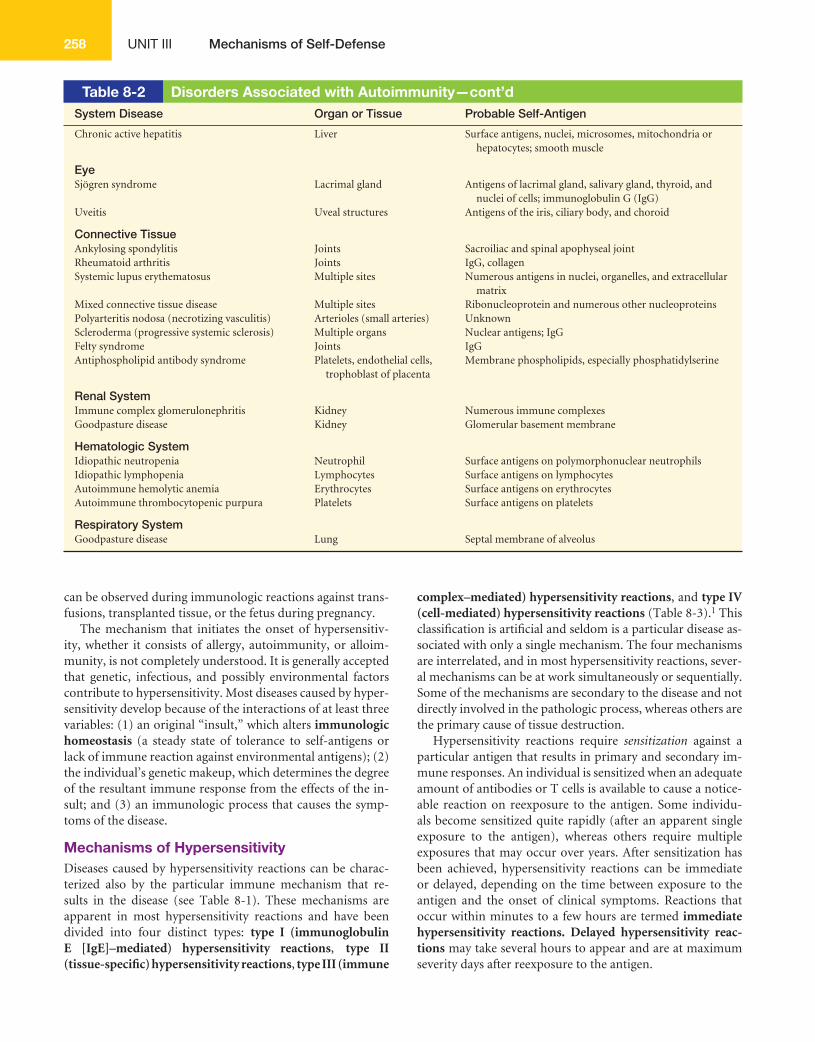
Chronic active hepatitis Liver Surface antigens, nuclei, microsomes, mitochondria or hepatocytes; smooth muscle
Eye Sjögren syndrome Lacrimal gland Antigens of lacrimal gland, salivary gland, thyroid, and
nuclei of cells; immunoglobulin G (IgG)Uveitis Uveal structures Antigens of the iris, ciliary body, and choroid
Connective Tissue Ankylosing spondylitis Joints Sacroiliac and spinal apophyseal jointRheumatoid arthritis Joints IgG, collagenSystemic lupus erythematosus Multiple sites Numerous antigens in nuclei, organelles, and extracellular
matrixMixed connective tissue disease Multiple sites Ribonucleoprotein and numerous other nucleoproteinsPolyarteritis nodosa (necrotizing vasculitis) Arterioles (small arteries) UnknownScleroderma (progressive systemic sclerosis) Multiple organs Nuclear antigens; IgGFelty syndrome Joints IgGAntiphospholipid antibody syndrome Platelets, endothelial cells,
trophoblast of placentaMembrane phospholipids, especially phosphatidylserine
Renal System Immune complex glomerulonephritis Kidney Numerous immune complexesGoodpasture disease Kidney Glomerular basement membrane
Hematologic System Idiopathic neutropenia Neutrophil Surface antigens on polymorphonuclear neutrophilsIdiopathic lymphopenia Lymphocytes Surface antigens on lymphocytesAutoimmune hemolytic anemia Erythrocytes Surface antigens on erythrocytesAutoimmune thrombocytopenic purpura Platelets Surface antigens on platelets
Respiratory System Goodpasture disease Lung Septal membrane of alveolus
Table 8-2 Disorders Associated with Autoimmunity—cont’d
can be observed during immunologic reactions against trans-fusions, transplanted tissue, or the fetus during pregnancy.
The mechanism that initiates the onset of hypersensitiv-ity, whether it consists of allergy, autoimmunity, or alloim-munity, is not completely understood. It is generally accepted that genetic, infectious, and possibly environmental factors contribute to hypersensitivity. Most diseases caused by hyper-sensitivity develop because of the interactions of at least three variables: (1) an original “insult,” which alters immunologic homeostasis (a steady state of tolerance to self-antigens or lack of immune reaction against environmental antigens); (2) the individual’s genetic makeup, which determines the degree of the resultant immune response from the effects of the in-sult; and (3) an immunologic process that causes the symp-toms of the disease.
Mechanisms of Hypersensitivity Diseases caused by hypersensitivity reactions can be charac-terized also by the particular immune mechanism that re-sults in the disease (see Table 8-1 ). These mechanisms are apparent in most hypersensitivity reactions and have been divided into four distinct types: type I (immunoglobulin E [IgE] – mediated) hypersensitivity reactions , type II (tissue-specifi c) hypersensitivity reactions , type III (immune
complex – mediated) hypersensitivity reactions , and type IV (cell-mediated) hypersensitivity reactions ( Table 8-3 ). 1 This classifi cation is artifi cial and seldom is a particular disease as-sociated with only a single mechanism. The four mechanisms are interrelated, and in most hypersensitivity reactions, sever-al mechanisms can be at work simultaneously or sequentially. Some of the mechanisms are secondary to the disease and not directly involved in the pathologic process, whereas others are the primary cause of tissue destruction.
Hypersensitivity reactions require sensitization against a particular antigen that results in primary and secondary im-mune responses. An individual is sensitized when an adequate amount of antibodies or T cells is available to cause a notice-able reaction on reexposure to the antigen. Some individu-als become sensitized quite rapidly (after an apparent single exposure to the antigen), whereas others require multiple exposures that may occur over years. After sensitization has been achieved, hypersensitivity reactions can be immediate or delayed, depending on the time between exposure to the antigen and the onset of clinical symptoms. Reactions that occur within minutes to a few hours are termed immediate hypersensitivity reactions. Delayed hypersensitivity reac-tions may take several hours to appear and are at maximum severity days after reexposure to the antigen.

Alterations in Immunity and Infl ammation CHAPTER 8 259
The most rapid and severe immediate hypersensitivity reaction is anaphylaxis. 2 Anaphylaxis occurs within min-utes of reexposure to the antigen and can be either systemic (generalized) or cutaneous (localized). 3 Symptoms of sys-temic anaphylaxis include itching, erythema, headaches, vomiting, abdominal cramps, diarrhea, and breathing dif-fi culties. In severe cases, contraction of bronchial smooth muscle, laryngeal edema, and vascular collapse may result in respiratory distress, decreased blood pressure, shock, and death. Examples of systemic anaphylaxis are allergic reac-tions to bee stings, peanuts, and fi sh. 4 Cutaneous anaphylaxis causes the less severe symptoms of local infl ammation.
Type I: IgE-Mediated Hypersensitivity Reactions Type I reactions are mediated by antigen-specifi c IgE and the products of tissue mast cells ( Figure 8-1 ). 5 Most common al-lergies (e.g., pollen allergies) are type I reactions. In addition, most type I reactions occur against environmental antigens and are therefore allergic. Because of this strong association, many healthcare professionals use the term allergy to indicate only IgE-mediated reactions. However, IgE can contribute to a few autoimmune and alloimmune diseases, and many com-mon allergies (e.g., poison ivy) are not mediated by IgE.
In some individuals, exposure to an environmental antigen causes primarily IgE production. 6 Repeated exposure to the antigen usually is required to elicit enough IgE so that the per-son becomes “sensitized.” IgE has a relatively short life span in the blood because it rapidly binds to very-high-affi nity Fc receptors on the plasma membranes of mast cells (see Figure 8-1 ). 7 The subclass IgG4 also has specifi c receptors on the mast cell and may contribute to the type I mechanism. Antibody that binds to mast cells is termed cytotropic antibody (able to bind to cell surfaces) or reagin (skin-sensitizing antibody). Unlike Fc receptors on phagocytes, which bind IgG that has reacted with antigen, the Fc receptors on mast cells bind with IgE that has not previously interacted with antigen.
If further exposure of a sensitized individual to the antigen occurs, one molecule of antigen may bind simultaneously to two molecules of IgE-Fc receptor complexes on the mast cell’s surface (cross-link) resulting in activation of intracellular signaling pathways and mast cell degranulation (see Figure
7-1, B , and Chapter 6). The antigen that triggers cross-linking must have at least two antigenic determinants on the same molecule. Sometimes an IgE-mediated response is benefi cial to the host, as is the case of some immune reactions against parasites. (This mechanism is described in Chapter 7 and il-lustrated in Figure 7-26.)
The products of mast cell degranulation can modulate almost all aspects of an acute infl ammatory response. 8 (The effects of biochemical mediators released by mast cells are illustrated in Figure 6-9). The most potent mediator is his-tamine, which affects several key target cells. 9 Acting through the H1 receptors, histamine contracts bronchial smooth muscles, causing bronchial constriction; increases vascular permeability, causing edema; and causes vasodilation, in-creasing blood fl ow into the affected area (see Figures 6-3 and 6-10). The interaction of histamine with H2 receptors results in increased gastric acid secretion and a decrease of histamine released from mast cells and basophils. The action of hista-mine through H2 receptors suggests an important negative-feedback mechanism that stops degranulation. That is, the released histamine inhibits release of additional histamine by interacting with H2 receptors on the mast cells. Histamine also may affect control of the immune response through H2 receptors on most cells of the immune system. 10 Another im-portant activity of histamine is enhancement of the chemo-tactic activity of other factors, such as eosinophil chemotactic factor of anaphylaxis (ECF-A), which attracts eosinophils into sites of allergic infl ammatory reactions and prevents them from migrating out of the infl ammatory site. (The role of the eosinophil in infl ammation is discussed in Chapter 6.)
Type II: Tissue-Specifi c Hypersensitivity Reactions Type II hypersensitivity reactions are generally characterized by a specifi c cell or tissue being the target of an immune re-sponse. In addition to major histocompatibility locus anti-gens (HLAs; discussed in Chapter 7), most cells have other antigens on their surfaces. Some of these other antigens are called tissue-specifi c antigens because they are expressed on the plasma membranes of only certain cells in specifi c tissues. Platelets, for example, have groups of antigens that are found on no other cells of the body. The symptoms of many type II
Table 8-3 Immunologic Mechanisms of Tissue Destruction
Type NameRate of Development
Class of Antibody Involved
Principal Effector Cells Involved
Complement Participation Examples of Disorders
I IgE-mediated reaction Immediate IgE Mast cells No Seasonal allergic rhinitisII Tissue-specifi c reaction Immediate IgG
IgM Macrophages in
tissuesFrequently Autoimmune thrombocytopenic
purpura, Graves disease, autoim-mune hemolytic anemia
III Immune complex – mediated reaction
Immediate IgG IgM
Neutrophils Yes Systemic lupus erythematosus
IV Cell-mediated reaction Delayed None Lymphocytes, macrophages
No Contact sensitivity to poison ivy and metals (jewelry)
Ig, Immunoglobulin.

UNIT III Mechanisms of Self-Defense260
diseases are determined by which tissue or organ expresses the particular antigen. Environmental antigens (e.g., drugs or their metabolites) may bind to the plasma membranes of spe-cifi c cells (especially erythrocytes and platelets) and function as targets of type II reactions.
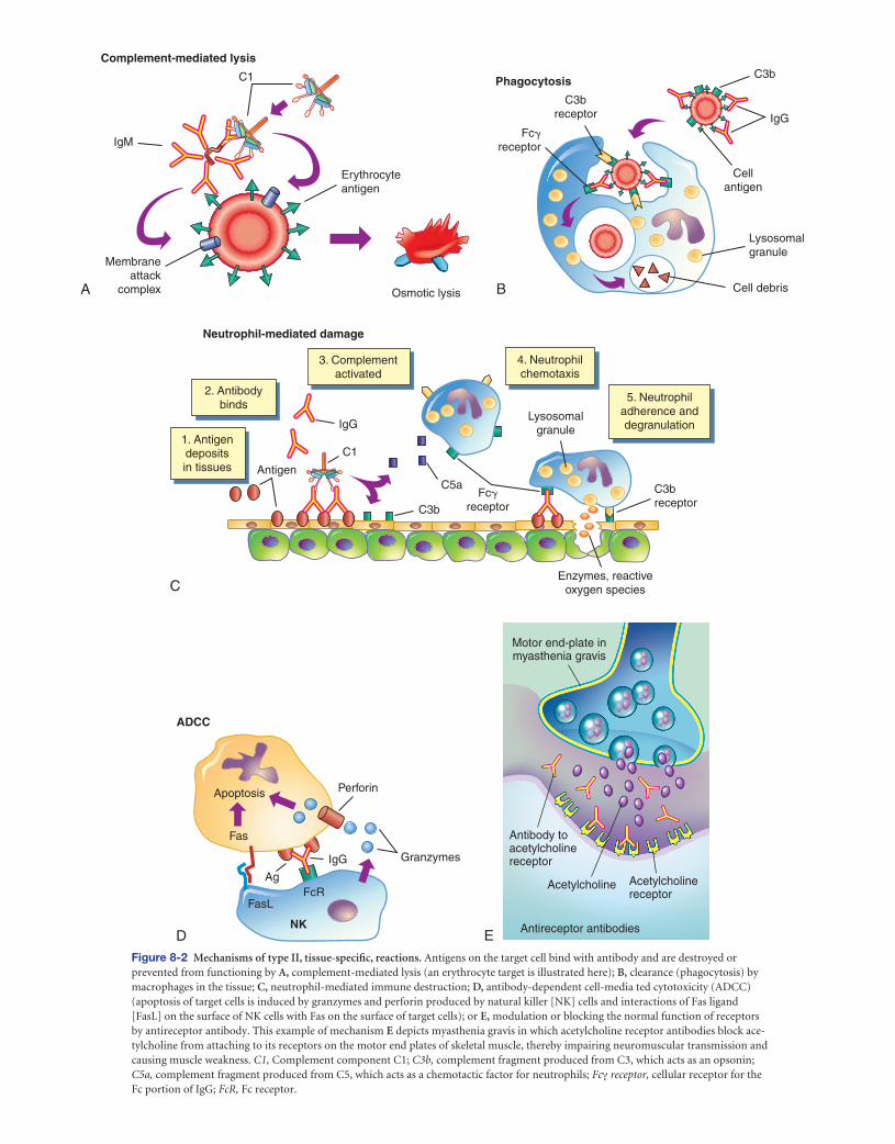
The fi ve general mechanisms by which type II hypersen-sitivity reactions can affect cells are shown in Figure 8-2 . All of these mechanisms begin with antibody binding to tissue-specifi c antigens or antigens that have attached to particular
tissues. First, the cell can be destroyed by antibody (IgG or IgM) and activation of the complement cascade through the classical pathway. Formation of the membrane attack com-plex (C5-9) damages the membrane and may result in lysis of the cell (see Figure 8-2 , A ). For example, erythrocytes are destroyed by complement-mediated lysis in individuals with autoimmune hemolytic anemia (see Chapter 26) or as a result of an alloimmune reaction to ABO-mismatched transfused blood cells.
Firs
t exp
osur
e se
nsiti
zatio
nS
ubse
quen
t exp
osur
e: a
llerg
ic r
eact
ion
Blood vessel
Edema
Th2 cellPollen
Antigen
Mucosallining
Smooth muscle spasm
IgE Fcreceptor
IgE antibody
Epithelial damage
Epithelial damage
Dendriticcell
IgE B cell
Eosinophilrecruitment
Release of granulesand mediators
Release of primary and secondary mediators
Mast cell
Leukocyteinfiltration
T cell receptor
IL-4, IL-5
IL-3, IL-5GM-CSF
IL-3, IL-5
Activation
Mucus secretion
A Figure 8-1 Mechanism of type I IgE – mediated reactions. A, Th2 cells are activated by antigen-presenting dendritic cells to produce cytokines, including IL-3, IL-4, IL-5, and granulocyte-macrophage colony-stimulating factor (GM-CSF). IL-3, IL-5, and GM-CSF attract and promote the survival of eosinophils. Other cytokines (e.g., IL-4) induce B cells to class-switch to IgE-producing plasma cells. The IgE coats the surface of the mast cell by binding with IgE-specifi c Fc receptors on the mast cell’s plasma membrane (sensitization). Further exposure to the same allergen cross-links the surface-bound IgE and activates signals from the cytoplasmic portion of the IgE Fc recep-tors. These signals initiate two parallel and interdependent processes: mast cell degranulation and discharge of preformed mediators (e.g., histamine, eosinophil-chemotactic factor of anaphylaxis) and production of newly formed mediators such as arachidonic metabo-lites (leukotrienes, prostaglandins). Many local type I hypersensitivity reactions have two well-defi ned phases. The initial phase is char-acterized by vasodilation, vascular leakage, and depending on the location, smooth muscle spasm or glandular secretions. These changes usually become evident within 5 to 30 minutes after exposure to the antigen. The late phase occurs 2 to 8 hours later without additional exposure to the antigen. The late phase has more intense infi ltration of tissues with eosinophils, neutrophils, basophils, monocytes, and Th cells and tissue destruction in the form of mucosal epithelial cell damage.

Alterations in Immunity and Infl ammation CHAPTER 8 261
Second, antibody may cause cell destruction through phagocytosis by macrophages. IgG, as well as C3b of the complement system, are opsonins that bind to receptors on the macrophage (see Figure 6-2, B ). Phagocytosis of the target cell follows. (Phagocytosis is illustrated in Figures 6-11 and 6-13.) For example, antibodies against platelet-specifi c antigens or against red blood cell antigens of the Rh system coat those cells at low density, resulting in their preferential removal by phagocytosis in the spleen, rather than by complement-mediated lysis.
Third, antibody and complement may attract neutrophils. Either antigen expressed normally on the vessel walls or solu-ble antigen in the circulation (e.g., released from cells within the body or from infectious agents or by way of drugs or med-ications) that has been deposited on the surface of endothe-lial cells may bind antibody (see Figure 8-2 , C ). The antibody initiates the complement cascade, resulting in the release of C3a and C5a, which are chemotactic for neutrophils, and de-position of complement component C3b. Neutrophils bind to the tissues through receptors for the Fc portion of antibody
(Fc receptor) or for C3b and attempt to phagocytose the tissue. Because the tissue is large, phagocytosis cannot be completed; even so, neutrophils release their granules onto the healthy tissue. The components of neutrophil granules, as well as the several toxic oxygen products produced by these cells, will damage the tissue.
The fourth mechanism is antibody-dependent cell- mediated cytotoxicity (ADCC) (see Figure 8-2 , D ). This mechanism involves a subpopulation of cytotoxic cells that are not antigen specifi c (natural killer [NK] cells). Antibody on the target cell is recognized by Fc receptors on the NK cells, which release toxic substances that destroy the target cell.
The fi fth mechanism does not destroy the target cell, but rather causes it to malfunction. In this mechanism of type II injury, the antibody is usually directed against antigenic de-terminants associated with specifi c cell-surface receptors, and the symptoms of the disease are a result of a direct effect of antibody binding alone (see Figure 8-2 , E ). 11 The antibody reacts with the receptors on the target cell surface and modu-lates the function of the receptor by preventing interactions with their normal ligands, replacing the ligand and inappro-priately stimulating the receptor, or destroying the receptor. For example, in the hyperthyroidism (excessive thyroid activ-ity) of Graves’ disease, autoantibody binds to and activates re-ceptors for thyroid-stimulating hormone (TSH) (a pituitary hormone that controls the production of the hormone thy-roxine by the thyroid). 12 In this way the antibody stimulates the thyroid cells to produce thyroxine. Under normal condi-tions, the increasing levels of thyroxine in the blood would signal the pituitary to decrease TSH production, which would result in less stimulation of the TSH receptor in the thyroid and a concomitant decrease in thyroxine production. Because the level of anti-TSH receptor antibody is not controlled by the pituitary, increasing amounts of thyroxine in the blood have no effect on antibody levels, and thyroxine production continues to increase despite decreasing amounts of TSH (see Chapter 21). 13
Type III: Immune Complex – Mediated Hypersensitivity Reactions Mechanisms of Type III Hypersensitivity Most type III hypersensitivity diseases are caused by antigen-antibody (immune) complexes that are formed in the circula-tion and deposited later in vessel walls or extravascular tissues ( Figure 8-3 ). 14 The primary difference between type II and type III mechanisms is that in type II hypersensitivity anti-body binds to the antigen on the cell surface, whereas in type III the antibody binds to soluble antigen that was released into the blood or body fl uids, and the complex is then deposited in the tissues. Type III reactions are not organ specifi c, and symptoms have little to do with the particular antigenic tar-get of the antibody. The harmful effects of immune complex deposition are caused by complement activation, particularly through the generation of chemotactic factors for neutro-phils. The neutrophils bind to antibody and C3b contained in the complexes and attempt to ingest the immune complexes.
B
Granule contents • Histamine • Proteases • Chemotactic factors (ECF, NCF)
Antigen
IgEIgE receptor
Signals fordegranulation
Signals forcytokinegeneactivation
Signals foractivation ofphospho-lipase A2
Secretedcytokines
Arachidonic acid PAF
Primary mediators Secondary mediators
LeukotrienesB4, C4, D4
ProstaglandinD2
MembranephospholipidsDegranulation
Nucleus
Figure 8-1, cont’d B, Activation of mast cells leading to degranulation of preformed mediators (primary mediators) and synthesis of newly formed (de novo) mediators (secondary mediators). ECF, Eosinophilic chemotactic factor; NCF, neutrophil chemotactic factor; PAF, platelet-activating factor.
Membraneattack
complex
IgM
C1
Erythrocyteantigen
Osmotic lysis
Complement-mediated lysis
IgG
Lysosomalgranule
Cell debris
Fc�receptor
C3breceptor
C3b
Cellantigen
Phagocytosis
IgGLysosomal
granule
Fc�receptorC3b
C5a
C1
Neutrophil-mediated damage
3. Complementactivated
2. Antibodybinds
4. Neutrophilchemotaxis
5. Neutrophiladherence anddegranulation
1. Antigendepositsin tissues
C3breceptor
Antigen
Enzymes, reactiveoxygen species
IgG
Ag
Perforin
Granzymes
Apoptosis
Fas
FasLFcR
NK
ADCC
Acetylcholine
Motor end-plate inmyasthenia gravis
Antibody toacetylcholinereceptor
Acetylcholinereceptor
Antireceptor antibodies
A B
C
D E Figure 8-2 Mechanisms of type II, tissue-specifi c, reactions. Antigens on the target cell bind with antibody and are destroyed or prevented from functioning by A, complement-mediated lysis (an erythrocyte target is illustrated here); B, clearance (phagocytosis) by macrophages in the tissue; C, neutrophil-mediated immune destruction; D, antibody-dependent cell-media ted cytotoxicity (ADCC) (apoptosis of target cells is induced by granzymes and perforin produced by natural killer [NK] cells and interactions of Fas ligand [FasL] on the surface of NK cells with Fas on the surface of target cells); or E, modulation or blocking the normal function of receptors by antireceptor antibody. This example of mechanism E depicts myasthenia gravis in which acetylcholine receptor antibodies block ace-tylcholine from attaching to its receptors on the motor end plates of skeletal muscle, thereby impairing neuromuscular transmission and causing muscle weakness. C1, Complement component C1; C3b, complement fragment produced from C3, which acts as an opsonin; C5a, complement fragment produced from C5, which acts as a chemotactic factor for neutrophils; Fc γ receptor, cellular receptor for the Fc portion of IgG; FcR, Fc receptor.

Alterations in Immunity and Infl ammation CHAPTER 8 263
They are often unsuccessful because the complexes are bound to large areas of tissue. During the attempted phagocytosis, large quantities of lysosomal enzymes are released into the infl ammatory site instead of into phagolysosomes. The attrac-tion of neutrophils and the subsequent release of lysosomal enzymes cause most of the resulting tissue damage.
Immune complexes can be of various sizes, depending on the relative amounts of antigen and antibody. Fairly large im-mune complexes are cleared rapidly from the circulation by tissue macrophages, whereas very small complexes eventu-ally are fi ltered from blood through the kidneys, without any pathologic consequences. Intermediate-sized immune com-plexes (formed at a ratio of antigen to antibody that has a slight excess of antigen) are likely to be deposited in certain target tissues, where they have severe pathologic consequences, such as infl ammation in the kidneys (glomerulonephritis), the ves-sels (vasculitis), or the joints (arthritis or degenerative joint disease).
Immune Complex Disease The nature of the immune complexes may change during
the progression of the disease, with resultant changes in the severity of the symptoms. Immune complex formation is dy-namic as variations in the ratio of antigen to antibody, the class and subclass of antibody, and the quantity and quality of circulating antigen occur. Thus complexes formed early in a disease process may differ from those formed later, and several types of immune complexes may be present simultaneously. With the tremendous potential heterogeneity of immune
complexes, it is not surprising that immune-complex diseases are characterized by a variety of symptoms and periods of remission or exacerbation of symptoms.
Because many immune complexes activate complement very effectively, complement levels in the blood may decrease during active disease. At times the individual’s blood may become hypocomplementemic (i.e., contains below normal amounts of complement activity). During type I, II, or IV hypersensitivity reactions, complement levels are unaffected, or some components of the complement cascade, such as C3, may even be increased.
Two prototypic models of type III hypersensitivity help ex-plain the variety of diseases in this category. Serum sickness is a model of systemic type III hypersensitivities, and the Arthus reaction is a model of localized or cutaneous reactions.
Serum Sickness . The systemic prototype of immune complex – mediated disease is called serum sickness because it was initially described as being caused by the therapeutic administration of foreign serum, such as horse serum that contained antibody against tetanus toxin. 15 Foreign serum generally is not administered to individuals today, although serum sickness reactions can be caused by the repeated intra-venous administration of other antigens, such as drugs, and the characteristics of serum sickness are observed in systemic type III autoimmune diseases. Serum sickness – type reactions are caused by the formation of immune complexes in the blood and their subsequent generalized deposition in target tissues. Typically affected tissues are the blood vessels, joints,
IgG
Lysosomalgranule
Large immunecomplexSmall immune
complex
Intermediateimmune
complexes
Fc�receptor
C3b
C5a
C1
2. Complementactivated
3. Neutrophilchemotaxis
4. Neutrophiladherence anddegranulation
1. Intermediate-sizedimmune complexes
deposited in the tissue
C3breceptor
Antigen
Enzymes, reactiveoxygen species
Figure 8-3 Mechanism of type III, immune complex – mediated reactions. Immune complexes form in the blood from circulating antigen and antibody. Both small and large immune complexes are removed successfully from the circulation and do not cause tissue damage. Intermediate-sized complexes are deposited in certain target tissues in which the circulation is slow or fi ltration of the blood occurs. The complexes activate the complement cascade through C1 and generate fragments including C5a and C3b. C5a is chemotactic for neutrophils, which migrate into the infl amed area and attach to the IgG and C3b in the immune complexes. The neutrophils attempt unsuccessfully to phagocytose the tissue and in the process release a variety of degradative enzymes that destroy the healthy tissues. Fc γ receptor is the cellular receptor for the Fc portion of IgG.

UNIT III Mechanisms of Self-Defense264
and kidneys. Other symptoms include fever, enlarged lymph nodes, rash, and pain at sites of infl ammation.
A form of serum sickness is Raynaud phenomenon, a con-dition caused by the temperature- dependent deposition of immune complexes in the capillary beds of the peripheral cir-culation. Certain immune complexes precipitate at tempera-tures below normal body temperature, particularly in the tips of the fi ngers, toes, and nose and are called cryoglobulins. The precipitates block the circulation and cause localized pal-lor and numbness, followed by cyanosis (a bluish tinge result-ing from oxygen deprivation) and eventually gangrene if the circulation is not restored.
Arthus Reaction . An Arthus reaction is the prototypic example of a localized immune complex – mediated infl am-matory response. 16 It is caused by repeated local exposure to an antigen that reacts with preformed antibody and forms im-mune complexes in the walls of the local blood vessels. Symp-toms of an Arthus reaction begin within 1 hour of exposure and peak 6 to 12 hours later. The lesions are characterized by a typical infl ammatory reaction, with increased vascular permeability, an accumulation of neutrophils, edema, hem-orrhage, clotting, and tissue damage.
Type IV: Cell-Mediated Hypersensitivity Reactions Whereas types I, II, and III hypersensitivity reactions are me-diated by antibody, type IV reactions are mediated by T lym-phocytes and do not involve antibody ( Figure 8-4 ). Type IV mechanisms occur through either cytotoxic T lymphocytes (Tc cells) or lymphokine-producing Th1 cells. 17 Tc cells at-tack and destroy cellular targets directly. Th1 cells produce cytokines that recruit and activate phagocytic cells, especially macrophages. Destruction of the tissue is usually caused by direct killing by toxins from Tc cells or the release of soluble factors, such as lysosomal enzymes and toxic reactive oxygen species (ROS), from activated macrophages.
Clinical examples of type IV hypersensitivity reactions include graft rejection and allergic reactions resulting from contact with such substances as poison ivy and metals. A type IV component also may be present in many autoimmune dis-eases. For example, T cells against type II collagen (a protein present in joint tissues) contribute to the destruction of joints in rheumatoid arthritis; T cells against a thyroid cell surface antigen contribute to the destruction of the thyroid in auto-immune thyroiditis (Hashimoto disease); and T cells against an antigen on the surface of pancreatic beta cells (the cell that normally produces insulin) are responsible for beta-cell de-struction in insulin-dependent (type 1) diabetes mellitus.
A type IV hypersensitivity reaction in the skin was thor-oughly described fi rst by Ehrlich in 1891 and led to the devel-opment of a diagnostic skin test for tuberculosis. 18 The reaction follows an intradermal injection of tuberculin antigen into a suitably sensitized individual and is called a delayed hypersen-sitivity skin test because of its slow onset—24 to 72 hours to reach maximum intensity. The reaction site is infi ltrated with T lymphocytes and macrophages, resulting in a clear hard cen-ter (induration) and a reddish surrounding area (erythema).
Antigenic Targets of Hypersensitivity Reactions Allergy Allergy is a hypersensitivity response against an environmen-tal antigen (allergen). Although the most common allergies are type I hypersensitivities, any of the other three mecha-nisms may cause allergic responses. 19
Typical allergens that induce type I hypersensitivity include pollens (e.g., ragweed), molds and fungi (e.g., Penicillium no-tatum ), foods (e.g., milk, eggs, fi sh), animals (e.g., cat dan-der, dog dander), cigarette smoke, components of house dust (e.g., fecal pellets of house mites), and almost anything else we may encounter in our environment. Allergens that primar-ily elicit type IV allergic hypersensitivities include plant resins (e.g., poison ivy, poison oak), metals (e.g., nickel, chromium), acetylates and chemicals in rubber, cosmetics, detergents, and topical antibiotics (e.g., neomycin). Type II and type III allergic hypersensitivities are relatively rare but may include antibiotics (e.g., penicillin, sulfonamides) and soluble anti-gens produced by infectious agents (e.g., hepatitis B).
Usually a sensitization process involving multiple exposures to the allergen occurs before adequate amounts of antibody or T cells are available to elicit a hypersensitivity response. In some instances, exposure to a particular allergen may not be apparent in the case of allergens that are drugs, additives, or preservatives in food. For example, milk may contain trace amounts of penicillin used for treating cows for mastitis. Thus, the fi rst therapeutic exposure to penicillin may cause an unexpected hypersensitivity reaction. Additionally, penicillin
Perforin
Targetcell
Granzymes
Apoptosis
Fas
FasL TCR
CD8
CD4
Tc cellTh1 cell
MHCclass II
INF�R
INF-�
Activatedmacrophage
Lysosomal granule
Lysosomal enzymes andtoxic oxygen species
MHCclass I
TCR
Figure 8-4 Mechanism of type IV, cell-mediated, reactions. Antigens from target cells stimulate T cells to differentiate into cytotoxic T cells (Tc cells), which have direct cytotoxic activity, and helper T cells (Th1 cells) involved in delayed hypersensitivity. The Th1 cells produce lymphokines (especially interferon- γ [IFN- γ ]) that activate the macrophage through specifi c recep-tors (e.g. IFN- γ receptor [IFN γ R]). The macrophages can attach to targets and release enzymes and reactive oxygen species that are responsible for most of the tissue destruction.

Alterations in Immunity and Infl ammation CHAPTER 8 265
shares a β -lactam structure with cephalosporin, so that one antibiotic may be sensitive against another. 20
Genetic Predisposition Certain individuals are genetically predisposed to develop
allergies, particularly type I allergies, and are called atopic. 21,22 In families in which one parent has an allergy, allergies develop in about 40% of the offspring. If both parents have allergies, the incidence in the offspring may be as high as 80%. 23 (Prin-ciples of genetic inheritance are discussed in Chapter 4.)
Atopic individuals tend to produce higher quantities of IgE and to have more Fc receptors for IgE on their mast cells. The airways and the skin of atopic individuals are also more responsive to a wide variety of both specifi c and nonspecifi c stimuli than are the airways and skin of individuals who are not atopic. Multiple genes have been associated with the atopic state, including polymorphisms in a large variety of cytokines that regulate IgE synthesis (e.g., interleukin [IL]-4, IL-5, IL-12, IL-13) and cellular receptors.
Clinical Symptoms of Type I Allergies The clinical manifestations of type I reactions are attributable mostly to the biologic effects of histamine. 24 Tissues most commonly affected contain large numbers of mast cells and are sensitive to the effects of histamine released from them. 25 These tissues are found in the gastrointestinal tract, the skin, and the respiratory tract ( Figure 8-5 and Table 8-4 ). The par-ticular symptoms frequently refl ect the main portal of entry for the allergen. For instance, pollens and other airborne al-lergens usually cause respiratory symptoms.
Effects of allergens on the mucosa of the eyes, nose, and respiratory tract include conjunctivitis (infl ammation of the membranes lining the eyelids), rhinitis (infl ammation of the mucous membranes of the nose), and asthma (constriction of the bronchi). Symptoms are caused by vasodilation, hyperse-cretion of mucus, edema, and swelling of the respiratory mu-cosa. Because the mucous membranes lining the respiratory tract (accessory sinuses, nasopharynx, and upper and lower respiratory tract) are continuous, they are all adversely af-fected. The degree to which each is affected determines the symptoms of the disease.
Gastrointestinal allergies are caused primarily by aller-gens that enter through the mouth—usually foods or medi-cines. Symptoms include vomiting, diarrhea, or abdominal pain and may be severe enough to result in malabsorption or protein-losing enteropathy, if the reactions are prolonged or recurrent. Foods most often implicated in gastrointestinal allergies are milk, chocolate, citrus fruits, eggs, wheat, nuts, peanut butter, and fi sh. When food is the allergen, the active immunogen may be a product of food breakdown by diges-tive enzymes.
Urticaria, or hives, is a dermal (skin) manifestation of type I allergic reactions (see Figure 8-5 ). The underlying mechanism is the localized release of histamine and increased vascular permeability, resulting in limited areas of edema. Urticaria is characterized by white fl uid-fi lled blisters (wheals) surround-ed by areas of redness (fl ares). The wheal and fl are reaction is
usually accompanied by itching. Not all urticarial symptoms are caused by allergic (immunologic) reactions. Some, termed nonimmunologic urticaria, result from exposure to cold tem-peratures, emotional stress, medications, systemic diseases, hyperthyroidism, or malignancies (e.g., lymphomas).
If possible, avoidance of the allergen is the best method to limit allergic responses. Approximately 30% of laboratory animal handlers have allergies to animal dander and must use face masks or other devices to avoid contact. 26
Although some type I allergic responses can be controlled by blocking histamine receptors with antihistamines, the pri-mary mechanism of control is the autonomic nervous system. The autonomic nervous system includes biochemical media-tors (e.g., epinephrine, acetylcholine) that, like the mediators of the infl ammatory response, have profound effects on cells. These mediators bind to appropriate receptors on mast cells and the target cells of infl ammation (e.g., smooth muscle), thereby controlling (1) release of infl ammatory mediators from mast cells and (2) the degree to which target cells re-spond to infl ammatory mediators (see Chapter 6).
Allergic Disease: Bee Sting Allergy An example of a life-threatening allergy is an anaphylac-
tic reaction to a bee sting. Bee venoms contain a mixture of enzymes and other proteins that may serve as allergens. About 1% of children may have an anaphylactic reaction to bee venom. Within minutes they may develop excessive swelling (edema) at the bee sting site, followed by generalized hives, itching, and swelling in areas distal from the sting (e.g., eyes, lips), and other systemic symptoms including fl ushing, sweat-ing, dizziness, and headache. The most severe symptoms may include gastrointestinal (e.g., stomach cramps, vomiting), respiratory (e.g., tightness in the throat, wheezing, diffi cul-ties breathing), and vascular (e.g., low blood pressure, shock) reactions. Severe respiratory and vascular reactions may lead to death.
If a child has had a previous anaphylactic reaction to bee stings, the chance of having another is about 60%. During the reaction the administration of antihistamines has little effect because histamine has already bound H1 receptors and initi-ated severe bronchial smooth muscle contraction. Most indi-viduals carry self-injectable epinephrine. Autonomic nervous system mediators, such as epinephrine, bind to specifi c recep-tors on smooth muscle and reverse the effects of histamine and result in muscle relaxation. Similar anaphylactic reactions have been described against peanuts and other nuts, shellfi sh, fi sh, milk, eggs, and some medications.
Tests of IgE-Mediated Allergy Allergic reactions can be life threatening; therefore, it is
essential that severely allergic individuals be made aware of the specifi c allergen against which they are sensitized and in-structed to avoid contact with that material. Several tests are available, including food challenges, skin tests with allergens, and laboratory tests for total IgE and allergen-specifi c IgE in the blood.
Reactivity to a particular food allergen may be tested by controlled administration of small doses of the suspected

UNIT III Mechanisms of Self-Defense266
allergen in order to evoke a mild allergic response. This approach can be dangerous if the individual has a history of anaphylactic responses. A safer approach is injection of an al-lergen into (intradermal) or onto (epicutaneous or prick test) the skin. If the individual is allergic to a particular allergen,
a local wheal and fl are reaction may occur within a few min-utes at the site of injection. The diameter of the fl are reaction is usually indicative of the individual’s degree of sensitivity to that allergen. 27 In the most severely allergic individuals, even the extremely small amounts of allergen used for the
Itching
Angioedema
Hypotension
Gastrointestinalcramps and malabsorption
Angioedema
ConjunctivitisRhinitis
Laryngeal edema
Urticaria
Bronchospasm(asthma)
Dysrhythmias
Figure 8-5 Type I hypersensitivity reactions. Manifestations of allergic reactions as a result of type I hypersensitivity include itching, angioedema (swelling caused by exudation), edema of the larynx, urticaria (hives), bronchospasm (constriction of airways in the lungs), hypotension (low blood pressure) and dysrhythmias (irregular heartbeat) because of anaphylactic shock, and gastrointestinal cramp-ing caused by infl ammation of the gastrointestinal mucosa. Photographic inserts show a diffuse allergic-like eye and skin reaction on an individual. The skin lesions have raised edges and develop within minutes or hours, with resolution occurring after about 12 hours. (From Roitt I, Brostoff J, Male D: Immunology, ed 6, St. Louis, 2001, Mosby.)
Table 8-4 Causes of Clinical Manifestations of Allergy
Typical AllergenMechanism of Hypersensitivity Clinical Manifestation
Ingestants Foods Type I Gastrointestinal allergyDrugs Types I, II, III Urticaria, immediate drug reaction, hemolytic anemia, serum sickness
Inhalants Pollens, dust, molds Type I Allergic rhinitis, bronchial asthma Aspergillus fumigatus Types, I, III Allergic bronchopulmonary aspergillosisThermophilic actinomycetes * Types III, IV Extrinsic allergic alveolitis
Injectants Drugs Types, I, II, III Immediate drug reaction, hemolytic anemia, serum sicknessBee venom Type I AnaphylaxisVaccines Type III Localized Arthus reactionSerum Types I, III Anaphylaxis, serum sickness
Contactants Poison ivy, metals Type IV Contact dermatitis
* An order of fungi that is stimulated by warmth to grow and proliferate. Modifi ed from Bellanti JA: Immunology III, Philadelphia, 1985, Saunders.
Alterations in Immunity and Infl ammation CHAPTER 8 267
skin test may evoke a systemic anaphylaxis. Skin test is also contraindicated if the patient is using medications that may affect the test or has diffuse dermatitis, which would make the reaction diffi cult to interpret. 28
A variety of laboratory tests can detect IgE antibodies in serum. These assays have various commercial acronyms, de-pending on whether they are radioimmunoassays (RIAs; reac-tivity detected by measuring a radioactive reagent) or enzyme immunoassays (EIAs or ELISA [enzyme-linked immunosor-bent assay]; reactivity detected by measuring a color change caused by an enzyme-labeled reagent). One set of assays measures circulating levels of total IgE, with atopic individu-als usually having elevated levels. Other assays are capable of measuring circulating levels of specifi c IgE antibodies against selected allergens. The amount of IgE against a specifi c aller-gen correlates well with the degree of skin test reactivity and the severity of clinical symptoms related to the same allergen, although the laboratory text is less sensitive.
Desensitization Clinical desensitization to allergens can be achieved in
some individuals. 29 Minute quantities of the allergen are in-jected in increasing doses over a prolonged period. The pro-cedure may reduce the severity of the allergic reaction in the treated individual. However, this form of therapy is associated with a risk of systemic anaphylaxis, which can be severe and life threatening. This approach works best for allergies against some food allergens and with biting insect allergies (80% to 90% rate of desensitization over 5 years of treatment). 30
The mechanisms by which desensitization occurs may be several, one of which is the production of large amounts of so-called blocking antibodies, usually circulating IgG. A blocking antibody presumably competes in the tissues or in the circulation for binding with antigenic determinants on the allergen so that the allergen is “neutralized” and is unable to bind with IgE on mast cells. Sublingual desen-sitization (another approach that works best with some food allergies) produces sIgA and circulating IgG that may prevent the allergen from accessing mast cells. Desensitiza-tion injections also may stimulate the generation of clones of regulatory T lymphocytes, which inhibit hypersensi-tivity by suppressing the production of IgE or modifying the Th1/Th2 interactions in favor of production of anti- infl ammatory cytokines.
Other approaches to suppressing type I allergic responses have been tested, with some preliminary success. An example is injection of anti-IgE antibody directed against the Fc por-tion of the IgG in order to decrease binding of IgE to mast cells.
Type IV Allergic Hypersensitivities The allergens that induce a type IV allergic reaction are
mostly haptens that react with normal self-proteins in the skin. When presented in this fashion, these antigens induce a cell-mediated response. The primary result is an allergic con-tact dermatitis that is confi ned to the area of contact with the allergen. The best-known example is poison ivy ( Figure 8-6 ). The antigen in that instance is a plant catechol, urushiol, that
reacts with normal skin proteins and evokes a cell-mediated immune response.
As noted, type I hypersensitivity reactions may result in a skin reaction (e.g., hives formed during an allergic reaction to a particular food). 31 The distribution of the lesions may suggest whether the reaction is caused by immediate (type I) or delayed (type IV) hypersensitivity mechanisms. Immediate hypersensitivity reactions, termed atopic dermatitis, are usu-ally characterized by widely distributed lesions, whereas con-tact dermatitis (delayed hypersensitivity) consists of lesions only at the site of contact with the allergen, such as a metal allergy to jewelry (see Figure 8-6 ).
Types II and III Allergic Hypersensitivities Type II allergic hypersensitivities are usually against aller-
gic haptens that bind to the surface of cells and elicit an IgG or IgM response. For instance, allergic reactions against many drugs (e.g., penicillin, sulfonamides) occur after the drug binds to proteins on the plasma membranes of a person’s cells and becomes immunogenic. 32 The immune system attacks the allergen on the cell membrane and destroys the cell as well. In allergic reactions to penicillin, the immunogenic an-tigen is a metabolite of penicillin catabolism that binds to the plasma membranes of erythrocytes or platelets and induces an antibody response that destroys the cells (type II hyper-sensitivity), causing anemia or thrombocytopenia. Type II al-lergic reactions also can occur against antigens of infectious diseases. For instance, encephalitis secondary to a rubella in-fection may result from damage to cells of the nervous system by an immune response against rubella virus antigen on the cell’s plasma membrane.
Type III allergic reactions occur after the formation of im-mune complexes containing soluble allergens. For instance, Arthus reactions may be observed after injection, ingestion, or inhalation of allergens. Skin reactions can follow subcutane-ous or intradermal inoculation with drugs, fungal extracts, or antigens used in skin tests. Gastrointestinal reactions, such as gluten-sensitive enteropathy (celiac disease), follow ingestion of antigen, usually gluten from wheat products (see Chapter 39). Allergic alveolitis is a type III acute hemorrhagic infl am-mation of the air sacs (alveoli) of the lungs resulting from in-halation of fungal antigens, usually particles from moldy hay (farmer’s lung) or pigeon feces (pigeon breeder’s disease) (see Chapter 33). Circulating drugs (e.g., penicillin) or antigens produced from infectious diseases (e.g., hepatitis B, strepto-coccal infection) may form circulating immune complexes that are deposited in the circulation (vasculitis) or the kidneys (glomerulonephritis).
Autoimmunity Breakdown of Tolerance Self-antigens are usually in a state of tolerance, or immu-nologic homeostasis, with the host’s own immune system. 33 Central tolerance develops in humans during the embryonic period as autoreactive lymphocytes are either eliminated or suppressed in the primary lymphoid organs during differ-entiation and proliferation of immature T or B lymphocytes

UNIT III Mechanisms of Self-Defense268
(see Figures 7-10 and 7-12). Clones of cells with antigen recep-tors for self-antigens are deleted. Peripheral tolerance is main-tained in the secondary lymphoid organs through the action of regulatory T lymphocytes or antigen-presenting dendritic cells. Autoimmunity is a breakdown of tolerance in which the body’s immune system begins to recognize self-antigens as foreign. 34 In most autoimmune conditions the mechanism of tolerance breakdown is unknown, although several potential mechanisms have been suggested.
Sequestered Antigen . The induction of central toler-ance requires that the self-antigen be present in the fetus and exposed to the developing fetal immune system. Some self-antigens may not normally encounter the immune system in
either fetal or adult life, but are sequestered or hidden from the immune system in immunologically privileged sites, so named because foreign tissues can be transplanted into these sites with less chance of immunologic rejection. For exam-ple, several sites (e.g., anterior chamber of the eye, the brain) are separated from the circulation by barriers (blood-ocular and blood-brain barriers) that offer protection against many immune cells and to lead to relatively poor lymphatic drain-age. Lymphocytes that enter these sites encounter tissue that expresses Fas ligand (FasL) and tumor necrosis factor (TNF) – related apoptosis-inducing ligand (TRAIL). 35 These molecules induce the lymphocytes to undergo apoptosis, thus protecting the tissue. Self-antigens in these sites are not normally seen
Catecholmolecules
Catechols combinedwith skin proteins
Skinprotein
1–2 days7–10 days
T memory cells Many active cells
Secondary contact
T cells T memory cells
Primary contact
No dermatitis Dermatitis
A
B Figure 8-6 Development of allergic contact dermatitis, a delayed hypersensitivity reaction. A, Shown here is the development of allergy to catechols from poison ivy. No dermatitis results from the primary contact because the antigens (catechols) are sensitizing the immune response and producing memory T cells. Secondary contact, however, quickly activates a type IV, cell-mediated reaction that causes dermatitis. B, This contact dermatitis was caused by a delayed hypersensitivity reaction that led to vesicles and scaling at the sites of contact. (From Damjanov I, Linder J: Anderson’s pathology, ed 10, St Louis, 1996, Mosby.)

Alterations in Immunity and Infl ammation CHAPTER 8 269
by the immune system and are therefore not immunogenic. However, if the barriers are damaged, antigenic sensitization can occur, and the resultant antibodies and lymphocytes can enter the site and cause additional damage to the tissue. For instance, physical trauma to one eye may result in release of sequestered antigen into the blood or lymphatics, resulting in immunologic injury to the other eye (sympathetic uveitis).
Infectious Disease . A long-standing hypothesis is that foreign antigens from infectious microorganisms can initiate autoimmune disease through a process of molecular mim-icry. 36 Some antigens of infectious agents so closely resemble (mimic) a particular self-antigen that antibodies or T cells produced to protect against the infection also recognize the self-antigen as foreign ( cross-reactive antibody or T cell ). Although the relationship between many autoimmune dis-eases and predisposing infections is being investigated, the only clearly defi ned example so far is acute rheumatic fever that may occur after a group A streptococcal sore throat (see following).
Neoantigen . In certain situations a neoantigen that in-duces an allergic reaction may lead also to autoimmunity. Many neoantigens (new antigens) are haptens, which be-come immunogenic after binding to self-proteins. The immune reaction against the neoantigen may lead to an immunologic reaction against normal antigenic determi-nants on the protein. Many experimental autoimmune diseases (e.g., experimental autoimmune thyroiditis) can be initiated by this mechanism.
Forbidden Clone . During differentiation and prolifera-tion of lymphoid stem cells into immature T and B lympho-cytes (see Figures 7-10 and 7-12), some lymphocytes produce receptors that react with self-antigens. Many autoreactive lymphocytes interact with self-antigens and other co-stimula-tory molecules on the surface of thymic epithelial cells and are induced to undergo clonal deletion by a process of apopto-sis. 37 Thus lymphocytes reactive against self-antigen are pre-vented, or “forbidden,” from maturing. Autoimmunity may result from the survival of a forbidden clone and its prolifera-tion later in life.
Defective Peripheral Tolerance . Tolerance to some self-antigens is controlled in the secondary lymphoid organs. This process is controlled by a variety of cells, including antigen-presenting dendritic cells and members of a family of regulatory T lymphocytes (Treg cells) that normally suppress immune responses against self. Defects in particular regula-tory cells may result in expansion of clones of autoreactive cells and the development of autoimmune disease. Systemic lupus erythematosus, which is characterized by the produc-tion of a large array of autoantibodies, may be caused by a general breakdown in the regulatory network.
Original Insult Although many theories exist, the initial cause of most
autoimmune diseases is unknown (see What’s New? Maternal Microchimerism and Autoimmune Disease). It is suspected that some autoimmune diseases are initiated by infections that have resolved without leaving evidence that would lead to
identifi cation of the particular infectious agent. The evidence for an infectious causation is clear for only one autoimmune disease: acute rheumatic fever. 38 In a small number of indi-viduals with group A streptococcal sore throats, the M pro-teins in the bacterial capsule induce antibodies that also react with proteins in the heart valve, damaging the valve.
Additionally, some streptococcal skin or throat infections result in the release of bacterial antigens into the blood and the formation of circulating immune complexes. The complexes
Maternal Microchimerism and Autoimmune Disease
Half of the genes of a child are from its father. Therefore, fetal cells express antigens that are foreign to the mother. The placenta was once considered an immunologic “barrier” that protected the fetus from the mother’s immune rejection. New understand-ings, however, reveal the barrier is porous. Both maternal and fetal cells routinely cross the placenta so that fetal cells can be found in the mother’s blood, and maternal cells can be found in the child’s blood. The possible long-term implications of that ex-change have only recently been described. It now seems that the child’s cells can take up long-term residence in the mother’s tis-sues and develop a state of microchimerism (mixing of cells of different origins).
Microchimerism is documented usually by the presence of male (Y chromosome) deoxyribonucleic acid (DNA) or male cells in the mother’s blood or tissues. Using fl uorescent probes that are spe-cifi c for markers on the X or Y chromosome, male cells can be de-tected in about 90% of a woman’s blood and tissues for decades after her last pregnancy with a male child. 39 Maternal cells also cross the placenta and can persist in the child into adulthood. 40 Because the techniques used in these studies differentiate be-tween cells with or without the Y chromosome, there is no clear information on the degree of microchimerism resulting from car-rying a female child.
Increased amounts of male DNA or cells are linked to several autoimmune diseases, such as scleroderma, dermatomyositis, Sjögren’s syndrome, thyroiditis, primary biliary cirrhosis, and sys-temic lupus erythematosus. Healthy individuals have low levels of fetal microchimerism. In a study of systemic sclerosis, the level of male DNA in the circulation was much higher in patients with scle-rosis than in healthy controls. 41 Elevated levels of male DNA were also found in skin lesions in patients with this disease. 42
Maternal microchimerism in the offspring also may increase their risk for autoimmune disease. Increased levels of maternal cells in the child’s blood have been reported in cases of juvenile infl ammatory myopathy and neonatal lupus syndrome. 43
Alternatively, fetal cells may benefi t the mother by providing a source of pluripotent stem cells for tissue regeneration. 44 Several chimeric cell types have been detected, including liver cells, epi-thelial cells, lymphocytes, and others. 45 Many of these cell types may originate from stem cells that cross the placenta, take up residence in various organs, and differentiate into cells character-istic of that organ.
The primary question is the signifi cance of microchimerism. To date, increased indications of microchimerism have been associ-ated with autoimmune disease in the mother and the child. How-ever, it cannot yet be determined whether the foreign cells are initiators of autoimmune damage or whether injury to the tissue results in their increased proliferation. Thus these observations remain intriguing but of unknown signifi cance.
WHAT’S
NEW?

UNIT III Mechanisms of Self-Defense270
may deposit in the kidneys and initiate an immune complex glomerulonephritis (infl ammation of the kidney). Thus cap-sular antigens of the group A Streptococcus may mimic (an-tigenic mimicry) normal heart antigens resulting in a type II autoimmune hypersensitivity (rheumatic fever), whereas in another person this infection may release bacterial antigen (an environmental antigen) into the blood, resulting in a type III allergic hypersensitivity (poststreptococcal glomerulone-phritis).
Genetic Factors Genetic factors that contribute to autoimmunity are easier
to identify than the original insult that initiates the disease. 46 It is fairly well established that autoimmune diseases can be familial. Affected family members may not all develop the same disease, but several members may have different disor-ders characterized by a variety of hypersensitivity reactions, including autoimmune and allergic.
Associations with particular autoimmune diseases have been identifi ed for a variety of major histocompatibility com-plex (MHC) alleles (see Chapter 7) or non-MHC genes. The specifi c HLA alleles of susceptible and resistant individuals have been analyzed for almost every known disease, and al-most universally individuals with certain diseases are more likely than the general population to have a specifi c HLA al-lele or set of alleles. Some associations are strong; others are more tenuous ( Table 8-5 ). The reason some HLA alleles are associated with inappropriate immune function is unclear, but it may directly involve the ability of particular HLA mol-ecules to present antigen or the use of particular HLAs as re-ceptors for disease-causing microorganisms. These genes may determine an individual’s susceptibility to specifi c infectious agents or the capacity of that individual to mount an immune response against specifi c antigens. Therefore, an individual of a specifi c HLA type may have inappropriate or exaggerated immune responses against a microorganism, resulting in a hypersensitivity reaction.
A large variety of non-MHC genes also have been identi-fi ed as risk factors for the development of specifi c autoim-mune diseases. Most of these genes encode for infl ammatory cytokines or co-stimulatory molecules found on the cell surface.
Alloimmunity Alloimmunity occurs when an individual’s immune system reacts against antigens on the tissues of other members of the same species. The two clinically relevant examples of this re-activity are (1) several transient neonatal diseases (in which the maternal immune system becomes sensitized against antigens expressed by the fetus) and (2) transplant rejection and transfusion reactions (in which the immune system of a recipient of an organ transplant or blood transfusion reacts against antigens on the donor cells).
Transient Neonatal Alloimmunity Because the fetus is a hybrid between the mother and fa-
ther, it expresses paternal antigens that are not found in the mother. Occasionally these fetal antigens cross the placenta
and elicit an immune response in the mother (e.g., produc-tion of alloantibodies against the fetal antigens). The maternal alloantibody may be transported across the placenta into the fetal circulation, bind to the fetal cells, and produce alloim-mune disease in the fetus and neonate. The mother’s immune system produces the antibody, but because her cells do not ex-press the target antigen, she has no symptoms of the disease.
Neonatal alloimmune disease may be secondary to ma-ternal autoimmune diseases in which the mother produces an IgG autoantibody specifi c for maternal self-antigens that are found on fetal cells as well. Therefore, symptoms of the same autoimmune disease may affect mother and child, even though the autoantibody is being produced only by the moth-er’s immune system. This form of disease usually occurs only in association with type II (tissue-specifi c) hypersensitivity reactions. It does not occur in association with IgE-mediated (type I) reactions, immune complex – mediated (type III) reac-tions, or cell-mediated (type IV) reactions because the immu-nologic factors (IgE, immune complexes, T cells) that cause these reactions do not readily cross the placenta and enter the fetal circulation in suffi cient quantity.
Symptoms of the alloimmune disease may be present in utero or immediately after birth and may be fatal to the fe-tus or neonate. At birth, maternal circulating antibody can no longer enter the child, and if symptoms are successfully treated, the disease will disappear as the maternal antibody is catabolized.
Table 8-5 Examples of Associations Between Specifi c HLA Alleles and Disease
Disease HLA Allele RR
Acute anterior uveitis B27 14Addison disease DR3 6Ankylosing spondylitis B27 90Behçet syndrome B51 4Celiac disease DR3 11Chronic active hepatitis DR3 13Dermatitis herpetiformis DR3 16Diabetes (type 1) DR3 5
DR4 6DR3/DR4 20
Goodpasture syndrome DR2 16Graves disease DR3 4Hashimoto disease DR11 3Multiple sclerosis DR2 4Myasthenia gravis DR3 3Pemphigus vulgaris DR4 13Postgonococcal arthritis B27 14Reiter syndrome B27 37Rheumatoid arthritis DR4 4Sjögren syndrome DR3 9Systemic lupus erythematosus DR3 6
HLA, Human leukocyte antigen; RR, the approximate relative risk, which is the frequency of a disease in individuals with the particular HLA allele compared with individuals without that allele.
Alterations in Immunity and Infl ammation CHAPTER 8 271
Examples of maternal immunologic hypersensitivity dis-eases in which the child can be affected include the following antibody-mediated diseases: 1. Graves disease —an autoimmune disease in which ma-
ternal antibody against the receptor for TSH causes neonatal hyperthyroidism
2. Myasthenia gravis —an autoimmune disease in which maternal antibody binds with receptors for neural transmitters on muscle cells (acetylcholine receptors), causing neonatal muscular weakness (see Chapter 17)
3. Immune thrombocytopenic purpura —both autoimmune and alloimmune variants in which maternal antiplate-let antibody destroys platelets in the fetus and neonate (see Chapter 27)
4. Alloimmune neutropenia —in which maternal antibody against neutrophils destroys neutrophils in the neonate
5. Systemic lupus erythematosus —autoimmune disease in which diverse maternal autoantibodies induce anoma-lies (e.g., congenital heart defects) in the fetus or cause pregnancy loss
6. Rh and ABO alloimmunization (e.g., erythroblastosis fetalis) —in which maternal antibody against erythrocyte antigens induces anemia in the child (see Chapter 28).
Autoimmune and Alloimmune Diseases Many examples of autoimmune or alloimmune diseases have been described. Several basic principles are exemplifi ed by two examples, systemic lupus erythematosus (an autoim-mune disease) and tissue rejection (i.e., transplant rejection or transfusion reaction) (an alloimmune phenomenon). Most of the classic autoimmune diseases, including disorders of the endocrine system (autoimmune thyroiditis and Graves disease), hematologic system (the hemolytic and pernicious anemias), nervous system (myasthenia gravis), and connec-tive tissue in joints (rheumatoid arthritis), are discussed in Unit II of this book.
Systemic Lupus Erythematosus Systemic lupus erythematosus (SLE) is a chronic, multisys-tem, infl ammatory disease and is one of the most common, complex, and serious of the autoimmune disorders. 47,48 SLE is characterized by the production of a large variety of autoan-tibodies against nucleic acids, erythrocytes, coagulation pro-teins, phospholipids, lymphocytes, platelets, and many other self-components. 49 The most characteristic autoantibodies produced in SLE are against nucleic acids (e.g., single- stranded deoxyribonucleic acid [DNA], double-stranded DNA), his-tones, ribonucleoproteins, and other nuclear materials.
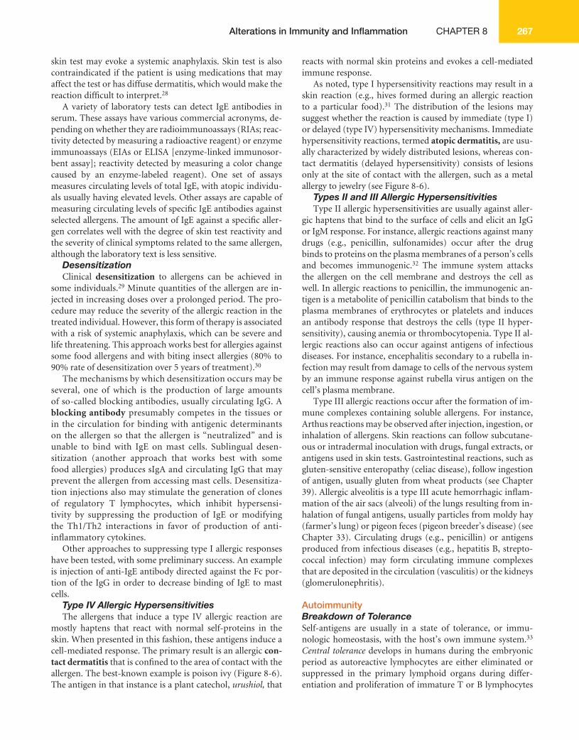
Deposition of circulating immune complexes containing antibody against DNA produces tissue damage in individu-als with SLE. DNA and DNA-containing immune complexes have a high affi nity for glomerular basement membranes and therefore may be selectively deposited in the glomerulus ( Figure 8-7 ). (Kidney structures are described in Chapter 35.) The presence of DNA in the circulation increases from cellular damage in response to trauma, drugs, or infections and is
usually removed in the liver. Removal of circulating DNA is slowed in the presence of immune complexes, thereby in-creasing the potential for deposition in the kidney. (The liver’s role in removing waste products from the blood is discussed in Chapter 35.) Deposition of immune complexes composed of DNA and antibody also causes infl ammatory lesions in the renal tubular basement membranes, brain (choroid plexus), heart, spleen, lung, gastrointestinal tract, skin (see Figure 8-7 ), and peritoneum.
SLE, as with most autoimmune diseases, occurs more often in women (approximately a 10:1 predominance of females), especially in the 20- to 40-year-old age group. Blacks are af-fected more often than whites (about an eightfold increased risk). A genetic predisposition for the disease has been im-plicated on the basis of increased incidence in twins and the existence of autoimmune disease in the families of individuals with SLE. 50
A transient lupus-like syndrome that is indistinguish-able both clinically and in the laboratory from spontane-ously occurring SLE can develop from the prolonged use of drugs. The drugs most often implicated are hydralazine
A
B Figure 8-7 Deposition of IgG in the kidney and skin of individuals with lupus. These photographs of tissue were obtained from individuals with lu-pus and stained with fl uorescent anti-IgG. A, Section from a kidney showing a glomerulus with deposits of IgG ( arrow, indicating bright areas of stain-ing). B, Section of the skin showing deposition of IgG along the dermal-epidermal junction ( arrow, indicating bright green staining). ( A courtesy of Dr. Helmut Rennke, Department of Pathology, Brigham and Women’s Hospital, Boston; B courtesy of Dr. Richard Sontheimer, Department of Dermatology, University of Texas Southwestern Medical School, Dallas. Modifi ed from Kumar V, Abbas A, Fausto N: Robbins and Cotran pathologic basis of disease, ed 7, Philadelphia, 2005, Saunders.)

UNIT III Mechanisms of Self-Defense272
(an antihypertensive agent) and procainamide (an antidys-rhythmic drug). In genetically susceptible individuals, certain environmental agents, such as ultraviolet light, and several in-fectious agents may trigger lupus-like immune reactions.
Clinical manifestations of SLE include arthralgias or arthri-tis (90% of individuals), vasculitis and rash (70% to 80% of individuals), renal disease (40% to 50% of individuals), hema-tologic abnormalities (50% of individuals, with anemia being the most common complication), and cardiovascular diseases (30% to 50% of individuals). As with most autoimmune dis-eases, the disease process develops slowly (up to 10 years from occurrence of the fi rst autoantibody until diagnosis) 51 and is characterized by frequent remissions and exacerbations. Be-cause the signs and symptoms affect almost every body system and tend to come and go, SLE is extremely diffi cult to diag-nose. This has led to the development of a list of 11 common clinical fi ndings. The serial or simultaneous presence of at least four of them indicates that the individual has SLE. 52 1. Facial rash confi ned to the cheeks (malar rash) 2. Discoid rash (raised patches, scaling) 3. Photosensitivity (skin rash developed as a result of ex-
posure to sunlight) 4. Oral or nasopharyngeal ulcers 5. Nonerosive arthritis of at least two peripheral joints 6. Serositis (pleurisy, pericarditis) 7. Renal disorder (proteinuria of 0.5 g/day or cellular
casts) 8. Neurologic disorders (seizures or psychosis) 9. Hematologic disorders (hemolytic anemia, leukope-
nia, lymphopenia, or thrombocytopenia) 10. Immunologic disorders (positive lupus erythemato-
sus [LE] cell preparation, anti – double-stranded DNA, anti-Smith [Sm] antigen, false-positive serologic test for syphilis, or antiphospholipid antibodies [anticar-diolipin antibody or lupus anticoagulant])
11. Presence of antinuclear antibody (ANA) There is no cure for SLE or most other autoimmune dis-
eases. The goals of treatment are to control symptoms and prevent further damage by suppressing the autoimmune re-sponse. Nonsteroidal anti-infl ammatory drugs, such as aspi-rin, ibuprofen, or naproxen, reduce infl ammation and relieve pain. Corticosteroids are often prescribed for more serious active disease. Immunosuppressive drugs (e.g., methotrexate, azathioprine, or cyclophosphamide) are used to treat severe symptoms involving internal organs. Ultraviolet light can worsen symptoms (known as fl ares), and protection from sun exposure is helpful. Prolonged use of certain drugs can cause transient SLE-like symptoms, and the medication history is important for diagnostic evaluation. Improved outcomes may be available in the future with the continued advances in medical research and the use of stem cell treatments. 53
Transfusion Reactions Red blood cells (erythrocytes) express several important sur-face antigens, known collectively as the blood group anti-gens, which can be targets of alloimmune reactions. 54 More
than 80 different red cell antigens are grouped into several dozen blood group systems, each determined by a different locus or set of loci. The most important of these, because they provoke the strongest humoral alloimmune response, are the ABO and Rh systems.
ABO System Human blood transfusions were carried out as early as
1818, but they were often unsuccessful. Sometimes after a transfusion, the recipient’s red blood cells would clump to-gether, thereby blocking the capillaries and causing death in some instances. In 1901, Karl Landsteiner reported that this reaction was related to the ABO antigens located on the sur-face of erythrocytes.
The ABO blood group consists of two major carbohydrate antigens, labeled A and B ( Figure 8-8 ). These two carbohydrate antigens are codominant, which means that both A and B can be simultaneously expressed, resulting in an individual hav-ing any one of four different blood types. The erythrocytes of persons with blood type A have the type A carbohydrate antigen (i.e., carry the A antigen), those with blood type B carry the B antigen, those with blood type AB carry both A and B antigens, and those of blood type O carry neither the A nor the B antigen. A person with type A blood also has circu-lating antibodies to the B carbohydrate antigen. If this person receives blood containing B antigens (i.e., blood from a type AB or B individual), a severe transfusion reaction occurs and the transfused erythrocytes are destroyed by agglutination ( Figure 8-9 ) or complement-mediated lysis. Similarly, a type B individual (whose blood contains anti-A antibodies) cannot receive blood from a type A or AB donor. Type O individuals, who have neither A or B antigen but have both anti-A and anti-B antibodies, cannot accept blood from any of the other three types. These naturally occurring antibodies, called iso-hemagglutinins, are immunoglobulins of the IgM class and are induced by similar antigens expressed on naturally occur-ring bacteria in the intestinal tract.
Because individuals with type O blood lack both types of antigens, they are considered universal donors, meaning that anyone can accept their red blood cells. Similarly, type AB individuals are considered universal recipients because they lack both anti-A and anti-B antibodies and can be transfused with any ABO blood type. When large volumes of whole blood (i.e., cells plus plasma) are transfused, however, antibodies in the donor’s blood can bind to antigenic determinants on the recipient’s erythrocytes, causing agglutination of the recipi-ent’s own cells. Agglutination and lysis cause harmful trans-fusion reactions that can be prevented only by complete and careful ABO matching between donor and recipient.
Rh System The Rh blood group is the most polymorphic system of
red cell antigens, consisting of at least 50 separate antigens. 55 At least fi ve major antigens and a large number of rare vari-ants have been identifi ed and are expressed primarily on erythrocytes. The major antigens are contained on two proteins encoded from two closely linked genes, RHD and RHCE . The RhD protein expresses the dominant antigen,
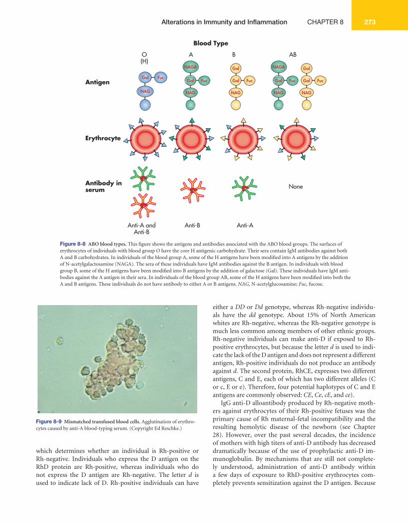
Alterations in Immunity and Infl ammation CHAPTER 8 273
which determines whether an individual is Rh-positive or Rh- negative. Individuals who express the D antigen on the RhD protein are Rh-positive, whereas individuals who do not express the D antigen are Rh-negative. The letter d is used to indicate lack of D. Rh-positive individuals can have
either a DD or Dd genotype, whereas Rh-negative individu-als have the dd genotype. About 15% of North American whites are Rh-negative, whereas the Rh-negative genotype is much less common among members of other ethnic groups. Rh- negative individuals can make anti-D if exposed to Rh-positive erythrocytes, but because the letter d is used to indi-cate the lack of the D antigen and does not represent a different antigen, Rh-positive individuals do not produce an antibody against d . The second protein, RhCE, expresses two different antigens, C and E, each of which has two different alleles (C or c, E or e). Therefore, four potential haplotypes of C and E antigens are commonly observed: CE , Ce , cE , and ce ).
IgG anti-D alloantibody produced by Rh-negative moth-ers against erythrocytes of their Rh-positive fetuses was the primary cause of Rh maternal-fetal incompatibility and the resulting hemolytic disease of the newborn (see Chapter 28). However, over the past several decades, the incidence of mothers with high titers of anti-D antibody has decreased dramatically because of the use of prophylactic anti-D im-munoglobulin. By mechanisms that are still not complete-ly understood, administration of anti-D antibody within a few days of exposure to RhD-positive erythrocytes com-pletely prevents sensitization against the D antigen. Because
Figure 8-9 Mismatched transfused blood cells. Agglutination of erythro-cytes caused by anti-A blood-typing serum. (Copyright Ed Reschke.)
AntigenGal
Gal Fuc
NAG NAG
Gal
Gal
Fuc
NAG
NAGA
Gal Fuc
NAG
Gal
Gal
Fuc
NAG
NAGA
Fuc
Erythrocyte
Antibody inserum None
Blood Type
AO(H)
B AB
Anti-A andAnti-B
Anti-B Anti-A
Figure 8-8 ABO blood types. This fi gure shows the antigens and antibodies associated with the ABO blood groups. The surfaces of erythrocytes of individuals with blood group O have the core H antigenic carbohydrate. Their sera contain IgM antibodies against both A and B carbohydrates. In individuals of the blood group A, some of the H antigens have been modifi ed into A antigens by the addition of N-acetylgalactosamine (NAGA). The sera of these individuals have IgM antibodies against the B antigen. In individuals with blood group B, some of the H antigens have been modifi ed into B antigens by the addition of galactose (Gal). These individuals have IgM anti-bodies against the A antigen in their sera. In individuals of the blood group AB, some of the H antigens have been modifi ed into both the A and B antigens. These individuals do not have antibody to either A or B antigens. NAG, N-acetylglucosamine; Fuc, fucose.

UNIT III Mechanisms of Self-Defense274
hemolytic disease of the newborn related to the D antigen has been controlled, alloantibodies against the other Rh an-tigens (usually C, c, or E) have become more important. In general, these alloantibodies are associated with a less severe hemolytic disease.
A form of autoimmune hemolytic anemia is often caused by autoantibodies against Rh antigens, especially e. This vari-ant is caused by IgG antibodies that react with erythrocytes at normal body temperature (thus called warm autoimmune hemolytic anemia ) and increase phagocytic destruction of the red cell. This characteristic differentiates the warm vari-ant from another form of autoimmune hemolytic anemia, which is caused by IgM autoantibodies that react optimally with erythrocytes in the cooler portions of the body (e.g., fi n-gers, toes) and is referred to as cold autoimmune hemolytic anemia .
Graft Rejection Transplantation of organs commonly is complicated by an immune response against antigens—primarily HLA—on the donated tissue. Most of our knowledge on the transplantation of organs is based on renal transplant studies. The primary mechanism of the rejection of transplanted organs is a type IV cell-mediated reaction. Two randomly chosen individuals are almost certainly antigenically different to some degree. Organ transplants between them could be rejected in approximate-ly 2 weeks without the extensive use of immunosuppressive drugs.
After the donor and recipient are matched for ABO anti-gens, HLAs are the principal targets of the rejection reaction; HLA matching of donor and recipient enhances the probabil-ity of acceptance of the graft. 56 Not all HLA loci are equally important; matching at the HLA-DR locus appears to be the most critical for graft acceptance, and matching at HLA-A and HLA-B of slightly lesser importance. (These loci are dis-cussed in Chapter 7.)
Transplant rejection may be classifi ed as hyperacute, acute, or chronic, depending on the amount of time that elapses be-tween transplantation and rejection. Hyperacute rejection is immediate and rare. When the circulation is reestablished to the grafted area, the graft may immediately turn white (the so-called white graft ) instead of a normal pink. Hyperacute re-jection usually occurs in recipients with preexisting antibody to antigens in the graft. The antibodies may have resulted from rejection of a previous graft or from prior blood trans-fusions that contained platelets and white blood cells with foreign HLA. Additionally, about half of women who have had multiple pregnancies have circulating antibodies against their husband’s HLA antigens. As the circulation to the graft is established, antibodies bind to the vascular endothelial cells in the grafted tissue and activate the infl ammatory re-sponse, including the coagulation cascade, which results in stasis of blood fl ow into the tissue ( Figure 8-10 ). (Coagula-tion is described in Chapters 6 and 25.) Biopsies of the graft often show deposits of antibody (IgG and IgM), complement, and neutrophils. This condition is rare because of effective
pretransplantation cross-matching during which a recipient is tested for antibodies against the HLA antigens of the po-tential donor.
Acute rejection is primarily a cell-mediated immune re-sponse that occurs within days to months after transplanta-tion. This type of rejection occurs when the recipient develops an immune response against unmatched HLAs after trans-plantation. Sensitization is usually initiated by the recipient’s lymphocytes interacting with the donor’s dendritic cells with-in the transplanted tissue, resulting in induction of recipient Th1 and Tc cells against the donor’s antigens. The Th1 cells re-lease cytokines that activate infi ltrating macrophages, and the Tc cells directly attack the endothelial cells in the transplanted tissue. A biopsy of the rejected organ usually shows an infi ltra-tion of lymphocytes and macrophages characteristic of a type IV reaction. Immunosuppressive drugs may delay or lessen the intensity of acute rejection.
A B
C Figure 8-10 Examples of hyperacute and acute rejection of renal allografts. A, Hyperacute antibody-mediated damage to the blood vessel of a renal allograft. The blood vessel is thickened, and the lumen (arrow) is obstructed by proliferating fi broblasts and macrophages. B, Acute cellular rejection of a renal allograft with intense mononuclear cell infi ltrate (arrow). C, Acute cellular rejection stained with immunoperoxidase reagent (brown) against T cells, which are infi ltrating the tissue. ( A courtesy of Dr. Ihsan Housini, Department of Pathology, University of Texas Southwestern Medi-cal School, Dallas; B and C courtesy of Dr. Robert Colvin, Department of Pathology, Massachusetts General Hospital, Boston. Modifi ed from Kumar V, Abbas A, Fausto N: Robbins and Cotran pathologic basis of disease, ed 7, Philadelphia, 2005, Saunders.)
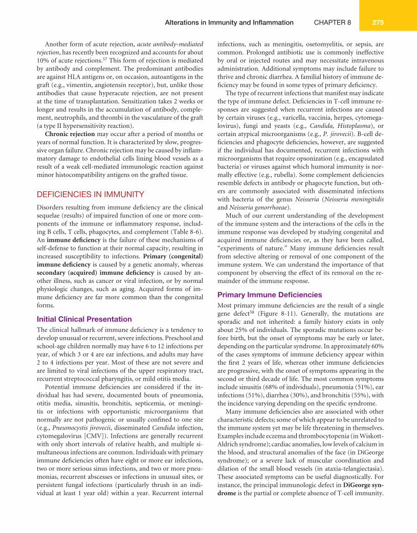
Alterations in Immunity and Infl ammation CHAPTER 8 275
Another form of acute rejection, acute antibody-mediated rejection , has recently been recognized and accounts for about 10% of acute rejections. 57 This form of rejection is mediated by antibody and complement. The predominant antibodies are against HLA antigens or, on occasion, autoantigens in the graft (e.g., vimentin, angiotensin receptor), but, unlike those antibodies that cause hyperacute rejection, are not present at the time of transplantation. Sensitization takes 2 weeks or longer and results in the accumulation of antibody, comple-ment, neutrophils, and thrombi in the vasculature of the graft (a type II hypersensitivity reaction).
Chronic rejection may occur after a period of months or years of normal function. It is characterized by slow, progres-sive organ failure. Chronic rejection may be caused by infl am-matory damage to endothelial cells lining blood vessels as a result of a weak cell-mediated immunologic reaction against minor histocompatibility antigens on the grafted tissue.
DEFICIENCIES IN IMMUNITY
Disorders resulting from immune defi ciency are the clinical sequelae (results) of impaired function of one or more com-ponents of the immune or infl ammatory response, includ-ing B cells, T cells, phagocytes, and complement ( Table 8-6 ). An immune defi ciency is the failure of these mechanisms of self-defense to function at their normal capacity, resulting in increased susceptibility to infections. Primary (congenital) immune defi ciency is caused by a genetic anomaly, whereas secondary (acquired) immune defi ciency is caused by an-other illness, such as cancer or viral infection, or by normal physiologic changes, such as aging. Acquired forms of im-mune defi ciency are far more common than the congenital forms.
Initial Clinical Presentation The clinical hallmark of immune defi ciency is a tendency to develop unusual or recurrent, severe infections. Preschool and school-age children normally may have 6 to 12 infections per year, of which 3 or 4 are ear infections, and adults may have 2 to 4 infections per year. Most of these are not severe and are limited to viral infections of the upper respiratory tract, recurrent streptococcal pharyngitis, or mild otitis media.
Potential immune defi ciencies are considered if the in-dividual has had severe, documented bouts of pneumonia, otitis media, sinusitis, bronchitis, septicemia, or meningi-tis or infections with opportunistic microorganisms that normally are not pathogenic or usually confi ned to one site (e.g., Pneumocystis jirovecii , disseminated Candida infection, cytomegalovirus [CMV]). Infections are generally recurrent with only short intervals of relative health, and multiple si-multaneous infections are common. Individuals with primary immune defi ciencies often have eight or more ear infections, two or more serious sinus infections, and two or more pneu-monias, recurrent abscesses or infections in unusual sites, or persistent fungal infections (particularly thrush in an indi-vidual at least 1 year old) within a year. Recurrent internal
infections, such as meningitis, osetomyelitis, or sepsis, are common. Prolonged antibiotic use is commonly ineffective by oral or injected routes and may necessitate intravenous administration. Additional symptoms may include failure to thrive and chronic diarrhea. A familial history of immune de-fi ciency may be found in some types of primary defi ciency.
The type of recurrent infections that manifest may indicate the type of immune defect. Defi ciencies in T-cell immune re-sponses are suggested when recurrent infections are caused by certain viruses (e.g., varicella, vaccinia, herpes, cytomega-lovirus), fungi and yeasts (e.g., Candida, Histoplasma ), or certain atypical microorganisms (e.g., P. jirovecii ). B-cell de-fi ciencies and phagocyte defi ciencies, however, are suggested if the individual has documented, recurrent infections with microorganisms that require opsonization (e.g., encapsulated bacteria) or viruses against which humoral immunity is nor-mally effective (e.g., rubella). Some complement defi ciencies resemble defects in antibody or phagocyte function, but oth-ers are commonly associated with disseminated infections with bacteria of the genus Neisseria ( Neisseria meningitidis and Neisseria gonorrhoeae ).
Much of our current understanding of the development of the immune system and the interactions of the cells in the immune response was developed by studying congenital and acquired immune defi ciencies or, as they have been called, “experiments of nature.” Many immune defi ciencies result from selective altering or removal of one component of the immune system. We can understand the importance of that component by observing the effect of its removal on the re-mainder of the immune response.
Primary Immune Defi ciencies Most primary immune defi ciencies are the result of a single gene defect 58 ( Figure 8-11 ). Generally, the mutations are sporadic and not inherited: a family history exists in only about 25% of individuals. The sporadic mutations occur be-fore birth, but the onset of symptoms may be early or later, depending on the particular syndrome. In approximately 60% of the cases symptoms of immune defi ciency appear within the fi rst 2 years of life, whereas other immune defi ciencies are progressive, with the onset of symptoms appearing in the second or third decade of life. The most common symptoms include sinusitis (68% of individuals), pneumonia (51%), ear infections (51%), diarrhea (30%), and bronchitis (55%), with the incidence varying depending on the specifi c syndrome.
Many immune defi ciencies also are associated with other characteristic defects; some of which appear to be unrelated to the immune system yet may be life threatening in themselves. Examples include eczema and thrombocytopenia (in Wiskott-Aldrich syndrome); cardiac anomalies, low levels of calcium in the blood, and structural anomalies of the face (in DiGeorge syndrome); or a severe lack of muscular coordination and dilation of the small blood vessels (in ataxia- telangiectasia). These associated symptoms can be useful diagnostically. For instance, the principal immunologic defect in DiGeorge syn-drome is the partial or complete absence of T-cell immunity.
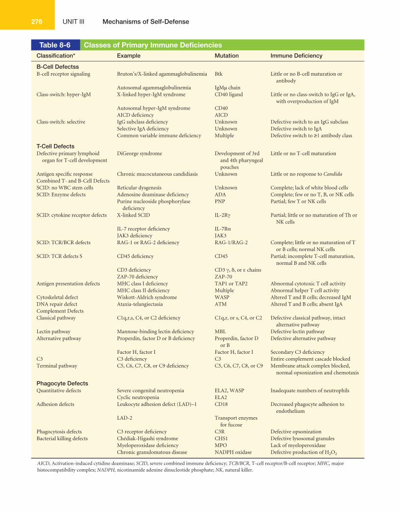
UNIT III Mechanisms of Self-Defense276
Table 8-6 Classes of Primary Immune Defi ciencies Classifi cation* Example Mutation Immune Defi ciency
B-Cell Defectss B-cell receptor signaling Bruton’s/X-linked agammaglobulinemia Btk Little or no B-cell maturation or
antibodyAutosomal agammaglobulinemia IgM μ chain
Class-switch: hyper-IgM X-linked hyper-IgM syndrome CD40 ligand Little or no class-switch to IgG or IgA, with overproduction of IgM
Autosomal hyper-IgM syndrome CD40AICD defi ciency AICD
Class-switch: selective IgG subclass defi ciency Unknown Defective switch to an IgG subclassSelective IgA defi ciency Unknown Defective switch to IgACommon variable immune defi ciency Multiple Defective switch to ≥ 1 antibody class
T-Cell Defects Defective primary lymphoid
organ for T-cell developmentDiGeorge syndrome Development of 3rd
and 4th pharyngeal pouches
Little or no T-cell maturation
Antigen specifi c response Chronic mucocutaneous candidiasis Unknown Little or no response to Candida Combined T- and B-Cell DefectsSCID: no WBC stem cells Reticular dysgenesis Unknown Complete; lack of white blood cellsSCID: Enzyme defects Adenosine deaminase defi ciency ADA Complete; few or no T, B, or NK cells
Purine nucleoside phosphorylase defi ciency
PNP Partial; few T or NK cells
SCID: cytokine receptor defects X-linked SCID IL-2R γ Partial; little or no maturation of Th or NK cells
IL-7 receptor defi ciency IL-7R α JAK3 defi ciency JAK3
SCID: TCR/BCR defects RAG-1 or RAG-2 defi ciency RAG-1/RAG-2 Complete; little or no maturation of T or B cells; normal NK cells
SCID: TCR defects S CD45 defi ciency CD45 Partial; incomplete T-cell maturation, normal B and NK cells
CD3 defi ciency CD3 γ , δ , or ε chainsZAP-70 defi ciency ZAP-70
Antigen presentation defects MHC class I defi ciency TAP1 or TAP2 Abnormal cytotoxic T cell activityMHC class II defi ciency Multiple Abnormal helper T cell activity
Cytoskeletal defect Wiskott-Aldrich syndrome WASP Altered T and B cells; decreased IgMDNA repair defect Ataxia-telangiectasia ATM Altered T and B cells; absent IgAComplement DefectsClassical pathway C1q,r,s, C4, or C2 defi ciency C1q,r, or s, C4, or C2 Defective classical pathway, intact
alternative pathwayLectin pathway Mannose-binding lectin defi ciency MBL Defective lectin pathwayAlternative pathway Properdin, factor D or B defi ciency Properdin, factor D
or BDefective alternative pathway
Factor H, factor I Factor H, factor I Secondary C3 defi ciencyC3 C3 defi ciency C3 Entire complement cascade blockedTerminal pathway C5, C6, C7, C8, or C9 defi ciency C5, C6, C7, C8, or C9 Membrane attack complex blocked,
normal opsonization and chemotaxis
Phagocyte Defects Quantitative defects Severe congenital neutropenia ELA2, WASP Inadequate numbers of neutrophils
Cyclic neutropenia ELA2Adhesion defects Leukocyte adhesion defect (LAD) – 1 CD18 Decreased phagocyte adhesion to
endotheliumLAD-2 Transport enzymes
for fucosePhagocytosis defects C3 receptor defi ciency C3R Defective opsonizationBacterial killing defects Chédiak-Higashi syndrome CHS1 Defective lysosomal granules
Myeloperoxidase defi ciency MPO Lack of myeloperoxidaseChronic granulomatous disease NADPH oxidase Defective production of H 2 O 2
AICD, Activation-induced cytidine deaminase; SCID, severe combined immune defi ciency; TCB/BCR, T-cell receptor/B-cell receptor; MHC, major histocompatibility complex; NADPH, nicotinamide adenine dinucleotide phosphate; NK, natural killer.
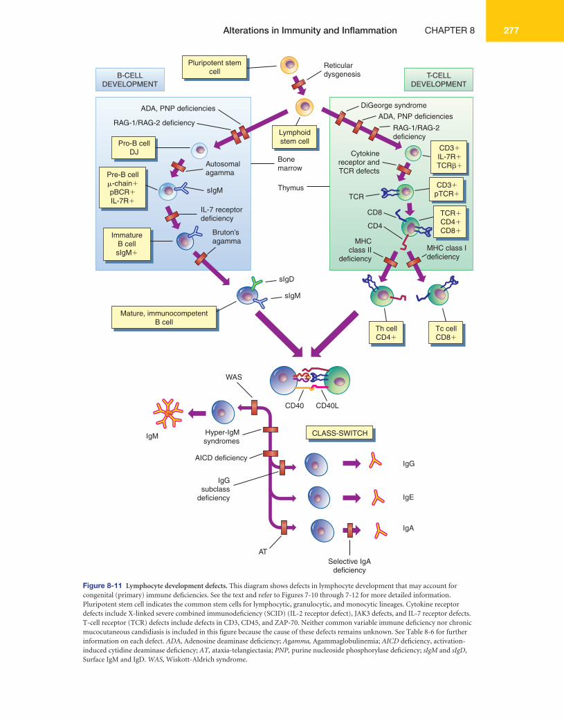
Alterations in Immunity and Infl ammation CHAPTER 8 277
Pluripotent stemcellB-CELL
DEVELOPMENTT-CELL
DEVELOPMENT
Lymphoidstem cell
Th cellCD4�
CLASS-SWITCH
Mature, immunocompetentB cell
Tc cellCD8�
CD3�pTCR�
Pro-B cellDJ
Pre-B cell�-chain�pBCR�IL-7R�
ImmatureB cellsIgM�
ADA, PNP deficiencies
RAG-1/RAG-2 deficiency
Autosomalagamma
Bonemarrow
TCR
CD8
Cytokinereceptor andTCR defects
MHCclass II
deficiency
MHC class Ideficiency
CD4
Reticulardysgenesis
DiGeorge syndrome
ADA, PNP deficiencies
Thymus
IL-7 receptordeficiency
sIgM
sIgD
IgG
IgM
IgE
IgA
sIgM
Hyper-IgMsyndromes
CD40
WAS
CD40L
Bruton’sagamma
TCR�CD4�CD8�
CD3�IL-7R�TCR��
RAG-1/RAG-2deficiency
AICD deficiency
IgGsubclass
deficiency
Selective IgAdeficiency
AT
Figure 8-11 Lymphocyte development defects. This diagram shows defects in lymphocyte development that may account for congenital (primary) immune defi ciencies. See the text and refer to Figures 7-10 through 7-12 for more detailed information. Pluripotent stem cell indicates the common stem cells for lymphocytic, granulocytic, and monocytic lineages. Cytokine receptor defects include X-linked severe combined immunodefi ciency (SCID) (IL-2 receptor defect), JAK3 defects, and IL-7 receptor defects. T-cell receptor (TCR) defects include defects in CD3, CD45, and ZAP-70. Neither common variable immune defi ciency nor chronic mucocutaneous candidiasis is included in this fi gure because the cause of these defects remains unknown. See Table 8-6 for further information on each defect. ADA, Adenosine deaminase defi ciency; A g amma, Agammaglobulinemia; AICD defi ciency, activation- induced cytidine deaminase defi ciency; AT, ataxia-telangiectasia; PNP, purine nucleoside phosphorylase defi ciency; sIgM and sIgD , Surface IgM and IgD. WAS, Wiskott-Aldrich syndrome.
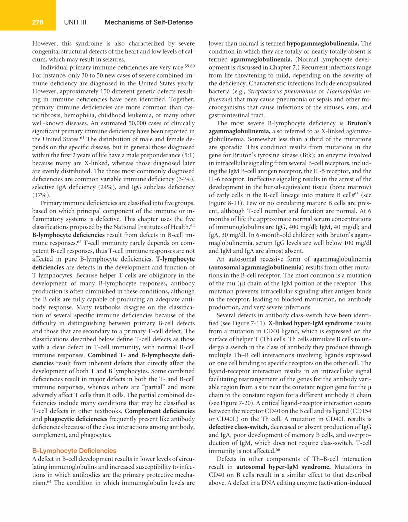
UNIT III Mechanisms of Self-Defense278
However, this syndrome is also characterized by severe congenital structural defects of the heart and low levels of cal-cium, which may result in seizures.
Individual primary immune defi ciencies are very rare. 59,60 For instance, only 30 to 50 new cases of severe combined im-mune defi ciency are diagnosed in the United States yearly. However, approximately 150 different genetic defects result-ing in immune defi ciencies have been identifi ed. Together, primary immune defi ciencies are more common than cys-tic fi brosis, hemophilia, childhood leukemia, or many other well-known diseases. An estimated 50,000 cases of clinically signifi cant primary immune defi ciency have been reported in the United States. 61 The distribution of male and female de-pends on the specifi c disease, but in general those diagnosed within the fi rst 2 years of life have a male preponderance (5:1) because many are X-linked, whereas those diagnosed later are evenly distributed. The three most commonly diagnosed defi ciencies are common variable immune defi ciency (34%), selective IgA defi ciency (24%), and IgG subclass defi ciency (17%).
Primary immune defi ciencies are classifi ed into fi ve groups, based on which principal component of the immune or in-fl ammatory systems is defective. This chapter uses the fi ve classifi cations proposed by the National Institutes of Health. 62 B-lymphocyte defi ciencies result from defects in B-cell im-mune responses. 63 T-cell immunity rarely depends on com-petent B-cell responses, thus T-cell immune responses are not affected in pure B-lymphocyte defi ciencies. T-lymphocyte defi ciencies are defects in the development and function of T lymphocytes. Because helper T cells are obligatory in the development of many B-lymphocyte responses, antibody production is often diminished in these conditions, although the B cells are fully capable of producing an adequate anti-body response. Many textbooks disagree on the classifi ca-tion of several specifi c immune defi ciencies because of the diffi culty in distinguishing between primary B-cell defects and those that are secondary to a primary T-cell defect. The classifi cations described below defi ne T-cell defects as those with a clear defect in T-cell immunity, with normal B-cell immune responses. Combined T- and B-lymphocyte defi -ciencies result from inherent defects that directly affect the development of both T and B lymphocytes. Some combined defi ciencies result in major defects in both the T- and B-cell immune responses, whereas others are “partial” and more adversely affect T cells than B cells. The partial combined de-fi ciencies include many conditions that may be classifi ed as T-cell defects in other textbooks. Complement defi ciencies and phagocytic defi ciencies frequently present like antibody defi ciencies because of the close interactions among antibody, complement, and phagocytes.
B-Lymphocyte Defi ciencies A defect in B-cell development results in lower levels of circu-lating immunoglobulins and increased susceptibility to infec-tions in which antibodies are the primary protective mecha-nism. 64 The condition in which immunoglobulin levels are
lower than normal is termed hypogammaglobulinemia. The condition in which they are totally or nearly totally absent is termed agammaglobulinemia. (Normal lymphocyte devel-opment is discussed in Chapter 7.) Recurrent infections range from life threatening to mild, depending on the severity of the defi ciency. Characteristic infections include encapsulated bacteria (e.g., Streptococcus pneumoniae or Haemophilus in-fl uenzae ) that may cause pneumonia or sepsis and other mi-croorganisms that cause infections of the sinuses, ears, and gastrointestinal tract.
The most severe B-lymphocyte defi ciency is Bruton’s agammaglobulinemia, also referred to as X-linked agamma-globulinemia. Somewhat less than a third of the mutations are sporadic. This condition results from mutations in the gene for Bruton’s tyrosine kinase (Btk); an enzyme involved in intracellular signaling from several B-cell receptors, includ-ing the IgM B-cell antigen receptor, the IL-5 receptor, and the IL-6 receptor. Ineffective signaling results in the arrest of the development in the bursal-equivalent tissue (bone marrow) of early cells in the B-cell lineage into mature B cells 65 (see Figure 8-11 ). Few or no circulating mature B cells are pres-ent, although T-cell number and function are normal. At 6 months of life the approximate normal serum concentrations of immunoglobulins are IgG, 400 mg/dl; IgM, 40 mg/dl; and IgA, 30 mg/dl. In 6-month-old children with Bruton’s agam-maglobulinemia, serum IgG levels are well below 100 mg/dl and IgM and IgA are almost absent.
An autosomal recessive form of agammaglobulinemia (autosomal agammaglobulinemia) results from other muta-tions in the B-cell receptor. The most common is a mutation of the mu ( μ ) chain of the IgM portion of the receptor. This mutation prevents intracellular signaling after antigen binds to the receptor, leading to blocked maturation, no antibody production, and very severe infections.
Several defects in antibody class-switch have been identi-fi ed (see Figure 7-11). X-linked hyper-IgM syndrome results from a mutation in CD40 ligand, which is expressed on the surface of helper T (Th) cells. Th cells stimulate B cells to un-dergo a switch in the class of antibody they produce through multiple Th – B cell interactions involving ligands expressed on one cell binding to specifi c receptors on the other cell. The ligand-receptor interaction results in an intracellular signal facilitating rearrangement of the genes for the antibody vari-able region from a site near the constant region gene for the μ chain to the constant region for a different antibody H chain (see Figure 7-20). A critical ligand-receptor interaction occurs between the receptor CD40 on the B cell and its ligand (CD154 or CD40L) on the Th cell. A mutation in CD40L results is defective class-switch, decreased or absent production of IgG and IgA, poor development of memory B cells, and overpro-duction of IgM, which does not require class-switch. T-cell immunity is not affected. 66
Defects in other components of Th – B-cell interaction result in autosomal hyper-IgM syndrome. Mutations in CD40 on B cells result in a similar effect to that described above. A defect in a DNA editing enzyme (activation-induced

Alterations in Immunity and Infl ammation CHAPTER 8 279
cytidine deaminase; AICD) also inhibits class-switch. During class-switch and movement of the H chain genetic informa-tion for the variable region to a different constant region gene, the double-stranded DNA must be cut and mended. This en-zyme is responsible for cutting and mending the DNA.
Defi ciencies in certain subclasses of antibody (IgG sub-class defi ciency), particularly IgG2, may result from a defect in switch to a particular subclass constant region (see Figure 8-11 ). The IgG2 subclass is often increased in response to polysaccharide antigens such as those on the surface of en-capsulated bacteria. Low levels of IgG2 may be responsible for recurrent risk for pneumonias caused by these bacteria. Whether IgG subclass defi ciencies are unique immune de-fi ciency conditions is unclear because many are apparently early indications of the development of common variable im-mune defi ciency (see following) or are secondary to selective IgA defi ciency.
One of the most common primary immune defi ciencies is a selective IgA defi ciency. Because many affected individuals are asymptomatic, the true incidence is uncertain, although estimates of 1 person in 300 to 1 in 3000 have been made. Individuals with selective IgA defi ciency are able to produce other classes of immunoglobulins but fail to produce IgA (see Figure 8-11 ). Many will have B cells that have undergone class-switch to IgA, but for unknown reasons, cannot undergo the terminal steps of differentiation to IgA-secreting plasma cells. Although many individuals are asymptomatic, others present with a history of severe recurring sinus, lung, and gastrointes-tinal infections. They commonly also have chronic intestinal candidiasis (infection with Candida albicans ). (The secretory, or mucosal, immune system is described in Chapter 7.)
Complications of IgA defi ciency include severe atopic dis-ease and autoimmune diseases; selective IgA defi ciency is two or three times more common in atopic individuals than in others. Secretory IgA normally may prevent the uptake of al-lergens from the environment so that IgA defi ciency may lead to increased allergen uptake and a more intense challenge to the immune system because of prolonged exposure to envi-ronmental antigens. One of the most severe complications of IgA defi ciency is an anaphylactic reaction that can follow administration of blood products that contain IgA. Serious anaphylactic reactions can occur in individuals totally lack-ing IgA because the immune system recognizes donor IgA as a foreign antigen. Initial sensitization can occur in fetal life through exposure to maternal IgA that leaks across the pla-centa or later through the ingestion of maternal IgA in breast milk or bovine IgA in cow’s milk. Sensitization also can occur with initial administration of blood products containing IgA. The individual’s primed immune system then acts against do-nor IgA on subsequent exposure.
Common variable immune defi ciency is the most com-monly diagnosed immune defi ciency. As the name implies, the presentation is very heterogeneous. It is characterized by hypogammaglobulinemia, but the particular class of antibody that is decreased varies: most have low amounts of IgG, which may or may not be accompanied by decreased levels of IgA
or IgM, or both, with normal numbers of B cells. Some may have accompanying T-cell defects. Multiple genetic defects in terminal differentiation account for this condition, although the specifi c defects have not been identifi ed in most patients. The age of onset of symptoms, such as recurrent bacterial re-spiratory tract infections, is generally later than most primary immune defi ciencies (late 20s). Secondary complications in-clude arthritis (infectious and noninfectious), gastrointestinal symptoms (malabsorption, chronic diarrhea) autoimmune disease (anemia, thrombocytopenia, endocrine diseases), and cancer (of the lymphoid system, skin, and gastrointestinal tract).
T-Lymphocyte Defi ciencies Two well-studied examples of T-lymphocyte defects that represent different ends of the T-cell differentiation process include DiGeorge syndrome and chronic mucocutaneous can-didiasis. Lymphoid stem cells begin maturing into functional T lymphocytes in the thymus. DiGeorge syndrome (congenital thymic aplasia or hypoplasia) is caused by the lack, or more commonly partial lack, of the thymus, resulting in greatly de-creased T-cell numbers and function and in life- threatening viral, fungal, and intracellular bacterial infections 67,68 (see Figure 8-11 ). The defect is attributed usually to deletions on chromosome 22 (some deletions also have been identifi ed on chromosome 10); about 25% of which are inherited. The de-leted region encodes information for formation of organs that originate from the third and fourth pharyngeal pouches during the twelfth week of gestation. In addition to the lack of thymus development, the individual may present with a partial or com-plete absence of the parathyroid gland (resulting in decreased blood calcium levels), major structural defects in the heart and the aorta (resulting in inadequate blood fl ow and inadequate oxygenation of the tissues), and abnormal facial characteristics (e.g., underdeveloped chin, low-set ears, shortened structure of the upper lip) ( Figure 8-12 ).
Chronic mucocutaneous candidiasis is a primary defect of T lymphocytes in response to a specifi c infectious agent, the yeast C. albicans . At least seven variants of this condition have been described. All are characterized by mild to extremely severe chronic mucocutaneous candidiasis: Candida infec-tions that involve the mucous membranes, nails, and skin. Invasive candidiasis is extremely rare. Although most B- and T-cell immune responses may be normal, most individuals with this defect cannot react to antigens from Candida. The cause of this defect is unknown.
Combined T- and B-Lymphocyte Defi ciencies The most severe defi ciencies usually occur when both the B- and T-cell immune responses are affected. A great deal of knowledge about the evolution of bone marrow stem cells into functional B- and T-cell effectors came from studying chil-dren with the most severe immune defi ciency, severe com-bined immune defi ciency (SCID). 69 The most severe form of SCID is reticular dysgenesis (failure of blood cells to de-velop), in which a common stem cell for all white blood cells is
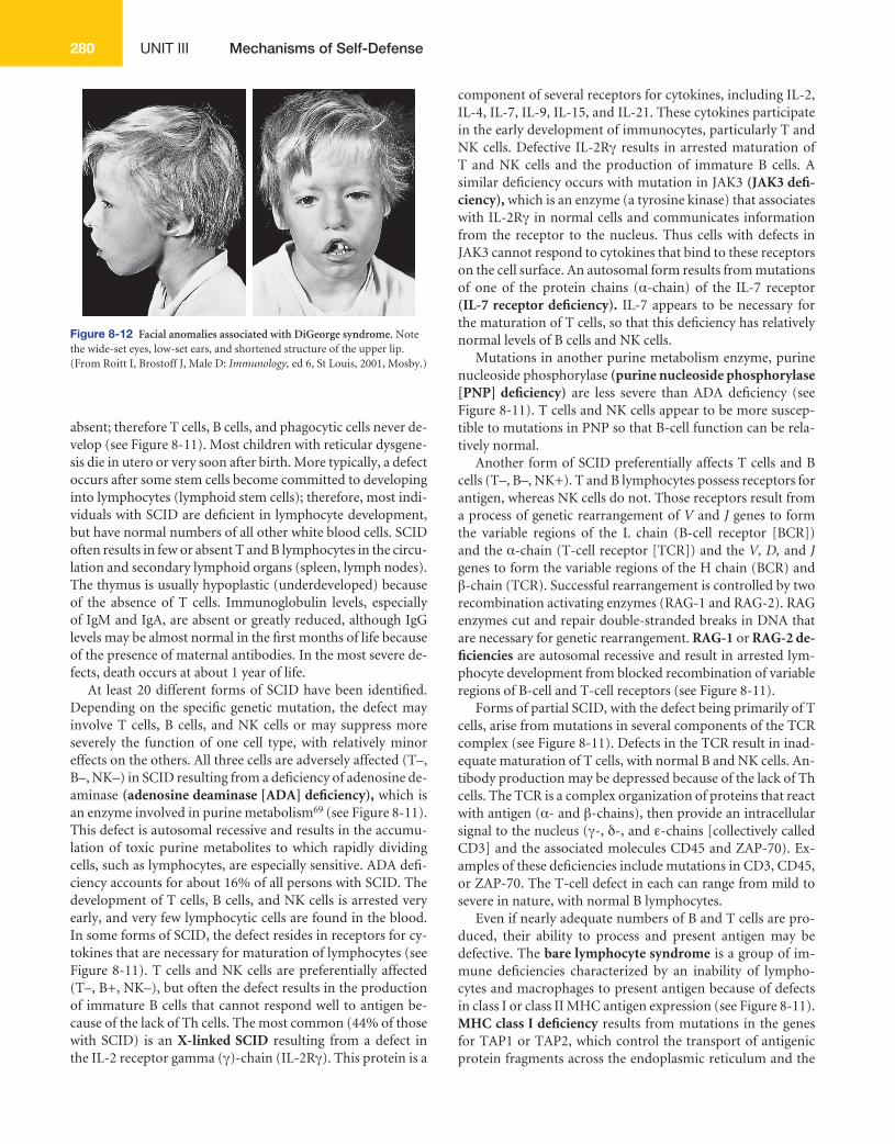
UNIT III Mechanisms of Self-Defense280
absent; therefore T cells, B cells, and phagocytic cells never de-velop (see Figure 8-11 ). Most children with reticular dysgene-sis die in utero or very soon after birth. More typically, a defect occurs after some stem cells become committed to developing into lymphocytes (lymphoid stem cells); therefore, most indi-viduals with SCID are defi cient in lymphocyte development, but have normal numbers of all other white blood cells. SCID often results in few or absent T and B lymphocytes in the circu-lation and secondary lymphoid organs (spleen, lymph nodes). The thymus is usually hypoplastic (underdeveloped) because of the absence of T cells. Immunoglobulin levels, especially of IgM and IgA, are absent or greatly reduced, although IgG levels may be almost normal in the fi rst months of life because of the presence of maternal antibodies. In the most severe de-fects, death occurs at about 1 year of life.
At least 20 different forms of SCID have been identifi ed. Depending on the specifi c genetic mutation, the defect may involve T cells, B cells, and NK cells or may suppress more severely the function of one cell type, with relatively minor effects on the others. All three cells are adversely affected (T – , B – , NK – ) in SCID resulting from a defi ciency of adenosine de-aminase (adenosine deaminase [ADA] defi ciency), which is an enzyme involved in purine metabolism 69 (see Figure 8-11 ). This defect is autosomal recessive and results in the accumu-lation of toxic purine metabolites to which rapidly dividing cells, such as lymphocytes, are especially sensitive. ADA defi -ciency accounts for about 16% of all persons with SCID. The development of T cells, B cells, and NK cells is arrested very early, and very few lymphocytic cells are found in the blood. In some forms of SCID, the defect resides in receptors for cy-tokines that are necessary for maturation of lymphocytes (see Figure 8-11 ). T cells and NK cells are preferentially affected (T – , B+, NK – ), but often the defect results in the production of immature B cells that cannot respond well to antigen be-cause of the lack of Th cells. The most common (44% of those with SCID) is an X-linked SCID resulting from a defect in the IL-2 receptor gamma ( γ )-chain (IL-2R γ ). This protein is a
component of several receptors for cytokines, including IL-2, IL-4, IL-7, IL-9, IL-15, and IL-21. These cytokines participate in the early development of immunocytes, particularly T and NK cells. Defective IL-2R γ results in arrested maturation of T and NK cells and the production of immature B cells. A similar defi ciency occurs with mutation in JAK3 (JAK3 defi -ciency), which is an enzyme (a tyrosine kinase) that associates with IL-2R γ in normal cells and communicates information from the receptor to the nucleus. Thus cells with defects in JAK3 cannot respond to cytokines that bind to these receptors on the cell surface. An autosomal form results from mutations of one of the protein chains ( α -chain) of the IL-7 receptor (IL-7 receptor defi ciency). IL-7 appears to be necessary for the maturation of T cells, so that this defi ciency has relatively normal levels of B cells and NK cells.
Mutations in another purine metabolism enzyme, purine nucleoside phosphorylase (purine nucleoside phosphorylase [PNP] defi ciency) are less severe than ADA defi ciency (see Figure 8-11 ). T cells and NK cells appear to be more suscep-tible to mutations in PNP so that B-cell function can be rela-tively normal.
Another form of SCID preferentially affects T cells and B cells (T – , B – , NK+). T and B lymphocytes possess receptors for antigen, whereas NK cells do not. Those receptors result from a process of genetic rearrangement of V and J genes to form the variable regions of the L chain (B-cell receptor [BCR]) and the α -chain (T-cell receptor [TCR]) and the V, D, and J genes to form the variable regions of the H chain (BCR) and β -chain (TCR). Successful rearrangement is controlled by two recombination activating enzymes (RAG-1 and RAG-2). RAG enzymes cut and repair double-stranded breaks in DNA that are necessary for genetic rearrangement. RAG-1 or RAG-2 de-fi ciencies are autosomal recessive and result in arrested lym-phocyte development from blocked recombination of variable regions of B-cell and T-cell receptors (see Figure 8-11 ).
Forms of partial SCID, with the defect being primarily of T cells, arise from mutations in several components of the TCR complex (see Figure 8-11 ). Defects in the TCR result in inad-equate maturation of T cells, with normal B and NK cells. An-tibody production may be depressed because of the lack of Th cells. The TCR is a complex organization of proteins that react with antigen ( α - and β -chains), then provide an intracellular signal to the nucleus ( γ -, δ -, and ε -chains [collectively called CD3] and the associated molecules CD45 and ZAP-70). Ex-amples of these defi ciencies include mutations in CD3, CD45, or ZAP-70. The T-cell defect in each can range from mild to severe in nature, with normal B lymphocytes.
Even if nearly adequate numbers of B and T cells are pro-duced, their ability to process and present antigen may be defective. The bare lymphocyte syndrome is a group of im-mune defi ciencies characterized by an inability of lympho-cytes and macrophages to present antigen because of defects in class I or class II MHC antigen expression (see Figure 8-11). MHC class I defi ciency results from mutations in the genes for TAP1 or TAP2, which control the transport of antigenic protein fragments across the endoplasmic reticulum and the
Figure 8-12 Facial anomalies associated with DiGeorge syndrome. Note the wide-set eyes, low-set ears, and shortened structure of the upper lip. (From Roitt I, Brostoff J, Male D: Immunology, ed 6, St Louis, 2001, Mosby.)

Alterations in Immunity and Infl ammation CHAPTER 8 281
formation of MHC class I/antigen complexes for transporta-tion to the cell surface (see Figure 7-16). Because MHC class I molecules preferentially present antigen to CD8+ Tc cells, the resultant defi ciency is of CD8+ cytotoxic cells, with normal levels of CD4+ helper cells and normal antibody production. MHC class II defi ciency is more severe. A variety of muta-tions prevent normal production of MHC class II molecules, which present antigen to CD4+ helper cells. Because of defec-tive recruitment of helper T cells, normal antibody responses are greatly suppressed. Children with this defi ciency develop life-threatening infections and usually die before age 5 years.
Some combined immune defi ciencies are secondary to mutations that affect a variety of cells other than immuno-cytes. For instance, Wiskott-Aldrich syndrome (WAS; an X-linked recessive disorder) results from sporadic mutations in the WAS protein (WASP), which is involved in intracellular signaling and regulation of the organization of the cell’s actin cytoskeleton 70 (see Figure 8-11 ). The defects in the cytoskel-eton lead to the classic symptoms of thrombocytopenia (with resultant bleeding disorders), scaly eczema, and defective T and B cells. IgA and IgG levels are usually normal, but IgM responses are highly depressed. Antibody responses against antigens that elicit primarily an IgM response, such as poly-saccharide antigens from bacterial cell walls (e.g., of Pseudo-monas aeruginosa, S. pneumoniae, H. infl uenzae, and other microorganisms with polysaccharide outer capsules), are de-fi cient. Persons with WAS have a very high risk of lymphoid malignancies (leukemias and lymphomas).
Ataxia-telangiectasia (AT) is an autosomal recessive dis-order resulting from a large variety of sporadic mutations in the ATM gene, which encodes a protein involved in repair of double-stranded breaks in DNA. Affected infants often de-velop ataxia (unsteady gait), which usually becomes apparent when the child is learning to walk. The neurologic defect may eventually lead to confi nement in a wheelchair. Telangiectasia (dilation of capillaries) can occur in the eyes and skin, espe-cially on the ears, neck, and extremities. Both B and T cells are variably affected and unrepaired double-stranded DNA breaks are commonly observed in the regions encoding the T-cell and B-cell receptors. About 70% of those with AT are IgA defi cient, occasionally accompanied by defi ciencies in IgG (see Figure 8-11 ). Individuals with AT are at high risk for developing leukemias and lymphomas.
Complement Defi ciencies Complement activation is a necessary component of protec-tion against many infectious agents. As a result, some defects in the complement cascade often resemble antibody defi cien-cies, with recurrent infections with encapsulated bacteria (e.g., H. infl uenzae and S. pneumoniae ). 71 Additionally, the Fc portion of IgG and some activated complement components, such as C3b, function as opsonins and facilitate phagocytosis by neutrophils and macrophages. In addition to recurrent in-fections, defi ciencies in the classical pathway commonly lead to a SLE-like syndrome. As noted, excessive levels of circu-lating complexes of antibody, antigen, and complement may
lead to type III hypersensitivity diseases (immune complex diseases). However, healthy individuals release small amounts of soluble intracellular antigens into the blood during normal cell turnover. Low levels of naturally occurring autoantibodies and limited activation of the classical pathway of the comple-ment system through C3 facilitate the removal of this debris by phagocytes. Thus some complement defects may slow the clearance from the blood of natural immune complexes, lead-ing to SLE-like symptoms.
C3 defi ciency is the most severe complement defect ( Figure 8-13 ). C3 is the component that unites all pathways of comple-ment activation, and complement component C3b is a major opsonin. Persons with C3 defi ciency are at risk for recurrent life-threatening infections with encapsulated bacteria at an early age, as well as a SLE-like syndrome that may be compli-cated by kidney disease (glomerulonephritis). 72 C2 defi ciency, more so than C1 or C4 defi ciencies, also has an increased risk for recurrent respiratory infections with encapsulated bacteria (e.g., S. pneumoniae , H. infl uenzae ).
Mannose-binding lectin (MBL) defi ciency is the primary defect of the lectin pathway of complement activation. The defect results in increased risk of infection with microorgan-isms that have polysaccharide capsules rich in mannose, par-ticularly the yeast Saccharomyces cerevisiae and encapsulated bacteria such as N. meningitidis and S. pneumoniae.
Defi ciencies in the alternative pathway also result in recur-rent infections with encapsulated bacteria. Properdin defi -ciency is associated with recurrent meningococcal infections and is X-linked, whereas all other complement defi ciencies are autosomal recessive. Symptoms generally appear in the second decade of life. Factor I and factor H are major regula-tors of the complement cascade and control the level of spon-taneous activation of C3. Factor I defi ciency and factor H defi ciency can be severe because they lead to increased spon-taneous destruction of C3 and a secondary C3 defi ciency.
Defi ciencies of components of the terminal portion of the complement cascade (C5, C6, C7, C8, or C9 defi ciencies) are associated with increased infections with only one group of bacteria; those of the genus Neisseria ( N. meningitidis or N. gonorrhoeae ). Neisseria usually cause localized infections (meningitis or gonorrhea), but those individuals with termi-nal pathway defects have more than an 8000-fold increased risk for systemic infections with atypical strains of these mi-croorganisms. C9 defi ciency is the most common terminal pathway defect, appears primarily in Japanese populations, and is generally asymptomatic. The other defi ciencies of the terminal pathway are extremely rare, but are characterized by more aggressive infections. The risk for systemic infections with Neisseria is also increased in those with defi ciencies of C2, factor D, factor B, and properdin.
Phagocytic Defi ciencies Phagocytosis is generally aided by bacterial opsonization with IgG or C3b; therefore, defects in phagocytic killing usually re-sult in recurrent infections with the same group of microor-ganisms (encapsulated bacteria) associated with antibody and
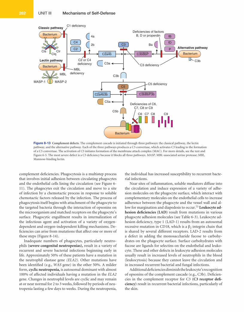
UNIT III Mechanisms of Self-Defense282
complement defi ciencies. Phagocytosis is a multistep process that involves initial adhesion between circulating phagocytes and the endothelial cells lining the circulation (see Figure 6-11 ). The phagocytes exit the circulation and move to a site of infection by a chemotactic process in response to soluble chemotactic factors released by the infection. The process of phagocytosis itself begins with attachment of the phagocyte to the targeted bacteria through the interaction of opsonins on the microorganism and matched receptors on the phagocyte’s surface. Phagocytic engulfment results in internalization of the infectious agent and activation of a variety of oxygen- dependent and oxygen-independent killing mechanisms. De-fi ciencies can arise from mutations that affect one or more of these steps ( Figure 8-14 ).
Inadequate numbers of phagocytes, particularly neutro-phils (severe congenital neutropenias), result in a variety of recurrent and severe bacterial infections beginning early in life. Approximately 50% of these patients have a mutation in the neutrophil elastase gene (ELA2). Other mutations have been identifi ed (e.g., WAS gene) in the other 50%. A milder form, cyclic neutropenia, is autosomal dominant with almost 100% of affected individuals having a mutation in the ELA2 gene. Changes in neutrophil levels are cyclic and may remain at or near normal for 2 to 3 weeks, followed by periods of neu-tropenia lasting a few days to weeks. During the neutropenia,
the individual has increased susceptibility to recurrent bacte-rial infections.
Near sites of infl ammation, soluble mediators diffuse into the circulation and induce expression of a variety of adhe-sion molecules on the phagocyte surface, which interact with complementary molecules on the endothelial cells to increase adherence between the phagocyte and the vessel wall and al-low for margination and diapedesis to occur. 73 Leukocyte ad-hesion defi ciencies (LAD) result from mutations in various phagocyte adhesion molecules (see Table 6-3 ). Leukocyte ad-hesion defi ciency, type 1 (LAD-1) results from an autosomal recessive mutation in CD18, which is a β 2 integrin chain that is shared by several different receptors. LAD-2 results from a defect in adding the monosaccharide fucose to carbohy-drates on the phagocyte surface. Surface carbohydrates with fucose are ligands for selectins on the endothelial and leuko-cyte. These and other defects in leukocyte adhesion molecules usually result in increased levels of neutrophils in the blood (leukocytosis) because they cannot leave the circulation and in increased recurrent bacterial and fungal infections.
Additional defi ciencies diminish the leukocyte’s recognition of opsonins of the complement cascade (e.g., C3b). Defi cien-cies in the complement receptor for C3 (C3 receptor defi -ciency) result in recurrent bacterial infections, particularly of the skin.
Classic pathway
Bacterium
Bacterium
Bacterium
BacteriumCls Clr
C1 deficiency
C2 or C4deficiency
Deficiencies of factorsB, D or properdin
Deficiencies of C6,C7, C8 or C9
C3 deficiency
C5 deficiency
MBLdeficiency
Clq
C4
C2
4a
2b C3
C3a
C3b
C3b
C2a4b
C5b
C5
C5a
Lectin pathway
MASP-1
MBL
MASP-2
Ba
fB
fD
P Alternative pathway
C6 C7 C8 C9
C2a4b3b C3bBbP3b
C3bBbP
Figure 8-13 Complement defects. The complement cascade is initiated through three pathways: the classical pathway, the lectin pathway, and the alternative pathway. Each of the three pathways produces a C3 convertase, which activates C3 leading to the formation of a C5 convertase. The activation of C5 initiates formation of the membrane attack complex (MAC). For more details, see the text and Figure 6-5. The most severe defect is a C3 defi ciency because it blocks all three pathways. MASP, MBL-associated serine protease; MBL, Mannose-binding lectin.
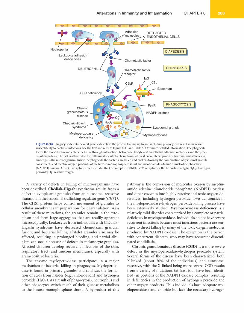
Alterations in Immunity and Infl ammation CHAPTER 8 283
A variety of defects in killing of microorganisms have been described. Chédiak-Higashi syndrome results from a defect in cytoplasmic granules from an autosomal recessive mutation in the lysosomal traffi cking regulator gene (CHS1). The CHS1 protein helps control movement of granules to cellular membranes in preparation for degranulation. As a result of these mutations, the granules remain in the cyto-plasm and form large aggregates that are readily apparent microscopically. Leukocytes from individuals with Chédiak- Higashi syndrome have decreased chemotaxis, granular fusion, and bacterial killing. Platelet granules also may be affected, resulting in prolonged bleeding, and partial albi-nism can occur because of defects in melanocyte granules. Affected children develop recurrent infections of the skin, respiratory tract, and mucous membranes, especially with gram-positive bacteria.
The enzyme myeloperoxidase participates in a major mechanism of bacterial killing in phagocytes. Myeloperoxi-dase is found in primary granules and catalyzes the forma-tion of acids from halides (e.g., chloride ion) and hydrogen peroxide (H 2 O 2 ). As a result of phagocytosis, neutrophils and other phagocytes switch much of their glucose metabolism to the hexose-monophosphate shunt. A byproduct of this
pathway is the conversion of molecular oxygen by nicotin-amide adenine dinucleotide phosphate (NADPH) oxidase and other enzymes into highly reactive and toxic oxygen de-rivatives, including hydrogen peroxide. Two defi ciencies in the myeloperoxidase – hydrogen peroxide killing process have been extensively studied. Myeloperoxidase defi ciency is a relatively mild disorder characterized by a complete or partial defi ciency in myeloperoxidase. Individuals do not have severe recurrent infections because most infectious bacteria are sen-sitive to direct killing by many of the toxic oxygen molecules produced by NADPH oxidase. The exception is the person with concurrent diabetes, who may have recurrent dissemi-nated candidiasis.
Chronic granulomatous disease (CGD) is a more severe defect in the myeloperoxidase – hydrogen peroxide system. Several forms of the disease have been characterized, both X-linked (about 70% of the individuals) and autosomal recessive, with the X-linked being more severe. CGD results from a variety of mutations (at least four have been identi-fi ed) in portions of the NADPH oxidase complex, resulting in defi ciencies in the production of hydrogen peroxide and other oxygen products. Thus individuals have adequate my-eloperoxidase and chloride but lack the necessary hydrogen
IgGC3bC3bR
Bacterium
Chemotactic factor
Adhesionmolecules
RETRACTEDENDOTHELIAL CELLS
NEUTROPHIL
Leukocyte adhesiondeficiencies
Neutropenia
Chemotactic factorreceptor
Lysosomal granule
Fc�R
NADPH oxidase
MyeloperoxidaseMyeloperoxidasedeficiency
Chédiak-Higashisyndrome
Chronicgranulomatous
disease
C3R deficiency
H2O2
O2�
DIAPEDESIS
CHEMOTAXIS
PHAGOCYTOSIS
Figure 8-14 Phagocytic defects. Several genetic defects in the process leading up to and including phagocytosis result in increased susceptibility to bacterial infections. See the text and refer to Figure 6-11 and Table 6-3 for more detailed information. The phagocyte leaves the bloodstream and enters the tissue through interactions between leukocyte and endothelial adhesion molecules and the proc-ess of diapedesis. The cell is attracted to the infl ammatory site by chemotaxis, where it encounters opsonized bacteria, and attaches to and engulfs the microorganism. Inside the phagocyte the bacteria are killed and broken down by the combination of lysosomal granule constituents and reactive oxygen products of the hexose-monophosphate shunt and nicotinamide adenine dinucleotide phosphate (NADPH) oxidase. C3R , C3 receptor, which includes the C3b receptor (C3bR); Fc γ R , receptor for the Fc portion of IgG; H 2 O 2 , hydrogen peroxide; O 2 − , reactive oxygen.
UNIT III Mechanisms of Self-Defense284
peroxide. Individuals with CGD have recurrent severe pneumonias; tumor-like granulomas in lungs, skin, and bones; and other infections with some normally relative innocuous microorganisms, such as Staphylococcus aureus, Serratia marcescens, Aspergillus spp., and others. These are catalase-positive microorganisms. Infections with more vir-ulent, but catalase-negative, microorganisms (e.g., S. pneu-moniae ) are rare. Most microorganisms produce their own hydrogen peroxide as a byproduct, which accumulates in the phagocytic vacuole and can be used by the phagocyte’s myelo-peroxidase to kill the microorganism. Some microorganisms also produce the enzyme catalase, which breaks down hydro-gen peroxide. Thus catalase-negative microorganisms donate hydrogen peroxide to the phagocyte’s myeloperoxidase, lead-ing to their own death. Catalase-positive microorganisms, however, destroy the bacterial hydrogen peroxide and survive and cause infection.
Secondary Immune Defi ciencies Secondary, or acquired, immune and infl ammatory defi cien-cies are far more common than primary defi ciencies. 74 These defi ciencies are not related to genetic defects, but are com-plications of other physiologic or pathophysiologic condi-tions. Some conditions that are known to be associated with acquired defi ciencies include:
Normal physiologic conditions������� ��•��� ���Pregnancy ��� ��•��� ���Infancy ��� ��•��� ���Aging
Psychologic stress������� ��•��� ���Emotional trauma ��� ��•��� ���Eating disorders
Dietary insuffi ciencies������� ��•��� ���Malnutrition caused by insuffi cient intake of large cat-
egories of nutrients, such as protein or calories ��� ��•��� ���Insuffi cient intake of specifi c nutrients, such as vitamins,
iron, or zinc Malignancies
������� ��•��� ���Malignancies of lymphoid tissues, such as Hodgkin dis-ease, acute or chronic leukemia, or myeloma
��� ��•��� ���Malignancies of nonlymphoid tissues, such as sarcomas and carcinomas
Metabolic diseases or genetic syndromes������� ��•��� ���Diabetes ��� ��•��� ���Cystic fi brosis ��� ��•��� ���Alcoholic cirrhosis ��� ��•��� ���Sickle cell disease ��� ��•��� ���SLE ��� ��•��� ���Chromosome abnormalities, such as trisomy 21 (Down
syndrome) Environmental
������� ��•��� ���Ultraviolet (UV) light ��� ��•��� ���Ionizing radiation ��� ��•��� ���Chronic hypoxia
Physical trauma������� ��•��� ���Burns
Medical treatments������� ��•��� ���Stress caused by surgery ��� ��•��� ���Anesthesia ��� ��•��� ���Immunosuppressive treatment with corticosteroids or
antilymphocyte antibodies ��� ��•��� ���Splenectomy ��� ��•��� ���Cancer treatment with cytotoxic drugs or ionizing ra-
diation Infections
������� ��•��� ���Congenital infections, such as rubella, cytomegalovirus, hepatitis B
��� ��•��� ���Acquired infections, such as acquired immunodefi cien-cy syndrome (AIDS)
Although secondary defi ciencies are common, many are not clinically relevant. In many cases, the degree of the im-mune defi ciency is relatively minor and without any apparent increased susceptibility to infection. Alternatively, the im-mune system may be substantially suppressed, but only for a short duration, thus minimizing the incidence of clinically relevant infections. Some secondary immune defi ciencies, however, are extremely severe and may result in recurrent life-threatening infections.
Normal Physiologic Conditions The competence of an individual’s immune system varies throughout life. Pregnancy itself is considered by many to be an immunocompromised condition. Pregnant women may have decreased reactivity or altered results in several tests of the immune system, including skin tests against various anti-gens, circulating numbers of T lymphocytes, and other very general tests. Pregnancy itself, however, is not associated with a marked change in infections, suggesting that the mother’s immune system is not severely altered.
The newborn child is immunologically immature. Although T-cell immune responses may be normal or near normal, other components of the immune system (especially antibody production) are just beginning to mature. Beginning at about 32 weeks of pregnancy, the placenta transports maternal anti-bodies into the fetal blood to protect the child during the fi rst months of life (see Figure 7-30). After the delivery, the level of the mother’s antibodies slowly decreases in the newborn so that maternal antibodies no longer protect the child by about 6 months of life. By 6 to 8 months, the newborn should be ef-fi ciently protected by antibodies produced by its own B cells. In some infants, the development of antibody production is delayed, and a transient low level of antibody may persist for several months (transient hypogammaglobulinemia of infancy), during which the child has increased susceptibility to infections. Premature infants are particularly immunologi-cally immature and are at increased risk for neonatal infec-tions. The blood of infants born before 32 weeks’ gestation is generally devoid of maternal antibody.
Aging is also associated with a progressive depression in immune responses. 75,76 Older adults generally have more severe bacterial and fungal infections, greater diffi culty re-solving those infections, and lower responses to vaccination.
Alterations in Immunity and Infl ammation CHAPTER 8 285
Several meaningful changes occur during aging, although variations in the degree of change and a corresponding in-creased susceptibility to infection can be considerable among individuals. The thymus involutes over time, resulting in decreased production of fresh T cells. A concurrent depletion of memory T cells results in depressed responses to both new and “recall” antigens. A shift toward Th2 cells also may occur with a resultant decrease in Th1 cytokines. Total numbers of B cells may decrease. Numbers of NK cells may remain nor-mal, although their activity is decreased. Similarly, neutrophil numbers may remain normal, with decreased phagocytosis and killing.
Psychologic Stress The relationship between emotional stress and depressed immune function has become an area of intense clinical and research interest. For many decades anecdotal reports have suggested that increased incidences of infection and malig-nancy are associated with periods of both intense stress (e.g., the loss of a loved one, divorce) and relatively minor stress (e.g., fi nal examination periods at colleges and universities). In addition, early studies showed that immune function, as demonstrated by delayed hypersensitivity skin test results, could be depressed through posthypnotic suggestion.
We are now beginning to understand the mechanisms of the relationship between emotional stress and the immune sys-tem. Many lymphoid organs are innervated and can be affected by nerve stimulation. In addition, lymphocytes have receptors for many hormones (e.g., sex hormones, neurotransmit-ters, and neuropeptides) and can respond to changing levels of these chemicals with increased or decreased function. For instance, stress-induced catecholamines affect the expression of adhesion molecules and movement of lymphocytes among lymphoid organs. (Further discussion of the effects of stress on susceptibility to disease is the subject of Chapter 10.)
Dietary Insuffi ciencies Nutritional status can have a profound effect on immune function, and malnutrition is the predominant cause of sec-ondary immune defi ciencies worldwide. Severe defi cits in calorie or protein intake lead to defi ciencies in T-cell function and numbers. The humoral immune response is less affected by starvation, although complement activity, neutrophil che-motaxis, and bacterial killing within neutrophils often are de-pressed, resulting in infections with microorganisms that are normally destroyed by opsonization and phagocytosis.
Defi cient zinc intake can profoundly depress both T- and B-cell function. Zinc is required as a cofactor for at least 70 dif-ferent enzymes, some of which are found in lymphocytes and are necessary for their function. Secondary zinc defi ciencies may be associated with malabsorption syndrome (failure to absorb zinc), chronic renal disease (loss of zinc in the urine), chronic diarrhea (loss of zinc through the gut), or burns or severe psoriasis (loss of zinc through the skin). Defi ciencies of other enzyme cofactors, such as vitamins (e.g., pyridoxine, pantothenic acid, folic acid, vitamins A, C, E, and B 12 ), also
may result in severe depressions of B- and T-cell function, phagocytosis, and complement activity.
Malignancies Many malignancies are complicated by a wasting syndrome (cachexia) in the later stages, which can suppress the immune system secondary to the resultant malnutrition. Additionally, a very close relationship exists between the immune system and the development of malignancies. It is generally accepted that successful malignancies have developed mechanisms to avoid rejection by the individual’s immune system. Persons with primary immune defi ciencies are usually at greater risk for developing malignancies, particularly malignancies of lymphoid tissues, such as leukemias or lymphomas. Malig-nancies aggressively depress the individual’s immune system. The effect is commonly nonspecifi c, resulting in a generalized defi ciency of the immune response and a greatly increased susceptibility to developing life-threatening infections. In fact, many people with malignancies die from infection rather than from direct effects of the tumor.
Malignancies of lymphoid tissues, such as Hodgkin disease, acute or chronic leukemia, or myeloma, result in depletion of normal lymphocytes and their replacement by the malignant cells. Thus the number of B or T cells capable of responding to infections is depleted. Many malignancies, even those of nonlymphoid tissues, produce cytokines (e.g., transforming growth factor-beta [TGF- β ] and vascular endothelial growth factor [VEGF]) that nonspecifi cally suppress the immune re-sponses.
Metabolic Diseases or Genetic Syndromes Diabetes suppresses many aspects of the immune and infl ammatory responses, including phagocytosis and che-motaxis, lymphocyte proliferation, and glucose metabolism. The effects of trisomy 21 are less severe, but primarily include diminished neutrophil function. Patients with cystic fi brosis have decreased airway clearance of bacteria, thus increasing the probability of major respiratory tract infections.
Environmental Individuals are constantly exposed to environmental agents that affect the immune system. UV light from sun exposure or tanning salons induces apoptosis of lymphoid stem cells, in-creases production of Treg cells that suppress defenses against cancer, and increases the production of anti-infl ammatory cytokines. Ionizing radiation affects rapidly dividing cells, including those of the immune system. At very high doses, the entire immune system can be depleted.
Physical Trauma Trauma that compromises the epithelial barrier also predis-poses an individual to infection. Burn victims are susceptible to severe bacterial infections. Thermal burns appear to be associated with suppressed neutrophil function (especially chemotaxis), complement levels, cell-mediated immunity, and primary humoral responses, although secondary humoral

UNIT III Mechanisms of Self-Defense286
responses are normal. The mechanism of this immunosup-pression may be twofold. Blood from burned individuals contains nonspecifi c immunosuppressive factors (all immune responses are suppressed, regardless of the antigen involved). In addition, burn victims also have increased regulatory T-cell function, which may increase antigen-specifi c suppression.
Medical Treatments Medical treatments themselves may produce suppression of immune responses. Depression of B- and T-cell formation is manifested as a progressive increase in infections with oppor-tunistic microorganisms (especially P. jirovecii, cytomegalovi-rus, C. albicans, and other fungi), the extent and location of which are unusual.
Many drugs that are used to fi ght cancer (e.g., cancer che-motherapeutic agents) are not specifi c for cancer cells, but are designed to attack cells in susceptible stages in their cell cycles or rapidly proliferating cells, which includes cells of the immune system as well as malignant cells. The immunosup-pressive effects of chemotherapeutic drugs are exacerbated by concurrent treatment with ionizing radiation (x-rays), which also affect cells that are rapidly making new DNA. Therefore, a person’s immune response can be profoundly depressed as a result of the therapy. Other drugs, such as corticosteroids, are intentionally used to suppress the immune system and control hypersensitivity diseases (especially autoimmune disease) or prevent rejection of transplants. Because of their nonspecifi c activity, however, immune responses against infectious agents also can be suppressed, increasing an individual’s susceptibil-ity to infection. The list of drugs that affect the immune re-sponse is ever increasing and includes analgesics, antithyroid medications, anticonvulsants, antihistamines, antimicrobial agents, antilymphocyte antibodies, and tranquilizers.
Surgery and anesthesia also can suppress T- and B-cell function. Transient, severe lymphopenia is a common post-operative condition that can last as long as 1 month. Surgery to remove the spleen (splenectomy) can result in a depressed humoral response against encapsulated bacteria (especially S. pneumoniae, H. infl uenzae, S. aureus, group A streptococci, and N. meningitidis ), depressed serum IgM levels, and de-creased levels of opsonins.
Infections Many infectious microorganisms are successful at invad-ing the human body because they have evolved mechanisms for fi ghting off specifi c immune/infl ammatory responses against themselves (discussed in Chapter 9). However, some infectious agents (e.g., human immunodefi ciency virus [HIV], Epstein-Barr virus [EBV], CMV, herpes simplex virus type 6, measles) can generally suppress the immune response. HIV is one of the few microorganisms that directly attacks the central processes involved in the development of an im-mune response (discussed in detail in Chapter 9). It infects and destroys the T-helper cell, which is necessary to provide help for the maturation of both plasma cells and cytotoxic T cells. Therefore, HIV suppresses the immune response against
itself and secondarily creates a generalized immune defi ciency by suppressing the development of immune responses against other pathogens and opportunistic microorganisms.
Several viruses (e.g., hepatitis B, rubella, CMV) can estab-lish congenital infections through transmission from an in-fected mother to her child at birth when the child’s immune system is immature. These children may have suppressed im-mune responses, although the degree of the defi ciency is not usually severe, but as the child’s immune system develops, the viral antigens may be partially seen as “self” so that a chronic infection is established.
Clinical Evaluation of Immunity Evaluation and Care of Those with Immune Defi ciency Routine care of individuals with immune defi ciencies must be tempered with the knowledge that the immune system may be totally ineffective. 61 Administration of conventional im-munizing agents or blood products to these individuals may be unsafe because of the risk that the immunizing agent will cause an uncontrolled infection. Attenuated vaccines contain live but weakened microorganisms (e.g., live polio vaccine, vaccines against measles, mumps, and rubella) that can cause disseminated infection. Although the vaccine virus is attenu-ated enough to be destroyed by a normal immune system, it can survive, multiply, and cause severe disease in an immune-defi cient recipient. Additionally, even healthy recipients of vaccines containing live microorganisms can shed those mi-croorganisms for a short time, increasing the risk of infection to family members or other close associates who are immune defi cient. Even simple procedures, such as penetrating the skin for routine blood tests, may lead to fatal septicemia (bacterial infection of the blood) in the immune-defi cient person.
Individuals with immune defi ciencies are also at risk for graft-versus-host disease (GVHD) . 77 Mature T cells in a trans-planted graft (e.g., transfused blood) are capable of a destructive cell-mediated reaction against unmatched histocompatibility antigens on the tissues in the graft recipient. Symptoms of an acute graft-versus-host reaction usually appear within 10 to 30 days after the transplant. The primary targets for GVHD are the skin (e.g., rash, loss or increase of pigment, thickening of skin), liver (e.g., damage to bile duct, hepatomegaly), mouth (e.g., dry mouth, ulcers, infections), eyes (e.g., burning, irrita-tion, dryness), and gastrointestinal tract (e.g., severe diarrhea) and may lead to death from infections.
GVHD is not a problem when the recipient is immuno-competent, that is, has an immune system that can control the donor’s lymphocytes. If, however, the recipient’s immune system is defi cient, the grafted T cells remain unchecked and attack the recipient’s tissues. Most GVHD is prevented by treating whole blood with irradiation to kill white blood cells before transfusion.
The most common presenting symptom of immune defi -ciencies is recurrent severe infections. Signifi cant information concerning the nature of the specifi c immune defi ciency can be obtained by noting the types of infection, as well as certain
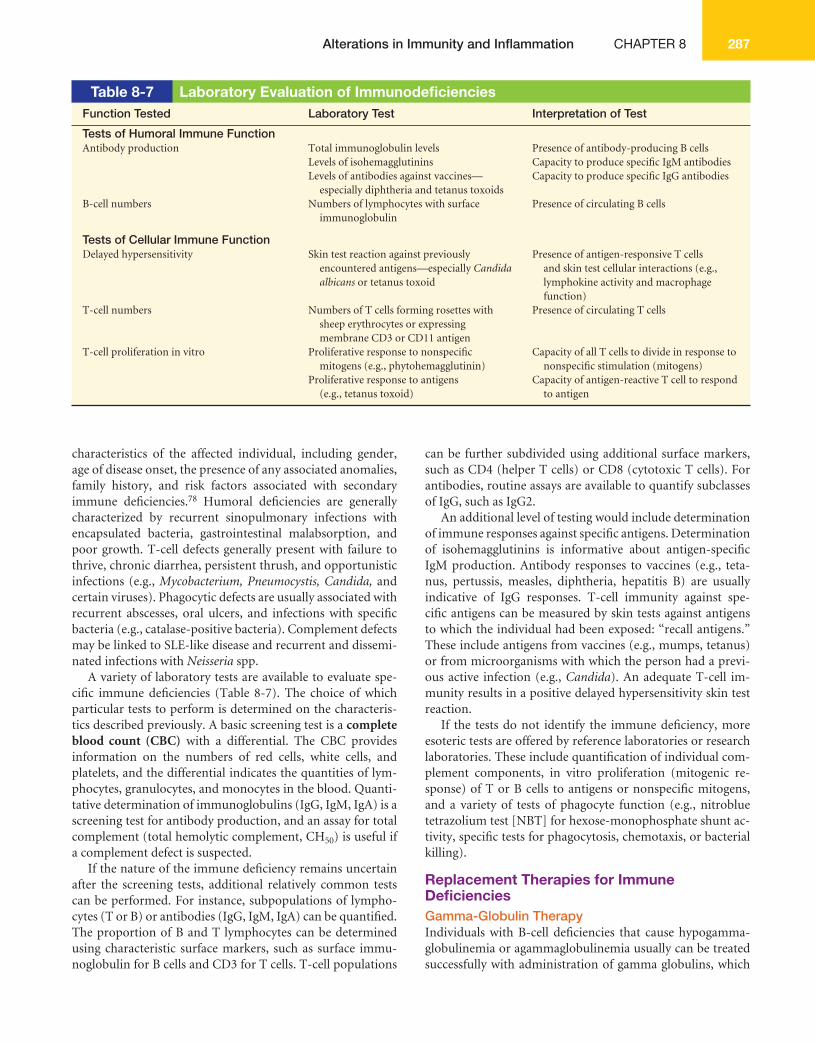
Alterations in Immunity and Infl ammation CHAPTER 8 287
characteristics of the affected individual, including gender, age of disease onset, the presence of any associated anomalies, family history, and risk factors associated with secondary immune defi ciencies. 78 Humoral defi ciencies are generally characterized by recurrent sinopulmonary infections with encapsulated bacteria, gastrointestinal malabsorption, and poor growth. T-cell defects generally present with failure to thrive, chronic diarrhea, persistent thrush, and opportunistic infections (e.g., Mycobacterium, Pneumocystis, Candida, and certain viruses). Phagocytic defects are usually associated with recurrent abscesses, oral ulcers, and infections with specifi c bacteria (e.g., catalase-positive bacteria). Complement defects may be linked to SLE-like disease and recurrent and dissemi-nated infections with Neisseria spp.
A variety of laboratory tests are available to evaluate spe-cifi c immune defi ciencies ( Table 8-7 ). The choice of which particular tests to perform is determined on the characteris-tics described previously. A basic screening test is a complete blood count (CBC) with a differential. The CBC provides information on the numbers of red cells, white cells, and platelets, and the differential indicates the quantities of lym-phocytes, granulocytes, and monocytes in the blood. Quanti-tative determination of immunoglobulins (IgG, IgM, IgA) is a screening test for antibody production, and an assay for total complement (total hemolytic complement, CH 50 ) is useful if a complement defect is suspected.
If the nature of the immune defi ciency remains uncertain after the screening tests, additional relatively common tests can be performed. For instance, subpopulations of lympho-cytes (T or B) or antibodies (IgG, IgM, IgA) can be quantifi ed. The proportion of B and T lymphocytes can be determined using characteristic surface markers, such as surface immu-noglobulin for B cells and CD3 for T cells. T-cell populations
can be further subdivided using additional surface markers, such as CD4 (helper T cells) or CD8 (cytotoxic T cells). For antibodies, routine assays are available to quantify subclasses of IgG, such as IgG2.
An additional level of testing would include determination of immune responses against specifi c antigens. Determination of isohemagglutinins is informative about antigen- specifi c IgM production. Antibody responses to vaccines (e.g., teta-nus, pertussis, measles, diphtheria, hepatitis B) are usually indicative of IgG responses. T-cell immunity against spe-cifi c antigens can be measured by skin tests against antigens to which the individual had been exposed: “recall antigens.” These include antigens from vaccines (e.g., mumps, tetanus) or from microorganisms with which the person had a previ-ous active infection (e.g., Candida ). An adequate T-cell im-munity results in a positive delayed hypersensitivity skin test reaction.
If the tests do not identify the immune defi ciency, more esoteric tests are offered by reference laboratories or research laboratories. These include quantifi cation of individual com-plement components, in vitro proliferation (mitogenic re-sponse) of T or B cells to antigens or nonspecifi c mitogens, and a variety of tests of phagocyte function (e.g., nitroblue tetrazolium test [NBT] for hexose-monophosphate shunt ac-tivity, specifi c tests for phagocytosis, chemotaxis, or bacterial killing).
Replacement Therapies for Immune Defi ciencies Gamma-Globulin Therapy Individuals with B-cell defi ciencies that cause hypogamma-globulinemia or agammaglobulinemia usually can be treated successfully with administration of gamma globulins, which
Table 8-7 Laboratory Evaluation of Immunodefi ciencies Function Tested Laboratory Test Interpretation of Test
Tests of Humoral Immune Function Antibody production Total immunoglobulin levels Presence of antibody-producing B cells
Levels of isohemagglutinins Capacity to produce specifi c IgM antibodiesLevels of antibodies against vaccines—
especially diphtheria and tetanus toxoidsCapacity to produce specifi c IgG antibodies
B-cell numbers Numbers of lymphocytes with surface immunoglobulin
Presence of circulating B cells
Tests of Cellular Immune Function Delayed hypersensitivity Skin test reaction against previously
encountered antigens—especially Candida albicans or tetanus toxoid
Presence of antigen-responsive T cells and skin test cellular interactions (e.g., lymphokine activity and macrophage function)
T-cell numbers Numbers of T cells forming rosettes with sheep erythrocytes or expressing membrane CD3 or CD11 antigen
Presence of circulating T cells
T-cell proliferation in vitro Proliferative response to nonspecifi c mitogens (e.g., phytohemagglutinin)
Capacity of all T cells to divide in response to nonspecifi c stimulation (mitogens)
Proliferative response to antigens (e.g., tetanus toxoid)
Capacity of antigen-reactive T cell to respond to antigen
UNIT III Mechanisms of Self-Defense288
are antibody-rich fractions prepared from plasma pooled from large numbers of donors. Administration of gamma globulin temporarily replaces the individual’s antibodies. An-tibodies from these preparations are removed slowly from the person’s blood, with half of the antibodies being removed by 3 to 4 weeks. Thus individuals must be treated repeatedly to maintain a protective level of antibodies in the blood.
Commercial gamma-globulin preparations are usually ad-ministered intramuscularly or by intravenous (IV) infusion. The dosage varies among individuals and is primarily deter-mined by body weight. The schedule and dosage are also de-termined according to titers of circulating immunoglobulins and the incidence of infections in the individual. Commercial gamma-globulin preparations usually contain small amounts of IgM and IgA. Individuals with selective IgA defi ciency oc-casionally develop allergic reactions to IgA in gamma- globulin preparations.
Individuals who need larger amounts of IgM or IgA can be given fresh frozen plasma in monthly IV infusions. Compli-cations associated with plasma therapy include the potential transmission of hepatitis or AIDS. The plasma is irradiated to destroy immunocompetent T cells and to avoid GVHD in individuals with accompanying T-cell defi ciencies. Adminis-tration of fresh frozen plasma is successful in individuals with WAS (IgM defi cient), AT (IgA defi cient), or complement component defi ciencies.
Transplantation and Transfusion Several primary immune defi ciencies originate from defects in lymphoid stem cells that interfere with their development in the primary lymphoid organs. Some of these (e.g., SCID, WAS, leukocyte adhesion defect) have benefi ted from replace-ment of stem cells through transplantation of bone marrow, umbilical cord cells, or other cell populations that are rich in stem cells. 79
The source of donor cells, particularly bone marrow, may contain a mixed population of stem cells and more mature T lymphocytes. In order to avoid GVHD, the preferred do-nor would be matched with the recipient for HLA antigens. Several other diseases involving depletion of the bone mar-row (i.e., aplastic anemia, leukemia requiring eradication of tumor cells in the marrow) also are treated by bone marrow transplantation. At least 75% of bone marrow transplants between individuals who are matched for HLA-A, HLA-B, HLA-C, and HLA-DR are accepted. In immunocompetent recipients, most rejections of HLA-matched transplants occur because of recognition of minor histocompatibility antigens by individuals who have received multiple blood transfusions and are, as a result, sensitized against those antigens, which are not evaluated in tissue typing. For stem cell transplants, differences in minor histocompatibility antigens may lead to GVHD. Because HLA antigens are inherited in a codominant fashion, the preferred donor would be a relative, especially a sibling. Although the donor is not tested for minor histocom-patibility antigens, the use of a close relative also would mini-mize differences at those loci.
Chronic GVHD appears in 30% to 50% of transplants be-tween HLA-matched siblings and 60% to 70% of transplants between unrelated donors. Symptoms may appear about 4 to 7 months after the transplant, but may begin much earlier or later. Depletion of T cells from bone marrow before trans-plantation signifi cantly lowers the incidence of both acute and chronic GVHD. One method of doing this is to infuse the graft with monoclonal antibody against plasma membrane antigens found only on mature T cells. Another is to use fe-tal tissue as the graft. For example, fetal liver, which contains stem cells but not immunocompetent lymphocytes, is some-times grafted in place of bone marrow if an HLA-matched donor cannot be found.
One therapy for defi ciency diseases in which the individual lacks a thymus or thymic function (e.g., DiGeorge syndrome, ataxia-telangiectasia, or chronic mucocutaneous candidia-sis) is reconstitution of thymic function. The procedure is to transplant fetal thymus tissue, which lacks immunocompe-tent T cells, or thymic epithelial cells (the cells that produce the thymic hormones) from which mature T cells have been removed. In some individuals transplantation increases the number of circulating mature T cells, but in most cases im-provement is only temporary.
Enzymatic defects that cause SCID (e.g., adenosine de-aminase defi ciency) have been treated successfully with trans-fusions of glycerol frozen-packed erythrocytes. The donor erythrocytes contain the needed enzyme and can, at least tem-porarily, provide suffi cient enzyme for normal lymphocyte function. An alternative method is administration of purifi ed adenosine deaminase that has been stabilized with polyethyl-ene glycol (PEG).
Treatment with Soluble Immune Modulators The administration of soluble materials that affect lympho-cyte function can restore T-cell function, especially in indi-viduals with WAS or chronic mucocutaneous candidiasis. Successful for some individuals is the use of transfer factor, a low-molecular-weight nucleoprotein prepared from lym-phocyte lysates, which can confer specifi c reactivity against certain antigens. Thymosin, a thymic hormone, also has been used, although with limited success. Cytokine therapy also has been effective in some cases of chronic granulomatous disease.
Gene Therapy The fi rst successful therapeutic replacement of defective genes was performed in two girls with SCID caused by an ADA de-fi ciency. 80,81 The normal gene for ADA was cloned and insert-ed into a retroviral vector. The gene for ADA replaced some retroviral genes, resulting in a virus that carried the normal human gene but did not cause disease. The virus was used to infect bone marrow stem cells from these children. The retrovirus inserted the normal ADA gene into the individu-als’ genetic material. The genetically altered stem cells were infused into the children, resulting in reconstitution of their immune systems.
Alterations in Immunity and Infl ammation CHAPTER 8 289
SUMMARY REVIEW
Hypersensitivity: Allergy, Autoimmunity, and Alloimmunity 1. Inappropriate immune responses are misdirected against
the host’s own tissues (autoimmunity); directed against benefi cial foreign tissues, such as transfusions or transplants (alloimmunity); exaggerated responses against environ-mental antigens (allergy); or insuffi cient to protect the host (immune defi ciency).
2. Allergy, autoimmunity, and alloimmunity are collectively known as hypersensitivity reactions.
3. Mechanisms of hypersensitivity are classifi ed as type I (IgE-mediated) reactions, type II (tissue-specifi c) reactions, type III (immune complex – mediated) reactions, and type IV (cell-mediated) reactions.
4. Hypersensitivity reactions can be immediate (develop-ing within minutes to a few hours) or delayed (developing within several hours or days).
5. Anaphylaxis, the most rapid immediate hypersensitivity re-action, is an explosive reaction that occurs within minutes of reexposure to the antigen and can lead to cardiovascular shock.
6. Allergens are antigens that cause allergic responses. 7. Type I (IgE-mediated) reactions are mediated through
the binding of IgE to Fc receptors on mast cells and cross- linking of IgE by antigens that bind to the Fab portions of IgE. Cross-linking causes mast cell degranulation and the release of histamine (the most potent mediator) and other infl ammatory substances.
8. Histamine enhances the chemotaxis of eosinophils into sites of type I allergic reactions
9. Atopic individuals tend to produce higher quantities of IgE and to have more Fc receptors for IgE on their mast cells.
10. Type II (tissue-specifi c) reactions are caused by fi ve possi-ble mechanisms: complement-mediated lysis, opsonization and phagocytosis, neutrophil-mediated tissue damage, an-tibody-dependent cell-mediated cytotoxicity, and modula-tion of cellular function.
11. Type III (immune complex – mediated) reactions are caused by the formation of immune complexes that are deposited in target tissues, where they activate the complement cascade, generating chemotactic fragments that attract neutrophils into the infl ammatory site. Neutrophils release lysosomal enzymes that result in tissue damage.
12. Intermediate-sized immune complexes are the most likely to have severe pathologic consequences.
13. Immune complex disease can be a systemic reaction, such as serum sickness, or localized, such as the Arthus reaction.
14. Type IV (cell-mediated) reactions are caused by either cy-totoxic T lymphocytes (Tc cells) or lymphokine-producing Th1 cells.
15. Typical allergens include pollen, molds and fungi, certain foods (milk, eggs, fi sh), animals, certain drugs, cigarette smoke, and house dust.
16. Clinical manifestations of allergic reactions usually are con-fi ned to the areas of initial intake or contact with the aller-gen. Ingested allergens induce gastrointestinal symptoms, airborne allergens induce respiratory or skin manifesta-tions, and contact allergens induce allergic responses at the site of contact.
17. Autoimmunity is a breakdown of immunologic homeostasis, the immune system’s tolerance of self-antigens. Central toler-ance develops during the embryonic period. Peripheral toler-ance is maintained in secondary lymphoid organs by regulatory T lymphocytes or antigen-presenting dendritic cells.
18. Autoimmune disease can be caused by the exposure of a previously sequestered antigen, the development of a neoantigen, the complications of infectious disease, the emergence of a forbidden clone of lymphocytes, or ineffec-tive peripheral tolerance.
19. Alloimmunity is the immune system’s reaction against anti-gens on the tissues of other members of the same species.
20. Alloimmune disorders include transient neonatal disease, in which the maternal immune system becomes sensitized against antigens expressed by the fetus, transplant rejection, and transfusion reactions, in which the immune system of the recipient of an organ transplant or blood transfusion reacts against foreign antigens on the donor’s cells.
21. SLE is a chronic, multisystem, infl ammatory disease and is one of the most serious of the autoimmune disorders. SLE is characterized by the production of a large variety of autoantibodies.
22. Hyperacute graft rejection (preexisting antibody) is imme-diate and rare, acute rejection is cell mediated and occurs days to months after transplantation, and chronic rejection is caused by infl ammatory damage to endothelial cells as a result of a weak cell-mediated reaction.
23. Red blood cell antigens may be the targets of autoimmune or alloimmune reactions. The most important of these, because they provoke the strongest humoral immune re-sponse, are the ABO and Rh systems.
Defi ciencies in Immunity 1. Disorders resulting from immune defi ciency are the clinical
sequelae of impaired function of components of the immune or infl ammatory response, phagocytes, or complement.
2. Immune defi ciency is the failure of mechanisms of self- defense to function in their normal capacity.
3. Immune defi ciencies are either congenital (primary) or acquired (secondary). Primary immune defi ciencies are caused by genetic defects that disrupt lymphocyte develop-ment, whereas secondary immune defi ciencies are second-ary to disease or other physiologic alterations.
4. The clinical hallmark of immune defi ciency is a propensity to unusual or recurrent severe infections. The type of infec-tion usually refl ects the immune system defect.
5. The most common infections in individuals with defects of cell-mediated immune response are fungal and viral, whereas infections in individuals with defects of the humoral immune response or complement function are primarily bacterial.
6. Defects in B-cell function are diverse, ranging from a complete lack of the human bursal equivalent function, the lymphoid organs required for B-cell maturation (as in Bruton’s agammaglobulinemia), to defi ciencies in a single class of immunoglobulins (e.g., selective IgA defi ciency).
7. DiGeorge syndrome (congenital thymic aplasia or hypoplasia) is characterized by complete or partial lack of the thymus (resulting in depressed T-cell immunity) and the parathyroid glands (resulting in hypocalcemia) and the presence of cardiac anomalies.
8. SCID is a total lack of T-cell function and a severe (either partial or total) lack of B-cell function. SCID can result from mutations in critical enzymes (ADA defi ciency, PNP defi ciency), in cytokine receptors (X-linked SCID, JAK3 defi ciency, IL-7 receptor defi ciency), or in antigen recep-tors (RAG-1/RAG-2 defi ciencies, CD45 defi ciency, CD3 defi ciency, ZAP-70 defi ciency). Other combined defects may result from defi ciencies in antigen-presenting molecules (bare lymphocyte syndrome), cytoskeletal proteins (WAS), or DNA repair (ataxia-telangiectasia).
UNIT III Mechanisms of Self-Defense290
9. Almost any portion of the complement cascade may be defective. The most severe defect is C3 defi ciency, which results in recurrent life-threatening bacterial infections. Defects in proteins of the membrane-attack complex usually result in unusual disseminated infections with bacteria of the Neisseria spp.
10. Defects in phagocyte function, which include insuffi cient numbers of phagocytes or defects of chemotaxis, phago-cytosis, or killing, can result in recurrent life-threatening infections such as septicemia and disseminated pyogenic lesions.
11. Acquired immunodefi ciencies are caused by superimposed conditions, such as aging, malnutrition, infections, malignancies, physical or psychologic trauma, environmental factors, some medical treatments, or other diseases.
12. Defi ciencies in immunity usually are treated by replacement therapy. Defi cient antibody production is treated by replacement of missing immunoglobulins with commercial gamma-globulin preparations. Lymphocyte defi ciencies are treated with the replacement of host lymphocytes with transplants of bone marrow, fetal liver, or fetal thymus from a donor.
SUMMARY REVIEW—cont’d
KEY TERMS
ABO blood group, XX Acute rejection, XX Adenosine deaminase (ADA) defi ciency, XX Agammaglobulinemia, XX Allergen, XX Allergy, XX Alloimmune disease, XX Alloimmunity, XX, XX Anaphylaxis, XX Antibody-dependent cell-mediated
cytotoxicity (ADCC), XX Arthus reaction, XX Ataxia-telangiectasia (AT), XX Atopic dermatitis, XX Atopic, XX Autoimmune disease, XX Autoimmunity, XX, XX Autosomal agammaglobulinemia, XX Autosomal hyper-IgM syndrome, XX Bare lymphocyte syndrome, XX Blood group antigen, XX Blocking antibody, XX B-lymphocyte defi ciency, XX Bruton’s agammaglobulinemia, XX C1 defi ciency, XX C2 defi ciency, XX C3 defi ciency, XX C4 defi ciency, XX C9 defi ciency, XX C3 receptor defi ciency, XX Chédiak-Higashi syndrome, XX Chronic granulomatous disease (CGD), XX Chronic mucocutaneous candidiasis, XX Chronic rejection, XX Combined T- and B-lymphocyte
defi ciency, XX Common variable immune defi ciency, XX
Complement defi ciency, XX Complete blood count (CBC), XX Contact dermatitis, XX Cross-reactive antibody (T cell), XX Cryoglobulin, XX Cyclic neutropenia, XX Cytotropic antibody, XX Defective class-switch, XX Delayed hypersensitivity reaction, XX Desensitization, XX DiGeorge syndrome, XX Factor H defi ciency, XX Factor I defi ciency, XX Forbidden clone, XX Graft-versus-host disease (GVHD), XX Hyperacute rejection, XX Hypersensitivity, XX Hypocomplementemic, XX Hypogammaglobulinemia, 270 IgG subclass defi ciency, XX IL-7 receptor defi ciency, XX Immediate hypersensitivity reaction, XX Immune defi ciency, XX Immunologically privileged site, XX Immunologic homeostasis, XX Isohemagglutinin, XX JAK3 defi ciency, XX Leukocyte adhesion defi ciency (LAD), XX Mannose-binding lectin (MBL)
defi ciency, XX MHC class I defi ciency, XX MHC class II defi ciency, XX Microchimerism, XX Molecular mimicry, XX Myeloperoxidase defi ciency, XX Neoantigen, XX Phagocytic defi ciency, XX
Primary (congenital) immune defi ciency, XX Properdin defi ciency, XX Purine nucleoside phosphorylase (PNP)
defi ciency, XX RAG-1 defi ciency, XX RAG-2 defi ciency, XX Raynaud phenomenon, XX Reagin, XX Reticular dysgenesis, XX Rh blood group, XX Secondary (acquired) immune
defi ciency, XX Selective IgA defi ciency, XX Serum sickness, XX Severe combined immune
defi ciency (SCID), XX Severe congenital neutropenia, XX Systemic lupus erythematosus (SLE), XX Tissue-specifi c antigen, XX T-lymphocyte defi ciency, XX Transient hypogammaglobulinemia of
infancy, XX Type I (IgE-mediated) hypersensitivity reac-
tion, XX Type II (tissue-specifi c) hypersensitivity
reaction, XX Type III (immune complex-mediated)
hypersensitivity reaction, XX Type IV (cell-mediated) hypersensitivity
reaction, XX Universal donor, XX Universal recipient, XX Urticaria (hives), XX Wheal and fl are reaction, XX Wiskott-Aldrich syndrome (WAS), XX X-linked hyper-IgM syndrome, XX X-linked SCID, XX
REFERENCES 1. Gell P G H , Coombs R R A , Lachman P T : Clinical aspects of immunology ,
Oxford, England , 1975 , Blackwell Scientifi c . 2. Simons F E R : Anaphylaxis , J Allergy Clin Immunol 121 ( 2 Suppl ) : S402 -
S407 , 2008 . 3. Portier P , Richet C : De l’action anaphylactique de certains venins ,
Comptes Rendus Societie Biologie (Paris) 54 : 170 , 1902 . 4. Roberts G : Anaphylaxis to foods , Pediatr Allergy Immunol 18 ( 6 ) : 543 -
548 , 2007 .
5. Prussin C , Metcalfe D D : IgE, mast cells, basophils, and eosinophils , J Allergy Clin Immunol 117 ( 2 Suppl ) : S450 - S456 , 2006 .
6. Saini S S , MacGlashan D : How IgE upregulates the allergic response , Curr Opin Immunol 14 ( 6 ) : 694 - 697 , 2002 .
7. Gould H J , Sutton B J : IgE in allergy and asthma today , Nature Rev Immunol 8 ( 3 ) : 205 - 217 , 2008 .
8. Robbie-Ryan M , Brown M A : The role of the mast cells in allergy and autoimmunity , Curr Opin Immunol 14 ( 6 ) : 728 - 733 , 2002 .
9. Nelson H S : Allergen immunotherapy: where is it now? J Allergy Clin Immunol 119 ( 4 ) : 769 - 779 , 2007 .
Alterations in Immunity and Infl ammation CHAPTER 8 291
10. Jutel M et al: Immune regulation by histamine , Curr Opin Immunol 14 ( 6 ) : 735 - 740 , 2002 .
11. Romi F , Gilhus N E , Aarli J A : Myasthenia gravis: clinical, immunologic, and therapeutic advances , Acta Neurol Scand 111 ( 2 ) : 134 - 141 , 2005 .
12. Rapoport B , McLachian S M : The thyrotropin receptor in Graves’ disease , Thyroid 17 ( 10 ) : 911 - 922 , 2007 .
13. Reid J R , Wheeler S F : Hyperthyroidism: diagnosis and treatment , Am Fam Physician 72 ( 4 ) : 635 - 636 , 2005 .
14. Jancar S , Crespo M S : Immune complex-mediated tissue injury: a multistep paradigm , Trends Immunol 26 ( 1 ) : 48 - 55 , 2005 .
15. Pirquet C , Schick B : Serum sickness , Leipzig, Germany , 1905 , Franz Denticke .
16. Arthus M , Breton M : Lésions cutanées produites par les injections de sérum , Comptes Rendus Societe de Biologie 55 : 817 , 1903 .
17. Blanco P et al: Cytotoxic T lymphocytes and autoimmunity , Curr Opin Rheumatol 17 ( 6 ) : 731 - 734 , 2005 .
18. Koch R : Fortsetzung der mitteilungen, ber ein heilmittel gegen tuberkulose , Deutsche Med Wochenschr 9 : 101 , 1891 .
19. Sicherer S H , Leung D Y M : Advances in allergic skin disease, anaphylaxis, and hypersensitivity reactions to foods, drugs, and insects , J Allergy Clin Immunol 119 ( 6 ) : 1462 - 1469 , 2007 .
20. Thien F C K : Drug hypersensitivity , Med J Aust 185 ( 6 ) : 333 - 338 , 2006 . 21. Geha R S : Allergy and hypersensitivity: nature versus nurture in allergy
and hypersensitivity . Curr Opin Immunol 15 ( 6 ) : 603 - 608 , 2003 . 22. Akbari O et al: Role of regulatory T cells in allergy and asthma , Curr
Opin Immunol 15 ( 6 ) : 627 - 633 , 2003 . 23. Vercelli D : Discovering susceptibility genes for asthma and allergy , Nat
Rev Immunol 8 ( 3 ) : 169 - 182 , 2008 . 24. Kinet J -P : Allergy and hypersensitivity , Curr Opin Immunol 14 ( 6 ) : 685 -
687 , 2002 . 25. Brown J M , Wilson T M , Metcalfe D D : The mast cell and allergic
diseases: role in pathogenesis and implications for therapy , Clin Exp Allergy 38 ( 1 ) : 4 - 18 , 2007 .
26. Bardana E J Jr : Occupational asthma , J Allergy Clin Immunol 121 ( 2 Suppl ) : S408 - S411 , 2008 .
27. Haydon R C : Allergic rhinitis-current approaches to skin and in vitro testing , Otolaryngol Clin North Am 41 ( 2 ) : 331 - 346 , 2008 .
28. Ahlstedt S , Murray C S : In vitro diagnosis of allergy: how to interpret IgE antibody results in clinical practice , Primary Care Resp J 15 ( 4 ) : 228 - 236 , 2006 .
29. Douglass J A , O’Hehir R E : Diagnosis, treatment and prevention of allergic disease: the basics , Med J Aust 185 ( 4 ) : 228 - 233 , 2006 .
30. Burks A W , Laubach S , Jones S M : Oral tolerance, food allergy, and immunotherapy: implications for future treatment , J Allergy Clin Immunol 121 ( 6 ) : 1344 - 1350 , 2008 .
31. Bieber T : Atopic dermatitis , N Engl J Med 358 ( 14 ) : 1483 - 1494 , 2008 . 32. Averbeck M et al: Immunologic principles of allergic disease , J Dtsch
Dermatol Ges 5 ( 11 ) : 1015 - 1028 , 2007 . 33. Schwartz R S : Shattuck lecture: diversity of the immune repertoire and
immunoregulation , N Engl J Med 348 ( 11 ) : 1017 - 1026 , 2003 . 34. Ohashi P S , DeFranco A L : Making and breaking tolerance , Curr Opin
Immunol 14 ( 6 ) : 744 - 759 , 2002 . 35. Ferguson T A , Griffi th T S : A vision of cell death: Fas ligand and immune
privilege 10 years later , Immunol Rev 213 ( 1 ) : 228 - 238 , 2006 . 36. Fourneau J M et al: The elusive case for a role of mimicry in
autoimmune diseases , Mol Immunol 40 ( 14-15 ) : 1095 - 1102 , 2004 . 37. enanzi E S , Benoist C , Mathis D : Good riddance: thymocyte clonal
deletion prevents autoimmunity , Curr Opin Immunol 16 ( 2 ) : 197 - 202 , 2004 .
38. Cunningham M W : Pathogenesis of group A streptococcal infections and their sequelae , Adv Exp Med Biol 609 : 29 - 42 , 2000 .
39. Adams K M , Nelson J L : Microchimerism: an investigative frontier in autoimmunity and transplantation , JAMA 291 ( 9 ) : 1127 - 1131 , 2004 .
40. Maloney S et al: Microchimerism of maternal origin persists into adult life . J Clin Invest 104 ( 1 ) : 41 - 47 , 1999 .
41. Nelson J L et al: Microchimerism and HLA-compatible relationships of pregnancy in scleroderma , Lancet 351 ( 9102 ) : 559 - 562 , 1998 .
42. Ohtsuka T et al: Quantitative analysis of microchimerism in systemic sclerosis skin tissue , Arch Dermatol Res 293 ( 8 ) : 387 - 391 , 2001 .
43. Artlett C M et al: Chimeric cells of maternal origin in juvenile idiopathic infl ammatory myopathies , Lancet 356 ( 9248 ) : 2155 - 2156 , 2000 .
44. Lissauer D et al: Persistence of fetal cells in the mother: friend or foe? Brit J Obstet Gynecol 114 ( 11 ) : 1321 - 1325 , 2007 .
45. Khosrotehrani K et al: Transfer of fetal cells with multilineage potential to maternal tissue , JAMA 292 ( 1 ) : 75 - 80 , 2004 .
46. Morahan G , Morel L : Genetics of autoimmune diseases in humans and in animal models , Curr Opin Immunol 14 ( 6 ) : 803 - 811 , 2002 .
47. Shmerling R H : Autoantibodies in systemic lupus erythematosus—there before you know it , N Engl J Med 349 ( 16 ) : 1499 - 1500 , 2003 .
48. Rahman A , Isenberg D A : Systemic lupus erythematosus , N Engl J Med 358 ( 9 ) : 929 - 939 , 2008 .
49. Riemekasten G , Hahn B H : Key autoantigens in SLE , Rheumatol 44 ( 8 ) : 975 - 982 , 2005 .
50. Perdriger A , Werner-Leyval S , Rollot-Elamrani K : The genetic basis for systemic lupus erythematosus , Joint Bone Spine 70 ( 2 ) : 103 - 108 , 2003 .
51. Arbuckle M R et al: Development of autoantibodies before the clinical onset of systemic lupus erythematosus , N Engl J Med 349 ( 16 ) : 1526 - 1533 , 2003 .
52. American College of Rheumatology 2004 systemic lupus erythematosus. Available at www.rheumatology.org/public/factsheets/lupus.asp?aud=pat#4 .
53. Burt R K et al: Clinical applications of blood-derived and marrow-derived stem cells for nonmalignant diseases , JAMA 299 ( 8 ) : 925 - 936 , 2008 .
54. Nydegger U E et al: Histo-blood group antigens as allo- and autoantigens , Ann N Y Acad Sci 1050 : 40 - 51 , 2005 .
55. Westhoff C M : The structure and function of the Rh antigen complex , Semin Hematol 44 ( 1 ) : 42 - 50 , 2007 .
56. Sheldon S , Poulton K : HLA typing and its infl uence on organ transplantation , Methods Mol Biol 333 : 157 - 174 , 2006 .
57. Truong L D et al: Acute antibody-mediated rejection of renal transplant: pathogenetic and diagnostic considerations , Arch Pathol Lab Med 131 ( 8 ) : 1200 - 1208 , 2007 .
58. Cooper M A , Pommering T L , Koranyi K : Primary immunodefi ciencies , Am Fam Physician 68 ( 10 ) : 2001 - 2008 , 2003 .
59. Fischer A : Human primary immunodefi ciency diseases , Immunity 27 ( 12 ) : 835 - 845 , 2007 .
60. Fleisher T A : Primary immune defi ciencies: windows into the immune system , Pediatr Rev 27 ( 10 ) : 363 - 372 , 2006 .
61. Verbsky J W , Grossman W J : Cellular and genetic basis of primary immune defi ciencies , Pediatr Clin North Am 53 ( 4 ) : 649 - 684 , 2006 .
62. Primary Immunodefi ciency. National Institute of Child Health and Human Development, NIH, DHHS, Washington D.C., Government Printing Offi ce. Available at www.nichd.nih.gov/publications/pubs/primary_immuo.cfm , updated 04/07/2008.
63. Simonte S J , Cunningham-Rundles C : Update on primary immunodefi ciency: defects of lymphocytes , Clin Immunol 109 ( 2 ) : 109 - 118 , 2003 .
64. Cunningham-Rundles C , Ponda P P : Molecular defects in T- and B-cell primary immunodefi ciency diseases , Nature Rev Immunol 5 ( 11 ) : 880 - 892 , 2005 .
65. Bruton O C : Agammaglobulinemia . Pediatrics 9 ( 6 ) : 722 - 728 , 1952 . 66. Notarangelo L D et al: Defects in class-switch recombination , J Allergy
Clin Immunol 117 ( 4 ) : 855 - 864 , 2006 . 67. Demczuk S , Aurias A : DiGeorge syndrome and related syndromes
associated with 22q11.2 deletions , Annales Genetique 38 : 59 , 1995 . 68. DiGeorge A M : Congenital absence of the thymus and its immunologic
consequences . In Bergsma D , McKusick F A , editors: Immunologic defi ciency diseases in man, National Foundation—March of Dimes original article series , Baltimore , 1968 , Williams & Wilkins .
69. Fischer A : Have we seen the last variant of severe combined immunodefi ciency? N Engl J Med 349 ( 19 ) : 1789 - 1792 , 2003 .
70. Notarangelo L D , Miao C H , Ochs H D : Wiskott-Aldrich syndrome , Curr Opin Hematol 15 ( 1 ) : 30 - 36 , 2008 .
71. Sjöholm A G et al: Complement defi ciency and disease: an update , Mol Immunol 43 ( 1-2 ) : 78 - 85 , 2006 .
72. Lewis M J , Botto M : Complement defi ciencies in humans and animals: links to autoimmunity , Autoimmunity 39 ( 5 ) : 367 - 378 , 2006 .
73. Smith C W : Adhesion molecules and receptors , J Allergy Clin Immunol 121 ( 2 Suppl ) : S375 - S379 , 2008 .
74. Chinen J , Shearer W T : Secondary immunodefi ciencies, including HIV infection , J Allergy Clin Immunol 121 ( 2 Suppl ) : S388 - S392 , 2008 .
75. Schroder A K , Rink L : Neutrophil immunity of the elderly , Mech Ageing Dev 124 ( 4 ) : 419 - 425 , 2003 .
76. Hakim F T et al: Aging, immunity and cancer , Curr Opin Immunol 16 ( 2 ) : 151 - 156 , 2004 .

UNIT III Mechanisms of Self-Defense292
77. Bhushan V , Collins R H Jr : Chronic graft-vs-host disease , JAMA 290 ( 19 ) : 2599 - 2603 , 2003 .
78. Buckley R H : Primary immunodefi ciency or not? Making the correct diagnosis , J Allergy Clin Immunol 117 ( 4 ) : 756 - 758 , 2006 .
79. Buckley R H : Molecular defects in human severe combined immunodefi ciency and approaches to immune reconstitution , Ann Rev Immunol 22 ( 1 ) : 625 - 655 , 2004 .
80. Blaese R M : Development of gene therapy for immunodefi ciency: adenosine deaminase defi ciency , Pediatr Res 33 ( 1 Suppl ) : S49 - S53 , 1993 .
81. Onodera M et al: Gene therapy for severe combined immunodefi ciency caused by adenosine deaminase defi ciency: improved retroviral vectors for clinical trials , Acta Haematol 101 ( 2 ) : 89 - 96 , 1999 .
Related Documents