Akhileshwari Nath et al JMSCR Volume 3 Issue 1 January 2015 Page 3946 JMSCR Volume||03||Issue||01||Page 3946-3955||January 2015 Alteration of Gene Expression Due to Arsenic Induced Oxidative Stress Leading to Ovarian Cancer Authors Akhileshwari Nath 1 , Aseem Kumar Anshu 1 , Priyanka 1 , Simran Kriti 3 , Shailendra Kumar 1 , Rittika Pandey 4 , J.K Singh 2 , Richa Chauhan 2 , and Manisha Singh 2 1 Research Center, Mahavir Cancer Institute & Research Center, Patna, Bihar 2 Department of Oncology, Mahavir Cancer Institute & Research center, Panta, Bihar 3 Jaccob School of Cellular and Molecular Biology, SHIATS, Allahabad, U.P 4 Department of Zoology, S.K.M University, Dumaka, Bihar Corresponding Author Akhileshwari Nath Research Center, Mahavir Cancer Institute, Patna Email:[email protected] Abstract Epidemiological studies and preventive measures for cancer have engaged most of the scientists and doctors for several decades and the research has come forth a long way now. Several heavy metals like arsenic have been linked with different types of cancer, one of which is ovarian cancer (Oca). Higher mortality rate of Oca signifies the gravity of the case. Moreover, arsenic is also believed to invoke generation of free radical and the consequences are even exacerbated. Early diagnosis of Oca is presently quite feeble and CA-125 alone seems to be insufficient and capricious. However, CA-125 along with other diagnostic parameter satisfies most of the pathologists and clinicians. For determination of oxidative stress, Malondialdehyde (MDA) was chosen as the marker and arsenic estimation was performed with the help of Atomic Absorption Spectrometer (AAS). RBC count, Heamoglobin, SGPT, SGOT and Alkaline phosphatase levels were performed according to standard protocol. Higher level of arsenic increases MDA level, decreases CA-125 level, decreases RBC and Haemoglobin level and increases SGPT, SGOT, ALP levels and vice versa for no arsenic in the Oca patients with significance <0.0001, <0.001, <0.005, <0.020, <0.001, >0.05, and <0.002 respectively. Concludingly, arsenic is one of the major causative agents for oxidative stress and hence leading to cancer including Oca. CA-125 was estimated to be very higher in Oca patients than normal ones and RBC count, Heamoglobin, SGPT, SGOT, ALP levels also seemed to be varied from normal. Keywords- Arsenic, MDA, CA-125, RBC, WBC. www.jmscr.igmpublication.org Impact Factor 3.79 ISSN (e)-2347-176x

Welcome message from author
This document is posted to help you gain knowledge. Please leave a comment to let me know what you think about it! Share it to your friends and learn new things together.
Transcript
Akhileshwari Nath et al JMSCR Volume 3 Issue 1 January 2015 Page 3946
JMSCR Volume||03||Issue||01||Page 3946-3955||January 2015
Alteration of Gene Expression Due to Arsenic Induced Oxidative Stress
Leading to Ovarian Cancer
Authors
Akhileshwari Nath1, Aseem Kumar Anshu
1, Priyanka
1, Simran Kriti
3, Shailendra
Kumar1, Rittika Pandey
4, J.K Singh
2, Richa Chauhan
2, and Manisha Singh
2
1Research Center, Mahavir Cancer Institute & Research Center, Patna, Bihar
2Department of Oncology, Mahavir Cancer Institute & Research center, Panta, Bihar
3Jaccob School of Cellular and Molecular Biology, SHIATS, Allahabad, U.P
4Department of Zoology, S.K.M University, Dumaka, Bihar
Corresponding Author
Akhileshwari Nath
Research Center, Mahavir Cancer Institute, Patna
Email:[email protected]
Abstract
Epidemiological studies and preventive measures for cancer have engaged most of the scientists and
doctors for several decades and the research has come forth a long way now. Several heavy metals like
arsenic have been linked with different types of cancer, one of which is ovarian cancer (Oca). Higher
mortality rate of Oca signifies the gravity of the case. Moreover, arsenic is also believed to invoke
generation of free radical and the consequences are even exacerbated. Early diagnosis of Oca is presently
quite feeble and CA-125 alone seems to be insufficient and capricious. However, CA-125 along with other
diagnostic parameter satisfies most of the pathologists and clinicians. For determination of oxidative
stress, Malondialdehyde (MDA) was chosen as the marker and arsenic estimation was performed with the
help of Atomic Absorption Spectrometer (AAS). RBC count, Heamoglobin, SGPT, SGOT and Alkaline
phosphatase levels were performed according to standard protocol. Higher level of arsenic increases MDA
level, decreases CA-125 level, decreases RBC and Haemoglobin level and increases SGPT, SGOT, ALP
levels and vice versa for no arsenic in the Oca patients with significance <0.0001, <0.001, <0.005, <0.020,
<0.001, >0.05, and <0.002 respectively. Concludingly, arsenic is one of the major causative agents for
oxidative stress and hence leading to cancer including Oca. CA-125 was estimated to be very higher in Oca
patients than normal ones and RBC count, Heamoglobin, SGPT, SGOT, ALP levels also seemed to be
varied from normal.
Keywords- Arsenic, MDA, CA-125, RBC, WBC.
www.jmscr.igmpublication.org Impact Factor 3.79
ISSN (e)-2347-176x
Akhileshwari Nath et al JMSCR Volume 3 Issue 1 January 2015 Page 3947
JMSCR Volume||03||Issue||01||Page 3946-3955||January 2015
1. INTRODUCTION
Deadliness of ovarian cancer is now evident as it
ranks fifth in cancer deaths among women. It has
been estimated that risk of conceiving ovarian
cancer during a woman’s life is about 1 in 73 and
her chance of dying of ovarian cancer is about 1 in
100. Ovarian cancer has been classified on the
basis of its origin: (a) surface epithelium derived
from either the coelomic epithelium or ectopic
endometrial epithelium, (b) germ cells, which
migrate unto the ovary from yolk sac and are
totipotential, and (c) the stroma of the ovary,
which include sex chords. Most primary types of
neoplasm in the ovary are categorized under the
tumour of mṻllerian epithelium. The three major
types of tumours of epithelial origin are serous,
mucinous, and endometroid.
CA-125 is a transmembrane glycoprotein
embedded on plasma membrane of cells that are
transformed to metaplastic to give rise to
mṻllerian epithelium [1]
. CA-125 is still the most
extensive studied biomarker and adopted
worldwide for early detection of Oca [2]
. But on
the other hand, elevated level of soluble CA-125
has been detected in other malignant cancers such
as breast cancer [3]
, mesothelioma, non-hodgkin
lymphoma, gastric cancer, and in other benign
conditions too, such as endometriosis [4]
,
pregnancy, ovulatory cycle, liver diseases and
congenital heart failure as well as infectious
diseases like tuberculosis.
Lipid peroxidation is perpetually occurring in cells
as normal cellular metabolism. LPO is one of the
most extensively studied consequences of
Reactive Oxygen Species (ROS), perturbing the
integrity of the plasma membrane and the function
of the cell. Polyunsaturated Fatty Acids (PUFAs)
are extremely susceptible to peroxidation. The
inkling that lipid peroxides and ROS participate in
inextricable signal transduction cascade
responsible for the control of the cell proliferation,
and the induction of differentiation, maturation
and apoptosis has been corroborated by several
literature [5]
. LPO mediated oxidative stress can
sabotage major cellular components and function
like DNA strand breakage, rises in intracellular
free ca2+ , damage to membrane ion transporters
and/or specific proteins [6]
. MDA, an aldehyde
product of LPO has been established to possess
mutagenic and carcinogenic effect. It appears to
bind with bases dG, dC and dA to build m1G,
m1C and m1A respectively [7]
.
Arsenic sits at 33rd
position on the periodic table
of chemical elements and it is categorized as a
metalloid, however it is most frequently referred
as heavy metal as in context of toxicology [8]
. A
number of results provide evidences that toxic and
carcinogenic metals like arsenic has capabilities to
bind with different nuclear proteins and DNA
causing oxidative deterioration of biological
macromolecules. Arsenic exposure has been
established to be one of the factors for generation
of ROS [9]
. Accumulation of arsenic in greater
quantity in the body has debilitating impact on
overall human health and has also been implicated
in cancer.
In our current investigation, correlation between
MDA, CA-125, Haemoglobin levels and RBC
count have been attempted to establish and

Akhileshwari Nath et al JMSCR Volume 3 Issue 1 January 2015 Page 3948
JMSCR Volume||03||Issue||01||Page 3946-3955||January 2015
besides that, alkaline phosphate, SGOT, and
SGPT levels provides panoptic view to this study.
2. MATERIALS and METHODS
The blood samples were collected intravenously
from the 110 ovarian cancer patients who came
for the treatment in Mahavir Cancer Institute and
Research Center, patna and 48 healthy women of
comparable age group, with their consent. Part of
blood was used for RBC count, estimation of
Haemoglobin level and arsenic concentration and
serum was prepared from rest of blood for the
analyses of SGPT, SGOT, Alkaline Phosphotase,
and MDA levels.
2.1 Haematological study
RBC count and Haemoglobin level were
performed by standard protocol using Cell
Counter (Medonic M- Series) model no- 18253.
2.2 MDA Assessment
Serum of OCa patients was prepared by
centrifuging blood sample at 3000g for 10
minutes. LPO assay was performed by a standard
protocol with slight modification [10].
2.3 ALP, SGPT and SGOT
Serum was used for liver function test which
included ALP, SGOT, and SGPT. Liver function
test was analysed by auto-analyzer (Vitalab-
selectra pro XL) model no- 8-7136.
2.4 CA-125 assay
CA-125 was estimated from the serum with the
help of ELISA kit method.
2.5 Estimation of Arsenic
1ml of blood was transferred to EDTA vials of
randomly selected 24 out of 110 ovarian cancer
patients for estimation of arsenic concentration.
Further, sample preparation and estimation of
arsenic was done by standard protocol of Atomic
Absorption Spectrometer graphite flame (Perkin
Elmer) model number 900T.
2.6 Statistical analysis
Mean±SD and p-value were obtained using SPSS
software (statistical package for social sciences,
version 16.0). A p-value less than 0.05 were
considered significant.
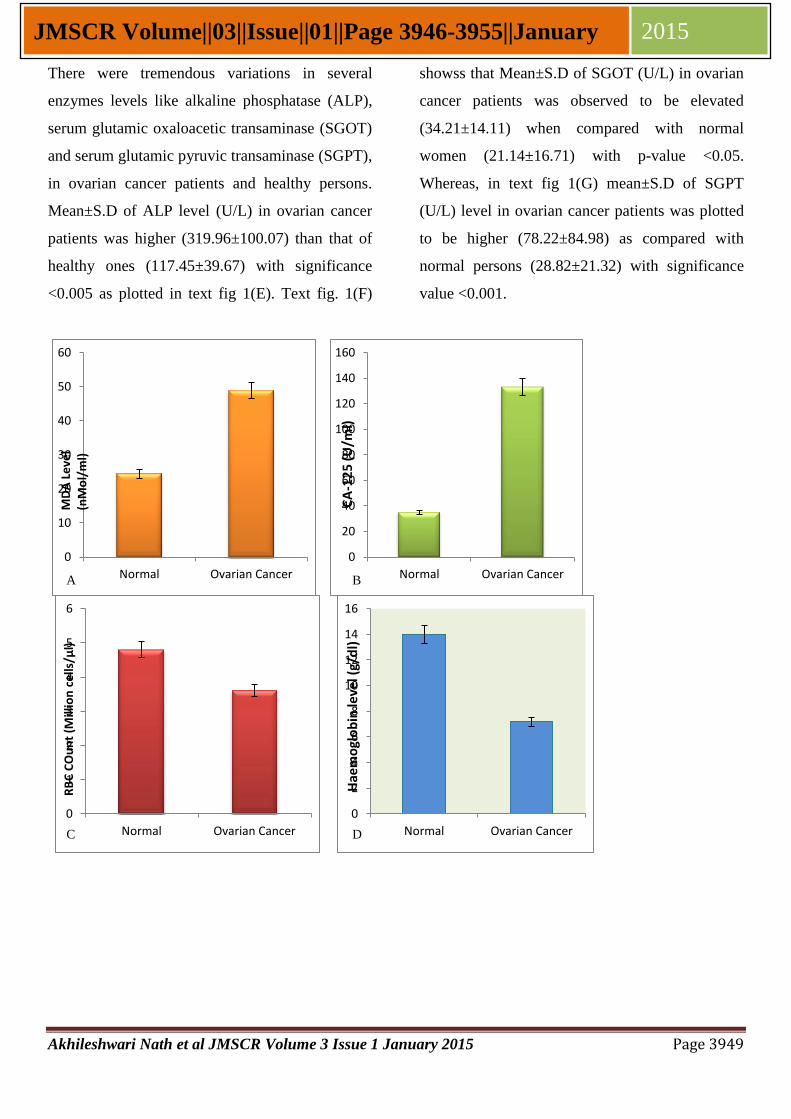
3. RESULTS
As per calculations, Mean±S.D of MDA level
(nMol/ml) in ovarian cancer patients and normal
persons was found to be 48.87±11.97 and
24.39±13.39, respectively with p-value <0.0001.
Text fig 1(A) demonstrates the comparative hike
in MDA level in ovarian cancer patients than
normal persons. Conventionally, CA-125 level in
ovarian cancer patients is higher than normal
persons, espousing quite an orthodox pattern.
Mean±S.D of CA-125 (U/ml) in ovarian cancer
patients was 133.11±293 and that of normal
persons was 31.81±9.56 with significance level
<0.0024, as depicted in text fig 1(B).
On the other hand, mean±S.D of haemoglobin
level (g/dl) in ovarian cancer patients (7.17±3.75)
was obtrusively lower than healthy women
(13.106±1.98) with p-value <0.001 as shown in
text fig 1(C). Whereas red blood cells count in
ovarian cancer patients and healthy women
yielded noticeable difference as seen in text fig
1(D). Mean±S.D of RBC count in ovarian cancer
and normal women was calculated to be 3.6±0.82
and 6.1±1.7, respectively which has significance
value <0.001.
Akhileshwari Nath et al JMSCR Volume 3 Issue 1 January 2015 Page 3949
JMSCR Volume||03||Issue||01||Page 3946-3955||January 2015
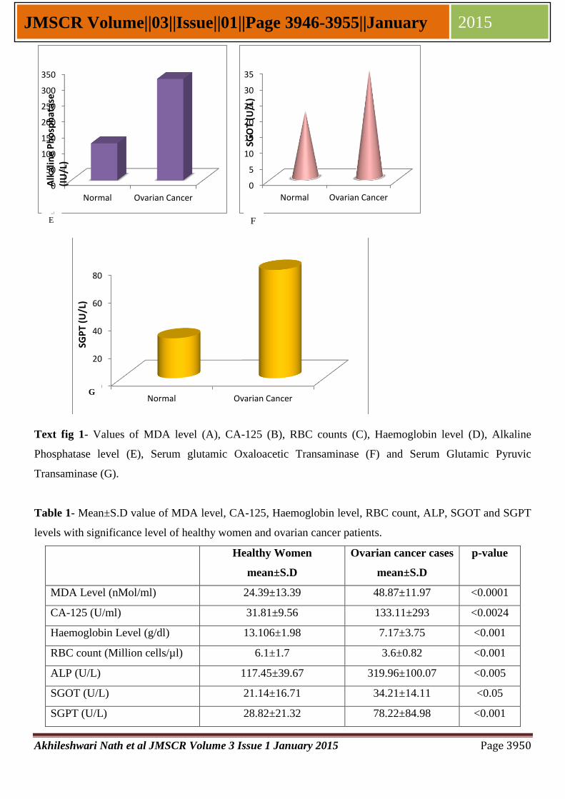
There were tremendous variations in several
enzymes levels like alkaline phosphatase (ALP),
serum glutamic oxaloacetic transaminase (SGOT)
and serum glutamic pyruvic transaminase (SGPT),
in ovarian cancer patients and healthy persons.
Mean±S.D of ALP level (U/L) in ovarian cancer
patients was higher (319.96±100.07) than that of
healthy ones (117.45±39.67) with significance
<0.005 as plotted in text fig 1(E). Text fig. 1(F)
showss that Mean±S.D of SGOT (U/L) in ovarian
cancer patients was observed to be elevated
(34.21±14.11) when compared with normal
women (21.14±16.71) with p-value <0.05.
Whereas, in text fig 1(G) mean±S.D of SGPT
(U/L) level in ovarian cancer patients was plotted
to be higher (78.22±84.98) as compared with
normal persons (28.82±21.32) with significance
value <0.001.
0
10
20
30
40
50
60
Normal Ovarian Cancer
MD
A L
eve
l (n
Mo
l/m
l)
0
20
40
60
80
100
120
140
160
Normal Ovarian Cancer
CA
-12
5 (U
/ml)
0
1
2
3
4
5
6
Normal Ovarian Cancer
RB
C C
Ou
nt
(Mill
ion
ce
lls/µ
l)
0
2
4
6
8
10
12
14
16
Normal Ovarian Cancer
Hae
mo
glo
bin
leve
l (g/
dl)
A B
C D
Akhileshwari Nath et al JMSCR Volume 3 Issue 1 January 2015 Page 3950
JMSCR Volume||03||Issue||01||Page 3946-3955||January 2015
Text fig 1- Values of MDA level (A), CA-125 (B), RBC counts (C), Haemoglobin level (D), Alkaline
Phosphatase level (E), Serum glutamic Oxaloacetic Transaminase (F) and Serum Glutamic Pyruvic
Transaminase (G).
Table 1- Mean±S.D value of MDA level, CA-125, Haemoglobin level, RBC count, ALP, SGOT and SGPT
levels with significance level of healthy women and ovarian cancer patients.
Healthy Women
mean±S.D
Ovarian cancer cases
mean±S.D
p-value
MDA Level (nMol/ml) 24.39±13.39 48.87±11.97 <0.0001
CA-125 (U/ml) 31.81±9.56 133.11±293 <0.0024
Haemoglobin Level (g/dl) 13.106±1.98 7.17±3.75 <0.001
RBC count (Million cells/µl) 6.1±1.7 3.6±0.82 <0.001
ALP (U/L) 117.45±39.67 319.96±100.07 <0.005
SGOT (U/L) 21.14±16.71 34.21±14.11 <0.05
SGPT (U/L) 28.82±21.32 78.22±84.98 <0.001
0
50
100
150
200
250
300
350
Normal Ovarian Cancer
Alk
alin
e P
ho
sph
atas
e (I
U/L
)
0
5
10
15
20
25
30
35
Normal Ovarian Cancer
SGO
T (U
/L)
0
20
40
60
80
Normal Ovarian Cancer
SGP
T (
U/L
)
E F
G
Akhileshwari Nath et al JMSCR Volume 3 Issue 1 January 2015 Page 3951
JMSCR Volume||03||Issue||01||Page 3946-3955||January 2015
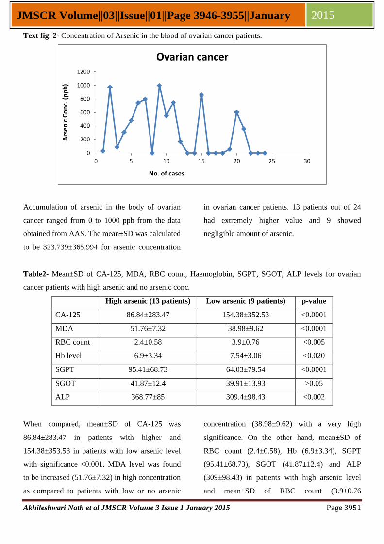
Text fig. 2- Concentration of Arsenic in the blood of ovarian cancer patients.
Accumulation of arsenic in the body of ovarian
cancer ranged from 0 to 1000 ppb from the data
obtained from AAS. The mean±SD was calculated
to be 323.739±365.994 for arsenic concentration
in ovarian cancer patients. 13 patients out of 24
had extremely higher value and 9 showed
negligible amount of arsenic.
Table2- Mean±SD of CA-125, MDA, RBC count, Haemoglobin, SGPT, SGOT, ALP levels for ovarian
cancer patients with high arsenic and no arsenic conc.
High arsenic (13 patients) Low arsenic (9 patients) p-value
CA-125 86.84±283.47 154.38±352.53 <0.0001
MDA 51.76±7.32 38.98±9.62 <0.0001
RBC count 2.4±0.58 3.9±0.76 <0.005
Hb level 6.9±3.34 7.54±3.06 <0.020
SGPT 95.41±68.73 64.03±79.54 <0.0001
SGOT 41.87±12.4 39.91±13.93 >0.05
ALP 368.77±85 309.4±98.43 <0.002
When compared, mean±SD of CA-125 was
86.84±283.47 in patients with higher and
154.38±353.53 in patients with low arsenic level
with significance <0.001. MDA level was found
to be increased (51.76±7.32) in high concentration
as compared to patients with low or no arsenic
concentration (38.98±9.62) with a very high
significance. On the other hand, mean±SD of
RBC count (2.4±0.58), Hb (6.9±3.34), SGPT
(95.41±68.73), SGOT (41.87±12.4) and ALP
(309±98.43) in patients with high arsenic level
and mean±SD of RBC count (3.9±0.76
0
200
400
600
800
1000
1200
0 5 10 15 20 25 30
Ovarian cancer A
rsen
ic C
on
c. (
pp
b)
No. of cases

Akhileshwari Nath et al JMSCR Volume 3 Issue 1 January 2015 Page 3952
JMSCR Volume||03||Issue||01||Page 3946-3955||January 2015
thousand/dl), Hb (7.54±3.06 g/dl), SGPT
(64.03±79.54), SGOT (39.91±13.93), and ALP
(309.4±98.43) in patients without arsenic level in
blood with significance <0.005, <0.020, <0.0001,
>0.05, <0.002 respectively.
4. DISCUSSION
Numerous environmental agents have been
inscribed to cause toxicity and cancer in body.
Arsenic has been indicated to be major causative
agent for oxidative stress. Inorganic arsenic
includes arsenite [As(III)] and arsenate [As(V)]
and can be in methylated form, either as
monomethylarsonic acid [MMA(V)] or
dimethylearsinic acid [DMA(V)]. Both in vitro
and in vivo studies of arsenic exposed animals and
humans have suggested involvement of higher
production of peoxyl radicals (ROO∙), superoxide
anion radicals (O2-∙), hydroxyl radical (OH
∙),
hydrogen peroxide (H2O2), dimethylarsenic
radical [(CH3)2As∙] and non-protein sulfhydryl and
oxidant induced DNA damage [11]
. Oxidative
stress has been found to be elevated in almost all
metabolic disorders and related as one of the
causative agent for cancer development. Most of
the ovarian cancer is derived from epithelial cells
and breast cancer of epithelial origin has been
asserted to produce abnormally high MDA [12]
.
Increased concentration of self-generated ROS in
the cells undermines the endurance of the plasma
membrane resulting into apoptosis inadvertently.
Such an endogenous stimulus is intensified not
only in several diseases [13]
and cancer but also
during the toxicity caused by accumulation of
pesticides [14]
and arsenic. MDA level almost
doubles in ovarian cancer patients when compared
with healthy persons as apparent in text fig 1A.
MDA is generated as the by-product of the lipid
peroxidation process, acts as mutagen and
possesses the capacity of activating proto-
oncogenes or tumour suppressor genes leading to
cancer.
Being the member of mucin family, glycoprotein,
CA-125 is usually embedded in plasma membrane
but due to neoplasm development in epithelial
ovarian cancer, it is relieved from the cell surface
by proteolytic action of certain enzyme.
Concentration of MUC16 increases in peripheral
blood due to which it acts as prognostic marker
for ovarian cancer. According to another
hypothesis, the release of CA-125 from the
surface of cell may also be due to loss of fluidity
of plasma membrane which abets escaping of this
transmembrane glycoprotein [15]
. As reflected by
text fig 1B, CA-125 is too high in ovarian cancer
patients as compared to healthy people which
affirm CA-125 as specific and sensitive marker
for early detection of ovarian cancer. Patients with
higher concentration of arsenic in their body
resulted with higher MDA and lower CA-125
level than patients with no arsenic level as shown
in table 02.
Anaemia in cancer is quite usual which can be
treated with re-oxygenation or erythropoietin
doses which are indicated to provide improved
survival rate in cancer [16]
. Kidney begins
manufacturing more erythropoietin on signal
received at the time of hypoxia condition. Low
level of haemoglobin and RBC count in blood
signifies that erythropoietin is hypoactive which
Akhileshwari Nath et al JMSCR Volume 3 Issue 1 January 2015 Page 3953
JMSCR Volume||03||Issue||01||Page 3946-3955||January 2015
means oxygen supply throughout the body is
sufficient. Higher the oxygen-supply to the organ,
greater the production of superoxides in the cell,
mainly due to incomplete electron transfer or
reduction of oxygen by damaged mitochondria.
Oxidative stress is observed to be higher whereas
RBC count and Hb level to be lower in Oca
patients, indicating increased oxidative stress
results in lower RBC count. Furthermore, patients
with higher arsenic level show higher MDA level
and lower RBC count as compared to patients
with no arsenic level, which signifies arsenic and
oxidative stress together have impact of RBC
count and haemoglobin level. It is possible that
arsenic and oxidative stress induce RBC death. In
addition, arsenic has binding affinity with
haemoglobin at its cys-rich area which might
affect Hb level.
In addition, Alkaline Phosphatase, serum glutamic
oxaloacetic transaminase and serum glutamic
pryruvic transaminase are made exclusively by
hepatocytes and released into blood stream. When
lever secretes SGOT and SGPT when it is chafed
or inflamed and releases into blood sream where it
can be measured. ALP, SGOT and SGPT can be
descried to be produced enormously. The reason
behind this might be attributed to the toxicity level
caused by several intrinsic and extrinsic factors.
Several extrinsic factors like arsenite, pesticides
and other toxic agents have been indicated to
cause discrepancies related to livers in animal
model [17]
. Liver damage due to oxidative stress,
apoptosis, necrosis, histological manifestations
which are intrinsic factors, would intervene with
normal function of hepatocytes. The increase in
ALP, SGOT and SGPT in serum is due to necrosis
in hepatocytes, which increases increase in
permeability of the cell membrane resulting in
secretion of transaminases in blood stream [18]
.
Liver cells are irritated by administration of
several drugs, as they all have more or less
negative effects on hepatocytes. SGPT, SGOT,
and ALP can be analysed to be higher in the
patients with higher arsenic level than patients
with nil arsenic, which further aids that arsenic
significantly affects the hepatocytes.
However oxidative stress is directly related to
necrosis and apoptosis which manifests several
malfunctions in the body but more study is needed
to establish a vital relationship which would help
develop better resolution for early detection of
ovarian cancer.
5. CONCLUSIONS
All such parameters have been evaluated and
studied extensively to establish correlation
between arsenic and oxidative stress, MUC16,
RBC count and Haemoglobin level and liver
function tests in ovarian cancer patients. It was
found that high level of arsenic increased
oxidative stress in the patients which led to high
MDA levels in such cases. Further, patients with
high arsenic level had a relatively lower level of
CA-125 as compared to ovarian cancer patients
with low or no arsenic on their blood. The above
findings suggest that the level of CA-125 in
patients with high level of arsenic in their blood
could be lower than that expected. However, this
needs further corroboration from trials with large
number of patients.

Akhileshwari Nath et al JMSCR Volume 3 Issue 1 January 2015 Page 3954
JMSCR Volume||03||Issue||01||Page 3946-3955||January 2015
ACKNOWLEDGEMENT
The authors express deep debt of gratitude to the
department of science and technology
(DST/SSTP/LSR division), Ministry of science
and Technology, Government of India for
financial support to Mahavir Cancer Institute and
Research Center, Patna for providing
infrastructure facilities and permission for doing
the research work.
REFERENCES
1. freeley KM, Wells M. Precurssor lesions
of ovarian epithelial malignancy.
Histopathology. 2001: 38:87-95.
2. Jacobs IJ, Menon U. Progress and
challenges in screening for early detection
of ovarian cancer. Mol Cell Proteomics.
2004: 3:355-366.
3. Norum LF, Erikstein B, Nustad K.
Elevated CA-125 in breast cancer-A sign
of advanced disease. Tumour Biol. 2001:
22:223-228.
4. Kitwaki J, Ishihara H, Koshiba H, et al.
Usefulness and limits of CA-125 in
diagnosis of without associated ovarian
endometriomas. Hum Reprod. 2005:
20:1990-2003.
5. Cejas P, Casado E, Belda-Iniesta C, De
Castro J, Espinosa E, Redondo A, Sereno
M, Garcia-cabezas MA, Vara JA,
Dominguez-Caceres A, et al. Implication
of oxidative stress and cell membrane lipid
peroxidation in human cancer (spain).
Cancer Causes Control. 2004: 25:707-
719.
6. Halliwell B, Auroma OI. DNA damage by
oxygen-derived species: its mechanism
and measurement in mammalian systems.
FEBS Lett. 1991: 281: 9-19.
7. Marnett, LJ. Lipid peroxidation – DNA
damage by malondialdehyde. Mut Res
Fund Mol Mech Mutagen. 1999: 424: 83–
95.
8. Mandal BK, Suzuki KT. Arsenic round the
world: a review. Talanta. 2002: 58: 201–
235.
9. Mascher R, Lippmann B, Holzinger S,
Bergmann H. Arsenate toxicity: effects on
oxidative stress response molecules and
enzymes in red clover plants. Plant Sci.
2002: 163: 961–969.
10. Ohkawa H, Ohishi N, Yagi K. Assay for
lipid peroxides in animal tissues by
thiobarbituric acid reaction. Anal
Biochem. 1979: 95: 351-8.
11. Flora SJS, Bhadauria S, Kannan GM,
Singh N. Arsenic induced oxidative stress
and the role of antioxidant
supplementation during chelation: a
review. J. Environ. Biol. 2007: 28: 333–
347.
12. Nath A, Priya P, Anshu AK, Priyanka,
Singh M, Chauhan R, Singh JK and Roy
SP, Escalated oxidative stress and estrogen
level stimulate invasion of tumor cells in
infiltrating ductal carcinoma, IJPSRR,
2014: 29(2):
13. Spiteller G. Are lipid peroxidation
processes induced by changes in the cell
wall structure and how are these processes
Akhileshwari Nath et al JMSCR Volume 3 Issue 1 January 2015 Page 3955
JMSCR Volume||03||Issue||01||Page 3946-3955||January 2015
connected with diseases? Med
Hypotheses. 2003 : 60(1): 69-83.
14. Nath A, Singh JK, Kumari P, Anshu AK,
Priyanka, Kumar S, Singh CK & Ojha J.
Synergistic effect of accumulated
chlorpyrifos and raised levels of MDA and
oestrogen induced ovarian cancer
progression, IOSR-JPBS, 2014: 9 (5) Ver.
IV: 55-63.
15. Nath A, Suman S, Anshu AK, Priyanka,
Sinha R, Mishra M, kumar S, Singh CK
and Singh JK. TBARS Assay and
Hematological Parameters in Relation to
Breast Cancer. J. Ecophysiol. Occup.
Hlth., 2014: 14(3&4): 110-116, 2014.
16. Bookemeyer C, Oechsle K, Hartmann JT.
Treatment-induced anemia and its
potential clinical impact in patients
receiving sequential high dose
chemotherapy for metastatic testicular
cancer. Br J Cancer. 2002: 87: 1066-1071.
17. Sharma A, Sharma MK, Kumar M.
Protective effect of menthe piperita against
arsenic-induced toxicity in liver of swiss
albino mice, Basic & Clinical
Pharmacology & Toxicology. 2007:
100(4), 249-257.
18. Rana SVS, Singh R, Verma S. Protective
effect of few antioxidants on liver function
in rats treated with cadmium and mercury.
Indian J Biol, 1996: 34: 177-9.
Related Documents