1989 73: 141-149 J Jandak, M Steiner and PD Richardson Alpha-tocopherol, an effective inhibitor of platelet adhesion http://bloodjournal.hematologylibrary.org/site/misc/rights.xhtml#repub_requests Information about reproducing this article in parts or in its entirety may be found online at: http://bloodjournal.hematologylibrary.org/site/misc/rights.xhtml#reprints Information about ordering reprints may be found online at: http://bloodjournal.hematologylibrary.org/site/subscriptions/index.xhtml Information about subscriptions and ASH membership may be found online at: Copyright 2011 by The American Society of Hematology; all rights reserved. 20036. the American Society of Hematology, 2021 L St, NW, Suite 900, Washington DC Blood (print ISSN 0006-4971, online ISSN 1528-0020), is published weekly by only. For personal use at PENN STATE UNIVERSITY on February 21, 2013. bloodjournal.hematologylibrary.org From

Welcome message from author
This document is posted to help you gain knowledge. Please leave a comment to let me know what you think about it! Share it to your friends and learn new things together.
Transcript

1989 73: 141-149
J Jandak, M Steiner and PD Richardson Alpha-tocopherol, an effective inhibitor of platelet adhesion
http://bloodjournal.hematologylibrary.org/site/misc/rights.xhtml#repub_requestsInformation about reproducing this article in parts or in its entirety may be found online at:
http://bloodjournal.hematologylibrary.org/site/misc/rights.xhtml#reprintsInformation about ordering reprints may be found online at:
http://bloodjournal.hematologylibrary.org/site/subscriptions/index.xhtmlInformation about subscriptions and ASH membership may be found online at:
Copyright 2011 by The American Society of Hematology; all rights reserved.20036.the American Society of Hematology, 2021 L St, NW, Suite 900, Washington DC Blood (print ISSN 0006-4971, online ISSN 1528-0020), is published weekly by
only.For personal use at PENN STATE UNIVERSITY on February 21, 2013. bloodjournal.hematologylibrary.orgFrom

a-Tocopherol, an Effective Inhibitor of Platelet Adhesion
Blood, Vol 73. No 1 (January), 1989: pp 14 1-149 141
By Jennifer Jandak, Manfred Steiner, and Peter D. Richardson
Platelet adhesiveness was tested ex vivo in a group of six
normal individuals receiving varying doses of a-tocopherol.
Adhesion to glass slides coated with fibronectin. collagen.
fibrinogen. or plasma proteins was studied by perfusing
platelet-rich plasma through a flow chamber that allowed
time- and space-resolved observations of platelet adhe-
sion. Platelet adherence was measured in an area of
parallel flow lines and low shear rate under standardized
conditions before and after dietary supplementation with
vitamin E at doses of 200 and 400 IU/d. Platelet adherence
differed in magnitude depending on the adhesive surface.
There was a distinct preference of platelets to adhere to
sites that had been previously occupied. A remarkable
M AJOR EMPHASIS of the pharmacologic modifica-
tion of platelet activation has been placed on inhibi-
tion of aggregation. Thus far very few if any effective
inhibitors of platelet adhesiveness have been described that
have potential for use in humans. In previous studies we were
able to demonstrate that a-tocopherol has a potent antago-
nistic effect on platelet adhesion to collagen when measured
in an EDTA-containing 5ystem.’ These ex vivo studies were
performed with platelets of normal individuals who hadingested vitamin E in doses ranging from 400 to 1,200 IU
daily. Acetylsalicylic acid, on the other hand, was completelyineffective in this assay system. The effect of these two
platelet antagonists on aggregation was completely opposite
to their action on adhesion.
In a follow-up investigation of those studies, we now
describe the effect of vitamin E administered in increasing
doses to a group of normal individuals on platelet adhesion
measured in a “dynamic” system. This system is based on the
Hele-Shaw flow chamber, which was modified by Rich-
ardson et al2 to allow time- and space-dependent observations
of platelet adhesion.
We were able to show that a-tocopherol is an effective
antiadhesive agent when measured by this device in an area
of parallel flow lines and low shear rate. The inhibitory
activity of a-tocopherol was dose dependent and correlated
well with the increase in tocopherol concentration in platelets
after supplementation.
MATERIALS AND METHODS
Preparation ofplatelets. Blood was obtained from a group of
normal, healthy volunteer donors who had abstained from anymedication including aspirin and other nonsteroidal antiinflamma-
tory agents for a period of no less than 2 weeks. Whole blood wascollected into 1/10 vol of3.8% sodium citrate by clean venipuncture.Platelet-rich plasma (PRP) was prepared as previously described.3The platelet count of the PRP was adjusted to 300,000 to 350,000/�L.
Experimental design. A total of six normal, healthy volunteers,
three men and three women, were studied. Each individual abstained
from all medications for the entire period ofthe study. After baseline
evaluation the volunteers began taking 200 IU D-a-tocopherolacetate for 2 weeks and then 400 lU for another 2 weeks. At the end
of each dosage period, platelet adhesion to four different adhesivesurfaces was studied. Adhesion was also measured at varying timesafter the vitamin E supplementation period had ended. Three of the
decrease in platelet adherence was observed after vitamin
E supplementation. The average decrease in adhesion after
2 weeks of 200 IU vitamin E was 75%. After 2 weeks of 400
lU vitamin E, platelet adhesion was reduced by 82%. The
inhibitory activity of a-tocopherol was dose dependent andcorrelated well with the increase in a-tocopherol concen-
tration in platelets after supplementation. Scanning elec-
tron microscopy revealed a striking decrease of pseudo-
podium formation in a-tocopherol-enriched platelets. Our
results suggest that vitamin E may also be an effective
antiadhesive agent in vivo.
S 1989 by Grune & Stratton. Inc.
six subjects in our study were tested at intervals up to 6 months after
vitamin E administration had been stopped. The age distribution of
the volunteers ranged from 28 to 52 years. All subjects werenonsmokers. Informed consent was obtained from all volunteers who
participated in this study.
Preparation of test surfaces. The glass slides used for test
surfaces were chemically cleaned and dried before coating with
protein by using the method of Cazenave et al.4 Slides were soaked
for five hours consecutively in detergent, 1 N KOH, and 1 N HCI,
rinsed thoroughly with distilled water between each chemical and
then air-dried. The following proteins were used to coat slides: type I
collagen (from rat tail) in 0.1% acetic acid, 2 mg/mL; fibronectin(human) in 0.05 mol/L Tris-HC1 and 0.14 mol/L NaCI, pH 7.2
(TBS), 0.4 mg/mL; and fibrinogen (human) in distilled water, 5
mg/mL. A few experiments were also performed with bovine serumalbumin (BSA) in TBS, 5 mg/mL. The glass slides were dip-coated,allowed to dry completely on a flat surface, and stored uprightat - 80#{176}C.All of the proteins were analyzed by sodium dodecylsulfate-polyacrylamide gradient gel electrophoresis5 and by reactingthe gels that had been transblotted onto nitrocellulose with specific
antibodies that were recognized with gold-conjugated antirabbit
IgG. There was no recognizable contamination of fibrinogen with
fibronectin and vice versa. von Willebrand factor did not appear to
be a contaminant of our proteins. The sensitivity of this methodshould have allowed us to detect contaminating proteins at a
concentration �2%. Two hours before the adhesion experiments, the
slides were removed from cryogenic storage and allowed to equili-
brate to room temperature in a Petri dish. The storage period of a
typical coated slide was approximately 1 week. Uncoated glass slides
were also used. Cleaning and storage conditions were as stated
earlier.
From the Division of Hematology/Oncology. Brown University,
Memorial Hospital of Rhode Island, Pawtucket, and the Depart-
ment ofBioengineering, Brown University, Providence, RI.
Submitted February 9, 1988; accepted August 10, 1988.
Supported by US Public Health Service Research Grant
HL22951 from the National Heart, Lung. and Blood Institute.
Address reprint requests to Manfred Steiner, MD. PhD, Box G.
Division of Hematology/Oncology, Brown University, Memorial
Hospital ofRhode Island, Pawtucket, RI 02860.
The publication costs ofthis article were defrayed in part by page
charge payment. This article must therefore be hereby marked
“advertisement” in accordance with 18 U.S.C. section 1 734 solely to
indicate this fact.
© 1 989 by Grune & Stratton, Inc.
0006-4971/89/7301-0004$3.00/0
only.For personal use at PENN STATE UNIVERSITY on February 21, 2013. bloodjournal.hematologylibrary.orgFrom
Ei�iToVacuum
Fig 1 . Schematic diagram of the experimental apparatus.
142 JANDAK. STEINER. AND RICHARDSON
Flow chamber. The flow chamber basically consists of a Hele-
Shaw channel between two flat parallel surfaces and has been
described in detail elsewhere.2 The chamber is 43 mm long and 14.2mm wide, with a 0.4-mm gap between the two surfaces. A silicone
rubber disk, 10 mm in diameter and of the same thickness as thechannel gap, was placed in the middle of the chamber to vary thelocal shear rate. The chamber itself is a machined piece of poly-methylmethacrylate (Plexiglass). The lower flat surface of thechannel is formed by the deck of the chamber, while a glass slide(75 x 38 mm) constitutes the upper surface. It is held onto thechamber by means of a perimeter vacuum pump. The glass slidesthat serve as the test surfaces were coated with one of the proteinslisted before. Inflow and outflow ports are at either end of thechamber. A thermistor was placed in an additional port opposite the
inlet port and attached to a telethermometer (model 43 Ta, YellowSprings Instruments Co. Yellow Springs, OH). This enabled contin-
uous monitoring of the temperature of the PRP perfusing the flow
chamber throughout the experiment.
We used a flow chamber with a centrally located obstacle having acircular cylindrical shape because this allows us to obtain measure-
ments of platelet adhesion over a range of different wall shear ratesby simply altering the x- and y-coordinates central to our place ofobservation with the microscope objective while keeping all otheraspects of the flow constant. The maximum wall shear stress that theflow experiences anywhere in the flow chamber is close to four times
what prevails at the site of our observations. The streamlines of the
flow that experience this maximum shear rate do not pass through
the place of our observations. We do not report here any measure-
ments at other x, y locations that would have other shear rates.Another advantage of the use of the obstacle is that at the end of theexperiment we can scan the entire slide for the relative distributionof platelet adhesion, which varies with local shear rate and thereforewith location on the surface. Very occasionally one can encounter a
donor with an abnormal pattern of adhesion that may cause one to
exclude such a donor from the study. We did not encounter such a
donor in this investigation.
Experimental procedure. The flow chamber was manuallyfilled with lactated Ringer’s solution, pH 7.2 (approximate mEq/L:
sodium, 130; potassium, 4; calcium, 3; chloride, 109; lactate, 28),containing 4 U heparin/mL. This ensured that no air bubbles were
present in either the chamber or the inflow line and eliminated theair-blood interface. A 35-mL plastic syringe containing PRP was
connected by silicone rubber tubing of I /8-inch internal diameter to
the inlet port. Syringe and inflow tube leading to the chamber weresurrounded by jackets that were perfused with water at 37#{176}C.The
prefilled chamber with the glass surface secured was inverted andmounted on the stage of a Nikon Diaphot inverted microscope that
was equipped with a Hoffman Modulation Contrast System (40x
objective, 0.55 numerical aperture). This optical system provided an
almost three-dimensional image of cells and thus facilitated recogni-
tion of platelets. PRP was pumped through the chamber at a flow
rate of 0.58 mI/mm by a syringe infusion pump (model 940,
Harvard Apparatus Co. Inc. Millis, MA). PRP that had circulated
through the flow chamber was drained by gravity through the
outflow line into a waste container (Fig 1).
To record the adhesion process, a Nikon FE 35-mm camera wasmounted on the microscope. The microscope was focused on the
inside glass surface of the chamber at a location that had previously
been etched onto the glass surface after chamber assembly (Fig 1A).This observation site was downstream from the disk and encom-passed an area of 7 1,000 �m2, which was approximately 1/8,000 ofthe total chamber area available for adhesion. The shear rate in this
area ranged from 20 to 25 s� ‘ . Shear rates were calculated accordingto methods described by Batchelor’ for Hele-Shaw cells, ie, flowchannels made from parallel walls and partly occupied by obstacles
in the form of cylinders with generators perpendicular to the walls.The wall shear rate can be calculated readily at any position x, y inthe chamber except for the narrow regions within one gap width
from the rectangular side walls and the side of the cylindrical
obstacle. Our observation point is outside these narrow regions, andtherefore, edge effects do not affect the wall shear rate determina-tion. The Reynold’s number, a dimensionless flow parameter, was
calculated to be 0.6 for our flow chamber system.
Photographs of the experimental run were taken at 30-secondintervals. Previous experiments by Richardson et a17 and Kane8 usingvideomicroscopy have shown that at times events do occur in less
than 30 seconds. However, these studies also demonstrated that a
30-second observation interval captures more than 80% of theevents. Technical considerations prompted us to choose 30-second
intervals as an acceptable compromise. A shutter speed of ‘/4 second
allowed stationary platelets on the surface to remain in focus whilemoving platelets were blurred. Each run lasted 13 minutes. A typicalexample of the appearance of adherent platelets on the adhesive
surface is shown in Fig 2.Data analysis. Computer-aided analysis was performed on the
series of developed photomicrographs resulting from each experi-
ment (black-and-white negatives of Tri-X Pan 400). Individualnegatives were projected onto a Summagraphics microgrid digitiz-
ing tablet by a 35-mm film projector. When using this method, theprojected pictures of platelets measured on average 3 mm indiameter, which is greater than the resolution limit of the digitizingtablet. A crosshair within the transparent window of the cursorenabled us to precisely identify the location of each platelet. Hence,platelets that had adhered side by side were registered as havingdistinct and separate coordinates. Using a Digital Equipment Corpo-ration VT 100 terminal connected to a VAX II computer system
made possible a site-by-site temporal and spatial analysis. A modi-fled computer program written in BASIC by Kane8 provided thefollowing information for each discrete time period: (a) occupied
sites, ie, the total number of sites currently occupied by platelets; (b)fresh adhesion sites, ie, the number of currently occupied sites thatwere not occupied during the immediately preceding (30-second)
only.For personal use at PENN STATE UNIVERSITY on February 21, 2013. bloodjournal.hematologylibrary.orgFrom

a-TOCOPHEROL AND PLATELET ADHESION 143
Fig 2. Photomicrograph of the area of observation in the flowchamber before starting the adhesion assay (A) and after 12minutes of perfusion (B). The adhesive surface is fibrinogen. Theirregularities that can be seen (A) are probably due to aggregatesof protein. In panel B they are less apparent because the flow hasdislodged them. The flow chamber and its operation have beendescribed in Methods. Adherent platelets are recognized. whereasmoving platelets appear blurred (Original magnification x 1040).
time period; (c) new sites, ie, the number of currently occupied sites
that had never been occupied until the present time; and (d)cumulative sites, ie, a running total of fresh adhesion. From these
data, it was possible to determine how many of the sites wereoccupied once, twice, three times, or more and thus provide an
indication of the relative reuse (reoccupation) of sites for each
experiment.From these results various indices were derived for statistical
comparisons and analyses. These included (a) adhesion rate, ie, theslope of the linear regression of cumulative sites v time; (b) thereuse-of-sites index, ie, the slope of the least-squares linear regres-sion line for the natural logarithm of the total number of sitesoccupied by platelets once, twice, etc. v the number of times of suchoccupations. These indices were compared with controls by using the
paired Student’s t test (baseline v vitamin E treatment).9
Extraction and measurement ofa-tocopherol. A 5-mL aliquot
of PRP was used to extract and quantify the amount of a-tocopherolincorporated into platelets. Platelets prepared from PRP as
described earlier were washed once in S mL of TBS plus 0.5 g/dLBSA and resedimented. More frequent washings of platelets werefound to be unnecessary because the content of a-tocopherol per
platelet remained stable after one washing step. Baseline values of
platelet a-tocopherol were 296.4 ± 124.0 (mean ± 1 SD; n = 3),
286.8 ± 115.6, and 290.4 ± 124.7 ng/109 platelets for 1, 2, and 3
washing steps, respectively. Comparable results were obtained after
200 IU vitamin E/d for 14 days, ie, 524.0 ± 157.2 (mean ± I SD;n = 3), 519.6 ± 148.9, and 518.8 ± 159.8 ng/i09 platelets for 1, 2,
and three washing steps, respectively. The resulting supernatant was
discarded, and the platelets were resuspended in 1 mL of TBS plusBSA containing ascorbic acid, 1 mg/mL. A small aliquot was taken
for determination of platelet count, the remaining platelet suspen-sion was extracted with 3 ml of n-hexane.’#{176}After vigorous shaking
for two minutes the mixture was allowed to settle. The organic toplayer was removed and dried under N2. When completely dry, 1 mL
methanol was added. After spin filtration through a 0.2-Mm filter(Millipore Corp, Bedford, MA), the extract was evaporated underN2 and redissolved in 0.1 mL high-performance liquid chromatogra-
phy-grade methanol. An aliquot of this solution was analyzed on an
Ultrasphere ODS 5-tim column (4.6 x 250 mm) (Rainin Instru-ment Co. Woburn, MA). The column was developed with methanol/water (95:5, vol/vol) at a flow rate of I mL/min. Optical absorbancewas measured at 230 and 290 nm. The retention time for a-
tocopherol was typically 16.8 minutes. The individual chromato-grams were analyzed and quantified by an IBM 9000 computer
using a chromatography application program (CAP3, IBM).
Plasma levels of a-tocopherol were measured according to Dc-Leenheer et al.’#{176}
Materials. D-a-tocopherol acetate (capsules of 200 and 400 IU)
was a product of Nature’s Bounty, Bohemia, NY. Rat tail collagen,type I, was obtained from Serva Biochemicals, Westbury, NY. BSAand human fibronectin were obtained from Sigma Chemical Co. StLouis. Human fibrinogen was purchased from Kabi Diagnostica,
Uppsala, Sweden. Heparin (from porcine intestine) was obtained
from Elkins-Sinn, Inc. Cherry Hill, Ni. Rabbit antisera to fibrino-gen, fibronectin, and coagulation factor VIlI-associated proteinwere purchased from Calbiochem-Behring, La iolla, CA; gold-
conjugated antirabbit IgG was obtained from Boehringer Mann-heim Biochemicals, Indianapolis.
RESULTS
In preliminary experiments reproducibility and reliability
of the adhesion test were assessed. Repeated measurement of
the platelet adhesion rate over a period of several months
revealed no significant change in the overall responsiveness
of the individual. Three individuals tested in this manner had
an average coefficient of variation equal to 7.75% ± 3.36%.
These subjects were tested before and after the period of
dietary vitamin E supplementation. Reproducibility of adhe-
sion on repeating the assay twice within three hours after
collection of the blood was good. The coefficient of variation
ranged from 2.0% to 8.3%.
In other preliminary experiments we established an opti-
mal time frame for collecting the blood, isolating PRP, and
measuring platelet adhesivity. Experiments in two individu-
als in whom adhesion assays were performed between one
and five hours after obtaining the blood showed no signifi-
cant difference between one and three hours, but a highly
significant (threefold) increase in platelet adhesion was
recorded after five hours. For this reason all experiments
were performed within three hours after collection of the
blood.
The rates of adhesion to the different surfaces evaluated
under baseline conditions, ie, before the initiation of the
vitamin E supplementation, showed considerable variation
(Fig 3). Fibrinogen-coated slides elicited greater platelet
only.For personal use at PENN STATE UNIVERSITY on February 21, 2013. bloodjournal.hematologylibrary.orgFrom
30
25
600
500
400
300 -
200
l00
.
. BaselineV 2001U
U 400W.
SS
S
S
S
S
S
S
S
S
S
SS yVY
S“V
Baseline 200 IU 400 IU
40
35
(0
C
5)0
g 20U)5):g is
C0 105)
5
Vitamin E Dose
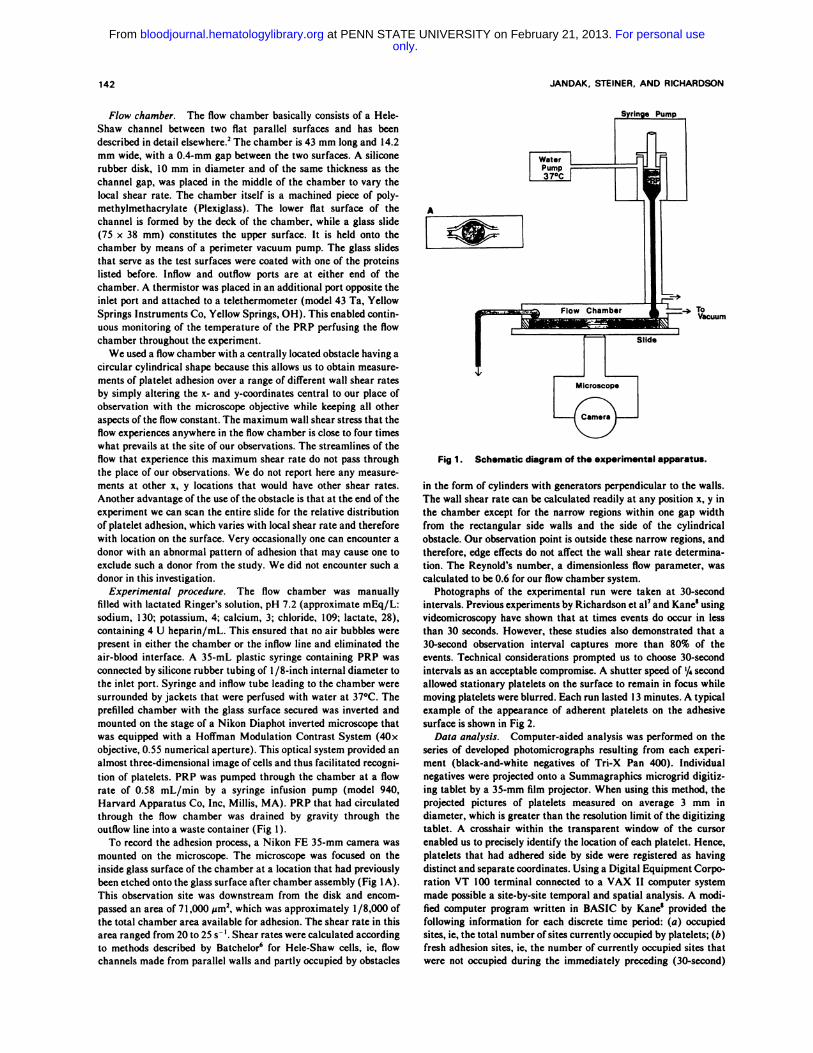
Fig 3. Adhesion trend lines for all four adhesive surfacesexamined in this study. Mean adhesion rates (cumulative plateletnumber x min’) ± SEM are shown for baseline and vitamin
E-supplemented periods. Differences between baseline and 200and 400 IU were significant at P < .01 for fibrinogen and glass andbetween baseline and 400 IU for collagen I. The remaining differ-ences in adhesion rate between baseline and vitamin E-supple-mented levels were significant at the P < .05 level.
adhesion than did any of the other three adhesive surfaces.
The lowest adhesion rates were recorded with noncoated
glass slides. A number of albumin-coated slides were
included as negative controls. The number of platelets that
adhered to this surface at any one time in the area of
observation was s2. The number of cumulative sites over the
entire period of observation was 10 ± 2 (mean ± SD; four
experiments). There was also very pronounced variability in
the rate of adhesion of platelets from different individuals to
the various adhesive surfaces. The large SE of the mean
adhesion rates reflect this individual variability. For this
reason statistics were computed by using individuals as their
own controls instead of comparing group means. The reuse-
of-sites index, on the other hand, showed far less variation
among individuals at baseline levels (Table 1).
A typical relationship between time and accumulation of
sites for a set of adhesion assays is shown in Fig 4. After an
initial lag time, the cumulative number of sites rises steadily
throughout the experimental run. Because the adhesion rate
was best represented by a linear regression curve, we deleted
Table 1 . Reuse-of-Sites Index Before
and After Dietary Supplementation
U)Ui
U)
Ui>
F-
-3:,
0
�b.o 2.0 4.0 6.0 8.0 0.0 2.0 14.0
TIME (minutes)
Fig 4. Cumulative number of adhesion sites over a 1 3-minuteperiod. Platelet adhesion to fibrinogen-coated glass slides wasstudied at baseline (circles). after 2 weeks of 200 lU vitamin E(triangles), and after an additional 2 weeks of 400 lU vitamin Esupplementation (squares). The data depicted are representativeof those obtained during a typical adhesion assay.
the first three points in our experiments and computed the
slope on the basis of the remaining observation points. The
initial time lag in platelet adhesion may be an important
phenomenon. For the purpose of using a consistent adhesion
rate parameter, however, the first two minutes were
ignored.
Platelets have a distinct tendency to reoccupy sites that
had been previously vacated by other platelets. Thus, a large
number of platelets occupied sites (an average of 44.5% for
all four adhesive surfaces) that had been visited twice, three
times, or up to eight times before. The reuse-of-sites data for
baseline conditions showed a strong exponential relationship
between the number of sites occupied v the number of times
of occupation (Fig 5).
Two experiments were performed with PRP obtained from
patients with severe and moderately severe type I von
5S
U)UiI-U)
0
a:Ui
z
C-I
0 #{149} � 2.0 � 3.0 � 4.0 � 5.0
NUMBER OF TIMES SITE WAS OCCUPIED
Fig 5. Graphic representation of the reuse of sites plottedagainst the number of times a specific adhesion site was occupiedduring an adhesion assay. The data shown in the figure wereobtained with platelets before any dietary vitamin E supplementa-tion was begun. The regression coefficient was �0.95.
Adhesive Surface Baseline
a-Tocopherol,
(200 lU,
Mean ± SD, n - 6)Supplementation
(400 IU)
Fibrinogen
Collagenl
Fibronectin
Glass
-1.0 ± 0.1
-0.7 ± 0.2
-1.3 ± 0.3
-0.9 ± 0.2
-1.3 ± 0.2
-1.1 ± 0.7
-1.6 ± 0.4
-1.2 ± 0.7
-1.5 ± 0.4
-1.5 ± 0.7
-1.0 ± 0.3
-2.0 ± 0.7
�I.0
Summary of the reuse-of-sites indices at baseline and after two
different levels of dietary vitamin E supplementation for four different
adhesive surfaces. The more negative the index. the smaller the numberof times a particular adhesion site was occupied. For further clarification
of this index, see Fig 5.
144 JANDAK, STEINER, AND RICHARDSON
only.For personal use at PENN STATE UNIVERSITY on February 21, 2013. bloodjournal.hematologylibrary.orgFrom

a-TOCOPHEROL AND PLATELET ADHESION 145
Mean ± 1 SD (n - 6).
tP < .05.
Willebrand’s disease. Both patients exhibited a severe reduc-
tion in the rate of platelet adhesion to collagen that was
found to be decreased by 92% in the severely affected patient
and 74% in the one with less severe disease. Adhesion to
fibronectin and fibrinogen was also reduced although not
quite to the degree as that of collagen. There was a decrease
of 56% and 37% with fibrinogen and 49% and 30%, respec-
tively, for the severe and less severe patient with fibronectin
as the adhesive surface.
Vitamin E supplementation of the diet produced a striking
decrease of the rate of adhesion to all four adhesive surfaces
(Fig 3). This decrease was statistically significant at both
levels of vitamin E supplementation. The baseline value for
each individual was compared with the value after each
supplementation level by using paired Student’s t tests. The
adhesion rate trend lines illustrate a consistent decrease in
platelet adhesion that is independent of the type of adhesive
surface studied as the dose of a-tocopherol increases. The
magnitude of reduction in the adhesive rate, however, was
found to be dependent on the adhesive surface. Collagen I
exhibited the largest decrease at both the 200-lU level (84%
decline) and at the 400-lU level (88% decline). Adhesion to
fibrinogen decreased by 82%, whereas adhesion to uncoated
glass slides declined by 77%. All of these declines were
statistically significant. The average decrease in adhesion
after 2 weeks of 200 IU vitamin E supplementation was 75%,
whereas at the 400-lU dose vitamin E reduced platelet
adhesion to the four surfaces by an average of 82%.
Vitamin E supplementation also decreased the reuse-
of-sites index (Table 1). The difference between baseline and
experimental values, however, did not reach statistical signif-
icance for any of the adhesive surfaces studied. Although the
number of times a particular site gets reoccupied decreases
after vitamin E supplementation, this decrease may be due to
the overall reduction in adhesion events. Since the reduction
is so steep, the number of times sites are reoccupied 3, 4, and
5 times becomes too small for meaningful statistical analyses
of differences in slope. For this reason, we determined the
ratio of the cumulative number of sites occupied once to the
cumulative number of sites occupied twice (01/02) (Table 2).
Because these ratios result from greater numbers of sites,
more reliance can be placed on them. Fibrinogen and
collagen showed increased ratios of 01/02 in platelets from
vitamin E-supplemented individuals. Fibronectin and glass,
on the other hand, did not produce a consistent change in
such ratios. Therefore, the effect of vitamin E on the rate of
second occupations of sites is very dependent on the adhesive
surface itself.
Table 2. Ratio of Once (0� ) to Twice (02) Occupied Platelet
Adhesion Sites at Baseline and After Vitamin E Supplementation
Adhesive Surface
Baseline(0,102) #{149}
200 IU(O,/O))
400 IU(0,102)
Fibrinogen 4.4 ± 3.1 4.7 ± 1.0 6.0 ± 2.7t
Collagen I 3.1 ± 0.5 5.3 ± 3.2t 6.2 ± 3.7t
Fibronectin 7.4 ± 2.7 9.5 ± 8.1 5.3 ± 2.7
Glass 8.6 ± 3.5 5.7 ± 2.7 13.4 ± 15.6
The high-pressure liquid chromatographic quantification
of the a-tocopherol content of platelets and plasma at
different levels of vitamin E supplementation is summarized
in Table 3. Considerable variability was found among mdi-
viduals before supplementation was begun as well as at the
different dose levels of vitamin E. Nevertheless, all individu-
als experienced a progressive increase in plasma and platelet
a-tocopherol levels when the dietary intake of vitamin E was
raised (Table 3). Evaluation of platelet a-tocopherol in
relation to the rate of adhesion to each surface is shown in
Fig 6. Even though the slopes of the lines plotted on a
logarithmic scale are quite different for each individual,
increased loading of platelets with a-tocopherol produced a
progressive decrease in platelet adhesion. In rare instances,
the adhesion rate actually showed a slight increase as the
vitamin E dose increased from 200 to 400 IU. This was found
in <10% of the measurements. It must be stressed that this
increase in the rate of adhesion that was occasionally regis-
tered was small and remained well below the baseline values.
To compare individuals, we normalized our results for two
arbitrary a-tocopherol levels and interpolated the corre-
sponding adhesion rates. Thus, rates corresponding to 360 ng
a-tocopherol/109 platelets were always higher than those at
500 ng/109 platelets (Table 4). There were no apparent
correlations between adhesion rates and sex or age of the
individuals. Correlation of plasma levels of a-tocopherol with
adhesion rates was very similar to that of platelet a-
tocopherol individual correlations not shown).
The appearance of platelets deposited on the adhesive
surfaces was also investigated by scanning electron micros-
copy (Fig 7). Under baseline conditions and before supple-
mentation of the diet with vitamin E, platelets exhibited
extensive pseudopodium formation when adherent to the
surface. Vitamin E administration resulted in a reduction of
pseudopodia at 200 IU/d and a virtual disappearance at 400
IU/d. All of the different coatings tested in our experiments
produced similar results.
DISCUSSION
The assessment of platelet adhesion by a reproducible
assay that mimics in vivo conditions of flow has been used in
this study. The flow chamber originally designed by Hele-
Shaw and subsequently modified by Richardson et a12 for the
evaluation of platelet adhesiveness provides a convenient
method for the in vivo assessment of platelet adhesiveness
and the cx vivo determination of pharmacologic and nutri-
tional factors affecting this platelet function. The simple
design and operation of this flow chamber makes it easy to
set up the system. A particularly attractive feature of this
Table 3. a-Tocopherol Content of Platelets and Plasma Before
and After Dietary Vitamin E Supplementation
Vitamin E Platelet a-Tocopherol Plasma a-TocopherolSupplementation (ng/lOa Platelets) (jig/mL)
0 294.3 ± 122.9 6.9 ± 1.1
200 IU/d 528.3 ± 155.2 9.3 ± 1.4
400IU/d 745.5 ± 253.1 10.8 ± 1.7
Mean ± SD (n - 6).
only.For personal use at PENN STATE UNIVERSITY on February 21, 2013. bloodjournal.hematologylibrary.orgFrom

10.0a:
C0
5�.C
Platelet Alpha - Tocopherol
0a:C0
U,
a,�04
a,
0a:C0U)0
.c�04
Platelet Alpha- Tocopherol
50
40
30
20
5) 0.00a:C
U)a,
V4
3.0
2.0 -
0\
\
\
50 � � I I
40-0=1, 53,F I
30 - +=2,4O,FIA =3, 25,M IO=4,29,F I
20 - D=5�2�j
I�O � \\�� � I
\
\
\
CA
I I
Platelet Alpha-Tocopherol
100 300 500 1000 2000 100 300 500 1000 2000
Fig 6. Relation of adhesion rate (cumulative number of platelet x min’) to a-tocopherol content of platelets (ng/10 platelets).Results obtained with fibrinogen as an adhesive surface are shown in panel A. with fibronectin in B. collagen in C. and with glass in D. Dataare shown for five of the six individuals tested. One subject in the study was omitted because his baseline value of platelet a-tocopherolwas >3 SD from the mean of the other five individuals. Age and sex of the subjects are indicated in the inset.
Platelet Alpha- Tocopherol
146 JANDAK. STEINER. AND RICHARDSON
only.For personal use at PENN STATE UNIVERSITY on February 21, 2013. bloodjournal.hematologylibrary.orgFrom
a-TOCOPHEROL AND PLATELET ADHESION 147
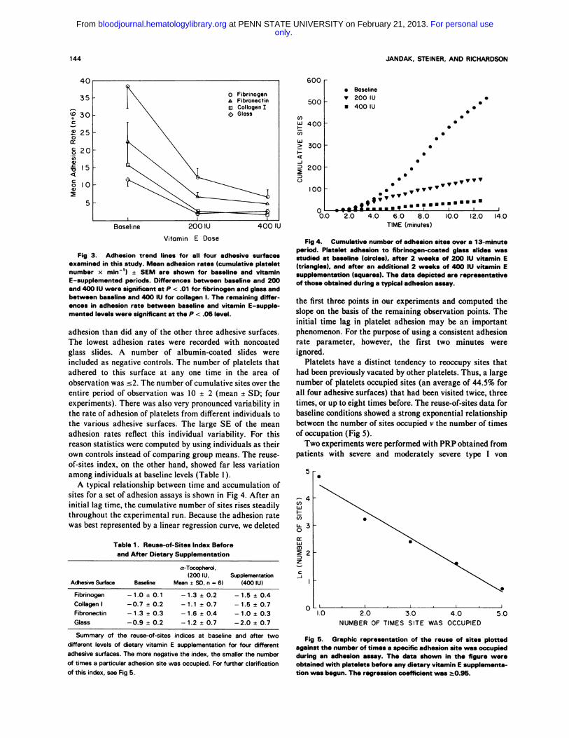
Table 4. Interpolated Adhesion Rates
at Two a-Tocopherol Levels
Subject
Fibrin
360
ogen
500
Colla
360
ger, I
500
Fjbr
360
onectin
500
GI
360
ass
500
J.N. 42.0 32.0 3.9 3.3 5.2 2.8 5.1 2.1
SB. 9.1 4.4 3.4 1.8 6.6 7.0 2.5 2.0
KM. 46.5 16.0 7.6 3.8 47.0 12.5 10.7 4.5
A.A. 5.3 3.0 3.5 2.3 11.0 7.6 5.1 3.2
Data for two of the individuals tested are not shown because their
baseline values of platelet a-tocopherol were >360 ng/ 1O� platelets.
a-Tocopherol, 360 or 500 ng/109 platelets.
adhesion assay is the ability to make time-resolved observa-
tions that can be evaluated by computerized methods and
give a permanent record of the adhesion process. A further
advantage is the ability to make observations on different
adhesive surfaces. The reproducibility of the assay is good,
provided adhesion measurements are done within a three-
hour period after the collection of blood.
For these studies we chose fibrinogen, fibronectin, col-
lagen I, and glass as adhesive surfaces. The plain glass
surface of course becomes coated by plasma proteins within a
short period of time after starting the perfusion of the
chamber. Our choices were determined by the ready avail-
ability of pure proteins and the relevance of these proteins to
platelet adhesion. The individual variation of platelet adhe-
sion to the different adhesive surfaces was quite high upon
repeated testing, while those who had a low adhesion rate
continued to have such when tested again.
Evaluation of platelet adhesion in relation to time showed
a distinct initial delay phase that under baseline conditions
was two minutes. During this interval few platelets adhere to
the adhesive surface. This initial time lag was noticed by
previous investigators who used this flow chamber.8
Although it is probably not the dominant factor, there is
some competitive adsorption of plasma proteins, especially
albumin, gamma globulin, and fibrinogen, to form a protein-
aceous layer on the surface.” Also, there may be a lower
concentration of platelets near the wall at early times
because the priming solution is displaced first near the center
of the flow channel and more slowly near the walls.’2 Until
the surface is coated, the conditions result in an initially low
adhesion rate. Another explanation for the delay phases is
that the adhesion of platelets and the accompanying release
of their granular contents leaves a concentration of agonists
in the immediate surroundings of such platelets that may
induce shape change in other platelets that stray into their
vicinity. Diffusion of agonists from the adherent platelets
should be relatively little disturbed by the flow because the
flow close to the adhesive surface would be almost station-
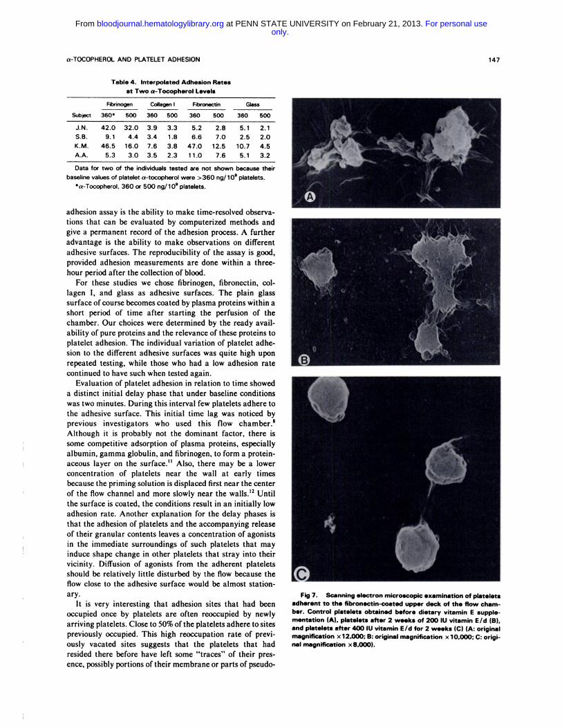
ary. Fig 7. Scanning electron microscopic examination of platelets
It is very interesting that adhesion sites that had been adherent to the fibronectin-coated upper deck of the flow cham-
occupied once by platelets are often reoccupied by newly ber. Control platelets obtained before dietary vitamin E supple-. . . mentation (A). platelets after 2 weeks of 200 lU vitamin E/d (B).
arriving platelets. Close to 50% ofthe platelets adhere to sites and platelets after 400 IU vitamin E/d for 2 weeks (C) (A: original
previously occupied. This high reoccupation rate of previ- magnification x 12.000; B: original magnification x 10.000; C: origi-
ously vacated sites suggests that the platelets that had nal magnification xB.000).
resided there before have left some “traces” of their pres-
ence, possibly portions of their membrane or parts of pseudo-
only.For personal use at PENN STATE UNIVERSITY on February 21, 2013. bloodjournal.hematologylibrary.orgFrom
REFERENCES
148 JANDAK. STEINER, AND RICHARDSON
podia at those sites. One can speculate that newly arriving
platelets will be more likely to adhere to such sites than to a
random area of the adhesive surfaces. Especially the glyco-
protein(s) involved in the adhesion process, eg, glycoprotein
lb’3”4 could be a possible candidate for such a trace-leaving
process. An alternative explanation could be the existence of
“hot spots,” very reactive sites of the adhesive surface.
It should be pointed out that the shear rates at which
adhesion was measured (20 to 25 s� I) do not correspond to
those normally present in coronary or other major arteries
(200 to 800 s_i) Because the primary purpose ofour studies
was to obtain time-resolved measurements of platelet adhe-
sion under conditions where arrival and departure of plate-
lets on the adhesive surface could be observed, the use of
whole blood that would have greatly increased platelet
transport to the adhesive surface had to be excluded. Only
the terminal deposition of platelets can be evaluated in the
latter case. High shear rates enhance the frequency of
platelet arrival at the surface,iS�7 but the process of removal
of adherent platelets also appears to be enhanced, which
results in fact in a net decrease in adhesion.2”8 When
observations were made in areas of the flow chamber where
higher shear rates (100 to 120 s_i) exist, ie, the narrow
channel adjacent to the central obstruction in the chamber,
lower adhesion rates were actually found than at our site of
observation. There are also technical difficulties in trying to
simulate high shear rate conditions in the flow chamber we
used. Although the chamber can withstand shear rates of 600
to 800 s-� extremely large volumes of PRP would be
required to perform an experiment.
Nevertheless, we believe that our studies are of clinical
relevance. Recent investigations by Davies and Thomas’9�#{176}
have demonstrated that a majority of patients with coronary
th�ombosis sustain the thrombotic event at a rupture site of
an arteriosclerotic plaque. Platelets were found in the cavi-
ties of these rupture sites, with the evolving thrombus
growing out into the lumen of the blood vessel. Flow rates at
such sites are very low, and equally low are the shear rates.
Platelet adhesivity was decreased in the patients with von
Willebrand’s disease that we were able to test. Although we
cannot make a general statement about the behavior of such
platelets, having studied only two patients with this disease,
our findings differ somewhat from those reported by Turitto
et al2i who observed decreased platelets adherence but only
at high shear rates (�800 �_i)#{149} We do not know the reason
for this difference, but the respective methods of adhesion
measurement are quite dissimilar: not only the shear rates
but also the suspension media (whole blood v PRP) are
different.
Supplementation of the diet with vitamin E produced a
striking reduction in platelet adhesiveness. It was interesting
to note that all six individuals studied showed a decrease not
only after 400 IU but also after 200 IU vitamin E daily for 2
weeks. Especially remarkable and sharp was the reduction in
I . Steiner M: Effect of alpha-tocopherol administration on plate-let function in man. Thromb Haemost 49:73, 1983
2. Richardson PD, Mohammed SF, Mason RG: Flow chamber
platelet adhesivity after 200 IU (up to 80% from the original
baseline value). The further decline in adhesivity upon
increasing vitamin E to 400 IU was relatively modest in
comparison. Although our study did not include a placebo
group, we tested platelet adhesion in several of the subjects
after the vitamin E supplementation had ended. All individu-
als showed a complete return to their presupplementation
adhesion rates.
The reutilization of once-occupied sites also changed after
vitamin E supplementation. The 01/02 ratio relates the
number of second occupations to the number of primary
occupations. In this way a change in the rate of reoccupation
is scaled to any change seen in the rate of primary adhesion,
Oi. By omitting consideration of 03, 04, etc, the effects from
small counts are eliminated. A decrease in the reoccupation
of sites is shown by an increase in this ratio. Judging by the
#{176}i/#{176}2ratios (Table 2), only collagen I and fibrinogen demon-strated reduced reoccupation of sites that had been vacated
by platelets. This would suggest a reduced amount of platelet
“traces” left by the first occupants of specific adhesion sites.
The results were not as clear for glass and fibronectin.
Even though the individual levels of vitamin E in platelets
were quite variable, all individuals showed a very marked
and progressive increase in platelet tocopherol levels upon
increasing the dietary supplementation of vitamin E (Table
3). Although the baseline levels of platelet a-tocopherol
showed no distinct relation to the adhesiveness of the plate-
lets, supplementary vitamin E did. This finding suggests that
a high platelet tocopherol level by itself does not produce any
inhibition of platelet adhesiveness. Only a-tocopherol
ingested in excess of the amount taken in with the individu-
al’s “normal” diet will affect platelet adhesivity. Dietary
abundance of a-tocopherol is closely related to the content of
polyunsaturated fatty acids.22 We believe that this balance
has to be tilted in favor of a-tocopherol to observe the
inhibitory action of platelet adhesivity.
The dose-related reduction of pseudopodium formation in
a-tocopherol-enriched platelets may be responsible for the
observed decrease in platelet adhesivity. The reason for this
phenomenon is not clear at this time, but a causal relation
between the a-tocopherol supplementation and the inhibition
of pseudopodium formation appears to exist.
We believe that our studies give convincing evidence of the
antiadhesive properties of vitamin E supplementation of the
diet. It was especially gratifying that “reasonable” doses of
vitamin E that had no untoward side effects were able to
accomplish such reduction. Even though vitamin E has not
shown to be an effective antiaggregating agent when tested
ex vivo,’ from our data it would appear that it could be an
effective antiadhesive agent in vivo.
ACKNOWLEDGMENT
We wish to thank Georg Panol of Brown University for his
technical assistance and design of the flow chamber system.
studies of platelet adhesion at controlled spatially varied shear rates.Proc Eur Soc ArtifOrgans 4:175, 1977
3. Landolfi R, Mower RL, Steiner M: Modification of platelet
only.For personal use at PENN STATE UNIVERSITY on February 21, 2013. bloodjournal.hematologylibrary.orgFrom
a-TOCOPHEROL AND PLATELET ADHESION 149
function and arachidonic acid metabolism by bioflavonoids. Bio-
chem Pharmacol 33:1525, 19844. Cazenave JP, Packham MA, Mustard iF: Adherence of
platelets to a collagen-coated surface: Development ofa quantitative
method. J Lab Clin Med 82:978, 19735. Steiner M, Luscher EF: Identification of the immunoglobulin
G receptor of human platelets. J Biol Chem 261 :7230, 19866. Batchelor GK: An Introduction to Fluid Dynamics. Cam-
bridge, UK, Cambridge University Press, 1967, p 2227. Richardson PD, Mohammed SF, Mason RG, Steiner M, Kane
R: Dynamics of platelet interaction with surfaces in steady flowconditions. Trans Am Soc Artif Intern Organs 25:147, 1979
8. Kane RL: Spatio-temporal dynamics of human platelet adhe-sion in a Hele-Shaw flow chamber. Master Thesis, Brown Universi-ty, 1981
9. Sokal RR, Rohlf FJ: The Principles and Practice of Statistics
in Biological Research. San Francisco, Freeman, 1969, p 143
10. DeLeenheer AP, DeBevere VO, DeRuyter MG, Claeys AE:Simultaneous determination of retinol and alpha-tocopherol in
human serum by high performance liquid chromatography. J Chro-
matogr 162:408, 1979I 1 . Park K, Cooper SL: Importance of composition of the initial
protein layer and platelet spreading in acute surface-induced throm-bosis. Trans Am Soc Artif Intern Organs 31:484, 1985
12. Levich VG: Physiochemical Hydrodynamics. Englewood
Cliffs, NJ, Prentice-Hall.13. Berndt MC, Phillips DR: Platelet membrane proteins: Com-
position and receptor function, in Gordon JL (ed): Platelets in
Biology and Pathology, vol 2. Amsterdam, Elsevier North Holland,1981, p44
14. George iN, Nurden AT, Phillips DR: Molecular defects ininteractions of platelets with the vessel wall. N EngI J Med311:1084, 1984
15. Grabowski EF, Friedman LI, Leonard EF: Effects of shearrate on diffusion and adhesion of blood platelets to a foreign surface.
Ind Eng Chem Fundam 1 I :224, 197216. Turitto VT, Weiss Hi, Baumgartner HR: The effect of shear
rate on platelet interaction with subendothelium exposed to citratedhuman blood. Microvasc Res 19:352, 1980
I 7. Turitto VT, Weiss Hi: Red blood cells: Their dual role inthrombus formation. Science 207:541, 1980
18. Butruille Ya, Leonard EF, Litwak RS: Platelet-platelet inter-
actions and non-adhesive encounters on biomaterials. Trans Am SocArtif Intern Organs 21:609, 1975
19. Davies Mi, Thomas T: The pathological basis and micro-anatomy of occlusive thrombus formation in human coronaryarteries. Philos Trans R Soc London [Biol] 294:225, 1981
20. Davies Mi, Thomas AC: Plaque fissuring-The cause ofacute myocardial infarction, sudden ischemic death, and crescendoangina. Br Heart J 53:363, 1985
21 . Turitto VT, Weiss Hi, Baumgartner HR: Decreased plateletadhesion on vessel segments in von Willebrand’s disease: A defect ininitial platelet attachment. i Lab Clin Med 102:551, 1983
22. Hove EL, Harris PL: Linoleic acid-tocopherol relation in fats
and oils. i Am Oil Chem Soc 28:405, 1951
only.For personal use at PENN STATE UNIVERSITY on February 21, 2013. bloodjournal.hematologylibrary.orgFrom
Related Documents