Alliny de Souza Bastos AVALIAÇÃO DA CORRELAÇÃO ENTRE PEROXIDAÇÃO LIPÍDICA E O PERFIL DE MARCADORES INFLAMATÓRIOS EM PACIENTES COM DIABETES MELLITUS TIPO 2, DISLIPIDEMIA E PERIODONTITE CRÔNICA. Araraquara 2012 UNIVERSIDADE ESTADUAL PAULISTA “JÚLIO DE MESQUITA FILHO” - UNESP FACULDADE DE ODONTOLOGIA DE ARARAQUARA

Welcome message from author
This document is posted to help you gain knowledge. Please leave a comment to let me know what you think about it! Share it to your friends and learn new things together.
Transcript
Alliny de Souza Bastos
AVALIAÇÃO DA CORRELAÇÃO ENTRE PEROXIDAÇÃO LIPÍDICA E O PERFIL DE MARCADORES INFLAMATÓRIOS EM
PACIENTES COM DIABETES MELLITUS TIPO 2, DISLIPIDEMIA EPERIODONTITE CRÔNICA.
Araraquara
2012
UNIVERSIDADE ESTADUAL PAULISTA “JÚLIO DE MESQUITA FILHO” - UNESP
FACULDADE DE ODONTOLOGIA DE ARARAQUARA
Tese apresentada ao programa de pós-graduação em Odontologia, Área de concentração Periodontia, da Faculdade de Odontologia de Araraquara, da Universidade Estadual Paulista, para obtenção do título de Doutor em Odontologia.
Orientador:Prof. Dra. Silvana Regina Perez Orrico
Co-Orientador:Prof. Dr. Carlos Rossa Júnior
Bastos, Alliny de Souza
Avaliação da correlação entre peroxidação lipídica e o perfil de marcadores inflamatórios em pacientes com diabetes mellitus tipo 2, dislipdemia e periodontite crônica / Alliny de Souza Bastos.-- Araraquara: [s.n.], 2012.
196 f. ; 30 cm. Tese (Doutorado) – Universidade Estadual Paulista,
Faculdade de Odontologia Orientadora: Profa. Dra. Silvana Regina Perez Orrico Co-orientador: Prof. Dr. Carlos Rossa Junior
1. Peroxidação de lipídeos 2. Diabetes mellitus tipo 2 3. Doenças periodontais 4. Citocinas I. Título
Ficha catalográfica elaborada pela Bibliotecária Marley C. Chiusoli Montagnoli, CRB-8/5646
Serviço Técnico de Biblioteca e Documentação da Faculdade de Odontologia de Araraquara / UNESP
ALLINY DE SOUZA BASTOS
AVALIAÇÃO DA CORRELAÇÃO ENTRE PEROXIDAÇÃO LIPÍDICA E O PERFIL DE MARCADORES INFLAMATÓRIOS EM PACIENTES
PORTADORES DE DIABETES MELLITUS TIPO 2 COM PERIODONTITE CRÔNICA.
COMISSÃO JULGADORA
DEFESA DE TESE PARA OBTENÇÃO DO GRAU DE DOUTOR
Presidente e Orientador: Profa. Dra.Silvana Regina Perez Orrico
2º Examinador: Profa. Dra. Margarete Aparecida Meneses de Almeida
3º Examinador: Prof. Dr. José Roberto Cortelli
4º Examinador: Profa. Dra. Maria Teresa Pepato
5º Examinador: Prof. Dr. Luís Carlos Spolidório
Araraquara, 12 de março de 2012.
U
DADOS CURRICULARES
Alliny de Souza Bastos NASCIMENTO: 03/01/1983- RIO BRANCO/AC
FILIAÇÃO: Antonio Bastos
Maria Franscismar de Souza Bastos
2000-2005 Curso de Graduação
Universidade Federal de Sergipe- UFS
2006-2008 Curso de Especialização em Implantodontia
Faculdade de Odontologia de Araraquara- UNESP
2008-2012 Doutorado em Odontologia- Área de Concentração Periodontia
Faculdade de Odontologia de Araraquara- UNESP
U
Com amor, dedico este trabalho...
Aos meus pais Tobias e Meire que fizeram dos meus sonhos seus
próprios objetivos e de meus objetivos suas próprias lutas. Chegamos
aqui juntos e esta conquista é nossa!
Aos pacientes que fizeram parte desta pesquisa, que foram receptivos e
tornaram possível a realização deste estudo. Obrigada pela paciência e
confiança!
U
U
AGRADEÇO ESPECIALMENTE... A Deus à luz do Espírito Santo que me iluminaram nos momentos mais difíceis em
que achei que poderia fraquejar, dando-me força e discernimento para tornar tudo
possível. Minha vida segue entregue a ti.
Ao amor e incentivo dos meus pais Tobias e Meire, ao apoio incondicional, por
estarem ao meu lado em todas as decisões e momentos e por entenderem minha
ausência por todo este tempo. Obrigada por investirem em minha educação e em
meus sonhos. AMO VOCÊS. Ao meu irmão Rafael que do seu jeito único, sempre
torce por minhas conquistas, apoiando e incentivando tudo que faço.
Ao meu namorado, amigo, companheiro Nicolau por ser minha fortaleza, minha paz e
meu refúgio. Ter você em minha vida foi um verdadeiro presente divino. Muito
obrigada pelos conselhos, por entender meus anseios e compartilhar sonhos, vitórias e
momentos difíceis. Agradeço a paciência e incentivo ao longo destes anos, espero que
saiba o quanto de você tem neste trabalho. Agradeço ainda a toda família pelo apoio.
À minha prima-irmã Grazianny, meu anjinho da guarda, que vibra com minhas
conquistas e sofre junto nos momentos difíceis que com Leonardo me receberam em
sua casa durante as análises realizadas em São Paulo. Obrigada por me acolher, pelo
apoio e carinho.
À minha orientadora Profa. Dra. Silvana Regina Perez Orrico por ter me aceitado
como estagiária e pós-graduanda. Pelos ensinamentos durante a execução desta
pesquisa, pelas palavras de sinceridade e pela maneira especial e direta de me fazer
aceitar quando eu estava errada e por me ensinar a ser mais clara e objetiva. Muito
obrigada por entender minhas ansiedades, por acreditar em mim, pela amizade,
competência e paciência com que me orientou durante a realização desta tese.
AGRADECIMENTOS À Faculdade de Odontologia de Araraquara, nas pessoas de seu Diretor, Prof. Dr.
José Cláudio Martins Segalla e Vice-Diretora Profa. Dra. Andreia Affonso Barreto
Montandon.
Ao Prof. Dr. Mário Tanomaru Filho, pela competência, dedicação e responsabilidade
com que coordena o Curso de Pós-Graduação em Odontologia.
Aos docentes do Departamento de Periodontia, Prof. Dr. Joni Augusto Cirelli, Profa.
Dra. Silvana Regina Perez Orrico, Profa. Dra. Rosemary Adriana Chiérici
Marcantonio, Prof. Dr. Élcio Marcantonio Júnior, Prof. Dr. José Eduardo Cezar
Sampaio, Prof. Dr. Carlos Rossa Junior pela convivência diária, amizade, formação
profissional e pelos exemplos de competência e dedicação à Periontia e à pesquisa.
Ao Prof Dr.Élcio Marcantonio Júnior por ter sido meu contato inicial e por ter me
recebido em Araraquara. Obrigada pelos conselhos e por me receber sempre com
tanto carinho.
À Profa. Dra. Rosemary Adriana Chiérici Marcantonio meu agradecimento especial
pelo carinho e acolhimento nos momentos difíceis, pela amizade fiel e ensinamentos
profissionais e de vida com que compartilhamos durante todos estes anos. Me sinto
privilegiada por te ter em minha vida.
Ao Prof Dr. Carlos Rossa Junior pelos sábios conselhos sempre vindos nos
momentos que mais precisava. Obrigada pela amizade e conhecimentos transmitidos.
Ao Prof. Dr. Luís Carlos Spolidório pela amizade e por ser uma inspiração a seguir a
carreira acadêmica. Sua vibração e amor pela pesquisa me fazer ter certeza de que
estou no caminho certo.
Ao Prof Dr Dana T Graves por ter me recebido em seu laboratório na Filadélfia
durante a realização do meu estágio de Doutorado. Obrigada pela confiança, por me
ensinar tanto em pouco tempo. Aos alunos do laboratório da Universidade da
Pensilvânia, Sandra, Bhaskar, Mireille, Guangyu, Kathy, Precious e Tina por
compartilhar uma fase da vida que jamais esquecerei.
À Profa. e amiga Elaine Maria Sgavioli Massucato por sempre me receber com um
olhar que praticamente já sabia o que eu estava sentindo. Por me apresentar um lado
da vida positivo e a lidar com os problemas de maneira sábia.
À Profa. Dra. Raquel Mantuaneli Scarel-Caminaga pela amizade, carinho e por ser
um exemplo de dedicação ao ensino e à pesquisa.
À Profa. Dra. Ana Paula de Melo Loureiro (Laboratório de Análises Toxicológicas
da Faculdade de Ciências Farmacêuticas, (FCF) Universidade de São Paulo- USP)
por participar ativamente das análises de peroxidação lipídica, ao meu lado na
bancada. Agradeço o carinho com que sempre me recebeu em seu laboratório, a
curiosidade e boa vontade de entender um pouquinho da Periodontia, a atenção em
me escutar e a paciência em me ensinar. Você fez toda a diferença. Aos alunos do
laboratório da Profa Ana Paula, em especial ao Tiago Franco de Oliveira pela
amizade, disposição, paciência e ajuda na realização das análises.
À Profa. Dra. Dulcinéia Saes Parra Abdalla (Laboratório de Bioquímica da FCF-
USP), por ter aberto as portas do seu laboratório para a realização do meu trabalho.
Às meninas do laboratório Tanize, Fernanda, Patrícia, Marcela, Martina e Elaine
pelo carinho nos momentos de desespero e por serem sempre tão prestativas. À
Tanize, um agradecimento especial por ser um anjo com coração bondoso que
apareceu em minha vida na hora que mais precisei. Obrigada por tornar as análises
possíveis e por ser um exemplo de calma, competência e dedicação à vida acadêmica.
Ao Prof. Dr. Niels Olsen Câmara (Departamento de Imunologia, Instituto de Ciências
Biomédicas, Universidade de São Paulo- USP) por ter me recebido em seu
laboratório para as análises de marcadores inflamatórios e à Meire Hiyane pela
dedicação e acolhimento durante a realização destas análises. Meire, você
literalmente abraçou a causa. Muito obrigada!
Ao Prof Dr Paulo Inácio da Costa, coordenador do CACH/NAC, por permitir a
realização dos exames dos pacientes nos laboratórios do NAC (Núcleo de
Antendimento à Comunidade de Araraquara).
Aos funcionários do NAC e do laboratório de bioquímica da Faculdade de Ciências
Farmacêuticas da UNESP nas pessoas de Dalva, Marisa, Mathilde, Fátima, Rose,
Flávia, Tânia, Edson. Obrigada por receber a mim e aos meus pacientes durante dois
anos e por tornar esta pesquisa possível. Vocês são um exemplo de bondade,
competência e dedicação ao trabalho que desempenham.
Aos funcionários da Periodontia, Maria do Rosário, Maria José, Ester, Claudinha,
Leandro pela amizade e carinho que sempre me dispensaram. Que Deus os abençoe!
À Regina Lúcia, pela paciência e amizade sempre disponíveis. Por ser meu refúgio e
me transmitir paz sempre que precisei. Obrigada por estar sempre presente!
A todos os funcionários da Secção de Pós-Graduação, Mara, Alexandre e Sérgio
vocês são especiais e essenciais na vida de um pós-graduando. Obrigada por tudo!
Aos funcionários da biblioteca, Ceres, Adriano, Marley e a todos os funcionários da
FOAr que colaboraram com o estudo fazendo parte dele em um momento que
precisei de pacientes para terminar a pesquisa em tempo hábil. Obrigada por ter
cedido seu tempo e confiança para finalização desta etapa!
À CAPES, CNPq e FAPESP pelo apoio financeiro para realizar esta pesquisa.
À Mariana Ferrari pela amizade e apoio que me ofereceu nos momentos em que
pensei que não fosse ser possível. Obrigada por me ajudar a ser uma pessoa melhor.
Ao meu querido Prof. Dr. Luiz Carlos Ferreira da Silva (Universidade Federal de
Sergipe- UFS) meu agradecimento especial por ter me incentivado a buscar algo a
mais da Odontologia. O grande responsável por ter iniciado esta jornada em
Araraquara. Obrigada por estar sempre disponível quando precisei
À Profa e amiga Kildane Maria Guedes, com quem fiz meu primeiro trabalho de
pesquisa. Obrigada por acreditar em mim, por ter me dar forças para continuar
explorando o meu potencial.
À Profa Dra Margarete A. Meneses de Almeida, minha orientadora de trabalho de
conclusão de curso e alguém que me mostrou que a Periodontia é uma especialidade
diferenciada e que se perseveramos e acreditarmos iremos colher bons frutos.
Obrigada pelos ensinamentos e por me ser incentivar à carreira acadêmica. À Profa
Tânia Fortes por ter me iniciado à Periodontia, a senhora é um exemplo de
competência e força.
Ao Departamento de Odontologia da UFS, nas pessoas da Profa Dra Marta Piva,
que só a presença já transmite paz e tranqüilidade, obrigada pelas palavras de
incentivo e apoio e à Profa Dra Rosa Bragança pela competência e seriedade com
que conduz nosso curso de Odontologia. Ao Prof Mirabeau Ramos por acreditar em
mim e me inspirar a ir em busca dos meus objetivos.
À Profa. Andréa Marcaccini pela atenção sempre disponibilizada quando precisei.
Aos amigos do curso de Pós-Graduação que fizeram parte da minha vida nos meus
primeiros anos de Araraquara Daniela Zandim, Fernanda Bello, Daniela Gonçalves,
Maurício, Ana Emília, Fernando, Rafael Sartori, Rafael Faeda. Obrigada pelo
acolhimento e amizade. À Aline Cavalcanti por ter me apresentado Araraquara, por
ter me ajudado no primeiro ano tão difícil, jamais esquecerei o que fez por mim.
Aos amigos do curso de Pós-Graduação em Periodontia, Leila, Chaine, Telma,
Michelle, Sâmia, Guilherme, Andressa, Lívia, Mariana, João, Lucas, Marina, Shelon,
Nicolau, Morgana, Sabrina, Rubens, Andrés, Naná, Roberta, Humberto, Yeon,
Wagner, Rodrigo, Nicole, Fausto, Giovana, Lívia Finoti, Luana, Fabiana, Pablo,
Jõao Paulo, Luiz Guilherme, Fernanda pelo convívio agradável e enriquecedor.
Às minhas amigas de pós-graduação e da vida inteira, Leila, Marina, Morgana, Lívia,
Sabrina, Roberta, Carol, Naná e Yeon por tornar tudo mais fácil e compartilhar de
momentos de descontração e dificuldades. Vocês moram no meu coração!
À Leila amiga fiel, por participar tão ativamente da minha vida pessoal e profissional.
Muito obrigada pelo companheirismo, por se preocupar comigo e por acreditar em
mim. Você estará sempre em minha vida, pois não dá para seguir mais sem você. À
Marina pela convivência diária, por compartilhar angústias e alegrias. À Lívia
Perussi por ser uma amiga tão fiel e verdadeira. À Morgana pela sinceridade e por
estar presente em momentos tão delicados com positivismo e tranqüilidade. Vocês
tornaram esta caminhada possível. Quero tê-las sempre perto!
À Sâmia que esteve comigo nos momentos mais difíceis deste doutorado. Quando o
cansaço já tomava de conta, sempre tínhamos uma a outra para nos apoiarmos. Muito
obrigada por poder contar com você sempre!
Ao amigo Fausto pelo companheirismo e ajuda no início da realização desta
pesquisa, pelo positivismo transmitido e por mostrar a importância de conduzir a vida
de uma maneira mais leve. Ao amigo Guilherme que tanto me ajudou no atendimento
dos meus pacientes em momentos em que estive ausente, sem forças e sem tempo
para dar conta de tudo. Você tem um coração e uma competência admiráveis.
Ao meu querido amigo Rubens por ser a palavra e a ação nos momentos que tanto
precisei. Você é único e a vida te recompensará por ser tão prestativo e fiel.
Ao amigo Bruno por compartilhar conhecimento e experiências de vida. Obrigada por
me incentivar a perseverar na pesquisa e a buscar algo maior.
À Gabriela Giro pelo apoio e incentivo pessoal e profissional sempre que precisei.
À família Castanharo, Sabrina, Cristiane, Elzio e Maria pelo acolhimento, confiança
e amizade. Muito obrigada por tudo que fizeram por mim e por minha família.
À Profa Sílvia Cavalheiros pelo apoio e conhecimentos transmitidos.
Aos amigos de Araraquara Michel, Paula, Fábio, Adriana, Rosângela, Elaine,
Liliane, Giovana, Cassiano, Flávia pelo confiança, carinho e amizade.
À minha querida Fábia por ser tão companheira e presente em minha vida, por tentar
entender minha rotina, por me dar forças e por tornar tudo mais leve e prazeiroso. À
Camilla pela companhia e energia positiva. Conviver com você é enriquecedor.
Às amigas da especialização em Implantodontia Sheila e Keidy, pelo incentivo e
companheirismo. Sheila, ter sua presença em nossa casa nos trouxe luz e alegria!
À minha grande amiga Emanuella por se esforçar para entender o que faço, por
sempre acreditar e me incentivar. Amizade que transcende o tempo e a distância.
A toda minha família, avós, tios, primos que mesmo de longe sempre torceram por
mim. Obrigada pela preocupação, apoio e carinho.
Aos meus amigos-irmãos Djalmyr e Vitor que compartilharam do meu primeiro ano
de Araraquara. Vocês são um exemplo de que honestidade e acreditar faz a diferença.
Aos meus amigos de graduação Sara (eterna dupla), Marina, Júlio, Wesley, Itamar,
Christiano, obrigada pela verdadeira amizade, pelo carinho e palavras de incentivo.
A todos os meus amigos em Aracaju, em especial Emanuella, Eliana, Evelyne,
Thereza, Luana, André. À distância às vezes me obriga a estar ausente em momentos
importantes...mas não tenham dúvidas que sempre trago vocês em meu coração.
A todos aqueles que não foram citados, porém têm conhecimento deste trabalho e de
sua importância, bem como aqueles que me ajudaram em momentos específicos e
essenciais, minha gratidão.
FILOSOFIA DO SUCESSO Napoleon Hill
Se você pensa que é um derrotado,
você será derrotado.
Se não pensar “quero a qualquer custo!”
Não conseguirá nada.
Mesmo que você queira vencer,
mas pensa que não vai conseguir,
a vitória não sorrirá para você.
Se você fizer as coisas pela metade,
você será fracassado.
Nós descobrimos neste mundo
que o sucesso começa pela intenção da gente
e tudo se determina pelo nosso espírito.
Se você pensa que é um malogrado,
você se torna como tal.
Se almeja atingir uma posição mais elevada,
deve, antes de obter a vitória,
dotar-se da convicção de que
conseguirá infalivelmente.
A luta pela vida nem sempre é vantajosa
aos fortes nem aos espertos.
Mais cedo ou mais tarde, quem cativa a vitória
é aquele que crê plenamente
EU CONSEGUIREI!
SUMÁRIO
Lista de abreviaturas.................................................................................................13
Resumo ......................................................................................................................16
Abstract ....................................................................................................................18
1 Introdução ..............................................................................................................20
2 Proposição ..............................................................................................................43
3 Capítulos .................................................................................................................45
3.1 Capítulo 1 ............................................................................................................46
3.2 Capítulo 2 ............................................................................................................70
3.3 Capítulo 3...........................................................................................................101
4 Discussão ...............................................................................................................131
5 Conclusão .............................................................................................................147
6 Referências ...........................................................................................................149
7 Apêndice................................................................................................................166
Apêndice 1 Material e Método ................................................................................167
Apêndice 2 Resultados Adicionais...........................................................................182
8 Anexos ...................................................................................................................187
Anexo A Certificado Comitê de Ética......................................................................188
Anexo B Termo de consentimento Livre e Esclarecido...........................................189
Anexo C Documentos comprobatórios....................................................................193
LISTA DE ABREVIATURAS
AGE- advanced glycation end-products
BHT- Butylated hydroxytoluene
BMI – Body Mass Index
BOP – Bleeding on Probing
CAL – Clinical attachment Level
CEP- Comitê de Ética em Pesquisa
CT- Colesterol Total
DAD – Diode array detector
DM2 – Type 2 Diabetes mellitus
ELISA- Enzyme-Linked Immunoabsorbent Assay
EROs- Espécies reativas de oxigênio
FSG- Fluido sulcular gengival
GBI – Gingival bleeding index
GCF – Gingival Crevicular Fluid
GM-CSF Fator Estimulador de Colônias de Macrófagos e Granulócitos
H3PO4 - ácido fosfórico
HbA1c – Glycated Hemoglobin A1c fraction
HDL – High density lipoprotein
HOMA IR- Homeostatasis model assessment of insulin resistance
HPLC- High performance liquid chromatography
hs CRP – High Sensitive C-reactive Protein
IL- Interleukine
IMC- Índice de massa corporal
IPV- Índice de placa visível
ISM- Índice de sangramento marginal
KI - iodeto de potássio
LDL – Low Density Lipoprotein
LDL (-) – LDL eletronegativa
LDL ox – LDL oxidada
LPO – Lipid peroxidation
MDA - Malondialdehyde
NaOH- hidróxido de sódio
NF-kB – nuclear factor kappa B
NI- Nível de inserção
OPG – osteoprotegerina
Ox-LDL – oxidized LDL
PBS- Phosphate-buffered saline
PD – Probing Depth
PL- peroxidação lipídica
PS- Profundidade de Sondagem
RAGE- receptor of advanced glycation end-products
RANKL- receptor activator of nuclear factor kappa B
ROS- Reactive oxygen species
SS – Sangramento à sondagem
TBA- ácido tiobarbitúrico
TBARs - thiobarbituric acid-reactive substances
TCA- ácido tricloracético
TCLE- Termo de consentimento livre e esclarecido
TG- Triglicérides
TNF-α: Fator de necrose tumoral alpha
VPI – Visible Plaque Index
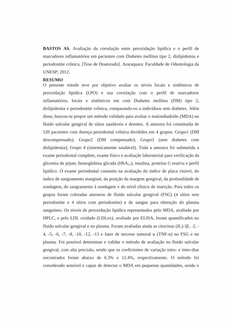
BASTOS AS. Avaliação da correlação entre peroxidação lipídica e o perfil de
marcadores inflamatórios em pacientes com Diabetes mellitus tipo 2, dislipidemia e
periodontite crônica. [Tese de Doutorado]. Araraquara: Faculdade de Odontologia da
UNESP; 2012.
RESUMO O presente estudo teve por objetivo avaliar os níveis locais e sistêmicos de
peroxidação lipídica (LPO) e sua correlação com o perfil de marcadores
inflamatórios, locais e sistêmicos em com Diabetes mellitus (DM) tipo 2,
dislipidemia e periodontite crônica, comparando-os a indivíduos sem diabetes. Além
disso, buscou-se propor um método validado para avaliar o malondiadeído (MDA) no
fluido sulcular gengival de sítios saudáveis e doentes. A amostra foi constituída de
120 pacientes com doença periodontal crônica divididos em 4 grupos: Grupo1 (DM
descompensado); Grupo2 (DM compensado); Grupo3 (sem diabetes com
dislipidemia); Grupo 4 (sistemicamente saudável). Toda a amostra foi submetida a
exame periodontal completo, exame físico e avaliação laboratorial para verificação da
glicemia de jejum, hemoglobina glicada (HbA1c), insulina, proteína C-reativa e perfil
lipídico. O exame periodontal consistiu na avaliação do índice de placa visível, do
índice de sangramento marginal, da posição da margem gengival, da profundidade de
sondagem, do sangramento à sondagem e do nível clínico de inserção. Para todos os
grupos foram coletadas amostras de fluido sulcular gengival (FSG) (4 sítios sem
periodontite e 4 sítios com periodontite) e de sangue para obtenção do plasma
sanguíneo. Os níveis de peroxidação lipídica representados pelo MDA, avaliado por
HPLC, e pelo LDL oxidado (LDLox), avaliado por ELISA, foram quantificados no
fluido sulcular gengival e no plasma. Foram avaliadas ainda as citocinas (IL)-1 , -2, -
4, -5, -6, -7, -8, -10, -12, -13 e fator de necrose tumoral α (TNF-α) no FSG e no
plasma. Foi possível determinar e validar o método de avaliação no fluido sulcular
gengival, com alta precisão, sendo que os coeficientes de variação intra- e inter-dias
encontrados foram abaixo de 6.3% e 12.4%, respectivamente. O método foi
considerado sensível e capaz de detectar o MDA em pequenas quantidades, sendo o
limite de quantificação (S/N = 5) de 0.03 μM, permitindo avaliação deste marcador
no fluido de sítios saudáveis. Foi observado que os sítios com doença periodontal
apresentavam-se com valores maiores de MDA do que os sítios saudáveis. Os
resultados mostraram que o grupo 1 apresentou maior severidade da doença
periodontal quando comparado aos demais grupos, em especial quanto ao
sangramento à sondagem, profundidade de sondagem≥6mm, nível de inserção
clínica≥5mm e supuração. Em relação aos parâmetros de LPO local e sistêmico, os
grupos com diabetes apresentaram maiores níveis de peroxidação quando
comparados aos grupos sem diabetes, sendo que o grupo 1 apresentou diferença
significativa também em relação ao grupo 2. Em relação às citocinas inflamatórias no
FSG, foi observado que o TNF-α, IL-6 e IL-10 estavam significativamente
aumentadas no fluido de pacientes com diabetes, sendo esta diferença maior no grupo
1. Foram verificadas correlações positivas e significantes entre os níveis de
peroxidação lipídica e citocinas no FSG (IL6, IL-10, TNF-α) bem como com
parâmetros de doença periodontal, dentre estes sangramento à sondagem,
profundidade de sondagem ≥ 6mm, nível de inserção clínica ≥ 5mm e presença de
supuração. Já no plasma, a expressão das citocinas seguiu um padrão decrescente do
grupo 1 ao grupo 4 tanto para citocinas pró- quanto anti-inflamatórias, sendo esta
diferença significante para (IL)-1 , -4, -6, -8 e TNF-α. Esta diferença ocorreu em
maior magnitude no grupo 1, embora nos pacientes portadores de diabetes com
adequado controle metabólico os níveis destas citocinas também estivessem mais
elevados em relação aos pacientes saudáveis. Os níveis de LPO plasmática
apresentaram correlação intermediária e positiva (p<0.0001) com níveis de IL1-
β(0.50), TNF-α.(0.40) e IL-6(0.40). Concluimos que a peroxidaçao lipídica pode ser
um mecanismo importante na expressão aumentada de marcadores inflamatórios e
maior severidade da doença periodontal no paciente com DM e dislipidemia.
Palavras-chave:Peroxidação de lipídeos, diabetes mellitus tipo 2, doenças
periodontais, citocinas
BASTOS AS. Correlation between lipid peroxidation and inflammatory markers
profile in Diabetes mellitus type 2 patients with dyslipidemia and chronic periodontal
disease. [Tese de Doutorado]. Araraquara: Faculdade de Odontologia da UNESP;
2012.
ABSTRACT The aim of this study is to evaluate the levels of the lipid peroxidation and its
correlation with systemic and local inflammatory markers profile in patients with
type 2 diabetes mellitus (DM2) dyslipidemia and chronic periodontitis compared to
systemically healthy patients. We also aimed to quantify a specific product of the
lipid peroxidation process, malondialdehyde (MDA), in gingival crevicular fluid and
to describe the validation of this method in this matrix using HPLC. The study
sample comprised 120 patients divided into four groups with 30 patients in each
group: group 1- diabetes with poor metabolic control with dyslipidemia, group 2-
diabetes with good metabolic control, group 3- without diabetes with dyslipidemia
and group 4- systemically healthy. All the subjects will go through a complete
periodontal examination and physical and laboratory evaluation in order to verify
fasting plasma glucose, glycated hemoglobin (HbA1c), lipid profile, insulin and high
sensitivity C-reactive protein. Periodontal examination will consist of visible plaque
index, gingival bleeding index, bleeding on probing, probing depth (PD) and clinical
attachment levels (CAL). Plasma and samples of gingival crevicular fluid (GCF) will
be collected in 4 sites without periodontal disease and 4 sites with periodontal
disease. The lipid peroxidation levels, evaluated by oxidized LDL (ox LDL) and
MDA were measured in GCF and in plasma. Inflammatory cytokines, (IL)-1 , -2, -4,
-5, -6, -7, -8, -10, -12, -13 and tumor necrosis factor-alpha α (TNF-α) were also
evaluated in plasma and GCF. It was possible to validate a HPLC-based method to
identify and quantify the MDA in GCF with sensitivity, precision, and accuracy even
in small concentrations as observed in healthy sites GCF. Samples’ intra- and
interday coefficients of variation were below 6.3% and 12.4%, respectively. The limit
of quantitation (S/N = 5) was 0.03 μM. GCF in the periodontal diseased sites
presented higher values of MDA than healthy sites for all groups. The results also
showed that all the periodontal parameters were considered worse in group 1 (DM
with inadequate metabolic control) when compared to the others groups, particularly
BOP, PD ≥ 6mm, CAL ≥ 5mm and suppuration. Regarding lipid peroxidation
evaluated by MDA in plasma and in gingival crevicular fluid (GCF), significant
differences were observed between the groups with diabetes when compared to the
groups without DM and G1 presented higher values of MDA when compared to G2
(p<0.05). The local inflammation assessed by cytokines in GCF(IL6, IL-10, TNF-α)
increased in all diabetic patients, being significant in the group with poorly controlled
diabetes and dyslipidemia. It was possible to verify significant and positive
correlations between GCF lipid peroxidation markers, GCF cytokines (IL6, IL-10,
TNF-α) and periodontal parameters such as bleeding on probing, PD ≥ 6mm, CAL ≥
5mm and presence of suppuration. The cytokines panel in plasma showed a
decreasing pattern from the poorly controlled diabetes to the healthy group being
significant to (IL)-1 , -4, -6, -8 e TNF-α. This difference was more evident is group 1
but even in well-controlled DM with dyslipidemia the levels of MDA, OxLDL and
inflammatory cytokines were significantly increased when compared to the non
DM/dyslipidemic group. There was a wide range of, moderate positive, correlations
observed between DM status, LPO markers and inflammatory cytokines expression,
such as MDA and IL1-β(0.50); MDA and TNF-α.(0.40); MDA and IL-6(0.40). These
findings indicate an important role for LPO in the severity of the local inflammatory
response to bacteria and the susceptibility to periodontal disease in DM patients and
suggest that LPO may represent an additive effect in the aggravation of inflammation
in DM and a role of increased oxidative metabolism in the inflammatory response.
Keywords: Lipid peroxidation, diabetes mellitus type 2, periodontal diseases,
cytokines

20
INTRODUÇÃO
U U
21
INTRODUÇÃO
A relação entre diabetes e doenças bucais não tem sido amplamente discutida
na literatura da mesma forma como se tem discutido os efeitos do diabetes em demais
órgãos 128. No entanto, evidências mostram que a periodontite é uma importante
complicação do diabetes 141 e uma das primeiras manifestações clínicas da doença 129.
Sendo a periodontite uma infecção, pode induzir ou perpetuar uma condição
inflamatória crônica 142 com possível impacto sobre o controle metabólico do
diabetes 73, 158. Desta forma, além da busca por um adequado controle glicêmico, a
prevenção e controle da periodontite são essenciais no tratamento integral do paciente
com diabetes 5, 87.
Diversos estudos buscam investigar as razões pelas quais a periodontite é mais
prevalente e severa em pacientes com diabetes quando comparados a pacientes sem
diabetes31, 60, 88, 112, 212, 227 e atribui-se ao pobre controle metabólico o papel de ser um
dos principais fatores de maior severidade136, 187, 227, 229. No entanto, as anormalidades
no metabolismo dos lipídios102, o estresse oxidativo6, 175, 201 e a peroxidação
lipídica210, 220 podem, caracterizar-se como importantes mecanismos relacionados à
patogênese da periodontite crônica nestes pacientes.
Recentemente estudos têm mostrado o papel das espécies reativas de oxigênio
(ROS), peroxidação lipídica e de atividade antioxidante nas complicações do diabetes
92, 110 e na patogênese da periodontite176, 246. No entanto, a avaliação da peroxidação
U

22
lipídica em ambas condições, diabetes e periodontite crônica, não tem sido
investigada.
Considerando que aumentos dos níveis de peroxidaçao lipídica podem
contribuir para danos significativos na integridade tecidual do hospedeiro 137, 153 por
diferentes mecanismos como dano ao DNA, oxidação de importantes enzimas e
estímulo à liberação de citocinas inflamatórias 66, 97, entende-se que estudos devem
ser conduzidos para investigar a influência da peroxidaçao lipídica sistêmica e
localmente, sobre o periodonto de pacientes com diabetes.
Doença periodontal
A doença periodontal é uma desordem inflamatória induzida por bactérias e
que afeta as estruturas de suporte dos dentes 1, 81, 129. É a maior causa de perdas
dentárias, acometendo aproximadamente 10-15 % da população mundial 14.
Apresenta-se mais severa em um grupo determinado da população, mais predisposto
à doença devido a uma reação inflamatória exacerbada frente a interação entre
hospedeiro e patógenos, causando dano desproporcional aos tecidos de suporte,
mobilidade e eventual perda do elemento dentário 82, 86, 128.
O fator etiológico primário é o biofilme bacteriano, sendo que a bolsa
periodontal pode conter diversas espécies microbianas diferentes, das quais uma
pequena quantidade é considerada periodontopatogênica, podendo participar
diretamente da doença periodontal cada uma com seu potencial de indução e
virulência 125, 219. O início e a progressão da periodontite envolvem a colonização
23
bacteriana da superfície dentária, penetração das bactérias e seus produtos no tecido
conjuntivo e estímulo da resposta inflamatória, a qual induz destruição do tecido
conjuntivo e osso alveolar e limita o processo de reparo tecidual 54.
A avaliação de marcadores inflamatórios relacionados à destruição do
periodonto tem sido constante na busca de elucidar os mecanismos de início e
progressão da doença periodontal. Estudos mostram que indivíduos com periodontite
apresentam níveis elevados de citocinas inflamatórias no plasma sanguíneo 10, 45 e no
periodonto, este último avaliado por meio de biópsia do tecido gengival 25, 194 ou de
coleta do fluido sulcular gengival 46. O fluido sulcular gengival (FSG) é considerado
um transudato sérico em condições de saúde dos tecidos e quando do estabelecimento
de condições inflamatórias periodontais é considerado um exsudato inflamatório
oriundo dos tecidos que circundam o periodonto 129. Tal fluido pode conter células
inflamatórias do hospedeiro e fatores derivados do plasma bem como produtos dos
microrganismos que compõem o biofilme supra e subgengival 59, 129. O valor do
fluido sulcular gengival como potencial fluido de diagnóstico é reconhecido por
estudos prévios que constataram que sua composição pode contribuir para a detecção
de alterações relacionadas ao início da doença periodontal 180, sendo uma amostra de
natureza sítio-específica 85 e que pode ser coletada usando métodos não invasivos 129,
servindo como uma excelente fonte de informação do paciente 41,190.
Atualmente, é bem estabelecido que o dano tecidual que ocorre na
periodontite é atribuído mais à resposta inflamatória do hospedeiro do que ao próprio
24
efeito direto das bactérias e seus produtos 12, 249, 257. Desta forma, passou-se a estudar
a influência de diversos fatores, além da microbiota, na instalação, progressão e
etiopatogênese da doença periodontal. Em especial, a influência de condições
sistêmicas, como o diabetes mellitus 115, 130, 158 e as anormalidades do metabolismo de
lipídios 47, 65, 162 têm sido avaliados como fatores modificadores do processo
imunoinflamatório da periodontite.
Diabetes mellitus
O Diabetes mellitus é um grupo de doenças metabólicas, que envolve
primariamente os carboidratos, seguido de lipídios e proteínas, sendo caracterizado
pela hiperglicemia resultante de defeitos na secreção de insulina, em sua ação ou em
ambos. Como resultado das alterações metabólicas e do desequilíbrio osmótico, uma
tríade clínica clássica de sintomas é desenvolvida e inclui polifagia, polidipsia e
poliúria 3.
O diagnóstico do Diabetes mellitus é baseado na história médica, exames
clínico e laboratoriais, como o exame de glicemia de jejum ( 126 mg/dL), valores
casuais de glicose sangüínea ( 200 mg/dL) ou teste de tolerância à glicose anormal.
Entretanto, para monitorar o controle glicêmico do paciente já diagnosticado como
portador de diabetes, o exame de hemoglobina glicada A1c (HbA1c) é validado como a
ferramenta de maior confiabilidade, fornecendo um registro de aproximadamente 90
dias da condição glicêmica do paciente, devendo ser inferior a 7% 3, 49, 235.
25
As formas mais comuns da doença são o tipo 1 (DM1) e tipo 2 (DM2). O
Diabetes mellitus tipo 1 é resultante da destruição auto-imune das células β
pancreáticas, responsáveis pela produção de insulina, enquanto o tipo 2 é causado
pela resistência das células alvo à ação da insulina com utilização deficiente do
hormônio, podendo estar combinada a um defeito na secreção do mesmo. A maior
parte dos pacientes portadores de DM2 são obesos e possuem um aumento do
percentual de gordura corporal distribuída principalmente na região abdominal 3.
O aparecimento da doença está relacionado a fatores de risco genéticos,
ambientais e comportamentais 116 com aumento da prevalência em todo o mundo,
sendo estimado que mais de 300 milhões de indivíduos serão afetados até 2030 248.
Muitos destes pacientes, particularmente do tipo 2, permanecem sem diagnóstico por
muitos anos, uma vez que a hiperglicemia aparece gradualmente gerando pouca
sintomatologia no início da doença 51.
As complicações do diabetes elevam os custos econômicos associados às
despesas com assistência médica e redução da capacidade produtiva destes paciente,
sendo uma das principais causas do aumento da mortalidade e morbidade destes
pacientes 62. Tais complicações estão associadas a hiperglicemia crônica
desencadeando uma cascata de eventos que levam a mudanças estruturais dos tecidos
afetados 101resultando em prejuízo a diversos órgãos em especial olhos, rins, coração,
nervos e vasos sangüíneos 159,.
Sabe-se que quando o excesso de glicose interage de forma não-enzimática e,
26
frequentemente, irreversível com proteínas, lipídios e ácidos nucléicos, ocorre a
formação de produtos finais da glicação avançada (AGE-advanced glycation end-
products) 28, sendo um mecanismo importante no desenvolvimento das complicações
do DM. Tanto macrófagos como células endoteliais apresentam receptores
específicos para AGES denominados RAGES e esta interação resulta em diversas
alterações celulares como o aumento da permeabilidade vascular e estímulo à
secreção de citocinas pró-inflamatórias, dentre elas a interleucina 1 (IL-1 ) e o fator
de necrose tumoral (TNF- ) 127, 253. A relação causal entre o aumento de AGEs e as
complicações do diabetes foi demonstrada em modelos animais em que a inibição
destes reduziu significativamente o processo de aterosclerose em animais com
diabetes induzido sem afetar o controle da glicemia 67.
Outro mecanismo biológico envolvido nas complicações associadas ao
diabetes, especialmente as complicações vasculares, é a alteração do metabolismo
lipídico. Portadores de diabetes podem apresentar resistência insulínica154,
adiposidade visceral e alteração do metabolismo lipídico 155, esta última sendo
definida na literatura como dislipidemia diabética. Apesar da possibilidade de
alteração de todas as frações de lipídios, o fenótipo mais comum é caracterizado pela
redução do colesterol de alta densidade (HDL), e elevação de triglicérides e do
colesterol de baixa densidade (LDL) 36, 121, 167.
A patogênese da dislipidemia no diabetes não é bem compreendida, no
entanto sabe-se que as anormalidades no metabolismo dos lipídios podem estar
27
presentes antes mesmo do estabelecimento da hiperglicemia, devido à possível
associação com a resistência insulínica 75, 90, 122. Acredita-se que a resistência
insulínica prejudica a capacidade dos adipócitos de armazenar triglicérides e pode
levar ao aumento da enzima lipoproteína lipase, que resulta no aumento da liberação
de ácidos graxos livres. O maior influxo destes ácidos graxos no fígado na presença
de estoques de glicogênio adequados promove a produção de triglicérides, a qual
estimula a secreção de apolipoproteína B e LDL colesterol 160.
A dislipidemia diabética desempenha um importante papel na resposta imune,
predispondo o paciente a um estado hiperinflamatório por modular a função e
atividade dos macrófagos 106, 230, 238, desencadeando a ativação de diversas vias
relacionadas à liberação de mediadores oxidativos e inflamatórios e fatores de
crescimento que levam a danos em diversos órgãos e tecidos 47, 101.
A relação entre diabetes e resposta imunológica é bidirecional e com
possibilidades de convergência das vias metabólica e inflamatória em múltiplos
níveis 99. Sabe-se que a resposta imunoinflamatória pode inibir eventos moleculares
relacionados à regulação da insulina, exercendo um papel fundamental no
metabolismo glicêmico 99. Reciprocamente, o diabetes pode modular a resposta
imune, levando a um aumento da magnitude da resposta inflamatória frente a
patógenos e seus produtos 43, 171, persistência do infiltrado inflamatório 139, aumento
do metabolismo oxidativo 84, inibição da função de polimorfonucleares 53, transtornos
no metabolismo do tecido conjuntivo 54, 79, inibição da proliferação de células
28
osteoblásticas e diminuição da formação óssea21, 144, menor resistência a infecções 177
e dificuldade na cicatrização de feridas 215.
Sendo assim, o controle de infecções, adequado controle glicêmico e a
manutenção dos níveis dos lipídicos séricos dentro da normalidade é uma meta a ser
atingida nos pacientes com Diabetes mellitus, principalmente pelo fato de tais
alterações estarem associadas com o desenvolvimento de complicações que elevam a
morbidade e mortalidade do paciente portador de DM 3.
Doença periodontal e diabetes
Atualmente, a periodontite pode ser reconhecida como a sexta complicação
associada ao diabetes 141 e, de fato, o diabetes é um fator de risco estabelecido para a
periodontite com maior prevalência, severidade e progressão em indivíduos com
diabetes em comparação a indivíduos saudáveis 41, 60, 115, 119, 158, 187, 226, 227. Tal relação
foi confirmada em recente revisão sistemática na qual foi constatado, após rigorosos
ajustes para potenciais fatores de confundimento, a ocorrência de maior extensão e
severidade da periodontite em pacientes com DM quando comparados a pacientes
sem DM 39.
O efeito negativo e recíproco da periodontite sobre o diabetes foi demonstrado
em estudos clínicos e longitudinais que observaram que a inflamação do periodonto
pode estar relacionada a um risco aumentado de desenvolver complicações do
diabetes como proteinúria, angina, infarto do miocárdio, derrame 231, falência renal
29
198, nefropatia 213, tolerância à glicose diminuída 189, retinopatia 174 e maiores níveis
de glicose sanguínea quando comparados a paciente sem diabetes 143.
Relata-se ainda que pacientes com inadequado controle metabólico (HbA1c ≥
7%) apresentam progressão mais rápida da periodontite em comparação a pacientes
com adequado controle metabólico (HbA1c < 7%), como demonstrado em diversos
estudos 16, 136, 187, 227, 229, que mostram o impacto do pobre controle metabólico sobre a
severidade da periodontite.
Os estudos têm demonstrado que a resposta imunoinflamatória do hospedeiro
frente ao desafio bacteriano representa um dos fatores mais importantes para a maior
destruição periodontal e que esta pode ser a responsável pela maior severidade da
doença periodontal no diabetes 43, 128, 171. É bem estabelecido em estudos in vitro, em
animais e em seres humanos que o diabetes induz à exacerbação da resposta
inflamatória, demonstrada por alterações celulares e teciduais. Evidências mostram
que no diabetes existe aumento da permeabilidade vascular do tecido gengival 214 e
que os neutrófilos apresentam-se com deficiência de suas funções em nível de
aderência, quimiotaxia e fagocitose 78, 148. Já monócitos e macrófagos demonstram
fenótipo hiperinflamatório, o que resulta na produção de mediadores e citocinas pró-
inflamatórias em níveis aumentados 191, mesmo em sítios com periodontite leve 192.
Além disso, tem sido sugerido que no diabetes existe uma diminuição da
quantidade de colágeno nos tecidos gengivais pela redução da síntese 54, 79 e aumento
da degradação 124, 204, 208, deficiência na cicatrização de feridas 140, 215, 216 e
30
persistência do infiltrado inflamatório por um período mais longo 83, contribuindo
para o extenso dano tecidual observado na periodontite de pacientes com diabetes.
Em se tratando de tecido ósseo, diversos autores investigaram a influência do
diabetes sobre fatores relacionados à osteoclastogênese nas infecções periodontais,
tendo sido demonstrado que a hiperglicemia pode modular as taxas de RANKL
(receptor activator of nuclear factor kappa B) e OPG (osteoprotegerina) nos tecidos
periodontais 57, 96, 131, 147, 197, além de aumentar a apoptose de células ósseas,
prejudicando a formação e a reabsorção óssea 96. Estudos mostram que, além da
reduzida formação óssea alveolar, há também um limitado reparo ósseo 54, 139 o que
poderia explicar, pelo menos em parte, o aumento da destruição óssea alveolar
observada em pacientes com DM.
Assim como nas demais complicações do diabetes, o papel da interação dos
produtos de glicação avançada (AGE) com seus receptores RAGEs também tem sido
investigado na doença periodontal. Estudos demonstram que existem receptores
RAGEs no tecido gengival 109, 202 de pacientes com diabetes e que níveis séricos de
AGEs foram associados com a extensão da doença periodontal em adultos com DM
tipo 2 224. Níveis aumentados de AGEs e sua interação com seus receptores estão
associados com a supressão da produção de colágeno por fibroblastos da gengiva e do
ligamento periodontal 164, 183, liberação de mediadores inflamatórios como
interleucina 1 (IL-1 ) e fator de necrose tumoral (TNF- ) 127, 253 promoção da
31
osteoclastogênese 55, 255 e atraso da reparação tecidual 80, sendo tais eventos cruciais
no desenvolvimento e progressão da doença periodontal.
Alguns estudos sugerem ainda que as dislipidemias 47, 69, 102 e o aumento do
metabolismo oxidativo 6, 175, 202, possam ser possíveis mecanismos que podem levar a
uma maior severidade da doença periodontal em pacientes com diabetes
principalmente pelo fato de estimularem a liberação de citocinas pró-inflamatórias 17,
76, 97, 135, 179. Pacientes com DM tipo 2 e elevados níveis séricos de triglicérides podem
apresentar piores parâmetros clínicos periodontais 47 enquanto níveis elevados de
colesterol podem estar correlacionados com a média aumentada da profundidade de
sondagem periodontal 69. Além disso, marcadores de estresse oxidativo foram
detectados no tecido gengival 202 e no plasma de pacientes 6 com diabetes e com
periodontite. No entanto, estes estudos são escassos e falham em correlacionar os
níveis de oxidação encontrados com dados clínicos periodontais e ainda com a
expressão de marcadores inflamatórios locais e sistêmicos nestes pacientes.
Estudos clínicos e epidemiológicos fornecem evidências científicas para
estabelecer a relação entre doença periodontal e diabetes. Contudo, as investigações
sobre o impacto do diabetes na periodontite, deveriam levar também em consideração
fatores de risco adicionais à doença periodontal, como a síndrome metabólica 48, 126,
163, 169, 232, a obesidade 35, 114, 200, 211 e a dislipidemia 102, condições que estão
intimamente ligadas ao DM tipo 2 128, 188 e que demonstram ter um efeito potencial
32
sobre o estado inflamatório local e sistêmico do paciente com diabetes, podendo
interferir na patogênese da periodontite.
Estresse oxidativo e peroxidação lipídica
As espécies reativas de oxigênio (EROs) (reactive oxygen species- ROS) são
produzidas continuamente pelas células sob condições fisiológicas, sendo
consideradas bioprodutos normais do metabolismo 145 e, durante a fagocitose, esses
radicais livres são liberados em maior quantidade como parte da reação bactericida 13,
207. No entanto, apesar de desempenharem funções fisiológicas, sabe-se que ocorre
uma produção de espécies reativas de oxigênio acima do normal em sítios de
inflamação crônica 92 e que tais compostos deixam de exercer uma função meramente
fisiológica, passando a contribuir para a injúria inflamatória dos tecidos do
hospedeiro 207.
As células desenvolvem diversos mecanismos de defesa antioxidante para
neutralizar o dano causado pelas ROS no organismo 94, 145. Tal sistema antioxidante
interage de forma conjunta e é composto de sistema enzimático (enzima superóxido
dismutase (SOD), catalase (CAT) e glutationa peroxidase (GPx)) e pelo sistema não
enzimático (vitamina C-ácido ascórbico, vitamina E, ácido úrico, selênio,
carotenóides, entre outros) 72. No entanto, quando uma condição patológica se
estabelece, ocorre a saturação da capacidade antioxidante celular devido ao freqüente
estímulo do eixo de sinalização sensível a alterações no mecanismo de oxidação-
33
redução. Este desequilíbrio no sentido pró-oxidativo 56, 236 desencadeia vias
bioquímicas associadas com o desenvolvimento de processos inflamatórios 92, 181 e de
doenças crônicas, como a aterosclerose, o diabetes e suas complicações 29.
Os radicais livres, em especial o radical hidroxila e o peroxinitrito, têm a
tendência de afetar os diferentes tipos de ácidos graxos poliinsaturados das
membranas celulares e lipídios circulantes (colesterol e lipoproteína de baixa
densidade) resultando em um processo conhecido como peroxidação lipídica (PL) 76,
105, 236. A interação das espécies reativas de oxigênio com os ácidos graxos das
membranas celulares altera a estrutura dos lipídios, causando modificações biológicas
e fisiológicas 170, 222 como a diminuição da fluidez e alteração da atividade de ligação
entre a membrana, enzimas e receptores 152. O aumento da peroxidação de lípídios em
nível sanguíneo atinge primeiramente as células endoteliais e, a seguir, tecidos e
órgãos intactos 252, gerando indesejáveis produtos oxidados responsáveis por causar
ou contribuir para a progressão de doenças 105, 236.
A produção descontrolada de peróxidos de lipídios pode provocar dano à
integridade celular e tecidual 137, 153 por mecanismos diferentes incluindo alterações
do DNA e proteínas, oxidação de enzimas e estímulo à liberação de citocinas pró-
inflamatórias 17, 76, 97, 135, 179, 240. Desta forma, a peroxidação lipídica tem sido
considerada um importante mecanismo patogênico em doenças associadas com
mecanismos de infiltração fagocitária em resposta a patógenos 37, 92, no
envelhecimento 151, na aterosclerose 92, no desenvolvimento de complicações do
34
diabetes como alterações micro e macrovasculares 110, 241, na artrite reumatóide 149, na
doença pulmonar obstrutiva crônica 146, na AIDS 58 e na doença periodontal 4, 38, 233,
246.
Diversos marcadores de peroxidação lipídica têm sido utilizados para
monitorar este processo, empregando diferentes metodologias 2, 4, uma vez que
espécies reativas de oxigênio são difíceis de serem detectadas por serem instáveis e
com meia-vida curta 52. No entanto, para um método ser considerado ideal na
mensuração da peroxidação lipídica deve: identificar e quantificar produtos derivados
especificamente do processo de peroxidação (estáveis in vivo e in vitro); ter baixo
coeficiente de variação; não estar sujeito a interferências de outras biomoléculas;
empregar técnicas confiáveis; não sofrer interferência direta da dieta e ter
sensibilidade para avaliação de níveis basais dos produtos 2, 37.
O processo de peroxidação lipídica como uma medida geral de oxidação de
lipídios, sem considerar a peroxidação de uma fração de lipoproteína específica 52, 92,
pode ser avaliado por meio do malondialdeído (MDA). Este é o principal e o mais
estudado produto da peroxidação lipídica de ácidos graxos poliinsaturados 50, 52 e tem
sido descrito como um importante indicador de injúria à membrana celular, estando
aumentado em situações de estresse oxidativo 52, 153. Diversos métodos têm sido
utilizados para mensurar os níveis de peroxidação lipídica por meio do MDA 4, 64, 153,
176, 221, 233, 234, 252. No entanto, alguns destes métodos, em especial a reação chamada de
“teste das substâncias reativas com ácido tiobarbitúrico (SRATB)” ou TBARs, têm
35
recebido críticas devido à falta de especificidade na avaliação efetiva dos níveis de
MDA sem interferências de outras moléculas 2, 118. Sendo assim, o método para
avaliação do MDA por meio de cromatografia líquida de alta eficiência (HPLC) tem
sido considerado mais específico, confiável e reprodutível, uma vez que o MDA é
eficientemente separado de outros cromógenos que poderiam vir a interferir na reação
4, 37, 40, 68, 89, 98, 256.
No entanto, existe ainda a mensuração da peroxidação de frações lipídicas
específicas como a oxidação da LDL (low density lipoprotein). A LDL oxidada
(LDL-ox) é uma forma modificada da LDL com importantes propriedades pró-
inflamatórias 195, 196 e também é utilizada como um biomarcador de peroxidação
lipídica 42, 173. Está relacionada com níveis aumentados da HbA1c 100 e com o
desenvolvimento de complicações vasculares do diabetes 20. A LDL-ox tem efeitos
importantes sobre as células, sendo relatado maior recrutamento de linfócitos T e de
monócitos circulantes assim como apoptose de linfócitos T, ambos os eventos
estando relacionados ao desenvolvimento de lesões ateroscleróticas 157. Outros
eventos incluem: citotoxicidade para fibroblastos 161, aumento da expressão de
metaloproteinase- 9 (MMP-9) e aumento da secreção de citocinas como IL-1 , IL-12
223 e TNF- 106.
Peroxidação lipídica e diabetes
Níveis altos de glicose no sangue podem levar à maior formação de radicais
36
livres 9, 111, 234 aumentando o estresse oxidativo 22, a peroxidação lipídica 103, 152 e
diminuindo os níveis de defesa antioxidante 11, 34, 108. Reciprocamente, evidências
sugerem que níveis elevados de estresse oxidativo e peroxidação lipídica podem ser
importantes mecanismos na produção, liberação ou função da insulina 24, 247, na
disfunção e morte de células beta pancreáticas 133, no estabelecimento de condições
de pré-diabetes e diabetes, podendo ainda ser responsável por complicações
associadas à doença 77, 111, 150, 152, 237. No entanto, ainda existem controvérsias se os
níveis aumentados do estresse oxidativo e da peroxidação lipídica estão meramente
associados ao DM ou se apresentam relação causal com o diabetes 152.
Apesar de alguns autores não encontrarem aumento dos níveis de peroxidação
lipídica no diabetes 241, diversos estudos 9, 104, 107, 160, 234 observaram que pacientes
com diabetes estão expostos a um aumento do estresse oxidativo por meio da
peroxidação lipídica 152. Estudos mostram que pacientes com DM tipo 2 apresentam
elevados níveis de marcadores de peroxidação lipídica como a LDL-oxidada 42, 100,
166, 193, o malondialdeído (MDA) 9, 84, 107, 160, 234 e o TBARs, quando comparados a
pacientes sistemicamente saudáveis 111, 165. Além disso, níveis elevados de PL foram
correlacionados positivamente com a glicemia de jejum, HbA1c e colesterol total 111,
165, sugerindo que pacientes com diabetes descompensado e com complicações têm
aumento sérico de PL 152.
As alterações no metabolismo de lipídios e os mecanismos de formação dos
peróxidos de lipídios no diabetes bem como os seus efeitos na função e estrutura
37
celular ainda não estão completamente elucidados 247, mas têm recebido significativa
importância no estudo da diabetogênese 152, principalmente quanto ao entendimento
do papel da dislipidemia diabética e da peroxidação de lipídios na modulação da
resposta imunoinflamatória.
No diabetes, o microambiente dislipidêmico pode ser alterado também pela
presença de produtos finais da glicação avançada (AGEs), os quais podem agir como
co-estimuladores das células. A própria LDL-colesterol, como qualquer outra
proteína plasmática, é susceptível à glicação formando um AGE 202. Os AGEs
presentes no plasma sanguíneo interagem com seus receptores específicos (RAGEs) e
esta interação resulta em diversas alterações patológicas, basicamente envolvendo a
modulação da resposta imune.
Desta forma, a hiperglicemia associada à dislipidemia e ao diabetes
descompensado aumentam a formação de AGEs, incluindo LDL-AGE, bem como
implicam em maior peroxidação lipídica representada por um aumento na quantidade
de LDL oxidada 117, 202, 225. Tanto AGEs como LDL-ox, presentes concomitantemente
no sangue de pacientes com diabetes, apresentam diversos efeitos biológicos sobre
linfócitos e monócitos, aumentando a expressão de citocinas inflamatórias devido à
ativação de fatores de transcrição como o NF-κB 254 que são conhecidos por serem
sensíveis a alterações do metabolismo oxidativo 205, 253. Tais eventos resultam na
regulação do crescimento e proliferação celular bem como no início de um processo
inflamatório difuso 43, 95, os quais podem ter impacto significativo na patogênese de
38
diferentes complicações associadas ao diabetes, como insuficiência renal 67, doenças
vasculares e menor resistência a infecções 177.
Peroxidação lipídica e doença periodontal
Estudos recentes têm demonstrado que pacientes com doença periodontal
crônica têm níveis de peroxidação lipídica aumentados no plasma 4, 246, na saliva 32, 37,
113, no tecido gengival 25, 202 e no fluido sulcular gengival 4, 233, 246.
Alguns autores verificaram uma correlação positiva entre os níveis de MDA
plasmáticos 4 e do fluido sulcular gengival 4, 233 com parâmetros clínicos periodontais
como índice gengival, profundidade de sondagem e nível de inserção clínica 4, 233,
indicando que níveis aumentados de peroxidação lipídica podem ter um papel
importante na inflamação e destruição na periodontite.
O efeito do aumento do metabolismo oxidativo no tecido periodontal e em
seus componentes celulares têm sido investigado em estudos in vivo e in vitro 6, 175,
201, 205, 210, 218. Evidências têm mostrado que níveis aumentados de peroxidação
lipídica podem contribuir para danos significativos ao periodonto 38, levando a maior
inflamação e destruição tecidual e a alterações degenerativas, como necrose e
destruição do osso alveolar na região do infiltrado inflamatório 218.
Localmente, o dano tecidual ao periodonto pode ser causado diretamente por
um desequilíbrio do sistema antioxidante nos locais de inflamação 27, 176 e pelo
aumento do metabolismo oxidativo durante a ativação de neutrófilos e macrófagos
39
por periodontopatógenos e seus produtos 25, 209, 210, 233. A presença do ânion
superóxido, potente indutor de peroxidação lipídica 123, 155 foi demonstrada em áreas
de reabsorção adjacente a osteoclastos, levando à reabsorção óssea 245. Tem sido
demonstrado que o colágeno, juntamente com outras proteínas, é susceptível ao
ataque direto de espécies reativas do oxigênio podendo interagir com produtos da
peroxidação lipídica como o malondialdeído 217, levando a dano aos tecidos
periodontais de proteção e sustentação. Durante o processo inflamatório e oxidativo
ocorre degradação de proteoglicanas e componentes extracelulares da matriz 59, 244,
alterações nas funções dos fibroblastos como adesão, proliferação e longevidade 185 e
estímulo à diferenciação 71 e ativação 19, 91 de osteoclastos.
O dano causado pela oxidação aos tecidos periodontais também pode ocorrer
indiretamente via desequilíbrio do eixo oxidação-redução ativando fatores de
transcrição como a proteína-1 (AP-1) e fator nuclear-kB (NF-kB) 7, 178,
desencadeando a liberação de mediadores inflamatórios e apoptóticos como citocinas
e moléculas de adesão celular 92, mecanismos considerados importantes na
patogênese da doença periodontal 132, 168.
No entanto, estudos mais recentes que investigaram o papel da peroxidação
lipídica em pacientes com periodontite 4, 25, 32, 113, 202, 233, 246, não consideraram o perfil
imunoinflamatório local e/ou sistêmico do indivíduo, tornando difícil, estabelecer
uma correlação entre achados clínicos periodontais e de peroxidação lipídica com a
magnitude da resposta inflamatória gerada.
40
Peroxidação lipídica e severidade da doença periodontal no paciente com
diabetes
As evidências atuais de fato comprovam a relação bidirecional entre diabetes
e doença periodontal 88, 112, 158, 227. No entanto os mecanismos precisos pelos quais
esta interação ocorre ainda permanecem incertos. O estado hiperinflamatório que
existe em ambas condições tem sido proposto como sendo um elo comum. No
entanto, suspeita-se que algum mecanismo adicional esteja atuando para amplificar
sinergisticamente a bioquímica e o curso clínico destas doenças 7.
Estudos recentes têm demonstrado que as anormalidades no metabolismo dos
lipídios e a peroxidação lipídica podem, além do pobre controle glicêmico,
caracterizar-se como um importante mecanismo associado à maior severidade e
extensão da doença periodontal em pacientes com diabetes 43, 202, 220. Sugere-se que
espécies reativas de oxigênio, por meio da peroxidação lipídica, cause danos diretos e
indiretos ao tecido periodontal e seus componentes celulares e teciduais, conforme
relatado acima.
O efeito da peroxidação lipídica sobre o periodonto pode ser ainda mais
severo em pacientes que apresentam condições sistêmicas que predispõem o
indivíduo à produção de PL em níveis exacerbados, como é o caso de pacientes com
dislipidemia 86 e diabetes, em especial com inadequado controle metabólico 172. No
41
entanto, os estudos clínicos que investigaram a peroxidação lipídica em pacientes
com diabetes e periodontite crônica são escassos 43, 202, 220.
O estudo da peroxidação lipídica plasmática, mensurada por meio do TBARs,
em pacientes com diabetes e doença periodontal demonstrou uma correlação fraca
entre PL e expressão de metaloproteinases da matriz-8 (MMPs), que dentre outras
funções são responsáveis por prejudicar a formação do colágeno 43.
Além disso, o acúmulo de AGEs e de produtos de peroxidação lipídica foi
demonstrado em diversos órgãos, inclusive na gengiva de pacientes com diabetes 202
e no endotélio e epitélio vascular gengival de pacientes portadores de diabetes com
periodontite 109. Sendo a doença periodontal uma condição que se desenvolve frente a
um desafio bacteriano, o acúmulo periférico ou local de AGE e de produtos de
peroxidação lipídica, pode prejudicar a capacidade antioxidante 156 ao mesmo tempo
em que aumenta a liberação de mediadores inflamatórios 29, 63, contribuindo para
dano aos tecidos periodontais 6. Avaliando a peroxidação lipídica por meio do
anticorpo anti-malondialdeído modificado pela LDL (MDA-LDL), em uma amostra
reduzida de pacientes, foram constatados níveis aumentados de PL em indivíduos
com diabetes e doença periodontal e que após o tratamento periodontal os níveis de
peroxidação lipídica foram significativamente reduzidos 220.
No entanto, os estudos acima citados apresentam certas deficiências
metodológicas que dificultam a interpretação dos dados de maneira conjunta, pois
alguns deles não consideraram os níveis de dislipidemia destes pacientes 43, 202, 220 ou
42
avaliaram a PL por meio de metodologias pouco específicas 43, apenas um estudo
avaliou os níveis de peroxidação lipídica no tecido periodontal 202 e nenhum
correlacionou tais achados com a resposta inflamatória sistêmica e local.
O paciente com diabetes e dislipidemia apresenta por si só um estado
hiperinflamatório em nível celular e molecular, que leva à maior destruição tecidual e
dificuldade de reparo. Tais condições combinadas, em especial em pacientes com
pobre controle glicêmico, predispõem a níveis de peroxidaçao lipídica aumentados
agravando o processo inflamatório e o dano tecidual. No entanto não existem estudos
que avaliem o estado periodontal e o papel da peroxidação lipídica em pacientes com
diabetes, dislipidemia e doença periodontal. Sendo que tais condições clínicas são, na
maioria das vezes, indissociáveis 75, 90, 122.
Neste contexto, sugere-se que a LPO possui um papel importante na
severidade do processo inflamatório relacionado à periodontite crônica em pacientes
com DM e dislipidemia. Em virtude do exposto acima, parece-nos oportuno estudar,
em pacientes portadores de Diabetes mellitus tipo 2, com dislipidemia, a correlação
entre peroxidação lipídica e o perfil imunoinflamatório, local e sistêmico.

43
PROPOSIÇÃO
44
2 PROPOSIÇÃO
2 Objetivo geral
O presente estudo teve por objetivo avaliar a correlação entre peroxidação
lipídica e o perfil de marcadores inflamatórios, locais e sistêmicos, em pacientes com
Diabetes mellitus (DM) tipo 2, dislipidemia e periodontite crônica, comparando-os a
indivíduos sem diabetes.
2.3 Objetivos específicos
- Avaliar a peroxidação lipídica plasmática e seu papel na modulação da
resposta imune, por meio do estímulo à liberação de citocinas inflamatórias;
- Validação de um método de avaliação dos níveis de peroxidação lipídica no
fluido sulcular gengival de sítios saudáveis e com periodontite crônica;
- Avaliar o impacto da peroxidação lipídica local e sistêmica sobre parâmetros
de severidade da periodontite crônica e expressão de citocinas inflamatórias no fluido
sulcular gengival.

45
CAPÍTULOS
U
U
46
CAPÍTULO 1
Quantitation of malonaldehyde in gingival crevicular fluid by
a high-performance liquid-chromatography-based method
Alliny de Souza Bastos, Ana Paula de Melo Loureiro, Tiago Franco de Oliveira,
Sâmia Cruz Tfaile Corbi, Raquel Mantuaneli Scarel Caminaga, Carlos Rossa Júnior,
Silvana Regina Perez Orrico
Artigo publicado na revista Analytical Biochemistry
Publisehd online 28 Jan 2012
PMID: 22330745
47
Quantitation of malonaldehyde in gingival crevicular fluid by a high-
performance liquid-chromatography-based method
Alliny de Souza Bastos1, Ana Paula de Melo Loureiro3, Tiago Franco de Oliveira3,
Sâmia Cruz Tfaile Corbi1, Raquel Mantuaneli Scarel Caminaga2, Carlos Rossa
Júnior1, Silvana Regina Perez Orrico1
1UNESP – Univ Estadual Paulista – Araraquara School of Dentistry, Department of
Diagnosis and Surgery, Araraquara, Brazil.
2UNESP – Univ Estadual Paulista – Araraquara School of Dentistry, Department of
Morphology, Araraquara, Brazil.
3USP – Univ São Paulo- Faculty of Pharmaceutical Sciences, Department of Clinical
and Toxicological Analyses, São Paulo, Brazil.
Short Title: Malonaldehyde in gingival crevicular fluid by HPLC
Corresponding author:
Profª. Drª. Silvana Regina Perez Orrico,
UNESP – Univ Estadual Paulista; Araraquara School of Dentistry
Department of Diagnosis and Surgery
Rua Humaitá, 1680, 2º andar
CEP: 14801-903, Araraquara, São Paulo, Brazil.
Phone: +55 (16) 3301-6377 Fax: +55 (16) 3301-6369
E-mail: [email protected]
Table of Contents: Chromatographic Techniques
48
ABSTRACT
Lipid peroxidation has been associated with periodontal disease, and the evaluation of
malonaldehyde (MDA) in the gingival crevicular fluid (GCF), an inflammatory
exudate from the surrounding tissue of the periodontium, may be useful to clarify the
role of lipid peroxidation in the pathogenesis of periodontal disease. We describe the
validation of a method to measure MDA in the gingival crevicular fluid using HPLC.
MDA calibration curves were prepared with PBS solution spiked with increasing
known concentrations of MDA. Healthy and diseased GCF samples were collected
from the same patient to avoid interindividual variability. MDA response was linear
in the range measured, and excellent agreement was observed between added and
detected concentrations of MDA. Samples’ intra- and interday coefficients of
variation were below 6.3% and 12.4%, respectively. The limit of quantitation (S/N =
5) was 0.03 μM. When the validated method was applied to the gingival crevicular
fluid, excellent agreement was observed in the MDA quantitation from healthy and
diseased sites, and diseased sites presented more MDA than healthy sites (p < 0.05).
In this study a validated method for MDA quantitation in gingival crevicular fluid
was established with satisfactory sensitivity, precision, and accuracy.
Keywords: Lipid peroxidation, malondialdehyde, periodontal diseases, gingival
crevicular fluid, HPLC.
49
INTRODUCTION
Reactive oxygen species (ROS) are produced as an integral feature of normal
cellular metabolism under physiological conditions [1]. The generation of ROS is an
important mechanism during phagocytosis as part of the bactericidal reaction [2,3,4].
However, it has been well-established that over-production of ROS occurs at sites of
chronic inflammation [4], and uncontrolled production of lipid peroxides can result in
oxidative stress. These conditions can contribute to injury of the host tissue [5] and
significant damage to cell integrity [6,7] by different mechanisms such as DNA
damage, oxidation of important enzymes, lipid peroxidation, and stimulation of
proinflammatory cytokine release [8,9].
Lipid peroxidation (LPO) is thought to play an important role in ageing,
atherosclerosis [4], late complications of diabetes mellitus [10] such as micro- and
macrovascular alterations [11,12], rheumatoid arthritis [13], chronic obstructive
pulmonary disease [14], and periodontitis [3,15,16,17,18].
Periodontitis is an infectious inflammatory disease involving the connective
tissue and bone that support the teeth, and its primary etiological factor is the biofilm
constituted by several pathogenic bacteria. The severity of periodontal disease is
determined by the interactions between host defense and pathogens and can lead to
periodontal destruction and lost teeth [19,20]. There have been many investigations
regarding the systemic conditions and modifier factors that can be involved in the
pathogenesis of periodontitis. Recently, more studies have focused on the roles of
50
antioxidant activity, reactive oxygen species, and lipid peroxidation products in the
pathogenesis of periodontitis [18,21]
It has been demonstrated that patients with periodontitis have increased levels
of lipid peroxidation in plasma [17,18], saliva [3,22,23], gingival tissue [24], and
gingival crevicular fluid [16,17,18]. In addition, these levels have been correlated
with periodontal parameters such as gingival index, probing pocket depth, and
gingival crevicular fluid volume [16,17]. Interventional studies have demonstrated
that conventional periodontal treatment resulted in decreased levels of lipid
peroxidation in gingival crevicular fluid, saliva [16,18], and plasma [25], suggesting
important roles of ROS and lipid peroxidation in the pathogenesis of periodontal
disease.
Gingival crevicular fluid (GCF) is considered as a serum transudate or, more
commonly, as an inflammatory exudate from the surrounding tissue of the
periodontium [26]. GCF contains substances from the host such as inflammatory cells
and serum-derived factors as well as from microorganisms in the subgingival and
supragingival plaque [26,27]. The potential diagnostic value of the gingival crevicular
fluid has been well-recognized since early studies reported that the composition of
gingival fluid seems promising as a potential medium for the detection of early
changes that could indicate the onset of disease [28]. The major interest in GCF as a
diagnostic marker is due to the site-specific nature of the sample [29]. Further, it can
51
be collected from the gingival sulcus surrounding the teeth [26] by a non-invasive
procedure, serving as an expedient biological source of patient information [30].
Numerous markers have been investigated for monitoring the production of
reactive oxygen species. Malonaldehyde (MDA) is the most-studied product of
polyunsaturated fatty acid peroxidation and can be an indicator of oxidative stress
increase [7,31]. Several methods have been described in the literature to measure the
levels of the adduct formed between MDA and two molecules of thiobarbituric acid,
MDA-(TBA)2 (Fig. 1) [7,16,32,33,34]. However, spectrophotometer or
spectrofluorimeter determination of thiobarbituric acid-reactive substances (TBARS)
has been criticized for a lack of specificity in the precise evaluation of MDA without
the interference of other molecules [21,35].
Of the studies that measured MDA in GCF, some used a method to evaluate
TBARS or colorimetric assays [16], and two recent studies described a lipid
peroxidation assay by a high-performance liquid chromatography (HPLC)-based
method [17,18]. However, this is the first study to demonstrate the measurement and
validation of MDA in healthy and diseased gingival crevicular fluid including
modifications in the assay described by the abovementioned authors, to optimize
chromatographic conditions, eliminate interferents that could influence the results of
the assay and improve the method sensitivity.
Considering the importance of having a reliable and validated method to
identify and quantify a specific product of the lipid peroxidation process in human
52
gingival crevicular fluid and the possibility to use this measurement as a risk marker
of disease, we describe here the validation of a method to quantitate MDA in this
matrix using HPLC with photodiode array detection.
MATERIALS AND METHODS
The present study was approved by the Ethics in Human Research Committee
of the Araraquara School of Dentistry (UNESP – Univ Estadual Paulista, Araraquara,
Brazil; Protocol number 50/06).
Sample selection and assessment of periodontal disease
The clinical measurements were performed by a single calibrated examiner
(kappa = 0.89) using a Williams periodontal probe PCPUNC15-6 (Hu-Friedy®,
Chicago, IL, USA). The periodontal status was evaluated by probing depth (PD -
distance from the gingival margin to the most apical penetration of the probe),
clinical attachment level (CAL - distance from the cementoenamel junction to the
most apical penetration of the probe), and bleeding on probing (BOP). All
measurements were evaluated at six sites per tooth. In the second clinical session, the
GCF was collected, to avoid stimulation of the fluid or bleeding during the probing,
which would interfere with the sample collection process.
53
mm, without clinical
attachment loss and without bleeding on probing. The sites with periodontal disease
mm, and bleeding on probing.
Gingival crevicular fluid collection
The GCF samples were collected with standardized paper strips (Periopaper®,
Oraflow Inc., Smithtown, NY, USA), and the volume of GCF in each strip was
measured with specific precalibrated equipment (Periotron® 8000; Oraflow Inc.).
After supragingival plaque was removed, the sites were isolated with cotton rolls, to
avoid saliva contamination, and gently air-dried. GCF was collected by means of a
paper strip inserted into each sulcus/pocket and left in place for 1 min. The GCF
collection was performed in different nonadjacent teeth. Samples contaminated with
blood or saliva were discarded.
Samples from four different healthy sites were collected and pooled in a
microtube with 300 μl of phosphate buffered solution (PBS), and the same procedure
was performed in 4 different diseased sites. This procedure was repeated 4 times with
new paper strips and the same methodology in the same sites with 5-min intervals. In
total, 32 samples were collected, resulting in 16 samples from 4 healthy sites (4
samples at each site) and 16 samples from 4 diseased sites (4 samples at each site) of
the same patient, to avoid interindividual variability. Samples from the same site were

54
pooled in the same PBS solution, resulting in 4 different healthy-site and 4 different
diseased-site solutions for MDA quantitation.
Samples were eluted for 40 min on ice and centrifuged at 3000 g for 5 min.
The supernatants were transferred to new microtubes and stored at -80°C until the
MDA analysis.
MDA assay
Chemicals and reagents
All chemicals used here were of the highest-grade purity commercially
available. Chromatography grade methanol was obtained from Merck (Darmstadt,
Germany). All other reagents used were acquired from Sigma-Aldrich® (St. Louis,
MO, USA). Water was purified in a Milli-Q system (Millipore, Billerica, MA, USA).
Chromatographic conditions
HPLC analyses were carried out with a Shimadzu (Kyoto, Japan) system
equipped with two LC-20AT pumps, a photo diode array detector (PDA-20AV), an
auto-injector (Proeminence SIL-20AC), and a column oven (CTO-10AS/VP)
controlled by a CBM-20A communication module and LC-Solution software. The
elution system was as follows: A 150 mm x 4.6 mm i.d., 5 m, Luna C18 (2) column
(Phenomenex, Torrance, CA, USA) with a C18 security guard cartridge, 4.0 mm x
3.0 mm (Phenomenex) was eluted in isocratic mode with a mobile phase consisting of
55
35% CH3OH and 65% potassium phosphate buffer (50 mM, pH 7.0) filtered through
a Millipore membrane filter (0.22 m; Millipore), at a flow rate of 1 ml/min and
30°C. The diode array detector (DAD) was set at 532 nm for detection of the adduct
TBA-MDA-TBA obtained from the reaction.
Preparation of MDA stock solution
A solution was prepared with 22 μl of 1,1,3,3-tetramethoxypropane added to
10 ml of H2SO4 (1%), and it was left to stand at room temperature for 2 h in a place
protected from light to generate MDA from acid hydrolysis of the standard. Then, 10
μl of the MDA stock solution were added to 3 ml of H2SO4 (1%). We determined the
concentration of the MDA stock solution by reading the absorbance at 245 nm in the
spectrophotometer ( 245 nm = 13700 M-1 cm-1).
Calibration curves
MDA calibration curves were prepared with PBS solutions spiked with known
concentrations of MDA. Increasing concentrations of MDA were used—0, 0.05, 0.1,
0.5, and 1.0 μM—and these curves were included in parallel with all samples. These
solutions were prepared on the same day of the experiment.
Sample preparation
MDA levels were determined by the method of Hong et al. [36], with slight
modification to measure the MDA in the gingival crevicular fluid. The tubes
56
l of the gingival crevicular fluid in PBS were vortexed, and a volume
l o l l of H3PO4,
0.44 M. The tubes were vortexed and left to stand at room temperature for 10 min.
The tubes utilized in this experiment had screws to avoid inadvertent opening during
the incubation l of a solution containing 0.6% of
thiobarbituric acid (TBA) in H3PO4, 0.44 M, were added to all tubes, vortexed, and
heated in a dry bath for 45 min at 90°C. After the incubation period, the tubes were
cooled at room temperature, and the extraction with 150 μl of n-butanol was carried
out by vortex-mixing for 1 min, followed by centrifugation at 3000 x g for 10 min. A
l of the butanol layer was transferred to a vial, and 40 μl were
injected into the HPLC system.
RESULTS
A typical calibration curve in the 0.05 – 1 μM range is presented in Fig. 2.
The DAD response was linear in the range measured. Method accuracy was
determined by the addition of MDA standard to the PBS solution to achieve different
samples within this concentration range. Samples were then processed in two
different days as described above, and 40 μl of the butanol layer were injected into
the HPLC system. Representative chromatograms are shown in Fig. 3. Excellent
57
agreement was observed between added and detected concentrations of MDA (Table
1).
Method precision was determined by quadruplicate analysis of the same
samples used for method accuracy determination. Intra- and interday coefficients of
variation (CV) were below 6.3% and 12.4%, respectively (Table 1). The limit of
quantitation (S/N = 5) was 0.03 μM.
The validated method was then applied to gingival crevicular fluid in PBS.
The periodontal clinical characteristics of the sites from which GCF was collected are
presented in Table 2. Four healthy and four diseased samples from the same person
were processed. As presented in Table 3 and Fig. 4, a significant increase in MDA
concentration was observed in the periodontally diseased gingival crevicular fluid
when compared with healthy fluid samples (p < 0.05). Similar MDA concentrations
were also observed for different sites within each group, showing the method
precision when applied to real samples (Table 3).
DISCUSSION
It has been postulated that the levels of oxidative markers are increased in
periodontally diseased sites [16,33,37,38]. Further, several studies have shown that
increased levels of MDA in gingival crevicular fluid significantly correlate with
clinical parameters of periodontal disease [16,17,18]. Investigators have been
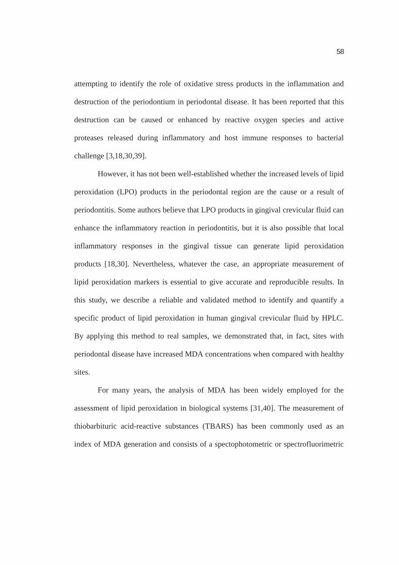
58
attempting to identify the role of oxidative stress products in the inflammation and
destruction of the periodontium in periodontal disease. It has been reported that this
destruction can be caused or enhanced by reactive oxygen species and active
proteases released during inflammatory and host immune responses to bacterial
challenge [3,18,30,39].
However, it has not been well-established whether the increased levels of lipid
peroxidation (LPO) products in the periodontal region are the cause or a result of
periodontitis. Some authors believe that LPO products in gingival crevicular fluid can
enhance the inflammatory reaction in periodontitis, but it is also possible that local
inflammatory responses in the gingival tissue can generate lipid peroxidation
products [18,30]. Nevertheless, whatever the case, an appropriate measurement of
lipid peroxidation markers is essential to give accurate and reproducible results. In
this study, we describe a reliable and validated method to identify and quantify a
specific product of lipid peroxidation in human gingival crevicular fluid by HPLC.
By applying this method to real samples, we demonstrated that, in fact, sites with
periodontal disease have increased MDA concentrations when compared with healthy
sites.
For many years, the analysis of MDA has been widely employed for the
assessment of lipid peroxidation in biological systems [31,40]. The measurement of
thiobarbituric acid-reactive substances (TBARS) has been commonly used as an
index of MDA generation and consists of a spectophotometric or spectrofluorimetric
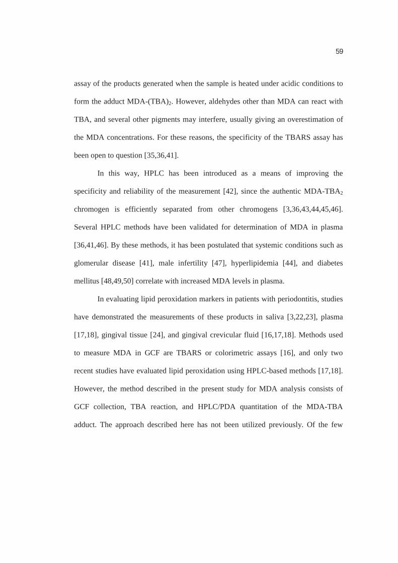
59
assay of the products generated when the sample is heated under acidic conditions to
form the adduct MDA-(TBA)2. However, aldehydes other than MDA can react with
TBA, and several other pigments may interfere, usually giving an overestimation of
the MDA concentrations. For these reasons, the specificity of the TBARS assay has
been open to question [35,36,41].
In this way, HPLC has been introduced as a means of improving the
specificity and reliability of the measurement [42], since the authentic MDA-TBA2
chromogen is efficiently separated from other chromogens [3,36,43,44,45,46].
Several HPLC methods have been validated for determination of MDA in plasma
[36,41,46]. By these methods, it has been postulated that systemic conditions such as
glomerular disease [41], male infertility [47], hyperlipidemia [44], and diabetes
mellitus [48,49,50] correlate with increased MDA levels in plasma.
In evaluating lipid peroxidation markers in patients with periodontitis, studies
have demonstrated the measurements of these products in saliva [3,22,23], plasma
[17,18], gingival tissue [24], and gingival crevicular fluid [16,17,18]. Methods used
to measure MDA in GCF are TBARS or colorimetric assays [16], and only two
recent studies have evaluated lipid peroxidation using HPLC-based methods [17,18].
However, the method described in the present study for MDA analysis consists of
GCF collection, TBA reaction, and HPLC/PDA quantitation of the MDA-TBA
adduct. The approach described here has not been utilized previously. Of the few
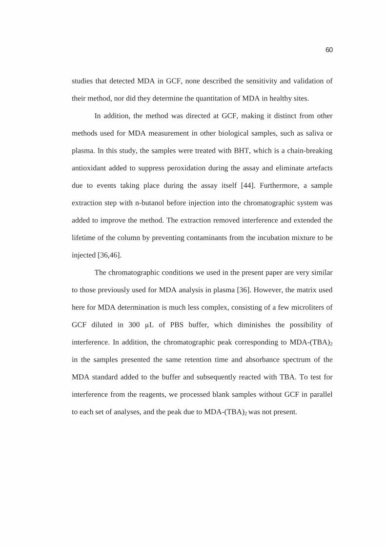
60
studies that detected MDA in GCF, none described the sensitivity and validation of
their method, nor did they determine the quantitation of MDA in healthy sites.
In addition, the method was directed at GCF, making it distinct from other
methods used for MDA measurement in other biological samples, such as saliva or
plasma. In this study, the samples were treated with BHT, which is a chain-breaking
antioxidant added to suppress peroxidation during the assay and eliminate artefacts
due to events taking place during the assay itself [44]. Furthermore, a sample
extraction step with n-butanol before injection into the chromatographic system was
added to improve the method. The extraction removed interference and extended the
lifetime of the column by preventing contaminants from the incubation mixture to be
injected [36,46].
The chromatographic conditions we used in the present paper are very similar
to those previously used for MDA analysis in plasma [36]. However, the matrix used
here for MDA determination is much less complex, consisting of a few microliters of
GCF diluted in 300 μL of PBS buffer, which diminishes the possibility of
interference. In addition, the chromatographic peak corresponding to MDA-(TBA)2
in the samples presented the same retention time and absorbance spectrum of the
MDA standard added to the buffer and subsequently reacted with TBA. To test for
interference from the reagents, we processed blank samples without GCF in parallel
to each set of analyses, and the peak due to MDA-(TBA)2 was not present.
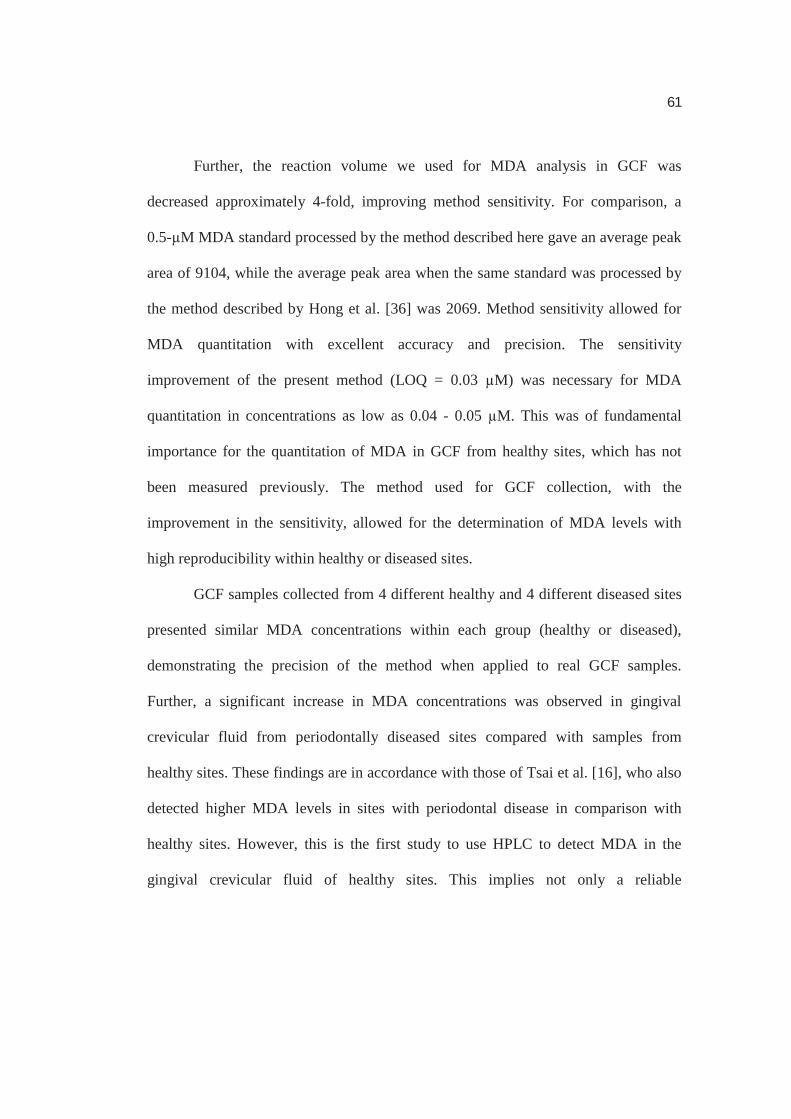
61
Further, the reaction volume we used for MDA analysis in GCF was
decreased approximately 4-fold, improving method sensitivity. For comparison, a
0.5-μM MDA standard processed by the method described here gave an average peak
area of 9104, while the average peak area when the same standard was processed by
the method described by Hong et al. [36] was 2069. Method sensitivity allowed for
MDA quantitation with excellent accuracy and precision. The sensitivity
improvement of the present method (LOQ = 0.03 μM) was necessary for MDA
quantitation in concentrations as low as 0.04 - 0.05 μM. This was of fundamental
importance for the quantitation of MDA in GCF from healthy sites, which has not
been measured previously. The method used for GCF collection, with the
improvement in the sensitivity, allowed for the determination of MDA levels with
high reproducibility within healthy or diseased sites.
GCF samples collected from 4 different healthy and 4 different diseased sites
presented similar MDA concentrations within each group (healthy or diseased),
demonstrating the precision of the method when applied to real GCF samples.
Further, a significant increase in MDA concentrations was observed in gingival
crevicular fluid from periodontally diseased sites compared with samples from
healthy sites. These findings are in accordance with those of Tsai et al. [16], who also
detected higher MDA levels in sites with periodontal disease in comparison with
healthy sites. However, this is the first study to use HPLC to detect MDA in the
gingival crevicular fluid of healthy sites. This implies not only a reliable
62
measurement of MDA even in low concentrations in these biological samples, but
also the possibility for use of this measurement as a risk marker of periodontal
disease.
In this study, using an HPLC-based assay, an improved method for MDA
quantitation in gingival crevicular fluid was established with satisfactory sensitivity,
precision, and accuracy. Such a reliable and validated method to measure MDA in
human GCF, collected non-invasively, may be very useful in clarifying the role of
lipid peroxidation in the pathogenesis of periodontal disease.
ACKNOWLEDGMENTS
This study was supported by the financial support from São Paulo State
Research Support Foundation (FAPESP) (grant 2007/08362-8) and the Coordination
for Improvement of Higher Education Students of the Brazilian Ministry of
Education (CAPES).
REFERENCES
[1] S. Luqman, S.I. Rizvi, Protection of lipid peroxidation and carbonyl formation in proteins by capsaicin in human erythrocytes subjected to oxidative stress, Phytother.Res. 20 (2006) 303-306.[2] B.M. Babior, Oxygen-dependent microbial killing by phagocytes (first of twoparts), N. Engl. J. Med. 298 (1978) 659-668.[3] I.L. Chapple, Reactive oxygen species and antioxidants in inflammatory diseases, J. Clin. Periodontol. 24 (1997) 287-296.[4] B. Halliwell, Oral inflammation and reactive species: a missed opportunity?, OralDis. 6 (2000) 136-137.[5] C.K. Sen, Oxygen toxicity and antioxidants: state of the art, Indian J. Physiol.Pharmacol. 39 (1995) 177-196.
63
[6] R.E. Little, B.C. Gladen, Levels of lipid peroxides in uncomplicated pregnancy: a review of the literature, Reprod. Toxicol. 13 (1999) 347-352.[7] L.J. Marnett, Chemistry and biology of DNA damage by malondialdehyde, IARC Sci. Publ. (1999) 17-27.[8] T. Hemnani, M.S. Parihar, Reactive oxygen species and oxidative DNA damage, Indian J. Physiol. Pharmacol. 42 (1998) 440-452.[9] T.A. Libermann, D. Baltimore, Activation of interleukin-6 gene expression through the NF-kappa B transcription factor, Mol. Cell Biol. 10 (1990) 2327-2334.[10] M.M. Kesavulu, R. Giri, B. Kameswara Rao, C. Apparao, Lipid peroxidation and antioxidant enzyme levels in type 2 diabetics with microvascular complications, Diabetes Metab. 26 (2000) 387-392.[11] E. Velazquez, P.H. Winocour, P. Kesteven, K.G. Alberti, M.F. Laker, Relation of lipid peroxides to macrovascular disease in type 2 diabetes, Diabet. Med. 8 (1991) 752-758.[12] K.Z. Kedziora-Kornatowska, M. Luciak, J. Blaszczyk, W. Pawlak, Lipid peroxidation and activities of antioxidant enzymes in erythrocytes of patients with non-insulin dependent diabetes with or without diabetic nephropathy, Nephrol. Dial.Transplant. 13 (1998) 2829-2832.[13] P.I. Mapp, M.C. Grootveld, D.R. Blake, Hypoxia, oxidative stress and rheumatoid arthritis, Br. Med. Bull. 51 (1995) 419-436.[14] W. Macnee, I. Rahman, Oxidants and antioxidants as therapeutic targets in chronic obstructive pulmonary disease, Am. J. Respir. Crit. Care Med. 160 (1999) S58-S65.[15] Y.T. Teng, The role of acquired immunity and periodontal disease progression, Crit. Rev. Oral Biol. Med. 14 (2003) 237-252.[16] C.C. Tsai, H.S. Chen, S.L. Chen, Y.P. Ho, K.Y. Ho, Y.M. Wu, C.C. Hung, Lipid peroxidation: a possible role in the induction and progression of chronic periodontitis, J. Periodontal Res. 40 (2005) 378-384.[17] F.A. Akalin, E. Baltacioglu, A. Alver, E. Karabulut, Lipid peroxidation levels and total oxidant status in serum, saliva and gingival crevicular fluid in patients with chronic periodontitis, J. Clin. Periodontol. 34 (2007) 558-565.[18] D. Wei, X.L. Zhang, Y.Z. Wang, C.X. Yang, G. Chen, Lipid peroxidation levels, total oxidant status and superoxide dismutase in serum, saliva and gingival crevicular fluid in chronic periodontitis patients before and after periodontal therapy, Aust.Dent. J. 55 (2010) 70-78.[19] D.F. Kinane, P.M. Bartold, Clinical relevance of the host responses of periodontitis, Periodontol. 2000. 43 (2007) 278-293.[20] D.N. Tatakis, P.S. Kumar, Etiology and pathogenesis of periodontal diseases, Dent. Clin. North Am. 49 (2005) 491-516.[21] P.M. Abuja, R. Albertini, Methods for monitoring oxidative stress, lipid peroxidation and oxidation resistance of lipoproteins, Clin. Chim. Acta. 306 (2001) 1-17.
64
[22] J. Khalili, H.F. Biloklytska, Salivary malondialdehyde levels in clinically healthy and periodontal diseased individuals, Oral Dis. 14 (2008) 754-760.[23] C.F. Canakci, Y. Cicek, V. Canakci, Reactive oxygen species and human inflammatory periodontal diseases, Biochemistry (Mosc). 70 (2005) 619-628.[24] I. Borges, Jr., E.A. Moreira, D.W. Filho, T.B. de Oliveira, M.B. da Silva, T.S. Frode, Proinflammatory and oxidative stress markers in patients with periodontal disease, Mediators Inflamm. 2007 (2007) 45794.[25] K. Sonoki, S. Nakashima, Y. Takata, T. Naito, K. Fujisawa, T. Ootsubo, M. Wakisaka, M. Iwase, M. Iida, M. Yokota, Decreased lipid peroxidation following periodontal therapy in type 2 diabetic patients, J. Periodontol. 77 (2006) 1907-1913.[26] I.B. Lamster, J.K. Ahlo, Analysis of gingival crevicular fluid as applied to the diagnosis of oral and systemic diseases, Ann. N. Y. Acad. Sci. 1098 (2007) 216-229.[27] R.J. Waddington, R. Moseley, G. Embery, Reactive oxygen species: a potential role in the pathogenesis of periodontal diseases, Oral Dis. 6 (2000) 138-151.[28] A.M. Polson, J.M. Goodson, Periodontal diagnosis. Current status and future needs, J. Periodontol. 56 (1985) 25-34.[29] G.S. Griffiths, Formation, collection and significance of gingival crevice fluid, Periodontol. 2000. 31 (2003) 32-42.[30] Y. Sakiyama, R. Kato, S. Inoue, K. Suzuki, H. Itabe, M. Yamamoto, Detection of oxidized low-density lipoproteins in gingival crevicular fluid from dental patients, J. Periodontal Res. 45 (2010) 216-222.[31] D. Del Rio, A.J. Stewart, N. Pellegrini, A review of recent studies on malondialdehyde as toxic molecule and biological marker of oxidative stress, Nutr.Metab. Cardiovasc. Dis. 15 (2005) 316-328.[32] K. Yagi, Lipid peroxides and human diseases, Chem. Phys. Lipids. 45 (1987) 337-351.[33] K. Panjamurthy, S. Manoharan, C.R. Ramachandran, Lipid peroxidation and antioxidant status in patients with periodontitis, Cell. Mol. Biol. Lett. 10 (2005) 255-264.[34] H. Esterbauer, K.H. Cheeseman, Determination of aldehydic lipid peroxidation products: malonaldehyde and 4-hydroxynonenal, Methods Enzymol. 186 (1990) 407-421.[35] J.A. Knight, R.K. Pieper, L. McClellan, Specificity of the thiobarbituric acid reaction: its use in studies of lipid peroxidation, Clin. Chem. 34 (1988) 2433-2438.[36] Y.L. Hong, S.L. Yeh, C.Y. Chang, M.L. Hu, Total plasma malondialdehyde levels in 16 Taiwanese college students determined by various thiobarbituric acid tests and an improved high-performance liquid chromatography-based method, Clin.Biochem. 33 (2000) 619-625.[37] H. Sobaniec, M.E. Sobaniec-Lotowska, Morphological examinations of hard tissues of periodontium and evaluation of selected processes of lipid peroxidation in blood serum of rats in the course of experimental periodontitis, Med. Sci. Monit. 6 (2000) 875-881.
65
[38] M. Sheikhi, R.K. Bouhafs, K.J. Hammarstrom, C. Jarstrand, Lipid peroxidation caused by oxygen radicals from Fusobacterium-stimulated neutrophils as a possible model for the emergence of periodontitis, Oral Dis. 7 (2001) 41-46.[39] M. Battino, M.S. Ferreiro, I. Gallardo, H.N. Newman, P. Bullon, The antioxidant capacity of saliva, J. Clin. Periodontol. 29 (2002) 189-194.[40] L.L. de Zwart, J.H. Meerman, J.N. Commandeur, N.P. Vermeulen, Biomarkers of free radical damage applications in experimental animals and in humans, Free Radic. Biol. Med. 26 (1999) 202-226.[41] J. Templar, S.P. Kon, T.P. Milligan, D.J. Newman, M.J. Raftery, Increased plasma malondialdehyde levels in glomerular disease as determined by a fully validated HPLC method, Nephrol. Dial. Transplant. 14 (1999) 946-951.[42] R.P. Bird, S.S. Hung, M. Hadley, H.H. Draper, Determination of malonaldehyde in biological materials by high-pressure liquid chromatography, Anal. Biochem. 128 (1983) 240-244.[43] I.S. Young, E.R. Trimble, Measurement of malondialdehyde in plasma by high performance liquid chromatography with fluorimetric detection, Ann. Clin. Biochem. 28 (Pt 5) (1991) 504-508.[44] S. Chirico, C. Smith, C. Marchant, M.J. Mitchinson, B. Halliwell, Lipid peroxidation in hyperlipidaemic patients. A study of plasma using an HPLC-based thiobarbituric acid test, Free Radic. Res. Commun. 19 (1993) 51-57.[45] K. Fukunaga, K. Takama, T. Suzuki, High-performance liquid chromatographic determination of plasma malondialdehyde level without a solvent extraction procedure, Anal. Biochem. 230 (1995) 20-23.[46] D. Grotto, L.D. Santa Maria, S. Boeira, J. Valentini, M.F. Charao, A.M. Moro, P.C. Nascimento, V.J. Pomblum, S.C. Garcia, Rapid quantification of malondialdehyde in plasma by high performance liquid chromatography-visible detection, J. Pharm. Biomed. Anal. 43 (2007) 619-624.[47] K. Li, X. Shang, Y. Chen, High-performance liquid chromatographic detection of lipid peroxidation in human seminal plasma and its application to male infertility, Clin. Chim. Acta. 346 (2004) 199-203.[48] S. Nacitarhan, T. Ozben, N. Tuncer, Serum and urine malondialdehyde levels in NIDDM patients with and without hyperlipidemia, Free Radic. Biol. Med. 19 (1995) 893-896.[49] M. Nakhjavani, O. Khalilzadeh, L. Khajeali, A. Esteghamati, A. Morteza, A. Jamali, S. Dadkhahipour, Serum oxidized-LDL is associated with diabetes duration independent of maintaining optimized levels of LDL-cholesterol, Lipids. 45 (2010) 321-327.[50] D.J. VanderJagt, J.M. Harrison, D.M. Ratliff, L.A. Hunsaker, D.L. Vander Jagt, Oxidative stress indices in IDDM subjects with and without long-term diabetic complications, Clin. Biochem. 34 (2001) 265-270.
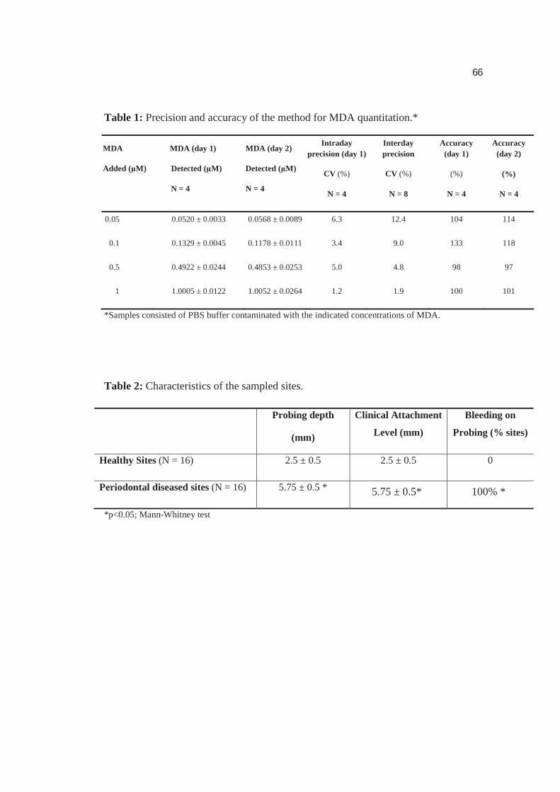
66
Table 1: Precision and accuracy of the method for MDA quantitation.*
MDA MDA (day 1)
Added (μM) Detected (μM)
N = 4
MDA (day 2)
Detected (μM)
N = 4
Intraday precision (day 1)
CV (%)
N = 4
Interday precision
CV (%)
N = 8
Accuracy (day 1)
(%)
N = 4
Accuracy (day 2)
(%)
N = 4
0.05 0.0520 ± 0.0033 0.0568 ± 0.0089 6.3 12.4 104 114
0.1 0.1329 ± 0.0045 0.1178 ± 0.0111 3.4 9.0 133 118
0.5 0.4922 ± 0.0244 0.4853 ± 0.0253 5.0 4.8 98 97
1 1.0005 ± 0.0122 1.0052 ± 0.0264 1.2 1.9 100 101
*Samples consisted of PBS buffer contaminated with the indicated concentrations of MDA.
Table 2: Characteristics of the sampled sites.
Probing depth
(mm)
Clinical Attachment
Level (mm)
Bleeding on
Probing (% sites)
Healthy Sites (N = 16) 2.5 ± 0.5 2.5 ± 0.5 0
Periodontal diseased sites (N = 16) 5.75 ± 0.5 * 5.75 ± 0.5* 100% *
*p<0.05; Mann-Whitney test
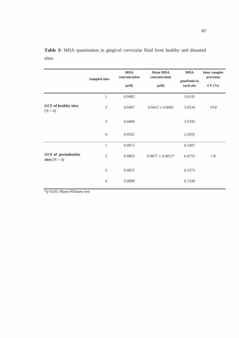
67
Table 3: MDA quantitation in gingival crevicular fluid from healthy and diseased
sites.
*p<0.05; Mann-Whitney test
Sampled sites
MDA concentration
(μM)
Mean MDA concentration
(μM)
MDA
pmol/min in each site
Inter samples precision
CV (%)
GCF of healthy sites(N = 4)
1 0.0482 3.6141
2 0.0407 0.0415 ± 0.0082 3.0534 19.8
3 0.0469 3.5193
4 0.0302 2.2655
GCF of periodontitis sites (N = 4)
1 0.0873 6.5497
2 0.0863 0.0877 ± 0.0015* 6.4755 1.8
3 0.0872 6.5373
4 0.0899 6.7439
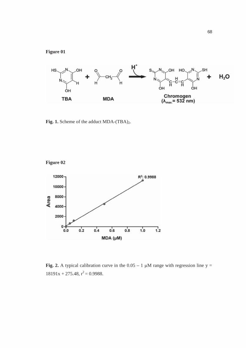
68
Figure 01
Fig. 1. Scheme of the adduct MDA-(TBA)2.
Figure 02
Fig. 2. A typical calibration curve in the 0.05 – 1 μM range with regression line y =
18191x + 275.48, r2 = 0.9988.
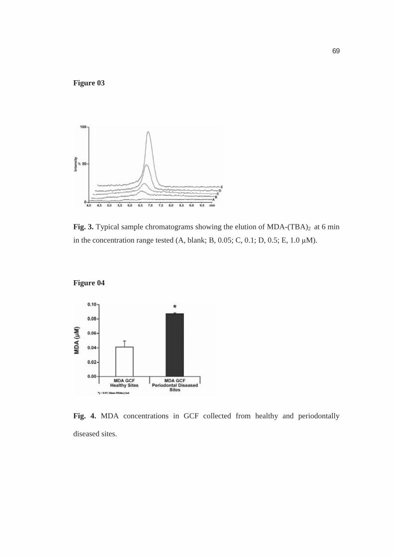
69
Figure 03
Fig. 3. Typical sample chromatograms showing the elution of MDA-(TBA)2 at 6 min
in the concentration range tested (A, blank; B, 0.05; C, 0.1; D, 0.5; E, 1.0 μM).
Figure 04
Fig. 4. MDA concentrations in GCF collected from healthy and periodontally
diseased sites.
70
CAPÍTULO 2
Lipid Peroxidation is Associated with the Severity of
Periodontal Disease and Local Inflammatory Markers in
Patients with Type 2 Diabetes
Alliny de Souza Bastos, Dana T. Graves, Ana Paula de Melo Loureiro, Carlos Rossa
Júnior, Dulcinéia Saes Parra Abdalla, Tanize do Espírito Santo Faulin, Niels Olsen
Câmara, Oelisoa M Andriankaja, and Silvana Regina Perez Orrico
Artigo submetido para revista
Journal of Clinical Endocrinology and Metabolism
Em revisão: JC-11-3397 (R1) 20 Dez 2011
U
71
Lipid Peroxidation is Associated with the Severity of Periodontal Disease and Local
Inflammatory Markers in Patients with Type 2 Diabetes
Alliny de Souza Bastos1, Dana T. Graves2, Ana Paula de Melo Loureiro3, Carlos Rossa
Júnior1, Dulcinéia Saes Parra Abdalla3, Tanize do Espírito Santo Faulin3, Niels Olsen
Câmara4, Oelisoa M Andriankaja2, and Silvana Regina Perez Orrico1
1Department of Diagnosis and Surgery, Araraquara School of Dentistry – UNESP-State
University of São Paulo, Araraquara, São Paulo, Brazil
2Department of Periodontics, School of Dental Medicine, University of Pennsylvania,
Philadelphia, PA, United States
3 Department of Clinical and Toxicological Analyses, Faculty of Pharmaceutical Sciences,
University of São Paulo (USP), São Paulo, Brazil
4 Department of Immunology, Institute of Biomedical Sciences, University of São Paulo
(USP), São Paulo, Brazil
Abbreviated Title: Lipid peroxidation, diabetes, periodontitis
Corresponding author:
Profª. Drª. Silvana Regina Perez Orrico, e-mail: [email protected]
UNESP – Univ Estadual Paulista; Araraquara School of Dentistry
Department of Diagnosis and Surgery Rua Humaitá, 1680, 2º andar
CEP: 14801-903, Araraquara, São Paulo, Brazil.
Phone: +55 (16) 3301-6377 Fax: +55 (16) 3301-6369
Grants- FAPESP 2007/08362-8; NIH DE017732
Disclosure Summary: The authors have nothing to disclose.
Key terms: Lipid peroxidation, diabetes mellitus type 2, periodontal diseases, cytokines
72
ABSTRACT
Context: Periodontitis is the most common lytic disease of bone and is recognized as
a common complication of diabetes. Lipid peroxidation (LPO) is increased in
diabetes and may be related to modulation of the inflammatory response. LPO levels
in patients with diabetes (DM) and periodontal disease have not been evaluated.
Objective: The aim of this study was to evaluate the association of LPO with
periodontal status and to establish a potential mechanism by assessing inflammatory
cytokines in type 2 diabetic and non-diabetic patients.
Design and Setting: This is a cross-sectional study of 18 months’ duration involving
Brazilian patients recruited at the Univ Estadual Paulista.
Patients: The sample was comprised of 120 patients divided into four groups based
upon diabetic and dyslipidemic status: poorly controlled diabetics with dyslipidemia,
well-controlled diabetics with dyslipidemia, normoglycemic individuals with
dyslipidemia, and healthy individuals.
Main Outcome Measure(s): Blood analyses were carried out for fasting plasma
glucose, HbA1c , and lipid profile. Periodontal examinations were performed, and
gingival crevicular fluid was collected. LPO levels were evaluated by measuring
oxLDL (ELISA) and malondialdehyde (MDA, HPLC). Cytokines were evaluated by
the multiplex bead technique.
73
Results: Lipid peroxidation evaluated by MDA in plasma and gingival crevicular
fluid was significantly increased in DM groups. Significant correlations between LPO
markers and periodontal parameters indicate a direct relationship between these levels
and the severity of inflammation and secretion of inflammatory cytokines,
particularly in diabetic patients.
Conclusion: These findings indicate an important role for LPO in the severity of the
local inflammatory response to bacteria and the susceptibility to periodontal disease
in diabetic patients.
Abbreviations: BMI – Body Mass Index; DM – Diabetes mellitus; GCF – Gingival
Crevicular Fluid; HbA1c – Glycated Hemoglobin A1c fraction; HDL – High-density
lipoprotein; LDL – Low-density Lipoprotein; LPO – Lipid peroxidation; MDA –
Malondialdehyde.
74
Introduction
Periodontitis is the most common lytic disease of bone and consists of a
bacteria-induced inflammatory disorder that involves the structures that support teeth
(1, 2). It is the most common cause of tooth loss in adults and affects approximately
10 to 15% of the world’s population (3). Periodontitis is initiated by bacteria that
induce an inflammatory response in the gingiva. It is the bacteria-stimulated
inflammation that leads to breakdown of bone that supports the teeth, which is a
hallmark of periodontitis (4-6).
The link between diabetes and oral diseases has not been widely discussed in
the medical literature (5). However, it has been reported that periodontitis is one of
the first clinical manifestations of diabetes (7) and is recognized as a common
complication in diabetic patients (8), particularly in poorly controlled diabetics (9).
As an infectious-initiated inflammatory condition, periodontal disease may result in a
systemic chronic inflammatory challenge that may have a significant impact on
metabolic control in diabetics (10).
A potential mechanism through which diabetes is linked to periodontitis and
other inflammatory conditions associated with diabetes is the increased production of
reactive oxygen species (ROS) and lipid peroxidation products (11, 12). However, the
role of lipid peroxidation in both conditions, diabetes and periodontal disease, has
received relatively little attention.
75
Diabetes may contribute to the destructive aspects of the host response that
leads to periodontal disease through the generation of reactive oxygen species (ROS)
(11, 13, 14) . Over-production of ROS is also thought to contribute to several diabetic
complications, including vascular disease (15-17). ROS are unstable, with a short
half-life, and thus are difficult to detect (14). Several markers of lipid peroxidation
have been studied to monitor ROS production. Malondialdehyde (MDA) is one of the
most studied end-products of lipid peroxidation and is often used as a surrogate
marker of oxidative stress (18) . To investigate the link among diabetes, lipid
peroxidation, and inflammation, we assessed periodontal disease as an indicator of
the severity of inflammation in a host-microbe interface. We examined the local and
systemic levels of lipid peroxidation in type 2 diabetic patients with dyslipidemia. We
also evaluated the effect of lipid peroxidation on periodontal status and investigated a
potential mechanism, the expression of inflammatory markers in local tissues.
Materials and Methods
Study population
The present study was approved by the Ethics in Human Research Committee
of the Araraquara School of Dentistry (UNESP – Univ. Estadual Paulista,
Araraquara, Brazil; Protocol number 50/06) and was carried out between May 2009
76
and November 2010. All volunteers were informed about the aims and methods of
this study, and they provided their written consent to participate.
As inclusion criteria, all study participants ranged in age from 35 to 60 yr and
had at least 15 natural teeth. All individuals had clinical chronic periodontal disease
as defined by the American Academy of Periodontology (19), with local signs of
inflammation and tissue destruction (presence of deep periodontal pockets
and loss of the connective tissue attachment of gingiva to teeth (clinical attachment
loss 4mm) in at least 4 non-adjacent teeth. Patients were excluded based on the
following criteria: history of antibiotic therapy in the previous 3 months and/or non-
steroidal anti-inflammatory drug therapy in the previous 6 months, pregnancy or use
of contraceptives or any other hormone, current smoker or former smoker, history of
anemia, periodontal treatment or surgery in the preceding 6 months, use of
hypolipemic drugs such as statins or fibrates, and history of diseases that interfere
with lipid metabolism, such as hypothyroidism and hypopituitarism.
A power analysis based on a pilot study determined that at least 20 patients in
each group would be sufficient to detect a 3-unit difference in MDA levels with 90%
power and 95% confidence interval. To compensate for the possibility of drop-out
during the experimental period, the experimental groups consisted of 30 patients
each. The population was divided into four groups based upon diabetic and
dyslipidemic status: poorly controlled diabetics with dyslipidemia (Group 1), well-
77
controlled diabetics with dyslipidemia (Group 2), normoglycemic individuals with
dyslipidemia (Group 3), and healthy individuals (Group 4) (Table 1).
Clinical record and physical evaluation
The individuals answered a structured questionnaire about demographic
characteristics, and a trained examiner collected information regarding time since
diabetes onset, medication used to control glucose, and the presence of at least one
complication associated with diabetes. Abdominal circumference (cm) and height (m)
were measured as part of a physical examination, which allowed the body mass index
(BMI) to be calculated.
Parameters of metabolic control and lipoprotein profile
Blood samples were collected after a 12-hour overnight fast for the evaluation
of fasting plasma glucose (mg/dl) by modified Bondar e Mead method, glycated
haemoglobin (HbA1c) by enzymatic immunoturbidimetry, high-sensitivity C-reactive
protein by the nephelomeric method, total cholesterol (TC), triglycerides (TGs), and
HDL by enzymatic methods. LDL was determined by the Friedewald formula. To
avoid the inclusion of individuals with transitory dyslipidemia, the cutoff points used
were the highest values according to the National Cholesterol Educational Program
(NCEP) Adult Treatment III (ATP III): TC 240 mg/dL, LDL
40 mg/dL, and TGs 200 mg/dL. Metabolic control was considered as adequate
when HbA1c < 8% and as inadequate when HbA1c
78
evaluated for insulin levels by the chemiluminescence method (uUI/mL) for
calculation of the HOMA index (HOMA IR) (20).
Periodontal clinical examination
All patients were subjected to a periodontal clinical examination performed in
six sites per tooth by a single trained calibrated examiner (kappa = 0.89). Periodontal
pocket depth, clinical attachment loss, and bleeding on probing were evaluated with a
periodontal probe PCPUNC15-6 (Hu-Friedy®). Severe periodontal disease was
s distributed in different
quadrants of the dentition (21).
Gingival crevicular fluid (GCF) collection
Gingival crevicular fluid (GCF) is considered as a serum transudate or, more
commonly, as an inflammatory exudate from the gingival sulcus surrounding the
teeth. GCF contains substances from the host, such as inflammatory cells and serum-
derived factors, as well as from microorganisms. The potential diagnostic value of the
GCF is due to the site-specific nature of the sample, and it can be collected non-
invasively, serving as an expedient biological source of patient information.
In each patient, 4 different healthy sites (probing depth 3mm, no loss of
attachment, and no bleeding on probing) and 4 periodontally diseased sites (probing
depth 5mm, loss of attachment 4mm, and with bleeding on probing) were
79
randomly chosen in different non-adjacent teeth for GCF collection. Collection of
gingival crevicular fluid was performed as previously described (22) with paper strips
(Periopaper®, Oraflow Inc., Smithtown, NY, USA), and the volume of gingival
crevicular fluid was measured with a specific instrument (Periotron®, Oraflow Inc.).
The samples from each patient were pooled in 2 different tubes, representing healthy
and diseased sites, centrifuged at 3000 g for 5 min, and the supernatants were stored
at -80 C until the analysis of MDA and cytokines.
Blood collection
Blood samples were collected in tubes with EDTA centrifuged at 3000 g for
10 min at 4°C. Plasma was obtained and stored at -80 C until the lipid peroxidation
analyses. The assays were performed by professionals blinded to the patients'
conditions.
Inflammatory biomarker assays
The concentration of 3 inflammatory cytokines in gingival crevicular fluid, as
- -10, and IL-6, was analyzed by multiplex beads
(Bio-Plex system, Bio-Rad Laboratories, Hercules, CA, USA), following the
manufacturer’s instructions.
Lipid peroxidation assays
Oxidized LDL assay in plasma samples
80
The levels of oxLDL in plasma were analyzed with a commercially available
ELISA kit (Mercodia AB®, Uppsala, Sweden) following the manufacturer’s
instructions.
MDA assay
The assessment of MDA in plasma and gingival crevicular fluid was
determined by HPLC (Shimadzu, Tokyo, Japan) with a reverse-phase HPLC column
(C18; 4.6 x 150 mm) (Phenomenex, Torrance, CA, USA) and compared with MDA
standard curves. Plasma and gingival crevicular fluid samples were prepared as
described previously (23).
Statistical analysis
The distribution and normality of the variables were evaluated by the
D’Agostino-Pearson test. The general characteristics of each group were described
with mean and standard deviation (SD), and the inflammatory cytokines were
expressed as median (25% /75% quartiles). The differences between the groups for
parametric data were evaluated by ANOVA, followed by Bonferroni’s post-test, and
non-parametric data were evaluated by the Mann-Whitney test or the Kruskall-Wallis
test, followed by Dunn’s post-test or. Pearson’s partial correlation coefficient was
used for normally distributed data, or Spearman’s partial correlation coefficient was
used for non-normally distributed data, to investigate the correlations between the
81
various parameters adjusted for age, gender, and BMI. To determine the influence of
lipid peroxidation and diabetic status on the severity of periodontal disease and on the
expression of local cytokines, separate multivariable logistic and linear regression
models were built for each independent variable. Variables found to be associated
with the independent and dependent variables, and thus possible confounding factors,
were included in the models. Moreover, regardless of their statistical significance, all
variables known to have biologic linkage with the outcomes were retained. The
software, IBM version 19.
Results
Sample population
In total, 1728 patients were screened, and 120 were identified as meeting the
study inclusion criteria; of these, 30 patients assigned to 4 different groups. The
general characteristics of the sample are demonstrated in Table 1. There were no age
and gender differences between the groups. Diabetics had significantly higher values
of BMI and abdominal circumference (p<0.05). Among all diabetics, there was no
difference in the age of diabetes onset. However, poorly controlled diabetics had
more diabetic complications, with the most common complication being retinopathy,
followed by nephropathy. Glucose metabolism, metabolic control, insulin resistance,
82
lipoprotein profile, and high-sensitivity C-reactive protein for all groups are presented
in Table 1.The groups with diabetes had increased levels of fasting glucose, HbA1c,
and insulin resistance compared with non-diabetic patients, and groups 1, 2, and 3 all
presented dyslipidemia with increased total cholesterol, LDL cholesterol, and
triglycerides compared with group 4 (healthy individuals) (p<0.05).
Periodontal evaluation
Periodontal tissue destruction and local inflammation were significantly more
severe in diabetics, particularly in group 1 (Fig. 1A and supplemental Table1),
indicating that poorly controlled diabetes aggravates local host response to
microorganisms in the dental biofilm. Relevant clinical periodontal parameters that
were significantly different between the groups are illustrated in Fig. 1B in terms of
fold change. Each patient group was compared with group 4 (healthy individuals).
Chronic periodontitis, as evaluated by the presence of deep periodontal pockets (
6mm), was increased four-fold in poorly controlled diabetics with dyslipidemia and
two-fold in well-controlled diabetics with dyslipidemia. Acute infection, evaluated by
the presence of suppuration, and acute inflammation, evaluated by bleeding on
probing, were increased 3.5- and 1.3-fold, respectively, in poorly controlled diabetics
with dyslipidemia.
83
Immunological analysis
To assess local inflammation, the expression of TNF- -6, and IL-10 was
measured in gingival crevicular fluid. The cytokine expression was normalized to the
volume of gingival crevicular fluid collected and expressed in concentration (pg/μl).
All cytokines were increased in the gingival crevicular fluid of diabetic patients,
being significant in the group with poorly controlled diabetes and dyslipidemia
(p<0.05), indicating that diabetes aggravates the local inflammatory response to
microorganisms in the dental biofilm (Table 2).
Lipid peroxidation levels
Plasma Malondialdehyde (MDA) and Oxidized LDL (oxLDL)
Plasma MDA levels were slightly increased in dyslipidemic patients, but the
presence of diabetes resulted in significantly increased systemic oxidative metabolism
(p<0.05) (Fig. 2A). However, the most increased levels of plasma MDA were observed
in poorly controlled diabetics with dyslipidemia who had about 2.5 times higher MDA
levels when compared with those in the non-diabetic groups and approximately 1.5
times higher MDA levels when compared with well-controlled diabetics with
dyslipidemia. OxLDL was increased in all groups with dyslipidemia (Groups 1, 2, and
3) (Fig. 2A).
In relation to local lipid peroxidation, evaluated by MDA in the gingival
crevicular fluid, the patients with diabetes presented significantly higher levels of
84
MDA when compared with those in the other groups (p<0.05) (Fig. 2B). The mean
levels of MDA in the diseased sites of the poorly controlled diabetics were 2.3 greater
than in the normoglycemic with dyslipidemia group (p<0.001), approximately 1.8
times higher than in the healthy group (p<0.05), and about 1.6 times higher in the well-
controlled diabetics with dyslipidemia (p<0.05) (Fig. 2B). Moreover, even the gingival
crevicular fluid of healthy sites in the groups with diabetes presented higher levels of
MDA when compared with the healthy gingival crevicular fluid of normoglycemic
groups.
Correlations
Correlations among lipid peroxidation markers, diabetes status, lipoprotein
profile, clinical periodontal parameters, and local cytokine levels were assessed
(Table 3). There was no independent association between lipoprotein profile and a
greater severity of periodontal disease in individuals with periodontitis. Diabetes was
significantly associated with lipid peroxidation markers, and this was the strongest
positive correlation observed (r=0.65, p<0.0001). Even though we cannot infer causal
relationships by correlations, this association could represent an additive effect in the
aggravation of inflammation in diabetics, as indicated by the positive correlations
observed between diabetes markers and periodontal disease parameters. The positive
associations between systemic levels of lipid peroxidation and the levels of
inflammatory cytokines in the gingival crevicular fluid (Table 3) support a role for
increased oxidative metabolism in the aggravation of inflammation.
85
Considering the correlation between systemic levels of lipid peroxidation, a
significant intermediate correlation between MDA in plasma and oxidized LDL
(r=0.460; p<0.01) was observed. In addition, there was a statistically significant but
weak correlation between plasma and local levels of MDA (r=0.33; p<0.01) and
between plasma-oxidized LDL and local levels of MDA (r=0.32; p<0.01).
Regressions
Regression analyses were performed to obtain insight into the role of diabetes and
lipid peroxidation in local inflammatory responses. Table 4A represents the results
from multiple regression analysis using separate models for independent variables,
lipid peroxidation parameters, and diabetes status. Lipid peroxidation markers were
directly related to the severity of local inflammation, represented by all clinical
measures of periodontal disease (except for bleeding on probing) after adjustment for
age, gender, BMI, and triglycerides. Diabetes was also directly related to the severity
of local inflammation. Table 4B represents the results from a multivariate logistic
regression analysis using separate models and taking into account the effects of age,
gender, BMI, and triglycerides. There was an approximate 1.81- and two-fold
increase in the odds of having severe bleeding on probing and deep periodontal
pockets, respectively, among those patients who have higher plasmatic levels of
MDA. This finding suggests a link between periodontal inflammation/destruction and
plasma MDA levels.
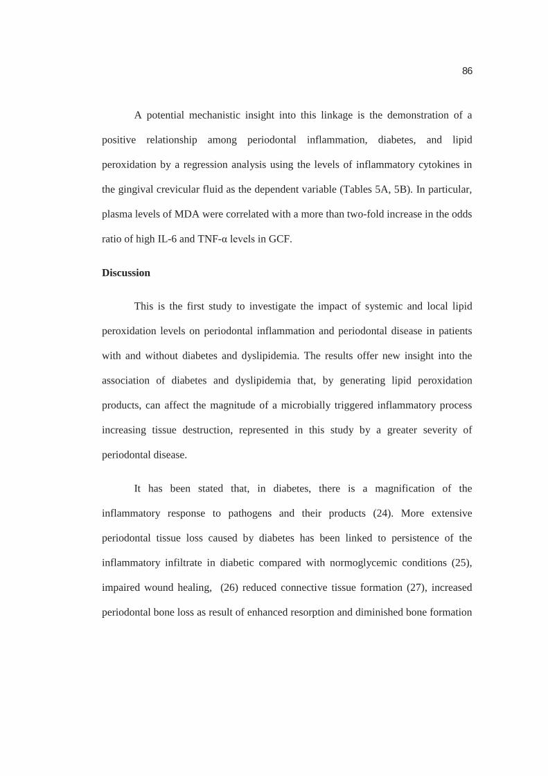
86
A potential mechanistic insight into this linkage is the demonstration of a
positive relationship among periodontal inflammation, diabetes, and lipid
peroxidation by a regression analysis using the levels of inflammatory cytokines in
the gingival crevicular fluid as the dependent variable (Tables 5A, 5B). In particular,
plasma levels of MDA were correlated with a more than two-fold increase in the odds
ratio of high IL-6 and TNF- in GCF.
Discussion
This is the first study to investigate the impact of systemic and local lipid
peroxidation levels on periodontal inflammation and periodontal disease in patients
with and without diabetes and dyslipidemia. The results offer new insight into the
association of diabetes and dyslipidemia that, by generating lipid peroxidation
products, can affect the magnitude of a microbially triggered inflammatory process
increasing tissue destruction, represented in this study by a greater severity of
periodontal disease.
It has been stated that, in diabetes, there is a magnification of the
inflammatory response to pathogens and their products (24). More extensive
periodontal tissue loss caused by diabetes has been linked to persistence of the
inflammatory infiltrate in diabetic compared with normoglycemic conditions (25),
impaired wound healing, (26) reduced connective tissue formation (27), increased
periodontal bone loss as result of enhanced resorption and diminished bone formation
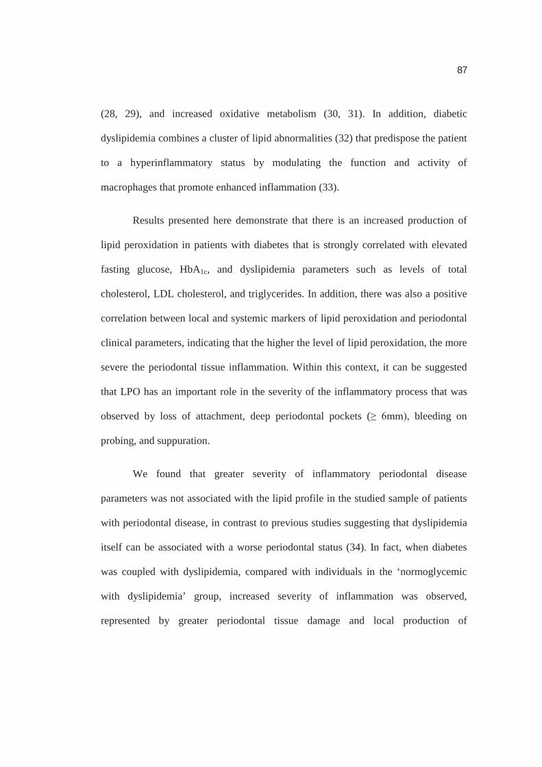
87
(28, 29), and increased oxidative metabolism (30, 31). In addition, diabetic
dyslipidemia combines a cluster of lipid abnormalities (32) that predispose the patient
to a hyperinflammatory status by modulating the function and activity of
macrophages that promote enhanced inflammation (33).
Results presented here demonstrate that there is an increased production of
lipid peroxidation in patients with diabetes that is strongly correlated with elevated
fasting glucose, HbA1c, and dyslipidemia parameters such as levels of total
cholesterol, LDL cholesterol, and triglycerides. In addition, there was also a positive
correlation between local and systemic markers of lipid peroxidation and periodontal
clinical parameters, indicating that the higher the level of lipid peroxidation, the more
severe the periodontal tissue inflammation. Within this context, it can be suggested
that LPO has an important role in the severity of the inflammatory process that was
observed by loss of attachment, deep periodontal pockets ( bleeding on
probing, and suppuration.
We found that greater severity of inflammatory periodontal disease
parameters was not associated with the lipid profile in the studied sample of patients
with periodontal disease, in contrast to previous studies suggesting that dyslipidemia
itself can be associated with a worse periodontal status (34). In fact, when diabetes
was coupled with dyslipidemia, compared with individuals in the ‘normoglycemic
with dyslipidemia’ group, increased severity of inflammation was observed,
represented by greater periodontal tissue damage and local production of
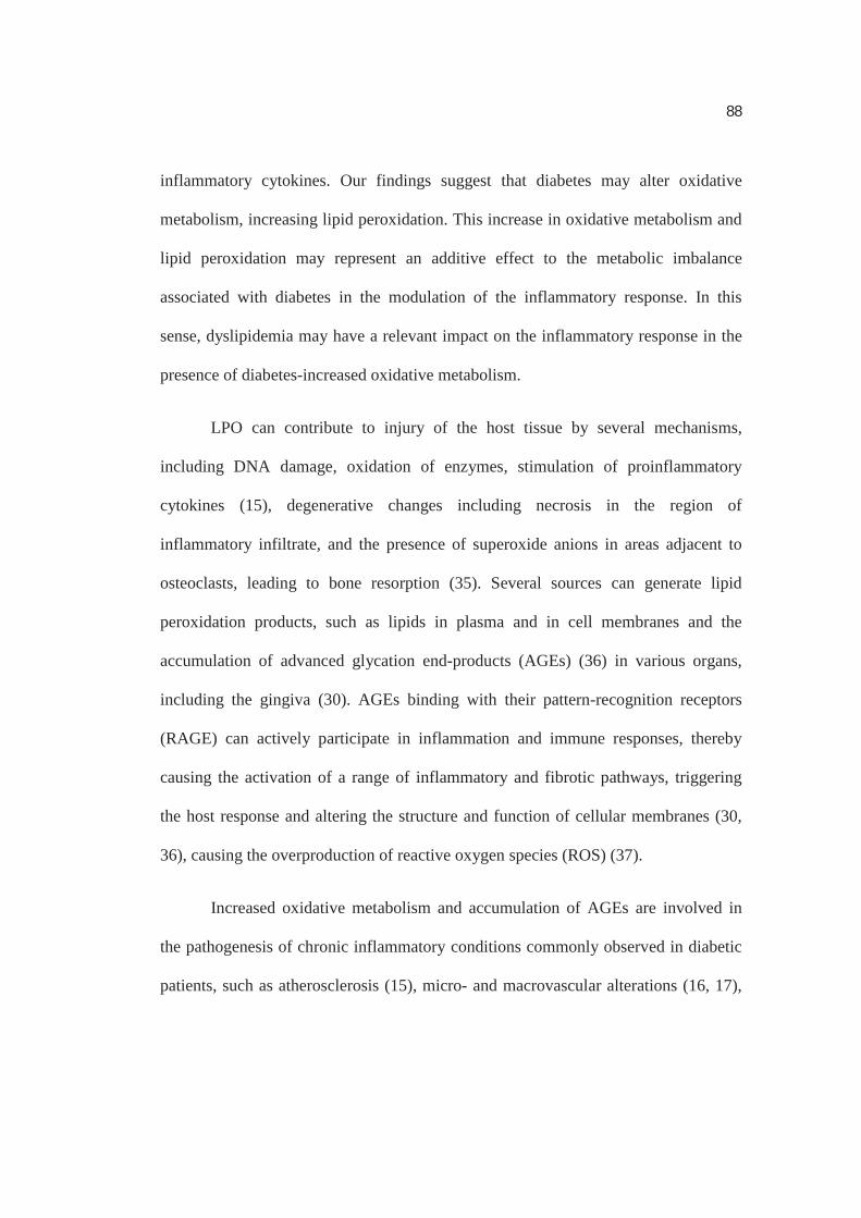
88
inflammatory cytokines. Our findings suggest that diabetes may alter oxidative
metabolism, increasing lipid peroxidation. This increase in oxidative metabolism and
lipid peroxidation may represent an additive effect to the metabolic imbalance
associated with diabetes in the modulation of the inflammatory response. In this
sense, dyslipidemia may have a relevant impact on the inflammatory response in the
presence of diabetes-increased oxidative metabolism.
LPO can contribute to injury of the host tissue by several mechanisms,
including DNA damage, oxidation of enzymes, stimulation of proinflammatory
cytokines (15), degenerative changes including necrosis in the region of
inflammatory infiltrate, and the presence of superoxide anions in areas adjacent to
osteoclasts, leading to bone resorption (35). Several sources can generate lipid
peroxidation products, such as lipids in plasma and in cell membranes and the
accumulation of advanced glycation end-products (AGEs) (36) in various organs,
including the gingiva (30). AGEs binding with their pattern-recognition receptors
(RAGE) can actively participate in inflammation and immune responses, thereby
causing the activation of a range of inflammatory and fibrotic pathways, triggering
the host response and altering the structure and function of cellular membranes (30,
36), causing the overproduction of reactive oxygen species (ROS) (37).
Increased oxidative metabolism and accumulation of AGEs are involved in
the pathogenesis of chronic inflammatory conditions commonly observed in diabetic
patients, such as atherosclerosis (15), micro- and macrovascular alterations (16, 17),
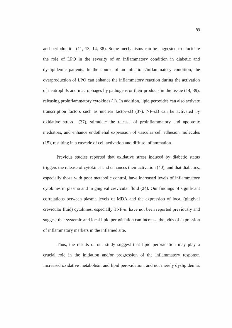
89
and periodontitis (11, 13, 14, 38). Some mechanisms can be suggested to elucidate
the role of LPO in the severity of an inflammatory condition in diabetic and
dyslipidemic patients. In the course of an infectious/inflammatory condition, the
overproduction of LPO can enhance the inflammatory reaction during the activation
of neutrophils and macrophages by pathogens or their products in the tissue (14, 39),
releasing proinflammatory cytokines (1). In addition, lipid peroxides can also activate
transcription factors such as nuclear factor- (37). NF-
oxidative stress (37), stimulate the release of proinflammatory and apoptotic
mediators, and enhance endothelial expression of vascular cell adhesion molecules
(15), resulting in a cascade of cell activation and diffuse inflammation.
Previous studies reported that oxidative stress induced by diabetic status
triggers the release of cytokines and enhances their activation (40), and that diabetics,
especially those with poor metabolic control, have increased levels of inflammatory
cytokines in plasma and in gingival crevicular fluid (24). Our findings of significant
correlations between plasma levels of MDA and the expression of local (gingival
crevicular fluid) cytokines, especially TNF- ously and
suggest that systemic and local lipid peroxidation can increase the odds of expression
of inflammatory markers in the inflamed site.
Thus, the results of our study suggest that lipid peroxidation may play a
crucial role in the initiation and/or progression of the inflammatory response.
Increased oxidative metabolism and lipid peroxidation, and not merely dyslipidemia,
90
aggravate the inflammatory response. If this association is proved causal by
experimental studies, targeting the oxidative metabolism and its downstream effects
may be an alternative therapeutic approach to manage diabetes-associated
inflammatory complications.
Acknowledgments
This study was supported by financial support from the São Paulo State Research
Support Foundation (FAPESP) (grant 2007/08362-8), by the Coordination for
Improvement of Higher Education Personnel of the Brazilian Ministry of Education
(CAPES), and by a grant from the NIH (DE017732).
References
1. Graves DT, Kayal RA 2008 Diabetic complications and dysregulated innate immunity. Front Biosci 13:1227-1239
2. Lamster IB, Ahlo JK 2007 Analysis of gingival crevicular fluid as applied to the diagnosis of oral and systemic diseases. Ann NY Acad Sci 1098:216-229
3. Baelum V, Lopez R 2004 Periodontal epidemiology: towards social science or molecular biology? Community Dent Oral Epidemiol 32:239-249
4. Griffiths R, Barbour S 2010 Lipoproteins and lipoprotein metabolism in periodontal disease. Clin Lipidol 5:397-411
5. Lalla E, Papapanou PN 2011 Diabetes mellitus and periodontitis: a tale of two common interrelated diseases. Nat Rev Endocrinol 7:738-748
6. Graves DT, Li J, Cochran DL 2011 Inflammation and uncoupling as mechanisms of periodontal bone loss. J Dent Res 90:143-153
91
7. Lamster IB, Lalla E, Borgnakke WS, Taylor GW 2008 The relationship between oral health and diabetes mellitus. J Am Dent Assoc 139(Suppl):19S-24S
8. Löe H 1993 Periodontal disease. The sixth complication of diabetes mellitus. Diabetes Care 16:329-334
9. Lim LP, Tay FB, Sum CF, Thai AC 2007 Relationship between markers of metabolic control and inflammation on severity of periodontal disease in patients with diabetes mellitus. J Clin Periodontol 34:118-123
10. Mealey BL, Oates TW 2006 Diabetes mellitus and periodontal diseases. J Periodontol 77:1289-1303
11. Chapple IL 1997 Reactive oxygen species and antioxidants in inflammatory diseases. J Clin Periodontol 24:287-296
12. Panjamurthy K, Manoharan S, Ramachandran CR 2005 Lipid peroxidation and antioxidant status in patients with periodontitis. Cell Mol Biol Lett 10:255-264
13. Akalin FA, Baltacioglu E, Alver A, Karabulut E 2007 Lipid peroxidation levels and total oxidant status in serum, saliva and gingival crevicular fluid in patients with chronic periodontitis. J Clin Periodontol 34:558-565
14. Tsai CC, Chen HS, Chen SL, Ho YP, Ho KY, Wu YM, Hung CC 2005 Lipid peroxidation: a possible role in the induction and progression of chronic periodontitis. J Periodontal Res 40:378-384
15. Halliwell B 2000 Oral inflammation and reactive species: a missed opportunity? Oral Dis 6:136-137
16. Velazquez E, Winocour PH, Kesteven P, Alberti KG, Laker MF 1991 Relation of lipid peroxides to macrovascular disease in type 2 diabetes. Diabet Med 8:752-758
17. Kedziora-Kornatowska KZ, Luciak M, Blaszczyk J, Pawlak W 1998 Lipid peroxidation and activities of antioxidant enzymes in erythrocytes of patients with non-insulin dependent diabetes with or without diabetic nephropathy. Nephrol Dial Transplant 13:2829-2832
18. Del Rio D, Stewart AJ, Pellegrini N 2005 A review of recent studies on malondialdehyde as toxic molecule and biological marker of oxidative stress. Nutr Metab Cardiovasc Dis 15:316-328
19. International Workshop for a Classification of Periodontal Diseases and Conditions. Papers. Oak Brook, Illinois, October 30-November 2, 1999. Ann Periodontol 4:i, 1-112
20. Matthews DR, Hosker JP, Rudenski AS, Naylor BA, Treacher DF, Turner RC1985 Homeostasis model assessment: insulin resistance and beta-cell function from fasting plasma glucose and insulin concentrations in man. Diabetologia 28:412-419
92
21. Koromantzos PA, Makrilakis K, Dereka X, Katsilambros N, Vrotsos IA, Madianos PN 2011 A randomized, controlled trial on the effect of non-surgical periodontal therapy in patients with type 2 diabetes. Part I: effect on periodontal status and glycaemic control. J Clin Periodontol 38:142-147
22. Gamonal J, Acevedo A, Bascones A, Jorge O, Silva A 2000 Levels of interleukin-1 beta, -8, and -10 and RANTES in gingival crevicular fluid and cell populations in adult periodontitis patients and the effect of periodontal treatment. J Periodontol 71:1535-1545
23. Hong YL, Yeh SL, Chang CY, Hu ML 2000 Total plasma malondialdehyde levels in 16 Taiwanese college students determined by various thiobarbituric acid tests and an improved high-performance liquid chromatography-based method. Clin Biochem 33:619-625
24. Nishimura F, Iwamoto Y, Soga Y 2007 The periodontal host response with diabetes. Periodontol 2000 43:245-253
25. Graves DT, Liu R, Oates TW 2007 Diabetes-enhanced inflammation and apoptosis: impact on periodontal pathosis. Periodontol 2000 45:128-137
26. Siqueira MF, Li J, Chehab L, Desta T, Chino T, Krothpali N, Behl Y, Alikhani M, Yang J, Braasch C, Graves DT 2010 Impaired wound healing in mouse models of diabetes is mediated by TNF-alpha dysregulation and associated with enhanced activation of forkhead box O1 (FOXO1). Diabetologia 53:378-388
27. Desta T, Li J, Chino T, Graves DT 2010 Altered fibroblast proliferation and apoptosis in diabetic gingival wounds. J Dent Res 89:609-614
28. Liu R, Bal HS, Desta T, Krothapalli N, Alyassi M, Luan Q, Graves DT 2006 Diabetes enhances periodontal bone loss through enhanced resorption and diminished bone formation. J Dent Res 85:510-514
29. Graves DT, Li J, Cochran DL Inflammation and uncoupling as mechanisms of periodontal bone loss. J Dent Res 90:143-153
30. Schmidt AM, Weidman E, Lalla E, Yan SD, Hori O, Cao R, Brett JG, Lamster IB 1996 Advanced glycation endproducts (AGEs) induce oxidant stress in thegingiva: a potential mechanism underlying accelerated periodontal disease associated with diabetes. J Periodontal Res 31:508-515
31. Allen EM, Matthews JB, DJ OH, Griffiths HR, Chapple IL 2011 Oxidative and inflammatory status in Type 2 diabetes patients with periodontitis. J Clin Periodontol 38:894-901
32. Chahil TJ, Ginsberg HN 2006 Diabetic dyslipidemia. Endocrinol Metab Clin North Am 35:491-510, vii-viii
93
33. Jovinge S, Ares MP, Kallin B, Nilsson J 1996 Human monocytes/macrophages release TNF-alpha in response to Ox-LDL. Arterioscler Thromb Vasc Biol 16:1573-1579
34. Fentoglu O, Oz G, Tasdelen P, Uskun E, Aykac Y, Bozkurt FY 2009 Periodontal status in subjects with hyperlipidemia. J Periodontol 80:267-273
35. Sobaniec H, Sobaniec-Lotowska ME 2000 Morphological examinations of hard tissues of periodontium and evaluation of selected processes of lipid peroxidation in blood serum of rats in the course of experimental periodontitis. Med Sci Monit 6:875-881
36. Gillery P, Monboisse JC, Maquart FX, Borel JP 1989 Does oxygen free radical increased formation explain long term complications of diabetes mellitus? Med Hypotheses 29:47-50
37. Yan SD, Schmidt AM, Anderson GM, Zhang J, Brett J, Zou YS, Pinsky D, Stern D 1994 Enhanced cellular oxidant stress by the interaction of advanced glycation end products with their receptors/binding proteins. J Biol Chem 269:9889-9897
38. Wei D, Zhang XL, Wang YZ, Yang CX, Chen G 2010 Lipid peroxidation levels, total oxidant status and superoxide dismutase in serum, saliva and gingival crevicular fluid in chronic periodontitis patients before and after periodontal therapy. Aust Dent J 55:70-78
39. Sheikhi M, Bouhafs RK, Hammarström KJ, Jarstrand C 2001 Lipid peroxidation caused by oxygen radicals from Fusobacterium-stimulated neutrophils as a possible model for the emergence of periodontitis. Oral Dis 7:41-46
40. Esposito K, Nappo F, Marfella R, Giugliano G, Giugliano F, Ciotola M, Quagliaro L, Ceriello A, Giugliano D 2002 Inflammatory cytokine concentrations are acutely increased by hyperglycemia in humans: role of oxidative stress. Circulation 106:2067-2072
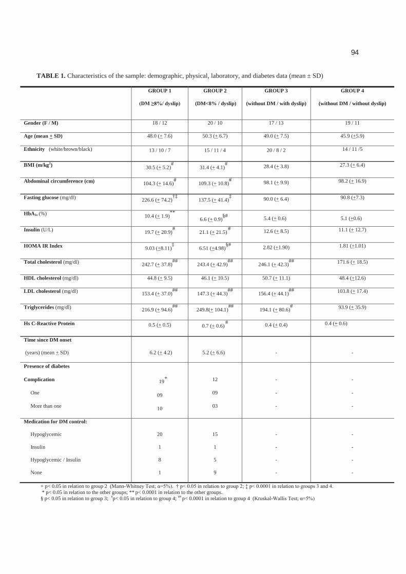
94
TABLE 1. Characteristics of the sample: demographic, physical, laboratory, and diabetes data (mean ± SD)
GROUP 1 GROUP 2
(DM<8% / dyslip)
GROUP 3
(without DM / with dyslip)
GROUP 4
(without DM / without dyslip)
Gender (F / M) 18 / 12 20 / 10 17 / 13 19 / 11
Age (mean + SD) 48.0 (+ 7.6) 50.3 (+ 6.7) 49.0 (+ 7.5) 45.9 (+5.9)
Ethnicity (white/brown/black) 13 / 10 / 7 15 / 11 / 4 20 / 8 / 2 14 / 11 /5
BMI (m/kg2) 30.5 (+ 5.2)#
31.4 (+ 4.1)# 28.4 (+ 3.8) 27.3 (+ 6.4)
Abdominal circumference (cm) 104.3 (+ 14.6)#
109.3 (+ 10.8)# 98.1 (+ 9.9) 98.2 (+ 16.9)
Fasting glucose (mg/dl) 226.6 (+ 74.2)†‡
137.5 (+ 41.4)‡ 90.0 (+ 6.4) 90.8 (+7.3)
HbA1c (%) 10.4 (+ 1.9)**
6.6 (+ 0.9)§# 5.4 (+ 0.6) 5.1 (+0.6)
Insulin (U/L) 19.7 (+ 20.9)#
21.1 (+ 21.5)# 12.6 (+ 8.5) 11.1 (+ 12.7)
HOMA IR Index 9.03 (+8.11)‡
6.51 (+4.98)§# 2.82 (+1.90) 1.81 (+1.01)
Total cholesterol (mg/dl) 242.7 (+ 37.8)##
243.4 (+ 42.9)##
246.1 (+ 42.3)## 171.6 (+ 18.5)
HDL cholesterol (mg/dl) 44.8 (+ 9.5) 46.1 (+ 10.5) 50.7 (+ 11.1) 48.4 (+12.6)
LDL cholesterol (mg/dl) 153.4 (+ 37.0)##
147.3 (+ 44.3)##
156.4 (+ 44.1)## 103.8 (+ 17.4)
Triglycerides (mg/dl) 216.9 (+ 94.6)##
249.8(+ 104.1)##
194.1 (+ 80.6)# 93.9 (+ 35.9)
Hs C-Reactive Protein 0.5 (+ 0.5) 0.7 (+ 0.6)# 0.4 (+ 0.4) 0.4 (+ 0.6)
Time since DM onset
(years) (mean + SD)
6.2 (+ 4.2)
5.2 (+ 6.6) - -
Presence of diabetes
Complication
One
More than one
19+
09
10
12
09
03
-
-
-
-
-
-
Medication for DM control:
Hypoglycemic
Insulin
Hypoglycemic / Insulin
None
20
1
8
1
15
1
5
9
-
-
-
-
-
-
-
-
+ p< 0.05 in relation to group 2 (Mann- * p< 0.05 in relation to the other groups; ** p< 0.0001 in relation to the other groups. § p< 0.05 in relation to group 3; #p< 0.05 in relation to group 4; ## p< 0.0001 in relation to group 4 (Kruskal-
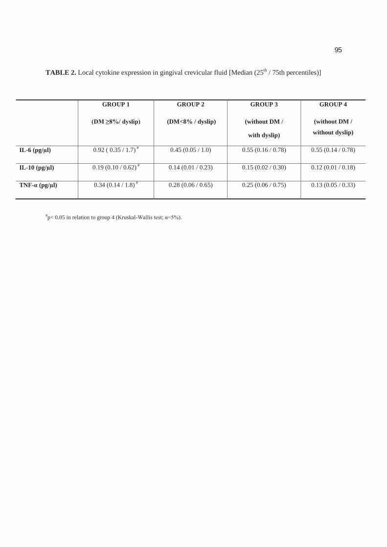
95
TABLE 2. Local cytokine expression in gingival crevicular fluid [Median (25th / 75th percentiles)]
GROUP 1 GROUP 2
(DM<8% / dyslip)
GROUP 3
(without DM /
with dyslip)
GROUP 4
(without DM /
without dyslip)
IL-6 (pg/μl) 0.92 ( 0.35 / 1.7) # 0.45 (0.05 / 1.0) 0.55 (0.16 / 0.78) 0.55 (0.14 / 0.78)
IL-10 (pg/μl) 0.19 (0.10 / 0.62) # 0.14 (0.01 / 0.23) 0.15 (0.02 / 0.30) 0.12 (0.01 / 0.18)
TNF- (pg/μl) 0.34 (0.14 / 1.8) # 0.28 (0.06 / 0.65) 0.25 (0.06 / 0.75) 0.13 (0.05 / 0.33)
#p< 0.05 in relation to group 4 (Kruskal-
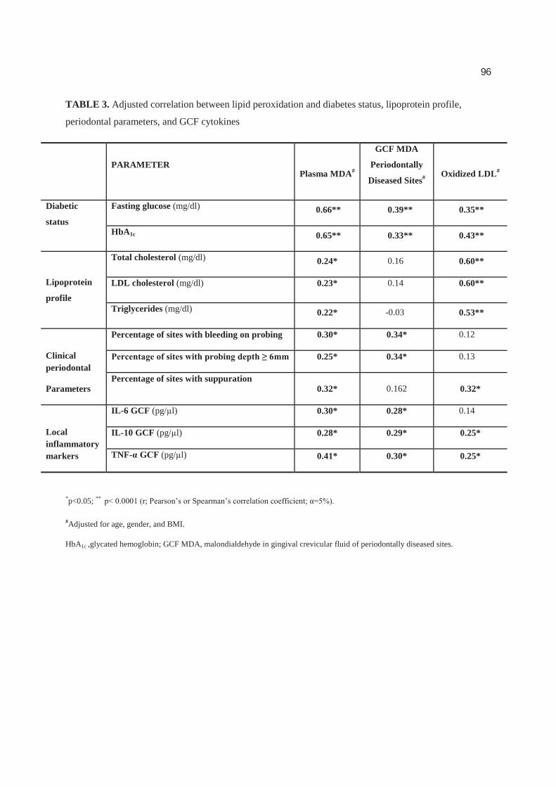
96
TABLE 3. Adjusted correlation between lipid peroxidation and diabetes status, lipoprotein profile,
periodontal parameters, and GCF cytokines
PARAMETERPlasma MDA#
GCF MDA
Periodontally
Diseased Sites# Oxidized LDL#
Diabetic
status
Fasting glucose (mg/dl) 0.66** 0.39** 0.35**
HbA1c 0.65** 0.33** 0.43**
Lipoprotein
profile
Total cholesterol (mg/dl) 0.24* 0.16 0.60**
LDL cholesterol (mg/dl) 0.23* 0.14 0.60**
Triglycerides (mg/dl) 0.22* -0.03 0.53**
Clinical periodontal
Parameters
Percentage of sites with bleeding on probing 0.30* 0.34* 0.12
0.25* 0.34* 0.13
Percentage of sites with suppuration 0.32* 0.162 0.32*
Local inflammatory markers
IL-6 GCF (pg/μl) 0.30* 0.28* 0.14
IL-10 GCF (pg/μl) 0.28* 0.29* 0.25*
TNF- (pg/μl) 0.41* 0.30* 0.25*
*p<0.05; **
#Adjusted for age, gender, and BMI.
HbA1c ,glycated hemoglobin; GCF MDA, malondialdehyde in gingival crevicular fluid of periodontally diseased sites.
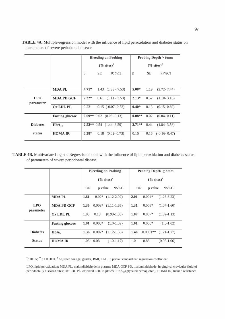
97
TABLE 4A. Multiple-regression model with the influence of lipid peroxidation and diabetes status on parameters of severe periodontal disease
TABLE 4B. Multivariate Logistic Regression model with the influence of lipid peroxidation and diabetes status of parameters of severe periodontal disease.
*p<0.05; ** p< 0.0001. # Adjusted for age, gender, BMI, TGL. partial standardized regression coefficient.
LPO, lipid peroxidation; MDA PL, malondialdehyde in plasma; MDA GCF PD, malondialdehyde in gingival crevicular fluid of periodontally diseased sites; Ox LDL PL, oxidized LDL in plasma; HbA1c (glycated hemoglobin); HOMA IR, Insulin resistance
Bleeding on Probing
(% sites)# (% sites)#
LPO parameter
MDA PL 4.71* 1.43 (1.88 - 7.53) 5.08* 1.19 (2.72- 7.44)
MDA PD GCF 2.32* 0.61 (1.11 - 3.53) 2.13* 0.52 (1.10- 3.16)
Ox LDL PL 0.23 0.15 (-0.07- 0.53) 0.40* 0.13 (0.15- 0.69)
Diabetes
status
Fasting glucose 0.09** 0.02 (0.05- 0.13) 0.08** 0.02 (0.04- 0.11)
HbA1c 2.52** 0.54 (1.44- 3.59) 2.71** 0.44 (1.84- 3.58)
HOMA IR 0.38* 0.18 (0.02- 0.73) 0.16 0.16 (-0.16- 0.47)
Bleeding on Probing
(% sites)#
OR p value 95%CI
(% sites)#
OR p value 95%CI
LPO parameter
MDA PL 1.81 0.02* (1.12-2.92) 2.01 0.004* (1.25-3.23)
MDA PD GCF 1.36 0.003* (1.11-1.65) 1.31 0.009* (1.07-1.60)
Ox LDL PL 1.03 0.13 (0.99-1.08) 1.07 0.007* (1.02-1.13)
Diabetes
Status
Fasting glucose 1.01 0.005* (1.0-1.02) 1.01 0.006* (1.0-1.02)
HbA1c 1.36 0.002* (1.12-1.66) 1.46 0.0001** (1.21-1.77)
HOMA IR 1.08 0.08 (1.0-1.17) 1.0 0.88 (0.95-1.06)
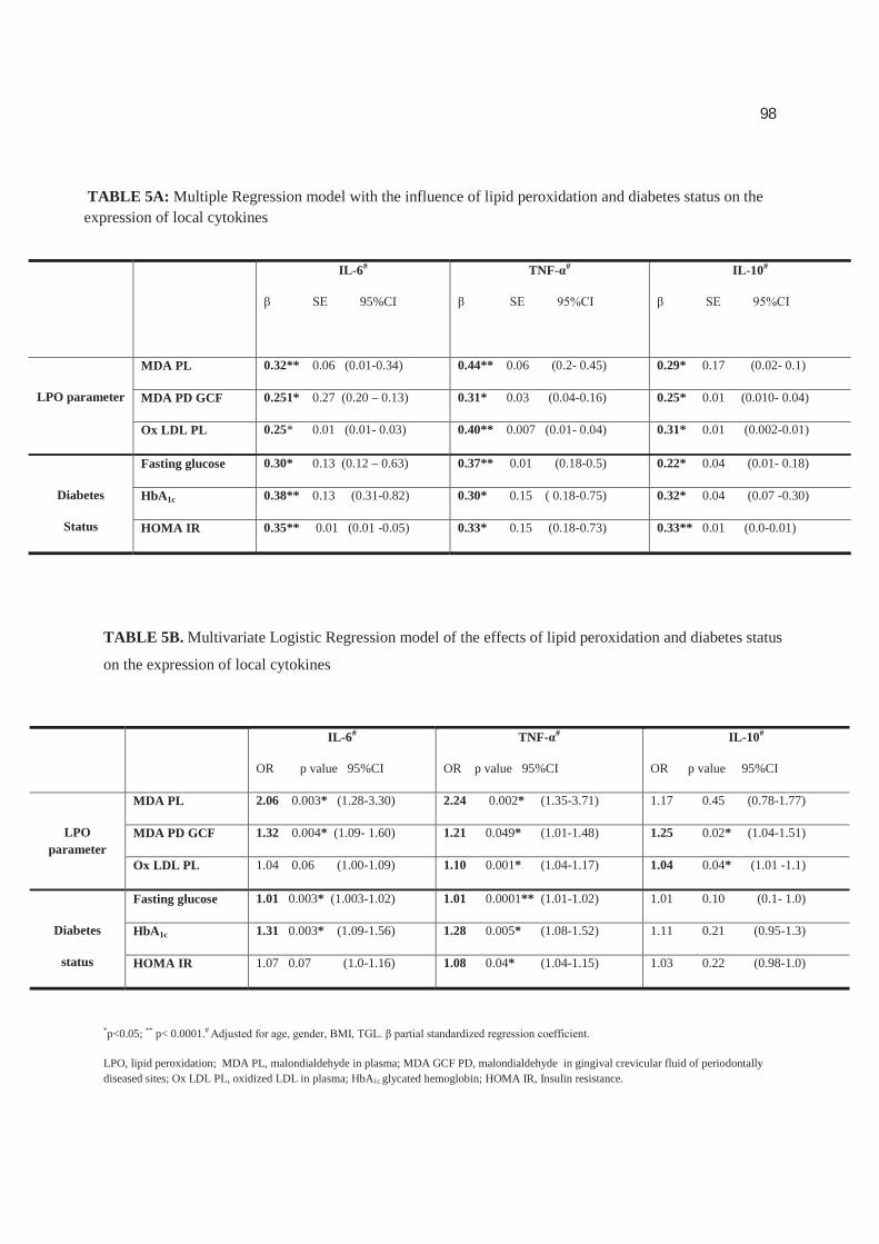
98
TABLE 5A: Multiple Regression model with the influence of lipid peroxidation and diabetes status on the expression of local cytokines
TABLE 5B. Multivariate Logistic Regression model of the effects of lipid peroxidation and diabetes status
on the expression of local cytokines
*p<0.05; ** p< 0.0001.#
LPO, lipid peroxidation; MDA PL, malondialdehyde in plasma; MDA GCF PD, malondialdehyde in gingival crevicular fluid of periodontally diseased sites; Ox LDL PL, oxidized LDL in plasma; HbA1c glycated hemoglobin; HOMA IR, Insulin resistance.
IL-6#
95%CI
TNF- # IL-10#
LPO parameter
MDA PL 0.32** 0.06 (0.01-0.34) 0.44** 0.06 (0.2- 0.45) 0.29* 0.17 (0.02- 0.1)
MDA PD GCF 0.251* 0.27 (0.20 – 0.13) 0.31* 0.03 (0.04-0.16) 0.25* 0.01 (0.010- 0.04)
Ox LDL PL 0.25* 0.01 (0.01- 0.03) 0.40** 0.007 (0.01- 0.04) 0.31* 0.01 (0.002-0.01)
Diabetes
Status
Fasting glucose 0.30* 0.13 (0.12 – 0.63) 0.37** 0.01 (0.18-0.5) 0.22* 0.04 (0.01- 0.18)
HbA1c 0.38** 0.13 (0.31-0.82) 0.30* 0.15 ( 0.18-0.75) 0.32* 0.04 (0.07 -0.30)
HOMA IR 0.35** 0.01 (0.01 -0.05) 0.33* 0.15 (0.18-0.73) 0.33** 0.01 (0.0-0.01)
IL-6#
OR p value 95%CI
TNF- #
OR p value 95%CI
IL-10#
OR p value 95%CI
LPO parameter
MDA PL 2.06 0.003* (1.28-3.30) 2.24 0.002* (1.35-3.71) 1.17 0.45 (0.78-1.77)
MDA PD GCF 1.32 0.004* (1.09- 1.60) 1.21 0.049* (1.01-1.48) 1.25 0.02* (1.04-1.51)
Ox LDL PL 1.04 0.06 (1.00-1.09) 1.10 0.001* (1.04-1.17) 1.04 0.04* (1.01 -1.1)
Diabetes
status
Fasting glucose 1.01 0.003* (1.003-1.02) 1.01 0.0001** (1.01-1.02) 1.01 0.10 (0.1- 1.0)
HbA1c 1.31 0.003* (1.09-1.56) 1.28 0.005* (1.08-1.52) 1.11 0.21 (0.95-1.3)
HOMA IR 1.07 0.07 (1.0-1.16) 1.08 0.04* (1.04-1.15) 1.03 0.22 (0.98-1.0)
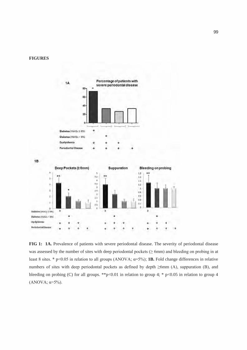
99
FIGURES
FIG 1: 1A. Prevalence of patients with severe periodontal disease. The severity of periodontal disease
was assessed by the number of sites with deep
1B. Fold change differences in relative
and
bleeding on probing (C) for all groups. **p<0.01 in relation to group 4; * p<0.05 in relation to group 4
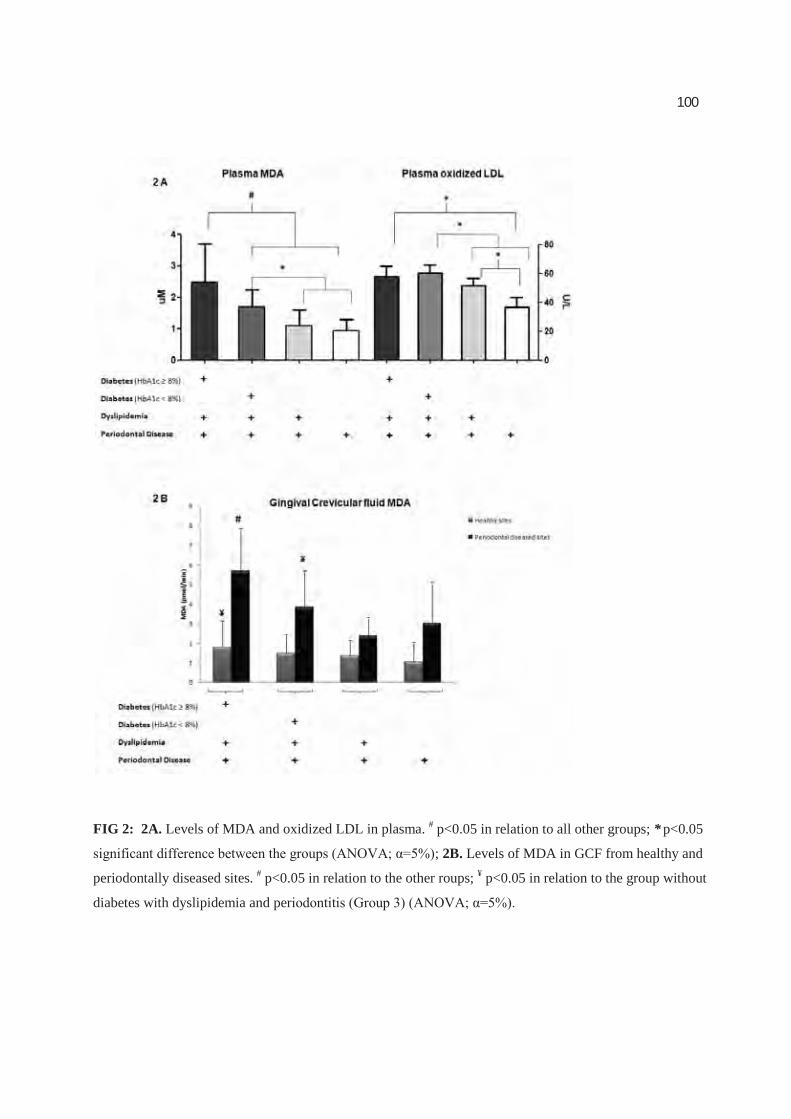
100
FIG 2: 2A. Levels of MDA and oxidized LDL in plasma. # p<0.05 in relation to all other groups; * p<0.05
; 2B. Levels of MDA in GCF from healthy and
periodontally diseased sites. # p<0.05 in relation to the other roups; ¥ p<0.05 in relation to the group without
diabetes with dyslipidemia and per
101
CAPÍTULO 3
Systemic inflammatory profile aggravated by lipid
peroxidation products in patients with type 2 Diabetes
Alliny de Souza Bastos, Dana T. Graves, Ana Paula de Melo Loureiro, Carlos Rossa
Júnior, Dulcinéia Saes Parra Abdalla, Tanize do Espírito Santo Faulin, Niels Olsen
Câmara, Oelisoa M Andriankaja, and Silvana Regina Perez Orrico
102
Systemic inflammatory profile aggravated by lipid peroxidation products in
patients with type 2 Diabetes
Alliny de Souza Bastos1, Dana T. Graves2, Ana Paula de Melo Loureiro3, Carlos
Rossa Júnior1, Dulcinéia Saes Parra Abdalla3, Tanize do Espírito Santo Faulin3, Niels
Olsen Câmara4, Oelisoa M Andriankaja2, and Silvana Regina Perez Orrico1
1Department of Diagnosis and Surgery, Araraquara School of Dentistry – UNESP-
State University of São Paulo, Araraquara, São Paulo, Brazil2Department of Periodontics, School of Dental Medicine, University of Pennsylvania,
Philadelphia, PA, United States3 Department of Clinical and Toxicological Analyses, Faculty of Pharmaceutical
Sciences, University of São Paulo (USP), São Paulo, Brazil 4 Department of Immunology, Institute of Biomedical Sciences, University of São
Paulo (USP), São Paulo, Brazil
Abbreviated Title: Lipid peroxidation, diabetes, periodontitis
Corresponding author:
Profª. Drª. Silvana Regina Perez Orrico, e-mail: [email protected]
UNESP – Univ Estadual Paulista; Araraquara School of Dentistry
Department of Diagnosis and Surgery Rua Humaitá, 1680, 2º andar
CEP: 14801-903, Araraquara, São Paulo, Brazil.
Phone: +55 (16) 3301-6377 Fax: +55 (16) 3301-6369
103
ABSTRACT
Lipid peroxidation (LPO) is increased in patients with diabetes. The
evaluation of diabetes, lipid peroxidation, dyslipidemia and inflammation has not
been evaluated simultaneously. The aim of this study is to investigate if diabetes
coupled with dyslipidemia may alter the oxidative metabolism, by increasing lipid
peroxidation products leading to the modulation of the inflammatory response. The
sample was comprised of 100 patients divided into four groups based upon diabetic
and dyslipidemic status: poorly controlled diabetics with dyslipidemia, well-
controlled diabetics with dyslipidemia, normoglycemic individuals with
dyslipidemia, and healthy individuals. Blood analyses were carried out for fasting
plasma glucose, HbA1c and lipid profile. LPO levels in plasma evaluated by
measuring oxLDL (ELISA) and malondialdehyde (MDA, HPLC). Plasma cytokines
were evaluated by the multiplex bead technique. Lipid peroxidation evaluated by
MDA was significantly increased in DM groups. The cytokines panel showed a
decreasing pattern from the poorly controlled diabetes (p<0.05) to the healthy group.
Even in well-controlled diabetics with dyslipidemia the levels of MDA, OxLDL and
inflammatory cytokines were significantly increased when compared to the non
diabetic/ dyslipidemic group. Moderate positive (p<0.0001) correlations were
observed between diabetic status, lipid peroxidation markers and proinflammatory
cytokines expression, such as MDA and HbA1c (0.64); MDA and IL1-
MDA and TNF- . (0.40); MDA and IL-6 (0.40).These findings suggest that LPO
may represent an additive mechanism in the aggravation of inflammation in diabetics
and may support a role for increased oxidative metabolism in the inflammatory
response.
Key terms: Lipid peroxidation, diabetes mellitus type 2, periodontal diseases,
cytokines
104
INTRODUCTION
Diabetes is a group of metabolic diseases that, by several mechanisms such as
the accumulation of advanced glycation end products (AGEs), leads to several events
that are involved in the pathogenesis of chronic inflammatory conditions, impaired
wound healing, higher susceptibility to infections1, atherosclerosis2, and micro- and
macrovascular alteration3-5. Considering that inflammation is an underlying cause of
several co-morbidities associated with poorly controlled diabetes6, several studies
demonstrated high levels of circulating inflammatory markers such as interleukin-6,
tumor necrosis factor (TNF- )6-10 in type 2 diabetic patients.
Systemically, several alterations can contribute to the hyperinflammatory
status of the patients with diabetes such as the binding of AGEs with their pattern-
recognition receptors (RAGE). The interaction AGE-RAGE participate actively in
inflammation and immune responses, dyslipidemia that modulates the function and
activity of macrophages that promote enhanced inflammation11 and an over-
production of ROS leading to peroxidation of lipid membranes. Lipid peroxidation
can contribute to injury of the host tissue by several mechanisms, including DNA
damage, oxidation of enzymes and stimulation of proinflammatory cytokines 2.
However the evaluation of these conditions, diabetes, lipid peroxidation,
dyslipidemia and inflammation have not been evaluated simultaneously. In this
context, even in well controlled diabetic patients the effects of dyslipidemia and lipid
peroxidation products should be considered in a more emphatic way helping to
105
prevent these patients to become a poorly controlled diabetic. In this context, the aim
of our study is to investigate if diabetes coupled with dyslipidemia may alter the
oxidative metabolism, by increasing lipid peroxidation products leading to the
modulation of the inflammatory response.
MATERIALS AND METHODS
Study population
The present cross-sectional study was approved by the Ethics in Human
Research Committee of the Araraquara School of Dentistry (UNESP – Univ. Estadual
Paulista, Araraquara, Brazil; Protocol number 50/06) and was conducted according to
the ethical principles of the Declaration of Helsinki. All volunteers were informed
about the aims and methods of this study, and they provided their written consent to
participate.
A simple questionnaire was applied initially to all subjects to identify the
patients that were eligible to be enrolled in the study, considering the
inclusion/exclusion criteria. All study participants should range in age from 35 to 60
years and have similar socio-economic level. None of the patients were pregnant or
were current or former smokers. Patients taking antibiotic, nonsteroidal anti-
inflammatory, hormones or hypolipidemic drugs such as statins or fibrates, were
excluded from the study. Particular care was taken to exclude subjects with history of
106
anemia and diseases known to influence lipoprotein metabolism, such as
hypothyroidism and hypopituitarism.
A power analysis determined that at least 25 patients in each group would be
sufficient to detect a 3-unit difference in MDA levels or a 2.4-unit difference in an
inflammatory cytokine with 90% power and 95% confidence interval. The population
was divided into four groups based upon diabetic and dyslipidemic status: poorly
controlled diabetics with dyslipidemia (Group 1), well-controlled diabetics with
dyslipidemia (Group 2), normoglycemic individuals with dyslipidemia (Group 3),
and healthy individuals (Group 4).
Clinical record and physical evaluation
After been enrolled in the study, all participants answered a face-to-face
structured questionnaire about socio-demographic data, personal and family medical
history, and use of medications. A trained examiner collected information from
diabetics patients regarding time since diabetes onset, medication used to control
glucose, and the presence of complications associated with diabetes. Subjects
completed a physical examination including anthropometric data such as abdominal
circumference (cm), hip (cm), waist (cm) and height (m), weight (kg), and body mass
index (BMI).
Laboratory measurements: metabolic control and lipoprotein profile
107
Blood samples were collected after a 12-hour overnight fast for the evaluation
of fasting plasma glucose (mg/dL) by modified Bondar e Mead method, glycated
haemoglobin (HbA1c) by enzymatic immunoturbidimetry, high-sensitivity C-reactive
protein by the nephelomeric method and lipid profile (total cholesterol (TC),
triglycerides (TGs), and HDL) by enzymatic methods. LDL was determined by the
Friedewald formula. To avoid the inclusion of individuals with transitory
dyslipidemia, the cutoff points used were the highest values according to the National
Cholesterol Educational Program (NCEP) Adult Treatment III (ATP III)12: TC 240
mg/dL, LDL 160 mg/dL , and TGs 200 mg/dL. Metabolic
control was considered as adequate when HbA1c 7.0% and as inadequate when
HbA1c -
controlled. Insulin levels were determined by the chemiluminescence method
(uUI/ml) and homeostatasis model assessment of insulin resistance (HOMA IR) was
calculated by the formula: fasting insulin concentration (μU/ml) x fasting glucose
concentration (mmol/L) 13.
Sample collection
Blood samples were collected in tubes with EDTA centrifuged at 3000 g for
10 min at 4°C, plasma was obtained, promptly aliquoted and stored at -80 C until the
lipid peroxidation and cytokine analyses. The following assays were performed by
professionals blinded to allocation of the patients into the groups.
108
Analysis of cytokines in plasma
Eleven cytokines (interleukin-1 IL-1 , IL-2,-4,-5,-6,-7,-8,-10,-12(p70)-IL-
13 and tumor necrosis factor-alpha TNF- ) were measured in plasma samples using a
multiplex bead analysis (Bio-Plex system, Bio-Rad Laboratories, Hercules, CA,
USA), following the manufacturer’s instructions.
Lipid peroxidation assays
Oxidized LDL assay: The levels of oxLDL in plasma were analyzed with a
commercially available ELISA kit (Mercodia AB®, Uppsala, Sweden) following the
manufacturer’s instructions.
MDA assay: The assessment of MDA in plasma was determined by HPLC
(Shimadzu, Tokyo, Japan) with a reverse-phase HPLC column (C18; 4.6 x 150 mm)
(Phenomenex, Torrance, CA, USA) and compared with MDA standard curves.
Plasma samples were prepared as described previously 14.
Statistical analysis
The distribution and normality of the variables were evaluated by the
D’Agostino-Pearson test. The general characteristics of each group were described
with mean and standard deviation (SD), and the inflammatory cytokines were
expressed as median (25% /75% quartiles). The differences between the groups for
parametric data were evaluated by ANOVA followed by Bonferroni’s post-test, and
109
non-parametric data were evaluated by Kruskall-Wallis test, followed by Dunn’s
post-test. Pearson’s partial correlation coefficient was used to investigate the
correlations between the various parameters adjusted for age, gender, and BMI. To
determine the influence of lipid peroxidation and diabetic status on the expression of
plasma cytokines, separate multivariable logistic and linear regression models were
built for each independent variable. Variables found to be associated with the
independent and dependent variables, and thus possible confounding factors, were
included in the models. Moreover, regardless of their statistical significance, all
variables known to have biologic linkage with the outcomes were retained. The
significance level was set at =0.05. All analyses were carried out with SPSS
software, IBM version 19.
RESULTS
Sample population
The general characteristics of the sample are demonstrated in Table 01. From
the screened patients, 100 were identified as meeting the study inclusion criteria; of
these, 25 patients assigned to 4 different groups. There was no statistical significant
difference between the participants with respect to gender, age and socioeconomic
status. In relation to physical evaluation, all individuals were overweight and the
diabetic groups were obese and presented higher values of BMI (p<0.05), waist/hip
proportion and abdominal circumference. The groups with diabetes had significantly
110
increased levels of fasting glucose, HbA1c, and insulin resistance compared with non-
diabetic patients. Based on ATP III12 definition of dyslipidemia groups 1, 2, and 3
were dyslipidemic presenting increased levels of total cholesterol, LDL cholesterol,
and triglycerides compared with group 4 (healthy individuals) (p<0.05).
Among all diabetics, there was no difference in the age of diabetes onset.
However, poorly controlled diabetics were the most hypertensive patients and had
more diabetic complications (p<0.05), with the most common complication being
retinopathy, followed by nephropathy.
Lipid peroxidation levels
Plasma Malondialdehyde (MDA) and Oxidized LDL (oxLDL)
Plasma MDA levels were slightly increased in dyslipidemic patients (Fig. 1A).
However, the most increased levels of plasma MDA were observed in poorly
controlled diabetics with dyslipidemia who had about 2.9 times higher MDA levels
when compared with those in the non-diabetic groups and approximately 1.6 times
higher MDA levels when compared with well-controlled diabetics with dyslipidemia.
OxLDL was also increased in all groups with dyslipidemia (Groups 1, 2, and 3) and in
the plasma of diabetics were approximately 1.7 times higher than in the healthy group
(p<0.05) (Fig. 1B). Well-controlled diabetics with dyslipidemia (Group 2) presented
significantly increased levels of MDA and OxLDL (p<0.05) when compared to the non
diabetic/ dyslipidemic group (Group 3).
111
Immunological analysis
To assess inflammation, the expression of IL-1 , IL-2, IL-4, IL-5, IL-6, IL-7,
IL-8, IL-10, IL-12 (p70) e IL-13 and TNF- was measured in plasma. The cytokines
panel showed a decreasing pattern from the poorly controlled diabetes (p<0.05) to the
healthy group, indicating that diabetes aggravates the inflammatory response. The
significant differences between the groups according to the expression of pro- and
anti-inflammatory cytokines are illustrated in Figure 2. The cytokines that did not
have statistical significant difference between the groups are represented in Table 2.
The cytokines that are considered adipokines such as IL-6 and TNF-
significantly increased in the group with dyslipidemia.
Correlations
Adjusted correlation among diabetes status, lipoprotein profile,
anthropometric and lipid peroxidation markers, and plasma cytokine expression were
assessed taking into account the effect of age, gender and BMI (Table 3). There was a
wide range of, moderate positive, correlations observed between diabetic status, lipid
peroxidation markers and proinflammatory cytokines expression. Among these most
significant correlations (p<0.0001) were between IL1-
HbA1c (0.40), MDA (0.50), OxLDL (0.42). Moderate positive correlation was also
observed between TNF- and fasting glucose (0.50), HbA1c (0.50), Triglycerides
112
(0.35), MDA (0.44), OxLDL (0.48). Lastly, IL-6 was moderately and positively
correlated with OxLDL (0.40). Diabetes was significantly associated with lipid
peroxidation markers, and there was a strong positive correlation observed between
MDA and HbA1c (r=0.643, p<0.0001), suggesting that the higher the concentration
of HbA1c, the higher the expression of MDA (Figure 03).
Regressions
A potential mechanistic insight into the linkage among inflammation,
diabetes, and lipid peroxidation is demonstrated by a multiple regression analysis
using separate models for independent variables, lipid peroxidation parameters,
diabetes status, lipoprotein profile and physical parameters. Lipid peroxidation
markers and diabetic status were directly related to the expression of inflammatory
cytokines, especially IL-1 - -6 was significantly
related to lipoprotein profile rather than diabetic or inflammatory status (Table 4)
The multivariate logistic regression analysis using separate models for
dependent variables, also evaluated the expression of cytokines taking into account
the effects of age, gender and BMI. There was an approximate 3.2-, 2.0- 1.9- and 2.6-
fold increase in the odds of having increased levels of IL-1 , IL-6, IL-8, and TNF- ,
respectively, among those patients who have higher plasmatic levels of MDA. In
addition, increased levels of HbA1c were correlated with, approximately, 1.5-fold
increase in the odds ratio of high of IL-1 , IL-8, and TNF- . Physical parameters and
113
lipoprotein profile did not demonstrate significant relationship with increased
expression of inflammatory cytokines (Table 5).
Discussion
The evaluation of diabetes, lipid peroxidation, dyslipidemia and inflammatory
profile has not been evaluated simultaneously. In the present study we evaluated the
impact of lipid peroxidation on the inflammatory status in patients with diabetes
coupled with dyslipidemia. Our results suggest that lipid peroxidation can have an
impact on the modulation of the inflammatory response, even in well controlled
diabetics.
Previous studies evaluated cytokines in plasma of patients with diabetes6, 8, 15
and its influence in diabetic complications16, 17. However to the best of our
knowledge, this is the first study to measure 11 cytokines simultaneously and
correlate them with the several systemic parameters. Elevated proinflammatory
cytokines (IL-1 , IL-6, IL-8, TNF- ) were significantly detected in diabetics patients.
Antinflammatory cytokines (IL-4, IL-10) were also elevated in these patients, but the
difference was statistically significant only to IL-4. The presence of complications
related to diabetes was positively associated with increased levels of TNF- . Thus,
the expression of adipocytokines, IL-6 and TNF- , were significantly increased in
patients with dyslipidemia (Gr 1, 2 and 3) and were positively correlated with total
cholesterol and triglycerides.
114
It is known that obesity promotes dyslipidemia and may impact insulin action
via additional pathways, including through the release of leptin, IL-6 and TNF- 9. In
the present study the individuals with DM were considered obese (BMI> 30), had a
large waist circumference, an increased insulin resistance index (HOMA IR)
compared to healthy patients. However, in contrast with some studies9, 18, 19 we did
not find a statistic significant correlation between the cytokines and physical
parameters, suggesting that even in individuals with the physical parameters
considered in a normal range, the cytokines expression can be elevated leading to an
increased inflammatory response in these patients. Besides, positive but weak
correlations (0.2) were observed between cytokines (IL-6 e o TNF- ) and insulin
resistance (HOMA IR). In spite of the fact that previous studies demonstrated the
association between insulin resistance and the increased expression of inflammatory
cytokines9, 10, other authors did not find this correlation and affirm that cytokines
circulating levels are more related to hyperglycemia20 or to the presence of adipose
tissue18, 21 rather than insulin resistance itself.
The pathogenesis of diabetic dyslipidemia is not completely elucidated,
however, it is stated that lipid abnormalities are part of primary features of metabolic
syndrome and can precede the clinical manifestation of hyperglicemia, probably due
to its association with insulin resistance22-25. It is important to notice that even with
the well-established effect of dyslipidemia on diabetes23, 24, 26, 27, several studies with
diabetic population do not consider the lipid profile of the patients8, 17, 20, 25, 28 or do
115
not categorize them according to their dyslipidemic status6, 19, 20, 29-31. In the present
study we evaluated the lipid profile of the whole sample and included in diabetic
groups only patients with established dyslipidemia12 to avoid the inclusion of diabetic
patients with a transitory state, considering that diabetes is a metabolic syndrome and
a cluster of lipid abnormalities32 is a part of it. To check real the impact of
dyslipidemia alone on lipid peroxidation markers and on inflammatory cytokines
we’ve included a group of patients only with dyslipidemia (group 3).
In the present study we observed significant but weak correlation between
dyslipidemic status and inflammatory cytokines suggesting that in spite of the fact
that abnormalities in lipids can contribute to the production of lipid peroxides,
dyslipidemia itself was not responsible to major alterations on oxidative and
inflammatory profile. In fact, when diabetes was coupled with dyslipidemia (Gr 1 and
2), compared with normoglycemic /dyslipidemic individuals (Gr 3), an increased
expression of LPO products and inflammatory cytokines was observed, suggesting
that not only the dyslipidemia itself but the presence of diabetes resulted in
significantly increased systemic oxidative metabolism. Our findings are in
accordance with other authors that showed an increased oxidative metabolism levels
in diabetic patients 28-30, 33, 34.Besides, previous studies reported that hyperglycemia
and dyslipidemia together predisposes the patient to an inflammatory status by
modulating the function and activity of macrophages and promote enhanced
inflammation increasing the expression of inflammatory cytokines 11, 35, 36.
116
Another relevant issue is that only few studies consider the metabolic control,
based on HbA1c levels, and separate the sample into different groups7, 30, 31, 37, 38. In
the present study, we’ve analyzed the DM patients as separate groups based on the
metabolic control. It was observed a significant increase on lipid peroxidation and
proinflammatory levels in poorly controlled diabetics when compared to the other
groups. These results indicated that the higher the concentration of blood glucose,
evaluated by fasting glucose and HbA1c, the higher the expression of MDA, oxLDL
and inflammatory cytokines. Consistent with this, previously reported increased
levels of inflammatory cytokines6, 39, 40, high levels of LPO27, 30, 33, 41, 42 in patients
whose diabetes is poorly controlled.
Interesting findings were observed in well-controlled diabetics. This group of
patients is usually considered at a lower risk to develop cardiovascular disease and
diabetic complications when compared to patients with inadequate metabolic
control12, 43 and for these reasons is not often evaluated regarding lipid profile and
acute phase proteins. However, in the present study, well-controlled diabetics showed
the higher levels of high sensitivity C-reactive protein and the larger abdominal
circumference from the whole sample, high lipid peroxidation levels and high
inflammatory cytokines levels (IL-1 , IL-6, IL-8, TNF- ). Thus, the inflammatory
and lipid peroxidation levels of the well-controlled diabetics were closer to the levels
observed in the group with poorly controlled DM when compared to the LPO levels
in the ‘normoglycemic with dyslipidemia’ group, suggesting a possible risk of these
117
patients to develop complications and a imbalance in the metabolic control. Other
authors also emphasize the hypothesis that lipid peroxidation may cause the
beginning and progression of diabetes44, 45 and that oxidative stress can be an
important early event in the pathogenesis of complications secondary to diabetes and
may indicate an underlying subclinical pathology despite the good metabolic
control31. These results point out the importance to establish strict goals to achieve
lower lipids levels not only in patients with high levels of HbA1c but also in patients
with adequate metabolic control, preventing them to become patients with poor
metabolic control in a near future.
Several studies show that the accumulation of advanced glycation end-
products (AGEs)46 and their binding with their pattern-recognition receptors (RAGE)
can actively participate in imunoinflammatory responses, being involved in the
development of diabetic complications2, 4, 47, 48. However, the precise mechanism by
LPO products, possibly serves as a key activator leading to the induction of
inflammatory cytokines release has not been directly studied in humans. Some
experimental studies point out to the direction that an increased oxidative
metabolism20 seems to be a likely mechanism linking acute hyperglycemia to diabetic
complications45, by activating nuclear factor- - 49 and increasing the
cytokines expression (IL-6 and TNF- ) 20. At the same time it was showed that the
inhibition of the lipid peroxidation can significantly improve the impaired wound
118
healing in diabetic mice through the normalization of the defect in VEGF
expression50.
Consistent with this, in the present study lipid peroxidation markers were
positively and significantly correlated with the expression of inflammatory cytokines,
such as IL1- - -6. It also demonstrated that, among those patients with
elevated plasmatic levels of MDA, there is a positive correlation with having
complications related to diabetes and there is a significant increase in the odds having
higher levels of cytokines release, particularly IL-1 and TNF- .
The present study has the limitation to have a cross-sectional design and its
inherent restriction to infer causality or allow to generalize the results observed.
However, in an effort to minimize the influence of lifestyle behaviors and the effects
of confounding factors, particular care was taken to use a strict inclusion criteria,
having subjects of similar socio economic level, age and gender-matched, who were
not currently taking medication that could influence inflammatory and oxidative
markers (i.e., statins) and who did not differ in habitual activity. Nevertheless, our
study introduces an additional aspect of the influence of the lipid peroxidation to
increase circulating inflammatory cytokines levels and also favor the modulation of
inflammatory response even in well controlled diabetics and dyslipidemic patients.
Taken together, such findings suggest that the lipid peroxidation may
represent an additive effect to the metabolic imbalance associated with diabetes in the
aggravation of inflammation in diabetics. In this sense, dyslipidemia may have a
119
relevant impact on the inflammatory response in the presence of diabetes-increased
oxidative metabolism.
Acknowledgments
This study was supported by financial support from the São Paulo State
Research Support Foundation (FAPESP) (grant 2007/08362-8), by the Coordination
for Improvement of Higher Education Personnel of the Brazilian Ministry of
Education (CAPES)
REFERENCES
1. Hirsch T, Spielmann M, Zuhaili B, Koehler T, Fossum M, Steinau HU, et al. Enhanced susceptibility to infections in a diabetic wound healing model. BMC surgery. 2008; 8: 5.2. Halliwell B. Oral inflammation and reactive species: a missed opportunity? Oral Dis. 2000; 6(3): 136-7.3. Ahmed N. Advanced glycation endproducts--role in pathology of diabetic complications. Diabetes Res Clin Pract. 2005; 67(1): 3-21.4. Velazquez E, Winocour PH, Kesteven P, Alberti KG, Laker MF. Relation of lipid peroxides to macrovascular disease in type 2 diabetes. Diabet Med. 1991; 8(8): 752-8.5. Mooradian AD. Dyslipidemia in type 2 diabetes mellitus. Nat Clin Pract Endocrinol Metab. 2009; 5(3): 150-9.6. Mirza S, Hossain M, Mathews C, Martinez P, Pino P, Gay JL, et al. Type 2-diabetes is associated with elevated levels of TNF-alpha, IL-6 and adiponectin and low levels of leptin in a population of Mexican Americans: a cross-sectional study. Cytokine. 2012; 57(1): 136-42.7. Rytter E, Vessby B, Asgard R, Johansson C, Sjodin A, Abramsson-Zetterberg L, et al. Glycaemic status in relation to oxidative stress and inflammation in well-controlled type 2 diabetes subjects. The British journal of nutrition. 2009; 101(10): 1423-6.8. Pickup JC, Chusney GD, Thomas SM, Burt D. Plasma interleukin-6, tumour necrosis factor alpha and blood cytokine production in type 2 diabetes. Life sciences. 2000; 67(3): 291-300.9. Vinik AI. The metabolic basis of atherogenic dyslipidemia. Clinical cornerstone. 2005; 7(2-3): 27-35.
120
10. Wisse BE. The inflammatory syndrome: the role of adipose tissue cytokines in metabolic disorders linked to obesity. Journal of the American Society of Nephrology : JASN. 2004; 15(11): 2792-800.11. Jovinge S, Ares MP, Kallin B, Nilsson J. Human monocytes/macrophages release TNF-alpha in response to Ox-LDL. Arterioscler Thromb Vasc Biol. 1996; 16(12): 1573-9.12. Third Report of the National Cholesterol Education Program (NCEP) Expert Panel on Detection, Evaluation, and Treatment of High Blood Cholesterol in Adults (Adult Treatment Panel III) final report. Circulation. 2002; 106(25): 3143-421.13. Matthews DR, Hosker JP, Rudenski AS, Naylor BA, Treacher DF, Turner RC. Homeostasis model assessment: insulin resistance and beta-cell function from fasting plasma glucose and insulin concentrations in man. Diabetologia. 1985; 28(7): 412-9.14. Hong YL, Yeh SL, Chang CY, Hu ML. Total plasma malondialdehyde levels in 16 Taiwanese college students determined by various thiobarbituric acid tests and an improved high-performance liquid chromatography-based method. Clin Biochem. 2000; 33(8): 619-25.15. Arnalich F, Hernanz A, Lopez-Maderuelo D, Pena JM, Camacho J, Madero R, et al. Enhanced acute-phase response and oxidative stress in older adults with type II diabetes. Hormone and metabolic research = Hormon- und Stoffwechselforschung = Hormones et metabolisme. 2000; 32(10): 407-12.16. Ridker PM, Rifai N, Pfeffer M, Sacks F, Lepage S, Braunwald E. Elevation of tumor necrosis factor-alpha and increased risk of recurrent coronary events after myocardial infarction. Circulation. 2000; 101(18): 2149-53.17. Liu J, Shi B, He S, Yao X, Willcox MD, Zhao Z. Changes to tear cytokines of type 2 diabetic patients with or without retinopathy. Molecular vision. 2010; 16:2931-8.18. Carey AL, Bruce CR, Sacchetti M, Anderson MJ, Olsen DB, Saltin B, et al. Interleukin-6 and tumor necrosis factor-alpha are not increased in patients with Type 2 diabetes: evidence that plasma interleukin-6 is related to fat mass and not insulin responsiveness. Diabetologia. 2004; 47(6): 1029-37.19. Van Guilder GP, Hoetzer GL, Greiner JJ, Stauffer BL, Desouza CA. Influence of metabolic syndrome on biomarkers of oxidative stress and inflammation in obese adults. Obesity (Silver Spring). 2006; 14(12): 2127-31.20. Esposito K, Nappo F, Marfella R, Giugliano G, Giugliano F, Ciotola M, et al. Inflammatory cytokine concentrations are acutely increased by hyperglycemia in humans: role of oxidative stress. Circulation. 2002; 106(16): 2067-72.21. Vozarova B, Weyer C, Hanson K, Tataranni PA, Bogardus C, Pratley RE. Circulating interleukin-6 in relation to adiposity, insulin action, and insulin secretion. Obesity research. 2001; 9(7): 414-7.22. Ginsberg HN. Lipoprotein physiology in nondiabetic and diabetic states. Relationship to atherogenesis. Diabetes Care. 1991; 14(9): 839-55.
121
23. Kreisberg RA. Diabetic dyslipidemia. Am J Cardiol. 1998; 82(12A): 67U-73U; discussion 85U-6U.24. Haffner SM. Lipoprotein disorders associated with type 2 diabetes mellitus and insulin resistance. Am J Cardiol. 2002; 90(8A): 55i-61i.25. Davi G, Chiarelli F, Santilli F, Pomilio M, Vigneri S, Falco A, et al. Enhanced lipid peroxidation and platelet activation in the early phase of type 1 diabetes mellitus: role of interleukin-6 and disease duration. Circulation. 2003; 107(25): 3199-203.26. Krauss RM, Siri PW. Dyslipidemia in type 2 diabetes. Med Clin North Am. 2004; 88(4): 897-909, x.27. Chisolm GM, Irwin KC, Penn MS. Lipoprotein oxidation and lipoprotein-induced cell injury in diabetes. Diabetes. 1992; 41 Suppl 2: 61-6.28. Pandey KB, Mishra N, Rizvi SI. Protein oxidation biomarkers in plasma of type 2 diabetic patients. Clin Biochem. 2010; 43(4-5): 508-11.29. Nakhjavani M, Khalilzadeh O, Khajeali L, Esteghamati A, Morteza A, Jamali A, et al. Serum oxidized-LDL is associated with diabetes duration independent of maintaining optimized levels of LDL-cholesterol. Lipids. 2010; 45(4): 321-7.30. Hussein OA, Gefen Y, Zidan JM, Karochero EY, Luder AS, Assy NN, et al. LDL oxidation is associated with increased blood hemoglobin A1c levels in diabetic patients. Clin Chim Acta. 2007; 377(1-2): 114-8.31. Cakatay U. Protein oxidation parameters in type 2 diabetic patients with good and poor glycaemic control. Diabetes Metab. 2005; 31(6): 551-7.32. Chahil TJ, Ginsberg HN. Diabetic dyslipidemia. Endocrinol Metab Clin North Am. 2006; 35(3): 491-510, vii-viii.33. Colas R, Pruneta-Deloche V, Guichardant M, Luquain-Costaz C, Cugnet-Anceau C, Moret M, et al. Increased lipid peroxidation in LDL from type-2 diabetic patients. Lipids. 45(8): 723-31.34. Sampi M, Veneskoski M, Ukkola O, Kesaniemi YA, Horkko S. High plasma immunoglobulin (Ig) A and low IgG antibody titers to oxidized low-density lipoprotein are associated with markers of glucose metabolism. J Clin Endocrinol Metab. 95(5): 2467-75.35. Thomas CE, Jackson RL, Ohlweiler DF, Ku G. Multiple lipid oxidation products in low density lipoproteins induce interleukin-1 beta release from human blood mononuclear cells. J Lipid Res. 1994; 35(3): 417-27.36. van der Poll T, Braxton CC, Coyle SM, Boermeester MA, Wang JC, Jansen PM, et al. Effect of hypertriglyceridemia on endotoxin responsiveness in humans. Infect Immun. 1995; 63(9): 3396-400.37. Gambino R, Uberti B, Alemanno N, Pisu E, Pagano G, Cassader M. In vivo oxidizability of LDL in type 2 diabetic patients in good and poor glycemic control. Atherosclerosis. 2004; 173(1): 103-7.38. Kostolanska J, Jakus V, Barak L. HbA1c and serum levels of advanced glycation and oxidation protein products in poorly and well controlled children and
122
adolescents with type 1 diabetes mellitus. Journal of pediatric endocrinology & metabolism : JPEM. 2009; 22(5): 433-42.39. Nishimura F, Iwamoto Y, Soga Y. The periodontal host response with diabetes. Periodontol 2000. 2007; 43: 245-53.40. Dennis RJ, Maldonado D, Rojas MX, Aschner P, Rondon M, Charry L, et al. Inadequate glucose control in type 2 diabetes is associated with impaired lung function and systemic inflammation: a cross-sectional study. BMC pulmonary medicine. 2010; 10: 38.41. Sundaram RK, Bhaskar A, Vijayalingam S, Viswanathan M, Mohan R, Shanmugasundaram KR. Antioxidant status and lipid peroxidation in type II diabetes mellitus with and without complications. Clin Sci (Lond). 1996; 90(4): 255-60.42. Nishigaki I, Hagihara M, Tsunekawa H, Maseki M, Yagi K. Lipid peroxide levels of serum lipoprotein fractions of diabetic patients. Biochemical medicine. 1981; 25(3): 373-8.43. Standards of medical care in diabetes--2011. Diabetes Care. 2011; 34 Suppl 1: S11-61.44. Van Dam PS, Van Asbeck BS, Erkelens DW, Marx JJ, Gispen WH, Bravenboer B. The role of oxidative stress in neuropathy and other diabetic complications. Diabetes Metab Rev. 1995; 11(3): 181-92.45. Giugliano D, Ceriello A, Paolisso G. Oxidative stress and diabetic vascular complications. Diabetes Care. 1996; 19(3): 257-67.46. Gillery P, Monboisse JC, Maquart FX, Borel JP. Does oxygen free radical increased formation explain long term complications of diabetes mellitus? Med Hypotheses. 1989; 29(1): 47-50.47. Kedziora-Kornatowska KZ, Luciak M, Blaszczyk J, Pawlak W. Lipid peroxidation and activities of antioxidant enzymes in erythrocytes of patients with non-insulin dependent diabetes with or without diabetic nephropathy. Nephrol Dial Transplant. 1998; 13(11): 2829-32.48. Brownlee M. Lilly Lecture 1993. Glycation and diabetic complications. Diabetes. 1994; 43(6): 836-41.49. Yan SD, Schmidt AM, Anderson GM, Zhang J, Brett J, Zou YS, et al. Enhanced cellular oxidant stress by the interaction of advanced glycation end products with their receptors/binding proteins. J Biol Chem. 1994; 269(13): 9889-97.50. Altavilla D, Saitta A, Cucinotta D, Galeano M, Deodato B, Colonna M, et al. Inhibition of lipid peroxidation restores impaired vascular endothelial growth factor expression and stimulates wound healing and angiogenesis in the genetically diabetic mouse. Diabetes. 2001; 50(3): 667-74.
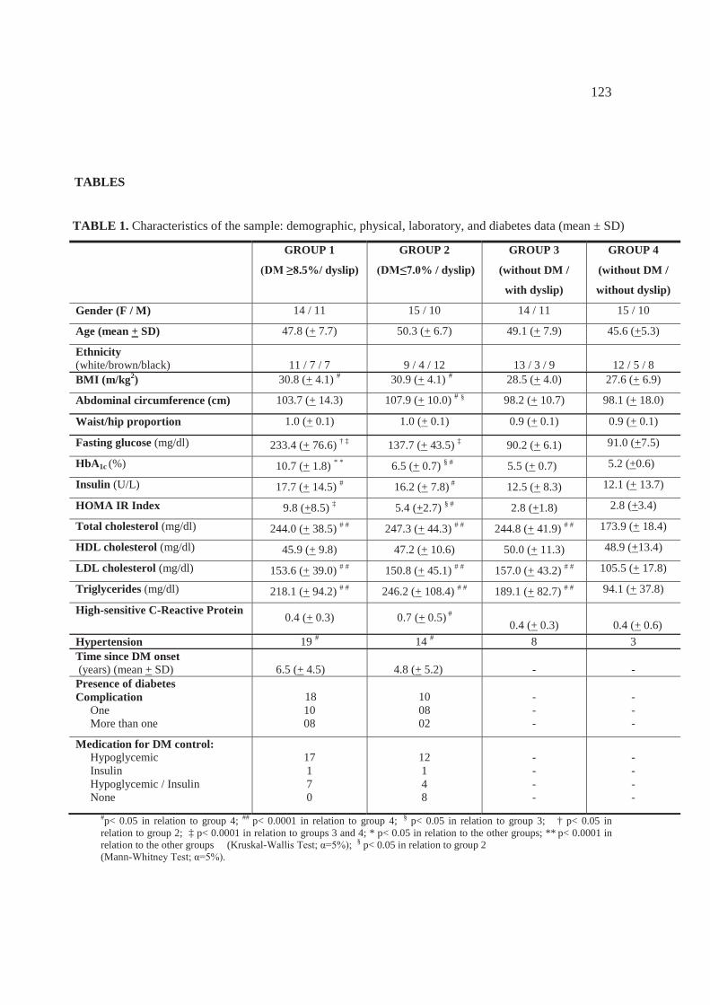
123
TABLES
TABLE 1. Characteristics of the sample: demographic, physical, laboratory, and diabetes data (mean ± SD)
GROUP 1
.5%/ dyslip)
GROUP 2
% / dyslip)
GROUP 3
(without DM /
with dyslip)
GROUP 4
(without DM /
without dyslip)
Gender (F / M) 14 / 11 15 / 10 14 / 11 15 / 10
Age (mean + SD) 47.8 (+ 7.7) 50.3 (+ 6.7) 49.1 (+ 7.9) 45.6 (+5.3)
Ethnicity(white/brown/black) 11 / 7 / 7 9 / 4 / 12 13 / 3 / 9 12 / 5 / 8BMI (m/kg2) 30.8 (+ 4.1) # 30.9 (+ 4.1) # 28.5 (+ 4.0) 27.6 (+ 6.9)
Abdominal circumference (cm) 103.7 (+ 14.3) 107.9 (+ 10.0) # § 98.2 (+ 10.7) 98.1 (+ 18.0)
Waist/hip proportion 1.0 (+ 0.1) 1.0 (+ 0.1) 0.9 (+ 0.1) 0.9 (+ 0.1)
Fasting glucose (mg/dl) 233.4 (+ 76.6) † ‡ 137.7 (+ 43.5) ‡ 90.2 (+ 6.1) 91.0 (+7.5)
HbA1c (%) 10.7 (+ 1.8) * * 6.5 (+ 0.7) § # 5.5 (+ 0.7) 5.2 (+0.6)
Insulin (U/L) 17.7 (+ 14.5) # 16.2 (+ 7.8) # 12.5 (+ 8.3) 12.1 (+ 13.7)
HOMA IR Index 9.8 (+8.5) ‡ 5.4 (+2.7) § # 2.8 (+1.8) 2.8 (+3.4)
Total cholesterol (mg/dl) 244.0 (+ 38.5) # # 247.3 (+ 44.3) # # 244.8 (+ 41.9) # # 173.9 (+ 18.4)
HDL cholesterol (mg/dl) 45.9 (+ 9.8) 47.2 (+ 10.6) 50.0 (+ 11.3) 48.9 (+13.4)
LDL cholesterol (mg/dl) 153.6 (+ 39.0) # # 150.8 (+ 45.1) # # 157.0 (+ 43.2) # # 105.5 (+ 17.8)
Triglycerides (mg/dl) 218.1 (+ 94.2) # # 246.2 (+ 108.4) # # 189.1 (+ 82.7) # # 94.1 (+ 37.8)
High-sensitive C-Reactive Protein 0.4 (+ 0.3) 0.7 (+ 0.5) #
0.4 (+ 0.3) 0.4 (+ 0.6)Hypertension 19 # 14 # 8 3Time since DM onset (years) (mean + SD) 6.5 (+ 4.5) 4.8 (+ 5.2) - -
Presence of diabetesComplication
OneMore than one
181008
100802
---
---
Medication for DM control:Hypoglycemic InsulinHypoglycemic / InsulinNone
17170
12148
----
----
#p< 0.05 in relation to group 4; ## p< 0.0001 in relation to group 4; § p< 0.05 in relation to group 3; † p< 0.05 in relation to group 2; ‡ p< 0.0001 in relation to groups 3 and 4; * p< 0.05 in relation to the other groups; ** p< 0.0001 in relation to the other groups (Kruskal- § p< 0.05 in relation to group 2(Mann-
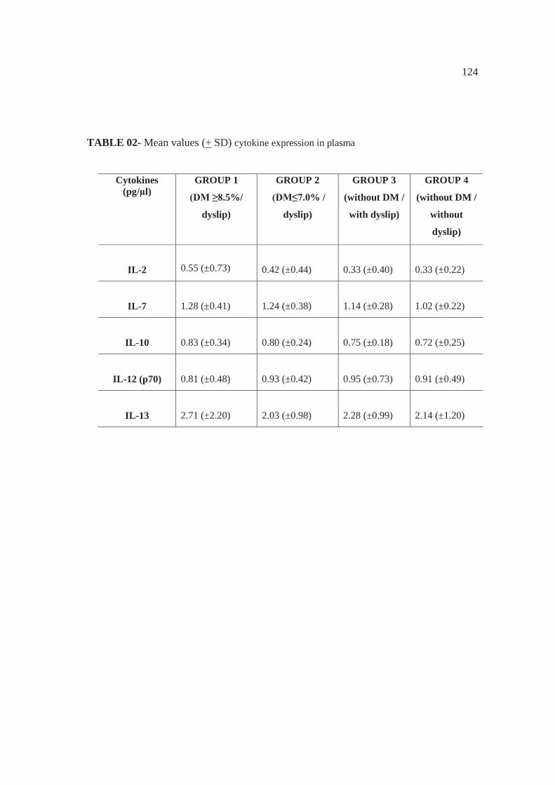
124
TABLE 02- Mean values (+ SD) cytokine expression in plasma
Cytokines (pg/μl)
GROUP 1
.5%/
dyslip)
GROUP 2
% /
dyslip)
GROUP 3
(without DM /
with dyslip)
GROUP 4
(without DM /
without
dyslip)
IL-2 0.55 (±0.73) 0.42 (±0.44) 0.33 (±0.40) 0.33 (±0.22)
IL-7 1.28 (±0.41) 1.24 (±0.38) 1.14 (±0.28) 1.02 (±0.22)
IL-10 0.83 (±0.34) 0.80 (±0.24) 0.75 (±0.18) 0.72 (±0.25)
IL-12 (p70) 0.81 (±0.48) 0.93 (±0.42) 0.95 (±0.73) 0.91 (±0.49)
IL-13 2.71 (±2.20) 2.03 (±0.98) 2.28 (±0.99) 2.14 (±1.20)
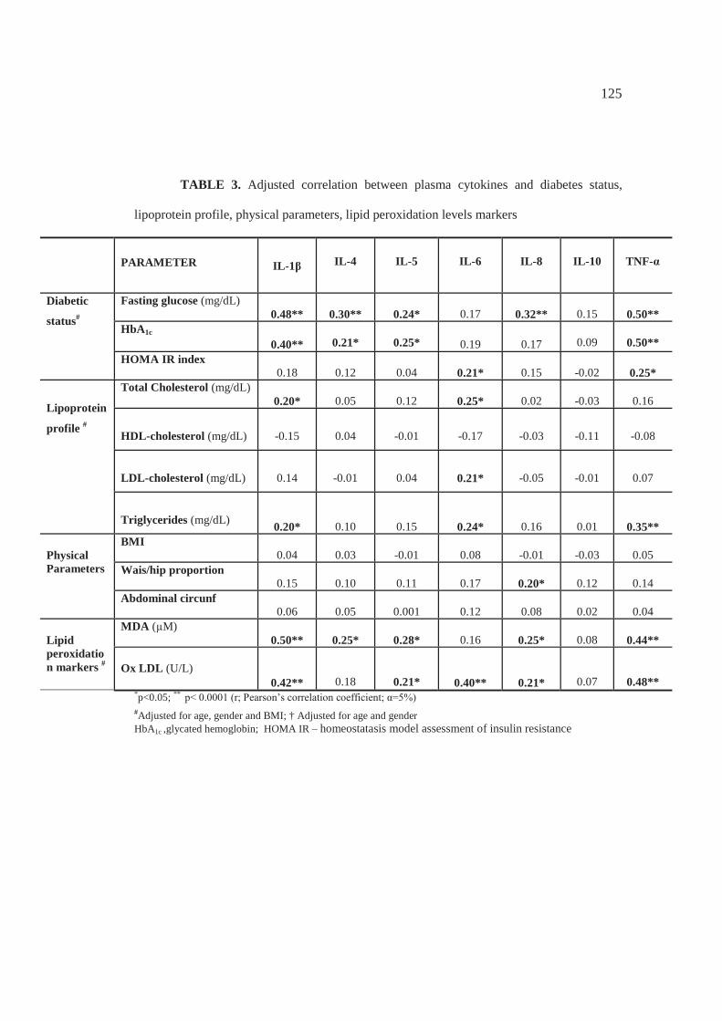
125
TABLE 3. Adjusted correlation between plasma cytokines and diabetes status,
lipoprotein profile, physical parameters, lipid peroxidation levels markers
PARAMETER IL-1 IL-4 IL-5 IL-6 IL-8 IL-10 TNF-
Diabetic
status#
Fasting glucose (mg/dL)0.48** 0.30** 0.24* 0.17 0.32** 0.15 0.50**
HbA1c
0.40** 0.21* 0.25* 0.19 0.17 0.09 0.50**HOMA IR index
0.18 0.12 0.04 0.21* 0.15 -0.02 0.25*
Lipoprotein
profile #
Total Cholesterol (mg/dL)0.20* 0.05 0.12 0.25* 0.02 -0.03 0.16
HDL-cholesterol (mg/dL) -0.15 0.04 -0.01 -0.17 -0.03 -0.11 -0.08
LDL-cholesterol (mg/dL) 0.14 -0.01 0.04 0.21* -0.05 -0.01 0.07
Triglycerides (mg/dL) 0.20* 0.10 0.15 0.24* 0.16 0.01 0.35**
PhysicalParameters
BMI0.04 0.03 -0.01 0.08 -0.01 -0.03 0.05
Wais/hip proportion0.15 0.10 0.11 0.17 0.20* 0.12 0.14
Abdominal circunf0.06 0.05 0.001 0.12 0.08 0.02 0.04
Lipid peroxidation markers #
MDA (μM)0.50** 0.25* 0.28* 0.16 0.25* 0.08 0.44**
Ox LDL (U/L)0.42** 0.18 0.21* 0.40** 0.21* 0.07 0.48**
*p<0.05; **
#Adjusted for age, gender and BMI; † Adjusted for age and gender HbA1c ,glycated hemoglobin; HOMA IR – homeostatasis model assessment of insulin resistance
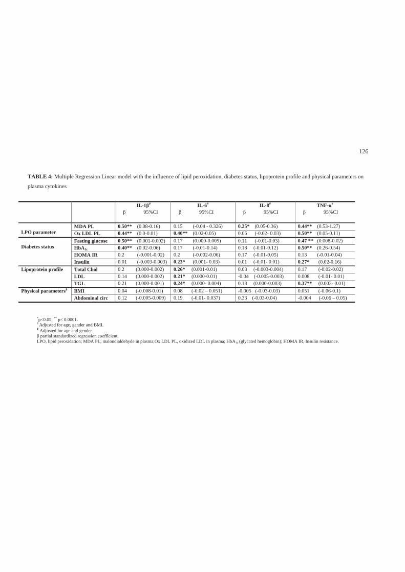
126
TABLE 4: Multiple Regression Linear model with the influence of lipid peroxidation, diabetes status, lipoprotein profile and physical parameters on
plasma cytokines
*p<0.05; ** p< 0.0001.# Adjusted for age, gender and BMI.§ Adjusted for age and gender
.LPO, lipid peroxidation; MDA PL, malondialdehyde in plasma;Ox LDL PL, oxidized LDL in plasma; HbA1c (glycated hemoglobin); HOMA IR, Insulin resistance.
IL- #
95%CIIL-6#
95%CIIL-8#
95%CITNF- #
95%CI
LPO parameterMDA PL 0.50** (0.08-0.16) 0.15 (-0.04 - 0.326) 0.25* (0.05-0.36) 0.44** (0.53-1.27)Ox LDL PL 0.44** (0.0-0.01) 0.40** (0.02-0.05) 0.06 (-0.02- 0.03) 0.50** (0.05-0.11)
Diabetes statusFasting glucose 0.50** (0.001-0.002) 0.17 (0.000-0.005) 0.11 (-0.01-0.03) 0.47 ** (0.008-0.02)HbA1c 0.40** (0.02-0.06) 0.17 (-0.01-0.14) 0.18 (-0.01-0.12) 0.50** (0.26-0.54)HOMA IR 0.2 (-0.001-0.02) 0.2 (-0.002-0.06) 0.17 (-0.01-0.05) 0.13 (-0.01-0.04)Insulin 0.01 (-0.003-0.003) 0.23* (0.001- 0.03) 0.01 (-0.01- 0.01) 0.27* (0.02-0.16)
Lipoprotein profile Total Chol 0.2 (0.000-0.002) 0.26* (0.001-0.01) 0.03 (-0.003-0.004) 0.17 (-0.02-0.02)LDL 0.14 (0.000-0.002) 0.21* (0.000-0.01) -0.04 (-0.005-0.003) 0.008 (-0.01- 0.01)TGL 0.21 (0.000-0.001) 0.24* (0.000- 0.004) 0.18 (0.000-0.003) 0.37** (0.003- 0.01)
Physical parameters§ BMI 0.04 (-0.008-0.01) 0.08 (-0.02 – 0.051) -0.005 (-0.03-0.03) 0.051 (-0.06-0.1)Abdominal circ 0.12 (-0.005-0.009) 0.19 (-0.01- 0.037) 0.33 (-0.03-0.04) -0.004 (-0.06 – 0.05)
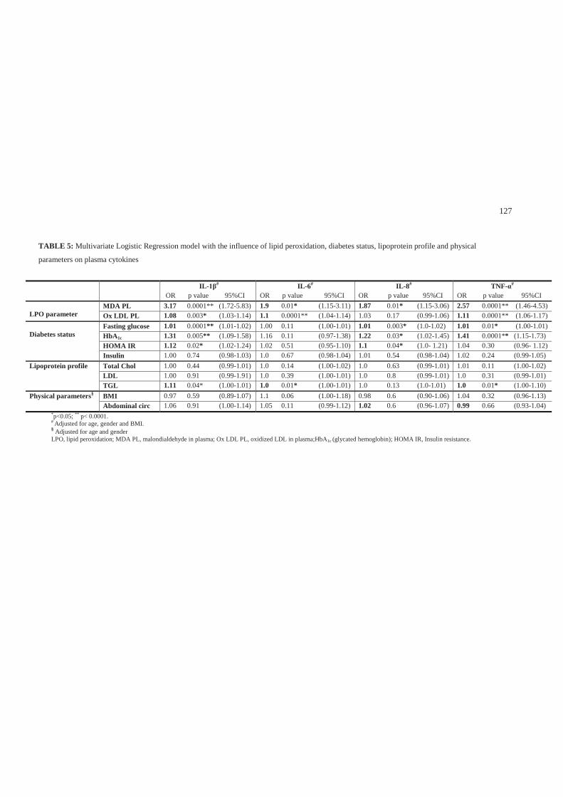
127
TABLE 5: Multivariate Logistic Regression model with the influence of lipid peroxidation, diabetes status, lipoprotein profile and physical
parameters on plasma cytokines
*p<0.05; ** p< 0.0001.# Adjusted for age, gender and BMI.§ Adjusted for age and genderLPO, lipid peroxidation; MDA PL, malondialdehyde in plasma; Ox LDL PL, oxidized LDL in plasma;HbA1c (glycated hemoglobin); HOMA IR, Insulin resistance.
IL- #
OR p value 95%CIIL-6#
OR p value 95%CIIL-8#
OR p value 95%CITNF- #
OR p value 95%CI
LPO parameterMDA PL 3.17 0.0001** (1.72-5.83) 1.9 0.01* (1.15-3.11) 1.87 0.01* (1.15-3.06) 2.57 0.0001** (1.46-4.53)Ox LDL PL 1.08 0.003* (1.03-1.14) 1.1 0.0001** (1.04-1.14) 1.03 0.17 (0.99-1.06) 1.11 0.0001** (1.06-1.17)
Diabetes statusFasting glucose 1.01 0.0001** (1.01-1.02) 1.00 0.11 (1.00-1.01) 1.01 0.003* (1.0-1.02) 1.01 0.01* (1.00-1.01)HbA1c 1.31 0.005** (1.09-1.58) 1.16 0.11 (0.97-1.38) 1.22 0.03* (1.02-1.45) 1.41 0.0001** (1.15-1.73)HOMA IR 1.12 0.02* (1.02-1.24) 1.02 0.51 (0.95-1.10) 1.1 0.04* (1.0- 1.21) 1.04 0.30 (0.96- 1.12)Insulin 1.00 0.74 (0.98-1.03) 1.0 0.67 (0.98-1.04) 1.01 0.54 (0.98-1.04) 1.02 0.24 (0.99-1.05)
Lipoprotein profile Total Chol 1.00 0.44 (0.99-1.01) 1.0 0.14 (1.00-1.02) 1.0 0.63 (0.99-1.01) 1.01 0.11 (1.00-1.02)LDL 1.00 0.91 (0.99-1.91) 1.0 0.39 (1.00-1.01) 1.0 0.8 (0.99-1.01) 1.0 0.31 (0.99-1.01)TGL 1.11 0.04* (1.00-1.01) 1.0 0.01* (1.00-1.01) 1.0 0.13 (1.0-1.01) 1.0 0.01* (1.00-1.10)
Physical parameters§ BMI 0.97 0.59 (0.89-1.07) 1.1 0.06 (1.00-1.18) 0.98 0.6 (0.90-1.06) 1.04 0.32 (0.96-1.13)Abdominal circ 1.06 0.91 (1.00-1.14) 1.05 0.11 (0.99-1.12) 1.02 0.6 (0.96-1.07) 0.99 0.66 (0.93-1.04)
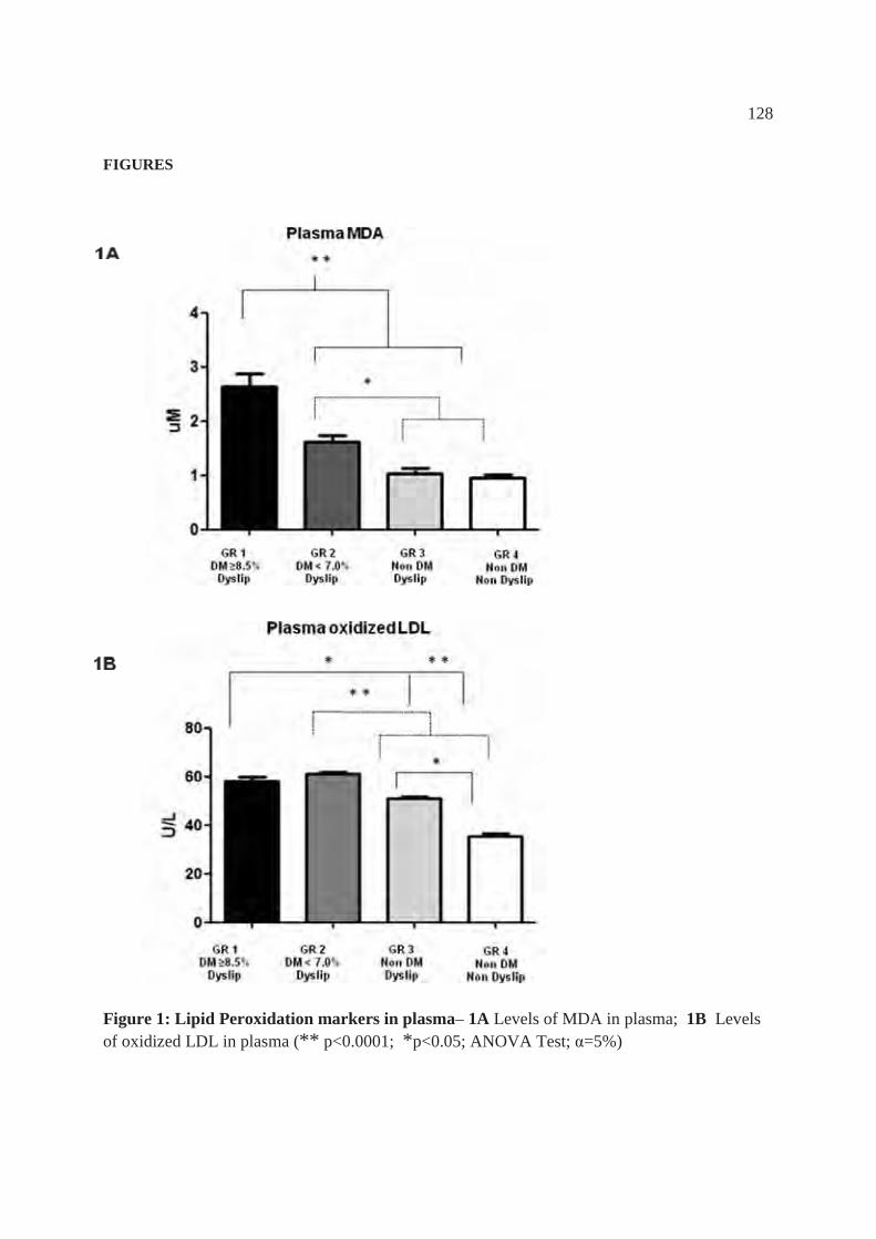
128
FIGURES
Figure 1: Lipid Peroxidation markers in plasma– 1A Levels of MDA in plasma; 1B Levels of oxidized LDL in plasma (** p<0.0001; *p<0.05; ANOVA Test; =5%)
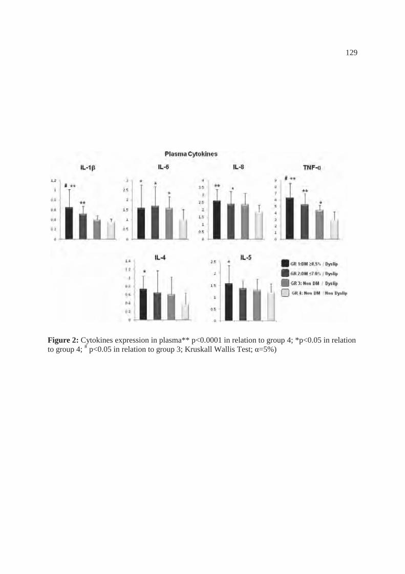
129
Figure 2: Cytokines expression in plasma** p<0.0001 in relation to group 4; *p<0.05 in relation to group 4; # p<0.05 in relation to group 3; Kruskall Wallis Test; =5%)
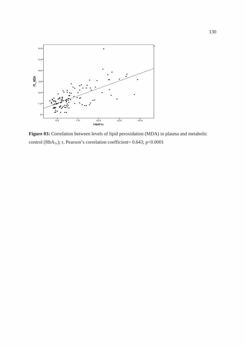
130
Figure 03: Correlation between levels of lipid peroxidation (MDA) in plasma and metabolic
control (HbA1c); r, Pearson’s correlation coefficient= 0.643; p<0.0001

131
DISCUSSÃO
U
132
4 DISCUSSÃO
Estudos prévios têm avaliado o impacto da peroxidação lipídica no
diabetes e na doença periodontal como condições independentes. Neste estudo
foi possível avaliar a peroxidação lipídica em pacientes com diabetes tipo 2,
com adequado e inadequado controle metabólico, e dislipidemia comparando-
os a pacientes sistemicamente saudáveis, bem como avaliar seu impacto sobre a
inflamação crônica no plasma e no fluido gengival. Além disso, foi possível
padronizar e validar a mensuração da peroxidação lipídica (PL) no fluido
sulcular gengival (FSG) de sítios saudáveis e com periodontite. Os resultados
demonstraram a presença de maiores níveis de peroxidação lipídica em
pacientes com diabetes e sugerem que a associação diabetes/dislipidemia pode
afetar a magnitude da inflamação local e sistêmica, aumentando a expressão de
citocinas inflamatórias plasmáticas e no periodonto, levando também à
destruição tecidual, representada neste estudo por maior severidade da
periodontite crônica.
A dislipidemia no diabetes pode estar presente antes mesmo do
estabelecimento da hiperglicemia75, 90, 122. Desempenha um importante papel na
resposta imune, predispondo o paciente a um estado hiperinflamatório por
modular a função e atividade dos macrófagos106, 230, 238, desencadeando a
ativação de diversas vias relacionadas à liberação de produtos oxidativos, de
peroxidação lipídica e de mediadores inflamatórios que levam a danos em
diversos órgãos e tecidos47, 101. O aumento do metabolismo oxidativo e o
acúmulo de AGEs estão envolvidos na patogênese de condições inflamatórias
133
crônicas observadas em pacientes com diabetes como a aterosclerose92, as
alterações micro e macrovasculares110, 241 e a doença periodontal4, 37, 233, 246.
Diversos autores observaram que pacientes com diabetes apresentam
aumento de marcadores de peroxidação lipídica como LDL-oxidada (LDL-
ox)42, 100, 104, 166, 193 e o malondialdeído (MDA)4, 9, 84, 107, 160. Em concordância
com tais autores, o presente resultado demonstrou que os níveis plasmáticos de
peroxidação lipídica (LDL-ox e MDA) estão aumentados em pacientes com
diabetes, sendo ainda mais elevados na presença de inadequado controle
metabólico e estão correlacionados com parâmetros de dislipidemia como
níveis elevados de colesterol total, LDL-colesterol e triglicérides. Em
concordância com os estudos de Kesavulu et al.111 e Nacitarhan et al.165, foi
observado que os níveis de peroxidação lipídica também apresentaram
correlação forte e significativa com elevados níveis de glicemia de jejum e
hemoglobina glicada (HbA1c). No entanto, alguns autores241 não encontraram
relação significativa entre níveis elevados de peroxidação lipídica e diabetes,
isso provavelmente esteve relacionado a diferenças nas populações de estudo.
Por muitos anos, a análise do MDA tem sido amplamente empregada
para verificar a peroxidação lipídica em amostras biológicas 50, 52. A reação
chamada de “teste das substâncias reativas com ácido tiobarbitúrico” (TBARS)
tem sido comumente utilizada como um índice de produção de MDA e consiste
de um ensaio espectrofotométrico ou espectrofluorimétrico de produtos gerados
quando a amostra é aquecida sob condições ácidas para formar o aduto MDA-
(TBA)2. No entanto, outros aldeídos além do MDA podem reagir com o TBA e
134
diversos outros cromógenos podem interferir, resultando dessa forma em uma
superestimação das concentrações de MDA. Por estas razões, a especificidade
do TBARs tem sido alvo de questionamentos 98, 118, 228.
Desta forma, a cromatografia líquida de alta performance (HPLC) tem
sido introduzida como uma maneira de melhorar a especificidade e
confiabilidade da avaliação23, uma vez que o autêntico cromógeno MDA-TBA2
é eficientemente separado de outros cromógenos que poderiam via a interferir
com a reação37, 40, 68, 89, 98, 256. Diversos autores demonstraram a validação de
métodos para avaliação de MDA em plasma89, 98, 228, mostrando que condições
sistêmicas como doença glomerular 228, infertilidade masculina 134,
hiperlipidemia 40 e Diabetes mellitus 165, 166, 239 apresentam níveis aumentados
de MDA plasmáticos.
Em se tratando de doença periodontal, estudos recentes têm investigado
o papel dos produtos do metabolismo oxidativo sobre a inflamação e dano ao
periodonto. Níveis aumentados de marcadores de estresse oxidativo foram
verificados em sítios com periodontite 176, 210, 218, 233 e produtos de peroxidação
lipídica no fluido sulcular gengival, como o malondialdeído (MDA), foram
significativamente correlacionados com parâmetros clínicos da doença4, 233, 246.
Alguns estudos sugerem que o início ou progressão da destruição periodontal
pode ser devido ao aumento de espécies reativas de oxigênio e proteases ativas,
liberadas durante o processo de resposta do hospedeiro frente a um desafio
bacteriano 18, 37, 190, 246.
135
Dessa forma, qualquer que seja o mecanismo, uma medida eficaz para
detectar os marcadores de peroxidação lipídica se faz necessária para fornecer
resultados precisos e reprodutíveis. Na avaliação da peroxidação lipídica em
pacientes com periodontite, os estudos têm realizado tal análise na saliva 32, 37,
113, no plasma 4, 246, no tecido gengival 25 e no fluido sulcular gengival 4, 233, 246.
No entanto, os métodos descritos para avaliar MDA no fluido gengival de sítios
com doença periodontal foram ensaios colorimétricos 233 e apenas dois autores
recentemente utilizaram cromatografia líquida de alta performance (HPLC) 4,
246. Deve-se ressaltar, entretanto, que a metodologia empregada para
mensuração no fluido sulcular gengival nestes estudos foi a mesma utilizada
para amostras de plasma e saliva e que não existe validação do método na
literatura. Considerando-se que o fluido sulcular gengival é uma amostra
biológica menos complexa em relação ao plasma e diferente da saliva quanto à
composição, o estabelecimento de uma metodologia específica para avaliação
do MDA no fluido gengival se faz necessária assim como a validação do
método para que esta análise seja precisa e reprodutível.
No presente estudo, foi descrito um método confiável o qual foi
validado para identificar e quantificar um produto específico da peroxidação
lipídica no fluido sulcular gengival, de sítios saudáveis ou com periodontite
crônica, utilizando HPLC. O método foi padronizado e direcionado
especificamente para o FSG, diferenciando-o da mensuração em outras
amostras biológicas. Além disso, este é o primeiro estudo a descrever a
quantificação do MDA no fluido sulcular gengival de sítios saudáveis, uma vez
136
que outros estudos apenas avaliaram MDA, por HPLC, em fluido de sítios com
doença periodontal 4, 246. Isto implica não somente na mensuração do MDA em
concentrações reduzidas, mas também na possibilidade de usar esta mensuração
como um marcador de risco para doença periodontal (Apêndice 1).
A sensibilidade do método empregado permitiu a quantificação do
MDA com excelente exatidão e precisão até mesmo em concentrações baixas
como 0.04 - 0.05 μM. Tal característica foi de fundamental importância na
quantificação do MDA no fluido de sítios saudáveis. Adicionalmente, as
diferentes amostras coletadas tanto de sítios saudáveis quanto com periodontite
crônica apresentaram concentrações semelhantes, a depender da característica
do sítio, demonstrando a precisão do método quando aplicado para amostras de
fluido sulcular gengival. Além disso, um significativo aumento nas
concentrações de MDA foi observado no fluido de sítios com periodontite
crônica quando comparados a sítios saudáveis, em concordância com o estudo
de Tsai et al. 233.
Além da possibilidade de detectar MDA no fluido de sítios saudáveis e
de se obter um método validado para mensurar a peroxidação lipídica no fluido
sulcular gengival utilizando a cromatografia líquida de alta performance
(Capítulo 1- Anexo C), o presente estudo avaliou o impacto da peroxidação
lipídica no plasma e fluido gengival sobre a inflamação crônica em pacientes
portadores ou não de diabetes e dislipidemia (Capítulo 2).
É reconhecido que o diabetes está associado a uma maior magnitude da
resposta inflamatória a patógenos e seus produtos43, 171. A periodontite crônica é
137
reconhecida como a sexta complicação do diabetes141 e é bem estabelecido que
há maior prevalência, severidade e extensão da doença periodontal nestes
pacientes41, 60, 115, 158, 187, 226, 227. Esta informação foi confirmada no presente
estudo mostrando que os grupos com diabetes (grupos 1 e 2) foi relacionado a
piores valores para todos os parâmetros periodontais quando comparados aos
grupos normoglicêmicos (grupos 3 e 4) (Apêndice 2).
O nível de controle metabólico tem sido extensivamente apontado como
um importante fator que leva à doença periodontal mais severa, tendo mais
impacto em pacientes com inadequado controle metabólico16, 136, 187, 227 apesar
de alguns estudos não observarem tal associação26, 199. No presente estudo foi
verificado que pacientes com inadequado controle metabólico e com
dislipidemia (grupo 1) apresentaram mais sítios com sangramento à sondagem,
supuração e perda de inserção quando comparados a pacientes sistemicamente
saudáveis (grupo 4). Além disso, uma correlação forte e positiva foi observada
entre glicemia de jejum e hemoglobina glicada e parâmetros clínicos de
periodontite crônica. Estes achados mostraram que, de fato, o controle
metabólico exerceu um impacto importante sobre a severidade da inflamação e
destruição periodontal.
No entanto, além do controle metabólico, fatores como alterações na
resposta imune, anormalidades no metabolismo de lipídios86, 218 e aumento do
metabolismo oxidativo6, 175, 202 podem caracterizar-se como um importante
mecanismo relacionado a maior destruição periodontal. Sabe-se que a
dislipidemia característica do diabetes36, 121 predispõe o paciente a um estado
138
hiperinflamatório pela modulação da função e atividade dos macrófagos106, 230,
238 desencadeando a ativação de vias associadas com a liberação de fatores de
crescimento e citocinas inflamatórias47, 101.
Estudos têm demonstrado a presença de produtos do metabolismo
oxidativo e de peroxidação lipídica no plasma4, 220, 246, saliva32, 37, 113 e fluido
sulcular gengival4, 233, 246. Ainda, alguns autores demonstraram a relação desses
produtos com a patogênese da doença periodontal18, 176, 206, 244. Neste aspecto, o
presente estudo verificou que independente da condição sistêmica, os sítios
com periodontite crônica apresentaram mais MDA do que os sítios saudáveis.
Nossos achados demonstram uma correlação positiva entre marcadores de
peroxidação lipídica local e sistêmica e parâmetros clínicos periodontais, como
sítios profundos (PD ≥ 6mm), sangramento à sondagem, nível de inserção
clínica (NI ≥5mm) e presença de supuração, estando em concordância com
outros estudos4, 233, o que indica que níveis aumentados de peroxidação lipídica
podem ter um papel importante na destruição periodontal. Além disso, também
foi verificada correlação positiva entre marcadores de peroxidação lipídica
local e sistêmica. Neste contexto, pode-se sugerir que a peroxidação lipídica
tem um importante papel na severidade do processo inflamatório periodontal e
sistêmico, frequentemente observado nestes pacientes.
Não foi encontrada correlação significativa entre dislipidemia e
parâmetros inflamatórios da periodontite crônica, em contraste com estudos
anteriores que sugerem que a dislipidemia por si só pode estar associada a um
pior status periodontal65, 102. O aumento da severidade da inflamação,
139
representada por maior destruição periodontal e produção aumentada de
citocinas inflamatórias locais ocorreu quando a dislipidemia esteve associada
ao diabetes (grupos 1 e 2), não havendo diferença na comparação entre
indivíduos com e sem dislipidemia não portadores de diabetes (grupos 3 e 4).
Nossos achados sugerem que o diabetes, na presença de um ambiente
dislipidêmico, pode alterar o metabolismo oxidativo, aumentando a
peroxidação lipídica. Tal aumento na peroxidação lipídica pode representar um
fator adicional no desequilíbrio metabólico associado ao diabetes com efeito
sobre a modulação da resposta inflamatória. Neste sentido, sugere-se que a
dislipidemia tenha impacto relevante sobre a resposta inflamatória na presença
de estresse oxidativo aumentado.
Até o momento, o efeito da peroxidação lipídica local e sistêmica em
pacientes com diabetes e periodontite crônica não havia sido estudado. No
presente estudo tais aspectos foram avaliados de maneira conjunta e foi
observado que apenas o fato de ter diabetes, independente do controle
metabólico, levou a um aumento dos níveis de peroxidação lipídica, avaliados
pela LDL-oxidada no plasma e pelo MDA no plasma e no fluido. Nesse
aspecto, um importante resultado que deve ser salientado foi a obtenção de
maiores valores de MDA no fluido sulcular gengival de sítios saudáveis para os
grupos com diabetes (grupo 1 e 2) quando comparado ao fluido de pacientes
normoglicêmicos (grupos 3 e 4). Além disso, os resultados de regressões,
logística e múltipla, sugerem que níveis aumentados de peroxidação lipídica
140
local e sistêmica podem representar um maior risco para a presença de
periodontite crônica considerada severa.
Alguns mecanismos podem ser sugeridos para elucidar o papel da
peroxidação lipídica sobre a severidade da condição inflamatória em pacientes
com diabetes e dislipidemia. Durante o curso de uma condição
inflamatória/infecciosa, após a ativação de neutrófilos e macrófagos por
patógenos209, 210, 233, ocorre a produção exagerada de produtos da peroxidação
lipídica no tecido como conseqüência do processo inflamatório200, 210. Por sua
vez, a presença de peroxidação lipídica nos locais inflamados pode, de maneira
oposta, aumentar a magnitude da reação inflamatória por meio da liberação de
citocinas pró-inflamatórias182.
Neste contexto, os peróxidos de lipídios podem ativar fatores de
transcrição como o fator nuclear-kB7, 178, 205, 253, o qual é sensível a alterações
no metabolismo oxidativo253. Tal ativação estimula a liberação de mediadores
apoptóticos e citocinas pró-inflamatórias e aumenta a expressão de moléculas
de adesão endotelial, como a VCAM-115, 93, 168, resultando numa cascata de
ativação celular e inflamação difusa44, 95.
A peroxidação lipídica pode ainda contribuir para a injúria tecidual por
outros mecanismos, incluindo dano ao DNA, oxidação de enzimas e alterações
degenerativas, como necrose e destruição do osso alveolar na região do
infiltrado inflamatório218. Tal condição de dano tecidual pode ser agravada em
pacientes com diabetes pelo fato de que, além dos lipídios no plasma e das
membranas celulares serem fontes de peróxido de lipídios, o acúmulo de AGEs
141
(produtos finais da glicação avançada) também pode gerar produtos de
peroxidação lipídica74. Sabe-se que os AGEs estão diretamente associados ao
nível de controle metabólico e à duração da hiperglicemia e têm a capacidade
de alterar a estrutura e função das membranas celulares, inclusive levando a um
aumento do metabolismo oxidativo (ROS) acima do fisiológico202. Tal
mecanismo de estresse oxidativo e produção de peróxido de lipídios induzido
por AGEs contribuem para a diminuição da capacidade antioxidante 156,
disfunção vascular difusa e ativação de células mononucleares44, 203. Sendo a
doença periodontal uma condição que se desenvolve frente a um desafio
bacteriano, o acúmulo periférico ou local de AGEs pode contribuir para dano
aos tecidos periodontais6 e por este motivo, tal acúmulo tem sido investigado na
doença periodontal202. Estudos demonstram que existem receptores RAGEs no
tecido gengival 109, 202 de pacientes com diabetes e a interação de AGEs com
seus receptores RAGEs, que ocorre em vários órgãos253, também pode ocorrer
no tecido gengival109, 202. Tal interação pode levar à supressão da produção de
colágeno por fibroblastos da gengiva e do ligamento periodontal164, 183,
liberação de mediadores inflamatórios como interleucina 1 (IL-1 ) e fator de
necrose tumoral (TNF- )127, 253, estímulo à osteoclastogênese55, 255 e atraso da
reparação tecidual80.
É bem estabelecido que pacientes com diabetes, especialmente com
inadequado controle metabólico, apresentam níveis aumentados de citocinas no
plasma e no fluido sulcular gengival61, 171, 188, 191 levando a dano tecidual38, 210 e
142
influenciando nas complicações relacionadas ao diabetes138, 184. No entanto, os
poucos estudos que investigaram a peroxidação lipídica como um possível
mecanismo adicional para a severidade da resposta inflamatória e da doença
periodontal em pacientes com diabetes 43, 202, 220, não consideraram o perfil
imunoinflamatório local e/ou sistêmico do indivíduo, tornando difícil,
estabelecer uma correlação entre achados clínicos periodontais e de
peroxidação lipídica com a magnitude da resposta inflamatória gerada.
No presente estudo, em relação aos marcadores inflamatórios (TNF-α,
IL-6 e IL-10) no fluido sulcular gengival, além de confirmar que pacientes com
DM e pobre controle metabólico apresentam maior expressão de tais
marcadores em sítios com periodontite crônica, também foi verificada uma
significativa correlação entre níveis plasmáticos de MDA e a expressão local de
citocinas, especialmente em relação ao TNF-α, dado este ainda não relatado na
literatura. Além disso, os resultados da regressão logística sugerem que o
aumento da peroxidação lipídica, local e sistêmica, pode aumentar o risco de
expressão de marcadores inflamatórios locais, particularmente TNF-α. Estes
achados demonstram um papel potencial da peroxidação lipídica quanto ao
aumento na expressão de citocinas no tecido inflamado e, conseqüente,
destruição periodontal.
Em se tratando de marcadores inflamatórios plasmáticos, no presente
estudo foram avaliadas 12 citocinas e estas foram correlacionadas com
condições sistêmicas. Poucos estudos que avaliaram marcadores inflamatórios
categorizaram os pacientes com DM de acordo com o controle metabólico,
143
baseado nos níveis de HbA1c30, 70, 100, 120, 186. No presente estudo, foi observado
que a expressão das citocinas seguiu um padrão decrescente do grupo 1 ao
grupo 4 tanto para citocinas pró- quanto anti-inflamatórias, sendo esta diferença
significante para IL-1 , IL-4, IL-5, IL-6, IL-8 e TNF-α. Esta diferença ocorreu
em maior magnitude na comparação do grupo 1 em relação aos demais grupos,
embora nos pacientes portadores de diabetes com adequado controle
metabólico os níveis destas citocinas também estivessem mais elevados em
relação aos pacientes saudáveis. Tais resultados estão em concordância com
outros estudos que mostraram um elevado padrão de citocinas inflamatórias em
pacientes com inadequado controle metabólico6, 39, 40. Além disso, conforme
demonstrado por outros autores70, 251, no presente estudo a expressão de
adipocitocinas, como IL-1 , IL-6 e TNF-α, foi maior em pacientes com
dislipidemia (grupos 1, 2 e 3).
Dados relevantes foram encontrados nos pacientes com adequado
controle metabólico. Este grupo de pacientes geralmente é considerado de
menor risco ao desenvolvimento de doenças cardiovasculares e complicações
relacionadas ao diabetes, quando comparados aos pacientes com inadequado
controle metabólico3, 167 e, por essas razões, não é avaliado freqüentemente em
relação ao perfil lipídico e às proteínas de fase aguda. Entretanto, os resultados
verificados neste estudo em relação ao grupo 2, como maiores níveis de
proteína C-reativa e de circunferência abdominal, altos níveis de peroxidação
lipídica e de citocinas inflamatórias plasmáticas (IL-1 , IL-6, IL-8, TNF-α),
144
demonstram que há um erro de premissa. Neste contexto, alguns autores
enfatizam que o aumento do metabolismo oxidativo pode levar ao início ou à
progressão do diabetes77, 237 e a que peroxidaçao lipídica pode ser um evento
precoce importante na patogênese do desenvolvimento das complicações
associadas ao DM, mesmo em pacientes com bom controle metabólico30. Desta
forma, os resultados do presente estudo apontam para a importância de que
sejam estabelecidas também para pacientes com adequado controle metabólico,
metas de prevenção devem ser tomadas buscando evitar um pobre controle
metabólico no futuro.
Apesar dos pacientes com DM serem considerados obesos (BMI>30) e
apresentarem altos valores de circunferência abdominal, de resistência
insulínica e de citocinas (IL-6 e TNF- α), não houve correlação significativa
entre citocinas plasmáticas e parâmetros físicos, diferente dos resultados de
alguns estudos33, 242. Além disso, foram observadas correlações significativas
fracas (0.2) citocinas (IL-6 e o TNF- α) com a resistência insulínica (HOMA
IR). Apesar de estudos prévios demonstrarem associação entre resistência
insulínica e a expressão de citocinas inflamatórias242, 250, 251, outros estudos não
encontraram tal relação e afirmam que o nível de citocinas circulantes está mais
relacionado à hiperglicemia63 ou à presença de tecido adiposo33, 243 do que à
resistência insulínica propriamente dita251.
No presente estudo foram verificadas correlações moderadas e
significantes entre citocinas, como IL1-β, TNF-α e IL-6, e marcadores de
145
peroxidação lipídica, como MDA e LDLox. As análises de regressão também
mostraram que a peroxidação lipídica possui um importante papel no aumento
do risco de expressão de citocinas plasmáticas, em especial IL1-β e TNF-α. No
entanto, foram observadas correlações significativas, porém fracas, entre a
expressão das citocinas plasmáticas e o perfil lipídico. Este achado sugere que,
apesar da dislipidemia contribuir efetivamente para a ocorrência de peroxidação
lipídica, seu papel no aumento da expressão de marcadores inflamatórios é
mais significativo quando esta é associada ao diabetes.
O mecanismo preciso pelo qual a peroxidação lipídica pode levar à
maior expressão de citocinas ainda não é bem elucidado em estudos clínicos.
No entanto, alguns estudos sugerem que o aumento do metabolismo oxidativo
ativa fatores de transcrição como o (NF-κB) 253 e aumenta a expressão de
citocinas inflamatórias (IL-6 e TNF- α) 63, sendo um mecanismo que pode levar
à hiperglicemia aguda e a complicações do diabetes77. Além disso, foi
demonstrado8 que a inibição da peroxidação lipídica é capaz de normalizar a
expressão de citocinas angiogênicas como o fator de crescimento vascular
endotelial (VEGF), melhorando a cicatrização de feridas em ratos diabéticos.
Desta forma, pode-se sugerir que a peroxidação lipídica pode ser um
mecanismo adicional importante na expressão aumentada de marcadores
inflamatórios em pacientes com diabetes e dislipidemia.
Apesar deste estudo apresentar limitações, inerentes a um estudo
transversal, que não permitem conclusões generalizadas ou de causalidade, foi
possível observar que marcadores de peroxidação lipídica no plasma e no

146
periodonto estão aumentados nos pacientes com diabetes que apresentam
dislipidemia, particularmente quando há pobre controle metabólico. Além
disso, níveis locais e sistêmicos de peroxidação lipídica aumentaram a
expressão de citocinas plasmáticas e locais, o que pode contribuir para a
ocorrência de inflamação difusa e dano a diversos tecidos, dentre esses o
periodonto.

147
CONCLUSÃO
U
148
5 CONCLUSÃO Os resultados obtidos permitem concluir que:
1- A peroxidação lipídica plasmática, mesmo em pacientes com
adequado controle metabólico, esteve associada a níveis elevados de
citocinas plasmáticas, sugerindo um possível mecanismo adicional
na modulação da resposta inflamatória em pacientes com diabetes e
dislipidemia.
2- Foi possível quantificar um importante marcador de peroxidação
lipídica no fluido sulcular gengival utilizando cromatografia líquida
de alta performance, com exatidão e precisão.
3- A avaliação do MDA no fluido sulcular gengival, inclusive de sítios
saudáveis, podendo ser avaliada como possível marcador de risco
para periodontite crônica.
4- A peroxidaçao lipídica pode ser um mecanismo importante para a
expressão aumentada de marcadores inflamatórios no fluido sulcular
gengival e maior severidade da periodontite crônica do paciente
portador de diabetes com dislipidemia.
5- A presença de maiores níveis de peroxidação lipídica, e não
meramente a dislipidemia, em pacientes com diabetes sugere que a
associação diabetes/dislipidemia possa afetar a magnitude da
inflamação local e sistêmica.
U

149
REFERÊNCIAS
150
6 REFERÊNCIAS**
1. AAP. International International Workshop for a Classification of Periodontal Diseases and Conditions. Papers. Ann Periodontol. 1999; 4: 1-112. 2. Abuja PM, Albertini R. Methods for monitoring oxidative stress, lipid peroxidation and oxidation resistance of lipoproteins. Clin Chim Acta. 2001; 306: 1-17. 3. ADA. Standards of medical care in diabetes--2011. Diabetes Care. 2011; 34 Suppl 1: S11-61. 4. Akalin FA, Baltacioglu E, Alver A, Karabulut E. Lipid peroxidation levels and total oxidant status in serum, saliva and gingival crevicular fluid in patients with chronic periodontitis. J Clin Periodontol. 2007; 34: 558-65. 5. Al Habashneh R, Khader Y, Hammad MM, Almuradi M. Knowledge and awareness about diabetes and periodontal health among Jordanians. J Diabetes Complications. 2009; 24: 409-14. 6. Allen EM, Matthews JB, DJ OH, Griffiths HR, Chapple IL. Oxidative and inflammatory status in Type 2 diabetes patients with periodontitis. J Clin Periodontol. 2011; 38: 894-901. 7. Allen EM, Matthews JB, O'Connor R, O'Halloran D, Chapple IL. Periodontitis and type 2 diabetes: is oxidative stress the mechanistic link? Scott Med J. 2009; 54: 41-7. 8. Altavilla D, Saitta A, Cucinotta D, Galeano M, Deodato B, Colonna M, et al. Inhibition of lipid peroxidation restores impaired vascular endothelial growth factor expression and stimulates wound healing and angiogenesis in the genetically diabetic mouse. Diabetes. 2001; 50: 667-74. 9. Altomare E, Vendemiale G, Chicco D, Procacci V, Cirelli F. Increased lipid peroxidation in type 2 poorly controlled diabetic patients. Diabete Metab. 1992; 18: 264-71. 10. Andriankaja OM, Barros SP, Moss K, Panagakos FS, DeVizio W, Beck J, et al. Levels of serum interleukin (IL)-6 and gingival crevicular fluid of IL-1beta and prostaglandin E(2) among non-smoking subjects with gingivitis and type 2 diabetes. J Periodontol. 2009; 80: 307-16. 11. Arai K, Maguchi S, Fujii S, Ishibashi H, Oikawa K, Taniguchi N. Glycation and inactivation of human Cu-Zn-superoxide dismutase. Identification of the in vitro glycated sites. J Biol Chem. 1987; 262: 16969-72. 12. Assuma R, Oates T, Cochran D, Amar S, Graves DT. IL-1 and TNF antagonists inhibit the inflammatory response and bone loss in experimental periodontitis. J Immunol. 1998; 160: 403-9. 13. Babior BM. Oxygen-dependent microbial killing by phagocytes (first of two parts). N Engl J Med. 1978; 298: 659-68. 14. Baelum V, Lopez R. Periodontal epidemiology: towards social science or molecular biology? Community Dent Oral Epidemiol. 2004; 32: 239-49.
* De acordo com o estilo Vancouver. Disponível em: http://www.nlm.nih.gov/bsd/uniform_requirements.html
151
15. Baeuerle PA. The inducible transcription activator NF-kappa B: regulation by distinct protein subunits. Biochimica et biophysica acta. 1991; 1072: 63-80. 16. Bandyopadhyay D, Marlow NM, Fernandes JK, Leite RS. Periodontal disease progression and glycaemic control among Gullah African Americans with type-2 diabetes. J Clin Periodontol. 2010; 37: 501-9. 17. Bartold PM, Wiebkin OW, Thonard JC. The effect of oxygen-derived free radicals on gingival proteoglycans and hyaluronic acid. J Periodontal Res. 1984; 19: 390-400. 18. Battino M, Ferreiro MS, Gallardo I, Newman HN, Bullon P. The antioxidant capacity of saliva. J Clin Periodontol. 2002; 29: 189-94. 19. Bax BE, Alam AS, Banerji B, Bax CM, Bevis PJ, Stevens CR, et al. Stimulation of osteoclastic bone resorption by hydrogen peroxide. Biochem Biophys Res Commun. 1992; 183: 1153-8. 20. Baynes JW. Role of oxidative stress in development of complications in diabetes. Diabetes. 1991; 40: 405-12. 21. Beam HA, Parsons JR, Lin SS. The effects of blood glucose control upon fracture healing in the BB Wistar rat with diabetes mellitus. J Orthop Res. 2002; 20: 1210-6. 22. Bierman EL. George Lyman Duff Memorial Lecture. Atherogenesis in diabetes. Arterioscler Thromb. 1992; 12: 647-56. 23. Bird RP, Hung SS, Hadley M, Draper HH. Determination of malonaldehyde in biological materials by high-pressure liquid chromatography. Anal Biochem. 1983; 128: 240-4. 24. Bonnefont-Rousselot D, Bastard JP, Jaudon MC, Delattre J. Consequences of the diabetic status on the oxidant/antioxidant balance. Diabetes Metab. 2000; 26: 163-76. 25. Borges I, Jr., Moreira EA, Filho DW, de Oliveira TB, da Silva MB, Frode TS. Proinflammatory and oxidative stress markers in patients with periodontal disease. Mediators Inflamm. 2007; 2007: 45794. 26. Bridges RB, Anderson JW, Saxe SR, Gregory K, Bridges SR. Periodontal status of diabetic and non-diabetic men: effects of smoking, glycemic control, and socioeconomic factors. J Periodontol. 1996; 67: 1185-92. 27. Brock GR, Butterworth CJ, Matthews JB, Chapple IL. Local and systemic total antioxidant capacity in periodontitis and health. J Clin Periodontol. 2004; 31: 515-21. 28. Brownlee M. Lilly Lecture 1993. Glycation and diabetic complications. Diabetes. 1994; 43: 836-41. 29. Brownlee M. Biochemistry and molecular cell biology of diabetic complications. Nature. 2001; 414: 813-20. 30. Cakatay U. Protein oxidation parameters in type 2 diabetic patients with good and poor glycaemic control. Diabetes Metab. 2005; 31: 551-7. 31. Campus G, Salem A, Uzzau S, Baldoni E, Tonolo G. Diabetes and periodontal disease: a case-control study. J Periodontol. 2005; 76: 418-25. 32. Canakci CF, Cicek Y, Canakci V. Reactive oxygen species and human inflammatory periodontal diseases. Biochemistry (Mosc). 2005; 70: 619-28.

152
33. Carey AL, Bruce CR, Sacchetti M, Anderson MJ, Olsen DB, Saltin B, et al. Interleukin-6 and tumor necrosis factor-alpha are not increased in patients with Type 2 diabetes: evidence that plasma interleukin-6 is related to fat mass and not insulin responsiveness. Diabetologia. 2004; 47: 1029-37. 34. Ceriello A, Giugliano D, Quatraro A, Dello Russo P, Lefebvre PJ. Metabolic control may influence the increased superoxide generation in diabetic serum. Diabet Med. 1991; 8: 540-2. 35. Chaffee BW, Weston SJ. Association between chronic periodontal disease and obesity: a systematic review and meta-analysis. J Periodontol. 2010; 81: 1708-24. 36. Chahil TJ, Ginsberg HN. Diabetic dyslipidemia. Endocrinol Metab Clin North Am. 2006; 35: 491-510. 37. Chapple IL. Reactive oxygen species and antioxidants in inflammatory diseases. J Clin Periodontol. 1997; 24: 287-96. 38. Chapple IL, Matthews JB. The role of reactive oxygen and antioxidant species in periodontal tissue destruction. Periodontol 2000. 2007; 43: 160-232. 39. Chavarry NG, Vettore MV, Sansone C, Sheiham A. The relationship between diabetes mellitus and destructive periodontal disease: a meta-analysis. Oral Health Prev Dent. 2009; 7: 107-27. 40. Chirico S, Smith C, Marchant C, Mitchinson MJ, Halliwell B. Lipid peroxidation in hyperlipidaemic patients. A study of plasma using an HPLC-based thiobarbituric acid test. Free Radic Res Commun. 1993; 19: 51-7. 41. Cianciola LJ, Park BH, Bruck E, Mosovich L, Genco RJ. Prevalence of periodontal disease in insulin-dependent diabetes mellitus (juvenile diabetes). J Am Dent Assoc. 1982; 104: 653-60. 42. Colas R, Pruneta-Deloche V, Guichardant M, Luquain-Costaz C, Cugnet-Anceau C, Moret M, et al. Increased lipid peroxidation in LDL from type-2 diabetic patients. Lipids. 2010; 45: 723-31. 43. Collin HL, Sorsa T, Meurman JH, Niskanen L, Salo T, Ronka H, et al. Salivary matrix metalloproteinase (MMP-8) levels and gelatinase (MMP-9) activities in patients with type 2 diabetes mellitus. J Periodontal Res. 2000; 35: 259-65. 44. Collins T. Endothelial nuclear factor-kappa B and the initiation of the atherosclerotic lesion. Lab Invest. 1993; 68: 499-508. 45. Correa FO, Goncalves D, Figueredo CM, Bastos AS, Gustafsson A, Orrico SR. Effect of periodontal treatment on metabolic control, systemic inflammation and cytokines in patients with type 2 diabetes. J Clin Periodontol. 2010; 37: 53-8. 46. Correa FO, Goncalves D, Figueredo CM, Gustafsson A, Orrico SR. The short-term effectiveness of non-surgical treatment in reducing levels of interleukin-1beta and proteases in gingival crevicular fluid from patients with type 2 diabetes mellitus and chronic periodontitis. J Periodontol. 2008; 79: 2143-50. 47. Cutler CW, Machen RL, Jotwani R, Iacopino AM. Heightened gingival inflammation and attachment loss in type 2 diabetics with hyperlipidemia. J Periodontol. 1999; 70: 1313-21. 48. D'Aiuto F, Sabbah W, Netuveli G, Donos N, Hingorani AD, Deanfield J, et al. Association of the metabolic syndrome with severe periodontitis in a large U.S. population-based survey. J Clin Endocrinol Metab. 2008; 93: 3989-94.
153
49. DCCT. The effect of intensive treatment of diabetes on the development and progression of long-term complications in insulin-dependent diabetes mellitus. The Diabetes Control and Complications Trial Research Group. N Engl J Med. 1993; 329: 977-86. 50. de Zwart LL, Meerman JH, Commandeur JN, Vermeulen NP. Biomarkers of free radical damage applications in experimental animals and in humans. Free Radic Biol Med. 1999; 26: 202-26. 51. DeFronzo RA, Ferrannini E. Insulin resistance. A multifaceted syndrome responsible for NIDDM, obesity, hypertension, dyslipidemia, and atherosclerotic cardiovascular disease. Diabetes Care. 1991; 14: 173-94. 52. Del Rio D, Stewart AJ, Pellegrini N. A review of recent studies on malondialdehyde as toxic molecule and biological marker of oxidative stress. Nutr Metab Cardiovasc Dis. 2005; 15: 316-28. 53. Delamaire M, Maugendre D, Moreno M, Le Goff MC, Allannic H, Genetet B. Impaired leucocyte functions in diabetic patients. Diabet Med. 1997; 14: 29-34. 54. Desta T, Li J, Chino T, Graves DT. Altered fibroblast proliferation and apoptosis in diabetic gingival wounds. J Dent Res. 2010; 89: 609-14. 55. Ding KH, Wang ZZ, Hamrick MW, Deng ZB, Zhou L, Kang B, et al. Disordered osteoclast formation in RAGE-deficient mouse establishes an essential role for RAGE in diabetes related bone loss. Biochem Biophys Res Commun. 2006; 340: 1091-7. 56. Djordjevic VB. Free radicals in cell biology. Int Rev Cytol. 2004; 237: 57-89. 57. Duarte PM, Neto JB, Casati MZ, Sallum EA, Nociti FH, Jr. Diabetes modulates gene expression in the gingival tissues of patients with chronic periodontitis. Oral Dis. 2007; 13: 594-9. 58. Elbim C, Pillet S, Prevost MH, Preira A, Girard PM, Rogine N, et al. Redox and activation status of monocytes from human immunodeficiency virus-infected patients: relationship with viral load. J Virol. 1999; 73: 4561-6. 59. Embery G, Waddington RJ, Hall RC, Last KS. Connective tissue elements as diagnostic aids in periodontology. Periodontol 2000. 2000; 24: 193-214. 60. Emrich LJ, Shlossman M, Genco RJ. Periodontal disease in non-insulin-dependent diabetes mellitus. J Periodontol. 1991; 62: 123-31. 61. Engebretson SP, Hey-Hadavi J, Ehrhardt FJ, Hsu D, Celenti RS, Grbic JT, et al. Gingival crevicular fluid levels of interleukin-1beta and glycemic control in patients with chronic periodontitis and type 2 diabetes. J Periodontol. 2004; 75: 1203-8. 62. Engelgau MM, Geiss LS, Saaddine JB, Boyle JP, Benjamin SM, Gregg EW, et al. The evolving diabetes burden in the United States. Ann Intern Med. 2004; 140: 945-50. 63. Esposito K, Nappo F, Marfella R, Giugliano G, Giugliano F, Ciotola M, et al. Inflammatory cytokine concentrations are acutely increased by hyperglycemia in humans: role of oxidative stress. Circulation. 2002; 106: 2067-72. 64. Esterbauer H, Cheeseman KH. Determination of aldehydic lipid peroxidation products: malonaldehyde and 4-hydroxynonenal. Methods Enzymol. 1990; 186: 407-21. 65. Fentoglu O, Oz G, Tasdelen P, Uskun E, Aykac Y, Bozkurt FY. Periodontal status in subjects with hyperlipidemia. J Periodontol. 2009; 80: 267-73. 66. Finkel T. Reactive oxygen species and signal transduction. IUBMB Life. 2001; 52: 3-6.
154
67. Forbes JM, Thallas V, Thomas MC, Founds HW, Burns WC, Jerums G, et al. The breakdown of preexisting advanced glycation end products is associated with reduced renal fibrosis in experimental diabetes. FASEB J. 2003; 17: 1762-4. 68. Fukunaga K, Takama K, Suzuki T. High-performance liquid chromatographic determination of plasma malondialdehyde level without a solvent extraction procedure. Anal Biochem. 1995; 230: 20-3. 69. Furukawa T, Wakai K, Yamanouchi K, Oshida Y, Miyao M, Watanabe T, et al. Associations of periodontal damage and tooth loss with atherogenic factors among patients with type 2 diabetes mellitus. Intern Med. 2007; 46: 1359-64. 70. Gambino R, Uberti B, Alemanno N, Pisu E, Pagano G, Cassader M. In vivo oxidizability of LDL in type 2 diabetic patients in good and poor glycemic control. Atherosclerosis. 2004; 173: 103-7. 71. Garrett IR, Boyce BF, Oreffo RO, Bonewald L, Poser J, Mundy GR. Oxygen-derived free radicals stimulate osteoclastic bone resorption in rodent bone in vitro and in vivo. J Clin Invest. 1990; 85: 632-9. 72. Gate L, Paul J, Ba GN, Tew KD, Tapiero H. Oxidative stress induced in pathologies: the role of antioxidants. Biomed Pharmacother. 1999; 53: 169-80. 73. Genco RJ, Grossi SG, Ho A, Nishimura F, Murayama Y. A proposed model linking inflammation to obesity, diabetes, and periodontal infections. J Periodontol. 2005; 76: 2075-84. 74. Gillery P, Monboisse JC, Maquart FX, Borel JP. Does oxygen free radical increased formation explain long term complications of diabetes mellitus? Med Hypotheses. 1989; 29: 47-50. 75. Ginsberg HN. Lipoprotein physiology in nondiabetic and diabetic states. Relationship to atherogenesis. Diabetes Care. 1991; 14: 839-55. 76. Girotti AW. Lipid hydroperoxide generation, turnover, and effector action in biological systems. J Lipid Res. 1998; 39: 1529-42. 77. Giugliano D, Ceriello A, Paolisso G. Oxidative stress and diabetic vascular complications. Diabetes Care. 1996; 19: 257-67. 78. Golub LM, Nicoll GA, Iacono VJ, Ramamurthy NS. In vivo crevicular leukocyte response to a chemotactic challenge: inhibition by experimental diabetes. Infect Immun. 1982; 37: 1013-20. 79. Gooch HL, Hale JE, Fujioka H, Balian G, Hurwitz SR. Alterations of cartilage and collagen expression during fracture healing in experimental diabetes. Connect Tissue Res. 2000; 41: 81-91. 80. Goova MT, Li J, Kislinger T, Qu W, Lu Y, Bucciarelli LG, et al. Blockade of receptor for advanced glycation end-products restores effective wound healing in diabetic mice. Am J Pathol. 2001; 159: 513-25. 81. Graves DT, Kayal RA. Diabetic complications and dysregulated innate immunity. Front Biosci. 2008; 13: 1227-39. 82. Graves DT, Li J, Cochran DL. Inflammation and uncoupling as mechanisms of periodontal bone loss. J Dent Res. 2011; 90: 143-53. 83. Graves DT, Liu R, Oates TW. Diabetes-enhanced inflammation and apoptosis: impact on periodontal pathosis. Periodontol 2000. 2007; 45: 128-37.
155
84. Griesmacher A, Kindhauser M, Andert SE, Schreiner W, Toma C, Knoebl P, et al. Enhanced serum levels of thiobarbituric-acid-reactive substances in diabetes mellitus. Am J Med. 1995; 98: 469-75. 85. Griffiths GS. Formation, collection and significance of gingival crevice fluid. Periodontol 2000. 2003; 31: 32-42. 86. Griffiths R, Barbour S. Lipoproteins and lipoprotein metabolism in periodontal disease. Clin Lipidol. 2010; 5: 397-411. 87. Grossi SG. Treatment of periodontal disease and control of diabetes: an assessment of the evidence and need for future research. Ann Periodontol. 2001; 6: 138-45. 88. Grossi SG, Genco RJ. Periodontal disease and diabetes mellitus: a two-way relationship. Ann Periodontol. 1998; 3: 51-61. 89. Grotto D, Santa Maria LD, Boeira S, Valentini J, Charao MF, Moro AM, et al. Rapid quantification of malondialdehyde in plasma by high performance liquid chromatography-visible detection. J Pharm Biomed Anal. 2007; 43: 619-24. 90. Haffner SM. Lipoprotein disorders associated with type 2 diabetes mellitus and insulin resistance. Am J Cardiol. 2002; 90: 55i-61i. 91. Hall TJ, Schaeublin M, Jeker H, Fuller K, Chambers TJ. The role of reactive oxygen intermediates in osteoclastic bone resorption. Biochem Biophys Res Commun. 1995; 207: 280-7. 92. Halliwell B. Oral inflammation and reactive species: a missed opportunity? Oral Dis. 2000; 6: 136-7. 93. Halliwell B. Oxidative stress in cell culture: an under-appreciated problem? FEBS Lett. 2003; 540: 3-6. 94. Halliwell B, Gutteridge JM. The antioxidants of human extracellular fluids. Arch Biochem Biophys. 1990; 280: 1-8. 95. Hayden MS, Ghosh S. Signaling to NF-kappaB. Genes Dev. 2004; 18: 2195-224. 96. He H, Liu R, Desta T, Leone C, Gerstenfeld LC, Graves DT. Diabetes causes decreased osteoclastogenesis, reduced bone formation, and enhanced apoptosis of osteoblastic cells in bacteria stimulated bone loss. Endocrinology. 2004; 145: 447-52. 97. Hemnani T, Parihar MS. Reactive oxygen species and oxidative DNA damage. Indian J Physiol Pharmacol. 1998; 42: 440-52. 98. Hong YL, Yeh SL, Chang CY, Hu ML. Total plasma malondialdehyde levels in 16 Taiwanese college students determined by various thiobarbituric acid tests and an improved high-performance liquid chromatography-based method. Clin Biochem. 2000; 33: 619-25. 99. Hotamisligil GS, Erbay E. Nutrient sensing and inflammation in metabolic diseases. Nat Rev Immunol. 2008; 8: 923-34. 100. Hussein OA, Gefen Y, Zidan JM, Karochero EY, Luder AS, Assy NN, et al. LDL oxidation is associated with increased blood hemoglobin A1c levels in diabetic patients. Clin Chim Acta. 2007; 377: 114-8. 101. Iacopino AM. Diabetic periodontitis: possible lipid-induced defect in tissue repair through alteration of macrophage phenotype and function. Oral Dis. 1995; 1: 214-29.
156
102. Iacopino AM, Cutler CW. Pathophysiological relationships between periodontitis and systemic disease: recent concepts involving serum lipids. J Periodontol. 2000; 71: 1375-84. 103. Jain SK. Hyperglycemia can cause membrane lipid peroxidation and osmotic fragility in human red blood cells. J Biol Chem. 1989; 264: 21340-5. 104. Jennings PE, McLaren M, Scott NA, Saniabadi AR, Belch JJ. The relationship of oxidative stress to thrombotic tendency in type 1 diabetic patients with retinopathy. Diabet Med. 1991; 8: 860-5. 105. Johansen JS, Harris AK, Rychly DJ, Ergul A. Oxidative stress and the use of antioxidants in diabetes: linking basic science to clinical practice. Cardiovasc Diabetol. 2005; 4: 5. 106. Jovinge S, Ares MP, Kallin B, Nilsson J. Human monocytes/macrophages release TNF-alpha in response to Ox-LDL. Arterioscler Thromb Vasc Biol. 1996; 16: 1573-9. 107. Kaji H, Kurasaki M, Ito K, Saito T, Saito K, Niioka T, et al. Increased lipoperoxide value and glutathione peroxidase activity in blood plasma of type 2 (non-insulin-dependent) diabetic women. Klin Wochenschr. 1985; 63: 765-8. 108. Kashiwagi A, Asahina T, Ikebuchi M, Tanaka Y, Takagi Y, Nishio Y, et al. Abnormal glutathione metabolism and increased cytotoxicity caused by H2O2 in human umbilical vein endothelial cells cultured in high glucose medium. Diabetologia. 1994; 37: 264-9. 109. Katz J, Bhattacharyya I, Farkhondeh-Kish F, Perez FM, Caudle RM, Heft MW. Expression of the receptor of advanced glycation end products in gingival tissues of type 2 diabetes patients with chronic periodontal disease: a study utilizing immunohistochemistry and RT-PCR. J Clin Periodontol. 2005; 32: 40-4. 110. Kedziora-Kornatowska KZ, Luciak M, Blaszczyk J, Pawlak W. Lipid peroxidation and activities of antioxidant enzymes in erythrocytes of patients with non-insulin dependent diabetes with or without diabetic nephropathy. Nephrol Dial Transplant. 1998; 13: 2829-32. 111. Kesavulu MM, Giri R, Kameswara Rao B, Apparao C. Lipid peroxidation and antioxidant enzyme levels in type 2 diabetics with microvascular complications. Diabetes Metab. 2000; 26: 387-92. 112. Khader YS, Dauod AS, El-Qaderi SS, Alkafajei A, Batayha WQ. Periodontal status of diabetics compared with nondiabetics: a meta-analysis. J Diabetes Complications. 2006; 20: 59-68. 113. Khalili J, Biloklytska HF. Salivary malondialdehyde levels in clinically healthy and periodontal diseased individuals. Oral Dis. 2008; 14: 754-60. 114. Kim EJ, Jin BH, Bae KH. Periodontitis and obesity: a study of the Fourth Korean National Health and Nutrition Examination Survey. J Periodontol. 2011; 82: 533-42. 115. Kinane DF, Mark Bartold P. Clinical relevance of the host responses of periodontitis. Periodontol 2000. 2007; 43: 278-93. 116. King GL, Shiba T, Oliver J, Inoguchi T, Bursell SE. Cellular and molecular abnormalities in the vascular endothelium of diabetes mellitus. Annu Rev Med. 1994; 45: 179-88.
157
117. Klein RL, Laimins M, Lopes-Virella MF. Isolation, characterization, and metabolism of the glycated and nonglycated subfractions of low-density lipoproteins isolated from type I diabetic patients and nondiabetic subjects. Diabetes. 1995; 44: 1093-8. 118. Knight JA, Pieper RK, McClellan L. Specificity of the thiobarbituric acid reaction: its use in studies of lipid peroxidation. Clin Chem. 1988; 34: 2433-8. 119. Koromantzos PA, Makrilakis K, Dereka X, Katsilambros N, Vrotsos IA, Madianos PN. A randomized, controlled trial on the effect of non-surgical periodontal therapy in patients with type 2 diabetes. Part I: effect on periodontal status and glycaemic control. J Clin Periodontol. 2011; 38: 142-7. 120. Kostolanska J, Jakus V, Barak L. HbA1c and serum levels of advanced glycation and oxidation protein products in poorly and well controlled children and adolescents with type 1 diabetes mellitus. Journal of pediatric endocrinology & metabolism: JPEM. 2009; 22: 433-42. 121. Krauss RM, Siri PW. Dyslipidemia in type 2 diabetes. Med Clin North Am. 2004; 88: 897-909, x. 122. Kreisberg RA. Diabetic dyslipidemia. Am J Cardiol. 1998; 82: 67U-73U; discussion 85U-6U. 123. Krinsky NI. Mechanism of action of biological antioxidants. Proc Soc Exp Biol Med. 1992; 200: 248-54. 124. Kumar MS, Vamsi G, Sripriya R, Sehgal PK. Expression of matrix metalloproteinases (MMP-8 and -9) in chronic periodontitis patients with and without diabetes mellitus. J Periodontol. 2006; 77: 1803-8. 125. Kumar PS, Leys EJ, Bryk JM, Martinez FJ, Moeschberger ML, Griffen AL. Changes in periodontal health status are associated with bacterial community shifts as assessed by quantitative 16S cloning and sequencing. J Clin Microbiol. 2006; 44: 3665-73. 126. Kushiyama M, Shimazaki Y, Yamashita Y. Relationship between metabolic syndrome and periodontal disease in Japanese adults. J Periodontol. 2009; 80: 1610-5. 127. Lalla E. Periodontal infections and diabetes mellitus: when will the puzzle be complete? J Clin Periodontol. 2007; 34: 913-6. 128. Lalla E, Papapanou PN. Diabetes mellitus and periodontitis: a tale of two common interrelated diseases. Nat Rev Endocrinol. 2011; 7: 738-48. 129. Lamster IB, Ahlo JK. Analysis of gingival crevicular fluid as applied to the diagnosis of oral and systemic diseases. Ann N Y Acad Sci. 2007; 1098: 216-29. 130. Lamster IB, Lalla E, Borgnakke WS, Taylor GW. The relationship between oral health and diabetes mellitus. J Am Dent Assoc. 2008; 139 Suppl: 19S-24S. 131. Lappin DF, Eapen B, Robertson D, Young J, Hodge PJ. Markers of bone destruction and formation and periodontitis in type 1 diabetes mellitus. J Clin Periodontol. 2009; 36: 634-41. 132. Lee JI, Burckart GJ. Nuclear factor kappa B: important transcription factor and therapeutic target. J Clin Pharmacol. 1998; 38: 981-93. 133. Lenzen S. The mechanisms of alloxan- and streptozotocin-induced diabetes. Diabetologia. 2008; 51: 216-26.
158
134. Li K, Shang X, Chen Y. High-performance liquid chromatographic detection of lipid peroxidation in human seminal plasma and its application to male infertility. Clin Chim Acta. 2004; 346: 199-203. 135. Libermann TA, Baltimore D. Activation of interleukin-6 gene expression through the NF-kappa B transcription factor. Mol Cell Biol. 1990; 10: 2327-34. 136. Lim LP, Tay FB, Sum CF, Thai AC. Relationship between markers of metabolic control and inflammation on severity of periodontal disease in patients with diabetes mellitus. J Clin Periodontol. 2007; 34: 118-23. 137. Little RE, Gladen BC. Levels of lipid peroxides in uncomplicated pregnancy: a review of the literature. Reprod Toxicol. 1999; 13: 347-52. 138. Liu J, Shi B, He S, Yao X, Willcox MD, Zhao Z. Changes to tear cytokines of type 2 diabetic patients with or without retinopathy. Molecular vision. 2010; 16: 2931-8. 139. Liu R, Bal HS, Desta T, Krothapalli N, Alyassi M, Luan Q, et al. Diabetes enhances periodontal bone loss through enhanced resorption and diminished bone formation. J Dent Res. 2006; 85: 510-4. 140. Liu R, Desta T, He H, Graves DT. Diabetes alters the response to bacteria by enhancing fibroblast apoptosis. Endocrinology. 2004; 145: 2997-3003. 141. Loe H. Periodontal disease. The sixth complication of diabetes mellitus. Diabetes Care. 1993; 16: 329-34. 142. Loos BG. Systemic markers of inflammation in periodontitis. J Periodontol. 2005; 76: 2106-15. 143. Losche W, Karapetow F, Pohl A, Pohl C, Kocher T. Plasma lipid and blood glucose levels in patients with destructive periodontal disease. J Clin Periodontol. 2000; 27: 537-41. 144. Lu H, Kraut D, Gerstenfeld LC, Graves DT. Diabetes interferes with the bone formation by affecting the expression of transcription factors that regulate osteoblast differentiation. Endocrinology. 2003; 144: 346-52. 145. Luqman S, Rizvi SI. Protection of lipid peroxidation and carbonyl formation in proteins by capsaicin in human erythrocytes subjected to oxidative stress. Phytother Res. 2006; 20: 303-6. 146. Macnee W, Rahman I. Oxidants and antioxidants as therapeutic targets in chronic obstructive pulmonary disease. Am J Respir Crit Care Med. 1999; 160: S58-65. 147. Mahamed DA, Marleau A, Alnaeeli M, Singh B, Zhang X, Penninger JM, et al. G(-) anaerobes-reactive CD4+ T-cells trigger RANKL-mediated enhanced alveolar bone loss in diabetic NOD mice. Diabetes. 2005; 54: 1477-86. 148. Manouchehr-Pour M, Spagnuolo PJ, Rodman HM, Bissada NF. Comparison of neutrophil chemotactic response in diabetic patients with mild and severe periodontal disease. J Periodontol. 1981; 52: 410-5. 149. Mapp PI, Grootveld MC, Blake DR. Hypoxia, oxidative stress and rheumatoid arthritis. Br Med Bull. 1995; 51: 419-36. 150. Maritim AC, Sanders RA, Watkins JB, 3rd. Diabetes, oxidative stress, and antioxidants: a review. J Biochem Mol Toxicol. 2003; 17: 24-38. 151. Marjani A. Age-related alterations of plasma lipid peroxidation and erythrocyte superoxide dismutase activity in different age groups of Gorgan City, Iran. Saudi Med J. 2005; 26: 1647-8.
159
152. Marjani A. Lipid peroxidation alterations in type 2 diabetic patients. Pak J Biol Sci. 2010; 13: 723-30. 153. Marnett LJ. Chemistry and biology of DNA damage by malondialdehyde. IARC Sci Publ. 1999: 17-27. 154. Matthews DR, Hosker JP, Rudenski AS, Naylor BA, Treacher DF, Turner RC. Homeostasis model assessment: insulin resistance and beta-cell function from fasting plasma glucose and insulin concentrations in man. Diabetologia. 1985; 28: 412-9. 155. Mattson MP. Roles of the lipid peroxidation product 4-hydroxynonenal in obesity, the metabolic syndrome, and associated vascular and neurodegenerative disorders. Exp Gerontol. 2009; 44: 625-33. 156. Maxwell SR, Thomason H, Sandler D, LeGuen C, Baxter MA, Thorpe GH, et al. Poor glycaemic control is associated with reduced serum free radical scavenging (antioxidant) activity in non-insulin-dependent diabetes mellitus. Ann Clin Biochem. 1997; 34 ( Pt 6): 638-44. 157. McMurray HF, Parthasarathy S, Steinberg D. Oxidatively modified low density lipoprotein is a chemoattractant for human T lymphocytes. J Clin Invest. 1993; 92: 1004-8. 158. Mealey BL, Oates TW. Diabetes mellitus and periodontal diseases. J Periodontol. 2006; 77: 1289-303. 159. Mealey BL, Rethman MP. Periodontal disease and diabetes mellitus. Bidirectional relationship. Dent Today. 2003; 22: 107-13. 160. Mooradian AD. Dyslipidemia in type 2 diabetes mellitus. Nat Clin Pract Endocrinol Metab. 2009; 5: 150-9. 161. Morel DW, Hessler JR, Chisolm GM. Low density lipoprotein cytotoxicity induced by free radical peroxidation of lipid. J Lipid Res. 1983; 24: 1070-6. 162. Morita M, Horiuchi M, Kinoshita Y, Yamamoto T, Watanabe T. Relationship between blood triglyceride levels and periodontal status. Community Dent Health. 2004; 21: 32-6. 163. Morita T, Yamazaki Y, Mita A, Takada K, Seto M, Nishinoue N, et al. A cohort study on the association between periodontal disease and the development of metabolic syndrome. J Periodontol. 2010; 81: 512-9. 164. Murillo J, Wang Y, Xu X, Klebe RJ, Chen Z, Zardeneta G, et al. Advanced glycation of type I collagen and fibronectin modifies periodontal cell behavior. J Periodontol. 2008; 79: 2190-9. 165. Nacitarhan S, Ozben T, Tuncer N. Serum and urine malondialdehyde levels in NIDDM patients with and without hyperlipidemia. Free Radic Biol Med. 1995; 19: 893-6. 166. Nakhjavani M, Khalilzadeh O, Khajeali L, Esteghamati A, Morteza A, Jamali A, et al. Serum oxidized-LDL is associated with diabetes duration independent of maintaining optimized levels of LDL-cholesterol. Lipids. 2010; 45: 321-7. 167. NCEP. Third Report of the National Cholesterol Education Program (NCEP) Expert Panel on Detection, Evaluation, and Treatment of High Blood Cholesterol in Adults (Adult Treatment Panel III) final report. Circulation. 2002; 106: 3143-421. 168. Neurath MF, Becker C, Barbulescu K. Role of NF-kappaB in immune and inflammatory responses in the gut. Gut. 1998; 43: 856-60.
160
169. Nibali L, D'Aiuto F, Griffiths G, Patel K, Suvan J, Tonetti MS. Severe periodontitis is associated with systemic inflammation and a dysmetabolic status: a case-control study. J Clin Periodontol. 2007; 34: 931-7. 170. Niki E, Yoshida Y, Saito Y, Noguchi N. Lipid peroxidation: mechanisms, inhibition, and biological effects. Biochem Biophys Res Commun. 2005; 338: 668-76. 171. Nishimura F, Iwamoto Y, Soga Y. The periodontal host response with diabetes. Periodontol 2000. 2007; 43: 245-53. 172. Niskanen LK, Salonen JT, Nyyssonen K, Uusitupa MI. Plasma lipid peroxidation and hyperglycaemia: a connection through hyperinsulinaemia? Diabet Med. 1995; 12: 802-8. 173. Njajou OT, Kanaya AM, Holvoet P, Connelly S, Strotmeyer ES, Harris TB, et al. Association between oxidized LDL, obesity and type 2 diabetes in a population-based cohort, the Health, Aging and Body Composition Study. Diabetes Metab Res Rev. 2009; 25: 733-9. 174. Noma H, Sakamoto I, Mochizuki H, Tsukamoto H, Minamoto A, Funatsu H, et al. Relationship between periodontal disease and diabetic retinopathy. Diabetes Care. 2004; 27: 615. 175. Ohnishi T, Bandow K, Kakimoto K, Machigashira M, Matsuyama T, Matsuguchi T. Oxidative stress causes alveolar bone loss in metabolic syndrome model mice with type 2 diabetes. J Periodontal Res. 2009; 44: 43-51. 176. Panjamurthy K, Manoharan S, Ramachandran CR. Lipid peroxidation and antioxidant status in patients with periodontitis. Cell Mol Biol Lett. 2005; 10: 255-64. 177. Peleg AY, Weerarathna T, McCarthy JS, Davis TM. Common infections in diabetes: pathogenesis, management and relationship to glycaemic control. Diabetes Metab Res Rev. 2007; 23: 3-13. 178. Pinkus R, Weiner LM, Daniel V. Role of oxidants and antioxidants in the induction of AP-1, NF-kappaB, and glutathione S-transferase gene expression. J Biol Chem. 1996; 271: 13422-9. 179. Poli G, Parola M. Oxidative damage and fibrogenesis. Free Radic Biol Med. 1997; 22: 287-305. 180. Polson AM, Goodson JM. Periodontal diagnosis. Current status and future needs. J Periodontol. 1985; 56: 25-34. 181. Pratico D, Iuliano L, Pulcinelli FM, Bonavita MS, Gazzaniga PP, Violi F. Hydrogen peroxide triggers activation of human platelets selectively exposed to nonaggregating concentrations of arachidonic acid and collagen. J Lab Clin Med. 1992; 119: 364-70. 182. Preshaw PM, Taylor JJ. How has research into cytokine interactions and their role in driving immune responses impacted our understanding of periodontitis? J Clin Periodontol. 2011; 38 Suppl 11: 60-84. 183. Ren L, Fu Y, Deng Y, Qi L, Jin L. Advanced glycation end products inhibit the expression of collagens type I and III by human gingival fibroblasts. J Periodontol. 2009; 80: 1166-73. 184. Ridker PM, Rifai N, Pfeffer M, Sacks F, Lepage S, Braunwald E. Elevation of tumor necrosis factor-alpha and increased risk of recurrent coronary events after myocardial infarction. Circulation. 2000; 101: 2149-53.
161
185. Rittie L, Monboisse JC, Gorisse MC, Gillery P. Malondialdehyde binding to proteins dramatically alters fibroblast functions. J Cell Physiol. 2002; 191: 227-36. 186. Rytter E, Vessby B, Asgard R, Johansson C, Sjodin A, Abramsson-Zetterberg L, et al. Glycaemic status in relation to oxidative stress and inflammation in well-controlled type 2 diabetes subjects. The British journal of nutrition. 2009; 101: 1423-6. 187. Safkan-Seppala B, Ainamo J. Periodontal conditions in insulin-dependent diabetes mellitus. J Clin Periodontol. 1992; 19: 24-9. 188. Saito T, Shimazaki Y. Metabolic disorders related to obesity and periodontal disease. Periodontol 2000. 2007; 43: 254-66. 189. Saito T, Shimazaki Y, Kiyohara Y, Kato I, Kubo M, Iida M, et al. The severity of periodontal disease is associated with the development of glucose intolerance in non-diabetics: the Hisayama study. J Dent Res. 2004; 83: 485-90. 190. Sakiyama Y, Kato R, Inoue S, Suzuki K, Itabe H, Yamamoto M. Detection of oxidized low-density lipoproteins in gingival crevicular fluid from dental patients. J Periodontal Res. 2010; 45: 216-22. 191. Salvi GE, Beck JD, Offenbacher S. PGE2, IL-1 beta, and TNF-alpha responses in diabetics as modifiers of periodontal disease expression. Ann Periodontol. 1998; 3: 40-50. 192. Salvi GE, Collins JG, Yalda B, Arnold RR, Lang NP, Offenbacher S. Monocytic TNF alpha secretion patterns in IDDM patients with periodontal diseases. J Clin Periodontol. 1997; 24: 8-16. 193. Sampi M, Veneskoski M, Ukkola O, Kesaniemi YA, Horkko S. High plasma immunoglobulin (Ig) A and low IgG antibody titers to oxidized low-density lipoprotein are associated with markers of glucose metabolism. J Clin Endocrinol Metab. 2010; 95: 2467-75. 194. Sanchez-Hernandez PE, Zamora-Perez AL, Fuentes-Lerma M, Robles-Gomez C, Mariaud-Schmidt RP, Guerrero-Velazquez C. IL-12 and IL-18 levels in serum and gingival tissue in aggressive and chronic periodontitis. Oral Dis. 2010; 17: 522-9. 195. Sanchez-Quesada JL, Benitez S, Ordonez-Llanos J. Electronegative low-density lipoprotein. Curr Opin Lipidol. 2004; 15: 329-35. 196. Santo Faulin Tdo E, de Sena KC, Rodrigues Telles AE, de Mattos Grosso D, Bernardi Faulin EJ, Parra Abdalla DS. Validation of a novel ELISA for measurement of electronegative low-density lipoprotein. Clin Chem Lab Med. 2008; 46: 1769-75. 197. Santos VR, Lima JA, Goncalves TE, Bastos MF, Figueiredo LC, Shibli JA, et al. Receptor activator of nuclear factor-kappa B ligand/osteoprotegerin ratio in sites of chronic periodontitis of subjects with poorly and well-controlled type 2 diabetes. J Periodontol. 2010; 81: 1455-65. 198. Saremi A, Nelson RG, Tulloch-Reid M, Hanson RL, Sievers ML, Taylor GW, et al. Periodontal disease and mortality in type 2 diabetes. Diabetes Care. 2005; 28: 27-32. 199. Sastrowijoto SH, Hillemans P, van Steenbergen TJ, Abraham-Inpijn L, de Graaff J. Periodontal condition and microbiology of healthy and diseased periodontal pockets in type 1 diabetes mellitus patients. J Clin Periodontol. 1989; 16: 316-22. 200. Saxlin T, Ylostalo P, Suominen-Taipale L, Mannisto S, Knuuttila M. Association between periodontal infection and obesity: results of the Health 2000 Survey. J Clin Periodontol. 2011; 38: 236-42.
162
201. Schmidt AM, Hori O, Chen JX, Li JF, Crandall J, Zhang J, et al. Advanced glycation endproducts interacting with their endothelial receptor induce expression of vascular cell adhesion molecule-1 (VCAM-1) in cultured human endothelial cells and in mice. A potential mechanism for the accelerated vasculopathy of diabetes. J Clin Invest. 1995; 96: 1395-403. 202. Schmidt AM, Weidman E, Lalla E, Yan SD, Hori O, Cao R, et al. Advanced glycation endproducts (AGEs) induce oxidant stress in the gingiva: a potential mechanism underlying accelerated periodontal disease associated with diabetes. J Periodontal Res. 1996; 31: 508-15. 203. Schmidt AM, Yan SD, Brett J, Mora R, Nowygrod R, Stern D. Regulation of human mononuclear phagocyte migration by cell surface-binding proteins for advanced glycation end products. J Clin Invest. 1993; 91: 2155-68. 204. Schneir M, Imberman M, Ramamurthy N, Golub L. Streptozotocin-induced diabetes and the rat periodontium: decreased relative collagen production. Coll Relat Res. 1988; 8: 221-32. 205. Schreck R, Rieber P, Baeuerle PA. Reactive oxygen intermediates as apparently widely used messengers in the activation of the NF-kappa B transcription factor and HIV-1. EMBO J. 1991; 10: 2247-58. 206. Sculley DV, Langley-Evans SC. Salivary antioxidants and periodontal disease status. Proc Nutr Soc. 2002; 61: 137-43. 207. Sen CK. Oxygen toxicity and antioxidants: state of the art. Indian J Physiol Pharmacol. 1995; 39: 177-96. 208. Seppala B, Sorsa T, Ainamo J. Morphometric analysis of cellular and vascular changes in gingival connective tissue in long-term insulin-dependent diabetes. J Periodontol. 1997; 68: 1237-45. 209. Shapira L, Gordon B, Warbington M, Van Dyke TE. Priming effect of Porphyromonas gingivalis lipopolysaccharide on superoxide production by neutrophils from healthy and rapidly progressive periodontitis subjects. J Periodontol. 1994; 65: 129-33. 210. Sheikhi M, Bouhafs RK, Hammarstrom KJ, Jarstrand C. Lipid peroxidation caused by oxygen radicals from Fusobacterium-stimulated neutrophils as a possible model for the emergence of periodontitis. Oral Dis. 2001; 7: 41-6. 211. Shimazaki Y, Egami Y, Matsubara T, Koike G, Akifusa S, Jingu S, et al. Relationship between obesity and physical fitness and periodontitis. J Periodontol. 2010; 81: 1124-31. 212. Shlossman M, Knowler WC, Pettitt DJ, Genco RJ. Type 2 diabetes mellitus and periodontal disease. J Am Dent Assoc. 1990; 121: 532-6. 213. Shultis WA, Weil EJ, Looker HC, Curtis JM, Shlossman M, Genco RJ, et al. Effect of periodontitis on overt nephropathy and end-stage renal disease in type 2 diabetes. Diabetes Care. 2007; 30: 306-11. 214. Sima C, Rhourida K, Van Dyke TE, Gyurko R. Type 1 diabetes predisposes to enhanced gingival leukocyte margination and macromolecule extravasation in vivo. J Periodontal Res. 2010; 45: 748-56. 215. Singer AJ, Clark RA. Cutaneous wound healing. N Engl J Med. 1999; 341: 738-46.
163
216. Siqueira MF, Li J, Chehab L, Desta T, Chino T, Krothpali N, et al. Impaired wound healing in mouse models of diabetes is mediated by TNF-alpha dysregulation and associated with enhanced activation of forkhead box O1 (FOXO1). Diabetologia. 2010; 53: 378-88. 217. Slatter DA, Paul RG, Murray M, Bailey AJ. Reactions of lipid-derived malondialdehyde with collagen. J Biol Chem. 1999; 274: 19661-9. 218. Sobaniec H, Sobaniec-Lotowska ME. Morphological examinations of hard tissues of periodontium and evaluation of selected processes of lipid peroxidation in blood serum of rats in the course of experimental periodontitis. Med Sci Monit. 2000; 6: 875-81. 219. Socransky SS, Haffajee AD. Evidence of bacterial etiology: a historical perspective. Periodontol 2000. 1994; 5: 7-25. 220. Sonoki K, Nakashima S, Takata Y, Naito T, Fujisawa K, Ootsubo T, et al. Decreased lipid peroxidation following periodontal therapy in type 2 diabetic patients. J Periodontol. 2006; 77: 1907-13. 221. Spitz DR, Malcolm RR, Roberts RJ. Cytotoxicity and metabolism of 4-hydroxy-2-nonenal and 2-nonenal in H2O2-resistant cell lines. Do aldehydic by-products of lipid peroxidation contribute to oxidative stress? Biochem J. 1990; 267: 453-9. 222. Stark G. Functional consequences of oxidative membrane damage. J Membr Biol. 2005; 205: 1-16. 223. Svensson PA, Asea A, Englund MC, Bausero MA, Jernas M, Wiklund O, et al. Major role of HSP70 as a paracrine inducer of cytokine production in human oxidized LDL treated macrophages. Atherosclerosis. 2006; 185: 32-8. 224. Takeda M, Ojima M, Yoshioka H, Inaba H, Kogo M, Shizukuishi S, et al. Relationship of serum advanced glycation end products with deterioration of periodontitis in type 2 diabetes patients. J Periodontol. 2006; 77: 15-20. 225. Tames FJ, Mackness MI, Arrol S, Laing I, Durrington PN. Non-enzymatic glycation of apolipoprotein B in the sera of diabetic and non-diabetic subjects. Atherosclerosis. 1992; 93: 237-44. 226. Taylor GW, Borgnakke WS. Periodontal disease: associations with diabetes, glycemic control and complications. Oral Dis. 2008; 14: 191-203. 227. Taylor GW, Burt BA, Becker MP, Genco RJ, Shlossman M, Knowler WC, et al. Non-insulin dependent diabetes mellitus and alveolar bone loss progression over 2 years. J Periodontol. 1998; 69: 76-83. 228. Templar J, Kon SP, Milligan TP, Newman DJ, Raftery MJ. Increased plasma malondialdehyde levels in glomerular disease as determined by a fully validated HPLC method. Nephrol Dial Transplant. 1999; 14: 946-51. 229. Tervonen T, Knuuttila M. Relation of diabetes control to periodontal pocketing and alveolar bone level. Oral Surg Oral Med Oral Pathol. 1986; 61: 346-9. 230. Thomas CE, Jackson RL, Ohlweiler DF, Ku G. Multiple lipid oxidation products in low density lipoproteins induce interleukin-1 beta release from human blood mononuclear cells. J Lipid Res. 1994; 35: 417-27. 231. Thorstensson H, Kuylenstierna J, Hugoson A. Medical status and complications in relation to periodontal disease experience in insulin-dependent diabetics. J Clin Periodontol. 1996; 23: 194-202.
164
232. Timonen P, Niskanen M, Suominen-Taipale L, Jula A, Knuuttila M, Ylostalo P. Metabolic syndrome, periodontal infection, and dental caries. J Dent Res. 2010; 89: 1068-73. 233. Tsai CC, Chen HS, Chen SL, Ho YP, Ho KY, Wu YM, et al. Lipid peroxidation: a possible role in the induction and progression of chronic periodontitis. J Periodontal Res. 2005; 40: 378-84. 234. Turk HM, Sevinc A, Camci C, Cigli A, Buyukberber S, Savli H, et al. Plasma lipid peroxidation products and antioxidant enzyme activities in patients with type 2 diabetes mellitus. Acta Diabetol. 2002; 39: 117-22. 235. UKPDS. Intensive blood-glucose control with sulphonylureas or insulin compared with conventional treatment and risk of complications in patients with type 2 diabetes (UKPDS 33). UK Prospective Diabetes Study (UKPDS) Group. Lancet. 1998; 352: 837-53. 236. Valko M, Leibfritz D, Moncol J, Cronin MT, Mazur M, Telser J. Free radicals and antioxidants in normal physiological functions and human disease. Int J Biochem Cell Biol. 2007; 39: 44-84. 237. Van Dam PS, Van Asbeck BS, Erkelens DW, Marx JJ, Gispen WH, Bravenboer B. The role of oxidative stress in neuropathy and other diabetic complications. Diabetes Metab Rev. 1995; 11: 181-92. 238. van der Poll T, Braxton CC, Coyle SM, Boermeester MA, Wang JC, Jansen PM, et al. Effect of hypertriglyceridemia on endotoxin responsiveness in humans. Infect Immun. 1995; 63: 3396-400. 239. VanderJagt DJ, Harrison JM, Ratliff DM, Hunsaker LA, Vander Jagt DL. Oxidative stress indices in IDDM subjects with and without long-term diabetic complications. Clin Biochem. 2001; 34: 265-70. 240. Varani J, Fligiel SE, Till GO, Kunkel RG, Ryan US, Ward PA. Pulmonary endothelial cell killing by human neutrophils. Possible involvement of hydroxyl radical. Lab Invest. 1985; 53: 656-63. 241. Velazquez E, Winocour PH, Kesteven P, Alberti KG, Laker MF. Relation of lipid peroxides to macrovascular disease in type 2 diabetes. Diabet Med. 1991; 8: 752-8. 242. Vinik AI. The metabolic basis of atherogenic dyslipidemia. Clinical cornerstone. 2005; 7: 27-35. 243. Vozarova B, Weyer C, Hanson K, Tataranni PA, Bogardus C, Pratley RE. Circulating interleukin-6 in relation to adiposity, insulin action, and insulin secretion. Obesity research. 2001; 9: 414-7. 244. Waddington RJ, Moseley R, Embery G. Reactive oxygen species: a potential role in the pathogenesis of periodontal diseases. Oral Dis. 2000; 6: 138-51. 245. Wang ML, Hauschka PV, Tuan RS, Steinbeck MJ. Exposure to particles stimulates superoxide production by human THP-1 macrophages and avian HD-11EM osteoclasts activated by tumor necrosis factor-alpha and PMA. J Arthroplasty. 2002; 17: 335-46. 246. Wei D, Zhang XL, Wang YZ, Yang CX, Chen G. Lipid peroxidation levels, total oxidant status and superoxide dismutase in serum, saliva and gingival crevicular fluid in chronic periodontitis patients before and after periodontal therapy. Aust Dent J. 2010; 55: 70-8.
165
247. West IC. Radicals and oxidative stress in diabetes. Diabet Med. 2000; 17: 171-80. 248. Wild S, Roglic G, Green A, Sicree R, King H. Global prevalence of diabetes: estimates for the year 2000 and projections for 2030. Diabetes Care. 2004; 27: 1047-53. 249. Williams RC, Jeffcoat MK, Kaplan ML, Goldhaber P, Johnson HG, Wechter WJ. Flurbiprofen: a potent inhibitor of alveolar bone resorption in beagles. Science. 1985; 227: 640-2. 250. Winkler G, Salamon F, Salamon D, Speer G, Simon K, Cseh K. Elevated serum tumour necrosis factor-alpha levels can contribute to the insulin resistance in Type II (non-insulin-dependent) diabetes and in obesity. Diabetologia. [Letter]. 1998; 41: 860-1. 251. Wisse BE. The inflammatory syndrome: the role of adipose tissue cytokines in metabolic disorders linked to obesity. Journal of the American Society of Nephrology: JASN. 2004; 15: 2792-800. 252. Yagi K. Lipid peroxides and human diseases. Chem Phys Lipids. 1987; 45: 337-51. 253. Yan SD, Schmidt AM, Anderson GM, Zhang J, Brett J, Zou YS, et al. Enhanced cellular oxidant stress by the interaction of advanced glycation end products with their receptors/binding proteins. J Biol Chem. 1994; 269: 9889-97. 254. Yeh CH, Sturgis L, Haidacher J, Zhang XN, Sherwood SJ, Bjercke RJ, et al. Requirement for p38 and p44/p42 mitogen-activated protein kinases in RAGE-mediated nuclear factor-kappaB transcriptional activation and cytokine secretion. Diabetes. 2001; 50: 1495-504. 255. Yoshida T, Flegler A, Kozlov A, Stern PH. Direct inhibitory and indirect stimulatory effects of RAGE ligand S100 on sRANKL-induced osteoclastogenesis. J Cell Biochem. 2009; 107: 917-25. 256. Young IS, Trimble ER. Measurement of malondialdehyde in plasma by high performance liquid chromatography with fluorimetric detection. Ann Clin Biochem. 1991; 28 ( Pt 5): 504-8. 257. Yu H, Li Q, Herbert B, Zinna R, Martin K, Junior CR, et al. Anti-inflammatory effect of MAPK phosphatase-1 local gene transfer in inflammatory bone loss. Gene Ther. 2011; 18: 344-53.

166
APÊNDICE
167
Apêndice 1 - MATERIAIS E MÉTODOS
Descrição da Amostra
O presente estudo foi aprovado pelo Comitê de Ética em Pesquisa em
Seres Humanos da Faculdade de Odontologia de Araraquara (Protocolo nº
50/06) (Anexo A) e foi conduzido entre Maio de 2009 e Novembro de 2010. Os
pacientes foram selecionados dentre os que buscam atendimento na Faculdade
de Odontologia de Araraquara e os que foram encaminhados pelas Unidades
Básicas de Saúde do município e região.
Após esclarecimento da natureza e objetivos da pesquisa, os pacientes
confirmaram sua aceitação para participar do estudo mediante a assinatura de
um Termo de Consentimento Livre e Esclarecido (Anexo B).
Os participantes desta pesquisa receberam orientação sobre a doença
periodontal e, após as coletas de material, foram encaminhados para tratamento
periodontal. Os pacientes portadores de diabetes, quando necessário, foram
encaminhados ao endocrinologista responsável para acompanhamento e
eventual tratamento da condição metabólica.
Grupos
O cálculo da amostra foi baseado em um estudo piloto conduzido pelo
nosso grupo de pesquisa, em que observando uma diferença de 3.0 unidades na
concentração de um determinado marcador de peroxidação lipídica (MDA) e
usando um desvio padrão de 1.4 unidades, poderia ser observada uma diferença
significativa entre o um grupo de pacientes com diabetes quando comparado a
168
um grupo sem diabetes. Desta forma, foi calculado que seriam necessários pelo
menos 20 pacientes em cada grupo para termos um poder de 90% com um
intervalo de confiança de 95%. Tendo em vista a possibilidade de desistência
dos pacientes em participar do estudo durante as fases de execução, cada grupo
foi constituído de 30 pacientes.
Sendo assim, a amostra foi composta por 120 pacientes, divididos em
quatro grupos:
GRUPO 01 (n=30)- Indivíduos portadores de Diabetes mellitus tipo 2,
descompensados metabolicamente (HbA1c < 8,0%), com periodontite crônica3
e com dislipidemia4;
GRUPO 02 (n=30)- Indivíduos portadores de Diabetes mellitus tipo 2,
compensados metabolicamente (HbA1c 8,0%), com periodontite crônica3 e
com dislipidemia4);
GRUPO 03 (n=30)- Indivíduos sistemicamente saudáveis, com periodontite
crônica3 e com dislipidemia4.
GRUPO 04 (n=30)- Indivíduos sistemicamente saudáveis, com periodontite
crônica3 e sem dislipidemia4.
Para o grupo de indivíduos portadores de diabetes, foi realizado exame
laboratorial para avaliação da HbA1c e, a partir deste resultado, o paciente foi
alocado no grupo 01 (descompensado metabolicamente) ou no grupo 02
169
(compensado metabolicamente). Para o grupo 03, indivíduos sistemicamente
saudáveis, o exame de glicemia de jejum foi realizado para confirmação da
ausência do diabetes, sendo considerados normoglicêmicos aqueles com
resultado inferior a 100mg/dl.
O critério de HbA1c 8,0%, no grupo de pacientes metabolicamente
descompensados, foi escolhido para evitar a inclusão de pacientes com
descompensação metabólica transitória e foi baseado em estudos prospectivos e
randomizados1,2 que definem o limite de 7% da HbA1c como controle
metabólico aceitável. Assim, a inclusão de pacientes com 1% acima do limite
superior objetivou eliminar a interferência de possíveis alterações metabólicas
transitórias.
Critérios de Inclusão
Ambos os sexos
Idade de 35 a 60 anos
Presença de, no mínimo, 15 dentes
Apresentar pelo menos 4 sítios com profundidade de sondagem (PS)
5mm, nível de inserção (NI) 4mm e sangramento à sondagem (SS),
em dentes não-adjacentes.
Critérios de Exclusão
170
Antibioticoterapia nos seis meses anteriores e de antiinflamatórios
esteróides ou não-esteróides nos três meses antecessores ao estudo
Gestação e lactação
Uso de anticoncepcional ou qualquer outra forma de hormônio
Tabagismo
Presença de anemia
Tratamento periodontal nos últimos 6 meses
Uso de drogas hipolipemiantes: vastatinas, estatinas, inibidores da
HMG-CoA redutase, resinas de troca, fibratos, ácido nicotínico, ômega-
3, ácido acetilsalicílico (AAS), inibidores da enzima de conversão
(IECA), betabloqueadores (BB)
Uso de drogas que interferem no metabolismo lipídico:
imunossupressores, esteróides, anticonvulsivantes, ácido acetilsalicílico,
ácido ascórbico, amiodarona, alopurinol, glitazonas
História de doenças que interferem no metabolismo lipídico:
hipotireoidismo, hipopituiritarismo, síndrome nefrótica, insuficiência
renal crônica, atresia biliar congênita, doenças de armazenamento, lúpus
eritematoso sistêmico.
Procedimentos Clínicos
Anamnese:
Todos os pacientes foram submetidos a um questionário estruturado
com relação aos dados demográficos, história de saúde geral e bucal. Os
171
pacientes portadores de diabetes foram avaliados quanto à história pregressa da
doença, presença de complicações associadas ao diabetes6, medicações
utilizadas para controle metabólico, dentre outras informações por meio de uma
ficha elaborada especialmente para a Clínica de Pacientes Portadores de
Diabetes da Faculdade de Odontologia de Araraquara (anexo 7.5).
Exame Físico
A amostra foi submetida a um exame físico no qual foram avaliados:
circunferência abdominal, proporção cintura/quadril e peso e altura para
obtenção do índice de massa corporal (IMC) de cada paciente.
Exame Clínico Intrabucal e Radiográfico
Todos os pacientes foram submetidos a exame periodontal completo,
realizado por um único examinador treinado e previamente calibrado
(kappa=0.89), usando instrumental da marca Hufriedy .
A avaliação periodontal foi composta de índice de placa visível (IPV),
índice de sangramento marginal (ISM), sangramento à sondagem (SS),
mensuração da profundidade de sondagem, posição da margem gengival e nível
clínico de inserção (NI). Foi realizado ainda um exame radiográfico de boca
toda para avaliação da perda óssea periodontal.
172
Índice de placa visível (IPV): presença ou não de placa bacteriana
visível a olho nu, após secagem da superfície dentária com jato de ar, em todas
as faces de todos os dentes.
Índice de sangramento marginal (ISM): presença ou ausência de
sangramento marginal após percorrer o espaço do sulco de uma proximal a
outra, com a sonda periodontal milimetrada inclinada em 60 graus em relação
ao dente.
Posição da margem gengival (NG): distância da margem gengival à
junção cemento-esmalte.
Mensuração da profundidade de sondagem(PS): distância da margem
gengival ao fundo do sulco gengival, medida com sonda periodontal
milimetrada em 6 sítios por dente: disto-vestibular, vestibular, mesio-vestibular,
disto-lingual, lingual e disto-lingual.
Sangramento à sondagem (SS): presença ou ausência de sangramento,
decorrido um tempo de 30 segundos depois de mensurada a profundidade de
sondagem.
Avaliação do nível clínico de inserção (NI): corresponde à somatória
das medidas da posição da margem gengival e profundidade de sondagem, para
cada sítio de cada elemento dentário.
Procedimentos radiográficos: o exame radiográfico intra-oral de cada
paciente foi composto por radiografias periapicais de boca toda realizadas com
auxílio de posicionadores autoclaváveis da marca RINN; filme periapical kodak
e um aparelho de raio-X odontológico convencional. Posteriormente, os filmes
173
radiográficos foram processados na processadora automática Dent – X 9000
utilizando soluções químicas kodak e montados em cartelas apropriadas para
interpretação das imagens em negatoscópio.
Uma classificação de periodontite crônica severa foi definida para
aqueles pacientes que apresentavam profundidade de sondagem (PS) ≥6mm,
nível de inserção (NI) ≥5mm e sangramento à sondagem em pelo menos 8
sítios distribuídos em diferentes quadrantes110.
Todas as mensurações foram realizadas em uma sessão anterior à coleta
do fluido sulcular gengival para evitar o estímulo do fluido ou sangramento o
que poderia interferir com o processo de coleta.
Coleta do Fluido Sulcular Gengical
Para cada paciente foram selecionados, em dentes não adjacentes:
- 4 sítios com periodontite crônica: PS 5mm, perda de inserção 4mm
e com sangramento à sondagem
- 4 sítios sem doença periodonal: PS 3mm, sem perda de inserção e
sem sangramento à sondagem,
A placa bacteriana supragengival foi removida dos sítios selecionados e
a região foi isolada com rolos de algodão estéreis e gentilmente seca com jato
de ar.

174
O fluido sulcular foi coletado com a introdução de um filtro especial*
para fluido gengival mantido durante 1 minuto, sendo descartada a amostra
contaminada visivelmente com sangue. A seguir, o volume de fluido foi
quantificado utilizando-se um equipamento† específico para esta metodologia.
Para a análise imunológica, as tiras contendo o fluido sulcular gengival
dos sítios selecionados foram acondicionadas em um único tubo (pool)
contendo 300 μl de solução de PBS (phosphate-buffered saline).
Após um intervalo de uma hora, outra amostra de fluido gengival foi
coletada para a análise do MDA, sendo que as tiras obtidas dos mesmos sítios
também foram acondicionadas em um único tubo (pool) contendo 300 μl de
solução de PBS.
Procedimentos Laboratoriais
Análise laboratorial do controle metabólico e perfil lipídico
Após um jejum de 12 horas, os pacientes foram encaminhados a um
Laboratório de Análises Clínicas com experiência na realização dos exames
solicitados, onde foram realizadas as seguintes análises: Hemograma‡,
* Periopaper , Oraflow Inc. (EUA)
† Periotron , Oraflow Inc. (EUA) ‡ ABX Micros 60
175
Glicemia de Jejum (método Bondar e Mead modificado)§, HbA1c (método de
imunoensaio de inibição turbidimétrica)**, Proteína C-reativa ultrasensível
(método nefelometria). O nível de insulina foi determinado pelo método de
quimioluminescência (uUI/mL), permitindo calcular o índice de resistência
insulínica HOMA (HOMA IR)141. O perfil lipídico foi avaliado por meio
triglicérides (método enzimático-Trinder)††, Colesterol Total (método
enzimático-Trinder)‡‡e HDL-colesterol (método enzimático)§§.
As frações do VLDL e LDL-colesterol foram calculadas segundo a
equação de Friedewald (válida se Triglicérides <400mg/dl)4:
Colesterol VLDL = triglicérides / 5
Colesterol LDL= Colesterol Total – (HDL+VLDL)
Para evitar a inclusão de pacientes com dislipidemia transitória, para os
grupos 1, 2 e 3, foram considerados os limites superiores de cada fração
lipídica, de acordo com o National Cholesterol Educational Program (NCEP)
Adult Treatment III (ATP III) 4:
Colesterol Total (CT): Considerado alto para valores 240 mg/dl.
LDL (Low density lipoprotein): Considerado alto para valores 160 mg/dl.
§ Kit Labtest ** Kit Roche †† Kit Labtest ‡‡ Kit Labtest §§ Kit Labtest
176
HDL (High density lipoprotein): Considerado baixo para valores ≤ 40 mg/dl.
Triglicérides (TG): Considerado alto para valores 200 mg/dl.
Coleta das Amostras de Plasma Sangüíneo
Foram coletados no mesmo Laboratório de Análises Clínicas sangue de
cada paciente em tubos com EDTA de cada paciente para posterior análise
imunológica e dos níveis de peroxidação lipídica (LDL oxidada e MDA).
Processamento das amostras para análise de Peroxidação Lipídica
Análise da LDL-oxidada
Amostras de plasma sangüíneo:
A amostra de sangue total foi centrifugada (3000g durante 10 minutos a
4°C) para obtenção do plasma sanguíneo e este foi aliquotado e congelado em
freezer a -80ºC para posterior determinação do LDL oxidada.
As amostras de plasma sangüíneo foram processadas por meio de kit
ELISA, disponível comercialmente (Mercodia AB oxidized LDL ELISA kits® -
Sweden) seguindo as recomendações do fabricante. A absorbância foi medida
por meio de espectofotômetro em 450 nm por um leitor de placas (Synergy
Mx,Biotek Instruments Inc, USA)
177
Análise do MDA
A avaliação das amostras foi realizada no Departamento de Análises
Clínicas e Toxicológicas da Faculdade de Ciências Farmacêuticas da
Universidade de São Paulo (USP), por meio de cromatografia líquida de alta
eficiência (HPLC-high perfomance cromatography), (Shimadzu, Tokyo, Japan)
quantificando a reação de MDA com ácido tiobarbitúrico (TBA), para
formação do produto dosado por cromatografia líquida de alta eficiência
(HPLC/PDA), com detecção na absorbância de 532 nm. Antes do
processamento das amostras foi construída uma curva de calibração utilizando
uma solução estoque de MDA, preparada no mesmo dia da avaliação e foi
incluída paralelamente às amostras para comparação. Os níveis de MDA no
fluido sulcular gengival e no plasma foram determinadas de acordo com o
método descrito por Hong et al.89.
Amostras de fluido sulcular gengival:
Após um período de 40 minutos da coleta das amostras do fluido sulcular
gengival, à temperatura ambiente, as tiras foram removidas dos tubos e as
amostras foram imediatamente congeladas em freezer a -80ºC para posterior
análise.
Brevemente, a cada tubo contendo 300 μL da solução de fluido sulcular
gengival e PBS foram adicionados 36 μL Butylated hydroxytoluene (BHT)
(0,2% em etanol 95%) e 100 μL de ácido fosfórico (H3PO4) 0.44 M. Os tubos
178
foram levados ao vórtex e deixados em temperatura ambiente por 10 minutos.
A seguir, 150 μL de uma solução preparada com 0.6% de ácido tiobarbitúrico
(TBA) em H3PO4 0.44 M foram adicionados a todos os tubos, levados ao
vórtex e incubadas em banho-maria a 90ºC por 45 minutos. Após incubação os
tubos foram deixados à temperatura ambiente e o aduto (fase rosa) foi extraído
com n-butanol (150 μL). A fase n-butanólica foi então injetada no sistema de
cromatografia líquida de alta eficiência HPLC/PDA.
Amostras de plasma sangüíneo:
As amostras de sangue total foram centrifugadas (3000g durante 10
minutos a 4°C) para obtenção do plasma sanguíneo, aliquotado e congelado em
freezer a -80ºC para posterior avaliação no mesmo laboratório em que foi
realizada análise do fluido sulcular gengival
Aos tubos contendo 250 μl de plasma foram adicionados 36 μL de BHT
(0,2% em etanol 95%) e 12.5 μl de 10N NaOH e incubados a 60 °C por 30 min
com agitação (Vortemp®). A esta solução foi adicionado 1.5 mL de 7.2% ácido
tricloracético (TCA) contendo 1% de iodeto de potássio (KI) e esta mistura foi
colocada em gelo por 10 minutos e centrifugada (1.000 xg, 10 min). 1ml do
sobrenadante foi transferido para um outro tubo com roscas, para evitar
abertura durante incubação, foi adicionado a 500 μl de 0.6% TBA e a mistura
foi incubada a 95 °C por 30 min. A fase n-butanólica foi extraida com 250μl de
n-butanol e injetada no sistema de cromatografia líquida de alta eficiência
HPLC/PDA.
179
Processamento das amostras para análise imunológica
Amostras de fluido sulcular gengival
Após 40 minutos da coleta das amostras do fluido sulcular gengival, à
temperatura ambiente, as tiras foram removidas dos tubos e as amostras foram
imediatamente centrifugadas a 6.000 rpm por 5 minutos. O sobrenadante foi
coletado e congelado em freezer a -80ºC para posterior análise.
No fluido sulcular gengival foi avaliada a quantidade total de
interleucina (IL)-6, IL-10 e TNF-α.
Amostras de plasma sangüíneo
A amostra de sangue total foi centrifugada (3000g durante 10 minutos a
4°C) para obtenção do plasma sanguíneo, o qual foi congelado em freezer a -
80ºC para posterior análise.
No plasma sangüíneo foi mensurada a quantidade total de: (IL)-1β, IL-
2, IL-4, IL-5, IL-6, IL-7, IL-8, IL-10, IL-12 (p70), IL-13 e TNF-α.
Tendo em vista a grande quantidade de marcadores a serem avaliados,
as amostras coletadas tanto de fluido sulcular gengival quanto de plasma
sangüíneo foram processadas no Departamento de Imunologia do Instituto de
Ciências Biomédicas da Universidade de São Paulo utilizando-se a tecnologia
Luminex***. Esta metodologia utiliza dois lasers, sendo um específico para
micropartícula (qualitativo) enquanto o outro determina a magnitude do sinal
*** Luminex Corporation (Austin, TX)
180
derivado da ficoeritrina (quantitativo). Todos os marcadores foram mensurados
por kits comercialmente disponíveis††† seguindo as orientações do fabricante.
Brevemente, micropartículas diluídas (50μL), padrão (50μL) e amostras
do plasma ou fluido sem diluição (50μL) foram pipetadas nos poços e
incubadas por duas horas. Após três lavagens, um coquetel de anticorpos
biotinilados específicos (50μL) foi adicionado e incubado por uma hora à
temperatura ambiente. Após outra lavagem, o conjugado estreptavidina-
ficoeritrina (50μL) foi adicionado, incubado por 30 minutos e as
micropartículas foram resuspensas em tampão. A análise dos dados foi
realizada com software Bio-Plex Manager®‡‡‡ utilizando o analisador Luminex.
ANÁLISE ESTATÍSTICA
Os dados foram tabulados no programa Excel® 2007 e foram
submetidos à análise estatística descritiva e analítica pelo programa GraphPad
Prism 5.0. A distribuição e normalidade dos dados foram avaliadas pelo teste
de D’Agostino & Pearson. As características gerais de cada grupo foram
descritas utilizando media e desvio padrão (DP) e as citocinas inflamatórias
foram expressas como mediana e 25% / 75% quartil. As diferenças entre os
grupos para dados paramétricos foram avaliadas pelo teste ANOVA, seguido
do pós-teste de Bonferroni e os dados não paramétricos foram avaliados pelo
teste Mann-Whitney ou Kruskall-Wallis seguido do pós-teste de Dunn. Testes ††† Bio-Plex system, Bio-Rad laboratories (Hercules, CA, EUA) ‡‡‡ Bio-Rad laboratories (Hercules, Califórnia, EUA)
181
de correlação de Pearson foram utilizados para dados com distribuição normal e
de Spearman para dados não-normais, para investigar a correlação entre as
diferentes variáveis, ajustando para possíveis fatores de confundimento como
idade, gênero e índice de massa corporal. Modelos de regressão logística e
linear foram construídos para cada variável independente separadamente para
avaliar a influência da peroxidação lipídica e do status do diabetes nos
parâmetros de severidade da periodontite crônica e na expressão de citocinas
inflamatórias locais e sistêmicas. Variáveis que estavam altamente
correlacionadas com as variáveis dependentes e independentes, e
consequentemente, como fatores de confundimento foram incluídas nos
modelos para avaliar o peso de cada uma delas. As análises de correlação e de
regressão foram realizadas no programa SPSS® IBM versão 19. O nível de
significância estabelecido foi de 5%. A hipótese de nulidade foi rejeitada se p ≤
0.05.
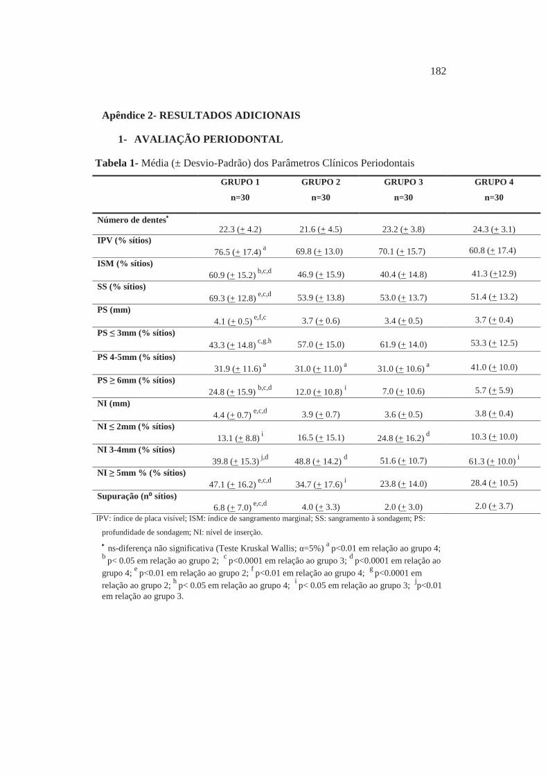
182
Apêndice 2- RESULTADOS ADICIONAIS
1- AVALIAÇÃO PERIODONTAL
Tabela 1- Média (± Desvio-Padrão) dos Parâmetros Clínicos Periodontais
GRUPO 1
n=30
GRUPO 2
n=30
GRUPO 3
n=30
GRUPO 4
n=30
Número de dentes 22.3 (+ 4.2)
21.6 (+ 4.5)
23.2 (+ 3.8)
24.3 (+ 3.1)
IPV (% sítios)
76.5 (+ 17.4) a
69.8 (+ 13.0)
70.1 (+ 15.7)
60.8 (+ 17.4)
ISM (% sítios)
60.9 (+ 15.2) b,c,d
46.9 (+ 15.9)
40.4 (+ 14.8)
41.3 (+12.9)
SS (% sítios) 69.3 (+ 12.8) e,c,d
53.9 (+ 13.8)
53.0 (+ 13.7)
51.4 (+ 13.2)
PS (mm) 4.1 (+ 0.5) e,f,c
3.7 (+ 0.6)
3.4 (+ 0.5)
3.7 (+ 0.4)
PS ≤ 3mm (% sítios)
43.3 (+ 14.8) c,g.h
57.0 (+ 15.0)
61.9 (+ 14.0)
53.3 (+ 12.5)
PS 4-5mm (% sítios)
31.9 (+ 11.6) a
31.0 (+ 11.0) a
31.0 (+ 10.6) a
41.0 (+ 10.0)
PS ≥ 6mm (% sítios) 24.8 (+ 15.9) b,c,d
12.0 (+ 10.8) i
7.0 (+ 10.6)
5.7 (+ 5.9)
NI (mm) 4.4 (+ 0.7) e,c,d
3.9 (+ 0.7)
3.6 (+ 0.5)
3.8 (+ 0.4)
NI ≤ 2mm (% sítios) 13.1 (+ 8.8) i
16.5 (+ 15.1)
24.8 (+ 16.2) d
10.3 (+ 10.0)
NI 3-4mm (% sítios) 39.8 (+ 15.3) j,d
48.8 (+ 14.2) d
51.6 (+ 10.7)
61.3 (+ 10.0) i
NI ≥ 5mm % (% sítios) 47.1 (+ 16.2) e,c,d
34.7 (+ 17.6) i
23.8 (+ 14.0)
28.4 (+ 10.5)
Supuração (n⁰ sítios) 6.8 (+ 7.0) e,c,d
4.0 (+ 3.3)
2.0 (+ 3.0)
2.0 (+ 3.7)
IPV: índice de placa visível; ISM: índice de sangramento marginal; SS: sangramento à sondagem; PS:
profundidade de sondagem; NI: nível de inserção. ns-diferença não significativa (Teste Kruskal Wallis; α=5%) a p<0.01 em relação ao grupo 4;
b p< 0.05 em relação ao grupo 2; c p<0.0001 em relação ao grupo 3; d p<0.0001 em relação ao grupo 4; e p<0.01 em relação ao grupo 2; f p<0.01 em relação ao grupo 4; g p<0.0001 em relação ao grupo 2; h p< 0.05 em relação ao grupo 4; i p< 0.05 em relação ao grupo 3; jp<0.01 em relação ao grupo 3.
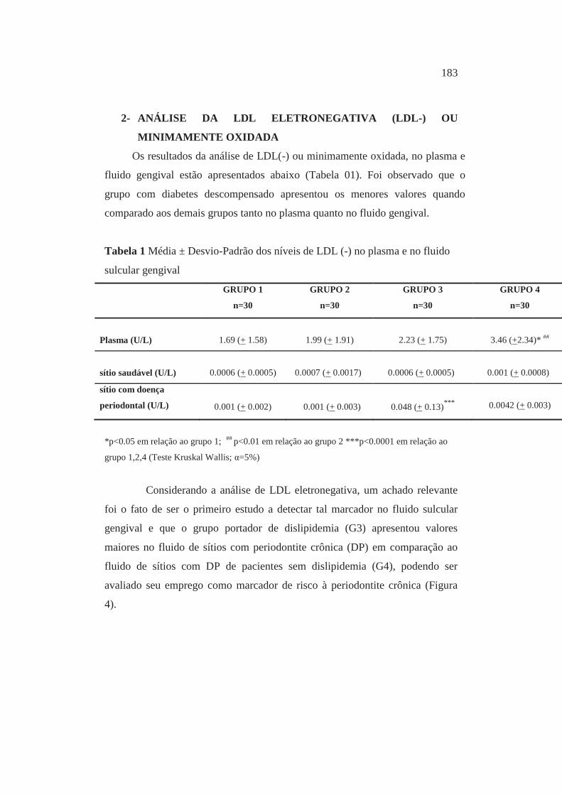
183
2- ANÁLISE DA LDL ELETRONEGATIVA (LDL-) OU
MINIMAMENTE OXIDADA
Os resultados da análise de LDL(-) ou minimamente oxidada, no plasma e
fluido gengival estão apresentados abaixo (Tabela 01). Foi observado que o
grupo com diabetes descompensado apresentou os menores valores quando
comparado aos demais grupos tanto no plasma quanto no fluido gengival.
Tabela 1 Média ± Desvio-Padrão dos níveis de LDL (-) no plasma e no fluido
sulcular gengival
GRUPO 1
n=30
GRUPO 2
n=30
GRUPO 3
n=30
GRUPO 4
n=30
Plasma (U/L)
1.69 (+ 1.58)
1.99 (+ 1.91)
2.23 (+ 1.75)
3.46 (+2.34)* ##
sítio saudável (U/L)
0.0006 (+ 0.0005)
0.0007 (+ 0.0017)
0.0006 (+ 0.0005)
0.001 (+ 0.0008)
sítio com doença
periodontal (U/L)
0.001 (+ 0.002)
0.001 (+ 0.003)
0.048 (+ 0.13)***
0.0042 (+ 0.003)
*p<0.05 em relação ao grupo 1; ## p<0.01 em relação ao grupo 2 ***p<0.0001 em relação ao
grupo 1,2,4 (Teste Kruskal Wallis; α=5%)
Considerando a análise de LDL eletronegativa, um achado relevante
foi o fato de ser o primeiro estudo a detectar tal marcador no fluido sulcular
gengival e que o grupo portador de dislipidemia (G3) apresentou valores
maiores no fluido de sítios com periodontite crônica (DP) em comparação ao
fluido de sítios com DP de pacientes sem dislipidemia (G4), podendo ser
avaliado seu emprego como marcador de risco à periodontite crônica (Figura
4).
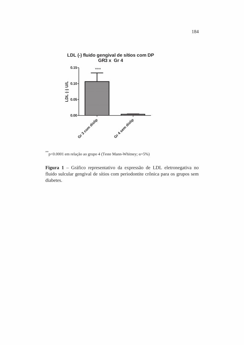
184
**p<0.0001 em relação ao grupo 4 (Teste Mann-Whitney; α=5%)
Figura 1 – Gráfico representativo da expressão de LDL eletronegativa no fluido sulcular gengival de sítios com periodontite crônica para os grupos sem diabetes.
***
LDL (-) fluido gengival de sítios com DPGR3 x Gr 4
Gr 3 co
m dislip
Gr 4
sem disl
ip0.00
0.05
0.10
0.15LD
L (-
) U/L
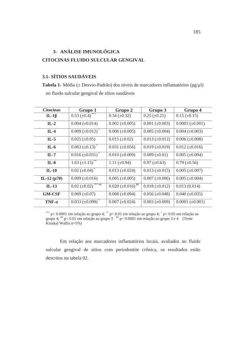
185
3- ANÁLISE IMUNOLÓGICA
CITOCINAS FLUIDO SULCULAR GENGIVAL
3.1- SÍTIOS SAUDÁVEIS
Tabela 1- Média (± Desvio-Padrão) dos níveis de marcadores inflamatórios (pg/μl)
no fluido sulcular gengival de sítios saudáveis
Citocinas Grupo 1 Grupo 2 Grupo 3 Grupo 4 IL-1β 0.53 (±0.4) *** 0.34 (±0.32) 0.25 (±0.21) 0.15 (±0.15) IL-2 0.004 (±0.014) 0.002 (±0.005) 0.001 (±0.003) 0.0003 (±0.001) IL-4 0.009 (±0.012) * 0.006 (±0.005) 0.005 (±0.004) 0.004 (±0.003) IL-5 0.025 (±0.05) 0.015 (±0.02) 0.013 (±0.012) 0.006 (±0.008)
IL-6 0.063 (±0.13) * 0.031 (±0.056) 0.019 (±0.019) 0.012 (±0.016) IL-7 0.016 (±0.031) * 0.010 (±0.009) 0.009 (±0.01) 0.005 (±0.004) IL-8 1.63 (±1.15) ** 1.11 (±0.94) 0.97 (±0.63) 0.79 (±0.56)
IL-10 0.02 (±0.04) * 0.013 (±0.024) 0.013 (±0.015) 0.005 (±0.007) IL-12 (p70) 0.009 (±0.016) 0.005 (±0.005) 0.007 (±0.006) 0.005 (±0.004)
IL-13 0.02 (±0.02) *** ## 0.020 (±0.016) ¥¥ 0.018 (±0.012) 0.013 (0.014) GM-CSF 0.069 (±0.07) 0.068 (±0.094) 0.056 (±0.048) 0.040 (±0.035)
TNF-α 0.033 (±0.099) * 0.007 (±0.024) 0.003 (±0.009) 0.0001 (±0.001)
*** p< 0.0001 em relação ao grupo 4; ** p< 0.01 em relação ao grupo 4; * p< 0.05 em relação ao grupo 4; ## p< 0.01 em relação ao grupo 3 ¥¥ p< 0.0001 em relação ao grupo 3 e 4 (Teste Kruskal Wallis α=5%)
Em relação aos marcadores inflamatórios locais, avaliados no fluido
sulcular gengival de sitios com periodontite crônica, os resultados estão
descritos na tabela 02.
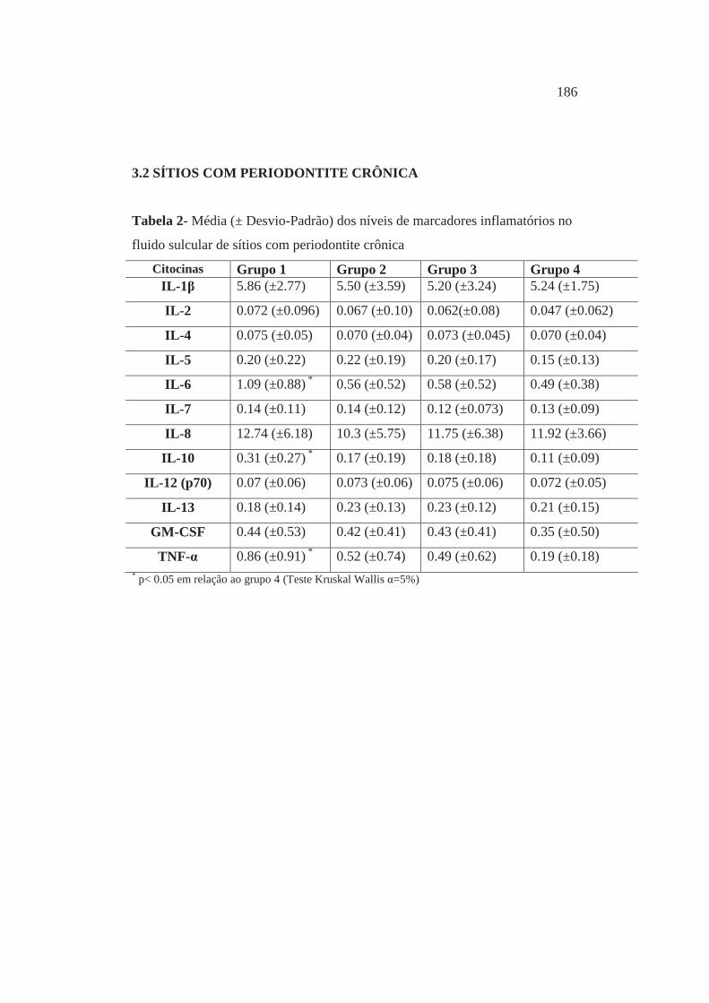
186
3.2 SÍTIOS COM PERIODONTITE CRÔNICA
Tabela 2- Média (± Desvio-Padrão) dos níveis de marcadores inflamatórios no
fluido sulcular de sítios com periodontite crônica
Citocinas Grupo 1 Grupo 2 Grupo 3 Grupo 4 IL-1β 5.86 (±2.77) 5.50 (±3.59) 5.20 (±3.24) 5.24 (±1.75)
IL-2 0.072 (±0.096) 0.067 (±0.10) 0.062(±0.08) 0.047 (±0.062)
IL-4 0.075 (±0.05) 0.070 (±0.04) 0.073 (±0.045) 0.070 (±0.04)
IL-5 0.20 (±0.22) 0.22 (±0.19) 0.20 (±0.17) 0.15 (±0.13)
IL-6 1.09 (±0.88) * 0.56 (±0.52) 0.58 (±0.52) 0.49 (±0.38)
IL-7 0.14 (±0.11) 0.14 (±0.12) 0.12 (±0.073) 0.13 (±0.09)
IL-8 12.74 (±6.18) 10.3 (±5.75) 11.75 (±6.38) 11.92 (±3.66)
IL-10 0.31 (±0.27) * 0.17 (±0.19) 0.18 (±0.18) 0.11 (±0.09)
IL-12 (p70) 0.07 (±0.06) 0.073 (±0.06) 0.075 (±0.06) 0.072 (±0.05)
IL-13 0.18 (±0.14) 0.23 (±0.13) 0.23 (±0.12) 0.21 (±0.15)
GM-CSF 0.44 (±0.53) 0.42 (±0.41) 0.43 (±0.41) 0.35 (±0.50)
TNF-α 0.86 (±0.91) * 0.52 (±0.74) 0.49 (±0.62) 0.19 (±0.18) * p< 0.05 em relação ao grupo 4 (Teste Kruskal Wallis α=5%)

187
ANEXOS
188
ANEXO A – CERTIFICADO DO COMITÊ DE ÉTICA
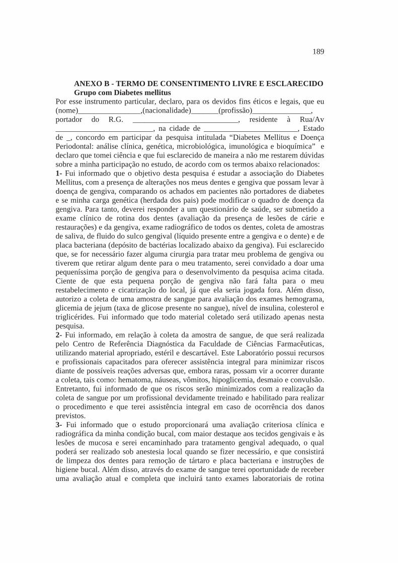
189
ANEXO B - TERMO DE CONSENTIMENTO LIVRE E ESCLARECIDO Grupo com Diabetes mellitus
Por esse instrumento particular, declaro, para os devidos fins éticos e legais, que eu (nome)________________,(nacionalidade)_______(profissão)_______________, portador do R.G. ___________________________, residente à Rua/Av _________________________, na cidade de ________________________, Estado de _, concordo em participar da pesquisa intitulada “Diabetes Mellitus e Doença Periodontal: análise clínica, genética, microbiológica, imunológica e bioquímica” e declaro que tomei ciência e que fui esclarecido de maneira a não me restarem dúvidas sobre a minha participação no estudo, de acordo com os termos abaixo relacionados: 1- Fui informado que o objetivo desta pesquisa é estudar a associação do Diabetes Mellitus, com a presença de alterações nos meus dentes e gengiva que possam levar à doença de gengiva, comparando os achados em pacientes não portadores de diabetes e se minha carga genética (herdada dos pais) pode modificar o quadro de doença da gengiva. Para tanto, deverei responder a um questionário de saúde, ser submetido a exame clínico de rotina dos dentes (avaliação da presença de lesões de cárie e restaurações) e da gengiva, exame radiográfico de todos os dentes, coleta de amostras de saliva, de fluido do sulco gengival (líquido presente entre a gengiva e o dente) e de placa bacteriana (depósito de bactérias localizado abaixo da gengiva). Fui esclarecido que, se for necessário fazer alguma cirurgia para tratar meu problema de gengiva ou tiverem que retirar algum dente para o meu tratamento, serei convidado a doar uma pequeníssima porção de gengiva para o desenvolvimento da pesquisa acima citada. Ciente de que esta pequena porção de gengiva não fará falta para o meu restabelecimento e cicatrização do local, já que ela seria jogada fora. Além disso, autorizo a coleta de uma amostra de sangue para avaliação dos exames hemograma, glicemia de jejum (taxa de glicose presente no sangue), nível de insulina, colesterol e triglicérides. Fui informado que todo material coletado será utilizado apenas nesta pesquisa. 2- Fui informado, em relação à coleta da amostra de sangue, de que será realizada pelo Centro de Referência Diagnóstica da Faculdade de Ciências Farmacêuticas, utilizando material apropriado, estéril e descartável. Este Laboratório possui recursos e profissionais capacitados para oferecer assistência integral para minimizar riscos diante de possíveis reações adversas que, embora raras, possam vir a ocorrer durante a coleta, tais como: hematoma, náuseas, vômitos, hipoglicemia, desmaio e convulsão. Entretanto, fui informado de que os riscos serão minimizados com a realização da coleta de sangue por um profissional devidamente treinado e habilitado para realizar o procedimento e que terei assistência integral em caso de ocorrência dos danos previstos. 3- Fui informado que o estudo proporcionará uma avaliação criteriosa clínica e radiográfica da minha condição bucal, com maior destaque aos tecidos gengivais e às lesões de mucosa e serei encaminhado para tratamento gengival adequado, o qual poderá ser realizado sob anestesia local quando se fizer necessário, e que consistirá de limpeza dos dentes para remoção de tártaro e placa bacteriana e instruções de higiene bucal. Além disso, através do exame de sangue terei oportunidade de receber uma avaliação atual e completa que incluirá tanto exames laboratoriais de rotina
190
quanto exames mais complexos, os quais por esta razão são esporadicamente solicitados pelos médicos. Assim, será dada ao paciente e ao médico a oportunidade de conhecer a real condição sistêmica do indivíduo e caso necessite de tratamento, será orientado e encaminhado ao médico. 4- Estou ciente de que os riscos frente aos procedimentos aos quais serei submetido estão relacionados à utilização de anestésicos locais de rotina no atendimento odontológico. Complicações relacionadas ao uso de anestésicos serão evitadas pela avaliação de história anterior de reações alérgicas a algum tipo de anestésico ou alterações na pressão arterial. Caso seja informada alguma alteração, serei encaminhado para avaliação médica e somente poderei participar do estudo quando houver autorização do profissional. Quanto à realização de radiografias e os riscos de exposição à radiação, usarei avental e colar de chumbo como medida de proteção, o exame será realizado dentro da quantidade de radiação permitida e por técnico capacitado, o que evitará as tomadas repetidas e desnecessárias. 5- Como existem riscos particulares para o grupo portador de Diabetes mellitus tipo 2, somente poderei participar da pesquisa com autorização por escrito do médico endocrinologista responsável pelo meu tratamento. Além disso, caso necessite tomar antibiótico para realização dos exames clínicos, não poderei ser incluído na pesquisa. Continuarei sob acompanhamento com o meu endocrinologista e este estará ciente que receberei tratamento periodontal. Se houver complicações metabólicas decorrente do Diabetes e meu médico julgar importante a minha exclusão da pesquisa, serei imediatamente excluído da mesma. 6- Torno ciente que recebi todas as informações sobre a minha participação nesta pesquisa e receberei novos esclarecimentos que julgar necessários durante o decorrer da mesma. Fui esclarecido que não terei gastos extras, a não ser o deslocamento, e que meu consentimento não tira a responsabilidade dos profissionais que estão realizando esta pesquisa. 7- Estou ciente que minha participação é voluntária e tenho plena liberdade para desistir da referida pesquisa, retirando o meu consentimento a qualquer momento, sem sofrer nenhum tipo de penalização. 8 - Autorizo, para os devidos fins, o uso, a divulgação e publicação em revistas científicas, brasileiras ou estrangeiras, dos dados obtidos na pesquisa. Recebi a garantia de que a minha identidade não será divulgada, assegurando a minha privacidade. Caso haja qualquer problema ou dúvida durante minha participação na pesquisa, terei plena liberdade de contactar o pesquisador responsável, pelo telefone Profa. Dra. Silvana, (16) 3301-6377 e que posso consultar o Comitê de Ética em Pesquisa, para qualquer informação em relação à pesquisa da qual participo, pelo telefone (16) 3301-6432/6434.
Desta forma, por estar de pleno acordo com o teor do mesmo, dato e assino.
Araraquara, ____ de _____________de_____
Assinatura do Paciente Assinatura do Pesquisador Responsável
191
Grupo sem Diabetes mellitus
TERMO DE CONSENTIMENTO LIVRE E ESCLARECIDO
Por esse instrumento particular, declaro, para os devidos fins éticos e legais, que eu (nome) ____________________________________,(nacionalidade)_______(profissão)_______________, portador do R.G. ___________________________, residente à Rua/Av _________________________, na cidade de ________________________, Estado de _, concordo em participar da pesquisa intitulada “Diabetes Mellitus e Doença Periodontal: análise clínica, genética, microbiológica, imunológica e bioquímica” e declaro que tomei ciência e que fui esclarecido de maneira a não me restarem dúvidas sobre a minha participação no estudo, de acordo com os termos abaixo relacionados: 1- Fui informado que o objetivo desta pesquisa é estudar a associação do Diabetes Mellitus, com a presença de alterações nos dentes e gengiva que possam levar à doença de gengiva, comparando os achados em pacientes sem diabetes e que apresentam doença de gengiva e se minha carga genética (herdada dos pais) pode modificar o quadro de doença da gengiva. Para tanto, deverei responder a um questionário de saúde, ser submetido a exame clínico de rotina dos dentes (avaliação da presença de lesões de cárie e restaurações) e da gengiva, exame radiográfico de todos os dentes, coleta de amostras de saliva, de fluido do sulco gengival (líquido presente entre a gengiva e o dente) e de placa bacteriana (depósito de bactérias localizado abaixo da gengiva). Fui esclarecido que, se for necessário fazer alguma cirurgia para tratar meu problema de gengiva ou tiverem que retirar algum dente para o meu tratamento, serei convidado a doar uma pequeníssima porção de gengiva para o desenvolvimento da pesquisa acima citada. Ciente de que esta pequena porção de gengiva não fará falta para o meu restabelecimento e cicatrização do local, já que ela seria jogada fora. Além disso, autorizo a coleta de amostra de sangue para avaliação dos exames hemograma, glicemia de jejum (taxa de glicose presente no sangue), nível de insulina, colesterol e triglicérides. 2- Fui informado que todo material coletado será utilizado apenas nesta pesquisa. 3- Em relação à coleta da amostra de sangue, estou ciente de que será realizada no Centro de Referência Diagnóstica da Faculdade de Ciências Farmacêuticas, utilizando material apropriado, estéril e descartável. Este Laboratório possui recursos e profissionais capacitados para oferecer assistência integral diante de possíveis reações adversas que, embora raras, possam vir a ocorrer durante a coleta, tais como: hematoma, náuseas, vômitos, hipoglicemia, desmaio e convulsão. Entretanto, fui informado de que os riscos serão minimizados com a realização da coleta de sangue por um profissional devidamente treinado e habilitado para realizar o procedimento e que terei assistência integral em caso de ocorrência dos danos previstos. 4- Fui informado também, que o estudo proporcionará uma avaliação criteriosa clínica e radiográfica da minha condição bucal, com maior destaque aos tecidos gengivais e às lesões de mucosa e serei encaminhado para tratamento gengival adequado, o qual poderá ser realizado sob anestesia local quando se fizer necessário,
192
e que consistirá de limpeza dos dentes para remoção de tártaro e placa bacteriana e instruções de higiene bucal. Além disso, através do exame de sangue terei oportunidade de receber uma avaliação atual e completa que incluirá tanto exames laboratoriais de rotina quanto exames mais complexos, os quais por esta razão são esporadicamente solicitados pelos médicos. Assim, será dada ao paciente e ao médico a oportunidade de conhecer a real condição sistêmica do indivíduo e caso necessite de tratamento, será orientado e encaminhado ao médico. 5- Estou ciente de que os riscos frente aos procedimentos aos quais serei submetido estão relacionados à utilização de anestésicos locais de rotina no atendimento odontológico. Complicações relacionadas ao uso de anestésicos serão evitadas pela avaliação de história anterior de reações alérgicas a algum tipo de anestésico ou alterações na pressão arterial. Caso seja informada alguma alteração, serei encaminhado para avaliação médica e somente poderei participar do estudo quando houver autorização do profissional. Quanto à realização de radiografias e os riscos de exposição à radiação, usarei avental e colar de chumbo como medida de proteção, o exame será realizado dentro da quantidade de radiação permitida e por técnico capacitado, o que evitará as tomadas repetidas e desnecessárias. 6- Torno ciente que recebi todas as informações sobre a minha participação nesta pesquisa e receberei novos esclarecimentos que julgar necessários durante o decorrer da mesma. Fui esclarecido que não terei gastos extras, a não ser o deslocamento, e que meu consentimento não tira a responsabilidade dos profissionais que estão realizando esta pesquisa. 7-Estou ciente que minha participação é voluntária e tenho plena liberdade para desistir da referida pesquisa, retirando o meu consentimento a qualquer momento, sem sofrer nenhum tipo de penalização. 8- Autorizo, para os devidos fins, o uso, a divulgação e publicação em revistas científicas, brasileiras ou estrangeiras, dos dados obtidos na pesquisa. Recebi a garantia de que a minha identidade não será divulgada, assegurando a minha privacidade. Caso haja qualquer problema ou dúvida durante minha participação na pesquisa, terei plena liberdade de contactar o pesquisador responsável, Profa. Dra. Silvana, pelo telefone: (16) 3301-6377 e que posso consultar o Comitê de Ética em Pesquisa, para qualquer informação em relação à pesquisa da qual participo, pelo telefone (16) 3301-6432/6434.
Desta forma, por estar de pleno acordo com o teor do mesmo, dato e assino.
Araraquara, ____ de ______________ de 20_.
___________________________ ________________________________ Assinatura do Paciente Assinatura da Pesquisador Responsável

193
ANEXO C (Documentos comprobatórios)

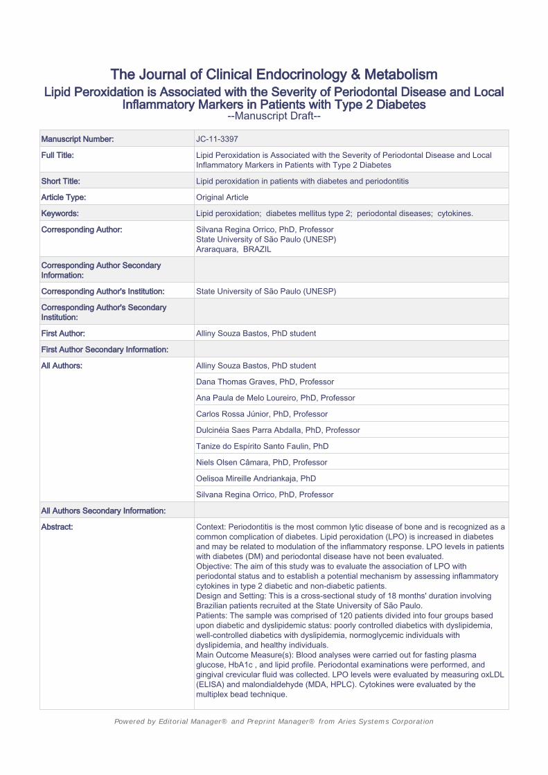
TThe Journal of Clinical Endocrinology & Metabolism
Lipid Peroxidation is Associated with the Severity of Periodontal Disease and LocalInflammatory Markers in Patients with Type 2 Diabetes
--Manuscript Draft--
Manuscript Number: JC-11-3397
Full Title: Lipid Peroxidation is Associated with the Severity of Periodontal Disease and LocalInflammatory Markers in Patients with Type 2 Diabetes
Short Title: Lipid peroxidation in patients with diabetes and periodontitis
Article Type: Original Article
Keywords: Lipid peroxidation; diabetes mellitus type 2; periodontal diseases; cytokines.
Corresponding Author: Silvana Regina Orrico, PhD, ProfessorState University of São Paulo (UNESP)Araraquara, BRAZIL
Corresponding Author SecondaryInformation:
Corresponding Author's Institution: State University of São Paulo (UNESP)
Corresponding Author's SecondaryInstitution:
First Author: Alliny Souza Bastos, PhD student
First Author Secondary Information:
All Authors: Alliny Souza Bastos, PhD student
Dana Thomas Graves, PhD, Professor
Ana Paula de Melo Loureiro, PhD, Professor
Carlos Rossa Júnior, PhD, Professor
Dulcinéia Saes Parra Abdalla, PhD, Professor
Tanize do Espírito Santo Faulin, PhD
Niels Olsen Câmara, PhD, Professor
Oelisoa Mireille Andriankaja, PhD
Silvana Regina Orrico, PhD, Professor
All Authors Secondary Information:
Abstract: Context: Periodontitis is the most common lytic disease of bone and is recognized as acommon complication of diabetes. Lipid peroxidation (LPO) is increased in diabetesand may be related to modulation of the inflammatory response. LPO levels in patientswith diabetes (DM) and periodontal disease have not been evaluated.Objective: The aim of this study was to evaluate the association of LPO withperiodontal status and to establish a potential mechanism by assessing inflammatorycytokines in type 2 diabetic and non-diabetic patients.Design and Setting: This is a cross-sectional study of 18 months' duration involvingBrazilian patients recruited at the State University of São Paulo.Patients: The sample was comprised of 120 patients divided into four groups basedupon diabetic and dyslipidemic status: poorly controlled diabetics with dyslipidemia,well-controlled diabetics with dyslipidemia, normoglycemic individuals withdyslipidemia, and healthy individuals.Main Outcome Measure(s): Blood analyses were carried out for fasting plasmaglucose, HbA1c , and lipid profile. Periodontal examinations were performed, andgingival crevicular fluid was collected. LPO levels were evaluated by measuring oxLDL(ELISA) and malondialdehyde (MDA, HPLC). Cytokines were evaluated by themultiplex bead technique.
Powered by Editorial Manager® and Preprint Manager® from Aries Systems Corporation

Autorizo a reprodução deste trabalho.
(Direitos de publicação reservado ao autor)
Araraquara, 12 de março de 2012.
ALLINY DE SOUZA BASTOS
Related Documents