Accepted Manuscript Title: ALISKIREN AFFECTS FATTY-ACID UPTAKE AND LIPID-RELATED GENES IN RODENT AND HUMAN CARDIOMYOCYTES. Authors: Diego Rodr´ ıguez-Penas, Sandra Feij ´ oo-Band´ ın, Pamela V Lear, Ana Mosquera-Leal, Vanessa Garc´ ıa-R´ ua, Manuel F Otero, Miguel Rivera, Oreste Gualillo, Jos´ e Ram ´ on Gonz´ alez-Juanatey, Francisca Lago PII: S0006-2952(11)00334-0 DOI: doi:10.1016/j.bcp.2011.05.021 Reference: BCP 10913 To appear in: BCP Received date: 19-1-2011 Revised date: 13-5-2011 Accepted date: 18-5-2011 Please cite this article as: Rodr´ ıguez-Penas D, Feij´ oo-Band´ ın S, Lear PV, Mosquera- Leal A, Garc´ ıa-R´ ua V, Otero MF, Rivera M, Gualillo O, Gonz´ alez-Juanatey JR, Lago F, ALISKIREN AFFECTS FATTY-ACID UPTAKE AND LIPID-RELATED GENES IN RODENT AND HUMAN CARDIOMYOCYTES., Biochemical Pharmacology (2010), doi:10.1016/j.bcp.2011.05.021 This is a PDF file of an unedited manuscript that has been accepted for publication. As a service to our customers we are providing this early version of the manuscript. The manuscript will undergo copyediting, typesetting, and review of the resulting proof before it is published in its final form. Please note that during the production process errors may be discovered which could affect the content, and all legal disclaimers that apply to the journal pertain. peer-00721645, version 1 - 29 Jul 2012 Author manuscript, published in "Biochemical Pharmacology 82, 5 (2011) 491" DOI : 10.1016/j.bcp.2011.05.021

Welcome message from author
This document is posted to help you gain knowledge. Please leave a comment to let me know what you think about it! Share it to your friends and learn new things together.
Transcript
Accepted Manuscript
Title: ALISKIREN AFFECTS FATTY-ACID UPTAKE ANDLIPID-RELATED GENES IN RODENT AND HUMANCARDIOMYOCYTES.
Authors: Diego Rodrıguez-Penas, Sandra Feijoo-Bandın,Pamela V Lear, Ana Mosquera-Leal, Vanessa Garcıa-Rua,Manuel F Otero, Miguel Rivera, Oreste Gualillo, Jose RamonGonzalez-Juanatey, Francisca Lago
PII: S0006-2952(11)00334-0DOI: doi:10.1016/j.bcp.2011.05.021Reference: BCP 10913
To appear in: BCP
Received date: 19-1-2011Revised date: 13-5-2011Accepted date: 18-5-2011
Please cite this article as: Rodrıguez-Penas D, Feijoo-Bandın S, Lear PV, Mosquera-Leal A, Garcıa-Rua V, Otero MF, Rivera M, Gualillo O, Gonzalez-Juanatey JR, Lago F,ALISKIREN AFFECTS FATTY-ACID UPTAKE AND LIPID-RELATED GENES INRODENT AND HUMAN CARDIOMYOCYTES., Biochemical Pharmacology (2010),doi:10.1016/j.bcp.2011.05.021
This is a PDF file of an unedited manuscript that has been accepted for publication.As a service to our customers we are providing this early version of the manuscript.The manuscript will undergo copyediting, typesetting, and review of the resulting proofbefore it is published in its final form. Please note that during the production processerrors may be discovered which could affect the content, and all legal disclaimers thatapply to the journal pertain.
peer
-007
2164
5, v
ersi
on 1
- 29
Jul
201
2Author manuscript, published in "Biochemical Pharmacology 82, 5 (2011) 491"
DOI : 10.1016/j.bcp.2011.05.021
Page 1 of 47
Accep
ted
Man
uscr
ipt
1 2 3 4 5 6 7 8 9 10 11 12 13 14 15 16 17 18 19 20 21 22 23 24 25 26 27 28 29 30 31 32 33 34 35 36 37 38 39 40 41 42 43 44 45 46 47 48 49 50 51 52 53 54 55 56 57 58 59 60 61 62 63 64 65
1
ALISKIREN AFFECTS FATTY-ACID UPTAKE AND LIPID-RELATED GENES IN RODENT
AND HUMAN CARDIOMYOCYTES.
Diego Rodríguez-Penas,1* Sandra Feijóo-Bandín,
1* Pamela V Lear,
1* Ana Mosquera-Leal,
1
Vanessa García-Rúa,1 Manuel F Otero,
1 Miguel Rivera,
2 Oreste Gualillo,
3 José Ramón González-
Juanatey,1 Francisca Lago.
1
*These authors contributed equally.
1Cellular and Molecular Cardiology Research Unit, Department of Cardiology and Institute of
Biomedical Research, and 3Neuroendocrine Interactions in Rheumatic Diseases Laboratory,
Institute of Biomedical Research, University Clinical Hospital, Santiago de Compostela, Spain;
2Cardiocirculatory Unit, Research Center, La Fe University Hospital, Valencia, Spain.
¶CORRESPONDENCE: Dr Francisca Lago
Laboratorio 7, Instituto de Investigaciones Sanitarias de Santiago de Compostela (IDIS),
Planta -2, Edificio de Consultas Externas, Hospital Clínico Universitario, Travesía Choupana s/n,
15706 Santiago de Compostela, Spain.
Tel: + 34 981 950 902
Fax: +34 981 950 905
Email: [email protected]
SHORT TITLE: Aliskiren affects cardiomyocyte metabolism.
KEY WORDS: Cardiomyocytes, aliskiren, metabolism, lipids, cholesterol, glucose.
peer
-007
2164
5, v
ersi
on 1
- 29
Jul
201
2
Page 2 of 47
Accep
ted
Man
uscr
ipt
1 2 3 4 5 6 7 8 9 10 11 12 13 14 15 16 17 18 19 20 21 22 23 24 25 26 27 28 29 30 31 32 33 34 35 36 37 38 39 40 41 42 43 44 45 46 47 48 49 50 51 52 53 54 55 56 57 58 59 60 61 62 63 64 65
2
ABSTRACT
PURPOSE: We investigated whether the direct renin inhibitor aliskiren can affect metabolism in
cardiomyocytes from rat, mouse and human sources. METHODS AND RESULTS: At 10-
50µmol/L, aliskiren significantly increased medium-chain-fatty-acid uptake in primary-cultured
neonatal-rat and HL-1 adult-mouse-derived cardiomyocytes (BODIPY-induced fluorescence
intensity). The fatty-acid transporter CD-36 was correspondingly translocated to, but the glucose
transporter Glut-4 away from, the sarcoplasmic reticulum/plasma membrane, in primary-cultured
neonatal-rat (CD-36, Glut-4) and adult-human (CD-36) cardiomyocytes (confocal
immunocytochemistry). Immunoblotting showed that aliskiren induced phosphorylation of
ERK1/2 in cardiomyocytes from all three sources; responses were dose- and time-dependent,
unaffected by renin treatment, and did not cause alterations in expression of (P)R or Igf2/M6P
receptors. Microarray analysis of the complete genome of aliskiren-treated neonatal-rat
cardiomyocytes, with RT-qPCR and immunoblot confirmation assays in rat and human primary
cardiomyocytes, showed that aliskiren up-regulated mRNA and increased protein expression of
several enzymes important in lipid and glucose metabolism and in cholesterol biosynthesis.
Cardiomyocyte cell-cycle and viability were unaffected by aliskiren. CONCLUSIONS: Aliskiren
can induce changes in fatty-acid and glucose uptake and expression of key enzymes of lipid and
cholesterol metabolism, which are not associated with increased expression of (P)R or Igf2/M6P
receptors, in cultured cardiomyocytes.
peer
-007
2164
5, v
ersi
on 1
- 29
Jul
201
2
Page 3 of 47
Accep
ted
Man
uscr
ipt
1 2 3 4 5 6 7 8 9 10 11 12 13 14 15 16 17 18 19 20 21 22 23 24 25 26 27 28 29 30 31 32 33 34 35 36 37 38 39 40 41 42 43 44 45 46 47 48 49 50 51 52 53 54 55 56 57 58 59 60 61 62 63 64 65
3
Introduction
Aliskiren is the first clinically effective anti-hypertensive agent to have been developed by
molecular modelling and to work by direct renin inhibition (DRI) in the renin-angiotensin-
aldosterone system (RAAS) [1-4]. Approved by the US FDA in 2007, aliskiren produces sustained
suppression of plasma renin activity when used in monotherapy in hypertensive patients [5,6], of
cardiac angiotensin in spontaneously hypertensive rats [7], and of renal angiotensin in human renal
podocytes [8]. In addition to its targetted anti-hypertensive action, aliskiren shares with
angiotensin-converting enzyme (ACE) inhibitors and angiotensin-receptor blockers (ARBs) effects
considered to be angiotensin- and blood-pressure-independent, notably cardiovascular and renal
protection, in hypertensive rats [7,9] and patients [2,4,10-12].
However, while ACE inhibitors and ARBs have some relatively minor differences in such non-
anti-hypertensive effects, several studies suggest that those of the DRI aliskiren are significantly
different in several ways [7,9-11]. First, aliskiren’s cardiovascular [2,7,9] and renal [2,4,12,13]
protection appear to be more complete, and possibly attributable to as-yet uncharacterised novel
means [14,15]. A recent report shows that aliskiren is able to reduce atherogenesis in mice
independent of anti-hypertensive effects [16]. Second, aliskiren also appears to be functionally
beneficial or protective in other tissues (pancreatic β-cell epithelium [17,18], white adipose [18],
immune and nervous tissues [19]) and might even reverse end-organ damage [3,7,20]. Third, DRI
appears to have haemodynamic effects, in spontaneously hypertensive rats [7], in healthy humans
on a low-sodium diet [21], and in patients with congestive heart failure [22]. Fourth, aliskiren’s
organ-protective effects have been particularly noted in a background of diabetes mellitus
[2,17,23,24], suggesting that it has one or more metabolic actions. Interestingly, recent clinical
studies have found aliskiren to be more effective than ACE inhibitors or ARBs at lowering blood
pressure and improving insulin sensitivity in hypertensive patients with metabolic syndrome
[6,25].
peer
-007
2164
5, v
ersi
on 1
- 29
Jul
201
2
Page 4 of 47
Accep
ted
Man
uscr
ipt
1 2 3 4 5 6 7 8 9 10 11 12 13 14 15 16 17 18 19 20 21 22 23 24 25 26 27 28 29 30 31 32 33 34 35 36 37 38 39 40 41 42 43 44 45 46 47 48 49 50 51 52 53 54 55 56 57 58 59 60 61 62 63 64 65
4
Metabolic syndrome is prevalent in hypertensive patients and strongly associated with
cardiovascular myopathies [26,27], while chronically elevated RAAS is implicated in many
features of metabolic syndrome [6]. The complexity of RAAS is evidenced by the discovery that
binding of (pro)renin to the (pro)renin receptor ((P)RR) [28] can produce non-angiotensin-II-
mediated effects which are not inhibited by aliskiren [29-31]. This latter finding underpins the
concept that renin can act not only as an enzyme but also as a hormone [10,20], providing a
putative mechanism for non-blood-pressure-related effects of all three classes of RAAS inhibitor,
and thus important insights into how to combine different RAAS inhibitors in tailored therapies
[3,10,32]. Yet, only five studies to date [7,9,17,32,33] have focussed on the cellular or molecular
mechanisms underlying organ protection by aliskiren, and none have directly investigated the
role(s) that metabolism might play.
Therefore, while it has been suggested that the use of aliskiren could add potential metabolic
benefits over other RAAS inhibitors, basic and clinical studies are still necessary to find a
mechanism that could link direct renin inhibition with metabolic regulation by this drug [34]. In the
current study, we have investigated some of aliskiren’s metabolic effects in cardiomyocytes and
determined whether they correlate with changes in expression of (P)Rr or the insulin-like growth
factor II/mannose-6-phosphate (Igf2/M6P) receptor, which can also internalise pro-renin and
activate it to renin [35]. In the absence of previous studies, our focus has been on cardiomyocytes
from rodent and human sources.
Methods
All reagents were from Sigma Chemical Co (MO, USA) unless otherwise stated.
peer
-007
2164
5, v
ersi
on 1
- 29
Jul
201
2

Page 5 of 47
Accep
ted
Man
uscr
ipt
1 2 3 4 5 6 7 8 9 10 11 12 13 14 15 16 17 18 19 20 21 22 23 24 25 26 27 28 29 30 31 32 33 34 35 36 37 38 39 40 41 42 43 44 45 46 47 48 49 50 51 52 53 54 55 56 57 58 59 60 61 62 63 64 65
5
Cardiomyocyte cultures, cell-viability/cell-cycle assays, and metabolic assays
HL-1 and primary cultures of neonatal-rat cardiomyocytes were cultured as previously [36]. The
investigation conforms with the Guide for Care and Use of Laboratory Animals published by the
US National Institutes of Health (NIH Publication No. 85-23, revised 1996). Approval for the
harvesting and use of cardiomyocytes from neonatal rats was also granted by the University of
Santiago de Compostela Ethics Review Board. Human cardiomyocytes (HCM) were obtained from
Promocell GmbH (Heidelberg, Germany) and cultured according to the supplier’s instructions.
Tissue used by PromoCell for the isolation of HCM is obtained from donors with informed consent
(by the donor or legally authorized agent) which includes the purpose of the donation and the
procedure for processing the tissue. PromoCell acts in strict compliance with the following: the
Convention for Protection of Human Rights and Dignity of the Human Being with Regard to the
Application of Biology and Medicine: Convention of Human Rights and Biomedicine (4 April,
1997, Council of Europe (European Treaty Series – no 164), the Human Tissue Act (15 November
2004, HM Government, United Kingdom (this Act aims to make consent a fundamental principle
underpinning the use and storage of human tissue), and the Declaration of Helsinki.
Due to patient confidentiality PromoCell does not supply clinical information except that that the
donors were not known to have any chronic disease. PromoCell states that: i.) their HCM are
suitable for in vitro studies of cardiac diseases such as hypertrophy and for physiological and
pharmacological studies; ii.) in contrast to freshly isolated myocytes, cultured HCM may be used
for longer term experiments such as investigating the effects of cytokines, mechanical strain, or
cell-cell interactions; iii.) the HCM have been tested immunohistochemically for cell-type-specific
markers, including: sarcomeric alpha-actinin positive, slow muscle myosin positive, CD-90
negative; iv.) rigorous quality-control tests are performed on each lot of PromoCell HCM,
including for cell morphology, adherence rate, and viability; v.) growth performance is tested
through multiple passages up to 15 population doublings (PD) under culture conditions without
peer
-007
2164
5, v
ersi
on 1
- 29
Jul
201
2
Page 6 of 47
Accep
ted
Man
uscr
ipt
1 2 3 4 5 6 7 8 9 10 11 12 13 14 15 16 17 18 19 20 21 22 23 24 25 26 27 28 29 30 31 32 33 34 35 36 37 38 39 40 41 42 43 44 45 46 47 48 49 50 51 52 53 54 55 56 57 58 59 60 61 62 63 64 65
6
antibiotics or antimycotics; vi.) all cells have been tested for the absence of HIV-1, HBV, HCV,
and microbial contaminants (fungi, bacteria, mycoplasma).
The range of doses of aliskiren (1nmol/L-50mol/L) was chosen on the basis of previous reports
[37,38]. For 3-[4,5-dimethylthiazol-2-yl]-2,5-diphenyltetrazolium bromide (MTT) mitochondrial
metabolic activity assays [36], serum-deprived HL-1 cardiomyocytes (104/P96 well) were treated
for 24 or 48h with aliskiren (a gift from Novartis Pharmaceuticals SA, Spain) and fetal bovine
serum (FBS) was used as a positive control due to its induction of cell proliferation. For cell-
viability assays [36] serum-deprived HL-1 cells (4×10
5/P96 well) were treated for 24h with 1-
50mol/L aliskiren. For fatty-acid uptake experiments, serum-deprived HL-1 cells or primary-
cultured neonatal-rat cardiomyocytes (4×10⁵/P6 well) were treated for 30 (HL-1) or 120min (rat)
with aliskiren (1-50μmol/L) or insulin (0.1μmol/L, positive control) in PBS containing 10μmol/L
4,4-difluoro-5-methyl-4-bora-3a,4a-diaza-s-indacene-3-dodecanoic acid (BODIPY® 500/510 C1,
C12; Molecular Probes, OR, USA) and 20μmol/L fatty-acid-free BSA. Cells were also stained with
propidium iodide (1μmol/L) to identify dead cells, and analyzed in a FACSCALIBUR flow
cytometer (Becton Dickinson, CA, USA) using the Cell Quest program.
Immunocytochemistry and confocal microscopy
Serum-deprived primary-cultured neonatal-rat (104/coverslip/P24 well) or adult-human (7000/well
in Lab-Tek®II Chamber Slides; Nunc, Denmark) cardiomyocytes were treated with 10 or
50μmol/L aliskiren for 15 or 30min, and fixed for 20min in 4% paraformaldehyde/PBS. Cells were
then permeabilised and blocked in PBS containing 0.2% tween-20, 5% heat-inactivated normal
goat serum and 0.2% electrophoresis-grade BSA, to which anti-CD-36 or Glut-4 (rabbit, 1:100;
Abcam, UK) was added, before incubating at 4C overnight. After washing, cells were blocked
and incubated in Cy3-conjugated donkey anti-rabbit IgG secondary antibody (1:500; Jackson
peer
-007
2164
5, v
ersi
on 1
- 29
Jul
201
2
Page 7 of 47
Accep
ted
Man
uscr
ipt
1 2 3 4 5 6 7 8 9 10 11 12 13 14 15 16 17 18 19 20 21 22 23 24 25 26 27 28 29 30 31 32 33 34 35 36 37 38 39 40 41 42 43 44 45 46 47 48 49 50 51 52 53 54 55 56 57 58 59 60 61 62 63 64 65
7
ImmunoResearch Inc, PA, USA) and TO-PRO-3-iodide (1:500; Invitrogen, Spain) for 45min at
37C. Coverslips were mounted in pre-mixed Mowiol (Calbiochem, CA, USA), glycerol, Tris-Cl
and 2.5% DABCO anti-fading agent (DABCO, Denmark) on cleaned glass slides.
Quantitative confocal analyses: For each coverslip, a minimum of six distinct and randomly
chosen visual fields were photographed and analysed as z-stacks using a Leica DMIRE2 confocal
microscope and software [39]. Mean intensity of Cy3 fluorecence indicated relative expression
levels of CD-36 and Glut-4. For CD-36, any cell containing a region of at least ¼ its total area with
a fluorescence intensity above the mean value for all control cells measured was considered to
show increased sarcoplasmic/plasma membrane expression. For Glut-4, whose plasmatic
expression is relatively more homogeneous than that of CD-36, changes in its distribution between
the nucleus/nuclear membrane and cytoplasm/plasma membrane compartments were determined as
previously [39]. Quantifications were carried out twice, by two independent observers, and
expressed as percentages of cells with altered CD-36 or Glut-4 distribution.
Micro-array analysis
Primary cultures (4×10⁵/P6 well) were serum-deprived for 12h before 3h treatment with 50μmol/L
aliskiren. Total RNA was extracted using an RNeasy Total RNA Extraction Kit (Quiagen KK, UK)
and its integrity assessed with an AGILENT 2100 Bioanalyzer and RNA Nano LabChip (Agilent
Technologies, Germany). cDNA was synthesized from 300ng of total RNA using a Whole
Transcript (WT) cDNA Synthesis and Amplification Kit (Affymetrix, CA, USA) and the
GeneChip WT Sense Target Labeling Assay Manual protocol (Affymetrix, CA, USA). The amount
of cDNA concentration was measured using a NanoDrop spectrophotometer (Thermo Scientific,
Spain). Correct cDNA fragmentation was checked with the AGILENT 2100
Bioanalyzer. Expression of the complete rat genome was evaluated using rat Gene 1.0 ST
microarrays (Affymetrix, CA, USA).
peer
-007
2164
5, v
ersi
on 1
- 29
Jul
201
2

Page 8 of 47
Accep
ted
Man
uscr
ipt
1 2 3 4 5 6 7 8 9 10 11 12 13 14 15 16 17 18 19 20 21 22 23 24 25 26 27 28 29 30 31 32 33 34 35 36 37 38 39 40 41 42 43 44 45 46 47 48 49 50 51 52 53 54 55 56 57 58 59 60 61 62 63 64 65
8
A bio-informatic analysis was performed by Progenika BioPharma SA (Spain). Change values
were determined as follows. 1) Quality control: array outlier analysis using the micro-array
normalization software DNA-Chip Analyzer (dChip); 2) pre-processing of data using the Robust
Multi-chip Average (RMA) (yielding 27,342 probe sets which after background normalization
were equivalent to 13,768 sets); 3) array global normalization using the Partek Genomics Suite
v7.3.1 program (Partek Inc, MO, USA); 4) second filtration of data, maintaining only sequences
with expression changes, measured as standard deviation of normalized intensity data (yielding
8,521 sequences); 5) statistical analysis, principal component analysis (PCA) and hierarchical
cluster analysis. To identify statistically significant changes between control and aliskiren-treated
groups, a linear regression model was used (Yi=conditionε, where Yi is intensity of Y for each
sequence, ‘condition’ is the effect of any particular treatment on intensity, and ε is the non-
measurable error). Results for individual sequences were expressed as fold-change relative to the
baseline (aliskiren vs control, FC). For validation by real-time PCR and western blotting, we
selected the genes most closely associated with lipid metabolism and with highly significant
(p<0.001) and/or large fold-changes in expression; additionally we included the gene with the
lowest ´p´ value in the array, AT-Rich Interaction Domain family member number 5a (Arid 5a,
also known as Modulator Recognition Factor 1 [40]). The micro-array was analysed in terms of
functional groups of genes, and genes closely related to cholesterol biosynthesis were also selected
for validation.
Real-time quantitative PCR
a) to validate microarray
RNA was extracted using a Qiagen RNeasy kit, following the manufacturer’s instructions (Qiagen
Iberia SL, Spain). For relative quantification, we performed an RT reaction with an RT² First
Strand Kit (SA Biosciences, Tebu-bio Iberia, Spain). Real-time PCR was performed using SA
peer
-007
2164
5, v
ersi
on 1
- 29
Jul
201
2

Page 9 of 47
Accep
ted
Man
uscr
ipt
1 2 3 4 5 6 7 8 9 10 11 12 13 14 15 16 17 18 19 20 21 22 23 24 25 26 27 28 29 30 31 32 33 34 35 36 37 38 39 40 41 42 43 44 45 46 47 48 49 50 51 52 53 54 55 56 57 58 59 60 61 62 63 64 65
9
Biosciences RT² SYBR Green/ROX qPCR Master Mix (SA Biosciences, Tebu-bio Iberia, Spain)
according to the manufacturer’s instructions, and the following specific primers for rat: Ras-related
associated with diabetes (Rrad), 191bp, PPR06499A, reference position 800-818, GenBank
accession no. NM053338.1; Insulin-induced gene-1 (Insig1), 103bp, PPR42307A, reference
position 557-576, GenBank accession no. NM022392.1; Lipin-1 (Lpin1), 63bp, PPR59564A,
reference position 2213-2234, GenBank accession no. NM001012111.1; Pyruvate dehydrogenase
kinase, isoenzyme-4 (Pdk4), 91bp, PPR48407A, reference position 1246-1268, GenBank accession
no. NM053551.1; 3-hydroxy-3-methylglutaryl-Coenzyme-A synthase-1 (Hmgcs1), 66bp,
PPR43438E, reference position 3138-3156, GenBank accession no. NM017268.1; Lanosterol
synthase (Lss), 182bp, PPR44730A, reference position 2747-2771, GenBank accession no.
NM031049.1; Farnesyl diphosphate farnesyl transferase-1 (Fdft1), 117bp, PPR43022A, reference
position 1250-1268, GenBank accession no. NM019238.2; 7-dehydrocholesterol reductase
(Dhcr7), 139bp, PPR42280A, reference position 1369-1387, GenBank accession no. NM022389.2;
3-hydroxy-3-methylglutaryl-Coenzyme-A reductase (Hmgcr), 93bp, PPR44338A, reference
position 2595-2614, GenBank accession no. NM013134.2; AT-Rich Interaction Domain family
member number 5a (Arid5A), 172bp, PPR46170A, reference position 554, GenBank accession no.
NM001034934.1.
b) to identify (P)R and Igf/M6P2 receptors
Real-time PCR for the identification of (P)R and Igf2/M6P receptors was performed using a
Stratagene Brilliant® II SYBR® Green QRT-PCR Master Mix Kit (Stratagene, CA, USA)
following the manufacter’s instructions and the following specific primers (TIB Molbiol,
Germany): for mouse: (P)Rr, 172bp, GenBank accession no. NM027439.4, Forward primer:
CCCAGCGAGGAGAGAGTGTATATG, Reverse primer: CGGAGAGAAAGAGCAGGTCAAC;
Igf2/M6Pr, 198bp, GenBank accession no. NM010515, Forward primer:
CAATGCCTGCCTGTGGAACG, Reverse primer: GATGATGAATGCTGTGCCTCTG; and for
peer
-007
2164
5, v
ersi
on 1
- 29
Jul
201
2

Page 10 of 47
Accep
ted
Man
uscr
ipt
1 2 3 4 5 6 7 8 9 10 11 12 13 14 15 16 17 18 19 20 21 22 23 24 25 26 27 28 29 30 31 32 33 34 35 36 37 38 39 40 41 42 43 44 45 46 47 48 49 50 51 52 53 54 55 56 57 58 59 60 61 62 63 64 65
10
rat: (P)Rr, 109bp, GenBank accession no. XM217592.5, Forward primer:
GGTCTGACTGCGGTGCTC, Reverse primer: AGAGAAGAGAGGAGAACGACAAG;
Igf2/M6Pr, 154bp, GenBank accession no. NM012756.1, Forward primer:
GCCACGAGACTGCTGACTG, Reverse primer: AGGAGGAGACTGAGGACTGC. PCR
products were separated by electrophoresis on a 2% agarose gel and stained with ethidium bromide
for UV visualization. Results of comparative real-time PCRs were analyzed using MxPro v.4
software (Stratagene, Spain).
Immunoblotting
Serum-deprived primary cultures of neonatal-rat cardiomyocytes (4×10⁵/P6 well) were treated with
50μmol/L aliskiren for 3-6h or 2-30min (for ERK1/2 phosphorylation) and/or with 0.01 μM
recombinant human (Anaspec, Inc. San José, CA, USA) or rat (Anaspec Inc. San José, CA, USA)
renin for 5min. Cells were then lysed with Triton X-100 (1% buffered in 50mmol/L Tris-HCl,
150mmol/L NaCl, 5mmol/L EDTA, 1mmol/L phenylmethylsulphonylfluoride, 10μg/ml leupeptin,
10μg/ml aprotinin, 10μg/ml trypsin inhibitor and 1mmol/L NaVO4). Samples were subjected to
SDS-PAGE under denaturing conditions on 10% gels and electroblotted onto PVDF membranes
(Amersham Pharmacia Biotech, Germany). Membranes were treated with the following primary
antibodies at 4ºC overnight: anti-Rrad, anti-Hmgcs1, anti-Insig1, anti-Lpin1, anti-Pdk4 (1:200;
Santa Cruz Biotechnology, CA, USA), Hmgcr (1:500; Upstate Biotechnology, Millipore
Corporate, MA, USA), anti-Fdft1 (1:200), anti-Lss (1:1000) (Abcam, MA, USA), anti-Dhcr7
(1:750; Abnova, Tebu-bio Iberia, Spain), anti-phospho-Erk1/2 and anti- Erk1/2 (1:1000; Cell
Signaling Technology, New England BioLabs, UK). Membranes were then incubated with
horeseradish-preoxidase-conjugated secondary antibody (1:2000; Santa Cruz Biotechnology, CA,
USA) followed by chemiluminescence detection (Millipore Corporate, MA, USA). Anti-β-actin
(1:1000; Santa Cruz Biotechnology, CA, USA) was used as a loading control. Densitometric
peer
-007
2164
5, v
ersi
on 1
- 29
Jul
201
2
Page 11 of 47
Accep
ted
Man
uscr
ipt
1 2 3 4 5 6 7 8 9 10 11 12 13 14 15 16 17 18 19 20 21 22 23 24 25 26 27 28 29 30 31 32 33 34 35 36 37 38 39 40 41 42 43 44 45 46 47 48 49 50 51 52 53 54 55 56 57 58 59 60 61 62 63 64 65
11
analyses were performed using a UVP EC3 Imaging System (Ultra-Violet Products Ltd, UK) and
the Image Master TotalLab program (Amersham Biosciences, GE Healthcare, Spain).
Statistical and gene network analyses
Statistical data are from at least three independent experiments (n=3) and expressed as meanSD
or as meanSEM (for confocal microscopy analysis). Statistical significance of the differences
between experimental values was determined using Mann-Whitney U, t-tests, Fisher’s exact test or
ANOVA as appropriate (p<0.05 considered significant). For the microarray, in addition to the
statistical analyses of individual genes, functional groups of genes were compared, using the
following databases: IPA (IPA® 8.8; Ingenuity Systems Inc., Redwood City, CA;
www.ingenuity.com), Gene Ontology (www.geneontology.org), Kyoto Encyclopedia of Genes and
Genomes (KEGG) metabolic pathways (www.genome.jp/kegg/), Obesity Gene Map Database
(www.obesitygene.pbrc.edu) and Database for Annotation, Visualization and Integrated Discovery
(DAVID, National Institute of Allergy and Infectious Diseases (NIAID, USA)).
In the IPA system, gene expression changes are considered in the context of physical,
transcriptional, or enzymatic interactions of the gene/gene products and are grouped according to
interacting gene networks. The score assigned to any given gene network takes into account the
total number of molecules in the data set, the size of the network, and the number of ‘network-
eligible’ genes/molecules in the data set. The network score is based on the hypergeometric
distribution and is calculated with the right-tailed Fisher’s exact test. The network score is the
negative log of that ´p´ value.
Results
Aliskiren treatment did not affect cardiomyocyte viability or the cell cycle.
peer
-007
2164
5, v
ersi
on 1
- 29
Jul
201
2
Page 12 of 47
Accep
ted
Man
uscr
ipt
1 2 3 4 5 6 7 8 9 10 11 12 13 14 15 16 17 18 19 20 21 22 23 24 25 26 27 28 29 30 31 32 33 34 35 36 37 38 39 40 41 42 43 44 45 46 47 48 49 50 51 52 53 54 55 56 57 58 59 60 61 62 63 64 65
12
Aliskiren (1nmol/L-50μmol/L) treatment for 24h or 48h did not alter the viability of, or cell-cycle,
assayed respectively by MTT and flow cytometry assays in HL-1 cardiomyocytes (Fig. 1), or
primary cultures of neonatal rat cardiomyocytes (data not shown).
Aliskiren increased dodecanoic-acid uptake and plasma membrane localization of the fatty-acid
transporter CD-36, but decreased cytoplasm/plasma membrane:nuclear distribution of the glucose
transporter Glut-4.
Flow cytometry showed that aliskiren induced uptake of the MCFA dodecanoic-acid after 30min in
HL-1 cardiomyocytes (Fig. 1C.1), and after 120min in primary cultures of neonatal-rat
cardiomyocytes (Fig. 1C.2). Immunocytochemistry and confocal analyses (Fig. 2) showed that
aliskiren also increased plasma membrane localization of the fatty-acid transporter CD-36, in
primary-cultured adult-human (Fig 2A-I) and neonatal-rat (Fig. 2J-O) cardiomyocytes after 30min.
Interestingly, aliskiren caused a complementary decrease in the localization of the glucose
transporter Glut-4 in the cytoplasm/plasma membrane compartment at 30min (Fig. 2P-U). These
changes were aliskiren-dose-dependent (Table 1), and not significant at 15min (data not shown). It
should be noted that the changes we observed in MCFA uptake and intracellular redistribution of
Glut-4 and CD-36 transporters after treatment with aliskiren were obtained with doses of 10μM
(see Figures 1 and 2, and Table 1), and that previous studies have shown that concentrations of
aliskiren in the low-μM range are present in the blood during treatment with aliskiren [41]. This
range of doses has also been used in several other recent studies on the effects of aliskiren at the
cellular level [8,37,38,41].
Aliskiren induced intracellular phosphorylation of ERK1/2 in a dose- and time-dependent manner
which was unaffected by renin treatment, and did not involve changes in expression levels of (P)R
or Igf2/M6P receptors.
peer
-007
2164
5, v
ersi
on 1
- 29
Jul
201
2

Page 13 of 47
Accep
ted
Man
uscr
ipt
1 2 3 4 5 6 7 8 9 10 11 12 13 14 15 16 17 18 19 20 21 22 23 24 25 26 27 28 29 30 31 32 33 34 35 36 37 38 39 40 41 42 43 44 45 46 47 48 49 50 51 52 53 54 55 56 57 58 59 60 61 62 63 64 65
13
Immunoblotting showed that aliskiren induced intracellular phosphorylation of ERK1/2 in HL-1,
neonatal-rat and adult-human cardiomyocytes. Experiments with primary-cultured neonatal-rat
(Fig. 3A, B, C.2, D, E) showed that ERK1/2 phosphorylation was dose- and time- dependent.
Using the optimal dose (50µmol/L) and time (5min) ascertained in the rat primary cultures, a
significant response was also seen in primary-cultured adult-human cardiomyocytes (Fig. 3C.1).
ERK1/2 phosphorylation was not affected by renin treatment (F.1, F.2). HL-1 cells showed similar
time- and dose-dependent ERK1/2 phosphorylation responses to aliskiren (Fig. 3: G.1, G.2). Real-
time qPCR experiments following 50μmol/L aliskiren treatments for 3h did not induce changes in
mRNA expression levels of (P)R or Igf2/M6P receptors in either neonatal-rat or HL-1
cardiomyocytes (data not shown).
Microarray Analysis
Bio-informatic analysis of the results of the microarray showed that 652 gene sequences were
significantly modified by a 3h treatment with 50µmol/L aliskiren in primary-cultured neonatal-rat
cardiomyocytes. We explored the functional interactions among the altered genes using IPA
(Ingenuity Pathway Analysis), as previously described [42,43], and which is integrated with
several other databases such as KEGG Metabolic Pathways, and the Gene Ontology and Obesity
Gene Map Database. A summary of the global results of the microarray obtained using Gene
Ontology and IPA databases is shown in Figure 4.A. Functional analysis of the entire data set using
IPA and restrictions set to ‘biological/cellular functions related to cardiovascular system cells’
(with a cut-off of p<0.001) and ‘cardiovascular function’ (with a cut-off of p<0.05) identified
biological functions and cardiac alterations that were most statistically significant (Table 2). Right-
tailed Fisher’s exact test was used to calculate p values which indicated the probability that each
biological function and/or disease assigned to the data set was not due to chance alone. Functional
classification of differentially expressed genes revealed a cluster of genes involved in lipid
metabolism as the altered biological function with the lowest p value (p<10-4
; Table 2). We next
peer
-007
2164
5, v
ersi
on 1
- 29
Jul
201
2
Page 14 of 47
Accep
ted
Man
uscr
ipt
1 2 3 4 5 6 7 8 9 10 11 12 13 14 15 16 17 18 19 20 21 22 23 24 25 26 27 28 29 30 31 32 33 34 35 36 37 38 39 40 41 42 43 44 45 46 47 48 49 50 51 52 53 54 55 56 57 58 59 60 61 62 63 64 65
14
explored the functional interactions among the altered genes using IPA. IPA identified a gene
interactive network containing all the genes related to lipid and/or fatty-acid metabolism that we
had found to be altered by treatment of cardiomyocytes with aliskiren (Fig.4. B).
Aliskiren up-regulated several enzymes of key importance to lipid metabolism and cholesterol
biosynthesis in neonatal-rat and adult-human cardiomyocytes.
For confirmation analyses we chose the three up-regulated genes implicated in lipid and/or fatty-
acid metabolism that showed the lowest p values (p<0.001): Insig1 (1.60 FC), Lpin1 (1.46 FC) and
Pdk4 (1.80 FC) and were also included in the most statistically significant network resulting from
the IPA analysis (lipid metabolism; score=15). We also added two more genes to our confirmation
analyses, namely Rrad, whose expression was diminished by aliskiren (p<0.001) with the greatest
FC after 3h aliskiren treatment (-2.46 FC) and which is involved in cardiomyocyte contractility,
viability and glucose metabolism [44-46], and Arid 5a, whose expression was also diminished by
aliskiren treatment (-2.16 FC) with the lowest p value in the whole array (p=7.38X10⁻⁵
).
Real-time qPCR and immunoblotting confirmed the microarray analysis, not only for all the
enzymes assayed in neonatal-rat primary cardiomyocytes, but for selected enzymes in adult-human
primary cardiomyocytes. Thus, in neonatal-rat and adult-human cardiomyocytes treated with
50μmol/L aliskiren, after 3h mRNA expression, and after 6h protein expression were increased for
Insig1, Lpin1 and Pdk4 (involved in lipid metabolism); by contrast, mRNA and protein expression
of Rrad (involved in glucose metabolism) and mRNA of Arid 5a were decreased (Fig. 5). Arid 5a
protein expression was not analyzed due to the lack of a good quality commercially available
antibody.
Analysis of the microarray in terms of functional groups of genes revealed that the cholesterol
biosynthesis pathway was markedly altered after 3h of aliskiren treatment in neonatal-rat
peer
-007
2164
5, v
ersi
on 1
- 29
Jul
201
2
Page 15 of 47
Accep
ted
Man
uscr
ipt
1 2 3 4 5 6 7 8 9 10 11 12 13 14 15 16 17 18 19 20 21 22 23 24 25 26 27 28 29 30 31 32 33 34 35 36 37 38 39 40 41 42 43 44 45 46 47 48 49 50 51 52 53 54 55 56 57 58 59 60 61 62 63 64 65
15
cardiomyocytes. In fact, the steroid biosynthesis pathway was selected by KEGG Metabolic
Pathways with p=10-6
and by IPA with p<10-4
. The following genes belonging to the cholesterol
metabolic pathway were therefore also selected from the microarray statistical analysis for further
confirmation by real-time PCR and immunoblotting: Hmgcs1 (1.43 FC, p<0.001), Hmgcr (1.31
FC, p<0.01), Fdft1 (1.22 FC, p<0.05) Lss (1.19 FC, p<0.05), and Dhcr7 (1.56 FC, p<0.01). It
should be noted that all the cholesterol-synthetic genes were also present in the lipid-metabolism
network generated by IPA with the highest significance score in the analysis of the whole array
(Fig.4.B). Real-time PCR and immunoblotting showed that expression of all these enzymes
involved in cholesterol biosynthesis were significantly increased at both RNA and protein levels at
3h (Fig. 6).
Discussion
Our principal finding is that in cardiomyocytes from rodent and human sources, aliskiren can
significantly increase uptake of the MCFA dodecanoic acid and modify intra-cellular distribution
of fatty-acid (CD-36) and glucose (Glut-4) transporters, as well as up-regulate key lipid- and
glucose-handling enzymes. Additionally, we found that aliskiren up-regulated enzymes along the
entire length of the cholesterol biosynthesis pathway, and that all of these were common to a lipid
metabolism gene network generated from our results.
Whilst our study appears to be the first to investigate directly a relationship between RAAS
inhibition and lipid or cholesterol metabolism in cardiomyocytes, activation of RAAS has
previously been implicated in lipid metabolism. Thus, mice lacking angiotensinogen have reduced
plasma levels of triglycerides and cholesterol which are associated with decreased fatty-acid-
synthase activity in epididymal fat [47]. Several studies have suggested that local adipose-tissue
effects of RAAS are important in lipid metabolism [48], while it has been shown that mice with an
peer
-007
2164
5, v
ersi
on 1
- 29
Jul
201
2
Page 16 of 47
Accep
ted
Man
uscr
ipt
1 2 3 4 5 6 7 8 9 10 11 12 13 14 15 16 17 18 19 20 21 22 23 24 25 26 27 28 29 30 31 32 33 34 35 36 37 38 39 40 41 42 43 44 45 46 47 48 49 50 51 52 53 54 55 56 57 58 59 60 61 62 63 64 65
16
ACE-gene deletion have less body fat and increased energy expenditure than their normal
littermates [49]. Type-2 diabetic mice treated with aliskiren also have substantially decreased body
fat [18] and, moreover, direct renin inhibition by aliskiren can reduce body-weight gain, adiposity
and plasma leptin levels in diet-induced obesity in mice [50]. RAAS activation in renal tissue has
also recently been linked to lipid disorders and fat accumulation in rodents [51].
At the cellular level, evidence from cultured adipocytes suggests that angiotensin II can directly
inhibit lipolysis while increasing fatty-acid-synthase activity [52]. On the other hand, a recent
study [31] has shown that rats over-expressing the human renin gene develop obesity and glucose
intolerance, but that these metabolic changes are not related to angiotensin II. This raises the
intriguing possibility of a new but as-yet unknown substrate for renin – other than angiotensinogen
– which could have an important role in energy homeostasis. Therefore, the increases in MCFA
cardiomyocyte uptake and expression of genes involved in subsequent intracellular lipid
processing that we have observed in the presence of the DRI aliskiren might be attributable either
to inhibition of RAAS, or to alterations involving a hypothetical new substrate
We have found that aliskiren modifies the expression of several enzymes closely related to lipid
metabolism, and these observations are closely correlated with our functional data showing that
aliskiren induces fatty-acid uptake and CD-36 and Glut-4 re-distribution in cultured
cardiomyocytes. Glut-4 and CD-36 dynamically traffic between subcellular compartments and the
plasma membrane in cardiomyocytes, increasing uptake of glucose and fatty acids, respectively
[53]. Our results therefore strongly suggest that CD-36 (and potentially other) fatty-acid
transporters execute the functional effect of aliskiren on increasing fatty-acid uptake that we
observed. In the healthy adult heart, the balance between fatty-acid and carbohydrate metabolism is
tightly controlled and optimises energy production [54]. We have focused our study on the concept
that an anti-hypertensive drug such as aliskiren, a direct renin inhibitor that acts through the
peer
-007
2164
5, v
ersi
on 1
- 29
Jul
201
2
Page 17 of 47
Accep
ted
Man
uscr
ipt
1 2 3 4 5 6 7 8 9 10 11 12 13 14 15 16 17 18 19 20 21 22 23 24 25 26 27 28 29 30 31 32 33 34 35 36 37 38 39 40 41 42 43 44 45 46 47 48 49 50 51 52 53 54 55 56 57 58 59 60 61 62 63 64 65
17
inhibition of renin-angiotensin system, can also affect other key physiological processes (in this
case nutrient metabolism) in a cell-type as highly differentiated as a cardiomyocyte. Regulation of
fatty-acid transport across the cardiomyocyte plasma membrane is essential to cardiovascular
health, and dysregulation can result in myocardial lipotoxicity [55]. In normal cardiac metabolism,
intracellular fatty-acid levels are thought to be maintained at low levels and thus provide the
driving force for transport into the cell [55]. In order to ascertain whether our observed increase in
fatty-acid uptake induced by aliskiren is beneficial or harmful, further experiments to measure
intracellular fatty-acid levels and rates of fatty-acid oxidation in aliskiren-treated cardiomyocytes
are required..
We found that expression of the intracellular enzyme Rrad, a small GTP-ase required for
excitation-contraction coupling and β-adrenergic signalling in the heart [44], was diminished by
aliskiren in cardiomyocytes. cGMP signaling is a cardinal regulator of the relative activities of β-
oxidation (energy production) and esterification to triglycerides (for storage) of fatty acids, also
critical to cardiac homeostasis [54]. Rrad is over-expressed in skeletal muscle of patients with type-
2 diabetes mellitus and/or obesity, and its over-expression has been associated with a decrease in
insulin-stimulated glucose uptake in cultured muscle and fat cells [56], as well as with alterations
in lipid metabolism characteristic of type-2 diabetes in vivo [57]. Therefore, our observed decrease
in expression of Rrad due to aliskiren makes sense, both in relation to previous studies and to the
aliskiren-induced increase in fatty-acid uptake that we have observed in cultured cardiomyocytes.
Whilst there do not appear to have been any previous investigations focussing on the effects of
Rrad on cardiac metabolism, it has been shown that enhanced cGMP signalling promotes
triglyceride synthesis in cardiomyocytes [54]. Our results with Pdk4 agree with those of Zhao et al
[58], in which mice selectively over-expressing Pdk4 in the heart showed increased fatty-acid and
decreased glucose oxidation. Reduced Pdk4 expression has also been associated with an increase in
glucose:fatty acid oxidation [59].
peer
-007
2164
5, v
ersi
on 1
- 29
Jul
201
2
Page 18 of 47
Accep
ted
Man
uscr
ipt
1 2 3 4 5 6 7 8 9 10 11 12 13 14 15 16 17 18 19 20 21 22 23 24 25 26 27 28 29 30 31 32 33 34 35 36 37 38 39 40 41 42 43 44 45 46 47 48 49 50 51 52 53 54 55 56 57 58 59 60 61 62 63 64 65
18
Up-regulation of Insig1, a feedback mediator of fatty-acid and cholesterol synthesis [60], has
previously been found to be anti-adipogenic [61,62], while Insig1-knock-out mice have raised liver
cholesterol and triglycerides [63], and Insig1 gene variants are associated with coronary heart
disease in humans [64]. Our results with Lpin1 could be favourable to cardiomyocyte lipid
homeostasis, as it has been suggested that down-regulation of Lpin1, seen in obesity-related insulin
resistance [65], in failing myocardium of type-2 diabetic rats, and in atrial tissue of type-2 diabetic
patients [66], can contribute to a worsened metabolic profile [65]. In the confirmation studies was
included the gene with the second highest change in expression (-2.16 FC) and the lowest ´p´ value
of the micro-array, Arid 5a, a member of the AT-Rich Interaction Domain family whose proteins
are characterized by their ability to bind preferentially to AT-rich DNA sequences and have roles
in regulating growth, differentiation, development and, interestingly, triglyceride metabolism
[40,67]. Intriguingly, Arid5a is abundantly expressed in the human heart, where it acts as a co-
repressor for oestrogen receptor-alpha [40]. Given the complex actions exerted by oestrogens on
the cardiovascular system, our expression data merit further investigation.
Alterations in the cellular content or intracellular localization of proteins involved in fatty-acid
uptake and transport can compromise cardiac function [68]. Conversely, cardiac diseases may lead
to alterations in the expression and/or localization of these fatty acid-handling proteins [68].
Further studies to elucidate the precise relationships between aliskiren-induced changes in proteins
involved in lipid metabolism and transport, and cardiac function under normal and pathological
conditions should therefore lead to new therapeutical strategies in the use of this drug. For these
reasons, we believe that the results of our study can serve as a useful starting point for the
interpretation and integration of the many potential intracellular responses that may be triggered by
aliskiren, a DRI previously considered to have only specific anti-hypertensive effects mediated by
inhibition of the renin-angiotensin system. Whether the additional effects of aliskiren that we have
peer
-007
2164
5, v
ersi
on 1
- 29
Jul
201
2

Page 19 of 47
Accep
ted
Man
uscr
ipt
1 2 3 4 5 6 7 8 9 10 11 12 13 14 15 16 17 18 19 20 21 22 23 24 25 26 27 28 29 30 31 32 33 34 35 36 37 38 39 40 41 42 43 44 45 46 47 48 49 50 51 52 53 54 55 56 57 58 59 60 61 62 63 64 65
19
described are also mediated by RAAS inhibition, or by other as-yet un-characterised mechanisms,
it is evident that this drug can regulate functions in cardiomyocytes (and potentially in other cell
types) as fundamental as lipid metabolism.
While the up-regulation by aliskiren of cholesterol-biosynthetic enzymes that we observed also
merits further investigation, it should be noted that we did not find changes in expression levels of
any genes known to be abnormally re-expressed in hypertrophied cardiomyocytes (smooth muscle
α-actin, atrial natiuretic peptide, or β-myosin heavy chain). In hypertensive patients, moreover,
monotherapy with aliskiren appears to have generally beneficial effects, with respect to cholesterol
metabolism and stabilising atherosclerotic plaques more completely, than with ACE inhibitors or
ARBs [69,70]. We can not at present say whether the up-regulation in the cholesterol biosynthetic
pathway observed at the cardiac level is harmful or beneficial, as the number of relevant published
studies is currently too small. In apparently the only other cardiomyocyte-level study to date to
describe cholesterol metabolism [71], induction of the cholesterol pathway caused a dramatic
increase in farnesylated and membrane-associated Ras, which has been shown to regulate proteins
that control signal transduction and excitability – in particular to increase
parasympathetic:sympathetic response [71]. Additionally, in rat ventricular cardiomyocytes,
cholesterol regulates L-type Ca2+
channel function [72], which is fundamental to excitation-
contraction coupling [72]. As shown in the network in Fig. 4, the cholesterol biosynthetic and
lipid-metabolism-related enzymes modified by aliskiren in cardiomyocytes are closely related.
Additional research will, however, be necessary to elucidate how the upregulation of cholesterol
biosynthesis is integrated with the control of lipid anabolism and/or catabolism in this cell type.
The major analysis in the current study has been centred on the changes in fatty-acid/lipid
metabolism that we found to be induced by aliskiren in cultured cardiomyocytes. Some other
interesting data, however, were also found in the functional analysis of the micro-array carried out
peer
-007
2164
5, v
ersi
on 1
- 29
Jul
201
2
Page 20 of 47
Accep
ted
Man
uscr
ipt
1 2 3 4 5 6 7 8 9 10 11 12 13 14 15 16 17 18 19 20 21 22 23 24 25 26 27 28 29 30 31 32 33 34 35 36 37 38 39 40 41 42 43 44 45 46 47 48 49 50 51 52 53 54 55 56 57 58 59 60 61 62 63 64 65
20
using IPA (Table 2). Notably, IPA identified aliskiren-induced alterations in the expression of
genes whose protein products are known to be involved in physiopathological processes leading to
several cardiovascular diseases. One example was the down-regulation by aliskiren of ERK1/2
dual-specificity phosphatase 6 (Dusp 6; FC -1.32, p<0.01) whose over-expression has been
associated with induction of heart failure in experimental animals [73]; another example was a
decrease in gene expression (FC -1.33; p<0.001) of connective tissue growth factor (Ctgf, the main
orchestrator of activity of important local factors that can evoke cardiac fibrosis [74]). These
results clearly also merit further investigation.
Cardiomyocytes, including from the same sources as in our study [75,76], possess all the
components of an operative intracellular RAAS, and bind (pro)renin to cell-surface (P)R [28] and
Igf2/M6P [77] receptors. Activation of (P)Rr induces intracellular signalling by at least two
pathways, one leading to angiotensin-II production which is inhibited by aliskiren [1,3], and
another which is angiotensin-II-independent, as shown by its not being inhibited by aliskiren [29-
31]. Igf2/M6P [77] receptor activation does not lead to angiotensin generation [35]. Since it has
been suggested that renin/prorenin signalling can have cardiac effects independent of angiotensin II
production [29], our aim was to determine whether in cardiomyocytes aliskiren could affect: a)
expression levels of the receptors that bind renin and prorenin in cardiomyocytes, and b)
intracellular signalling downstream of (P)Rr, specifically, ERK1/2 phosphorylation, a critical
downstream effector of renin-(P)Rr [28] binding. Our aliskiren treatments did not alter mRNA
expression levels of (P)Rr or Igf2/M6Pr, but in an initial series of experiments and in agreement
with Saris et al (2006) [29] we confirmed that in the cardiomyocytes we were using renin treatment
did not affect ERK1/2 phosphorylation (data not shown). Intriguingly, aliskiren alone was able to
induce ERK1/2 phosphorylation in our cardiomyocytes, suggesting that P(R)r could have been
activated but not up-regulated. By comparison, in the in vivo kidney, but not in renal cell types in
vitro [30,38], (P)Rr mRNA is down-regulated in response to aliskiren [38]; in human podocytes
peer
-007
2164
5, v
ersi
on 1
- 29
Jul
201
2
Page 21 of 47
Accep
ted
Man
uscr
ipt
1 2 3 4 5 6 7 8 9 10 11 12 13 14 15 16 17 18 19 20 21 22 23 24 25 26 27 28 29 30 31 32 33 34 35 36 37 38 39 40 41 42 43 44 45 46 47 48 49 50 51 52 53 54 55 56 57 58 59 60 61 62 63 64 65
21
(P)Rr levels are unchanged whilst those of ERK1/2 are decreased in response to aliskiren [8].
Increases in (P)Rr expression occur in diabetes and as a response to high glucose [78], and have
also been linked to hypertension and increased heart-rate in rats [79].
Whilst the mechanism by which aliskiren phosphorylated ERK1/2 in our study is therefore not
certain, we carried out experiments with aliskiren both in the presence and absence of exogenous
renin, but found no difference in effect (Fig. 3, and additional data not shown). One possible
explanation is that renin was produced by the cardiomyocytes but remained within the cytoplasm
[80], where it interacted with aliskiren. Notably, Ras is an upstream regulator of ERK1/2
phosphorylation, and is itself up-regulated by the cholesterol pathway [71]. Thus, aliskiren could
have induced activation of ERK 1/2 and the metabolic changes that we describe as a consequence
of cell-surface (P)Rr, intracellular (pro)renin, or by means of an as-yet unidentified, non-RAAS-
blockade mechanism [8]. The specific mechanism of action of aliskiren should therefore be the
subject of subsequent work. Finally, we can not at present clearly define the effects of our observed
increase in phosphorylation of ERK1/2 induced by aliskiren in cardiomyocytes. The potentially
harmful effects of an increase in renin caused paradoxically during renin inhibition have caused
much debate [81]. In our cardiomyocytes, aliskiren did not decrease expression of (P)Rr or
Igf2/M6Pr receptors, so the angiotensin II-independent effects of a potential renin increase at the
cardiac level should perhaps be further considered in relation to clinical treatment. It has also
recently been demonstrated that the ERK1/2 pathway activation is involved in regulating cardiac
fatty-acid metabolism [82] and in the contraction-induced increase in plasma-membrane CD-36
and fatty-acid uptake in rodent myocytes [83]. These latter findings could constitute a nexus
between our functional data showing that aliskiren stimulates fatty-acid uptake and increases
ERK1/2 phosphorylation. However, we can not at present determine whether ERK1/2
phosphorylation is required for the increase in fatty-acid uptake and changes in CD-36 and Glut-4
localization we found to be induced by aliskiren in cardiomyocytes.
peer
-007
2164
5, v
ersi
on 1
- 29
Jul
201
2

Page 22 of 47
Accep
ted
Man
uscr
ipt
1 2 3 4 5 6 7 8 9 10 11 12 13 14 15 16 17 18 19 20 21 22 23 24 25 26 27 28 29 30 31 32 33 34 35 36 37 38 39 40 41 42 43 44 45 46 47 48 49 50 51 52 53 54 55 56 57 58 59 60 61 62 63 64 65
22
In conclusion, in cultured rodent and human cardiomyocytes aliskiren has important regulatory
actions on several key areas of metabolism. Some of the mechanisms by which these actions occur
remain unclear, which highlights the increasing complexity of RAAS and especially the cellular
function of (P)Rr [20]. Our study therefore raises fundamental questions for an emerging area of
investigation into the metabolic effects of aliskiren and their relationship with organ protection and
excitation-contraction coupling. The main limitation of our study was not being able to positively
identify the mechanisms of action for the metabolic effects of aliskiren that we report and,
specifically, whether these effects are mediated by RAAS or non-RAAS components. Another
limitation was that we could not repeat all our experiments in human cardiomyocytes. Further
investigations in rodent and human cardiomyocytes should therefore establish whether angiotensin
I and/or angiotensin II are involved, and whether (P)Rr is activated without being up-regulated, in
the metabolic effects of aliskiren. As it is already known that RAAS affects lipid metabolism in
non-cardiomyocyte cell-types [18,48-52], studying the mechanisms of aliskiren on other cell types
might indicate components of its mechanism of action that are also possessed by cardiomyocytes.
Finally, it will be fundamentally important to link the metabolic mechanisms of action of aliskiren
with excitation-contraction coupling in cardiomyocytes from several sources.
Funding
This work was supported by a grant to FL and JRG-J from Novartis SA, Spain, via the Spanish
Society of Cardiology (SEC), and by the Health Research Fund of Instituto de Salud Carlos III
(FIS08/0044). FL is funded by the Health Research Fund of Instituto de Salud Carlos III (FIS) and
Xunta de Galicia (SERGAS). PL is funded by Red Temática de Investigación Cooperativa
(REDINSCOR).
peer
-007
2164
5, v
ersi
on 1
- 29
Jul
201
2
Page 23 of 47
Accep
ted
Man
uscr
ipt
1 2 3 4 5 6 7 8 9 10 11 12 13 14 15 16 17 18 19 20 21 22 23 24 25 26 27 28 29 30 31 32 33 34 35 36 37 38 39 40 41 42 43 44 45 46 47 48 49 50 51 52 53 54 55 56 57 58 59 60 61 62 63 64 65
23
Acknowledgements
We thank Marta Picado Barreiro and Manuel Calaza Cabanas for expert assistance, and Peter
Rothwell for critically reading an earlier version of the manuscript.
Potential conflict of interest
See Funding from Novartis SA, Spain (above).
References
[1] Nussberger J, Wuerzner G, Jensen C, Brunner HR. Angiotensin II suppression in humans by
the orally active renin inhibitor Aliskiren (SPP100): comparison with enalapril. Hypertension
2002;39:E1-E8.
[2] Pimenta E, Oparil S. Role of aliskiren in cardio-renal protection and use in hypertensives with
multiple risk factors. Ther Clin Risk Manag 2009;5:459-64.
[3] Israili ZH, Velasco M, Bermúdez V. Direct renin inhibitors as antihypertensive agents. Am J
Ther 2010;17:237-54.
[4] Hollenberg NK. Direct renin inhibition and the kidney. Nat Rev Nephrol 2010;6:49-55.
[5] Stanton AV, Gradman AH, Schmieder RE, Nussberger J, Sarangapani R, Prescott MF.
Aliskiren monotherapy does not cause paradoxical blood pressure rises: meta-analysis of data from
8 clinical trials. Hypertension 2010;55:54-60.
peer
-007
2164
5, v
ersi
on 1
- 29
Jul
201
2
Page 24 of 47
Accep
ted
Man
uscr
ipt
1 2 3 4 5 6 7 8 9 10 11 12 13 14 15 16 17 18 19 20 21 22 23 24 25 26 27 28 29 30 31 32 33 34 35 36 37 38 39 40 41 42 43 44 45 46 47 48 49 50 51 52 53 54 55 56 57 58 59 60 61 62 63 64 65
24
[6] Krone W, Hanefield M, Meyer H-F, Jung T, Bartlett M, Yeh C-M, et al. Comparative efficacy
and safety of aliskiren and irbesartan in patients with hypertension and metabolic syndrome. J Hum
Hypertens 2010;doi:10.1038/jhh.2010.38 (in press).
[7] van Esch JH, Moltzer E, van Veghel R, Garrelds IM, Leijten F, Bouhuizen AM, et al.
Beneficial cardiac effects of the renin inhibitor aliskiren in spontaneously hypertensive rats. J
Hypertens 2010; 28:2145-55.
[8] Sakoda M, Ichihara A, Kurauchi-Miho A, Narita T, Kinouchi K, Murohashi-Bokuda K, et al.
Aliskiren inhibits intracellular angiotensin II levels without affecting (pro)renin receptor signals in
human podocytes. Am J Hypertens 2010;23:575-80.
[9] Singh VP, Le B, Khode R, Baker KM, Kumar R. Intracellular angiotensin II production in
diabetic rats is correlated with cardiomyocyte apoptosis, oxidative stress, and cardiac fibrosis.
Diabetes 2008;57:3297-306.
[10] Paulis L, Unger T. Novel therapeutic targets for hypertension. Nat Rev Cardiol 2010;7:431-
41.
[11] Braga MF, Leiter LA. Role of renin-angiotensin system blockade in patients with diabetes
mellitus. Am J Cardiol 2009;104:835-9.
[12] Epstein BJ, Smith SM, Choksi R. Recent changes in the landscape of combination RAS
blockade. Expert Rev Cardiovasc Ther 2009;7:1373-84.
peer
-007
2164
5, v
ersi
on 1
- 29
Jul
201
2

Page 25 of 47
Accep
ted
Man
uscr
ipt
1 2 3 4 5 6 7 8 9 10 11 12 13 14 15 16 17 18 19 20 21 22 23 24 25 26 27 28 29 30 31 32 33 34 35 36 37 38 39 40 41 42 43 44 45 46 47 48 49 50 51 52 53 54 55 56 57 58 59 60 61 62 63 64 65
25
[13] Loriga G. Direct Renin inhibition: promising treatment in renoprotection? Recent Pat
Cardiovasc Drug Discov 2010;5:113-9.
[14] Mende CW. Application of direct renin inhibition to chronic kidney disease. Cardiovasc
Drugs Ther 2010;24:139-49.
[15] Feldman DL. New insights into the renoprotective actions of the renin inhibitor aliskiren in
experimental renal disease. Hypertens Res 2010;33:279-87.
[16] Pöss J, Werner C, Lorenz D, Gensch C, Böhm M, Laufs U. The renin inhibitor aliskiren
upregulates pro-angiogenic cells and reduces atherogenesis in mice. Basic Res Cardiol 2010;
105:725-35.
[17] Dong YF, Liu L, Kataoka K, Nakamura T, Fukuda M, Tokutomi Y, et al. Aliskiren prevents
cardiovascular complications and pancreatic injury in a mouse model of obesity and type 2
diabetes. Diabetologia 2010;53:180-91.
[18] Iwai M, Kanno H, Tomono Y, Inaba S, Senba I, Furuno M, et al. Direct renin inhibition
improved insulin resistance and adipose tissue dysfunction in type 2 diabetic KK-A(y) mice. J
Hypertens 2010;28:1471-81.
[19] Stegbauer J, Lee DH, Seubert S, Ellrichmann G, Manzel A, Kvakan H, et al. Role of the
renin-angiotensin system in autoimmune inflammation of the central nervous system. PNAS USA
2009;106:14942-7.
peer
-007
2164
5, v
ersi
on 1
- 29
Jul
201
2
Page 26 of 47
Accep
ted
Man
uscr
ipt
1 2 3 4 5 6 7 8 9 10 11 12 13 14 15 16 17 18 19 20 21 22 23 24 25 26 27 28 29 30 31 32 33 34 35 36 37 38 39 40 41 42 43 44 45 46 47 48 49 50 51 52 53 54 55 56 57 58 59 60 61 62 63 64 65
26
[20] Funke-Kaiser H, Zollmann FS, Schefe JH, Unger T. Signal transduction of the (pro)renin
receptor as a novel therapeutic target for preventing end-organ damage. Hypertens Res
2010;33:98-104.
[21] Splenser AE, Fisher ND, Danser AH, Hollenberg NK. Renal plasma flow: glomerular
filtration rate relationships in man during direct renin inhibition with aliskiren. J Am Soc
Hypertens 2009;3:315-20.
[22] Neuberg GW, Kukin ML, Penn J, Medina N, Yushak M, Packer M. Hemodynamic effects of
renin inhibition by enalkiren in chronic congestive heart failure. Am J Cardiol 1991;67:63-6.
[23] Kelly DJ, Zhang Y, Moe G, Naik G, Gilbert RE. Aliskiren, a novel renin inhibitor, is
renoprotective in a model of advanced diabetic nephropathy in rats. Diabetologia 2007;50:2398-
2404.
[24] Ogawa S, Nako K, Okamura M, Senda M, Mori T, Ito S. Aliskiren reduces albuminuria and
oxidative stress, and elevates glomerular filtration in japanese patients with advanced diabetic
nephropathy. Hypertens Res 2010; doi:10.1038/hr.2010.250 (in press).
[25] Fogari R, Zoppi A, Mugellini A, Derosa G. Different effects of aliskiren and losartan on
fibrinolysis and insulin sensitivity in hypertensive patients with metabolic syndrome. Horm Metab
Res 2010; 42:892-6.
[26] Dobrin JS, Lebeche D. Diabetic cardiomyopathy: signaling defects and therapeutic
approaches. Expert Rev Cardiovasc Ther 2010;8:373-91.
peer
-007
2164
5, v
ersi
on 1
- 29
Jul
201
2
Page 27 of 47
Accep
ted
Man
uscr
ipt
1 2 3 4 5 6 7 8 9 10 11 12 13 14 15 16 17 18 19 20 21 22 23 24 25 26 27 28 29 30 31 32 33 34 35 36 37 38 39 40 41 42 43 44 45 46 47 48 49 50 51 52 53 54 55 56 57 58 59 60 61 62 63 64 65
27
[27] Mancia G, Bombelli M, Facchetti R, Casati A, Ronchi I, Quarti-Trevano F, et al. Impact of
different definitions of the metabolic syndrome on the prevalence of organ damage,
cardiometabolic risk and cardiovascular events. J Hypertens 2010;28:999-1006.
[28] Nguyen G, Delarue F, Burcklé C, Bouzhir L, Giller T, Sraer JD. Pivotal role of the
renin/prorenin receptor in angiotensin II production and cellular responses to renin. J Clin Invest
2002;109:1417-27.
[29] Saris JJ, ‘t Hoen PAC, Garrelds IM, Dekkers DH, den Dunnen JT, Lamers JM, et al. Prorenin
induces intracellular signaling in cardiomyocytes independently of angiotensin II. Hypertension
2006;48:564-71.
[30] Schefe JH, Neumann C, Goebel M, Danser J, Kirsch S, Gust R, et al. Prorenin engages the
(pro)renin receptor like renin and both ligand activities are unopposed by aliskiren. J Hypertens
2008;26:1787-94.
[31] Gratze P, Boschmann M, Dechend R, Qadri F, Malchow J, Graeske S, et al. Energy
metabolism in human renin-gene transgenic rats. Does renin contribute to obesity? Hypertension
2009;53:516-23.
[32] Dong YF, Liu L, Lai ZF, Yamamoto E, Kataoka K, Nakamura T, et al. Aliskiren enhances
protective effects of valsartan against type 2 diabetic nephropathy in mice. J Hypertens
2010;28:1554-65.
peer
-007
2164
5, v
ersi
on 1
- 29
Jul
201
2

Page 28 of 47
Accep
ted
Man
uscr
ipt
1 2 3 4 5 6 7 8 9 10 11 12 13 14 15 16 17 18 19 20 21 22 23 24 25 26 27 28 29 30 31 32 33 34 35 36 37 38 39 40 41 42 43 44 45 46 47 48 49 50 51 52 53 54 55 56 57 58 59 60 61 62 63 64 65
28
[33] Rashikh A, Najmi AK, Akhtar M, Mahmood D, Pillai KK, Ahmad SJ. Protective effects of
aliskiren in doxorubicin-induced acute cardiomyopathy in rats. Hum Exp Toxicol
2010;doi:10.1177/0960327110369819 (in press).
[34] Abassi Z, Winaver J, Feuerstein GZ. The biochemical pharmacology of renin
inhibitors :implications for translational medicine in hypertension, diabetic nephropathy and heart
failure :expectations and reality. Biochem Pharmacol 2009;78:933-40.
[35] Saris JJ, van den Eijnden MM, Lamers JM, Saxena PR, Schalekamp MA, Danser AH.
Prorenin-induced myocyte proliferation: no role for intracellular angiotensin II. Hypertension
2002;39:573-7.
[36] González-Juanatey JR, Iglesias MJ, Alcaide C, Piñeiro R, Lago F. Doxazosin induces
apoptosis in cardiomyocytes by a mechanism that is independent of alpha1-adrenergic blockade.
Circulation 2003;107:127-31.
[37] Feldt S, Batenburg WW, Mazak I, Maschke U, Wellner M, Kvakan H, et al. Prorenin and
renin-induced extracellular signal-regulated kinase 1/2 activation in monocytes is not blocked by
aliskiren or the handle-region peptide. Hypertension 2008;51:682-8.
[38] Feldman DL, Jin L, Contrepas A, Zhou Y, Webb RL, Mueller DN, et al. Effects of aliskiren
on blood pressure, albuminuria, and (Pro)renin receptor expression in diabetic TG(mRen-2)27 rats.
Hypertension. 2008;52:130-6.
peer
-007
2164
5, v
ersi
on 1
- 29
Jul
201
2
Page 29 of 47
Accep
ted
Man
uscr
ipt
1 2 3 4 5 6 7 8 9 10 11 12 13 14 15 16 17 18 19 20 21 22 23 24 25 26 27 28 29 30 31 32 33 34 35 36 37 38 39 40 41 42 43 44 45 46 47 48 49 50 51 52 53 54 55 56 57 58 59 60 61 62 63 64 65
29
[39] Lear PV, Iglesias MJ, Feijóo-Bandín S, Rodríguez-Penas D, Mosquera-Leal A, García-Rúa V
et al. Des-acyl ghrelin has specific binding sites and different metabolic effects from ghrelin in
cardiomyocytes. Endocrinology 2010;151:3286-98.
[40] Georgescu SP, Li JH, Karas RH, Brown M, Mendelsohn ME. Modulator Recognition Factor
1, an AT-rich interaction domain family member, is a novel corepressor for estrogen receptor
alpha. Mol Endocrinol 2005;19:2491-501.
[41] Batenburg WW, de Bruin RJA, van Gool JMG, Müller DN, Bader M, Nguyen G, et al.
Aliskiren-binding increases the half life of renin and prorenin in rat aortic vascular smooth muscle
cells. Arterioscler Thromb Vasc Biol 2008;28:1151-7.
[42] Saban MR, O’Donnell MA, Hurst RE, Wu XR, Simpson C, Dozmorov I, et al. Molecular
networks discriminating mouse bladder responses to intravesical bacillus Calmette-Guerin (BCG),
LPS, and TNF-alpha. BMC Immunol 2008;9:4.
[43] Chen Y, Pat B, Zheng J, Cain L, Powell P, Shi K, et al. Tumor necrosis factor-alpha produced
in cardiomyocytes mediates a predominant myocardial inflammatory response to stretch in early
volume overload. J Mol Cell Cardiol 2010;49:70-8.
[44] Wang G, Zhu X, XieW, Han P, Li K, Sun Z, et al. Rad as a novel regulator of excitation-
contraction coupling and β-adrenergic signalling in heart. Circ Res 2010;106:317-27.
[45] Chang L, Zhang J, Tseng YH, Xie CQ, Ilany J, Brüning JC, et al. Rad GTPase deficiency
leads to cardiac hypertrophy. Circulation 2007;116:2976-83.
peer
-007
2164
5, v
ersi
on 1
- 29
Jul
201
2
Page 30 of 47
Accep
ted
Man
uscr
ipt
1 2 3 4 5 6 7 8 9 10 11 12 13 14 15 16 17 18 19 20 21 22 23 24 25 26 27 28 29 30 31 32 33 34 35 36 37 38 39 40 41 42 43 44 45 46 47 48 49 50 51 52 53 54 55 56 57 58 59 60 61 62 63 64 65
30
[46] Yada H, Murata M, Shimoda K, Yuasa S, Kawaguchi H, Ieda M, et al. Dominant negative
suppression of Rad leads to QT prolongation and causes ventricular arrhythmias via modulation of
L-type Ca2+ channels in the heart. Circ Res 2007;101:69-77.
[47] Massiera F, Seydoux J, Geloen A, Quignard-Boulange A, Turban S, Saint-Marc P, et al.
Angiotensinogen-deficient mice exhibit impairment of diet-induced weight gain. Endocrinology
2001;142:5220-5.
[48] Boschmann M, Ringel J, Klaus S, Sharma AM. Metabolic and hemodynamic responses of
adipose tissue to angiotensin II. Obes Res 2001;9:486-91.
[49] Jayasooriya AP, Mathai ML, Walker LL, Begg DP, Denton DA, Cameron-Smith D, et al.
Mice lacking angiotensin-converting enzyme have increased energy expenditure, with reduced fat
mass and improved glucose clearance. Proc Natl Acad Sci USA. 2008;105:6531-36.
[50] Stucchi P, Cano V, Ruiz-Gayo M, Fernández-Alonso MS. Aliskiren reduces body-weight
gain, adiposity and plasma leptin during diet-induced obesity. Br J Pharmacol 2009;158:771-8.
[51] Sui Y, Zhao HL, Fan RR, Guan J, He L, Lee HM, et al. Renin-angiotensin system activation
in renal adipogenesis. Am J Physiol Renal Physiol 2010;298:F391-400.
[52] Jones BH, Standrige MK, Moustaid N. Angiotensin II increases lipogenesis in 3T3-L1 and
human adipose cells. Endocrinology 1997;138:1512-9.
peer
-007
2164
5, v
ersi
on 1
- 29
Jul
201
2

Page 31 of 47
Accep
ted
Man
uscr
ipt
1 2 3 4 5 6 7 8 9 10 11 12 13 14 15 16 17 18 19 20 21 22 23 24 25 26 27 28 29 30 31 32 33 34 35 36 37 38 39 40 41 42 43 44 45 46 47 48 49 50 51 52 53 54 55 56 57 58 59 60 61 62 63 64 65
31
[53]Schwenk RW, Dirkx E, Coumans WA, Bonen A, Klip A, Glatz JFC, et al. Requierements for
distinct vesicle-associated membrane proteins in insulin- and AMP-activated protein kinase
(AMPK)-induced translocation of GLUT4 and CD36 in cultured cardiomyocytes. Diabetologia
2010;53:2209-19
[54] Khairallah RJ, Khairallah M, Gélinas R, Bouchard B, Young ME, Allen BG, et al. Cyclic
GMP signalling in cardiomyocytes modulates fatty acid trafficking and prevents triglyceride
accumulation. J Mol Cell Cardiol 2008;45:230-9.
[55]Glatz JF, Bonen A. Ouwens DM, Luiken JJ. Regulation of sarcolemmal transport of substrates
in the healthy and diseased heart. Cardiovasc Drug Ther 2006; 20:471-6
[56] Moyers JS, Bilan PJ, Reynet C, Kahn CR. Overexpression of RAD inhibits glucose uptake in
cultured muscle and fat cells. J Biol Chem 1996;271:23111-6.
[57] Ilany J, Bilan PJ, Kapur S, Caldwell JS, Patti ME, Marette A, et al. Overexpression of Rad in
muscle worsens diet-induced insulin resistance and glucose intolerance and lowers plasma
triglyceride levels. Proc Natl Acad Sci USA 2006;103:4481-6.
[58] Zhao G, Jeoung NH, Burgess SC, Rosaaen-Stowe KA, Inagaki T, Latif S, et al.
Overexpression of pyruvate dehydrogenase kinase 4 in heart perturbs metabolism and exacerbates
calcineurin-induced cardiomyopathy. Am J Physiol Heart Circ Physiol 2008;294:H936-43.
[59] Planavila A, Laguna JC, Vázquez-Carrera M. Nuclear factor-kappaB activation leads to
down-regulation of fatty acid oxidation during cardiac hypertrophy. J Biol Chem 2005;280:17464-
71.
peer
-007
2164
5, v
ersi
on 1
- 29
Jul
201
2
Page 32 of 47
Accep
ted
Man
uscr
ipt
1 2 3 4 5 6 7 8 9 10 11 12 13 14 15 16 17 18 19 20 21 22 23 24 25 26 27 28 29 30 31 32 33 34 35 36 37 38 39 40 41 42 43 44 45 46 47 48 49 50 51 52 53 54 55 56 57 58 59 60 61 62 63 64 65
32
[60] Smith EM, Zhang Y, Baye TM, Gawrieh S, Cole R, Blangero J, et al. INSIG1 influences
obesity-related hypertriglyceridemia in humans. J Lipid Res 2010;51:701-8.
[61] Ka SO, Kim KA, Kwon KB, Park JW, Park BH. Silibinin attenuates adipogenesis in 3T3-L1
preadipocytes through a potential upregulation of the insig pathway. Int J Mol Med 2009;23:633-7.
[62] Qin X, Xie X, Fan Y, Tian J, Guan Y, Wang X, et al. Peroxisome proliferator-activated
receptor-delta induces insulin-induced gene-1 and suppresses hepatic lipogenesis in obese diabetic
mice. Hepatology 2008;48:432-41.
[63] Engelking LJ, Liang G, Hammer RE, Takaishi K, Kuriyama H, Evers BM, et al.
Schoenheimer effect explained-feedback regulation of cholesterol synthesis in mice mediated by
Insig proteins. J Clin Invest 2005;115:2489-98.
[64] Liu X, Li Y, Wang L, Zhao Q, Lu X, Huang J, et al. The INSIG1 gene, not the INSIG2 gene,
associated with coronary heart disease: tagSNPs and haplotype-based association study. The
Beijing Atherosclerosis Study. Thromb Haemost 2008;100:886-92.
[65] Miranda M, Escoté X, Alcaide MJ, Solano E, Ceperuelo-Mallafré V, Hernández P, et al.
Lpin1 in human visceral and subcutaneous adipose tissue: similar levels but different associations
with lipogenic and lipolytic genes. Am J Physiol Endocrinol Metab 2010;299:E308-17.
[66] Burgdorf C, Hänsel L, Heidbreder M, Jöhren O, Schütte F, Schunkert H, et al. Suppression of
cardiac phosphatidate phosphohydrolase 1 activity and lipin mRNA expression in Zucker diabetic
peer
-007
2164
5, v
ersi
on 1
- 29
Jul
201
2
Page 33 of 47
Accep
ted
Man
uscr
ipt
1 2 3 4 5 6 7 8 9 10 11 12 13 14 15 16 17 18 19 20 21 22 23 24 25 26 27 28 29 30 31 32 33 34 35 36 37 38 39 40 41 42 43 44 45 46 47 48 49 50 51 52 53 54 55 56 57 58 59 60 61 62 63 64 65
33
fatty rats and humans with type 2 diabetes mellitus. Biochem Biophys Res Commun
2009;390:165-70.
[67] Yamakawa T, Sugimoto K, Whitson RH, Itakura K. Modulator recognition factor 2 regulates
triglyceride metabolism in adipocytes. Biochem Biophys Res Commun 2010;391:277-81.
[68]van der Vusse GJ, van Bilsen M, Glatz JFC. Cardiac fatty acid uptake and transport in health
and disease. Cardiovasc Res 2000;45:279-93.
[69] Nussberger J, Aubert JF, Bouzourene K, Pellegrin M, Hayoz D, Mazzolai L. Renin inhibition
by aliskiren prevents atherosclerosis progression: comparison with irbesartan, atenolol, and
amlodipine. Hypertension 2008;51:1306-11.
[70] Verdecchia P, Angeli F, Mazzotta G, Gentile G, Reboldi G. The renin angiotensin system in
the development of cardiovascular disease: role of aliskiren in risk reduction. Vasc Health Risk
Manag 2008;4:971-81.
[71] Gadbut AP, Wu L, Tang D, Papageorge A, Watson JA, Galper JB. Induction of the cholesterol
metabolic pathway regulates the farnesylation of Ras in embryonic chick heart cells: a new role for
Ras in regulating the expression of muscarinic receptors and G proteins. EMBO J 1997;16:7250-
60.
[72]Tsujikawa H, Song Y, Watanabe M, Masumiya H, Gupte SA, Ochi R, et al. Cholesterol
depletion modulates basal L-type Ca2+ current and abolishes its β-adrenergic enhancement in
ventricular myocytes. Am J Physiol Heart Circ Physiol 2008;294:H285-92.
peer
-007
2164
5, v
ersi
on 1
- 29
Jul
201
2
Page 34 of 47
Accep
ted
Man
uscr
ipt
1 2 3 4 5 6 7 8 9 10 11 12 13 14 15 16 17 18 19 20 21 22 23 24 25 26 27 28 29 30 31 32 33 34 35 36 37 38 39 40 41 42 43 44 45 46 47 48 49 50 51 52 53 54 55 56 57 58 59 60 61 62 63 64 65
34
[73]Purcell NH, Wilkins BJ, York A, Saba-El-Leil MK, Mecloche S, Robbins J, et al. Genetic
inhibition of cardiac ERK1/2 promotes stress-induced apoptosis and heart failure but has no effect
on hypertrophy in vivo. Proc Natl Acad Sci USA 2007;104:14074-9.
[74]Daniels A, van Bilsen M, Goldschmeding R, van der Vusse GJ, van Nieuwenhoven FA.
Connective tissue growth factor and cardiac fibrosis. Acta Physiol (Oxf) 2009;195:321-38.
[75] Singh VP, Le B, Bhat VB, Baker KM, Kumar R. High-glucose-induced regulation of
intracellular ANG II synthesis and nuclear redistribution in cardiac myocytes. Am J Physiol Heart
Circ Physiol 2007;293:H939-48.
[76] Tsai CT, Lai LP, Hwang JJ, Chen WP, Chiang FT, Hsu KL, et al. Renin-angiotensin system
component expression in the HL-1 atrial cell line and in a pig model of atrial fibrillation. J
Hypertens 2008;26:570-82.
[77] Saris JJ, Derkx FH, Lamers JM, Saxena PR, Schalekamp MA, Danser AH. Cardiomyocytes
bind and activate native human prorenin: role of soluble mannose 6-phosphate receptors.
Hypertension 2001;37:710-5.
[78] Huang J, Siragy HM. Regulation of (pro)renin receptor expression by glucose-induced
mitogen-activated protein kinase, nuclear factor-kappaB, and activator protein-1 signaling
pathways. Endocrinology 2010;151:3317-25.
[79] Burcklé CA, Danser JAH, Müller DN, Garrelds IM, Gasc JM, Popova E, et al. Elevated blood
pressure and heart rate in human renin receptor transgenic rats. Hypertension 2006;47:552-6.
peer
-007
2164
5, v
ersi
on 1
- 29
Jul
201
2
Page 35 of 47
Accep
ted
Man
uscr
ipt
1 2 3 4 5 6 7 8 9 10 11 12 13 14 15 16 17 18 19 20 21 22 23 24 25 26 27 28 29 30 31 32 33 34 35 36 37 38 39 40 41 42 43 44 45 46 47 48 49 50 51 52 53 54 55 56 57 58 59 60 61 62 63 64 65
35
[80] Clausmeyer S, Reinecke A, Farrenkopf R, Unger T, Peters J. Tissue-specific expression of a
rat renin transcript lacking the coding sequence for the prefragment and its stimulation by
myocardial infarction. Endocrinology 2000;141:2963-70.
[81] Danser AHJ. The increase in renin during renin inhibition:does it result in harmful effects by
the (pro)renin receptor?. Hypertension Res 2010;33:4-10.
[82] Bogazzi R, Raggi F, Ultimieri F, Russo D, D’Alessio A, Manariti A, et al. Regulation of
cardiac fatty acid metabolism in transgenic mice overexpressing bovine GH. J Endocrinol
2009;201:419-27.
[83] Turcotte LP, Raney MA, Todd MK. ERK1/2 inhibition prevents contraction-induced increase
in plasma membrane FAT/CD36 content and FA uptake in rodent muscle. Acta Physiol Scand
2005;184:131-9.
peer
-007
2164
5, v
ersi
on 1
- 29
Jul
201
2

Page 36 of 47
Accep
ted
Man
uscr
ipt
1 2 3 4 5 6 7 8 9 10 11 12 13 14 15 16 17 18 19 20 21 22 23 24 25 26 27 28 29 30 31 32 33 34 35 36 37 38 39 40 41 42 43 44 45 46 47 48 49 50 51 52 53 54 55 56 57 58 59 60 61 62 63 64 65
36
Figure Legends
Figure 1.A. MTT assays showed that 1nmol/L-50μmol/L of aliskiren did not affect viability of
HL-1 cells at 24 or 48h (n=3). FBS was used as a positive control. B. FACS analysis of HL-1 cells
treated with 50μmol/L aliskiren for 48h and stained with propidium iodide (representative
experiment of n=3 shown), showing number of cells vs DNA content (indicating cells that are
apoptotic (Apo), in G0/G1 (2n) or S/M (4n) mitotic stages. C. FACS analysis of BODIPY-labelled
dodecanoic acid by HL-1 and primary-cultured neonatal-rat cardiomyocytes after dose-response
(1-50μmol/L) treatments with aliskiren. Aliskiren induced dodecanoic-acid uptake after 30min in
HL-1 cardiomyocytes at doses of 20µmol/L (1.57±0.35, p<0.05, n=4) and 50µmol/L (1.60±0.38,
p<0.05, n=4) (C.1), and after 120min in primary cultures of neonatal-rat cardiomyocytes at doses
of 10µmol/L (1.47±0.14, p<0.05, n=4) and 50µmol/L (1.86±0.13, p<0.001, n=4). 100nmol/L
insulin was used as a positive control in neonatal-rat primary cardiomyocytes (1.53±0.21, p<0.05,
n=4) (C.2). Data are shown as mean±SD; *p<0.05; ***p<0.001.
Figure 2. Immunocytochemistry and confocal analyses showed that aliskiren increased the plasma-
membrane localization of the fatty-acid transporter CD-36, in primary-cultured adult-human (A-C:
controls; D-F: 50µmol/L aliskiren; examples indicated with arrows; G-I: 10μmol/L aliskiren;
examples indicated with arrows) and neonatal-rat (J-L: controls; M-O: 50µmol/L aliskiren;
examples indicated with arrows) cardiomyocytes after 30min. The dose of 10 µmol/L of aliskiren
caused a complementary decrease of the amount of the glucose transporter Glut-4 at the
cytoplasm/plasma membrane compartment of neonatal-rat cardiomyocytes at 30min (P-R:
controls; S-U; 10µmol/L aliskiren; examples indicated with arrows). MAIN PANEL: left column
(A, D, G, J, M, P, S): antibody expression; middle column (B, E, H, K, N, Q,T): TO-PRO-3
nuclear marker; right column (C, F, I, L, O, R, U): merge. LEFT OF MAIN PANEL: D, G, M
and S are shown at higher magnification. Scale bar: 50µm.
peer
-007
2164
5, v
ersi
on 1
- 29
Jul
201
2
Page 37 of 47
Accep
ted
Man
uscr
ipt
1 2 3 4 5 6 7 8 9 10 11 12 13 14 15 16 17 18 19 20 21 22 23 24 25 26 27 28 29 30 31 32 33 34 35 36 37 38 39 40 41 42 43 44 45 46 47 48 49 50 51 52 53 54 55 56 57 58 59 60 61 62 63 64 65
37
Figure 3. Aliskiren induced intracellular phosphorylation of ERK1/2 in primary-cultured
cardiomyocytes. Responses in neonatal-rat cardiomyocytes were dose-dependent at 5min (A:
statistical analysis which shows that 50µmol/L aliskiren produced the maximum response
(1.45±0.51, p<0.05, n=4); D: representative western blot), and time-dependent at 50µmol/L (B:
statistical analysis which shows that the maximum response was at 5min (1.42±0.33, p<0.001,
n=8); E: representative western blot). ERK1/2 was phosphorylated in adult-human cardiomyocytes
treated for 5min with 50µmol/L aliskiren; C: statistical analysis, showing that ERK1/2
phosphorylation was not significantly affected by renin treatment either in human (C.1) or in
neonatal rat (C.2) cardiomyocytes; F.1: representative western blot for C.1. F.2: representative
western blot for C.2. Aliskiren also induced phosphorylation of ERK1/2 dose- (G.1) and time-
dependently (G.2) in HL-1 cardiomyocytes. Data are expressed as mean±SD; *p<0.05; **p<0.01;
***p<0.001.
Figure 4. Global, functional and network analyses of gene expression data from the rat Gene 1.0
ST microarray using Gene Ontology and IPA software. A: global summary of results; left panel:
distribution of genes altered in cardiomyocytes by aliskiren treatment, according to biological
function; right panel: distribution of genes according to molecular type. B: interactive network of
most significantly altered genes (IPA network score=15) in cardiomyocytes treated with aliskiren
(relative to untreated cells). Genes and gene products are represented by their common abbreviated
names and lines represent established direct (solid lines) or indirect (dashed lines) interactions as
well as the nature of the interaction (e.g. binding (straight line), activation (arrow), inhibition
(truncated line)). Genes that were significantly over-expressed in the aliskiren-treated group
(relative to control) are highlighted in black; reduced expression is denoted in grey; white indicates
no expression change. The shape of each gene ’node’ denotes the function of the gene product.
peer
-007
2164
5, v
ersi
on 1
- 29
Jul
201
2
Page 38 of 47
Accep
ted
Man
uscr
ipt
1 2 3 4 5 6 7 8 9 10 11 12 13 14 15 16 17 18 19 20 21 22 23 24 25 26 27 28 29 30 31 32 33 34 35 36 37 38 39 40 41 42 43 44 45 46 47 48 49 50 51 52 53 54 55 56 57 58 59 60 61 62 63 64 65
38
Figure 5. Real-time qPCR and immunoblotting confirmed the microarray analysis for lipid and
glucose metabolism, in neonatal-rat cardiomyocytes with 50μmol/L aliskiren. After a 3h treatment
mRNA expression of Insig1, Lpin1 and Pdk4 (involved in lipid metabolism) were all significantly
increased (Insig1: 204±47% control, p<0.01; Lpin1: 212±80% control, p<0.05; Pdk4: 163±48%
control, p<0.05) (A), but of Rrad (involved in glucose metabolism) and Arid5a were decreased
(Rrad: 29±15% control, p<0.001; Arid5a 74±10% control, p<0.05) (B) (each n=5). Similar results
were obtained for protein expression of Insig1, Lpin1, Pdk4 and Rrad after 6h of treatment: Insig1
(183±11% control, p<0.01, n=4), Lpin1 (146±41% control, p<0.05, n=3), Pdk4 (179±60% control,
p<0.05, n=5) and Rrad (50±18% control, p<0.05, n=4) in neonatal rat cardiomyocytes (C.1:
representative western blots; D: statistical analysis;); and for the protein assayed, Lpin1, in human
primary cardiomyocytes (C.2). Data are expressed as mean±SD; *p<0.05; **p<0.01; ***p<0.001.
Figure 6. Real-time PCR and western blotting confirmed the microarray analysis with respect to
cholesterol biosynthesis. mRNA levels (A) of Hmgcs1 (199±38% control, p<0.05, n=5), Hmgcr
(167±34% control, p<0.05, n=5), Fdft1 (195±35% control, p<0.01, n=5), Lss (130±21% control,
p<0.05, n=5) and Dhcr7 (227±47% control, p<0.01, n=5) were increased in neonatal-rat
cardiomyocytes treated for 3h with 50μmol/L aliskiren. There were similar alterations in protein
levels at 3h (B): Hmgcs1 (185±31% control, p<0.05, n=3), Hmgcr (144±19% control, p<0.05,
n=4), Fdft1 (144±11% control, p<0.05, n=3), Lss (231±14% control, p<0.01, n=3), and Dhcr7
(161±23% control, p<0.05, n=3). Representative western blots are shown for neonatal-rat (C.1)
and adult-human (C.2) cardiomyocytes. Data are shown as mean±SD; *p<0.05; **p<0.01.
peer
-007
2164
5, v
ersi
on 1
- 29
Jul
201
2

Page 39 of 47
Accep
ted
Man
uscr
ipt
Aliskiren can induce changes in fatty-acid and glucose uptake and expression of key enzymes of
lipid and cholesterol metabolism in cultured cardiomyocytes
(S)-squalene-2,3-epoxide
Acetyl-CoA
Acetoacetyl-CoA
Mevalonate
HMG-CoA
reductase (Hmgcr)
+
Squalene
Farnesyl diphosphate
farnesyl transferase 1 (Fdft1)
Lanosterol
Lanosterol synthase
(Lss)
7-dehydrocholesterol Cholesterol
7-dehydrocholesterol
reductase (Dhcr7)
Protein
farnesylation
HMG-CoA
synthase1 (Hmgcs1)
Pathway of cholesterol biosynthesis
with the enzymes affected by aliskiren in bold
3-hydroxy-3-methylglutaryl-CoA
(HMG-CoA)
Farnesyl Pyrophosphate
(FPP)
Confocal microphotography showing upregulation of the expression
of fatty-acid transporter CD36 in the membrane of human cardiomyocytes
treated with aliskiren
*Graphical Abstractpe
er-0
0721
645,
ver
sion
1 -
29 J
ul 2
012
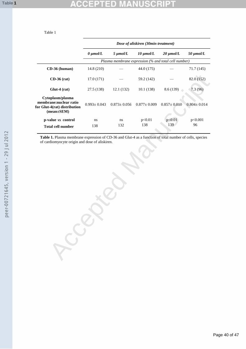
Page 40 of 47
Accep
ted
Man
uscr
ipt
Table 1
Table 1. Plasma membrane expression of CD-36 and Glut-4 as a function of total number of cells, species
of cardiomyocyte origin and dose of aliskiren.
8.6 (139)
82.0 (152)
CD-36 (human) 14.8 (210) — 44.0 (175)
10.1 (138) 7.3 (96)
— 71.7 (145)
—
Cytoplasm/plasma
membrane:nuclear ratio
for Glut-4(rat) distribution
(mean±SEM)
p-value vs control
0.993± 0.043
ns
0.873± 0.056
ns
0.877± 0.009
p<0.01
0.857± 0.010
p<0.01
0.804± 0.014
p<0.001
17.0 (171) —
12.1 (132)
59.2 (142)
Dose of aliskiren (30min treatment)
Glut-4 (rat) 27.5 (138)
CD-36 (rat)
Plasma membrane expression (% and total cell number)
0 µmol/L 5 µmol/L 10 µmol/L 20 µmol/L 50 µmol/L
Total cell number 138 132 138 139 96
Table 1pe
er-0
0721
645,
ver
sion
1 -
29 J
ul 2
012
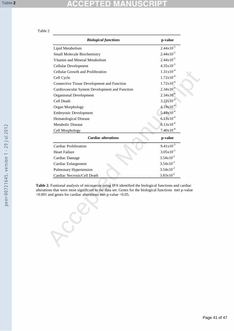
Page 41 of 47
Accep
ted
Man
uscr
ipt
Table 2
Table 2. Funtional analysis of microarray using IPA identified the biological functions and cardiac
alterations that were most significant to the data set. Genes for the biological functions met p-value
<0.001 and genes for cardiac alterations met p-value <0.05.
Lipid Metabolism 2.44x10⁻⁵
Small Molecule Biochemistry 2.44x10⁻⁵
Vitamin and Mineral Metabolism 2.44x10⁻⁵
Cellular Development 4.35x10⁻⁵
Cellular Growth and Proliferation 1.31x10⁻⁴
Cell Cycle 1.72x10⁻⁴
Connective Tissue Development and Function 1.72x10⁻⁴
Cardiovascular System Development and Function 2.34x10⁻⁴
Organismal Development 2.34x10⁻⁴
Cell Death 3.22x10⁻⁴
Organ Morphology 4.19x10⁻⁴
Embryonic Development 5.44x10⁻⁴
Hematological Disease 6.13x10⁻⁴
Metabolic Disease 6.13x10⁻⁴
Cell Morphology 7.40x10⁻⁴
Cardiac Proliferation 9.41x10⁻³
Heart Failure 3.05x10⁻²
Cardiac Damage 3.54x10⁻²
Cardiac Enlargement 3.54x10⁻²
Pulmonary Hypertension 3.54x10⁻²
Cardiac Necrosis/Cell Death 3.83x10⁻²
Biological functions p-value
Cardiac alterations p-value
Table 2pe
er-0
0721
645,
ver
sion
1 -
29 J
ul 2
012
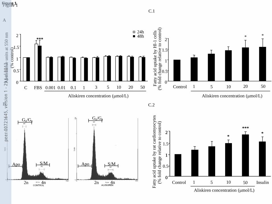
Page 42 of 47
Accep
ted
Man
uscr
ipt
0
0.5
1
1.5
2
Control 5 10 20 50
* *
1
(% f
old
chan
ge
rela
tive
to c
ontr
ol)
A
Figure 1
0
0.5
1
1.5
2
C FBS 0.001 0.01 0.1 1 3
24h 48h
***
Aliskiren concentration (μmol/L)
2n 4n 2n 4n
Apo
G0/G1G0/G1
S/M S/MApo
B
Fat
ty a
cid u
pta
ke
by r
at c
ardio
myo
cyte
s
0
0.5
1
1.5
2
Control 10 Insulin
***
0
1
2
1(% f
old
chan
ge
rela
tive
to c
ontr
ol)
**
5 50
C.1
C.2
Fat
ty a
cid u
pta
ke
by H
l-1 c
ells
5 10 20 50
Aliskiren concentration (μmol/L)
Aliskiren concentration (μmol/L)
Abso
rban
ce u
nit
s at
550 n
m
(% c
ontr
ol)
Figure 1pe
er-0
0721
645,
ver
sion
1 -
29 J
ul 2
012
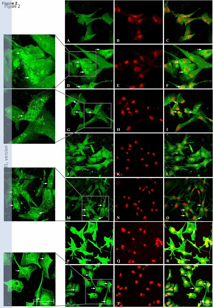
Page 43 of 47
Accep
ted
Man
uscr
ipt
Figure 2
A B C
D E F
G H I
J K L
M N O
P Q R
S T U50μm 50μm
Figure 2pe
er-0
0721
645,
ver
sion
1 -
29 J
ul 2
012
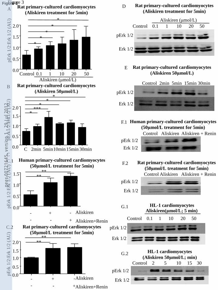
Page 44 of 47
Accep
ted
Man
uscr
ipt
F.1
pErk 1/2
Erk 1/2
E
pErk 1/2
Erk 1/2
Control 2min 5min 15min 30min
Control Aliskiren Aliskiren + Renin
Human primary-cultured cardiomyocytes
(50μmol/L treatment for 5min)
Figure 3
Rat primary-cultured cardiomyocytes
(50μmol/L treatment for 5min)
Control Aliskiren Aliskiren + Renin
pErk 1/2
Erk 1/2
F.2
D
pErk 1/2
Erk 1/2
Control 0.1 1 10 20 50
Rat primary-cultured cardiomyocytes
(Aliskiren treatment for 5min)
Aliskiren (μmol/L)
Aliskiren (μmol/L)
*
*
*
*
*
A Rat primary-cultured cardiomyocytes
(Aliskiren treatment for 5min)
Control 0.1 1 10 20 50 0.0
0.5
1.0
1.5
2.0
pE
rk 1
/2:E
rk 1
/2 (
AU
)
Rat primary-cultured cardiomyocytes
(Aliskiren 50μmol/L)
****
*
**
B Rat primary-cultured cardiomyocytes
(Aliskiren 50μmol/L)
C 2min 5min10min15min 30min0.0
0.5
1.0
1.5
2.0
pE
rk1/2
:Erk
1/2
(A
U)
****
Aliskiren
Aliskiren+Renin
-
- -
-+
+
C
Human primary-cultured cardiomyocytes
(50μmol/L treatment for 5min)
0.0
0.5
1.0
1.5
pE
rk 1
/2:E
rk 1
/2 (
AU
)
G.1
pErk 1/2
Erk 1/2
Control 0.1 1 10 20 50
HL-1 cardiomyocytes Aliskiren(μmol/L; 5 min)
G.2
Control 2 5 10 15 30
pErk 1/2
Erk 1/2
HL-1 cardiomyocytes
(Aliskiren 50μmol/L; min)
****
Aliskiren
Aliskiren+Renin
-
- -
-+
+
C.2 Rat primary-cultured cardiomyocytes
(50μmol/L treatment for 5min)
pE
rk 1
/2:E
rk 1
/2 (
AU
)
C.1
0.0
1.0
1.5
2.0
0.5
Figure 3pe
er-0
0721
645,
ver
sion
1 -
29 J
ul 2
012
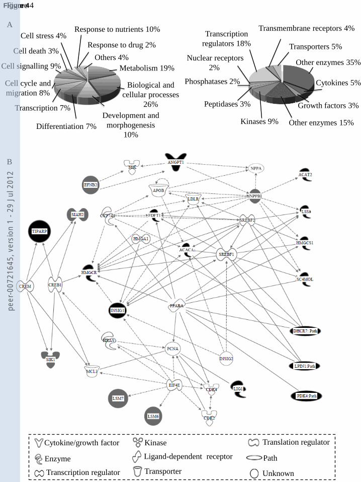
Page 45 of 47
Accep
ted
Man
uscr
ipt
Figure 4
A
Nuclear receptors
2%Metabolism 19%
Biological and
cellular processes
26%
Development and
morphogenesis
10%
Cell cycle and
migration 8%
Differentiation 7%
Transcription 7%
Cell signalling 9%
Cell death 3%
Cell stress 4%
Others 4%
Response to drug 2%
Response to nutrients 10%
Other enzymes 35%
Transporters 5%
Transmembrane receptors 4%Transcription
regulators 18%
Phosphatases 2% Cytokines 5%
Growth factors 3%
Other enzymes 15%Kinases 9%
Peptidases 3%
B
KinaseCytokine/growth factor
Enzyme
Transcription regulator
Ligand-dependent receptor
Transporter
Translation regulator
Path
Unknown
Figure 4pe
er-0
0721
645,
ver
sion
1 -
29 J
ul 2
012
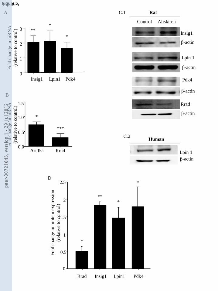
Page 46 of 47
Accep
ted
Man
uscr
ipt
Lpin 1
β-actin
Rrad
β-actin
Insig1
β-actin
Lpin 1
β-actin
Pdk4
β-actin
Human
Control Aliskiren
Rat
Insig1 Lpin1 Pdk4
Fold
chan
ge
in m
RN
A
(rel
ativ
e to
contr
ol) **
*
*
Fold
chan
ge
in m
RN
A
(rel
ativ
e to
contr
ol)
***
A
B
C.1
C.2
Figure 5
0
1
2
3
0.0
0.5
1.0
1.5
D
0
0.5
1
1.5
2
2.5
Rrad Insig1 Lpin1 Pdk4
***
*
*
Fold
chan
ge
in p
rote
in e
xpre
ssio
n
(rel
ativ
e to
contr
ol)
Rrad
*
Arid5a
Figure 5pe
er-0
0721
645,
ver
sion
1 -
29 J
ul 2
012
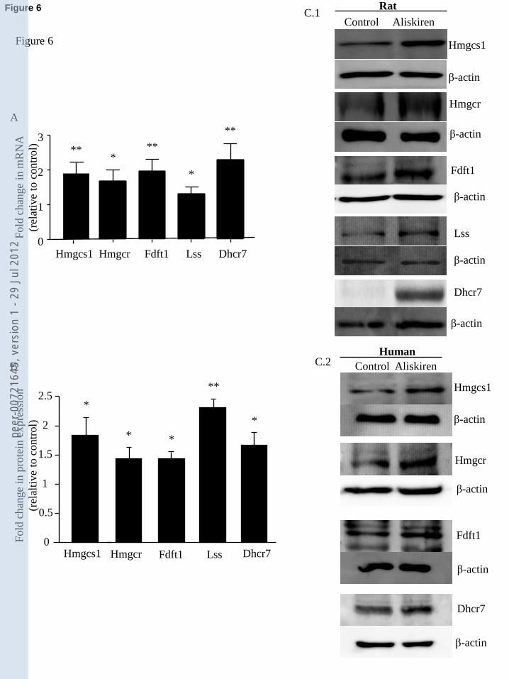
Page 47 of 47
Accep
ted
Man
uscr
iptHmgcr
Dhcr7
β-actin
Hmgcs1
β-actin
Control Aliskiren
Hmgcr
β-actin
Fdft1
Dhcr7
β-actin
Human
Fdft1
β-actin
Control Aliskiren
Rat
β-actin
Hmgcs1
β-actin
Lss
β-actinHmgcs1 Hmgcr Fdft1 Lss Dhcr7
β-actin
Figure 6
C.1
C.2
Fold
chan
ge
in m
RN
A
(rel
ativ
e to
co
ntr
ol) **
**
*
*
A
B
0
1
2
3**
0
0.5
1
1.5
2
2.5
Hmgcr Fdft1 Lss Dhcr7
**
**
Fold
chan
ge
in p
rote
in e
xpre
ssio
n
(rel
alti
ve
to c
ontr
ol) *
*
Hmgcs1
Figure 6pe
er-0
0721
645,
ver
sion
1 -
29 J
ul 2
012
Related Documents