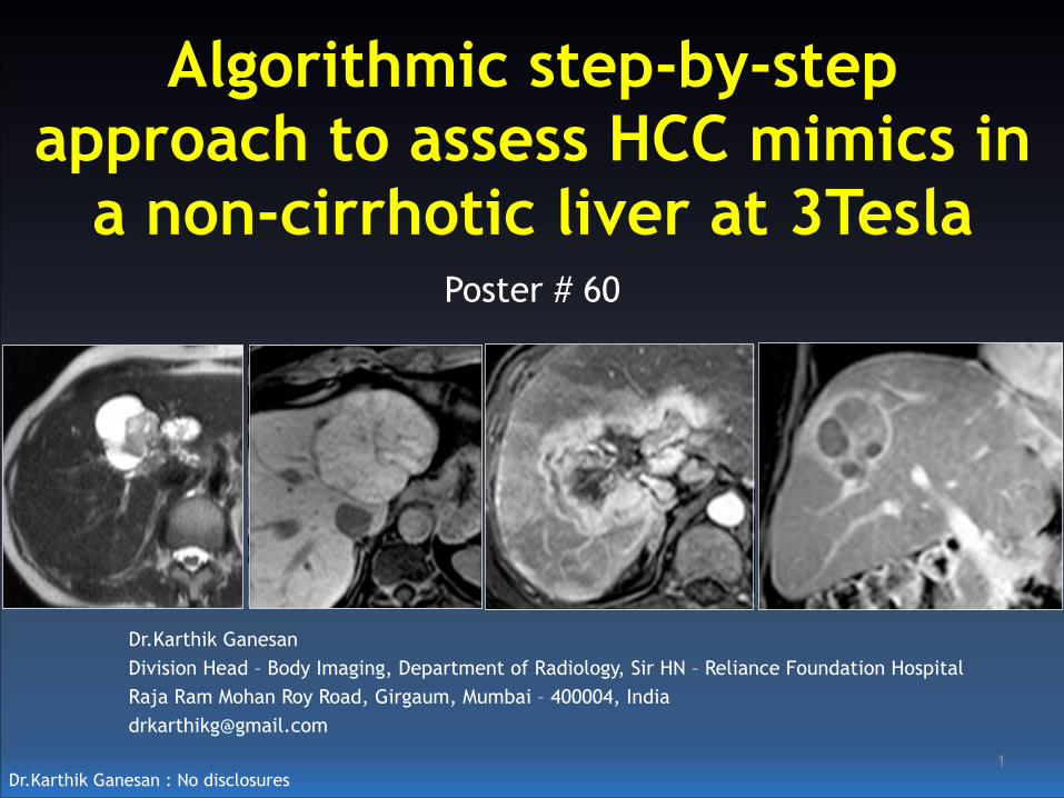
Algorithmic step-by-step approach to assess HCC mimics in a non-cirrhotic liver at 3Tesla Dr.Karthik Ganesan Division Head – Body Imaging, Department of Radiology, Sir HN – Reliance Foundation Hospital Raja Ram Mohan Roy Road, Girgaum, Mumbai – 400004, India [email protected] Poster # 60 Dr.Karthik Ganesan : No disclosures 1

Welcome message from author
This document is posted to help you gain knowledge. Please leave a comment to let me know what you think about it! Share it to your friends and learn new things together.
Transcript
Algorithmic step-by-step approach to assess HCC mimics in
a non-cirrhotic liver at 3Tesla
Dr.Karthik Ganesan Division Head – Body Imaging, Department of Radiology, Sir HN – Reliance Foundation Hospital Raja Ram Mohan Roy Road, Girgaum, Mumbai – 400004, India [email protected]
Poster # 60
Dr.Karthik Ganesan : No disclosures!1

!2
Learning Objectives
• Review key imaging criteria of HCC in a non-cirrhotic liver
• Review the MR imaging protocol for assessment of focal liver lesions at 3T
• Review a diagnostic algorithm to detect HCC mimics in non-cirrhotic liver
• Review potential pitfalls
PitfallsImaging ProtocolHCC - MR Criteria HCC Mimics

!3
HCC - MR CriteriaPitfallsImaging ProtocolHCC - MR Criteria HCC Mimics
T2-wTSE T2-wTSE T1 IP
T1 IP b = 0 b = 500
Pre HAP PVP HVP
3-min 5-min 10-min 60-min
Major Criteria • Arterial Hypervascularity
• Venous Washout
• Delayed enhancing rim
• Follow-up: Interval growth (>50% in ≤6-months)
Minor Criteria • Morphology - Mosaic / nodule-in-nodule
• T2 - Intermediate / Hyperintense
• Fat sparing or Fat containing
• Iron sparing or Hemorrhage
• Restricted diffusion
HCC mimics - MR Plays a Pivotal Role
Detection & Characterization
Road Map for Management
Follow-up of Treated Cases

!4!4
•T2TSE coronal •T2TSE axial •T1-w dual echo •T2TSE obl coronal / sagittal •T2TSE obl axial •DW (b = 0, 500)
Injection Buscopan – 0.5mg (Hyoscine butylbromide) Mechanism: antispasmodic - 5 min before procedure / (Optional)
Pre-contrast Post-contrast Delayed Phase at 60-min
•3D T1-w GRE (HAP – 10-min) •T1-w FS axial – 5min •T1-w FS cor / sag –optional •DW (b = 0, 500)
• 3D T1-w GRE • Useful to see GBCA uptake or lack of it
MRI
CECTPre-contrast Post-contrast
AP / PVPDelayed Post-
contrast
Useful adjunct to characterize
FLLs
MR Advantages • Tissue resolution • Contrast dynamics • Functional assessment (biliary excretion)
If CT is used.. • Temporal resolution • Calcification in lesion
Imaging Protocol @ 3TeslaPitfallsImaging ProtocolHCC - MR Criteria HCC Mimics
!5
PitfallsImaging ProtocolHCC - MR Criteria HCC Mimics
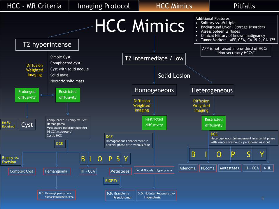
HCC MimicsT2 hyperintense
Simple Cyst
Complicated cyst
Cyst with solid nodule
Solid mass
Necrotic solid mass
DiffusionWeighted imaging
Prolonged diffusivity
Restricted diffusivity
Cyst
DCE
T2 Intermediate / low
Solid Lesion
DiffusionWeighted imaging
Restricted diffusivity
Homogeneous
Focal Nodular Hyperplasia
Heterogeneous
DiffusionWeighted imaging
Restricted diffusivity
Adenoma IH - CCAMetastasesPEcoma NHLComplex Cyst IH - CCA MetastasesHemangioma
DCE Homogeneous Enhancement in arterial phase with venous fade
Additional Features • Solitary vs. Multiple • Background Liver - Storage Disorders • Assess Spleen & Nodes • Clinical History of known malignancy • Tumor Markers - AFP, CEA, CA 19-9, CA-125
D.D: Nodular Regenerative Hyperplasia
D.D: Hemangiopericytoma Hemangioendothelioma
Complicated / Complex Cyst Hemangioma Metastases (neuroendocrine) IH-CCA (secretory) Cystic HCC
B I O P S Y B I O P S YBiopsy vs. Excision
BIOPSY
AFP is not raised in one-third of HCCs “Non-secretory HCCs”
No FU Required
DCE Heterogeneous Enhancement in arterial phase with venous washout / peripheral washout
D.D: Granuloma Pseudotumor
PitfallsImaging ProtocolHCC - MR Criteria HCC Mimics
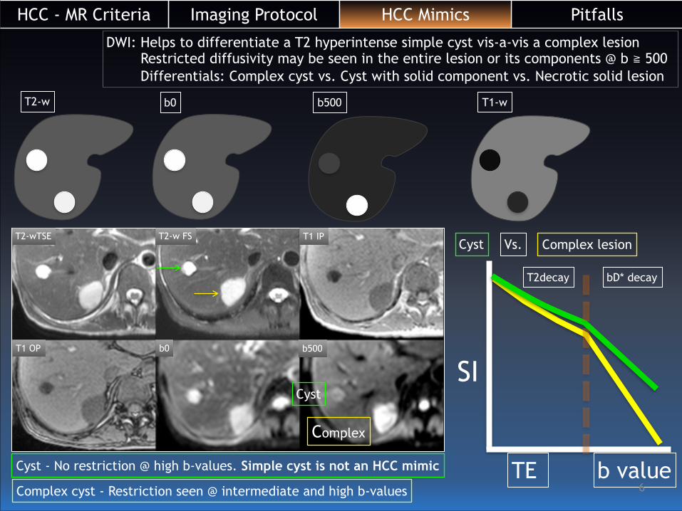
T2-w b0 b500 T1-w
Cyst Vs. Complex lesion
SI
TE b value
T2decay bD* decay
T2-wTSE T2-w FS T1 IP
T1 OP b0 b500
Cyst
Complex
DWI: Helps to differentiate a T2 hyperintense simple cyst vis-a-vis a complex lesion Restricted diffusivity may be seen in the entire lesion or its components @ b ≧ 500 Differentials: Complex cyst vs. Cyst with solid component vs. Necrotic solid lesion
Cyst - No restriction @ high b-values. Simple cyst is not an HCC mimic
Complex cyst - Restriction seen @ intermediate and high b-values !6

PitfallsImaging ProtocolHCC - MR Criteria HCC Mimics
!7
Complex Cystic Lesions
Complicated Cyst Infectious Cyst Cyst with solid nodule/s Necrotic Solid Mass
• Hemorrhage within cyst • Fibrocalcific cyst
• Wall enhancement ± • Debris / Fluid level • Incomplete / complete
septae • Non-enhancing mural
nodule (retracted blood clot)
• Biliopathy ± • Capsular bulge
• Multiplicity / Solitary • Thick walled • Enhancing Wall / septae • Daughter cysts • Intracystic debris • Biliopathy ± • Subcapsular Rupture • THID
• Cholangitic Abscess • Amebic Abscess • Hydatid cyst • Tubercular Abscess • Fungal granuloma
• Solitary • Thick walled • Enhancing Wall / septae • Debris • Mural / Papillary nodule • Venous invasion ± • Biliopathy ± • THID
• Cystadenocarcinoma • Neuroendocrine tumor • Necrotic HCC • Combined / Collision tumor
• Necrotic HCC • Neuroendocrine tumor • Hypervascular metastases
• Solitary / Multiple • Thick walled • Enhancing Wall / septae • Intracystic debris • Mural nodules • Venous invasion ± • Biliopathy ± • Subcapsular Rupture • THID • Extrahepatic disease
E T I O L O G Y
FEAT UR ES
• Asymptomatic: Follow-up • Symptomatic: Aspiration • Cyst Marsupialization or
excision for definitive diagnosis
• Treat Primary cause of obstruction
• PTC / PTBD • Antibiotics • Aspiration
T R E A T M E NT
• CYST Aspiration - poor yield (not preferred)
• BIOPSY of solid component • Excision preferred if
possible
• BIOPSY solid component • Excision preferred if possible • TACE / Portal Vein
Embolization followed by hepatectomy / OLT
PitfallsImaging ProtocolHCC - MR Criteria HCC Mimics
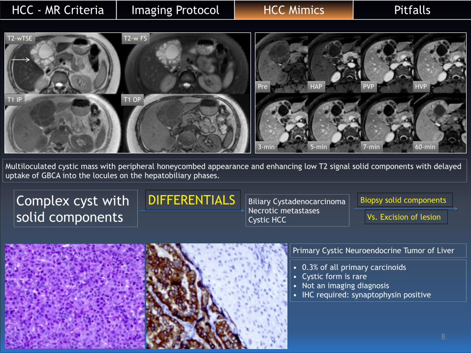
T2-wTSE
T1 IP
T2-w FS
T1 OP
Pre HAP PVP HVP
3-min 5-min 7-min 60-min
Primary Cystic Neuroendocrine Tumor of Liver
• 0.3% of all primary carcinoids • Cystic form is rare • Not an imaging diagnosis • IHC required: synaptophysin positive
Multiloculated cystic mass with peripheral honeycombed appearance and enhancing low T2 signal solid components with delayed uptake of GBCA into the locules on the hepatobiliary phases.
!8
Biliary Cystadenocarcinoma Necrotic metastases Cystic HCC
Complex cyst with solid components
DIFFERENTIALS Biopsy solid components
Vs. Excision of lesion

AA A A A
• Peripheral Discontinuous Puddling • Iso-intense to Aorta • Puddles Coalesce to form Blobs • Near Complete or Complete Fill-in
Pre-contrast Hepatic Arterial Phase Portal Venous Phase Hepatic Venous Phase Late Venous Phases
“Progressive Centripetal Enhancement + Incomplete Fill In”
Cavernous Hemangioma
“NO BIOPSY”
PitfallsImaging ProtocolHCC - MR Criteria HCC Mimics

Focal Nodular Hyperplasia
AA A A A
Pre-contrast Hepatic Arterial Phase Portal Venous Phase Hepatic Venous Phase Late Venous Phase
Lesion - early enhancement with venous fade
Vs.
Scar / Septa - late enhancement
“Contrast FADE”
“NO BIOPSY”
M
M
PitfallsImaging ProtocolHCC - MR Criteria HCC Mimics

FNH without scar ∼ 20% cases
Findings: Solitary, large exophytic intermediate T2 signal intensity mass with restricted diffusivity. Note absence of signal loss on T1-w OP signifying absence of intralesional fat.
T2-wTSE
T2-wTSE FS
T1-w IP T1-w OP
b=0 b=500
M
continued!11
PitfallsImaging ProtocolHCC - MR Criteria HCC Mimics

Homogeneous enhancement of the mass is seen on arterial phase, with progressive fade on venous phases, with delayed uptake of hepatocyte specific contrast agent on the hepatobiliary phase image obtained at 60-minutes. Differentials: Focal Nodular Hyperplasia versus Hepatic Adenoma
Pre HAP PVP HVP
3-min 5-min 10-min 60-minutes
M
!12
PitfallsImaging ProtocolHCC - MR Criteria HCC Mimics
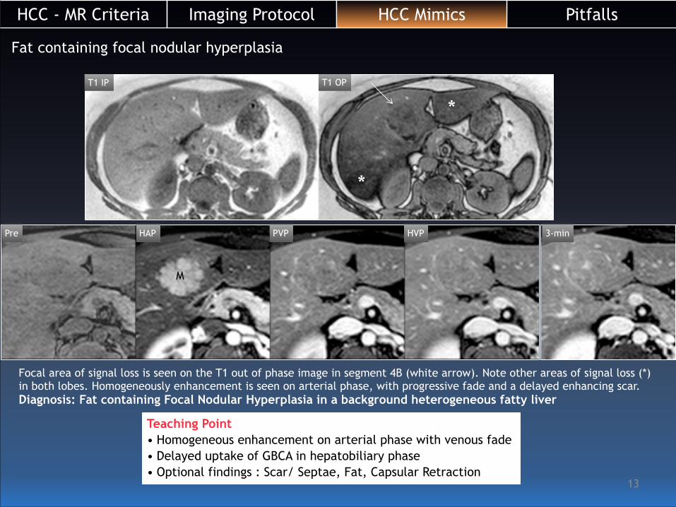
Fat containing focal nodular hyperplasia
Pre HAP PVP HVP 3-min
Focal area of signal loss is seen on the T1 out of phase image in segment 4B (white arrow). Note other areas of signal loss (*) in both lobes. Homogeneously enhancement is seen on arterial phase, with progressive fade and a delayed enhancing scar. Diagnosis: Fat containing Focal Nodular Hyperplasia in a background heterogeneous fatty liver
T1 OPT1 IP
*
*
Teaching Point • Homogeneous enhancement on arterial phase with venous fade • Delayed uptake of GBCA in hepatobiliary phase • Optional findings : Scar/ Septae, Fat, Capsular Retraction
M
!13
PitfallsImaging ProtocolHCC - MR Criteria HCC Mimics

Inflammatory Granuloma - Tuberculosis
Pre HAP PVP HVP
3-min 5-min 10-min 60-minutes
AA A A A
Pre-contrast Hepatic Arterial Phase Portal Venous Phase Hepatic Venous Phase Late Venous Phase
Granuloma • Intermediate T2 signal • Restricted diffusivity • Pattern of Enhancement a. “Ring / Rim like” with progressive central enhancement b. “Homogeneous enhancement with fade” c. SPIO uptake - Kupffer cells d. May or may not retain Gd- BOPTA or EOB-DTPA • PET uptake is variable • Biopsy required for definitive
diagnosis
!14
PitfallsImaging ProtocolHCC - MR Criteria HCC Mimics
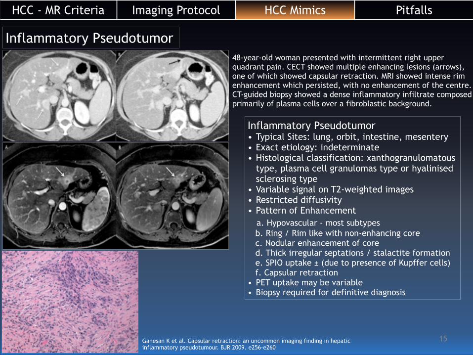
Inflammatory Pseudotumor
!15
48-year-old woman presented with intermittent right upper quadrant pain. CECT showed multiple enhancing lesions (arrows), one of which showed capsular retraction. MRI showed intense rim enhancement which persisted, with no enhancement of the centre. CT-guided biopsy showed a dense inflammatory infiltrate composed primarily of plasma cells over a fibroblastic background.
Inflammatory Pseudotumor • Typical Sites: lung, orbit, intestine, mesentery • Exact etiology: indeterminate • Histological classification: xanthogranulomatous
type, plasma cell granulomas type or hyalinised sclerosing type
• Variable signal on T2-weighted images • Restricted diffusivity • Pattern of Enhancement a. Hypovascular - most subtypes b. Ring / Rim like with non-enhancing core c. Nodular enhancement of core d. Thick irregular septations / stalactite formation e. SPIO uptake ± (due to presence of Kupffer cells) f. Capsular retraction • PET uptake may be variable • Biopsy required for definitive diagnosis
Ganesan K et al. Capsular retraction: an uncommon imaging finding in hepatic inflammatory pseudotumour. BJR 2009. e256-e260
PitfallsImaging ProtocolHCC - MR Criteria HCC Mimics

Hepatic Adenoma
!16
Pre HAP PVP
HVP 5-min 2-hours
SSFSE T1IP T1OP
Pre-contrast Hepatic Arterial Phase Portal Venous Phase Hepatic Venous Phase
A A A
Focal non fat containing hemorrhagic mass in a background fatty liver. The mass demonstrates heterogeneous enhancement on the arterial phase with suspicious washout. Note a central non-enhancing speck and a thin enhancing rim, representing compressed parenchyma / pseudo capsule. Diagnosis: Hepatic Adenoma Differential Diagnosis: AML, PEcoma, HCC
SSFSE FS T1-w OP T1-w IP Delayed T1-wFS Hepatic AML
Image courtesy Dr.Claude Sirlin
A
PitfallsImaging ProtocolHCC - MR Criteria HCC Mimics

48-year old woman presented with low grade fever since 2-months not responding treatment. Elevated ESR; Liver Function Tests: Normal; AFP / CEA / CA 19-9 are negative. Viral markers are negative. Patient is not on OCs. Screening USG (not shown) detected a focal SOL with heterogeneous echogenicity.
T2-wTSE T1-IP T1-OP
Findings: Solitary intermediate T2 signal intensity mass (M) with eccentric areas of low signal. The low T2 signal areas appear hyperintense on T1 in-phase without signal loss on T1 out of phase Background liver is mildly fatty Interpretation: Hemorrhagic focal mass in fatty liver
PEcoma - Perivascular Epitheloid Cell Tumor
M
continued
!17
PitfallsImaging ProtocolHCC - MR Criteria HCC Mimics

Pre HAP PVP
HVP 3-min 5-min
Findings: Hypervascular mass on arterial phase with venous washout and delayed enhancing rim. Differentials: Adenoma HCC PEcoma (mesenchymal tumor)
Tumor stained positive with HMB-45 and Desmin
• Zamboni et al described this entity in 1996. • Recognized in 2002 - 2003 by WHO • H.P shows “Radial arrangement of cells around a vessel” (perivascular epithelioid cell differentiation) • Epithelioid to spindle cells with eosinophilic to clear cytoplasm • Positivity with “Myoid markers” (smooth muscle actin, desmin, calponin, caldesmon) • Positivity with “Melanocytic markers”: HMB-45, Melan-A, tyrosinase and microphthalmia transcription factor • Locations: Uterus is the commonest (46% in uterus & 90% female predisposition) • Benign > > > Malignant • Other entities: AML, clear cell sugar tumor, Lymphangioleiomyomatosis
M
!18
PitfallsImaging ProtocolHCC - MR Criteria HCC Mimics

Intrahepatic (mass forming) Cholangiocarcinoma: IH-CCA
Mass forming Nodular Exophytic
Guglielmi A et al. Mass Forming subtype was associated with negative prognostic factors • Extrahepatic bile duct involvement • Nodal metastases • Macroscopic vascular invasion • Perineural invasion • Higher T stage
!19
T1-w OP T1-w IP T2TSE b=0 b=500
PitfallsImaging ProtocolHCC - MR Criteria HCC Mimics

AA A A A
Pre-contrast Hepatic Arterial Phase Portal Venous Phase Hepatic Venous Phase Late Venous Phase
Pre HAP PVP HVP
3-min 7-min 10-min 60-minutes
“Peripheral Washout Sign”
!20
PitfallsImaging ProtocolHCC - MR Criteria HCC Mimics
!21
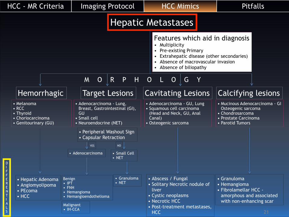
Hepatic Metastases
Features which aid in diagnosis • Multiplicity • Pre-existing Primary • Extrahepatic disease (other secondaries) • Absence of macrovascular invasion • Absence of biliopathy
Hemorrhagic • Melanoma • RCC • Thyroid • Choriocarcinoma • Genitourinary (GU)
D I F F E R E N T I A L S
• Hepatic Adenoma • Angiomyolipoma • PEcoma • HCC
Target Lesions • Adenocarcinoma - Lung,
Breast, Gastrointestinal (GI), GU
• Small cell • Neuroendocrine (NET)
Benign • IPT • FNH • Hemangioma • Hemangioendothelioma
Cavitating Lesions • Adenocarcinoma - GU, Lung • Squamous cell carcinoma
(Head and Neck, GU, Anal Canal)
• Osteogenic sarcoma
• Abscess / Fungal • Solitary Necrotic nodule of
liver • Cystic neoplasms • Necrotic HCC • Post-treatment metastases,
HCC
Calcifying lesions• Mucinous Adenocarcinoma - GI
Osteogenic sarcoma • Chondrosarcoma • Prostate Carcinoma • Parotid Tumors
• Granuloma • Hemangioma • Fibrolamellar HCC -
amorphous and associated with non-enhancing scar
• Peripheral Washout Sign • Capsular Retraction
• Small Cell • NET
• Adenocarcinoma
• Granuloma • NET
YES NO
M O R P H O L O G Y
Malignant • IH-CCA
PitfallsImaging ProtocolHCC - MR Criteria HCC Mimics
Pre
45-min3-minHVP
PVPHAP
!22
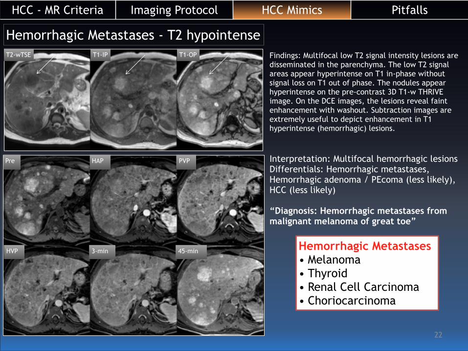
Hemorrhagic Metastases - T2 hypointenseFindings: Multifocal low T2 signal intensity lesions are disseminated in the parenchyma. The low T2 signal areas appear hyperintense on T1 in-phase without signal loss on T1 out of phase. The nodules appear hyperintense on the pre-contrast 3D T1-w THRIVE image. On the DCE images, the lesions reveal faint enhancement with washout. Subtraction images are extremely useful to depict enhancement in T1 hyperintense (hemorrhagic) lesions. !!Interpretation: Multifocal hemorrhagic lesions Differentials: Hemorrhagic metastases, Hemorrhagic adenoma / PEcoma (less likely), HCC (less likely) !“Diagnosis: Hemorrhagic metastases from malignant melanoma of great toe”
T2-wTSE T1-IP T1-OP
Hemorrhagic Metastases • Melanoma • Thyroid • Renal Cell Carcinoma • Choriocarcinoma
PitfallsImaging ProtocolHCC - MR Criteria HCC Mimics
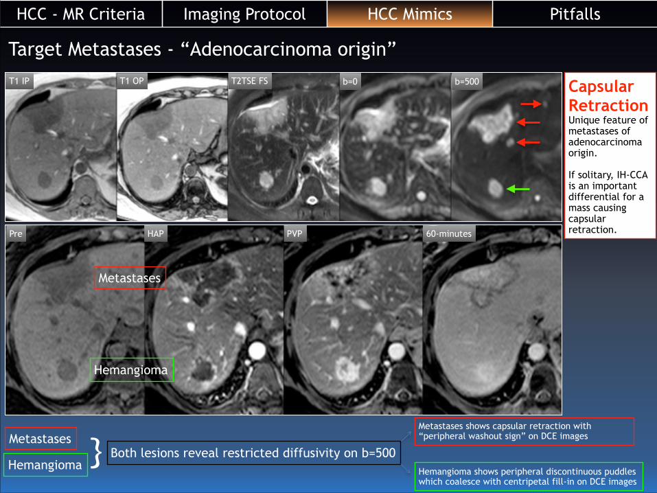
Target Metastases - “Adenocarcinoma origin”
Hemangioma
Metastases
Hemangioma
Metastases } Both lesions reveal restricted diffusivity on b=500
Metastases shows capsular retraction with “peripheral washout sign” on DCE images
Hemangioma shows peripheral discontinuous puddles which coalesce with centripetal fill-in on DCE images
T1 IP T1 OP T2TSE FS b=0 b=500
Pre HAP PVP 60-minutes
Capsular Retraction Unique feature of metastases of adenocarcinoma origin. !If solitary, IH-CCA is an important differential for a mass causing capsular retraction.
PitfallsImaging ProtocolHCC - MR Criteria HCC Mimics
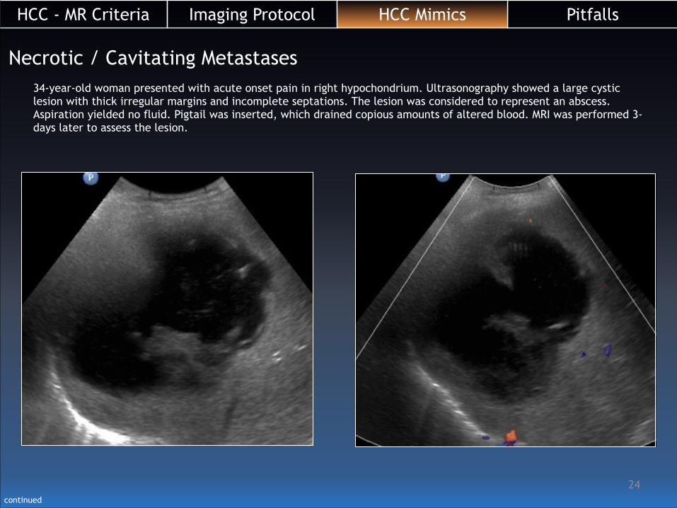
34-year-old woman presented with acute onset pain in right hypochondrium. Ultrasonography showed a large cystic lesion with thick irregular margins and incomplete septations. The lesion was considered to represent an abscess. Aspiration yielded no fluid. Pigtail was inserted, which drained copious amounts of altered blood. MRI was performed 3-days later to assess the lesion.
Necrotic / Cavitating Metastases
continued
!24
PitfallsImaging ProtocolHCC - MR Criteria HCC Mimics

A thick walled hemorrhagic mass (M) is seen in the right lobe with T1 hyperintense contents representing blood degradation products. On DCE images, enhancement of the thick irregular lesion wall is seen.
2nd non-hemorrhagic lesion (M2) in segment 6. The lesion wall shows a continuous rind of thick nodular enhancement, surrounding a hypoenhancing non-hemorrhagic core.
Biopsy showed necrotic metastases from “Endometroid Carcinoma of Uterus”
Teaching Point: Not all necrotic or liquefied masses are abscesses. Further imaging is required to characterize these lesions prior to any planned intervention
Pre HAP PVP HVP
3-min 5-min 10-min 60-minutes
Pre HAP PVP HVP
3-min 5-min 10-min 60-minutes
T2TSE T2TSE T1IP
T1-OP b=0 b=500
M1
M2
!25
PitfallsImaging ProtocolHCC - MR Criteria HCC Mimics
Pre HAP PVP HVP
3-min 5-min 10-min 60-minutes
M
!26
T2-wTSE T2-wTSE FS T1-w IP
T1-w OP b=0 b=500
M
M
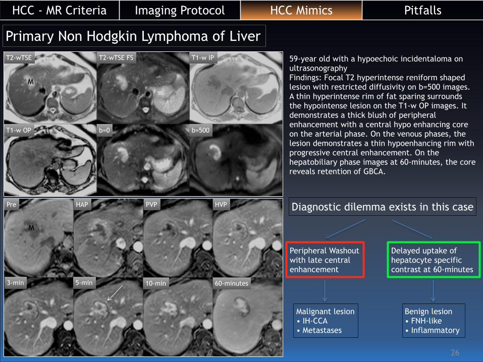
59-year old with a hypoechoic incidentaloma on ultrasonography Findings: Focal T2 hyperintense reniform shaped lesion with restricted diffusivity on b=500 images. A thin hyperintense rim of fat sparing surrounds the hypointense lesion on the T1-w OP images. It demonstrates a thick blush of peripheral enhancement with a central hypo enhancing core on the arterial phase. On the venous phases, the lesion demonstrates a thin hypoenhancing rim with progressive central enhancement. On the hepatobiliary phase images at 60-minutes, the core reveals retention of GBCA.
Primary Non Hodgkin Lymphoma of Liver
Diagnostic dilemma exists in this case
Peripheral Washout with late central enhancement
Delayed uptake of hepatocyte specific contrast at 60-minutes
Malignant lesion • IH-CCA • Metastases
Benign lesion • FNH-like • Inflammatory
PitfallsImaging ProtocolHCC - MR Criteria HCC Mimics

HP 40x Bcl 2 CD 3
CD 20 CK MIB 1
Teaching Point •Retention of GBCA within the lesion on hepatobiliary phase images can mislead the
radiologist. •Infiltrative tumors, such as NHL, may spread over an intact hepatic parenchymal framework.
In such instances, presence of functioning hepatocytes interspersed amongst abnormal tissue may lead to retention of hepatocyte specific contrast agents like Gd-BOPTA or EOB-DTPA.
!27
PitfallsImaging ProtocolHCC - MR Criteria HCC Mimics
PitfallsImaging ProtocolHCC - MR Criteria HCC Mimics
!28
• This imaging algorithm was proposed on the basis of a single centre study over 5-years. A multicenter study is required to refine and standardize this system.
• Substantial overlap in imaging findings exist which may lead to unnecessary and expensive work-up of HCC mimics.
• In the absence of clinical - biologic markers, no definitive and reliable MR imaging criteria exist to differentiate HCC mimics in these settings:
a. Complex cystic lesions vis-a-vis Necrotic HCC b. Hemorrhagic lesions (Adenoma / PEcoma) vis-a-vis Hemorrhagic HCC • Though PET-CT has a proven role in evaluating patients with suspected
metastases, its role in the initial assessment of primary intrahepatic HCC mimics is not been clearly defined.
• Most malignant HCC mimics (excluding metastases) may always require some form of histopathological confirmation, the current gold standard.
Pitfalls

!29!29
Conclusion
• MR imaging is sensitive and accurate enough to detect & diagnose HCC mimics and guide in appropriate management.
• Liver Biopsy - crucial role in Dx of indeterminate FLLs • FNA / biopsy in focal solid liver lesions in NCL – issues exist a. FNA is unreliable and should “ONLY” be done under image guidance
b. Targeted biopsy of solid non-hemorrhagic component is necessary c. If biopsy is performed, likely incidence of tract seeding must be considered ? d. If pre-op work-up fails, intraoperative biopsy with frozen section should be done, just in case additional nodal clearance is required.
• Proposed imaging algorithm may serve as a template for development of a comprehensive imaging system to accurately detect HCC mimics in non-cirrhotic livers

!30!30
References• Lantinga MA, Gevers TJG, Drenth JPH. Evaluation of hepatic cystic lesions. World J Gastroenterol 2013 June 21; 19(23):
3543-3554. • van der Hoeff M, Crook DW, Marincek B, Weishaupt D. Primary neuroendocrine tumors of the liver: MRI features in two cases.
Abdom Imaging 2004;29:77-81 • Shetty P, Baliga S, Balaiah K, Gnana P. Primary hepatic neuroendocrine tumor: an unusual cystic presentation. Indian J Pathol
Microbiol 2010; 53:760-2. • Yu RS, Zhang SZ, Wu JJ, Li RF. Imaging diagnosis of 12 patients with hepatic tuberculosis. World J Gastroenterol. 2004 Jun
1;10(11):1639-1642. • Ganesan K. Viamonte B, Peterson M, Kono Y, Santillan C, Middleton M, Sirlin C. Capsular retraction: an uncommon imaging
finding in hepatic inflammatory pseudotumor. Br J Radiol. Dec 2009;82(984):e256-260. • Yan FH, Zhou KR, Jiang YP, Shi WB. Inflammatory pseudotumor of the liver: 13 cases of MRI findings.World J Gastroenterol.
2001;7(3):422-424. • Katabathina VS, Menias CO, Shanbhogue AK, Jagiradr J, Paspulati RM, Prasad SR. Genetics and Imaging of hepatocellular
adenomas: 2011 update. Radiographics 2011 Oct;31(6):1529-1543 • Hepatic angiomyolipoma: CT and MR imaging findings with clinical-pathologic comparison. Abdom Imaging 2013;Jun 38(3):
482-489. • Tan Y, Ziao EH. Hepatic perivascular epithelioid cell tumor (PEComa): dynamic CT, MRI, ultrasonography, and pathologic
features--analysis of 7 cases and review of the literature. Abdom Imaging 2012 Oct;37(5):781-787. • Lim JH, et al. Cholangiocarcinoma: Morphologic Classification According to Growth Pattern and Imaging Findings. AJR
2003;181:819 -827. • Guglielmi A et al. Intrahepatic cholangiocarcinoma: prognostic factors after surgical resection. World J Surg. 2009 Jun;33(6):
1247-54. • Yoon KH, Yun KJ, Lee JM, Kim CG. Solitary Necrotic nodules of liver mimicking hepatic metastasis: report of Two Cases. Korean J
Radiol. 2000 Jul-Sep;1(3):165-168. • Maher MM, McDermott SR, Fenlon HM, Conroy D, O’Keane JC, Carney DN et al. Imaging of Primary non-Hodgkin’s lymphoma of
the liver. Clin Radiol 2011 Apr;56(4);295-301.
Dr.Karthik Ganesan Division Head – Body Imaging, Department of Radiology, Sir HN – Reliance Foundation Hospital Raja Ram Mohan Roy Road, Girgaum, Mumbai – 400004, India [email protected]
Related Documents