RESEARCH ARTICLE Open Access Alfalfa snakin-1 prevents fungal colonization and probably coevolved with rhizobia Araceli Nora García 1† , Nicolás Daniel Ayub 1† , Ana Romina Fox 1 , María Cristina Gómez 1 , María José Diéguez 1 , Elba María Pagano 1 , Carolina Andrea Berini 2 , Jorge Prometeo Muschietti 3,4 and Gabriela Soto 1* Abstract Background: The production of antimicrobial peptides is a common defense strategy of living cells against a wide range of pathogens. Plant snakin peptides inhibit bacterial and fungal growth at extremely low concentrations. However, little is known of their molecular and ecological characteristics, including origin, evolutionary equivalence, specific functions and activity against beneficial microbes. The aim of this study was to identify and characterize snakin-1 from alfalfa (MsSN1). Results: Phylogenetic analysis showed complete congruence between snakin-1 and plant trees. The antimicrobial activity of MsSN1 against bacterial and fungal pathogens of alfalfa was demonstrated in vitro and in vivo. Transgenic alfalfa overexpressing MsSN1 showed increased antimicrobial activity against virulent fungal strains. However, MsSN1 did not affect nitrogen-fixing bacterial strains only when these had an alfalfa origin. Conclusions: The results reported here suggest that snakin peptides have important and ancestral roles in land plant innate immunity. Our data indicate a coevolutionary process, in which alfalfa exerts a selection pressure for resistance to MsSN1 on rhizobial bacteria. The increased antimicrobial activity against virulent fungal strains without altering the nitrogen-fixing symbiosis observed in MsSN1-overexpressing alfalfa transgenic plants opens the way to the production of effective legume transgenic cultivars for biotic stress resistance. Keywords: Antimicrobial peptides, Alfalfa, Evolution, Land plants, Innate immunity, Snakin Background Alfalfa (Medicago sativa L.), known as the “Queen of Forages”, is a perennial legume. This species is native to Asia, and is considered one of the first known crops with a cultivation history of at least 3500 years. Due to its strong vitality, high nutritional quality, high yields, high adaptability and multiple uses, alfalfa is the main forage crop produced in temperate regions of the planet. Elite alfalfa cultivars must not only have high forage yields but also maintain their productivity and stands over sev- eral years to provide substantial economic benefits. Re- garding this complex topic, improved fungal disease resistance has been specifically identified as the critical trait in alfalfa persistence [1]. In this context, it is pro- posed that the use of snakin-1 peptide (SN1), a powerful but poorly studied antimicrobial compound, to improve alfalfa tolerance to virulent fungal pathogens should be explored. Antimicrobial peptides are present in virtually all or- ganisms and are an ancient and critical component of innate immunity. SN1, the first member of the snakin family to be characterized, was isolated from a crude cell wall preparation of potato (Solanum tuberosum) tubers (StSN1) [2]. This cysteine-rich peptide from potato was found to be active against bacterial and fungal pathogens at extremely low concentrations (EC 50 < 10 μM) [3]. The expression pattern of the StSN1 gene suggests that plant SN1 could be a component of constitutive defense bar- riers, especially those of storage and reproductive plant organs [3]. A second snakin peptide (StSN2), which has a high amino acid identity (30%) to StSN1, was also iso- lated from a crude cell wall preparation of potato tubers. Consistent with this high amino acid identity, StSN2 is also active at very low concentrations against a wide range of pathogens [4]. In contrast to StSN1, the expression * Correspondence: [email protected] † Equal contributors 1 Instituto de Genética Ewald A. Favret (CICVyA-INTA), De los Reseros S/N, Castelar C25 (1712), Buenos Aires, Argentina Full list of author information is available at the end of the article © 2014 García et al.; licensee BioMed Central Ltd. This is an Open Access article distributed under the terms of the Creative Commons Attribution License (http://creativecommons.org/licenses/by/4.0), which permits unrestricted use, distribution, and reproduction in any medium, provided the original work is properly credited. The Creative Commons Public Domain Dedication waiver (http://creativecommons.org/publicdomain/zero/1.0/) applies to the data made available in this article, unless otherwise stated. García et al. BMC Plant Biology 2014, 14:248 http://www.biomedcentral.com/1471-2229/14/248

Welcome message from author
This document is posted to help you gain knowledge. Please leave a comment to let me know what you think about it! Share it to your friends and learn new things together.
Transcript
García et al. BMC Plant Biology 2014, 14:248http://www.biomedcentral.com/1471-2229/14/248
RESEARCH ARTICLE Open Access
Alfalfa snakin-1 prevents fungal colonization andprobably coevolved with rhizobiaAraceli Nora García1†, Nicolás Daniel Ayub1†, Ana Romina Fox1, María Cristina Gómez1, María José Diéguez1,Elba María Pagano1, Carolina Andrea Berini2, Jorge Prometeo Muschietti3,4 and Gabriela Soto1*
Abstract
Background: The production of antimicrobial peptides is a common defense strategy of living cells against a widerange of pathogens. Plant snakin peptides inhibit bacterial and fungal growth at extremely low concentrations.However, little is known of their molecular and ecological characteristics, including origin, evolutionary equivalence,specific functions and activity against beneficial microbes. The aim of this study was to identify and characterizesnakin-1 from alfalfa (MsSN1).
Results: Phylogenetic analysis showed complete congruence between snakin-1 and plant trees. The antimicrobialactivity of MsSN1 against bacterial and fungal pathogens of alfalfa was demonstrated in vitro and in vivo. Transgenicalfalfa overexpressing MsSN1 showed increased antimicrobial activity against virulent fungal strains. However, MsSN1did not affect nitrogen-fixing bacterial strains only when these had an alfalfa origin.
Conclusions: The results reported here suggest that snakin peptides have important and ancestral roles in landplant innate immunity. Our data indicate a coevolutionary process, in which alfalfa exerts a selection pressure forresistance to MsSN1 on rhizobial bacteria. The increased antimicrobial activity against virulent fungal strains withoutaltering the nitrogen-fixing symbiosis observed in MsSN1-overexpressing alfalfa transgenic plants opens the way tothe production of effective legume transgenic cultivars for biotic stress resistance.
Keywords: Antimicrobial peptides, Alfalfa, Evolution, Land plants, Innate immunity, Snakin
BackgroundAlfalfa (Medicago sativa L.), known as the “Queen ofForages”, is a perennial legume. This species is native toAsia, and is considered one of the first known crops witha cultivation history of at least 3500 years. Due to itsstrong vitality, high nutritional quality, high yields, highadaptability and multiple uses, alfalfa is the main foragecrop produced in temperate regions of the planet. Elitealfalfa cultivars must not only have high forage yieldsbut also maintain their productivity and stands over sev-eral years to provide substantial economic benefits. Re-garding this complex topic, improved fungal diseaseresistance has been specifically identified as the criticaltrait in alfalfa persistence [1]. In this context, it is pro-posed that the use of snakin-1 peptide (SN1), a powerful
* Correspondence: [email protected]†Equal contributors1Instituto de Genética Ewald A. Favret (CICVyA-INTA), De los Reseros S/N,Castelar C25 (1712), Buenos Aires, ArgentinaFull list of author information is available at the end of the article
© 2014 García et al.; licensee BioMed Central LCommons Attribution License (http://creativecreproduction in any medium, provided the orDedication waiver (http://creativecommons.orunless otherwise stated.
but poorly studied antimicrobial compound, to improvealfalfa tolerance to virulent fungal pathogens should beexplored.Antimicrobial peptides are present in virtually all or-
ganisms and are an ancient and critical component ofinnate immunity. SN1, the first member of the snakinfamily to be characterized, was isolated from a crude cellwall preparation of potato (Solanum tuberosum) tubers(StSN1) [2]. This cysteine-rich peptide from potato wasfound to be active against bacterial and fungal pathogensat extremely low concentrations (EC50 < 10 μM) [3]. Theexpression pattern of the StSN1 gene suggests that plantSN1 could be a component of constitutive defense bar-riers, especially those of storage and reproductive plantorgans [3]. A second snakin peptide (StSN2), which hasa high amino acid identity (30%) to StSN1, was also iso-lated from a crude cell wall preparation of potato tubers.Consistent with this high amino acid identity, StSN2 isalso active at very low concentrations against a widerange of pathogens [4]. In contrast to StSN1, the expression
td. This is an Open Access article distributed under the terms of the Creativeommons.org/licenses/by/4.0), which permits unrestricted use, distribution, andiginal work is properly credited. The Creative Commons Public Domaing/publicdomain/zero/1.0/) applies to the data made available in this article,
García et al. BMC Plant Biology 2014, 14:248 Page 2 of 13http://www.biomedcentral.com/1471-2229/14/248
of StSN2 is locally induced by wounding and pathogen in-fection, suggesting a critical role of snakin-2 in both con-stitutive and inducible defense barriers of plants. Thesestrong antimicrobial activities of snakin peptides havebeen verified using bacterial and eukaryotic heterologousexpression systems [5-7]. Based on the presence of theGibberellic Acid Stimulated Arabidopsis (GASA) domainand the absence of bioinformatic (e.g. RGD residues) andfunctional (e.g. toxic activity) data supporting its relation-ship with cysteine-rich peptides from snakes venoms,StSN1 and StSN2 have been recently renamed as GSL1and GSL2, respectively [8,9].Overexpression of SN1 in potato and wheat (Triticum
aestivum), SN2 in potato and tomato (Solanum lycoper-sicum), and snakin-defensin hybrid protein in tobacco(Nicotiana tabacum) and potato restricts pathogen inva-siveness and enhances tolerance to bacterial and fungaldiseases, without altering the agronomic phenotype ofthese crops [6,9-12]. Furthermore, disease sensitivity isenhanced by silencing SN2 in wild tobacco (Nicotianabenthamiana), supporting the central role of snakin pep-tides in plant defense [13]. In addition, it has been re-cently shown that StSN1 is located in the plant cell wall[14], confirming that snakin peptides are components ofthe physical barrier and the first line of defense used byplant cells to prevent bacterial and fungal entry [3]. Fur-thermore, in concordance with the functional classifica-tion of snakin peptides as members of the GASA proteinfamily [4,15,16], StSN1 silencing affects cell division, pri-mary metabolism, and cell wall composition [14]. Snakinpeptides could have additional functions in plant growthand development beyond their demonstrated function inbiotic stress response. In spite of their hypothetical func-tional similarity, there is little to no phylogenetic reportson the relationship between StSN1 and GASA-relatedproteins.The aim of this study was to identify and characterize
snakin-1 (MsSN1) gene of alfalfa. The phylogenetic andfunctional analyses showed here, propose that MsSN1 isan ancestral plant gene involved in biotic stress resist-ance, suggesting a coevolutionary process, in which al-falfa exerts a selection pressure for resistance to MsSN1on rhizobial bacteria.
MethodsBacterial and fungal strainsThe bacterial strains (all Gram-negative bacteria) used inthis study were: Pseudomonas fluorescens Pf-5 [17], Sinor-hizobium meliloti BL225C [18], Sinorhizobium melilotiSM11 [19], Sinorhizobium medicae WSM419 [20], Sinor-hizobium fredii USDA 257 [21], Rhizobium sp. Or 191[22], Rhizobium etli CFN 42 [23], Mesorhizobium lotiMAFF303099 [24], Bradyrhizobium japonicum USDA110[25] and Agrobacterium tumefaciens LBA4404 [26]. The
fungal strains used in this work were: Phoma medicagi-nis strain CT1 and Colletotrichum trifolii strain CT2,isolated from INTA alfalfa cultivars and kindly providedby Dr. Ricardo Comerio (Instituto de Microbiología yZoología Agrícola, Instituto Nacional de TecnologiaAgropecuaria, Argentina).
Fungus materialTwo fungal pathogens, Phoma medicaginis strain CT1and Colletotrichum trifolii strain CT2, were grown onPDA (Cat. # B0216605, Britania) plates at room tem-perature for approximately 7 days before the start of thebioassay. For spore collection, the plates were flooded withsterile distilled water and scraped with a wire loop. Sporeconcentration was adjusted to 1 × 10⁶ spores/ml for C. tri-folii CT2 and to 1 × 10⁵ spores/ml for P. medicaginis CT1with sterile distilled water. The fungal strains were main-tained through sequential passages in plants.
Plant materialThe Medicago sativa plants used were the regenerativeclone C2-3, kindly provided by Drs. B. McKersie and S.Bowley (Plant Biotechnology Division, Department ofPlant Agriculture, University of Guelph, Canada), andthe regenerative clone 432-19-17, previously isolated inour laboratory.
Bacterial and plant RNA Isolation and cDNA synthesisTotal bacterial (E. coli) and plant tissues (roots, steam,leaflets) RNA was extracted by using an RNeasy MiniKit (Cat. # 74106, Qiagen) following the manufactures’instructions. Samples of 2 μg total RNA isolated frombacterial cells or plant tissues were reverse-transcribedin a 25 μl reaction using MMLV-RT (Cat. # M1701, Pro-mega). For PCR amplification in bacteria and plants,1 μl of RT reaction was used. The PCR reactions werecarried out in 25 μl with 0.5 μM of each primer [27],using Taq polymerase (Cat#. 11615010, Invitrogen) fol-lowing the manufactures’ instructions.
MsSN1 plasmid constructionThe MsSN1 cDNA (GenBank accession number JQ809686)was isolated using primers p1 FW and p2 RV (Additionalfile 1) designed against the 5′ and 3′ untranslated regions(UTR) of the putative SN1 gene (GenBank accession num-ber XM_003589066, MTR_1g018640) from Medicagotruncatula. Full-length cDNA was amplified by PCR andthis fragment was cloned into a pCR2.1TOPO vector (Cat.# K4500-01, Invitrogen). The pTOPO-MsSN1 plasmidwas digested with Not I (Cat. # R6431, Promega), treatedwith Klenow, and religated to destroy the polylinker Not Isite. The resulting plasmid was named pTOPO-NotI-MsSN1. The sequencing reactions of pTOPO-NotI-MsSN1were performed at INTA-Argentina (www.inta.gov.ar).
García et al. BMC Plant Biology 2014, 14:248 Page 3 of 13http://www.biomedcentral.com/1471-2229/14/248
The cDNA sequence was named MsSN1 (Medicago sativasnakin-1). To produce recombinant bacteria expressingMsSN1, plasmid pSJ33-MsSN1 carrying MsSN1 was con-structed by subcloning the 0.5-kb EcoRI (Cat. # R6011,Promega) fragment from pCR2.1TOPO-MsSN1 into pSJ33[28], and afterward introduced in Escherichia coli forheterologous expression of MsSN1. To produce transgenicalfalfa lines overexpressing MsSN1, pCR2.1TOPO-NotI-MsSN1 was digested with KpnI (Cat. # R6341, Promega)and XbaI (Cat. # R6181, Promega), and the MsSN1 restric-tion fragment was cloned into pKANNIBAL vector (Gen-Bank accession number AJ311873). The resulting plasmidwas digested with NotI, and the 35SMsSN1 restrictionfragment was cloned into pART27 binary vector [29]. Theresulting recombinant binary vector containing the MsSN1cDNA with its signal peptide under the CaMV 35S pro-moter was named pART-35S::MsSN1.
Bioinformatic analysis of MsSN1MsSN1 sequence of Medicago sativa (AFE82743), a pep-tide composed of 91 amino acids, was used as query tosearch against all available complete eukaryotic genomedatabases in NCBI with protein annotation in GenBank.The cut-off to obtain candidate orthologs was 20% ofamino acid identity (Additional file 2). Sequences weresearched by using BLASTP tools in NCBI and PLAZAdatabases (http://www.ncbi.nlm.nih.gov/blast; http://bioinformatics.psb.ugent.be/plaza). Protein identity calcula-tions were performed using MatGAT v2.02 [30]. Evolu-tionary analysis was conducted by using MEGA version5.0 [31]. Protein sequences were aligned using the ClustalWprogram. Phylogenetic trees were constructed using theneighbor-joining method with genetic distances computedusing the pairwise deletion model and bootstrap analysisof 500 values and root on midpoint (i.e. midpoint of thelongest pathway between two clusters of sequences). Insilico analysis of conserved motifs in putative snakin/GASA proteins, Pfam domains and signal peptides werepredicted by using Pfam and Signal-3 L with default pa-rameters, respectively [32,33].
DNA extraction and sequence analysis of fungal strainsFungal DNA was extracted as previously described byMoller [34] with modifications suggested by Dr. E.W.Boehm (http://www.eboehm.com/). Briefly, 100 mg offungal mycelia was scraped from 10-day-old PDA cul-tures, ground in a 1.5 ml tube with micropestle by add-ing 500 μl of Lysis Buffer (100 mM Tris pH 8, 10 mMEDTA, 2% SDS, 1% β-Mercaptoethanol, 100 μg/ml pro-teinase K). Lysate was incubated at 60°C for 60 min.Subsequently, 5 M NaCl was added to a final concentra-tion of 1.4 M and mixed before adding 0.1 vol of CTAB10% (w/v). The mixture was incubated at 65°C for 10 min.DNA was extracted by adding an equal volume of
chloroform:isoamyl alcohol (24:1), incubating for 30 minat 0°C and centrifuging at 14,000 × g for 10 min at 4°C.The aqueous phase was mixed with 0.5 vol of 5 M ammo-nium acetate, incubated at 0°C for 60 min and centrifugedat 14,000 × g for 1 min. DNA was precipitated from thesupernatant by adding 0.55 vol of isopropanol, centrifugedat 14,000 × g for 10 min and washed twice with 70% etha-nol. DNA pellet was air-dried and resuspended in 50 μl ofTE (10 mM Tris pH 8.0; 1 mM EDTA pH 8.0). The in-ternal transcribed spacer regions 1 & 2 and 5.8S nrDNA(ITS) and partial sequences of TUB genes were amplifiedand sequenced by Macrogen Inc. (Korea) service (http://www.macrogen.com/) using the primer pairs ITS-1FWand ITS-4RV and Btub2FW and Btub4RV, respectively(Additional file 1). The nucleotide sequences obtainedhere were deposited in the EMBL Nucleotide SequenceDatabase, accession numbers: KF846005 for C. trifoliiTUB, KF846006 for P. medicaginis TUB, KF846009 for C.trifolii ITS and KF846010 for P. medicaginis ITS.
In vitro antimicrobial activity assaysEscherichia coli recombinant strains containing the pSJ33empty vector or pSJ33-MsSN1 were grown overnight at30°C with shaking (250 rpm) in LB medium supplementedwith 1 mM isopropyl-β-D-thiogalactopyranoside in 250-ml Erlenmeyer flasks containing 50 ml of medium. Sam-ples of 25 ml of culture broth (O.D: 0.8) were centrifugedat 5,000 × g at 4°C for 10 min. The pellets were washedthree times with physiological solution (0.9% NaCl). Crudecell lysates were achieved by four consecutive cycles offreezing in liquid nitrogen followed by thawing at 37°C.After centrifugation at 14,000 × g at 4°C for 20 min, thesupernatant was resuspended in 400 μl of physiological so-lution. For the analysis of MsSN1 expression in E. coli,total bacterial RNA was extracted by using the RNeasyMini kit and treated with DNaseI (Cat. # M6101, Pro-mega). cDNA was obtained using random hexamers (Cat.# B070-40, Promega) and AMV Reverse Transcriptase(Cat. # M9004, Promega). For PCR amplification, 1 μl ofRT reaction was used. The PCR reactions were carried outin 25 μl with 0.5 μM of each primer according to Setten[27]. PCR conditions comprised: 1 cycle at 94°C for 3 min,34 cycles of 94°C for 45 s, 56°C for 1 min and 72°C for1 min. The expression of MsSN1 was analyzed usingprimers p3 FW and p4 RV which amplify the completeopen reading frame (Additional file 1).The disk inhibition assays were evaluated as described
by Ayub [35], with very slight modifications. Cultureswere performed in 125-ml Erlenmeyer flasks containing25 ml of TY medium [36] or LB medium for rhizobia orPseudomonas and Agrobacterium, respectively. Bacteriawere incubated overnight at 28°C with shaking (250 rpm).Sterile Whatman No. 1 filter disks (5 mm) impregnatedwith 5 μl of MsSN1-free (E. coli pSJ33) or MsSN1 (E. coli
García et al. BMC Plant Biology 2014, 14:248 Page 4 of 13http://www.biomedcentral.com/1471-2229/14/248
pSJ33-MsSN1) extracts were placed on top of bacteria-seeded plates. Zones of inhibition were measured after in-cubation at 28°C for 24 h. The antifungal activities of theMsSN1 extract were determined according to Kovalskaya[12] by counting germinating and non-germinating fungalspores. The fungal spores of Phoma medicaginis var. med-icaginis CBS 316.90 from CBS-KNAW Fungal BiodiversityCenter (www.cbs.knaw.nl/Collections) were prepared inPDB. For the inhibition assays, spore suspensions of 1×10⁵
spores/ml were used. Each antifungal assay was performedin triplicate.
Plant transformationThe recombinant binary vector pART-35S::MsSN1 wasintroduced into Agrobacterium tumefaciens LBA 4404by electroporation, by using the procedure described byShen & Forde [37]. Petioles of alfalfa clone C2-3 weretransformed with pART-35S::MsSN1 via A. tumefaciensand cultured in vitro as described by D’Halluin [38], withslight modifications (Additional file 3). Petiole tissueswere decontaminated by immersing in 70% ethanol for1 min and then in 2% sodium hypochlorite for 20 min.The petioles were washed 3 times in sterile distilledwater. Explants previously injured with a scalpel were inoc-ulated with a bacterial culture for 2 minutes (OD600 mm =0.5-0.8), and then dried in Whatman filter paper andtransferred onto a solid co-cultivation SHK medium [3%sucrose, 0.435% KSO4, 2 mg/l 2.4-D, 0.2 mg/l kinetine,6.5 g/l agar, 20% SHK stock solution (w/v) pH:5.8(300 mg/l NH4H2PO4, 2.5% KNO3, 200 mg/l CaCl2.2H2O,400 mg/l MgSO4.7H2O, 4.3% K2SO4, 1 mg/l KI, 5 mg/lH3BO3, 10 mg/l MnSO4.H2O, 1 mg/l ZnSO4.H2O, 1 mg/lNa2MoO4.2H2O, 1 mg/l CuSO4.5H2O, 0.1 mg/l CoCl2.6H2O, 26.29 mg/l NaFeEDTA.H2O, 288 mg/l proline,53 mg/l thioproline, 200 mg/l myo-inositol, 5 mg/l nico-tinic acid, 0.5 mg/l pyridoxine, 5 mg/l thiamine)] contain-ing 100 μM acetosyringone for 2 days at 25°C in the dark.The explants were then washed with 0.5 g/l cefotaximesupplemented with distilled water and transferred to selec-tion/induction medium SHK containing 25 mg/l kanamy-cin and 400 mg/l cefotaxime with routine transfers tofresh medium every 2 weeks, at 25°C and 16 h light (100μmoles m−2 s−1). Somatic embryos were obtained threemonths later and then transferred to MS rooting medium,composed of Murashige and Skoog Basal Medium (Cat. #M5519, Sigma) diluted 1:2 with water. Transgenic plantswere obtained about 6 months after callus induction. Theregenerated plantlets were transferred to the greenhouseonce they were well rooted. All plants were grown in agreenhouse at 25-20°C day/night temperature and watereddaily. Alfalfa transgenic events (named S1, S2 and S3) werepropagated by crossing with clone 432-19-17 and by cut-tings to increase the number of plants available for bio-chemical, physiological, and genetic analysis.
Identification of transgenic plantsDNA was isolated from leaf tissue with the DNeasy plantmini kit (Cat. # 69104, Qiagen). Transgenic plants werefirst identified by PCR with primers p5 FW and p6 RV(Additional file 1), designed against the 35S promoterand SN1 regions of recombinant binary vector. The ex-pression of the transgene was corroborated by RT-PCR,using primers p7 FW and p4 RV, designed against thepCR2-1-TOPO-MCS-derived 5′ UTR region and the 5′UTR of the MsSN1 gene (Additional file 1). All PCRreactions were performed with Taq (Cat. # 11615010,Invitrogen).For Southern hybridization analysis, genomic DNA was
isolated from leaf tissues of greenhouse-grown plantsusing the DNeasy Plant Maxi kit (Cat. # 68161, Qiagen)following the manufacture’s indications. DNA wasdigested with the KpnI restriction enzyme, which cleavesthe construct only once. Then, 20 μg of DNA from eachsample was digested overnight and blotted after separationon 1% (w/v) agarose gel 1× TAE. The DNA fragments ingels were transferred to a positively charged Nylon mem-brane (Cat. # 11209272001, Roche). Nylon membraneswere crosslinked and then used for hybridization with aDIG-labeled probe. Prehybridization and hybridizationwas carried out according to the manufacturer’s instruc-tions. The hybridization probe MsSN1digoxigenin-labeledDNA was generated by PCR by using the PCR DIG probesynthesis kit (Cat. # 11573152910, Roche), using primersp8 FW and p9 RV and then used as a probe (Additionalfile 1). PCR amplification was performed under standardconditions (25 μl volume using 0.8 μM of each primer, 1XPCR buffer, 0.2 mM each dNTP, 2 mM MgCl2 and 20 ngof template) with a program of 34 cycles of 94°C for1 min, 50°C for 30 s and 72°C for 2.5 min and a final cycleof 72°C for 10 min.
Real-time quantitative RT-PCR (RT-qPCR)For RT-qPCR, PCR amplification was performed with 5 μlof RT (1:5 diluted) per reaction, by using 1 U iQ SYBRgreen Supermix (Cat. # 170–8880, Bio-Rad) and 0.2 mMprimers, with the iCycler iQ system. Primers for the real-time qPCR were p10 FW and p11 RV (Additional file 1).qPCR conditions comprised: 1 cycle at 94°C for 5 min,34 cycles of 94°C for 45 s, 59.1°C for 30 s, and 72°C for30 s. At each cycle, accumulation of PCR products was de-tected. The amplification fragment was sequenced andfound to be identical (100% nucleotide identity) to theMsSN1 gene. The expression level of MsSN1 was normal-ized using aspartate aminotransferase (ATT) (AAB46610)as a housekeeping gene, using primers p12 FW and p13RV (Additional file 1). The efficiency of primer bindingwas determined by linear regression by plotting the cyclethreshold value versus the log of the cDNA dilution [39].RT-qPCR experiments were performed two or three times
García et al. BMC Plant Biology 2014, 14:248 Page 5 of 13http://www.biomedcentral.com/1471-2229/14/248
with independent RNA samples (biological replicates). Foreach biological replicate the qPCR reactions were carriedout in duplicate.
Bioassays. In vitro challengesAssays were performed in healthy 15 day-old non-fumigated leaflets. Leaflets were decontaminated wash-ing them in flasks with sterile water with Tween-20(0.01%) for 10 min. After three washes with sterile water,the leaflets were transferred to a 5 % hypochlorite solu-tion for 5 min. Finally, leaflets were washed three timeswith sterile water and transferred to agar-water petridishes. Plates were maintained in growth chambers pro-grammed for 16 h light at 23°C and 8 h dark at 20°C.Leaflets were inoculated by putting 5 μl of P. medicaginisCT1 or C. trifolii CT2 spore solution per leaflet. Infectionevolution was observed and documented by photos andthe following damage score was generated to quantify in-juries: 1. Healthy leaflet, 2. Countable injuries, 3. Uncount-able injuries, 4. Chlorosis, 5. Completely damaged.
Bioassays. In vivo challengesAlfalfa transgenic seeds were obtained by crossing trans-genic plants S1, S2 and S3 with 432-19-17 genotypeplants. Transgenic and wild type seeds were treated withsulfuric acid for 10 minutes, washed three times withsterile water and placed in petri dishes with 1% agarwater at 16 h of light (100 μmoles/m2s) and 25°C. Germi-nated plantlets were transferred to MS 0.5X flasks and in-cubated at 25°C with 16 h photoperiod for 1 month, afterwhich plants were transferred to 1:1 vermiculite:perliteand maintained in magenta vessels (SIGMA) to conservethe humidity. Two-month-old plants were inoculated withP. medicaginis CT1 by spraying spore suspension to allaerial tissues. Percentage of diseased leaflet was analyzed30-day post-inoculation and the number of plants with re-growth and the percentage of highly defoliated plants wascounted 60-days post-inoculation. As an additional sever-ity parameter of plant disease, the percentage of vigor af-fected plants (i.e. with visual signs of turgidity losscompared with non-inoculated plants) was estimated. Allexperiments were carried out with at least 10 independentplants per treatment and with at least 5 non inoculatedwild type plants used as control.
Evaluation of bacterial root colonizationFor the symbiosis assay, wild type and transgenic alfalfaseedlings were grown in 100% vermiculite and daily irri-gated with the minimal medium called “INTA13 withoutnitrogen” [27]. 10-days-old plants were inoculated withan early stationary-phase culture of rhizobial suspension(Sinorhizobium meliloti BL225C). After a month, theplants were harvested and the number of pink nodules wasanalyzed. Non-inoculated plants were used as controls.
Three replicates were analyzed for each treatment. The P.fluorescens Pf-5 colonization assay was evaluated accord-ing to Sanchez [40], with two slight modifications: alfalfawas grown in INTA13 supplemented with Ca (NO3)2 [27]and 0.5 NE2 medium plus sodium octanoate (0.25% w/v)was used to estimate the colony forming units [28].
Statistical analysisBiological measurements were repeated at least threetimes with at least 10 different plants. Significant differ-ences between treatments were calculated using Student’st-test. qPCR experiments were independently performedthree times, with comparable results. The three PCR reac-tions were carried out in duplicate. Significant differenceswere calculated using ANOVA followed by Tukey test.
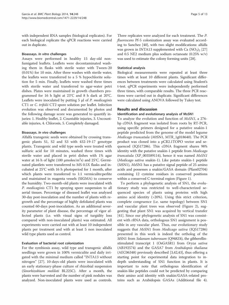
Results and discussionIdentification and evolutionary analysis of MsSN1To analyze the evolution and function of MsSN1, a 276-bp cDNA fragment was isolated from roots by RT-PCR,using specific primers designed for a putative snakin-1peptide predicted from the genome of the model legumeMedicago truncatula (MtSN1, MTR_1g018640). The PCRproduct was cloned into a pCR2.1TOPO vector and se-quenced (JQ517286). This cDNA fragment shares 98%identity with the putative snakin-1 peptide from Medicagotruncatula (XP_003589114), hence it was named MsSN1(Medicago sativa snakin-1). Like potato snakin-1 peptide(StSN1), MsSN1 has a putative signal peptide of 25 aminoacids and possesses a snakin/GASA domain (Pfam02704)containing 12 cysteine residues in conserved positionswithin a conserved C-terminal region (Figure 1).To perform a phylogenetic analysis of SN1, the evolu-
tionary study was restricted to well-characterized se-quenced species of plants using proteins with highamino acid identity (>20%). Using this strict criterion,complete congruence (i.e. same topology) between SN1and vascular plant trees was observed (Figure 2), sug-gesting that plant SN1 was acquired by vertical transfer[41]. Since our phylogenetic analysis of SN1 was consist-ent with rRNA data, orthologous SN1 assignment is pos-sible in any vascular plant. Thus, our evolutionary studysuggests that MsSN1 from Medicago sativa (JQ517286)presented in this work is indeed the ortholog of theStSN1 from Solanum tuberosum (Q948Z4), the gibberellin-stimulated transcript 1 (OsGASR1) from Oryza sativa(AB192574) and the GASA7 from Arabidopsis thaliana(AEC06348) previously described [3,42,43], thus offering astarting point for experimental data integration to in-depth understanding of SN1 function in plants. It isimportant to note that orthologous identification ofsnakin-like peptides could not be predicted by comparingtheir amino acid identity with snakin/GASA-related pro-teins such as Arabidopsis GASAs (Additional file 4).
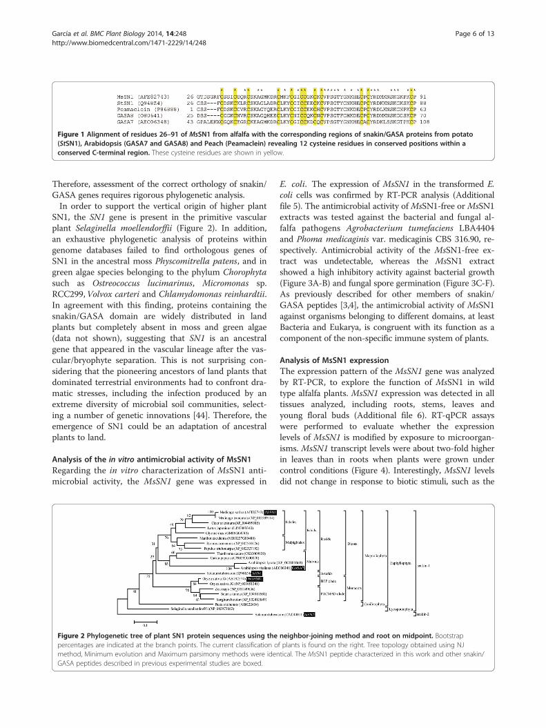
Figure 1 Alignment of residues 26–91 of MsSN1 from alfalfa with the corresponding regions of snakin/GASA proteins from potato(StSN1), Arabidopsis (GASA7 and GASA8) and Peach (Peamaclein) revealing 12 cysteine residues in conserved positions within aconserved C-terminal region. These cysteine residues are shown in yellow.
García et al. BMC Plant Biology 2014, 14:248 Page 6 of 13http://www.biomedcentral.com/1471-2229/14/248
Therefore, assessment of the correct orthology of snakin/GASA genes requires rigorous phylogenetic analysis.In order to support the vertical origin of higher plant
SN1, the SN1 gene is present in the primitive vascularplant Selaginella moellendorffii (Figure 2). In addition,an exhaustive phylogenetic analysis of proteins withingenome databases failed to find orthologous genes ofSN1 in the ancestral moss Physcomitrella patens, and ingreen algae species belonging to the phylum Chorophytasuch as Ostreococcus lucimarinus, Micromonas sp.RCC299,Volvox carteri and Chlamydomonas reinhardtii.In agreement with this finding, proteins containing thesnakin/GASA domain are widely distributed in landplants but completely absent in moss and green algae(data not shown), suggesting that SN1 is an ancestralgene that appeared in the vascular lineage after the vas-cular/bryophyte separation. This is not surprising con-sidering that the pioneering ancestors of land plants thatdominated terrestrial environments had to confront dra-matic stresses, including the infection produced by anextreme diversity of microbial soil communities, select-ing a number of genetic innovations [44]. Therefore, theemergence of SN1 could be an adaptation of ancestralplants to land.
Analysis of the in vitro antimicrobial activity of MsSN1Regarding the in vitro characterization of MsSN1 anti-microbial activity, the MsSN1 gene was expressed in
Figure 2 Phylogenetic tree of plant SN1 protein sequences using thepercentages are indicated at the branch points. The current classification omethod, Minimum evolution and Maximum parsimony methods were idenGASA peptides described in previous experimental studies are boxed.
E. coli. The expression of MsSN1 in the transformed E.coli cells was confirmed by RT-PCR analysis (Additionalfile 5). The antimicrobial activity of MsSN1-free or MsSN1extracts was tested against the bacterial and fungal al-falfa pathogens Agrobacterium tumefaciens LBA4404and Phoma medicaginis var. medicaginis CBS 316.90, re-spectively. Antimicrobial activity of the MsSN1-free ex-tract was undetectable, whereas the MsSN1 extractshowed a high inhibitory activity against bacterial growth(Figure 3A-B) and fungal spore germination (Figure 3C-F).As previously described for other members of snakin/GASA peptides [3,4], the antimicrobial activity of MsSN1against organisms belonging to different domains, at leastBacteria and Eukarya, is congruent with its function as acomponent of the non-specific immune system of plants.
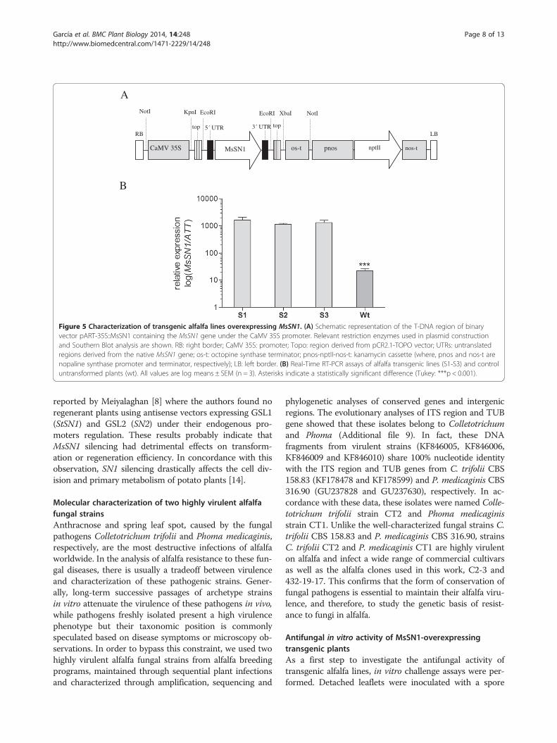
Analysis of MsSN1 expressionThe expression pattern of the MsSN1 gene was analyzedby RT-PCR, to explore the function of MsSN1 in wildtype alfalfa plants. MsSN1 expression was detected in alltissues analyzed, including roots, stems, leaves andyoung floral buds (Additional file 6). RT-qPCR assayswere performed to evaluate whether the expressionlevels of MsSN1 is modified by exposure to microorgan-isms. MsSN1 transcript levels were about two-fold higherin leaves than in roots when plants were grown undercontrol conditions (Figure 4). Interestingly, MsSN1 levelsdid not change in response to biotic stimuli, such as the
neighbor-joining method and root on midpoint. Bootstrapf plants is found on the right. Tree topology obtained using NJtical. The MsSN1 peptide characterized in this work and other snakin/
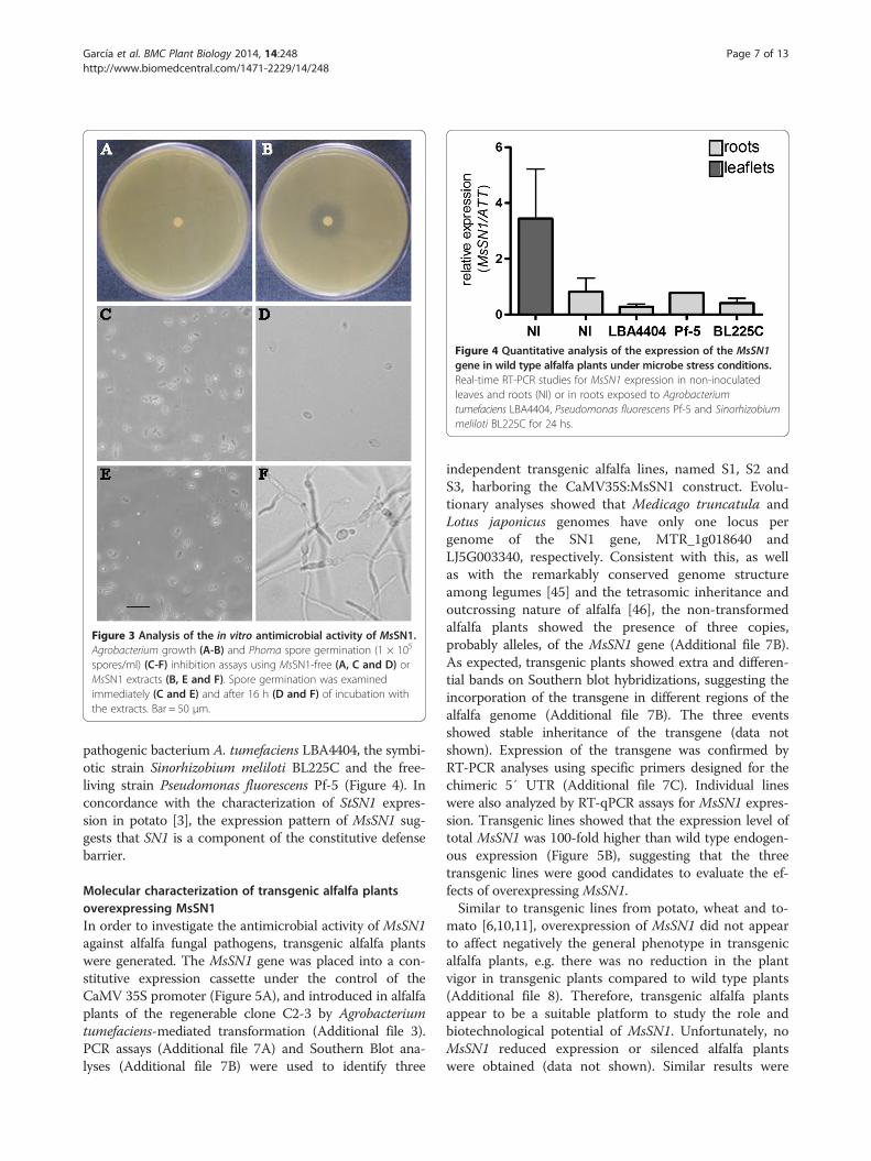
Figure 3 Analysis of the in vitro antimicrobial activity of MsSN1.Agrobacterium growth (A-B) and Phoma spore germination (1 × 10⁵
spores/ml) (C-F) inhibition assays using MsSN1-free (A, C and D) orMsSN1 extracts (B, E and F). Spore germination was examinedimmediately (C and E) and after 16 h (D and F) of incubation withthe extracts. Bar = 50 μm.
Figure 4 Quantitative analysis of the expression of the MsSN1gene in wild type alfalfa plants under microbe stress conditions.Real-time RT-PCR studies for MsSN1 expression in non-inoculatedleaves and roots (NI) or in roots exposed to Agrobacteriumtumefaciens LBA4404, Pseudomonas fluorescens Pf-5 and Sinorhizobiummeliloti BL225C for 24 hs.
García et al. BMC Plant Biology 2014, 14:248 Page 7 of 13http://www.biomedcentral.com/1471-2229/14/248
pathogenic bacterium A. tumefaciens LBA4404, the symbi-otic strain Sinorhizobium meliloti BL225C and the free-living strain Pseudomonas fluorescens Pf-5 (Figure 4). Inconcordance with the characterization of StSN1 expres-sion in potato [3], the expression pattern of MsSN1 sug-gests that SN1 is a component of the constitutive defensebarrier.
Molecular characterization of transgenic alfalfa plantsoverexpressing MsSN1In order to investigate the antimicrobial activity of MsSN1against alfalfa fungal pathogens, transgenic alfalfa plantswere generated. The MsSN1 gene was placed into a con-stitutive expression cassette under the control of theCaMV 35S promoter (Figure 5A), and introduced in alfalfaplants of the regenerable clone C2-3 by Agrobacteriumtumefaciens-mediated transformation (Additional file 3).PCR assays (Additional file 7A) and Southern Blot ana-lyses (Additional file 7B) were used to identify three
independent transgenic alfalfa lines, named S1, S2 andS3, harboring the CaMV35S:MsSN1 construct. Evolu-tionary analyses showed that Medicago truncatula andLotus japonicus genomes have only one locus pergenome of the SN1 gene, MTR_1g018640 andLJ5G003340, respectively. Consistent with this, as wellas with the remarkably conserved genome structureamong legumes [45] and the tetrasomic inheritance andoutcrossing nature of alfalfa [46], the non-transformedalfalfa plants showed the presence of three copies,probably alleles, of the MsSN1 gene (Additional file 7B).As expected, transgenic plants showed extra and differen-tial bands on Southern blot hybridizations, suggesting theincorporation of the transgene in different regions of thealfalfa genome (Additional file 7B). The three eventsshowed stable inheritance of the transgene (data notshown). Expression of the transgene was confirmed byRT-PCR analyses using specific primers designed for thechimeric 5′ UTR (Additional file 7C). Individual lineswere also analyzed by RT-qPCR assays for MsSN1 expres-sion. Transgenic lines showed that the expression level oftotal MsSN1 was 100-fold higher than wild type endogen-ous expression (Figure 5B), suggesting that the threetransgenic lines were good candidates to evaluate the ef-fects of overexpressing MsSN1.Similar to transgenic lines from potato, wheat and to-
mato [6,10,11], overexpression of MsSN1 did not appearto affect negatively the general phenotype in transgenicalfalfa plants, e.g. there was no reduction in the plantvigor in transgenic plants compared to wild type plants(Additional file 8). Therefore, transgenic alfalfa plantsappear to be a suitable platform to study the role andbiotechnological potential of MsSN1. Unfortunately, noMsSN1 reduced expression or silenced alfalfa plantswere obtained (data not shown). Similar results were
A
B
CaMV 35S MsSN1 os-t
NotI KpnI EcoRI NotIXbaIEcoRI
RB LB
pnos nptII nos-t
5´ UTR 3´ UTR toptop
Figure 5 Characterization of transgenic alfalfa lines overexpressing MsSN1. (A) Schematic representation of the T-DNA region of binaryvector pART-35S::MsSN1 containing the MsSN1 gene under the CaMV 35S promoter. Relevant restriction enzymes used in plasmid constructionand Southern Blot analysis are shown. RB: right border; CaMV 35S: promoter; Topo: region derived from pCR2.1-TOPO vector; UTRs: untranslatedregions derived from the native MsSN1 gene; os-t: octopine synthase terminator; pnos-nptII-nos-t: kanamycin cassette (where, pnos and nos-t arenopaline synthase promoter and terminator, respectively); LB: left border. (B) Real-Time RT-PCR assays of alfalfa transgenic lines (S1-S3) and controluntransformed plants (wt). All values are log means ± SEM (n = 3). Asterisks indicate a statistically significant difference (Tukey: ***p < 0.001).
García et al. BMC Plant Biology 2014, 14:248 Page 8 of 13http://www.biomedcentral.com/1471-2229/14/248
reported by Meiyalaghan [8] where the authors found noregenerant plants using antisense vectors expressing GSL1(StSN1) and GSL2 (SN2) under their endogenous pro-moters regulation. These results probably indicate thatMsSN1 silencing had detrimental effects on transform-ation or regeneration efficiency. In concordance with thisobservation, SN1 silencing drastically affects the cell div-ision and primary metabolism of potato plants [14].
Molecular characterization of two highly virulent alfalfafungal strainsAnthracnose and spring leaf spot, caused by the fungalpathogens Colletotrichum trifolii and Phoma medicaginis,respectively, are the most destructive infections of alfalfaworldwide. In the analysis of alfalfa resistance to these fun-gal diseases, there is usually a tradeoff between virulenceand characterization of these pathogenic strains. Gener-ally, long-term successive passages of archetype strainsin vitro attenuate the virulence of these pathogens in vivo,while pathogens freshly isolated present a high virulencephenotype but their taxonomic position is commonlyspeculated based on disease symptoms or microscopy ob-servations. In order to bypass this constraint, we used twohighly virulent alfalfa fungal strains from alfalfa breedingprograms, maintained through sequential plant infectionsand characterized through amplification, sequencing and
phylogenetic analyses of conserved genes and intergenicregions. The evolutionary analyses of ITS region and TUBgene showed that these isolates belong to Colletotrichumand Phoma (Additional file 9). In fact, these DNAfragments from virulent strains (KF846005, KF846006,KF846009 and KF846010) share 100% nucleotide identitywith the ITS region and TUB genes from C. trifolii CBS158.83 (KF178478 and KF178599) and P. medicaginis CBS316.90 (GU237828 and GU237630), respectively. In ac-cordance with these data, these isolates were named Colle-totrichum trifolii strain CT2 and Phoma medicaginisstrain CT1. Unlike the well-characterized fungal strains C.trifolii CBS 158.83 and P. medicaginis CBS 316.90, strainsC. trifolii CT2 and P. medicaginis CT1 are highly virulenton alfalfa and infect a wide range of commercial cultivarsas well as the alfalfa clones used in this work, C2-3 and432-19-17. This confirms that the form of conservation offungal pathogens is essential to maintain their alfalfa viru-lence, and therefore, to study the genetic basis of resist-ance to fungi in alfalfa.
Antifungal in vitro activity of MsSN1-overexpressingtransgenic plantsAs a first step to investigate the antifungal activity oftransgenic alfalfa lines, in vitro challenge assays were per-formed. Detached leaflets were inoculated with a spore

García et al. BMC Plant Biology 2014, 14:248 Page 9 of 13http://www.biomedcentral.com/1471-2229/14/248
suspension of two virulent and molecularly characterizedalfalfa fungal strains P. medicaginis CT1 and C. trifoliiCT2 incubating the leaves in petri plates for 7 days(Figure 6A-B). Disease severity was estimated by visuallyscoring disease symptoms in infected leaflets as a percent-age of leaflets showing disease lesions (Figure 6C). Thethree transgenic lines (S1- S3) showed significant lowerpercentage of diseased leaflets than wild type plants whenchallenged with both P. medicaginis CT1 (Figure 6A) andC. trifolii CT2 virulent strains (Figure 6B), suggesting anantimicrobial function of MsSN1 under biotic stress con-ditions. Moreover, this small-scale study could facilitaterapid visual screening of a large number of transgenicevents (Figure 6D), saving time, reducing cost, and speed-ing up the introgression of this antimicrobial transgeneinto commercial varieties, especially in outcrossing speciessuch as alfalfa [47].
Antifungal in vivo activity of MsSN1-overexpressingtransgenic plantsThe three transgenic alfalfa lines (S1-S3) were chosenfor progeny analysis of their potentially enhanced bioticresistance. The clone 432-19-17 was used as a pollendonor in a sexual cross with the transgenic lines to avoidthe common inbreeding depression of progeny fromself-crossed alfalfa plants. The presence and expressionof the transgene in leaves of the progeny plants were
Figure 6 Antifungal in vitro activity of MsSN1 overexpressing transgePhoma (#diseased leaflet/#total leaflet). (B) Diseased leaflets related to leafl(C) Damage score: 1. Healthy leaflet, 2. Countable injuries, 3. Uncountable iassay. Wt: wild type. S1-S3: MsSN1 transgenic plants. All values are log meadifference (Turkey: *p < 0.5; **p < 0.01 ***p < 0.001). Leaflets were considerescore. Disease severity was estimated from scoring 30 to 60 detached leafle
assayed by PCR and RT-qPCR analyses, respectively(data not shown). In vivo assays for antifungal activityagainst P. medicaginis CT1 were carried out spraying aspore solution to transgenic and wild type plants. Plantdisease severity was estimated by evaluating agronomicparameters for two months. Wild type plants manifestedall the symptoms of the disease [48,49], including darkbrown lesions on the stems, dark brown lesions on theleaflets that begin with specific dark color injuries, wilt-ing leaves, chlorosis, and defoliation. Transgenic linespresented scarce symptoms and usually looked like con-trol plants (data not shown).Firstly, leaflet disease was estimated by visually scoring
disease symptoms in infected plants. One month post-infection, the percentage of diseased leaflets was signifi-cantly lower in transgenic lines than in wild type plants(Figure 7A). Two months post-inoculation, disease se-verity was analyzed by evaluating the vigor of the plants,the number of plants with regrowth and the percentageof highly defoliated plants. Transgenic plants showedsignificantly higher levels of regrowth than wild typeplants (Figure 7B, F and G). In fact, disease symptomswere present in wild type regrowth but not in transgenicplants (Figure 7G). Overexpression of MsSN1 in alfalfaimproves plant vigor under fungal stress conditions(Figure 7C). In addition, wild type plants showed moredefoliation than transgenic plants but the differences
nic plants. (A) Diseased leaflets related to leaflets inoculated withets inoculated with Colletotrichum (#diseased leaflet/#total leaflet).njuries, 4. Chlorosis, 5. completely damaged. (D) Representative Phomans ± SEM (n = 10–30). Asterisks indicate a statistically significantd diseased when they showed symptoms 3, 4 or 5 in the damagets from three individual plants.
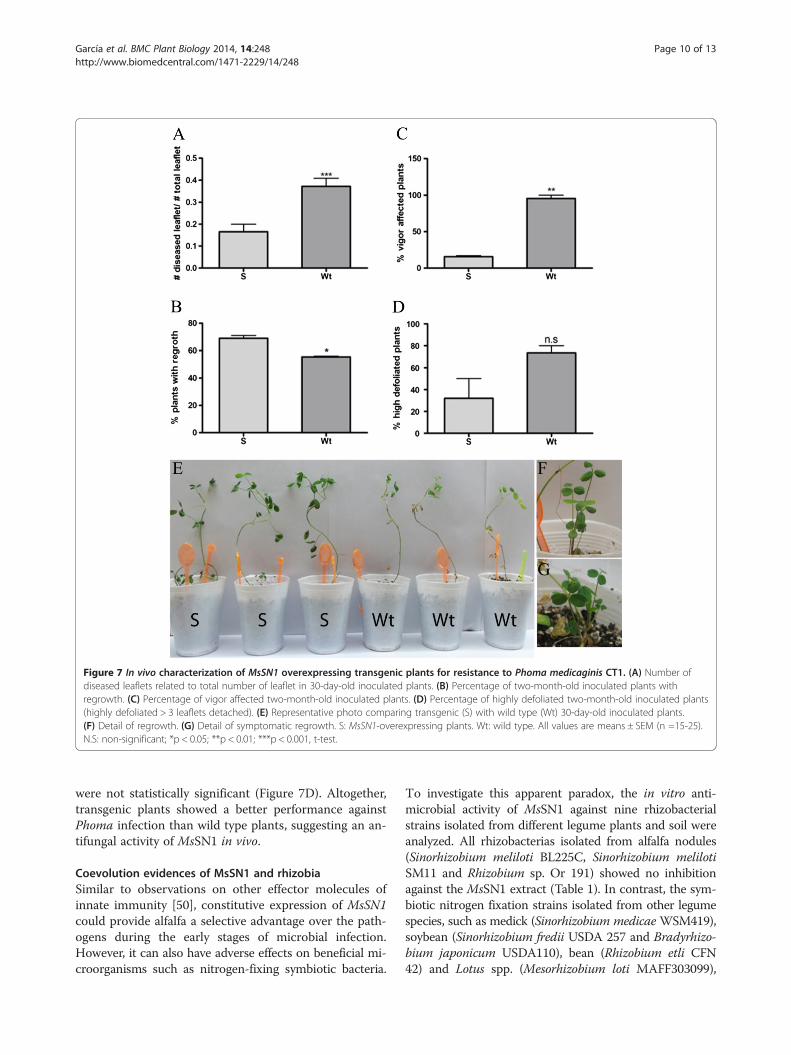
Figure 7 In vivo characterization of MsSN1 overexpressing transgenic plants for resistance to Phoma medicaginis CT1. (A) Number ofdiseased leaflets related to total number of leaflet in 30-day-old inoculated plants. (B) Percentage of two-month-old inoculated plants withregrowth. (C) Percentage of vigor affected two-month-old inoculated plants. (D) Percentage of highly defoliated two-month-old inoculated plants(highly defoliated > 3 leaflets detached). (E) Representative photo comparing transgenic (S) with wild type (Wt) 30-day-old inoculated plants.(F) Detail of regrowth. (G) Detail of symptomatic regrowth. S: MsSN1-overexpressing plants. Wt: wild type. All values are means ± SEM (n =15-25).N.S: non-significant; *p < 0.05; **p < 0.01; ***p < 0.001, t-test.
García et al. BMC Plant Biology 2014, 14:248 Page 10 of 13http://www.biomedcentral.com/1471-2229/14/248
were not statistically significant (Figure 7D). Altogether,transgenic plants showed a better performance againstPhoma infection than wild type plants, suggesting an an-tifungal activity of MsSN1 in vivo.
Coevolution evidences of MsSN1 and rhizobiaSimilar to observations on other effector molecules ofinnate immunity [50], constitutive expression of MsSN1could provide alfalfa a selective advantage over the path-ogens during the early stages of microbial infection.However, it can also have adverse effects on beneficial mi-croorganisms such as nitrogen-fixing symbiotic bacteria.
To investigate this apparent paradox, the in vitro anti-microbial activity of MsSN1 against nine rhizobacterialstrains isolated from different legume plants and soil wereanalyzed. All rhizobacterias isolated from alfalfa nodules(Sinorhizobium meliloti BL225C, Sinorhizobium melilotiSM11 and Rhizobium sp. Or 191) showed no inhibitionagainst the MsSN1 extract (Table 1). In contrast, the sym-biotic nitrogen fixation strains isolated from other legumespecies, such as medick (Sinorhizobium medicae WSM419),soybean (Sinorhizobium fredii USDA 257 and Bradyrhizo-bium japonicum USDA110), bean (Rhizobium etli CFN42) and Lotus spp. (Mesorhizobium loti MAFF303099),
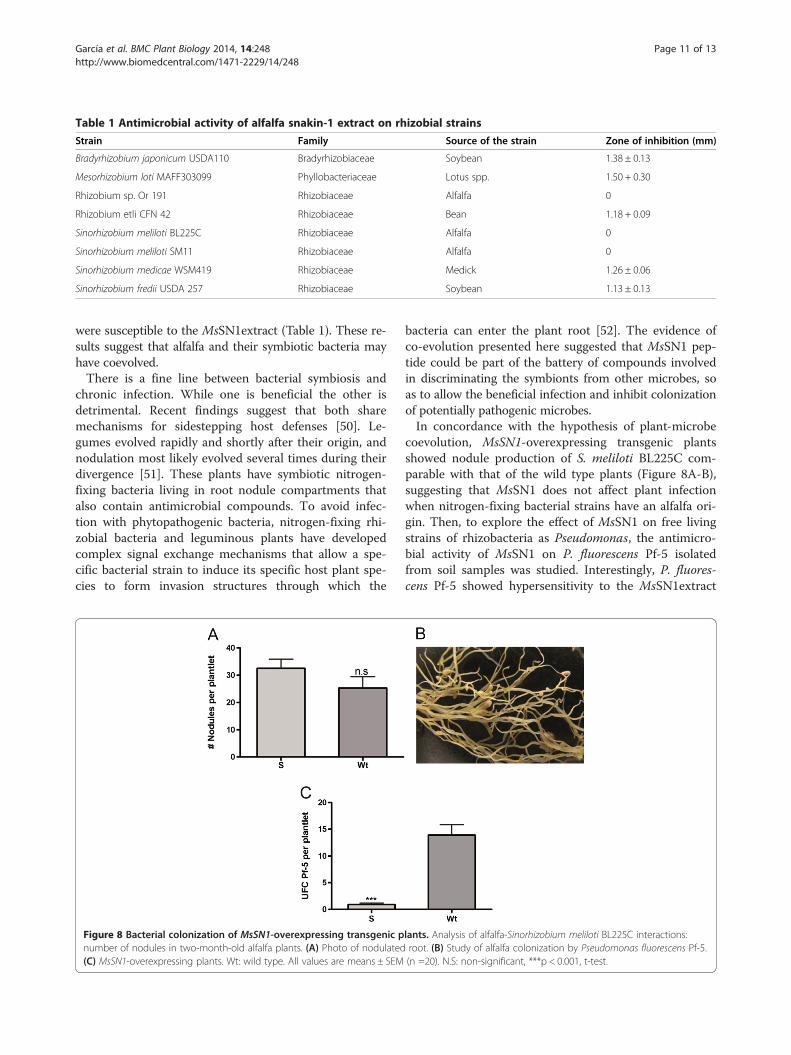
Table 1 Antimicrobial activity of alfalfa snakin-1 extract on rhizobial strains
Strain Family Source of the strain Zone of inhibition (mm)
Bradyrhizobium japonicum USDA110 Bradyrhizobiaceae Soybean 1.38 ± 0.13
Mesorhizobium loti MAFF303099 Phyllobacteriaceae Lotus spp. 1.50 + 0.30
Rhizobium sp. Or 191 Rhizobiaceae Alfalfa 0
Rhizobium etli CFN 42 Rhizobiaceae Bean 1.18 + 0.09
Sinorhizobium meliloti BL225C Rhizobiaceae Alfalfa 0
Sinorhizobium meliloti SM11 Rhizobiaceae Alfalfa 0
Sinorhizobium medicae WSM419 Rhizobiaceae Medick 1.26 ± 0.06
Sinorhizobium fredii USDA 257 Rhizobiaceae Soybean 1.13 ± 0.13
García et al. BMC Plant Biology 2014, 14:248 Page 11 of 13http://www.biomedcentral.com/1471-2229/14/248
were susceptible to the MsSN1extract (Table 1). These re-sults suggest that alfalfa and their symbiotic bacteria mayhave coevolved.There is a fine line between bacterial symbiosis and
chronic infection. While one is beneficial the other isdetrimental. Recent findings suggest that both sharemechanisms for sidestepping host defenses [50]. Le-gumes evolved rapidly and shortly after their origin, andnodulation most likely evolved several times during theirdivergence [51]. These plants have symbiotic nitrogen-fixing bacteria living in root nodule compartments thatalso contain antimicrobial compounds. To avoid infec-tion with phytopathogenic bacteria, nitrogen-fixing rhi-zobial bacteria and leguminous plants have developedcomplex signal exchange mechanisms that allow a spe-cific bacterial strain to induce its specific host plant spe-cies to form invasion structures through which the
Figure 8 Bacterial colonization of MsSN1-overexpressing transgenic pnumber of nodules in two-month-old alfalfa plants. (A) Photo of nodulated(C) MsSN1-overexpressing plants. Wt: wild type. All values are means ± SEM
bacteria can enter the plant root [52]. The evidence ofco-evolution presented here suggested that MsSN1 pep-tide could be part of the battery of compounds involvedin discriminating the symbionts from other microbes, soas to allow the beneficial infection and inhibit colonizationof potentially pathogenic microbes.In concordance with the hypothesis of plant-microbe
coevolution, MsSN1-overexpressing transgenic plantsshowed nodule production of S. meliloti BL225C com-parable with that of the wild type plants (Figure 8A-B),suggesting that MsSN1 does not affect plant infectionwhen nitrogen-fixing bacterial strains have an alfalfa ori-gin. Then, to explore the effect of MsSN1 on free livingstrains of rhizobacteria as Pseudomonas, the antimicro-bial activity of MsSN1 on P. fluorescens Pf-5 isolatedfrom soil samples was studied. Interestingly, P. fluores-cens Pf-5 showed hypersensitivity to the MsSN1extract
lants. Analysis of alfalfa-Sinorhizobium meliloti BL225C interactions:root. (B) Study of alfalfa colonization by Pseudomonas fluorescens Pf-5.(n =20). N.S: non-significant, ***p < 0.001, t-test.

García et al. BMC Plant Biology 2014, 14:248 Page 12 of 13http://www.biomedcentral.com/1471-2229/14/248
(Zone of inhibition = 1.76 ± 0.06 mm). In agreement withthe in vitro activity of MsSN1, P. fluorescens Pf-5 showeddifferential proliferation in the rhizosphere of wild typeand transgenic lines of alfalfa 30 days after inoculation,suggesting an increased antibacterial activity in MsSN1-overexpressing plants (Figure 8C). These results showadditional in vitro and in vivo evidence of MsSN1 as apart of the innate immune response and the wide rangeof activity of MsSN1.
ConclusionsPrevious studies and the results reported here show thatsnakin peptides have important and ancestral roles inland plant innate immunity. Interestingly, MsSN1-over-expressing alfalfa transgenic plants show increased anti-microbial activity against virulent fungal strains withoutaltering the nitrogen-fixing symbiosis, opening the wayto the production of effective alfalfa transgenic cultivarsfor biotic stress resistance. In this work, data also sug-gest a coevolutionary process, in which alfalfa exerts aselection pressure for resistance to MsSN1 on rhizobialbacteria.
Additional files
Additional file 1: Table of primers used in this work.
Additional file 2: Similarity of the product of MsSN1 gene to othersnakin/GASA proteins from plants.
Additional file 3: Alfalfa transformation.
Additional file 4: Identity of the product of MsSN1 gene to othersnakin/GASA proteins from Arabidopsis.
Additional file 5: RT-PCR studies of MsSN1 gene expression inE. coli pSJ33 (1) and E. coli pSJ33-MsSN (2) strains.
Additional file 6: RT-PCR assays showing MsSN1 expression inyoung floral buds (A-B), leaves (C-D), stems (E-F) and roots (G-H).
Additional file 7: Molecular characterization of transgenic alfalfalines overexpressing MsSN1.
Additional file 8: Vigor phenotype of transgenic (S1) and wild type(Wt) alfalfa plants.
Additional file 9: Phylogenetic analysis of fungal strains isolatedfrom alfalfa.
Competing interestsThe authors declare that they have no competing interests.
Authors’ contributionsConceived and designed the experiments: GCS; NDA; JPM; MEP. Performedthe experiments: ANG; NDA; GCS; ARF; MCG; MJD. Analyzed the data: GS;NDA. Wrote the paper: GS; NDA; CAB. All authors read and approved thefinal manuscript.
AcknowledgmentsIn memory of Ing. Agr. Raúl Ríos.We thank Carmen Soria, Juan Riquelme and Guillermo Piparola (IGEAF-INTA)for their technical support.This work was supported by Agencia Nacional de Promoción Científica yTecnológica (grant PICT 2011–1325). NDA; CAB; JPM and GCS are researchersof the National Scientific and Technical Research Council (CONICET).
Author details1Instituto de Genética Ewald A. Favret (CICVyA-INTA), De los Reseros S/N,Castelar C25 (1712), Buenos Aires, Argentina. 2Instituto de InvestigacionesBiomédicas en Retrovirus y SIDA (INBIRS), UBA-CONICET, Paraguay 2155,C1121ABG Ciudad Autónoma de Buenos Aires, Argentina. 3Departamento deBiodiversidad y Biología Experimental, Facultad de Ciencias Exactas yNaturales, Universidad de Buenos Aires, Intendente Güiraldes 2160, CiudadUniversitaria, Pabellón II, C1428EGA Ciudad Autónoma de Buenos Aires,Argentina. 4Instituto de Investigaciones en Ingeniería Genética y BiologíaMolecular, “Dr. Hector Torres”, (INGEBI-CONICET), Vuelta de Obligado 2490,C1428ADN Ciudad Autónoma de Buenos Aires, Argentina.
Received: 20 June 2014 Accepted: 11 September 2014
References1. Volenec JJ, Cunningham SM, Haagenson DM, Berg WK, Joern BC, Wiersma
DW: Physiological genetics of alfalfa improvement: past failures, futureprospects. Field Crops Res 2002, 75:97–110.
2. Lopez-Solanilla E, Garcia-Olmedo F, Rodriguez-Palenzuela P: Inactivation ofthe sapA to sapF locus of Erwinia chrysanthemi reveals commonfeatures in plant and animal bacterial pathogenesis. Plant Cell 1998,10(6):917–924.
3. Segura A, Moreno M, Madueno F, Molina A, Garcia-Olmedo F: Snakin-1, apeptide from potato that is active against plant pathogens. Mol PlantMicrobe Interact 1999, 12(1):16–23.
4. Berrocal-Lobo M, Segura A, Moreno M, Lopez G, Garcia-Olmedo F, Molina A:Snakin-2, an antimicrobial peptide from potato whose gene is locallyinduced by wounding and responds to pathogen infection. Plant Physiol2002, 128(3):951–961.
5. Kovalskaya N, Hammond RW: Expression and functional characterizationof the plant antimicrobial snakin-1 and defensin recombinant proteins.Protein Expr Purif 2009, 63(1):12–17.
6. Rong W, Qi L, Wang J, Du L, Xu H, Wang A, Zhang Z: Expression of apotato antimicrobial peptide SN1 increases resistance to take-allpathogen Gaeumannomyces graminis var. tritici in transgenic wheat.Funct Integr Genomics 2013, 13(3):403–409.
7. Guzman-Rodriguez JJ, Ibarra-Laclette E, Herrera-Estrella L, Ochoa-Zarzosa A,Suarez-Rodriguez LM, Rodriguez-Zapata LC, Salgado-Garciglia R, Jimenez-Moraila B, Lopez-Meza JE, Lopez-Gomez R: Analysis of expressed sequencetags (ESTs) from avocado seed (Persea americana var. drymifolia) revealsabundant expression of the gene encoding the antimicrobial peptidesnakin. Plant Physiol Biochem 2013, 70:318–324.
8. Meiyalaghan S, Thomson SJ, Fiers MW, Barrell PJ, Latimer JM, Mohan S,Jones EE, Conner AJ, Jacobs JM: Structure and expression of GSL1 andGSL2 genes encoding gibberellin stimulated-like proteins in diploid andhighly heterozygous tetraploid potato reveals their highly conservedand essential status. BMC Genomics 2014, 15:2.
9. Mohan S, Meiyalaghan S, Latimer JM, Gatehouse ML, Monaghan KS, VangaBR, Pitman AR, Jones EE, Conner AJ, Jacobs JM: GSL2 over-expressionconfers resistance to Pectobacterium atrosepticum in potato. Theor ApplGenet 2014, 127(3):677–689.
10. Almasia NI, Bazzini AA, Hopp HE, Vazquez-Rovere C: Overexpression ofsnakin-1 gene enhances resistance to Rhizoctonia solani and Erwiniacarotovora in transgenic potato plants. Mol Plant Pathol 2008, 9(3):329–338.
11. Balaji V, Smart CD: Over-expression of snakin-2 and extensin-like proteingenes restricts pathogen invasiveness and enhances tolerance toClavibacter michiganensis subsp. michiganensis in transgenic tomato(Solanum lycopersicum). Transgenic Res 2012, 21(1):23–37.
12. Kovalskaya N, Zhao Y, Hammond RW: Antibacterial and antifungal activityof a snakin-defensin hybrid protein expressed in tobacco and potatoplant. Open Plant Sci J 2011, 5:29–42.
13. Balaji V, Sessa G, Smart CD: Silencing of host basal defense response-related gene expression increases susceptibility of Nicotianabenthamiana to Clavibacter michiganensis subsp. michiganensis.Phytopathology 2011, 101(3):349–357.
14. Nahirñak V, Almasia NI, Fernandez PV, Hopp HE, Estevez JM, Carrari F,Vazquez-Rovere C: Potato snakin-1 gene silencing affects cell division,primary metabolism, and cell wall composition. Plant Physiol 2012,158(1):252–263.

García et al. BMC Plant Biology 2014, 14:248 Page 13 of 13http://www.biomedcentral.com/1471-2229/14/248
15. Nahirñak V, Almasia NI, Hopp HE, Vazquez-Rovere C: Snakin/GASA proteins:involvement in hormone crosstalk and redox homeostasis. Plant SignalBehav 2012, 7(8):1004–1008.
16. Porto WF, Franco OL: Theoretical structural insights into the snakin/GASAfamily. Peptides 2013, 44:163–167.
17. Howell CR, Stipanovic RD: Control of Rhizoctonia solani on cottonseedlings with Pseudomonas fluorescens with an antibiotic produced bythe bacterium. Phytopathology 1979, 69:480–482.
18. Carelli M, Gnocchi S, Fancelli S, Mengoni A, Paffetti D, Scotti C, BazzicalupoM: Genetic diversity and dynamics of Sinorhizobium meliloti populationsnodulating different alfalfa cultivars in Italian soils. Appl Environ Microbiol2000, 66(11):4785–4789.
19. Selbitschka W, Keller M, Miethling-Graff R, Dresing U, Schwieger F, Krahn I,Homann I, Dammann-Kalinowski T, Puhler A, Tebbe CC: Long-term field releaseof bioluminescent Sinorhizobium meliloti strains to assess the influence of arecA mutation on the strains’ survival. Microb Ecol 2006, 52(3):583–595.
20. Reeve W, Chain P, O’Hara G, Ardley J, Nandesena K, Brau L, Tiwari R, MalfattiS, Kiss H, Lapidus A, Copeland A, Nolan M, Land M, Hauser L, Chang YJ,Ivanova N, Mavromatis K, Markowitz V, Kyrpides N, Gollagher M, Yates R,Dilworth M, Howieson J: Complete genome sequence of the Medicagomicrosymbiont Ensifer (Sinorhizobium) medicae strain WSM419. StandGenomic Sci 2010, 2(1):77–86.
21. Keyser HH, Bohlool BB, Hu TS, Weber DF: Fast-growing rhizobia isolatedfrom root nodules of soybean. Science 1982, 215(4540):1631–1632.
22. Eardly BD, Hannaway DB, Bottomley PJ: Characterization of Rhizobia fromineffective Alfalfa nodules: ability to nodulate bean plants [Phaseolusvulgaris (L.) Savi.]. Appl Environ Microbiol 1985, 50(6):1422–1427.
23. Segovia L, Young JP, Martinez-Romero E: Reclassification of AmericanRhizobium leguminosarum biovar phaseoli type I strains as Rhizobiumetli sp. nov. Int J Syst Bacteriol 1993, 43(2):374–377.
24. Kaneko T, Nakamura Y, Sato S, Asamizu E, Kato T, Sasamoto S, Watanabe A,Idesawa K, Ishikawa A, Kawashima K, Kimura T, Kishida Y, Kiyokawa C, KoharaM, Matsumoto M, Matsuno A, Mochizuki Y, Nakayama S, Nakazaki N, ShimpoS, Sugimoto M, Takeuchi C, Yamada M, Tabata S: Complete genomestructure of the nitrogen-fixing symbiotic bacterium Mesorhizobium loti(supplement). DNA Res 2000, 7(6):381–406.
25. Mathis JN, Israel DW, Barbour WM, Jarvis BD, Elkan GH: Analysis of thesymbiotic performance of bradyrhizobium japonicum USDA 110 and itsderivative I-110 and discovery of a new mannitol-utilizing, nitrogen-fixing USDA 110 derivative. Appl Environ Microbiol 1986, 52(1):75–80.
26. Hoekema A, Hirsch PR, Hooykaas PJJ, Schilperoort RA: A binary plant vectorstrategy based on separation of vir- and T-region of the Agrobacteriumtumefaciens Ti-plasmid. Nature 1983, 303(5913):179–180.
27. Setten L, Soto G, Mozzicafreddo M, Fox AR, Lisi C, Cuccioloni M, AngelettiM, Pagano E, Diaz-Paleo A, Ayub ND: Engineering Pseudomonas protegensPf-5 for nitrogen fixation and its application to improve plant growth undernitrogen-deficient conditions. PLoS One 2013, 8(5):e63666.
28. Ayub ND, Julia Pettinari M, Mendez BS, Lopez NI: Impairedpolyhydroxybutyrate biosynthesis from glucose in Pseudomonas sp.14–3 is due to a defective beta-ketothiolase gene. FEMS Microbiol Lett2006, 264(1):125–131.
29. Gleave AP: A versatile binary vector system with a T-DNA organisationalstructure conducive to efficient integration of cloned DNA into the plantgenome. Plant Mol Biol 1992, 20(6):1203–1207.
30. Campanella JJ, Bitincka L, Smalley J: MatGAT: an application thatgenerates similarity/identity matrices using protein or DNA sequences.BMC Bioinformatics 2003, 4:29.
31. Tamura K, Peterson D, Peterson N, Stecher G, Nei M, Kumar S: MEGA5:molecular evolutionary genetics analysis using maximum likelihood,evolutionary distance, and maximum parsimony methods. Mol Biol Evol2011, 28(10):2731–2739.
32. Finn RD, Mistry J, Tate J, Coggill P, Heger A, Pollington JE, Gavin OL,Gunasekaran P, Ceric G, Forslund K, Holm L, Sonnhammer EL, Eddy SR:The Pfam protein families database. Nucleic Acids Res 2010,38(Database issue):D211–D222.
33. Shen HB, Chou KC: Signal-3 L: a 3-layer approach for predicting signalpeptides. Biochem Biophys Res Commun 2007, 363(2):297–303.
34. Moller EM, Bahnweg G, Sandermann H, Geiger HH: A simple and efficientprotocol for isolation of high molecular weight DNA from filamentousfungi, fruit bodies, and infected plant tissues. Nucleic Acids Res 1992,20(22):6115–6116.
35. Ayub ND, Pettinari MJ, Ruiz JA, Lopez NI: A polyhydroxybutyrate-producing Pseudomonas sp. isolated from Antarctic environments withhigh stress resistance. Curr Microbiol 2004, 49(3):170–174.
36. Beringer JE: R factor transfer in Rhizobium leguminosarum. J Gen Microbiol1974, 84(1):188–198.
37. Shen WJ, Forde BG: Efficient transformation of Agrobacterium spp. byhigh voltage electroporation. Nucleic Acids Res 1989, 17(20):8385.
38. D’Halluin K, Botterman J, De Greef W: Engineering of herbicide-resistantAlfalfa and evaluation under field conditions. Crop Sci 1990, 30:866–871.
39. Soto G, Fox R, Ayub N, Alleva K, Guaimas F, Erijman EJ, Mazzella A, AmodeoG, Muschietti J: TIP5;1 is an aquaporin specifically targeted to pollenmitochondria and is probably involved in nitrogen remobilization inArabidopsis thaliana. Plant J 2010, 64(6):1038–1047.
40. Sanchez L, Weidmann S, Arnould C, Bernard AR, Gianinazzi S, Gianinazzi-Pearson V: Pseudomonas fluorescens and Glomus mosseae triggerDMI3-dependent activation of genes related to a signal transductionpathway in roots of Medicago truncatula. Plant Physiol 2005,139(2):1065–1077.
41. Perez Di Giorgio J, Soto G, Alleva K, Jozefkowicz C, Amodeo G, MuschiettiJP, Ayub ND: Prediction of aquaporin function by integratingevolutionary and functional analyses. J Membr Biol 2014, 247(2):107–125.
42. Furukawa T, Sakaguchi N, Shimada H: Two OsGASR genes, rice GASThomologue genes that are abundant in proliferating tissues, showdifferent expression patterns in developing panicles. Genes Genet Syst2006, 81:171–180.
43. Roxrud I, Lid SE, Fletcher JC, Schmidt ED, Opsahl-Sorteberg HG: GASA4, oneof the 14-member Arabidopsis GASA family of small polypeptides,regulates flowering and seed development. Plant Cell Physiol 2007,48(3):471–483.
44. Bennici A: Origin and early evolution of land plants: Problems andconsiderations. Commun Integr Biol 2008, 1(2):212–218.
45. Choi HK, Kim D, Uhm T, Limpens E, Lim H, Mun JH, Kalo P, Penmetsa RV,Seres A, Kulikova O, Roe BA, Bisseling T, Kiss GB, Cook DR: A sequence-based genetic map of Medicago truncatula and comparison of markercolinearity with M. sativa. Genetics 2004, 166(3):1463–1502.
46. Cook DR: Medicago truncatula–a model in the making! Curr Opin PlantBiol 1999, 2(4):301–304.
47. McCann MC, Rogan GJ, Fitzpatrick S, Trujillo WA, Sorbet R, Hartnell GF,Riodan SG, Nemeth MA: Glyphosate-tolerant alfalfa is compositionallyequivalent to conventional alfalfa (Medicago sativa L.). J Agr Food Chem2006, 54(19):7187–7192.
48. Masoud SA, Zhu Q, Lamb C, Dixon RA: Constitutive expression of aninducible glucanase in alfalfa reduces disease severity caused by theoomycete pathogen Phytophthora megasperma f. sp medicaginis, butdoes not reduce disease severity of chitin-containing fungi. TransgenicRes 1996, 5:313–323.
49. Hipskind JD, Paiva NL: Constitutive accumulation of a resveratrol-glucoside in transgenic alfalfa increases resistance to Phoma medicaginis.Mol Plant Microbe Interact 2000, 13(5):551–562.
50. Gruenheid S, Le Moual H: Resistance to antimicrobial peptides in Gram-negative bacteria. FEMS Microbiol Lett 2012, 330(2):81–89.
51. Doyle JJ: Phylogenetic perspectives on the origins of nodulation.Mol Plant Microbe Interact 2011, 24(11):1289–1295.
52. Jones KM, Kobayashi H, Davies BW, Taga ME, Walker GC: How rhizobialsymbionts invade plants: the Sinorhizobium-Medicago model. Nat RevMicrobiol 2007, 5(8):619–633.
doi:10.1186/s12870-014-0248-9Cite this article as: García et al.: Alfalfa snakin-1 prevents fungalcolonization and probably coevolved with rhizobia. BMC Plant Biology2014 14:248.
Related Documents


