Aldehyde Oxidase Drug Metabolism: Evaluation of Drug Interaction Potential and Allometric Scaling Methods to Predict Human Pharmacokinetics By Rachel Denise Crouch Dissertation Submitted to the Faculty of the Graduate School of Vanderbilt University in partial fulfillment of the requirements for the degree of DOCTOR OF PHILOSOPHY in Pharmacology December, 2016 Nashville, Tennessee Approved: J. Scott Daniels, Ph.D. Joey V. Barnett, Ph.D. Colleen M. Niswender, Ph.D. C. David Weaver, Ph.D. Neil Osheroff, Ph.D. Wendell S. Akers, Pharm.D., Ph.D.

Welcome message from author
This document is posted to help you gain knowledge. Please leave a comment to let me know what you think about it! Share it to your friends and learn new things together.
Transcript

Aldehyde Oxidase Drug Metabolism: Evaluation of Drug Interaction Potential and
Allometric Scaling Methods to Predict Human Pharmacokinetics
By
Rachel Denise Crouch
Dissertation
Submitted to the Faculty of the
Graduate School of Vanderbilt University
in partial fulfillment of the requirements
for the degree of
DOCTOR OF PHILOSOPHY
in
Pharmacology
December, 2016
Nashville, Tennessee
Approved:
J. Scott Daniels, Ph.D.
Joey V. Barnett, Ph.D.
Colleen M. Niswender, Ph.D.
C. David Weaver, Ph.D.
Neil Osheroff, Ph.D.
Wendell S. Akers, Pharm.D., Ph.D.

ii
For Mom, Dad, and Robby
Colossians 3:17

iii
ACKNOWLEDGEMENTS
The research described herein was supported by the NIGMS Vanderbilt
Training Program in Pharmacological Sciences, the PhRMA Pre-Doctoral Fellowship
Program in Pharmacology/Toxicology, and the NIH Division of Loan Repayment
Program in Clinical Research. The commitment of these organizations to the
development of young scientists in biomedical, pharmaceutical, and translational
research is vital to the discovery and advancement of disease diagnosis, treatment,
and prevention, and I would like to thank them for the financial provisions they
have provided to me throughout my Ph.D. training.
In addition, I would like to thank the Lipscomb University College of
Pharmacy/Vanderbilt University Department of Pharmacology Pharm.D./Ph.D.
Degree Partnership Program for providing me the opportunity to train in an
environment of exceptional scientists and a program dedicated to high-quality
education. Most notably, I want to express my gratitude toward the individuals who
created this program, Drs. Joey Barnett and Scott Akers, for the countless avenues of
support they have provided to me. I would not be in the position to present this
work today without their support and encouragement—no one, perhaps, is owed
more credit for me taking the step toward this achievement than Dr. Akers.
Likewise, several prior teachers and mentors at Lipscomb have contributed
to my interest in science and my ultimate decision to pursue a career in research.
From my undergraduate professors and mentors Drs. Kent Clinger and Ronnie
Boone, to my pharmacy school professors and mentors Drs. Michael Fowler and

iv
Susan Mercer, I am thankful for the instruction, mentoring, and encouragement I
received from these individuals. Likewise, I would not have made it through
pharmacy school to reach this point without the support and encouragement of my
pharmacy classmates Drs. Katie Black and Chris Stokes.
In addition to Drs. Barnett and Akers, I am grateful for the others who have
graciously served on my dissertation committee. Insightful suggestions from Dr.
Colleen Niswender have been an essential contribution to improving the quality and
focus of my studies, as have fresh perspectives and ideas from Dr. Neil Osheroff. I
am also very appreciative of Dr. Dave Weaver for stepping in to serve as my co-chair
alongside Dr. Niswender. Each member of my committee has been tremendously
supportive and encouraging, and it has been a pleasure to work with them and gain
from their expertise. I would also like to take this time to thank Dr. Matthew Hutzler,
who has generously served as an additional mentor to me and a critical contributor
to completion of this work.
In addition to my committee members, I am especially thankful for the
opportunity to work with my mentor, Dr. Scott Daniels, who has been more than just
a research advisor, but a life coach, in support of my research, my career aims, as
well as my personal well-being. I am appreciative to Dr. Daniels for granting me the
freedom to take ownership of my research and for always encouraging me to
explore my own ideas. At the same time, I have been fortunate for the opportunity to
learn from his expertise and build a foundation for my career. Dr. Daniels has always
been my advocate, and I am eternally grateful for his dedicated commitment to my

v
success. It has truly been a privilege to train with Dr. Daniels and likewise to have
gained a lifelong mentor, colleague, and friend.
As for the DMPK lab members, past and present, I am thankful for their
friendship and for all that they have graciously taught me. I am especially grateful to
Dr. Tom Bridges, who I could always count on to take time to answer a question,
Frank Byers for that all he taught and assisted me with and, especially, for making
the lab a fun place to be, Jay Foster for always being willing to help and for keeping
me entertained in the lab, and Dr. Annie Blobaum and Dr. Chuck Locuson for all their
assistance and encouragement. I undoubtedly owe my gratitude to Ryan Morrison
for the hours of time he generously dedicated to teaching me everything I know
about LC/MS/MS, among countless other concepts and techniques relating to DMPK.
His instruction was essential to me reaching this point, as was his friendship. I also
thank Sichen Chang, Katrina Brewerer, and several other past labmates for all they
have done to aid me.
Thanks as well to those in the Lindsely Lab, especially Craig Lindsley, who
aided in facilitating the completion of my research. I am also particularly thankful to
Matt Mulder for the many ways in which he has assisted me, as well as being a great
desk neighbor, and Jeanette Bertron for being a fun, supportive, and encouraging
friend. Thanks to everyone else in Cool Springs for all thier help, especially Nathan
Kett for keeping the place up and running, as well as many others for creating a fun
environment in which to work. There are many others who are owed my gratitude
for various means of support in the Department of Pharmacology and VCNDD. I
specifically want to thank Karen Gieg, Donna Johnson, and Kristin Riggs for all their

vi
assistance, as well as Dr. Jeffrey Conn and his research group, many members of
which have aided me in one way or another.
Of course, I must also thank my friends and family, who are too many to
name, for all the moral support provided to me over the past several years. I most
certainly could not have made it through this challenging time without their support
and encouragement. My Mom and Dad have always supported me in whatever I
chose to pursue and always believed I could achieve anything I set out to do. I could
never repay them for all they have done for me, and I am forever indebted to them
for all their love and support. I also want to thank my brother Matt for his love and
encouragement, as well as my mother and father-in-law, Bob and Martha, who have
treated me like their own daughter. I lastly am infinitely grateful for my husband
and best friend, Robby, who has supported me through eight years of pharmacy
school and graduate school without complaint. He makes me laugh, makes me think,
makes me strong, makes me happy, and I could not make it through life without him.
Finally, for all these blessings I have received, I am eternally grateful to God,
who already knows the answer before we even conceive the question.

vii
TABLE OF CONTENTS
Page
DEDICATION ............................................................................................................................................. ii
ACKNOWLEDGEMENTS ...................................................................................................................... iii
LIST OF TABLES ...................................................................................................................................xiii
LIST OF FIGURES ............................................................................................................................... xvii
LIST OF EQUATIONS .......................................................................................................................... xxii
LIST OF ABBREVIATIONS .............................................................................................................. xxiii
Chapter
I. INTODUCTION TO ALDEHYDE OXIDASE ................................................................................. 1
Aldehyde Oxidase Structure .......................................................................................................... 1
Aldehyde Oxidase Substrate Specificity and Reactions ...................................................... 2
Aldehyde Oxidase Catalytic Mechanism ................................................................................... 3
Known Clinical Aldehyde Oxidase Substrates ........................................................................ 4
Known Clinical Aldehyde Oxidase Inhibitors ......................................................................... 7
Aldehyde Oxidase Expression ...................................................................................................... 8
Single human aldehyde oxidase isoform ........................................................................... 8
Species-specific aldehyde oxidase isoforms ..................................................................... 9
Age-dependent aldehyde oxidase expression ...............................................................11
Regulation of aldehyde oxidase expression ...................................................................11
Species-Specific Aldehyde Oxidase Activity ..........................................................................12
Sex Differences in Aldehyde Oxidase Activity ......................................................................13
Endogenous Aldehyde Oxidase Substrates and Physiological Relevance .................15
Human Aldehyde Oxidase Single Nucleotide Polymorphisms ......................................16
Identification of Aldehyde Oxidase Metabolism .................................................................17
Failed Clinical Aldehyde Oxidase Substrates ........................................................................20
Challenges in Predicting Human Pharmacokinetics of Aldehyde Oxidase ...............21

viii
II. MATERIALS AND METHODS .......................................................................................................23
Materials .............................................................................................................................................23
In Vitro Biotransformation and Clearance of VU0409106 ..............................................24
Biotransformation in hepatic microsomal and recombinant human P450
incubations ...................................................................................................................................24
Metabolite formation in hepatic S9 fractions .................................................................25
Intrinsic clearance in hepatic S9 fractions .......................................................................25
In Vivo Metabolism and Disposition of VU0409106 in Sprague-Dawley Rats ........26
Intravenous or intraperitoneal administration of VU0409106 ..............................27
Hepatic portal vein or mesenteric ileal vein administration of VU0409106 .....28
In Vitro Biotransformation and Clearance of Zaleplon, O6-Bezylguanine,
Zoniporide, BIBX1382, and SGX523 ........................................................................................29
Biotransformation in hepatic S9 incubations ................................................................29
Intrinsic clearance and estimated hepatic clearance from hepatic S9
incubations ..................................................................................................................................29
Estimation of fraction metabolized by AO (Fm,AO) in hepatic S9 ..............................30
Multispecies Determination of Pharmacokinetic Parameters of Zaleplon,
O6-Benzlguanine, Zoniporide, BIBX1382, and SGX523 ....................................................31
Intravenous cassette administration of zaleplon, O6-benzylguanine,
zonipoirde, BIBX1382, and SGX523...................................................................................31
Liquid Chromatography-Mass Spectrometry Methods ....................................................32
Quantitation from hepatic S9 incubations ......................................................................32
Quantitation from plasma ......................................................................................................34
Metabolite detection in hepatic microsomal, S9, and rhP450 incubations
and in rat plasma .......................................................................................................................35
Data Analysis .....................................................................................................................................38
In vitro clearance measurements .......................................................................................38
In vitro estimation of fraction metabolized by aldehyde oxidase (Fm,AO) ...........41
Pharmacokinetic parameters ...............................................................................................43
Area under the plasma concentration-time curve (AUC) ..........................................43
Plasma clearance (CLp) ...........................................................................................................44
Half-life (t1/2) ..............................................................................................................................44
Mean residence time (MRT) .................................................................................................45
Volume of distribution and steady state (Vss) ................................................................46
Maximum plasma concentration (Cmax) ...........................................................................46
Multispecies allometry (MA) ................................................................................................46
Single-species scaling (SSS) ..................................................................................................49
Success criteria for prediction of human clearance by MA or SSS .........................49
Statistical Analysis ..........................................................................................................................51

ix
Pharmacokinetic analysis of VU0409106 and metabolites ......................................51
Calculation of intrinsic clearance from incubations with hepatic S9 ...................52
SSS Correlation with Fm or E .................................................................................................52
III. EVALUATING THE DISPOSITION OF A MIXED ALDEHYDE
OXIDASE/CYTOCHROME P450 SUBSTRATE IN RATS WITH ATTENUATED
P450 ACTIVITY ................................................................................................................................55
INTRODUCTION ...............................................................................................................................55
RESULTS .............................................................................................................................................59
A Mixed AO:P450 Metabolism Phenotype of VU0409106 In Vitro ..............................59
Metabolism of VU0409106 in rat and human hepatic microsomes and
recombinant human P450s ...................................................................................................59
LC/MS/MS characterization of VU0409106 metabolite M6 ....................................62
Intrinsic Clearance of VU0409106 and Relative Formation of M1 in Rat and
Human S9 Fractions Implicate a Metabolic Shunting Mechanism Mediated
by Aldehyde Oxidase ......................................................................................................................66
M1 formation in hepatic S9 ...................................................................................................66
Concentration-dependence of total, NADPH-dependent, and NADPH-
independent hepatic S9 intrinsic clearance (CLint) of VU0409106 .......................68
ABT Pretreatment Results in Increased Exposure to Parent VU0409106 and
the AO Metabolite M1 In Vivo in SD Rats ...............................................................................70
VU0409106 .................................................................................................................................70
M1 ...................................................................................................................................................73
M4-M6 ...........................................................................................................................................77
Pretreatment of SD Rats with Hydralazine Mildly Increased Exposure to P450
Metabolites M4-M6 .........................................................................................................................77
M4, but not M6, was decreased in pooled plasma samples of rats pretreated
with ABT .......................................................................................................................................79
Similar Trends in VU0409106 and Metabolite Disposition in Response to
Inhibitors in a Crossover Experiment of Rats Receiving 1 mg/kg VU0409106
via Mesenteric Vein Administration ........................................................................................81
Mean pharmacokinetics of VU0409106 and metabolites .........................................82
Individual pharmacokinetics of VU0409106 and metabolites................................83
DISCUSSION .......................................................................................................................................86

x
IV. ALLOMETRIC SCALING OF IN VITRO HEPATIC CLEARANCE OF DRUGS
POSSESSING AND ALDEHYDE OXIDASE CLEARANCE PATHWAY IN HUMAN.......93
INTRODUCTION ...............................................................................................................................93
RESULTS .............................................................................................................................................98
Intrinsic Clearance in Hepatic S9 Fractions ..........................................................................98
Estimation of Fm,AO in Hepatic S9 Fractions ........................................................................ 101
Zaleplon Fm,AO ........................................................................................................................... 102
O6-benzlguanine Fm,AO ........................................................................................................... 103
Zoniporide Fm,AO....................................................................................................................... 104
BIBX1382 Fm,AO ........................................................................................................................ 105
SGX523 Fm,AO ............................................................................................................................ 107
Prediction of Human Hepatic S9 Clearance by Multi- or Single-Species
Allometry ......................................................................................................................................... 109
Multispecies allometry (MA) of CLint ............................................................................... 110
Single-species scaling (SSS) of CLint ................................................................................. 116
Multispecies allometry (MA) of CLHEP ............................................................................. 118
Single-species scaling (SSS) of CLHEP ............................................................................... 122
SSS Correlation with Fm or E .................................................................................................... 124
Sex Differences in Intrinsic Clearance .................................................................................. 128
Male and female rat S9 CLint ................................................................................................ 129
Male and female mouse S9 CLint ........................................................................................ 130
Male and female cynomolgus S9 CLint ............................................................................. 131
Male and female rhesus S9 CLint ........................................................................................ 132
Male and female guinea pig S9 CLint ................................................................................. 134
Male and female minipig S9 CLint ...................................................................................... 135
Single-species scaling (SSS) of female CLint ................................................................... 136
Multispecies allometry (MA) with female minipig CLint substitution for
male minipig CLint ................................................................................................................... 139
Multispecies Biotransformation ............................................................................................. 142
Zaleplon ...................................................................................................................................... 143
O6-benzylguanine .................................................................................................................... 153
Zoniporide ................................................................................................................................. 163
BIBX1382 ................................................................................................................................... 171
SGX523 ........................................................................................................................................ 188
DISCUSSION .................................................................................................................................... 209

xi
V. ALLOMETRIC SCALING OF IN VIVO HEPATIC CLEARANCE OF DRUGS
POSSESSING AN ALDEHYDE OXIDASE PATHWAY IN HUMAN ................................... 219
INTRODUCTION ............................................................................................................................ 219
RESULTS .......................................................................................................................................... 222
Pharmacokinetic Parameters in Preclinical Species ....................................................... 222
In vitro-in vivo correlation (IVIVC) ....................................................................................... 229
Single-Species Scaling (SSS) of Plasma Clearance ........................................................... 232
Multispecies Simple Allometry (MA) of Plasma Clearance .......................................... 235
In Vitro Allometry to Guide Species Selection for In Vivo PK Anyalsis ................... 237
Zaleplon ...................................................................................................................................... 238
O6-benzylguanine .................................................................................................................... 241
Zoniporide ................................................................................................................................. 243
BIBX1382 ................................................................................................................................... 247
SGX523 ........................................................................................................................................ 249
DISCUSSION .................................................................................................................................... 256
VI. CONCLUSIONS AND FUTURE DIRECTIONS ...................................................................... 264
Drug Interaction Liability Associated with AO-Mediated Drug Clearance .................. 264
Summary and conclusions ................................................................................................... 264
Future directions ..................................................................................................................... 265
Multispecies Allometry to Predict Human Clearance of Drugs Metabolized
by Aldehyde Oxidase ........................................................................................................................ 267
Summary and conclusions ................................................................................................... 267
Future directions ..................................................................................................................... 269
Application of In Vitro Intrinsic Clearance to Allometric Scaling ................................... 270
Summary and conclusions ................................................................................................... 270
Future directions ..................................................................................................................... 271
Influence of Fm,AO and E on SSS Prediction Accuracy ........................................................... 273
Summary and conclusions ................................................................................................... 273
Future directions ..................................................................................................................... 274
Interspecies evaluation of metabolism, clearance, and SSS to predict human
clearance of AO substrates ............................................................................................................. 274
Summary and conclusions ................................................................................................... 274
Future directions ..................................................................................................................... 278
Commentary ........................................................................................................................................ 279

xii
REFERENCES ....................................................................................................................................... 281

xiii
LIST OF TABLES
Chapter I
Table Page
I.1. Example AO inhibitors ........................................................................................................... 8
I.2. Aldehyde oxidase genes expressed in liver and other tissues of humans
and experimental animals ...................................................................................................10
I.3. Distinctive characteristics of cytochrome P450s versus aldehyde
oxidase ........................................................................................................................................19
I.4. Drugs that have failed in clinical trials due to
unidentified/underpredicted human AO-mediated metabolism ........................21
Chapter II
II.1. HPLC gradients and ion transitions monitored (multiple reaction
monitoring) during LC/MS/MS quantitative analysis .............................................34
II.2. Tune settings for ion trap mass spectrometers used in
LC/UV/MS/MS analysis of biotransformation ............................................................36
II.3. HPLC gradients for LC/UV/MS/MS analysis of biotransformation.....................37
II.4 Species-specific hepatic scaling factors .........................................................................40
II.5. Species-specific hepatic blood flow .................................................................................41
II.6. Standard body weights used for multispecies allometry and single-
species scaling of in vitro S9 intrinsic clearance ........................................................48
Chapter III
III.1. LC/MS detection of metabolites in vivo (SD rat) or in vitro in SD rat
and human microsomes or recombinant human P450 enzymes ........................61
III.2. Exposure of M1 Formed from human or rat hepatic S9 incubations of
VU0409106 in the presence or absence of NADPH...................................................67
III.3. Total, NADPH-dependent, and NADPH-independent rat and human
hepatic S9 intrinsic clearance of VU0409106 .............................................................68
III.4. Pharmacokinetic parameters of VU0409106 following the IV (1 mg/kg) or
IP (3 mg/kg) administration of VU0409106 to control rats or rats
pretreated with ABT ..............................................................................................................72
III.5. Pharmacokinetic parameters of metabolites M1, M2, and M4-M6
following an IP administration of VU0409106 (3 mg/kg) to control

xiv
rats or rats pretreated with ABT ......................................................................................74
III.6 Systemic exposure of metabolites M1 and M2 following an IP
administration of VU0409106 (10 mg/kg) to control rats or rats
pretreated with ABT, allopurinol, or allopurinol + ABT ..........................................76
III.7. Pharmacokinetic parameters of VU0409106 and metabolites M1, M2,
and M4-M6 following an IP administration of VU0409106 (10 mg/kg)
to control rats or rats pretreated with ABT or hydralazine. .................................79
III.8. Exposure of VU0409106 in SD rats receiving VU0409106 via the
hepatic portal vein or the mesenteric ileal vein .........................................................82
III.9. Mean pharmacokinetic parameters of VU0409106 and metabolites M1,
M2, and M4-M6 following an administration of VU0409106 (1 mg/kg)
via the mesenteric vein to control rats or rats pretreated with ABT or
hydralazine ...............................................................................................................................83
III.10. Individual pharmacokinetic parameters of VU0409106 and metabolites
M1, M2, and M4-M6 following an administration of VU0409106
(1 mg/kg) via the mesenteric vein to rats A, B, and C when pretreated
with either vehicle (control), ABT, or hydralazine ....................................................85
Chapter IV
IV.1. Multispecies intrinsic clearance of zaleplon, O6-benzylguanine,
zoniporide, BIBX1382, and SGX523 in incubations with hepatic S9 ..................99
IV.2. Multispecies hepatic clearance of zaleplon, O6-benzylguanine,
zoniporide, BIBX1382, and SGX523 in incubations with hepatic S9 ............... 100
IV.3. Substrate rank order of intrinsic clearance obtained from incubations
with multiple species’ hepatic S9 .................................................................................. 101
IV.4 Multispecies estimated fraction metabolized by AO (Fm,AO) of zaleplon ....... 102
IV.5. Multispecies estimated fraction metabolized by AO (Fm,AO) of
O6-benzylguanine ................................................................................................................ 103
IV.6. Multispecies estimated fraction metabolized by AO (Fm,AO) of
Zoniporide .............................................................................................................................. 104
IV.7. Multispecies estimated fraction metabolized by AO (Fm,AO) of
BIBX1382 ................................................................................................................................ 105
IV.8. Multispecies estimated fraction metabolized by AO (Fm,AO) of SGX523 ........ 107
IV.9. AAFE, AFE, and percentage of compounds predicted within 2 or 3
fold-error of observed human S9 CLint, as predicted by multispecies
allometry ................................................................................................................................. 112
IV.10. Multispecies allometry of zaleplon CLint .................................................................... 114
IV.11. Multispecies allometry of O6-benzylguanine CLint ................................................. 114
IV.12. Multispecies allometry of zonipoide CLint ................................................................. 115

xv
IV.13. Multispecies allometry of BIBX1382 CLint ................................................................. 115
IV.14. Multispecies allometry of SGX523 CLint ...................................................................... 116
IV.15. AAFE, AFE, and percentage of compounds predicted within 2 or 3
fold-error of observed human S9 CLint, as predicted by SSS ............................... 118
IV.16. SSS of zaleplon, O6-benzylguanine, zoniporide, BIBX1382, and
SGX523 CLint .......................................................................................................................... 118
IV.17. AAFE, AFE, and percentage of compounds predicted within 2 or 3
fold-error of observed human S9 CLHEP, as predicted by multispecies
allometry ................................................................................................................................. 121
IV.18. Multispecies allometry of zaleplon, O6-benzylguanine, zoniporide,
BIBX1382, and SGX523 CLHEP ......................................................................................... 122
IV.19. AAFE, AFE, and percentage of compounds predicted within 2 or 3
fold-error of observed human S9 CLHEP, as predicted by SSS ............................. 123
IV.20. SSS of zaleplon, O6-benzylguanine, zoniporide, BIBX1382, and
SGX523 CLHEP ........................................................................................................................ 124
IV.21. Female SSS of zaleplon, O6-benzylguanine, zoniporide, BIBX1382, and
SGX523 CLint .......................................................................................................................... 137
IV.22. Multispecies allometry of zaleplon, O6-benzylguanine, zoniporide,
BIBX1382, and SGX523 CLint using female minipig ................................................ 140
IV.23. Peak area ratio of M1 (presence of NADPH/ absence of NADPH)
detected in multispecies S9 ............................................................................................. 160
IV.24. Summary of AAFE, AFE, and percentage of compounds predicted
within 2 or 3 fold-error of observed human S9 CLint, as predicted by
MA or SSS ................................................................................................................................ 210
IV.25. Summary of AAFE, AFE, and percentage of compounds predicted
within 2 or 3 fold-error of observed human S9 CLHEP, as predicted by
MA or SSS ................................................................................................................................ 210
Chapter V
V.1. Pharmacokinetic parameters of zaleplon, O6-benzylguanine,
zoniporide, and SGX523 obtained from a cassette IV bolus dose to
mouse, rat, guinea pig and minipig ............................................................................... 228
V.2 In vitro-in vivo correlation (IVIVC) of S9 hepatic clearance and plasma
clearance in preclinical species for zaleplon, O6-benzylguanine,
zoniporide, BIBX1382, and SGX523 ............................................................................. 231
V.3. SSS of zaleplon, O6-benzylguanine, zoniporide, and BIBX1382 plasma
clearance (CLp) ..................................................................................................................... 233
V.4. SSS of SGX523 plasma clearance (CLp) ........................................................................ 234
V.5. Multispecies allometry of zaleplon, O6-benzylguanine, zoniporide, and

xvi
BIBX1382 plasma clearance (CLp) ............................................................................... 236
V.6. Multispecies allometry of SGX523 plasma clearance (CLp) ................................ 237
V.7. In vitro-to-in vivo comparison of zaleplon human CL (CLint or CLp)
predictions by multispecies allometry ........................................................................ 240
V.8. In vitro-to-in vivo comparison of O6-benzylguanine human CL
(CLint or CLp) predictions by multispecies allometry ............................................ 243
V.9. In vitro-to-in vivo comparison of zoniporide human CL (CLint or CLp)
predictions by multispecies allometry ........................................................................ 246
V.10. In vitro-to-in vivo comparison of BIBX1382 human CL (CLint or CLp)
predictions by multispecies allometry ........................................................................ 249
V.11. In vitro-to-in vivo comparison of SGX523 human CL (CLint or CLp)
predictions by multispecies allometry ........................................................................ 253
V.12. In vitro-to-in vivo comparison of allometric exponents (b) obtained from
multispecies allometry ...................................................................................................... 255

xvii
LIST OF FIGURES
Chapter I
Figure Page
I.1. An illustration of the aldehyde oxidase homodimer .................................................. 2
I.2. Example reactions catalyzed by aldehyde oxidase ..................................................... 3
I.3. Proposed mechanism and catalytic cycle of aldehyde oxidase-mediated
oxidation of an aromatic azaheterocycle ........................................................................ 4
I.4. Example marketed drugs or intermediate metabolites of marketed drugs
metabolized by aldehyde oxidase ...................................................................................... 6
Chapter II
II.1. Representative plot of the natural log of the percent remaining substrate
versus incubation time for the determination of in vitro half-life ......................39
II.2. Representative plot of CL vs Body Weight to obtain the allometric
coefficient and exponent used to calculate a predicted human CL with
the simple allometric equation .........................................................................................48
Chapter III
III.1. Metabolism of VU0409106 in vitro and in vivo in Sprague-Dawley rats
and in vitro in human ............................................................................................................59
III.2. The AO inhibitor hydralazine inhibited the formation of M1 in
incubations of VU0409106 with rat hepatic microsomes ......................................62
III.3. LC/MS/MS and proposed structure of metabolite, M6, detected in SD
rat and human hepatic microsomes and recombinant human P450
incubations ................................................................................................................................63
III.4. LC/MS3 and proposed structure of metabolite, M6, detected in SD rat
and human hepatic microsomes and recombinant human P450
incubations ................................................................................................................................64
III.5. Description of a full scan LC/MS deuterium exchange experiment ...................65
III.6. Full scan LC/MS deuterium exchange experiment of M6 .......................................66
III.7. Formation of M1 in incubations of VU0409106 with human hepatic S9
or SD rat hepatic S9 in the presence or absence of NADPH ...................................67
III.8. Mean plasma concentration-time profiles of VU0409106, M1, M4-M6,
and M2 following administration of VU0409106 to control or ABT
pretreated SD rats ..................................................................................................................71

xviii
III.9. Mean plasma concentration-time profiles of M2 or M1 after IP
administration of VU0409106 (10 mg/kg) to rats with or without an
inhibitor .....................................................................................................................................76
III.10. Mean plasma concentration-time profiles of VU0409106 , M1, M4-M6,
and M2 following administration of VU0409106 to control, ABT
pretreated, or hydralazine pretreated SD rats ............................................................78
III.11. Relative abundance of M4 and M6 detected in extracts of pooled
plasma from rats receiving 10 mg/kg of VU0409106 alone or
pretreated with 50 mg/kg ABT or 50 mg/kg hydralazine .....................................80
III.12. Individual plasma concentration-time profiles of VU0409106 following
administration of VU0409106 via the hepatic portal vein or the
mesenteric ileal vein to SD rats .........................................................................................81
III.13. Individual plasma concentration-time profiles of VU0409106, M1,
M4-M6, and M2 following MV administration of VU0409106 to control
ABT, or hydralazine pretreated SD rats .........................................................................84
III.14. Increase in M1 formation observed in rats following ABT inhibition
of P450 ........................................................................................................................................86
Chapter IV
IV.1. Structures of AO substrates subjected to in vitro allometric scaling .................97
IV.2. Plots of observed human S9 CLint vs that predicted from multispecies
allometry ................................................................................................................................. 111
IV.3 Plots of observed human S9 CLint vs that predicted from SSS ............................ 117
IV.4. Plots of observed human S9 CLHEP vs that predicted from multispecies
allometry ................................................................................................................................. 120
IV.5. Plots of observed human S9 CLHEP vs that predicted from SSS ....................... 123
IV.6. Correlation of CLint SSS fold-error and animal/human ratios of Fm, AO
or E ............................................................................................................................................ 126
IV.7. Correlation of CLHEP SSS fold-error and animal/human ratios of Fm, AO
or E ............................................................................................................................................ 127
IV.8. Correlation of SSS fold-error and animal/human ratios of Fm, UGT or CLp
as a percentage of liver blood flow for in vivo data of UGT substrates .......... 128
IV.9. Intrinsic clearance in male and female rat hepatic S9 ......................................... 130
IV.10. Intrinsic clearance in male and female mouse hepatic S9 .................................. 131
IV.11. Intrinsic clearance in male and female cynomolgus monkey hepatic S9 ...... 132
IV.12. Intrinsic clearance in male and female rhesus monkey hepatic S9 ................. 133
IV.13. Intrinsic clearance in male and female guinea pig hepatic S9 ........................... 134
IV.14. Intrinsic clearance in male and female minipig hepatic S9 ................................ 135
IV.15. Predicted human CLint of zaleplon, O6-benzylguanine, zoniporide,

xix
BIBX1382, or SGX523 from SSS with male and female hepatic S9 .................. 138
IV.16. Predicted human CLint of zaleplon , O6-benzylguanine, zoniporide, or
SGX523 from MA using all male data or male data for all species except
minipig ..................................................................................................................................... 141
IV.17. Proposed multi-species metabolism of zaleplon in hepatic S9 ......................... 145
IV.18. Representative LC-UV chromatograms depicting principal
metabolite(s) from multispecies S9 extracts incubated with zaleplon in
the absence of NADPH ....................................................................................................... 146
IV.19. Representative LC-UV chromatograms depicting principal
metabolite(s) from multispecies S9 incubations with zaleplon in the
presence of NADPH ............................................................................................................. 147
IV.20. LC/MS/MS spectra of zaleplon ...................................................................................... 148
IV.21. LC/MS/MS spectra of zaleplon metabolite M1 ........................................................ 149
IV.22. LC/MS/MS spectra of zaleplon metabolite M2 ........................................................ 150
IV.23. LC/MS/MS spectra of zaleplon metabolite M3 ........................................................ 151
IV.24. LC/MS/MS spectra of zaleplon metabolite M4 ........................................................ 152
IV.25. Proposed multispecies metabolism of O6-benzylguanine in hepatic S9 ....... 156
IV.26. Representative LC-UV chromatograms depicting principal metabolite
from multispecies S9 incubations with O6-benzylguanine in the absence
of NADPH ................................................................................................................................ 158
IV.27. Representative LC-UV chromatograms depicting principal metabolite
from multispecies S9 incubations with O6-benzylguanine in the
presence of NADPH ............................................................................................................. 157
IV.28. Extracted ion chromatograms of m/z 242 and m/z 258 revealing
elution times of 11.54 min and 11.60 min, respectively, for the
parent O6-benzylguanine and the AO metabolite M1 ............................................ 159
IV.29. Chromatograms representing the total ion current of fragment ions
produced by m/z 258 (M1) from multispecies S9 incubations with
O6-benzylguanine in the absence of NADPH ............................................................ .161
IV.30. Chromatograms representing the total ion current of fragment ions
produced by m/z 258 (M1) from multispecies S9 incubations with
O6-benzylguanine in the presence of NADPH ........................................................... 160
IV.31. LC/MS/MS spectra of O6-benzylguanine .................................................................... 161
IV.32. LC/MS/MS spectra of O6-benzylguanine metabolite M1 .................................... 162
IV.33. Proposed multispecies metabolism of zoniporide in hepatic S9 .................... 165
IV.34. Representative LC-UV chromatograms depicting principal metabolite
from multispecies S9 incubations with zoniporide in the
absence of NADPH .............................................................................................................. 166
IV.35. Representative LC-UV chromatograms depicting principal metabolite
from multispecies S9 incubations with zoniporide in the

xx
presence of NADPH............................................................................................................ 167
IV.36. LC/MS/MS spectra of zoniporide ................................................................................ 168
IV.37. LC/MS/MS spectra of zoniporide metabolite M1 .................................................. 169
IV.38. LC/MS/MS spectra of zoniporide metabolite M2 .................................................. 170
IV.39. Proposed multispecies metabolism of BIBX1382 in hepatic S9 ...................... 175
IV.40. Representative LC-UV chromatograms depicting principal metabolite
from multispecies S9 incubations with BIBX1382 in the absence
of NADPH ............................................................................................................................... 176
IV.41. Representative LC-UV chromatograms depicting principal metabolite
from multispecies S9 incubations with BIBX1382 in the presence
of NADPH ............................................................................................................................... 177
IV.42. LC/MS/MS spectra of BIBX1382 .................................................................................. 178
IV.43. LC/MS/MS spectra of BIBX1382 metabolite M1 ................................................... 179
IV.44. LC/MS/MS spectra of BIBX1382 metabolite M2 ................................................... 180
IV.45. LC/MS/MS spectra of BIBX1382 metabolite M3 ................................................... 181
IV.46. LC/MS/MS spectra of BIBX1382 metabolite M4 .................................................. 182
IV.47. LC/MS/MS spectra of BIBX1382 metabolite M5 ................................................... 183
IV.48. LC/MS/MS spectra of BIBX1382 metabolite M6 ................................................... 184
IV.49. LC/MS/MS spectra of BIBX1382 metabolite M7 ................................................... 185
IV.50. LC/MS/MS spectra of BIBX1382 metabolite M ...................................................... 186
IV.51. LC/MS/MS spectra of BIBX1382 metabolite M9 ................................................... 187
IV.52. Proposed multi-species metabolism of SGX523 in hepatic S9 ......................... 191
IV.53. Representative LC-UV chromatograms depicting principal
metabolite(s) from multispecies S9 incubations with SGX523 in the
absence of NADPH .............................................................................................................. 192
IV.54. Representative LC-UV chromatograms depicting principal
metabolite(s) from multispecies S9 incubations with SGX523 in the
presence of NADPH............................................................................................................ 193
IV.55. Representative extracted ion chromatograms (XIC, m/z 376, 392,
346, and 362) depicting principal metabolites from S9 extracts of
human incubated with SGX523 in the presence of NADPH ............................... 194
IV.56. LC/MS/MS spectra of SGX523 ...................................................................................... 195
IV.57. LC/MS/MS spectra of SGX523 metabolite M1 ........................................................ 196
IV.58. LC/MS/MS spectra of SGX523 metabolite M2 ........................................................ 197
IV.59. LC/MS/MS spectra of SGX523 metabolite M3 ........................................................ 198
IV.60. LC/MS/MS spectra of SGX523 metabolite M4 ........................................................ 199
IV.61. LC/MS/MS spectra of SGX523 metabolite M5 ........................................................ 200
IV.62. LC/MS/MS spectra of SGX523 metabolite M6 ........................................................ 201
IV.63. LC/MS/MS spectra of SGX523 metabolite M7 ........................................................ 202
IV.64. LC/MS/MS spectra of SGX523 metabolite M8 ........................................................ 203

xxi
IV.65. LC/MS/MS spectra of SGX523 metabolite M9 ........................................................ 204
IV.66. LC/MS/MS spectra of SGX523 metabolite M10 ..................................................... 205
IV.67. LC/MS/MS spectra of SGX523 metabolite M11 ..................................................... 206
IV.68. LC/MS/MS spectra of SGX523 metabolite M12 ..................................................... 207
Chapter V
V.1. Plasma concentration-time curves obtained from mouse plasma
following an IV cassette dose of zaleplon, SGX523, and
O6-benzylguanine ................................................................................................................ 224
V.2. Plasma concentration-time curves obtained from rat plasma
following an IV cassette dose of zaleplon, SGX523, and
O6-benzylguanine ................................................................................................................ 225
V.3. Plasma concentration-time curves obtained from guinea pig plasma
following an IV cassette dose of zaleplon, SGX523, and
O6-benzylguanine ................................................................................................................ 226
V.4. Plasma concentration-time curves obtained from minipig plasma
following an IV cassette dose of zaleplon, SGX523, O6-benzylguanine,
and zoniporide ...................................................................................................................... 227
V.5. Evaluation of in vitro data to select a preclinical species for in vivo PK
of zaleplon and subsequent SSS to predict human CLp ......................................... 239
V.6. Evaluation of in vitro data to select a preclinical species for in vivo PK
of O6-benzlguanine and subsequent SSS to predict human CLp ........................ 242
V.7. Evaluation of in vitro data to select a preclinical species for in vivo PK
of zoniporide and subsequent SSS to predict human CLp .................................... 245
V.8. Evaluation of in vitro data to select a preclinical species for in vivo PK
of BIBX1382 and subsequent SSS to predict human CLp ..................................... 248
V.9. Evaluation of in vitro data to select a preclinical species for in vivo PK
of SGX523 and subsequent SSS to predict human CLp .......................................... 251

xxii
LIST OF EQUATIONS
Chapter II
Equation Page
II.1. Determination of hepatic intrinsic clearance by the in vitro
half-life method .......................................................................................................................39
II.2. Determination of CLHEP .........................................................................................................40
II.3. Determination of hepatic extraction (E) ........................................................................41
II.4. Estimation of Fm, AO in hepatic S9 by method A ..........................................................42
II.5. Estimation of Fm, AO in hepatic S9 by method B ...........................................................42
II.6. Estimation of AUC by the linear trapezoidal rule. .................................................... 44
II.7. Estimation of AUC by the logarithmic trapezoidal rule ...........................................44
II.8. Estimation of CLp by noncompartmental analysis .....................................................44
II.9. Estimation of the terminal half-life ..................................................................................45
II.10. Estimation of AUMC by noncompartmental analysis ................................................45
II.11. Estimation of MRT by noncompartmental analysis ...................................................45
II.12. Estimation of Vss by noncompartmental analysis ..........................................................46
II.13. Simple allometric equation for the prediction of human clearance
by multispecies allometry ....................................................................................................48
II.14. Prediction of human clearance by single-species scaling ........................................49
II.15. Determination of fold-error in the prediction of CLint ,CLHEP, or CLp ...................50
II.16. Determination of absolute average fold-error .............................................................51
II.17. Determination of average fold-error ...............................................................................51
II.18. Determination of plasma clearance as a percentage of liver blood flow ...........53
Chapter III
III.1. Relationship between Michaelis-Menten parameters, Vmax and Km, and
intrinsic clearance ..................................................................................................................88
III.2. Well stirred model of hepatic clearance ........................................................................91

xxiii
LIST OF ABBREVIATIONS
AAFE absolute average fold error
ABT 1-aminobenzotriazole
AFE average fold error
AO aldehyde oxidase
AUC area under the concentration-time curve
AUMC area under the first moment curve oC degrees Celcius
CLHEP in vitro hepatic clearance
CLint in vitro intrinsic clearance
Cmax maximum plasma concentration
CLp plasma clearance
DDI drug-drug interaction
DME drug-metabolizing enzyme
DMPK drug metabolism and pharmacokinetics
E hepatic extraction ratio
ESI electrospray ionization
FAD Flavin adenine dinucleotide
FDA food and drug administration
Fm fraction metabolized
Fm, AO fraction metabolized by aldehyde oxidase
Fm, UGT fraction metabolized by uridine diphosphate gluruonosyl transferase
g gram
HPLC high performance liquid chromatography
HPV hepatic portal vein
IP intraperitoneal
IV intravenous
kg kilogram
kis dissociation constant of the enzyme-inhibitor complex
Km Michaelis constant
LC/MS liquid chromatography mass spectrometry
LC/MS/MS liquid chromatography tandem mass spectrometry
MA multispecies simple allometry
mg milligram
MgCl2 magnesium chloride
mL milliliter
mM millimolar

xxiv
M micromolar
MoCo molybdenum-containing tetracyclic pteridine complex
MRT mean residence time
MV mesenteric ileal vein
NADPH nicotinamide adenine dinucleotide phosphate
NCA noncompartmental analysis
NCE new chemical entity
ng nanogram
nM nanomolar
P450 cytochrome P450
pmol picomol
QH hepatic blood flow
rhP450 recombinant human P450
PK pharmacokinetics
S9 subcellular supernatant fraction resulting from 9000 g centrifugation
SAR structure activity relationship
SD Sprague-Dawley
SSS single-species scaling
t1/2 half-life
TIC total ion current
Tmax time at maximum plasma concentration
UGT uridine diphosphate glucuronosyltransferase
UV ultraviolet
Vmax maximum reaction velocity
Vss volume of distribution at steady state
XIC extracted ion chromatogram
XO xanthine oxidase

1
CHAPTER I
INTRODUCTION TO ALDEHYDE OXIDASE
Aldehyde Oxidase Structure
Aldehyde oxidase (AO) is a cytosolic enzyme and member of the molybdo-
flavoenzyme family, functioning as a homodimer with two identical subunits of
approximately 150 kDa. Each subunit is composed of a 20 kDa N-terminal domain
that binds two iron-sulfur clusters, a 40 kDa central domain containing a flavin-
adenine dinucleotide (FAD), and an 85 kDa C-terminal domain housing a
molybdenum-containing tetracyclic pteridine complex (MoCo) adjacent to the
substrate binding pocket, all of which are required for catalytic activity (Figure I.1)
(Terao et al., 2016). Two unstructured hinge regions link domains I and II and
domains II and III (Coelho et al., 2015). Crystal structures of mouse AOX3 and
human AOX1 (AO isoenzymes) have been solved, revealing high overall similarity
(65% sequence identity), but important differences, within the MoCo active site and
tunnel leading to the substrate binding site (Coelho et al., 2012; Coelho et al., 2015).
The deep substrate tunnel present in both structures is wide at the protein surface
and narrows approaching the active site and contains flexible gates that influence
substrate entry and product release.

2
Figure I.1. An illustration of the aldehyde oxidase homodimer. Adapted from Terao et al.
(Terao et al., 2016).
Aldehyde Oxidase Substrate Specificity and Reactions
As the nomenclature implies, aldehydes are among the functional groups
metabolized by AO, which are oxidized to carboxylic acids (Pryde et al., 2010). The
most commonly identified biotransformation catalyzed by AO, however, is oxidation
of nitrogen-containing aromatic heterocycles such as pyridines and pyrimidines,
which are the AO-susceptible moieties most frequently encountered in drugs or
drug-like molecules (Pryde et al., 2010; Garattini and Terao, 2012). In addition, AO
is capable of oxidizing iminium ions, as well as reducing amine-oxides (N-oxide) or
sulfoxides (S-oxide), nitro groups and aromatic heterocycles (Pryde et al., 2010). A
recent study has also demonstrated AO-mediated amide? hydrolysis (Sodhi et al.,
2015). Example biotransformations catalyzed by AO are depicted in Figure I.2.

3
Figure I.2. Example reactions catalyzed by aldehyde oxidase.
Aldehyde Oxidase Catalytic Mechanism
The catalytic mechanism of AO used to oxidize aromatic azaheterocyles has
been proposed and is depicted in Figure I.3 (Alfaro and Jones, 2008). The oxidation
of an electrophilic carbon (typically located adjacent to a nitrogen) is believed to
proceed via a concerted nucleophilic attack and hydride transfer to the sulfur of the
MoCo, resulting in reduction of the molybdenum from Mo(VI) to Mo(IV). The
reaction intermediate is hydrolyzed, releasing the oxidized product, and the
reducing equivalents are shuttled from the MoCo to FAD via the iron-sulfur clusters.
FADH2 is then reoxidized by molecular oxygen, generating H2O2.

4
Figure I.3. Proposed mechanism (A) and catalytic cycle (B) of aldehyde oxidase-mediated
oxidation of an aromatic azaheterocycle.
Known Clinical Aldehyde Oxidase Substrates
The number of drugs currently on the market known to be metabolized by
AO is relatively few (Pryde et al., 2010), but the list is comprised of drugs with a
variety of therapeutic targets. Both oxidative and reductive mechanisms are
represented among these drugs, with most undergoing oxidation of an aromatic
azaheterocycle (Pryde et al., 2010). The nonbenzodiazepine sedative hypnotic
zaleplon, for example, is extensively cleared via AO-mediated oxidation of the
pyrimidine ring (Lake et al., 2002a), while the second generation antipsychotic
ziprasidone is metabolized by AO via reduction of the thiazole ring (Beedham et al.,

5
2003). In some cases, AO catalyzes secondary metabolism of an intermediate P450
metabolite, where an aldehyde (e.g., citalopram) or imunium ion (e.g., nicotine) has
been generated, as well as intermediates produced by other drug metabolizing
enzymes (Pryde et al., 2010). Famciclovir, for example, is a prodrug requiring
activation by AO to penciclovir, proceeding through an ester hydrolysis
intermediate, 6-deoxy-penciclovir (Clarke et al., 1995; Rashidi et al., 1997). Figure
I.4 displays examples of marketed drugs with a primary or secondary AO-mediated
metabolite (Pryde et al., 2010). A 2010 report by Pryde et al. determined that
approximately 10% of drugs on the market contained structural components
expected to be potentially susceptible to AO metabolism, while an examination of
drugs in research and development revealed that 45% had the potential to be AO
substrates based on their structural components (i.e., nitrogen-containing aromatic
rings) (Pryde et al., 2010). The rise in AO-susceptible compounds encountered in
drug discovery programs has been attributed to aromatic azaheterocycle inclusion
in chemical scaffolds to achieve requisite target interactions (e.g. kinases) and/or as
a medicinal chemistry strategy to mitigate P450 metabolism (Pryde et al., 2010).
The most recent AO substrate to reach the market is the phosphoinositide-3-kinase
inhibitor, idelalisib (Figure I.4), which is cleared by both AO and P450 pathways and
was approved for the treatment of chronic lymphocytic leukemia (Ramanathan et
al., 2016).

6
Figure I.4. Example marketed drugs (A) or intermediate metabolites of marketed drugs (B)
metabolized by aldehyde oxidase. Arrows indicate the site of oxidation or reduction
(ziprasidone, zonisamide, and sulindac).

7
Known Clinical Aldehyde Oxidase Inhibitors
A report by Obach, et al. identified 36 drugs exhibiting greater than 80% inhibition
of AO-mediated phthalazine oxidation at 50 M in human liver cytosol (Obach et al.,
2004). The majority of these were compounds that target the central nervous
system (e.g. antipsychotics) or were estrogen-related drugs. Table I.1 depicts
examples of commonly used AO inhibitors, which illustrate a diversity of structural
features. The selective estrogen receptor modulator raloxifene is the most potent
known inhibitor of human AO (IC50 = 2.9 nM) (Obach, 2004; Obach et al., 2004).
Menadione is also known to potently inhibit AO ( IC50 = 200 nM) and is commonly
used to assess AO-mediated metabolism (Zientek and Youdim, 2015). Hydralazine is
reported to be the most selective AO inhibitor; however, there are reports
suggesting that hydralazine may also inhibit cytochrome P450 2D6 (Johnson et al.,
1985; Strelevitz et al., 2012; Zientek and Youdim, 2015). Of the AO inhibitors with
known modes of inhibition, most exhibit a mixed-mode type of inhibition with both
uncompetitive and competitive behavior, including menadione (Barr and Jones,
2011). However, studies with raloxifene suggest that the mode of AO inhibition may
be substrate-dependent, as Barr et al. demonstrated raloxifene to be a purely
competitive inhibitor of DACA oxidation, while Obach et al. observed only
uncompetitive inhibition with respect to phthalazine oxidation (Obach, 2004; Barr
and Jones, 2011). Hydralazine has been demonstrated to act as both a competitive
and time-dependent inhibitor of AO (Critchley et al., 1994; Strelevitz et al., 2012). To
date, the only clinical drug interaction that has been reported between a drug
metabolized by AO and a recognized AO inhibitor is a moderate interaction between

8
the AO substrate zaleplon and cimetidine; however, cimetidine inhibits not only AO,
but also the secondary clearance pathway for zaleplon, P450 3A4 (Renwick et al.,
2002).
Table I.1. Example AO inhibitors. IC50 values represent inhibitory potencies toward human
liver aldehyde oxidase. Potency of raloxifene and menadione obtained from Obach et al
(Obach et al., 2004). Hydralazine potency obtained from Zientek et al (Zientek and Youdim,
2015).
Aldehyde Oxidase Expression
Single human aldehyde oxidase isoform
Only a single functional AO isoform is expressed in human and is encoded by
the gene AOX1 (Garattini and Terao, 2012). Human AOX1 expression is widely

9
distributed, with high levels of mRNA found in the liver, adrenal gland, prostate and
adipose tissue (Terao et al., 2016). It has been detected in many other tissues as
well, including highly perfused organs such as the lung and kidney (Terao et al.,
2016). Although liver has been identified as the richest source of AOX1 protein
(Moriwaki et al., 2001), the enzyme’s broad tissue distribution presents the
potential for AO-susceptible drugs to undergo extra-hepatic metabolism in vivo.
Hutzler et al., for example, demonstrated metabolism of the AO substrate BIBX1382
in S9 fractions of human adrenal gland, kidney, and lung, possibly explaining the
rapid clearance observed in patients, which exceeded hepatic blood flow (Hutzler et
al., 2014a).
Species-specific aldehyde oxidase isoforms
AO expression is highly variable across species, with humans and most
primates expressing only a single AO enzyme (AOX1), while others express as many
as 4 isoforms (AOX1, AOX2, AOX3, and AOX4) (Garattini and Terao, 2012; Terao et
al., 2016). Table I.2 reveals the variability in expression of AO isoforms in species
commonly used for drug metabolism and pharmacokinetics (DMPK) and toxicology
studies (Garattini and Terao, 2012; Terao et al., 2016). Importantly, rat and mouse
express two liver isoforms (AOX1 and AOX3), while dog expresses no functional
liver isoform at all. Furthermore, AOX3 is the predominant isoform expressed in
mouse liver, bearing only 65% sequence identity with human AOX1 (Coelho et al.,
2015)(in contrast to 85% homology between human and mouse AOX1). Tissue
distribution of AO isoforms is also species-specific (Terao et al., 2016). AOX1 and
AOX3 mRNA, for example, has been detected in many tissues in humans and/or

10
mice (Terao et al., 2016). The mouse AOX2 isoform, however, is specifically
expressed in the nasal mucosa (Terao et al., 2000). Furthermore, the tissues
expressing the highest levels of AOX1 mRNA in mice do not overlap with those in
human except for the liver, and mouse AOX1 and AOX3 expression is not as widely
distributed as human AOX1 (Terao et al., 2016). Challenges in predicting human
clearance of drugs metabolized by AO have been attributed to these species
differences in AO expression, in addition to other factors, discussed later.
Table I.2. Aldehyde oxidase isoenzymes expressed in liver and other tissues of humans and
experimental animals. Inactive pseudogenes are also listed. Adapted from Garattini and
Terao (Garattini and Terao, 2012).

11
Age-dependent aldehyde oxidase expression
From a developmental perspective, AOX1 activity and protein levels have
been found to be low in liver cytosol of human infants under 4 months of age,
reaching adult levels by age 2 (Tayama et al., 2012). Likewise, AOX1 protein and
activity has been demonstrated to be low in rats until 4 weeks of age (Tayama et al.,
2007) and in mice until adulthood, while AOX3 plateaus in mice by 5 days (Terao et
al., 2016).
Regulation of aldehyde oxidase expression
Studies indicate that mouse (Hu et al., 2006), rat (Maeda et al., 2012), and
human (Shintani et al., 2015) AOX1/Aox1 gene expression is regulated by the
transcription factor Nrf2, which has been demonstrated to bind antioxidant
responsive elements (ARE) located in the 5’-upstream region of the rat AOX1 gene,
resulting in activation of gene expression (Maeda et al., 2012). Nrf2 is also
associated with regulating expression of other drug-metabolizing enzymes as well
as drug transporters (Huang et al., 2015). In addition, studies in rabbits (Johnson et
al., 1984), rats (Ohkubo et al., 1983), and mice (Rivera et al., 2005) suggest AO is an
inducible enzyme. Induction of mouse AOX1 and AOX3 in the presence of dioxin,
along with an absence of induction in cells lacking the aryl hydrocarbon receptor
(AHR) or aryl hydrocarbon receptor nuclear translocator (ARNT) proteins, indicate
that AO induction proceeds through the AHR pathway (Rivera et al., 2005).
However, another AHR ligand, 3-methylcholanthrene, has been shown to have no
inductive effect on mouse AO, indicating the possibility that other elements may
influence the AO inductive mechanism (Sugihara et al., 2001). Upregulation of

12
human AOX1 has also been observed in a human bronchial epithelial cell line
(16HBE cells) treated with the corticosteroid dexamethasone, which was found to
be associated with increased airway epithelial barrier integrity (Shintani et al.,
2015).
Species-Specific Aldehyde Oxidase Activity
As might be expected due to variation in the number of AO isoforms
expressed across species, differences in hepatic AO activity have been observed,
with human and monkey generally exhibiting higher activity than rats, and no
activity in dog (Pryde et al., 2010). However, with the exception of dog, the relative
activity between species is substrate-dependent and does not exclusively abide by
this trend. For example, while rat underpredicted the oral bioavailability of AO
substrates BIBX1382 (Dittrich et al., 2002) and FK3453 (Akabane et al., 2011),
metabolism of VU0409106 (Morrison et al., 2012) and zoniporide (Dalvie et al.,
2010) in Sprague Dawley (SD) rat were comparable to or greater than human.
Monkey has demonstrated similar AO-mediated metabolism to human for several
substrates (Kawashima et al., 1999; Itoh et al., 2006; Diamond et al., 2010a;
Morrison et al., 2012; Hutzler et al., 2014a), which is not surprising considering
monkeys, like humans, express only the AOX1 isoform (in the liver) (Garattini and
Terao, 2012), which bears 96% sequence identity to human AOX1 (Hoshino et al.,
2007). However, cynomolgus monkey failed to predict human clearance of the p38
kinase inhibitor RO1 (Zhang et al., 2011), and rhesus monkey cytosol exhibited 20-

13
fold higher intrinsic clearance of pthalazine versus human cytosol (Choughule et al.,
2013a), again reiterating the substrate dependency of AO metabolism across
species. Analysis of human AOX1 and mouse AOX3 crystal structures revealed
differences in the MoCo active site and substrate channel, helping to explain
differences in AO activity observed between species (Coelho et al., 2015). Likewise,
homology modeling of the four mouse AO isoforms also indicated differences in the
substrate-binding region (Terao et al., 2016). Accordingly, of the select substrates
evaluated in a report by Vila et al, most exhibited a higher intrinsic clearance by
mouse AOX1 relative to AOX3 (Vila et al., 2004).
Sex Differences in Aldehyde Oxidase Activity
Differences in AO activity have been reported in male and female nonclinical
species, albeit in a substrate and species-specific manner (Beedham, 1985; Klecker
et al., 2006; Akabane et al., 2011; Dalvie et al., 2013). For example, AO-mediated
metabolism of zoniporide measured in hepatic S9 fractions revealed higher activity
in males for some species (e.g., SD rat), while female activity was higher in other
species (e.g., Gottingen minipig) (Dalvie et al., 2013). Differences in zoniporide
metabolism were even noted across different strains of rat, with females exhibiting
elevated AO activity versus males in Wistar and Fischer rat strains in contrast to SD
rat (Dalvie et al., 2013). In addition to the species-dependent differences in AO
activity between the sexes, substrate-dependent differences have also been noted.
For example, while a 6-fold increase in the AO-mediated intrinsic clearance (CLint) of

14
zoniporide (Dalvie et al., 2013) was observed in liver S9 of male CD-1 mice relative
to female, a 50-fold difference was observed in the AO-mediated metabolism of
zebularine (Klecker et al., 2006) between male and female CD-1 mouse liver cytosol.
Alternatively, in SD rat liver fractions, male exhibited a 2-fold higher intrinsic
clearance of zoniporide versus female (Dalvie et al., 2013), while no difference was
observed for zebularine (Klecker et al., 2006). Furthermore, intrinsic clearance of
FK3453 was found to be 11-fold greater in female SD rats over males (Akabane et
al., 2011). Human, however, has been consistently reported to exhibit no
differences in AO activity between males and females (Al-Salmy, 2001; Klecker et al.,
2006; Dalvie et al., 2013). Studies in mice demonstrated that growth hormone and
testosterone increased hepatic AO activity, while it was decreased by estradiol
(Yoshihara and Tatsumi, 1997). Likewise, testosterone increased AOX1 and AOX3
mRNA and protein in mouse liver (Kurosaki et al., 1999) (Terao et al., 2000).
Conversely, testosterone was shown to decrease expression of AOX4 in the
Harderian gland of mice (Terao et al., 2009). Interestingly, despite no differences
observed between male and female AOX1 expression or activity in human liver,
estradiol, ethinyl estradiol, and estrogen-related drugs raloxifene and tamoxifen
were found to inhibit human AO in vitro (Obach et al., 2004). Estradiol, however,
was reported to inhibit AO predominantly through an uncompetitive mode of
inhibition, and although it exhibited some degree of competitive inhibition, plasma
concentrations in women are predicted to be well-below the estimated Kis
(dissociation constant for the enzyme-inhibitor complex) value, precluding the
likelihood of a significant inhibitory effect in vivo (Barr and Jones, 2011).

15
Endogenous Aldehyde Oxidase Substrates and Physiological Relevance
While several endogenous compounds have been identified as AO substrates,
including but not limited to retinaldehyde, indol, pyridoxal, and nicotinamide, the
physiological role of AO (aside from xenobiotic metabolism) remains poorly
understood (Garattini et al., 2008; Sanoh et al., 2015; Terao et al., 2016). The
differences in AO expression across species further complicate the elucidation of the
enzyme’s role in human physiology. For example, it is possible that AOX4 and AOX2
have species specific functions not carried out by AOX1 in humans, as AOX4 is
predominantly expressed in the Harderian gland (absent in humans) in the eye, and
AOX2 is exclusively expressed in the nasal cavity, possibly serving a role in olfaction
for mammals expressing this isoform (Terao et al., 2016). Many potential
physiological contributions of AOX1 have been proposed, including, but not limited
to, maintenance of airway epithelial barrier integrity, adipogenesis, myocyte
differentiation, and nitric oxide production under hypoxic conditions (Terao et al.,
2016), as well as metabolism of an endogenous DNA adduct following excision and
repair (Otteneder et al., 2006). However, studies are limited, and extrapolation of
data available on endogenous AO substrates across species and even interpretation
of physiological relevance within a single species is difficult due to a lack of data on
relative specificity of identified endogenous substrates for the different inter- and
intra-species AO isoforms.
The most data available regarding an endogenous role for AO surrounds
vitamin A metabolism. All-trans retinoic acid (ATRA) is the active form of vitamin A,

16
which is involved in development of many tissues and organs in vertebrate
embryos, as well as vision and growth/differentiation of skin epithelial and
hematopoietic cells in adult organisms (Terao et al., 2016). All-trans retinaldehyde
(RAL) has been identified as a substrate of rabbit liver AOX (Tomita et al., 1993;
Huang et al., 1999) as well as all four mouse AOX isoforms (Huang et al., 1999;
Kurosaki et al., 2004; Vila et al., 2004; Terao et al., 2009; Terao et al., 2016), with the
enzymes demonstrating conversion of RAL to ATRA. Furthermore, Aox4 knockout
mice exhibited decreased levels of ATRA in skin and Harderian gland and
downregulation of retinoid-dependent genes, as well as perturbations in lipid
homeostasis and thickening of the epidermis (Terao et al., 2009). However, kinetic
studies revealed that mouse liver AOX1 and AOX3 are unlikely to contribute
substantially to the formation of ATRA due to high levels of aldehyde
dehydrogenases exhibiting much higher affinity and activity towards RAL relative to
AOX1 and AOX3 (Garattini et al., 2008; Terao et al., 2009). As AO activity is only
detectible in mice after birth, a role for AO-mediated involvement of vitamin A in
developing mouse embryos is unlikely (Terao et al., 2016).
Human Aldehyde Oxidase Single Nucleotide Polymorphisms
Multiple single nucleotide polymorphisms (SNPs) have been identified for
the human AOX1 gene. The clinical significance of these SNPs in drug metabolism,
however, remains unclear. One study of a cohort of 180 Italian patients reported
three nonsynonymous SNPs associated with poor (rs56199635) and fast

17
(rs55754655 and rs3731722) metabolizers, determined using enzymes purified
after heterologous expression in Escherichia coli (Hartmann et al., 2012).
Accordingly, the two fast metabolizer variants have been associated with poor
response to the immunosuppressant azathioprine, which is inactivated by AO
(Smith et al., 2009). However, another study evaluating human hepatocytes from 75
donors reported no statistically significant differences in the intrinsic clearance
measured in hepatocytes from wild-type donors versus those with either the
rs55754655 or rs3731722 variants (Hutzler et al., 2014b). In addition, patients
treated with the topoisomerase II inhibitor XK469 exhibited a wide range of
clearance values, which were not explained by the SNPs identified in these patients;
however, a significant decrease in clearance was associated with the rs10931910
variant when a cohort of patients with solid tumors was considered in isolation
(Ramirez et al., 2014). A recent report by Foti et al. identified two AOX1 mutations
resulting in the inability to synthesize a stable protein due to loss of FeSII insertion
(C44W) and another resulting in a stable protein but complete loss of activity
(G1269R) (Foti et al., 2016).
Identification of Aldehyde Oxidase Metabolism
Strategies to identify AO-mediated metabolism have been established that
involve a variety of in vitro techniques. The structure of a compound offers an initial
indication as to the potential for AO metabolism, particularly the presence of an
aromatic nitrogen-containing heterocycle, as previously noted. In fact, an evaluation

18
by Pryde et al. found no correlation between AO-mediated metabolism and
physiochemical properties such as cLogP and polar surface area, but rather
concluded that the presence of an unsubstituted aromatic carbon adjacent to the
nitrogen of an aromatic ring is more likely to indicate a potential for AO
susceptibility (Pryde et al., 2010). In addition, AO exhibits several characteristics
that can be utilized to distinguish AO-mediated metabolism from that catalyzed by
other enzymes, particularly P450-mediated metabolism (Table I.3). First of all, AO
is a cytosolic enzyme, whereas P450s are localized to the endoplasmic reticulum.
Consequently, metabolism occurring in the cytosolic subcellular (liver) fraction, but
not the microsomal fraction, indicates the possibility of AO-mediated metabolism
(Dalvie and Zientek, 2015). Likewise, the S9 fraction, which contains both the
cytosolic and microsomal fractions, can be employed in the absence and presence of
NADPH to differentiate between AO and P450-mediated metabolism, respectively,
since, unlike cytochrome P450s, AO does not require the co-factor NADPH for
catalytic activity (Dalvie and Zientek, 2015). Accordingly, the recognition of NADPH-
independent metabolism indicates the potential involvement of AO, even when the
metabolism occurs in microsomes, which at times may be contaminated with small
amounts of cytosol (Diamond et al., 2010a; Dalvie and Zientek, 2015). Because the
closely related molybdo-flavoenzyme xanthine oxidase (XO) is also localized to the
cytosol and can catalyze NADPH-independent oxidation of aromatic
azaheterocycles, the use of AO and/or XO-specific inhibitors are required to confirm
AO metabolism (Dalvie and Zientek, 2015). As previously discussed, common AO
inhibitors include menadione, raloxifene, and hydralazine, the latter of which is

19
believed to be the most AO-specific (Obach et al., 2004; Strelevitz et al., 2012; Dalvie
and Zientek, 2015). Allopurinol is frequently used to identify XO-mediated
metabolism and is clinically marketed as an XO inhibitor (Kitamura et al., 2006).
Hepatocytes are commonly employed in drug metabolism assessments, as they
contain the full complement of hepatic drug-metabolizing enzymes and co-factors.
Hepatocytes, therefore, may also be employed to identify the involvement of AO via
the utilization of AO-specific inhibitors (Dalvie and Zientek, 2015). Finally, the
catalytic mechanism of molybdenum hydroxylases can also be exploited to isolate
AO/XO metabolism. Because P450s utilize molecular oxygen as their source for
oxidative metabolism (Guengerich, 2001), in contrast to AO/XO which derive their
oxygen from water (Alfaro and Jones, 2008), substrate incubation with hepatocytes
or S9/cytosol in the presence of 18O-labeled water will result in 18O-incorporation
into the metabolite if AO (or XO) is involved (Diamond et al., 2010a; Hutzler et al.,
2012; Morrison et al., 2012).
Table I.3. Distinctive characteristics of cytochrome P450s versus aldehyde oxidase.

20
Failed Clinical Aldehyde Oxidase Substrates
Several drug candidates have failed in clinical trials in recent years due to
unexpectedly rapid clearance or toxicity related to an AO-mediated metabolite that
went unrecognized in preclinical development (Table I.4) (Kaye et al., 1985; Dittrich
et al., 2002; Diamond et al., 2010a; Zhang et al., 2011; Lolkema et al., 2015). In these
cases, inappropriate use of microsomes (lacking cytosol, thus lacking AO) for in vitro
intrinsic clearance estimates resulted in the underprediction of human hepatic
clearance, and employment of preclinical species with decreased (e.g., rat and
mouse) or absent (dog) AO activity towards the candidate drug relative to humans
resulted in an overestimation of human oral exposure. Carbazeran, BIBX1382, RO1,
and FK3453 were all terminated during clinical assessment due to poor exposure
(Dittrich et al., 2002; Akabane et al., 2011). Two other clinical candidates, c-Met
inhibitors SGX523 and JNJ-38877605, were recently terminated not because of
unexpected rapid clearance, but due to renal toxicity associated with a poorly
soluble AO metabolite (Diamond et al., 2010a). Again, preclinical species (rat and
dog) employed in toxicology assessments produced lower quantities of the AO
metabolite relative to human, thus preventing occurrence of the AO metabolite-
associated renal toxicity in these animals (Diamond et al., 2010a; Lolkema et al.,
2015).

21
Table I.4. Drugs that have failed in clinical trials due to unidentified/underpredicted human
AO-mediated metabolism. Arrows indicate the site of AO oxidation. Adapted from Hutzler et
al. (Hutzler et al., 2013).
Challenges in Predicting Human Pharmacokinetics of Aldehyde Oxidase
Substrates
It is clear from the recent failures in clinical trials that traditional methods
for predicting human PK are often unsuitable for AO substrates. In addition to the
species differences in AO activity and expression, broad tissue distribution (i.e.,

22
potential for extra-hepatic metabolism), and AOX1 single nucleotide
polymorphisms, several other factors may contribute to the difficulty in predicting
the human disposition of drugs metabolized by AO. Several reports, for example,
have found that scaled in vitro estimates of intrinsic clearance for AO substrates
using human hepatic S9/cytosol or fresh/cryopreserved hepatocytes under-predict
the clearance observed in vivo (Zientek et al., 2010; Akabane et al., 2012; Hutzler et
al., 2012). In addition, a wide range in variability has been reported for in vitro
clearance estimates of AO substrates (Hutzler et al., 2014b). Uncertainty remains as
to which factor(s) are responsible for these observations. While polymorphisms
may contribute, it has also been proposed that the processing of liver tissue for
preparation of hepatocytes and liver fractions may destabilize the protein (e.g.,
dimer dissociation) or a deficiency of the MoCo may play a role (Fu et al., 2013).
Furthermore, two laboratories have reported a 50-fold difference in levels of AOX1
quantified by LC-MS from pooled human liver cytosol (21-40 pmol/mg)(Barr et al.,
2013) or cytosol of individual donors (0.74-2.3 pmol/mg) (Fu et al., 2013). Finally,
environmental factors and disease states may also influence AO expression and
activity, but at present are poorly understood (Fu et al., 2013; Hutzler et al., 2014b;
Terao et al., 2016). The recent increase in AO metabolism of compounds in drug
discovery and development (Pryde et al., 2010) highlights the importance of
continued attention to the study of AO function and regulation, and particularly to
improved methods for predicting human PK and disposition of AO substrates.

23
CHAPTER II
MATERIALS AND METHODS
Materials
VU0409106 was prepared and characterized by the Department of Medicinal
Chemistry within the Vanderbilt Center for Neuroscience Drug Discovery.
Potassium phosphate, ammonium formate, formic acid, magnesium chloride,
dimethyl sulfoxide, 1-aminobenzotriazole (ABT), hydralazine hydrochloride,
allopurinol, carbamazepine, O6-benzylguanine, and zaleplon were purchased from
Sigma-Aldrich (St. Louis, MO). Nicotinamide adenine dinucleotide phosphate
(NADPH) tetrasodium salt was purchased from VWR (Radnor, PA). Zoniporide
dihydrochloride, BIBX1382 dihydrochloride, and SGX523 were purchased from
Tocris Bioscience (R&D Systems, Minneapolis, MN). Pooled human (150-donor,
mixed gender) or male Sprague-Dawley (SD) rat hepatic microsomes and S9 were
obtained from BD Biosciences (San Diego, CA). Pooled male CD-1 mouse,
cynomolgus monkey, and SD rat hepatic S9 were obtained from Corning Inc.
(Tewksbury, MA). Pooled male rhesus monkey and Hartley guinea pig hepatic S9
were purchased from XenoTech (Lenexa, KS), and pooled male Gottingen minipig
hepatic S9 was purchased from BioreclamationIVT (Baltimore, MD). Pooled female
SD rat, Gottingen minipig, Hartley guinea pig, and cynomolgus monkey hepatic S9
were purchased from BioreclamationIVT, pooled female rhesus monkey hepatic S9
was purchased from XenoTech, and pooled female CD-1 mouse hepatic S9 was

24
purchased from Corning Inc. Male SD rat, CD-1 mouse, Hartley guinea pig, and
Gottingen minipig plasma was purchased from BioreclamationIVT. Bacterial
membranes (Escherichia coli) containing human cytochrome P450 enzymes (1A2,
2C9, 2C19, 2D6, and 3A4) co-expressed with human NADPH-cytochrome P450
reductase were obtained from Cypex Ltd (Dundee DD2 1NH, United Kingdom). All
solvents used for bioanalysis were purchased from Sigma-Aldrich or Fisher
Scientific (Waltham, MA) and were of high-performance liquid chromatography
(HPLC) grade.
In Vitro Biotransformation and Clearance of VU0409106
Biotransformation in hepatic microsomal and recombinant human P450 incubations
The in vitro metabolism of VU0409106 was investigated in rat and human
hepatic microsomal fractions and recombinant human P450 enzymes (rhP450; 1A2,
2C9, 2C19, 2D6, and 3A4). A potassium phosphate-buffered reaction (100 mM, pH
7.4) of VU0409106 (10 M), hepatic microsomes (1 mg/mL) or rhP450 (0.2
nmol/mL), MgCl2 (3 mM) and NADPH (2 mM) was incubated at 37°C in borosilicate
glass test tubes under ambient oxygenation for 60 minutes with or without pre-
incubation with the AO inhibitor hydralazine (25 M). The total incubation volume
was 0.5 mL. Reactions were initiated by the addition of VU0409106, terminated
with the addition of 2 volumes of acetonitrile, and subsequently centrifuged at 3500
rcf for 10 min. The resulting supernatant was dried under a stream of nitrogen and
reconstituted in 85:15 (v/v) water:acetonitrile in preparation for LC/MS analysis.

25
Metabolite formation in hepatic S9 fractions
Experiments in hepatic S9 were designed to measure formation of the
VU0409106 AO metabolite M1 in the presence and absence of P450 activity.
Formation of M1 was measured in incubations of VU0409106 (1 M) with rat and
human hepatic S9 fractions (2.5 mg/mL) containing potassium phosphate buffer
(100 mM, pH 7.4), and MgCl2 (3 mM), with or without NADPH (1 mM). Total
incubation volume was 200 L. Reactions were initiated with addition of
VU0409106, and at designated times (t = 0, 7, 15, 30, 45, and 60 minutes), aliquots
were removed and precipitated with ice-cold acetonitrile containing an internal
standard (carbamazepine, 50 nM). The mixture was centrifuged at 3500 rcf for 5
min and resulting supernatants diluted with water in preparation for LC/MS/MS
analysis. Experiments were performed in triplicate.
Intrinsic clearance in hepatic S9 fractions
Intrinsic clearance (CLint) of VU0409106 was determined in rat and human S9
fractions at three different concentrations of VU0409106, using NADPH and
hydralazine to isolated the NADPH-dependent (P450), NADPH-independent (AO),
and total (P450 + AO) hepatic S9 CLint for each species. For measurement of a) total,
b) NADPH-independent, and c) NADPH-dependent S9 CLint, VU0409106 (0.1 M, 1
M, or 10 M) was incubated in rat or human S9 fractions (2.5 mg/mL) containing
potassium phosphate buffer (100 mM, pH 7.4), and MgCl2 (3 mM), a) in the presence
of NADPH, b) in the absence of NADPH, or c) in the presence of NADPH after pre-
incubation with the AO-specific inhibitor hydralazine (50 M), respectively. Total

26
incubation volume was 200 L. Reactions were initiated with addition of
VU0409106, and at designated times (t = 0, 7, 15, 20, 30, and 45 minutes), aliquots
were removed and precipitated with ice-cold acetonitrile containing an internal
standard (carbamazepine, 50 nM). The mixture was centrifuged at 3500 rcf for 5
min and resulting supernatants diluted with water in preparation for LC/MS/MS
analysis of substrate depletion, by monitoring the analyte/internal standard ratio.
Experiments were performed in triplicate. Calculation of CLint (mL/min/kg) was
estimated using the substrate depletion method (see Figure II.1 and Equation II.1).
In Vivo Metabolism and Disposition of VU0409106 in Sprague-Dawley Rats
Animal studies were approved by the Vanderbilt University Medical Center
Institutional Animal Care and Use Committee. To evaluate the pharmacokinetics of
VU0409106, 8-12 week old male SD rats (n = 2) weighing between 250 and 325 g
were purchased from Harlan (Indianapolis, IN) with catheters surgically implanted
in the carotid artery and jugular vein for intravenous administration of 1 mg/kg
VU0409106. SD rats weighing between 350 and 415 g receiving 1 mg/kg
VU0409106 via the mesenteric ileal vein or the hepatic portal vein were surgically
implanted with catheters in either the mesenteric ilea vein or the hepatic portal vein
and the jugular vein. The cannulated animals were acclimated to their surroundings
for approximately 1 week before dosing and provided food and water ad libitum.
Similarly, in-life studies in SD rats receiving an intraperitoneal dose of 3 mg/kg (n =

27
3 for control group and n = 4 for ABT group) or 10 mg/kg (n = 2 for all groups) of
VU0409106 were conducted at Frontage Laboratories (Exton, PA).
Intravenous or intraperitoneal administration of VU0409106
Inhibitors of P450 (ABT), aldehyde oxidase (hydralazine), or xanthine oxidase
(allopurinol) were administered orally to male SD rats at 50 mg/kg at a dose volume
of 5 mL/kg (ABT, 1% methylcellulose), 2.5 mL/kg (allopurinol, 1% methylcellulose),
or 20 mL/kg (hydralazine, water). Two hours following inhibitor administration, a
dose of VU0409106 (10% ethanol/70% PEG400/20% saline) was administered at 3
or 10 mg/kg intraperitoneally (IP), or intravenously (IV) at 1 mg/kg. Blood (200 L)
was collected via the carotid artery at 0.0833, 0.25, 0.5, 1, 2, 4, 6, 8, 10, 12 and 24
hours post administration of VU0409106. Samples were collected in chilled, EDTA-
fortified tubes and centrifuged for 5 min (1700 rcf, 4°C), and the resulting plasma
was stored at - 80°C until LC/MS/MS analysis. The resulting plasma samples were
protein precipitated with ice-cold acetonitrile containing internal standard
(carbamazepine, 50 nM), centrifuged (3500 rcf for 5 min) and the resulting
supernatants diluted with water in preparation for LC/MS/MS analysis. For
individual analysis of M4 and M6, plasma aliquots (samples collected at 1, 2, 4, and 6
hours) from rats receiving 10 mg/kg VU0409106 were pooled and precipitated with
acetonitrile, then centrifuged at 3500 rcf for 10 min. Supernatants were dried under
a stream of nitrogen, and resulting residues reconstituted in 85:15 (v/v)
water:acetonitrile in preparation for LC/MS analysis.

28
Hepatic portal vein or mesenteric ileal vein administration of VU0409106
Studies were also conducted with VU0409106 dosed via the mesenteric ileal
vein, which drains into the hepatic portal vein. Vehicle (1% methylcellulose), ABT
(50 mg/kg ) or hydralazine (10 mg/kg) was administered orally at a dose volume of
5 mL/kg, 2 hours prior to administration of 1 mg/kg VU0409106 (dose volume of 5
mL/kg, 10% EtOH 70% PEG400 20% saline) via the mesenteric ileal vein. The study
was conducted in a crossover fashion, with rats (n =3) receiving vehicle pre-
treatment first, then crossed over to ABT pre-treatment, and finally hydralazine
pretreatment, each with a one week washout period in between inhibitor groups.
Deshmukh, et al previsouly reported ABT had no residual effects on drug disposition
in male SD rats following a 7-day washout (Deshmukh et al., 2008). Blood and
plasma collection was carried out as described above, with blood collected at 0,
0.033, 0.117, 0.25, 0.5, 1, 2, 4, and 8 hours after VU0409106 administration.
Administration was conducted via the mesenteric ileal vein as a substitution for the
hepatic portal vein in order to avoid potential problems with maintaining portal
vein cannulation throughout this extended study period, as hepatic portal vein
cannulation requires an invasive surgical procedure. Prior to conducting studies
with inhibitors, a pilot study was conducted to ensure that dosing via the mesenteric
ilea vein would produce similar pharmacokinetics to hepatic portal vein
administration. For this study, VU0409106 was formulated as described above and
administered to a mesenteric ileal vein cannulated rat (n =1) and to hepatic portal
vein cannulated rats ( n = 2), followed by blood collection at 0.033, 0.117, 0.25, 1, 2,
4, 8, and 24 hours.

29
In Vitro Biotransformation and Clearance of Zaleplon, O6-Benzylguanine,
Zoniporide, BIBX1382, and SGX523
Biotransformation in hepatic S9 incubations
The in vitro metabolism of zaleplon, O6-benzylguanine, zoniporide, BIBX1382,
and SGX523 was investigated in human (mixed gender), and male CD-1 mouse,
Sprague-Dawley rat, Hartley guinea pig, cynomolgus monkey, rhesus monkey, and
Gottingen minipig S9 fractions. A potassium phosphate-buffered reaction (100 mM,
pH 7.4) of substrate (25 M), hepatic S9 (5 mg/mL), and MgCl2 (3 mM), with or
without NADPH (2 mM) was incubated at 37°C in borosilicate glass test tubes under
ambient oxygenation for 60 minutes. The total incubation volume was 1 mL.
Reactions were initiated by the addition of substrate, terminated with the addition
of 2 volumes of acetonitrile, and subsequently centrifuged at 3500 rcf for 10 min.
The resulting supernatant was dried under a stream of nitrogen and reconstituted
in 85:15 (v/v) water:acetonitrile in preparation for LC/MS analysis.
Intrinsic clearance and estimated hepatic clearance from hepatic S9 incubations
Zaleplon, O6-benzylguanine, zoniporide, BIBX1382, or SGX523 (1 M) was
incubated at 37°C in a potassium phosphate-buffered reaction (100 mM, pH 7.4)
containing hepatic S9 from human (mixed gender) or male and female CD-1 mouse,
Sprague-Dawley rat, Hartley guinea pig, cynomolgus monkey, rhesus monkey, or
Gottingen minipig (2.5 mg/mL), MgCl2 (3 mM), with or without NADPH (1 mM).
Reactions were initiated with addition of substrate, and at designated times (t = 0, 7,
15, 30, 45, and 60 min), aliquots were removed and precipitated with ice-cold

30
acetonitrile containing an internal standard (carbamazepine, 50 nM). The mixture
was centrifuged at 3500 rcf for 5 min and resulting supernatants diluted with water
in preparation for LC/MS/MS analysis of substrate depletion, by monitoring the
analyte/internal standard ratio. Experiments were performed in triplicate. Hepatic
intrinsic clearance (CLint) was estimated using the substrate depletion method (see
Figure II.1 and Equation II.1). Hepatic clearance (CLHEP) was estimated using the
well-stirred model, described by Equation II.2, and the hepatic extraction ratio (E)
was estimated using Equation II.3. Subsequently, CLint obtained from incubations in
S9 of male preclinical species were used to predict human S9 CLint by multispecies
simple allometry (Equation II.13) and single-species scaling (Equation II.14).
Estimation of fraction metabolized by aldehyde oxidase (Fm, AO) in hepatic S9
Zaleplon, O6-benzylguanine, zoniporide, BIBX1382, or SGX523 (1 M) was
incubated at 37°C in a potassium phosphate-buffered reaction (100 mM, pH 7.4)
containing hepatic S9 from human (mixed gender) or male CD-1 mouse, Sprague-
Dawley rat, Hartley guinea pig, cynomolgus monkey, rhesus monkey, or Gottingen
minipig (2.5 mg/mL), MgCl2 (3 mM), with or without NADPH (1 mM), and with or
without 15 minute pre-incubation with the AO inhibitor hydralazine. Reactions
were initiated with addition of substrate, and at designated times (t = 0, 7, 15, 30,
45, and 60 min), aliquots were removed and precipitated with ice-cold acetonitrile
containing an internal standard (carbamazepine, 50 nM). The mixture was
centrifuged at 3500 rcf for 5 min and resulting supernatants diluted with water in
preparation for LC/MS/MS analysis of substrate depletion, by monitoring the
analyte/internal standard ratio. Experiments were performed in triplicate. Hepatic

31
intrinsic clearance (CLint) was estimated using the substrate depletion method
(Figure II.1 and Equation II.1). Subsequently, the Fm, AO of the five substrates was
estimated for each species, using two different methods (A and B), described by
Equations II.4 and II.5, respectively
Multispecies Determination of Pharmacokinetic Parameters of Zaleplon, O6-
Benzylguanine, Zoniporide, BIBX1382, and SGX523
Intravenous cassette administration of zaleplon, O6-benzylguanine, zoniporide,
BIBX1382, and SGX523
In-life studies in minipigs and guinea pigs were conducted at WIL Research
(Ashland, OH) and studies in rats and mice were conducted at Frontage
Laboratories (Exton, PA). Test articles were formulated (5% DMSO/5%
ethanol/40% PEG-400/50% phosphate buffered saline) and administered as a
cassette via an IV bolus dose at 0.2 mg/kg per compound. Male Gottingen minipigs
(n = 2) age 3-5 months weighing approximately 7.5 kg were purchased from
Marshall Bioresources and Hartley guinea pigs (n = 3) age 4-6 weeks weighing 400-
430 g were purchased from Charles River Laboratories. Minipigs received the test
article formulation (2 mL/kg) via ear vein administration and blood (1 mL) was
collected via the jugular vein. Guinea pigs were cannulated in the left jugular vein for
administration (2 mL/kg) and the right jugular vein for blood collection (500 L).
Blood samples were collected from minipigs and guinea pigs at 2, 7, 15, and 30
minutes and 1, 2, 4, 8, 12, and 24 hours post-dosing. Male Sprague-Dawley rats (n=

32
3) age 7-9 weeks weighing approximately 300-400 g and CD-1 mice (n = 3 per time
point, 30 mice in total) age 8-12 weeks weighing 19-25 g were purchased from
Charles River Laboratories. Juguar vein cannulated rats received the test article
formulation (2 mL/kg) via the femoral vein and blood (200 L) was collected via the
jugular vein. Mice were administered the test article formulation (4 mL/kg) via the
tail vein and blood was collected (100-200 L) via terminal cardiac puncture. Blood
samples were collected from rats and mice at 2, 5, 15, and 30 minutes and 1, 2, 4, 6,
8, and 24 hours post-dosing. Blood was collected over EDTA and centrifuged for
plasma isolation. Plasma was stored at -80oC until shipment on dry ice to Vanderbilt
for bioanalysis.
Liquid Chromatography-Mass Spectrometry Methods
Quantitation of analytes (substrate or metabolite) present in extracts of
plasma or hepatic S9 incubations was performed using liquid chromatography-
tandem mass spectrometry (LC/MS/MS). Qualitative detection/identification of
analytes (substrate or metabolite) present in extracts of plasma, hepatic
microsomes, S9, or recombinant human P450 incubations was performed using
liquid chromatography-ultraviolet detection-tandem mass spectrometry
(LC/UV/MS/MS).
Quantitation from hepatic S9 incubations
Substrate depletion or metabolite formation (VU0409106 metabolites M1
and M6) in S9 fraction incubations with VU0409106, zaleplon, O6-benzylguanine,

33
zoniporide, BIBX1382, or SGX523 was determined by quantitation of the
analyte:internal standard (carbamazepine, 237→194 ) peak area ratio using
LC/MS/MS via electrospray ionization on a Sciex API-4000 triple quadrupole
instrument (Sciex, Foster City, CA) coupled to LC-10AD pumps (Shimadzu,
Columbia, MD) and a CTC PAL autosampler (Leap Technologies, Carrboro, NC) or on
an AB Sciex API-5500 QTrap instrument (Applied Biosystems, Foster City, CA)
coupled to LC-20AD pumps (Shimadzu, Columbia, MD) and a CTC PAL autosampler
(Leap Technologies, Carrboro, NC). Analytes were separated by gradient elution
(Table II.1) using a Fortis C18 column (3 × 50 mm, 3 μm; Fortis Technologies Ltd.,
Cheshire, UK) warmed to 40°C. Mobile phase A was 0.1% formic acid in water (pH
unadjusted); mobile phase B was 0.1% formic acid in acetonitrile, and the flow rate
was 0.5 mL/min. Mass spectral analyses were performed using multiple reaction
monitoring (MRM), with transitions (Table II.1) and voltages specific for each
analyte using a Turbo Ion Spray source (source temp 500°C) in positive ionization
mode (5.0 kV spray voltage). Data were analyzed using Sciex Analyst 1.5.1 software.

34
Table II.1. HPLC gradients and ion transitions monitored (multiple reaction monitoring)
during LC/MS/MS quantitative analysis.
Quantitation from plasma
The quantitation of VU0409106 and its metabolites (M1, M2, M4-M6) from rat
plasma or zaleplon, O6-benzylguanine, zoniporide, BIBX1382, and SGX523 (cassette
dose) from plasma of mouse, rat, guinea pig, or minipig was conducted via
electrospray ionization on an AB Sciex API-5500 QTrap (Applied Biosystems, Foster

35
City, CA) instrument that was coupled with LC-20AD pumps (Shimadzu, Columbia,
MD). Analytes were separated by gradient elution using a Fortis C18 column (3 × 50
mm, 3 μm; Fortis Technologies Ltd., Cheshire, UK) warmed to 40°C. Mobile phase A
was 0.1% formic acid in water (pH unadjusted); mobile phase B was 0.1% formic
acid in acetonitrile. HPLC flow rate was 0.5 mL/min. The source temperature was
set at 500°C, and mass spectral analyses were performed using multiple reaction
monitoring, with transitions (Table II.1) and voltages specific for each analyte using
a Turbo Ion Spray source in positive ionization mode (5.0 kV spray voltage). Data
were analyzed using Sciex Analyst 1.5.1 software. Concentrations of analytes were
determined using matrix-matched standard curves (8 – 10 points) with a lower limit
of quantitation = 0.5 ng/mL for VU0409106, zaleplon, and O6-benzyguanine, 5
ng/mL for zoniporide, 5 ng/mL for BIBX523, and 1 ng/mL for SGX523. As no
authentic standard was available to determine VU0409106 metabolite
concentrations, a semi-quantitative analysis was expressed as the analyte:internal
standard (carbamazepine, 237→194) peak area ratio (Morrison et al., 2012).
Metabolite detection in hepatic microsomal, S9, and rhP450 incubations and in rat
plasma
Detection of VU0409106 metabolites generated in vitro or obtained from
pooled rat plasma samples was performed with an Agilent 1100 HPLC system
coupled to a LCQ Deca XPPLUS ion trap mass spectrometer (Thermoelectron Corp.,
San Jose, CA) and an Agilent 1100 diode array detector. Detection of metabolites of
zaleplon, O6-benzylguanine, zoniporide, BIBX1382, and SGX523 generated in S9
incubations was performed with an Agilent 1290 Infinity HPLC system coupled to a

36
Thermo LTQ XL ion trap mass spectrometer (Thermoelectron Corp., San Jose, CA)
and an Agilent 1290 Infinity diode array detector. Analytes were separated by
gradient elution using a Supelco Discovery C18 column (5 m, 2.1 150 mm; Sigma-
Aldrich, St. Louis, MO) over a total run time of 30 minutes (Table II.3). Solvent A was
10 mM (pH 4.1) ammonium formate, and solvent B was acetonitrile. For the
deuterium exchange experiment (VU0409106 metabolite M6), solvent A was 10 mM
(pH 4.1) ammonium formate prepared in deuterium hydroxide. The flow rate was
0.400 ml/min. The HPLC eluent was first introduced into the diode array detector
(254 nM) followed by electrospray ionization-assisted introduction into the ion trap
mass spectrometer operated in positive ionization mode. Ionization was assisted
with sheath and auxiliary gas (ultra-pure nitrogen), according to Table II.2.
Electrospray temperatures and voltages and heated ion transfer capillary
temperatures and voltages are also listed in Table II.2. Data were analyzed using
Thermo Xcalibur 2.0 (LCQ Deca XP) or Xcaliber 2.2 (LTQ XL) software.
Table II.2. Tune settings for ion trap mass spectrometers used in LC/UV/MS/MS analysis of
biotransformation.

37
Table II.3. HPLC gradients for LC/UV/MS/MS analysis of biotransformation.

38
Data Analyses
Plots and graphs were generated using GraphPad Prism version 5.04
(GraphPad Software, San Diego, CA). Area-under-the-curve (AUC) values of
VU0409106 metabolites M1 and M6 from S9 experiments were also generated in
GraphPad Prism (trapezoid rule). Pharmacokinetic (PK) parameters were obtained
using WinNonLin (noncompartmental analysis; Phoenix version 6.2; Pharsight,
Mountain View, CA). Allometric coefficients and exponents for multispecies
allometry were obtained from plots of body weight versus intrinsic clearance using
linear regression analysis in Microsoft Excel 2010 by fitting data points to the power
function described by Equation II.13.
In vitro clearance measurements
In vitro hepatic intrinsic clearance (CLint) was estimated from hepatic S9
incubations with substrate, using the substrate depletion method (in vitro half-life
method). The in vitro half-life is calculated from the elimination rate constant, k,
which represents the slope determined from linear regression analysis of the
natural log of the percent remaining substrate as a function of incubation time
(Figure II.1). Equation II.1 is then applied to calculate hepatic CLint (mL/min/kg)
from the in vitro half-life (Zientek et al., 2010).

39
Figure II.1. Representative plot of the natural log of the percent remaining substrate versus
incubation time for the determination of in vitro half-life.
𝐶𝐿𝑖𝑛𝑡 =𝑙𝑛2
𝑡1/2 (𝑚𝑖𝑛)×
𝑚𝐿
2.5 𝑚𝑔 𝑝𝑟𝑜𝑡𝑒𝑖𝑛𝑆9 ×
120.7 𝑚𝑔 𝑝𝑟𝑜𝑡𝑒𝑖𝑛𝑆9
𝑔 𝑙𝑖𝑣𝑒𝑟 𝑤𝑒𝑖𝑔ℎ𝑡×
(𝐴)𝑔 𝑙𝑖𝑣𝑒𝑟 𝑤𝑒𝑖𝑔ℎ𝑡
𝑘𝑔 𝑏𝑜𝑑𝑦 𝑤𝑒𝑖𝑔ℎ𝑡
Equation II.1. Determination of hepatic intrinsic clearance (CLint) by the in vitro half-life
method (substrate depletion method). The species-specific hepatic scaling factor, A, is listed
below in Table II.4.

40
Table II.4. Species-specific hepatic scaling factors (A) applied to Equation II.1 for
determination of hepatic intrinsic clearance (CLint). Cyno, cynomolgus monkey a Davies and Morris,(1993) Pharm Res. 10(7):1093-95. b Suenderhauf and Parrott,(2013) Pharm Res. 30(1):1-15. c Boxenbaum,(1980) J Pharmicokinet Biopharm. 8(2):165-76. d Lin et al,(1994) Drug Metab Dispos. 24(10):1111-20.
Subsequently, CLint was used to estimate hepatic clearance (CLHEP) according to the
well-stirred model, uncorrected for protein binding, described by Equation II.2
(Wilkinson and Shand, 1975; Pang and Rowland, 1977; Obach, 1999). The well-
stirred model assumes the liver to be a single, well-stirred, homogenous
compartment, where the rate of drug elimination is a function of the rate of drug
presentation to the liver (governed by hepatic blood flow) and the intrinsic
clearance of the drug (governed by drug metabolizing enzyme capacity).
𝐶𝐿𝐻𝐸𝑃 (𝑚𝐿 𝑚𝑖𝑛/𝑘𝑔⁄ ) =𝑄𝐻 × 𝐶𝐿𝑖𝑛𝑡
𝑄𝐻 + 𝐶𝐿𝑖𝑛𝑡
Equation II.2. Determination of CLHEP, where QH is the species-specific hepatic blood flow,
listed below in Table II.5.

41
Table II.5. Species-specific hepatic blood flow (QH) applied to Equation II.2 for estimation of
hepatic clearance. Cyno, cynomolgus monkey a Davies and Morris,(1993) Pharm Res. 10(7):1093-95. b Suenderhauf and Parrott,(2013) Pharm Res. 30(1):1-15. c Boxenbaum,(1980) J Pharmicokinet Biopharm. 8(2):165-76.
The hepatic extraction (E), which is a measure of the efficiency of the liver to
remove drug, can be estimated from the CLHEP and QH, and was calculated according
to Equation II.3 (Rane et al., 1977). E represents the fraction of drug removed during
one pass through the liver.
𝐸 = 𝐶𝐿𝐻𝐸𝑃
𝑄𝐻
Equation II.3. Determination of hepatic extraction (E).
In vitro estimation of fraction metabolized by aldehyde oxidase (Fm, AO)
The Fm,AO of zaleplon, O6-benzylguanine, zoniporide, BIBX1382, and SGX523 was
estimated in hepatic S9 of human (mixed gender) and male minipig, rhesus monkey,
cynomolgus monkey, guinea pig, rat, and mouse. Hydralazine has been proposed as
a selective AO inhibitor suitable for use in determination of Fm,AO in hepatocytes
(Strelevitz et al., 2012). Utilizing this method in hepatic S9 fractions, incubations

42
were fortified with NADPH in the presence or absence of hydralazine, and Equation
II.4 (method A) was applied to estimate the Fm,AO.
𝐹𝑚,𝐴𝑂 (𝐴) = 𝐶𝐿𝑖𝑛𝑡 − 𝐶𝐿𝑖𝑛𝑡 (+𝐻𝑦𝑑)
𝐶𝐿𝑖𝑛𝑡
Equation II.4. Estimation of Fm, AO in hepatic S9 by method A, where CLint is the intrinsic
clearance in S9 fortified with NADPH, and CLint (+Hyd) is the intrinsic clearance in S9
containing both NADPH and the AO inhibitor hydralazine.
Alternatively, the Fm,AO can also be estimated with Equation II.5 (method B), utilizing
S9 in the absence of NADPH. This method takes advantage of the NADPH-dependent
nature of P450 and the NADPH-independent nature of AO to distinguish AO-
mediated clearance from P450 (or other NADPH-dependent enzymes present in S9,
such as FMO). Because xanthine oxidase (XO) is also present in S9 and is NADPH-
independent, hydralazine serves to distinguish any potential XO activity (or other
NADPH-independent enzyme such as esterases) from AO activity.
𝐹𝑚,𝐴𝑂 (𝐵) = 𝐶𝐿𝑖𝑛𝑡 (𝑛𝑜 𝑁𝐴𝐷𝑃𝐻) − 𝐶𝐿𝑖𝑛𝑡 (𝑛𝑜 𝑁𝐴𝐷𝑃𝐻+𝐻𝑦𝑑)
𝐶𝑙𝑖𝑛𝑡
Equation II.5. Estimation of Fm, AO in hepatic S9 by method B, where CLint is the intrinsic
clearance in S9 fortified with NADPH, CLint (no NADPH) is the intrinsic clearance in S9 without
NADPH, and CLint (no NADPH+Hyd) is the intrinsic clearance in S9 containing the AO inhibitor
hydralazine without NADPH.

43
In some cases, low turnover prevented an estimation of CLint in S9 incubations
containing NADPH and hydralazine (CLint (+Hyd) = 0) or in S9 incubations absent
NADPH (CLint (no NADPH) = 0), which results in an Fm,AO estimation equal to 1 by
method A or 0 by method B, respectively. Due to this limitation, both methods A and
B were used to approximate Fm,AO in the event that at least one method would
permit a calculation of Fm,AO. In addition, agreement between the two methods
provides more confidence in the estimate. In some cases, low turnover prevented a
calculation of Fm,AO by both methods A and B.
Pharmacokinetic parameters
Pharmacokinetic parameters described below (AUC, CLp, t1/2, Vss, MRT, and
Cmax) reported for VU0409106 and its metabolites, and for zaleplon, O6-
benzylguanine, zoniporide, BIBX1382, and SGX523 were obtained by
noncompartmental analysis (NCA) of their plasma concentration versus time
profiles using WinNonLin (Phoenix version 6.2; Pharsight, Mountain View, CA)
pharmacokinetic analysis software.
Area under the plasma concentration-time curve (AUC)
The AUC is the area under the plasma concentration-time curve and is a
measure of the total drug exposure, expressed in units of amount x time/volume.
AUC was estimated by the linear trapezoidal rule (Equation II.6) or the logarithmic
trapezoidal rule (Equation II.7).

44
𝐴𝑈𝐶𝑡1
𝑡2 = 𝐶1 + 𝐶2
2 (𝑡2 − 𝑡1)
Equation II.6. Estimation of AUC by the linear trapezoidal rule
𝐴𝑈𝐶𝑡1
𝑡2 = 𝐶2 − 𝐶1
𝑙𝑛 (𝐶2𝐶1
) (𝑡2 − 𝑡1)
Equation II.7. Estimation of AUC by the logarithmic trapezoidal rule
Plasma clearance (CLp)
Plasma clearance (CLp) is the volume of plasma containing drug that is
eliminated per unit time, and is expressed in units of volume/time. CLp was
estimated from the AUC and the dose administered, according to Equation II.8.
𝐶𝐿𝑝 = 𝐷𝑜𝑠𝑒
𝐴𝑈𝐶
Equation II.8. Estimation of CLp by noncompartmental analysis (NCA)
Half-life (t1/2)
Half-life (t1/2) is the time it takes for the plasma drug concentration to
decrease by 50%. For the purpose of these studies, t1/2, represents the terminal
half-life, unless otherwise indicated, and is described by Equation II.9.

45
𝑡12⁄ =
𝑙𝑛2
𝑘𝑒𝑙
Equation II.9. Estimation of the terminal half-life (t1/2), where kel is the first order
elimination rate constant of the terminal exponential phase, estimated by linear regression
of time versus log concentration.
Mean residence time (MRT)
Mean residence time (MRT) represents the average amount of time a drug
molecule spends in the body, and is expressed in units of time. MRT was estimated
by dividing the area under the first moment curve (AUMC, Equation II.10) by the
AUC, as described by Equation II.11.
𝐴𝑈𝑀𝐶 = 𝐴𝑈𝑀𝐶𝑙𝑎𝑠𝑡 +𝑡𝑙𝑎𝑠𝑡 × 𝐶𝑙𝑎𝑠𝑡
𝑘𝑒𝑙+
𝐶𝑙𝑎𝑠𝑡
𝑘𝑒𝑙2
Equation II.10. Estimation of AUMC by NCA. AUMClast is equal to the area under the
moment curve from the time of dosing to the last measurable concentration, tlast is the time
of the last measurable concentration, Clast is the last measurable concentration, and kel is the
first order elimination rate constant of the terminal exponential phase.
𝑀𝑅𝑇 = 𝐴𝑈𝑀𝐶
𝐴𝑈𝐶
Equation II.11. Estimation of MRT by NCA

46
Volume of distribution at steady state (Vss)
The volume of distribution at steady state (Vss) describes the extent of drug
distribution into the blood and tissues and is expressed in units of volume. Vss was
estimated from the AUC and CLp, according to Equation II.12.
𝑉𝑠𝑠 = 𝑀𝑅𝑇 × 𝐶𝐿𝑝
Equation II.12. Estimation of Vss by NCA.
Maximum plasma concentration (Cmax)
The maximum plasma concentration (Cmax) is the peak plasma concentration
observed in a given plasma concentration-time curve.
Multispecies allometry (MA)
From a biological perspective, the term allometry, which literally means “a
different measure,” refers to the study of changes in biological characteristics as a
function of size (Mahmood, 2007). Allometry describes a parameter that is not
directly proportional to size (i.e., isometry), but rather changes with size according
to a power-law function (e.g., Equation II.13, where the parameter, clearance,
changes with body weight) (Mahmood, 2007). Allometry was originally used to
demonstrate an empirical relationship between body surface area of a species and
its body weight, and has since been widely used to quantitatively relate a range of
biological parameters to body weight, including pharmacokinetic parameters
(Adolph, 1949; Dedrick, 1973; Boxenbaum, 1982). Liver size and liver blood flow,

47
which govern hepatic drug clearance, are related to body weight according to an
allometric relationship. (Boxenbaum, 1982). Accordingly, an allometric relationship
has been demonstrated between drug clearance and body weight, and consequently,
allometry has been adopted as a common method to predict human clearance from
the clearance of preclinical species (Boxenbaum, 1982; Mahmood and Balian, 1996;
Nagilla and Ward, 2004; Hosea et al., 2009). In the present investigation, simple
allometry was used for the prediction of human clearance values, where preclinical
clearance values were plotted against the species’ body weight (Figure II.2) and
subsequently fit to the simple allometric equation (power function), described by
Equation II.13. Simple allometry was used to predict human plasma clearance (CLp,
total body clearance) from CLp values of three or four preclinical species (minipig,
cynomolgus, guinea pig, rat, mouse), which was obtained by IV administration of a
cassette dosing formulation containing zaleplon, O6-benzylguanine, zoniporide,
BIBX1382, and SGX523, or from CLp values in the literature, where indicated (e.g.,
cynomolgus monkey data). Simple allometry was also used to predict human in vitro
S9 intrinsic clearance (CLint) from in vitro CLint values obtained from hepatic S9
incubations of three or four preclinical species (minipig, rhesus, cynomolgus, guinea
pig, rat, mouse). When plotting in vitro CLint values, a standard body weight was
used for each species (Table II.6), whereas actual (mean) body weight was used
when plotting CLp, unless otherwise indicated.

48
Figure II.2. Representative plot of CL vs Body Weight to obtain the allometric coefficient
and exponent (a and b, respectively) used to calculate a predicted human CL with the simple
allometric equation (Equation II.13). Solid shapes represent experimental data obtained
from preclinical species. The open symbol represents the predicted human CL value. For
scaling of in vitro data, CL represents CLint obtained from hepatic S9 incubations. For scaling
of in vivo data, CL represents plasma clearance (CLp) obtained from in vivo administration of
drug.
𝐶𝐿 (𝑚𝐿 𝑚𝑖𝑛)⁄ = 𝑎 × 𝑊𝑏
Equation II.13. Simple allometric equation for the prediction of human clearance by
multispecies allometry (MA), where W is body weight, and a and b are the allometric
coefficient and exponent, respectively, obtained from a plot of CL versus W of 3 or 4 species
(Figure II.2). The allometric exponent, b, describes the rate of change between CL and W.
Table II.6. Standard body weights used for multispecies allometry and single-species
scaling of in vitro S9 intrinsic clearance data (CLint). Cyno, cynomolgus

49
Single-Species Scaling (SSS)
Following the same principle of multispecies allometry, Equation II.14 was
applied in single-species scaling (SSS) analyses, where human clearance is directly
extrapolated from the clearance of a single species (Hosea et al., 2009). A fixed
exponent of 0.75 has been proposed for prediction of CL by SSS, based on the
understanding that many physiological factors including basal metabolic rate and
passive renal clearance may be scaled using this exponent (Boxenbaum, 1982;
Hosea et al., 2009).
𝐶𝐿ℎ𝑢𝑚𝑎𝑛 (𝑚𝐿 𝑚𝑖𝑛)⁄ = 𝐶𝐿 𝑎𝑛𝑖𝑚𝑎𝑙 ×𝑊ℎ𝑢𝑚𝑎𝑛
𝑊𝑎𝑛𝑖𝑚𝑎𝑙
0.75
Equation II.14. Prediction of human clearance by single-species scaling (SSS), where
CLhuman is the predicted human clearance value (CLint or CLp), CLanimal is the observed
clearance value obtained from the preclinical species in vivo (CLp) or in vitro in hepatic S9
(CLint), Wanimal is the body weight of the preclinical species, and Whuman is the human body
weight.
Success criteria for prediction of human clearance by MA or SSS
The success of each individual human clearance prediction (in vitro or in
vivo) was assessed by calculation of the fold-error, described by Equation II.15. Due
to the variability in human pharmacokinetics, it is standard for a fold-error of ≤ 3 to
be considered successful in the prediction of human clearance and was therefore
used as a benchmark of success in these studies.

50
𝑓𝑜𝑙𝑑 𝑒𝑟𝑟𝑜𝑟 = 𝐶𝐿 𝑝𝑟𝑒𝑑
𝐶𝑙𝑜𝑏𝑠
Equation II.15. Determination of fold-error in the prediction of CLint ,CLHEP, or CLp, where
CLobs equals either the CLint or CLHEP measured in human hepatic S9 or the human CLp
obtained from the literature, and CLpred equals the predicted human hepatic S9 CLint or CLHEP
obtained from either MA or SSS of hepatic S9 CLint from preclinical species, or the predicted
human CLp obtained from either MA or SSS of CLp from preclinical species.
The success of each prediction method was assessed by calculation of the absolute
average fold-error (AAFE) and the average fold error (AFE), described by Equations
II.16 and II.17, respectively (Obach et al., 1997; Tang et al., 2007), for the fold-errors
in the human clearance predictions obtained for the five compounds, zaleplon, O6-
benzylguanine, zoniporide, BIBX1382, and SGX523. AFE is equal to the geometric
mean of the fold-error and represents a measurement of the overall bias in both
directions (above or below the reference value of 1), whereas the AAFE gives both
overpredictions and underpredictions equal value. Therefore the overall bias of the
prediction method towards under- or overprediction is represented by the AFE, and
the AAFE is an unbiased representation of the fold-error. An AAFE of ≤ 3 was
considered successful. The percentage of compounds within 2-fold error (fold error
= 0.5-2.0) and 3-fold error (fold error = 0.33-3.0) was also considered when
assessing each method.

51
𝐴𝐴𝐹𝐸 = 10∑|𝑙𝑜𝑔(𝑓𝑜𝑙𝑑 𝑒𝑟𝑟𝑜𝑟)|
𝑁
Equation II.16. Determination of absolute average fold-error (AAFE), where N equals the
total number of compounds, and fold error (Equation II.15) is the ratio of the CLpred to the
CLobs.
𝐴𝐹𝐸 = 10∑ 𝑙𝑜𝑔(𝑓𝑜𝑙𝑑 𝑒𝑟𝑟𝑜𝑟)
𝑁
Equation II.17. Determination of average fold-error (AFE), where N equals the total
number of compounds, and fold error (Equation II.15) is the ratio of the CLpred to the CLobs.
Statistical Analyses
All statistical analyses were conducted with GraphPad Prism version 5.04
(GraphPad Software, San Diego, CA), including calculations of standard deviations
(SD) and standard errors of the mean (SEM). Likewise all plasma concentration-
time curves and other data plots were generated in GraphPad Prism, unless
otherwise noted.
Pharmacokinetic analysis of VU0409106 and metabolites.
GraphPad Prism was employed in the statistical analyses of in vivo
pharmacokinetic parameters of VU0409106 and metabolites, using a two-tailed
unpaired t-test (paired t-test for the crossover study) with a significance level of p <
0.05.

52
Calculation of intrinsic clearance from incubations with hepatic S9
CLint was only calculated and reported if the mean ln[C] versus time slope
was significantly different from zero (determined using GraphPad Prism version
5.04 by an F test with a significance level of p < 0.05). If the slope was exclusively
dependent on the terminal time point in order to be considered different from zero,
CLint was not calculated.
SSS Correlation with Fm or E.
The fold-error for in vitro SSS predictions (CLint or CLHEP) was plotted against
either Fm, AO or E, with correlation coefficients (r2) determined using GraphPad
Prism (Figures IV.6-7). For plots involving Fm,AO, estimates calculated by method B
were used, with the exception of zaleplon for mouse and minipig, in which case
estimates calculated by method A were instead used since method B assumed an
Fm,AO = 0 (no measurable turnover in the absence of NADPH). Zaleplon and O6-
benzylguanine data for rat, O6-benzylguanine data for mouse and minipig, and
BIBX1382 data for mouse were excluded altogether because CLint was only
measurable in the presence of NADPH without hydralazine, resulting in an Fm,AO
estimation of 1 by method A and 0 by method B. Fm,AO estimations > 1, were
assumed to be equal to 1 for the purposes of this analysis. In addition, the same
correlation analyses were performed using in vivo data derived from a report by
Deguchi et al of SSS to predict human CLp of uridine diphosphate-
glucuronosltransferase (UGT) substrates (Figure IV.8) (Deguchi et al., 2011). For
twelve UGT substrates, Deguchi et al. reported CLp in each species (mouse, rat,
monkey, and dog), human predictions of CLp obtained from SSS, and Fm,UGT in each

53
species estimated from in vivo production of glucuronide metabolites excreted into
the bile and urine. For our correlation analyses using UGT substrate data from
Deguchi et al., fold-errors of the SSS predictions were calculated as described above
in Equation II.15 from the observed human CLp values and SSS CLp predictions
reported by Deguchi et al. Fm,UGT was not reported by Deguchi et al for imipramine
in mouse, nor was it reported for levofloxacin and telmisartan in human, resulting in
exclusion of levofloxacin and telmisartan from the Fm,UGT analysis. In place of a
correlation analysis involving E, a similar analysis was conducted using CLp as a
percentage of QH obtained from Deguchi et al’s report. Equation II.3 was adapted to
calculate CLp as a percentage of QH using CLp values reported by Deguchi et al. and
species-specific hepatic blood flow, resulting in Equation II.18:
𝐶𝐿𝑝 𝑎𝑠 𝑎 𝑝𝑒𝑟𝑐𝑒𝑛𝑡𝑎𝑔𝑒 𝑜𝑓 𝑄𝐻 = 𝐶𝐿𝑝
𝑄𝐻
Equation II.18. Determination of plasma clearance (CLp) as a percentage of liver blood
flow(QH), where QH is the species-specific hepatic blood flow listed in Table II.5.
When CLp is exclusively mediated by hepatic elimination, this value (CLp as a
percentage of QH) will be equal to E. If extra-hepatic elimination is present, this
value will be greater than E. Therefore, levofloxacin and furosemide, which are
predominantly excreted unchanged in the urine, were excluded from this analysis.

54
Portions of the following chaper have been reprinted with permission of the American
Society for Pharmacology
and Experimental Therapeutics. All rights reserved.
Rachel D. Crouch, Ryan D. Morrison, Frank W. Byers, Craig W.Lindsley, Kyle A. Emmitte and
J. Scott Daniels, Evaluating the Disposition of a Mixed Aldehyde Oxidase/Cytochrome P450
Substrate in Rats with Attenuated P450 Activity, Drug Metabolism and Disposition August
2016, 44(8):1296-1303;
DOI: http://dx.doi.org/10.1124/dmd.115.068338
Copyright 2016 by the American Society for Pharmacology and Experimental
Therapeutics

55
CHAPTER III
EVALUATING THE DISPOSITION OF A MIXED ALDEHYDE
OXIDASE/CYTOCHROME P450 SUBSTRATE IN RATS WITH ATTENUATED P450
ACTIVITY
INTRODUCTION
Our initial interest in AO stemmed from the prior discovery in our laboratory
that VU0409106, a novel negative allosteric modulator of the metabotropic
glutamate receptor subtype 5 (mGlu5 NAM) synthesized at the Vanderbilt Center for
Neuroscience Drug Discovery (VCNDD) (Felts et al., 2013), was metabolized in an
NADPH-independent manner in rat and human hepatic S9 and cytosolic fractions
(Morrison et al., 2012). Further exploration revealed that VU0409106 was oxidized
by aldehyde oxidase on the pyrimidine ring to the primary metabolite M1 (Morrison
et al., 2012). Our continued interest in VU0409106 has been driven largely by the
observation that P450 pathways also contribute to its biotransformation, resulting
in formation of metabolites M4, M5 and M6 (Figure III.1). This observation provided
us with a unique opportunity to investigate the consequences of inhibiting either the
P450 clearance pathway or the AO clearance pathway on the disposition of
VU0409106 and the associated P450 and AO metabolites.
The 2012 US Food and Drug Administration (FDA) draft guidance on defining
the drug interaction potential of new chemical entities (NCEs) in drug discovery and

56
development
(http://www.fda.gov/downloads/Drugs/GuidanceComplianceRegulatoryInformati
on/Guidances/ucm292362.pdf) focuses primarily on human in vitro and nonclinical
in vivo approaches to model clinical drug-drug interactions (DDIs) involving
cytochrome P450s and drug efflux proteins (Prueksaritanont et al., 2006; Di et al.,
2013; Prueksaritanont et al., 2013). However, sparse attention has been paid to the
potential for drug interactions involving compounds metabolized by enzymes falling
outside these two classes of drug disposition proteins, including AO. Significant
strides have been made towards understanding the structure-activity relationships
(SAR) of AO binding and metabolism (Beedham et al., 1995; Dalvie et al., 2012;
Coelho et al., 2015), species differences (Beedham et al., 1987; Garattini and Terao,
2012; Dalvie et al., 2013), human AO variability (Hartmann et al., 2012; Hutzler et
al., 2014b), and inhibition of AO in vitro (Obach et al., 2004; Barr and Jones, 2011),
while studies defining the importance of this enzyme in an in vivo drug interaction
scenario are lacking, perhaps owing to a deficiency of well-established specific AO
inhibitors considered suitable for in vivo pharmacokinetic studies.
To date, limited pharmacokinetic drug interactions involving the few marketed
[known] AO substrates have been recognized, despite identification of several
clinical drugs demonstrating AO inhibitory activity in vitro (Obach et al., 2004).
Rather, the few reported clinical interactions for AO-cleared drugs involve inhibition
or induction of a secondary non-AO metabolism pathway (Ramanathan et al., 2016).
For example, FDA labeling for the nonbenzodiazepine sedative hypnotic zaleplon
recommends a dose adjustment when co-administered with cimetidine, which

57
inhibits not only AO (primary route of zaleplon metabolism in humans), but also
P450 3A4 (secondary route of zaleplon metabolism)
(http://www.accessdata.fda.gov/drugsatfda_docs/label/2007/020859s011lbl.pdf).
Additionally, changes in exposure to the phosphatidylinositol 3-kinase- inhibitor
idelalisib were noted with co-administration of the P450 3A inhibitor ketoconazole,
or the inducer rifampin (Ramanathan et al., 2016). A noteworthy report by Li et al.
recently indicated an important role for AO in a drug interaction between BILR 355
and the P450 3A inhibitor ritonavir, where co-administration resulted in a
metabolic “switch” from P450 3A metabolism of BILR 355 to gut bacterial and
subsequent AO metabolism (Li et al., 2012a; Li et al., 2012b). Furthermore, Li’s
studies monitoring formation of this AO metabolite in human S9 in the presence and
absence of NADPH were consistent with a metabolic shunt towards AO when the
P450 pathway was inactive. This report brought attention to the possibility of a
drug interaction leading to a metabolic switch and the potential for AO to contribute
to such an event.
Drawing on observations from Li’s report demonstrating a drug interaction that
elicits a metabolic switch in a unique scenario requiring an intermediate gut
bacterial metabolism step, we hypothesized that hepatic P450 inhibition may result
in an elevated exposure to the AO metabolite of a drug cleared via both AO and P450
enzymes. Because AO and P450 commonly generate different metabolites due to
opposing substrate specificities (AO prefers to oxidize electron-deficient carbons
whereas P450 prefers electron-rich sites), this creates a potential scenario for
increased exposure to one metabolite when the other metabolic pathway is

58
inhibited. The observation that VU0409106 was metabolized exclusively by AO to
M1 and to M4-M6 by P450 enzymes presented an opportunity to investigate the
disposition of a mixed AO:P450 substrate and the corresponding AO and P450
metabolites in a drug interaction scenario of P450 inhibition. Given the similarities
in rodent metabolism and clearance of VU0409106 to that observed in human S9
and hepatocytes (Morrison et al., 2012) we designed this drug interaction scenario
in vivo in Sprague-Dawley (SD) rats, a conventional species historically employed in
nonclinical pharmacokinetic investigations (Di et al., 2013), via co-administration of
VU0409106 and the pan-P450 inactivator 1-aminobenzotriazole (ABT).
Observations from these in vitro and in vivo investigations indicate evidence of
metabolic shunting towards AO metabolism in the disposition of VU0409106 in rat
when P450 activity is attenuated, resulting in elevated exposure to the AO
metabolite M1. In addition, we investigated the impact of inhibiting AO metabolism
with the co-administration of the AO inhibitor hydralazine to rats receiving
VU0409106. Results from these studies indicate a similar pattern of increased
exposure to the P450 metabolite(s); however, as hydralazine possesses
characteristics that limit its utility in pharmacokinetic drug interaction studies,
identification of a more suitable in vivo AO inhibitor will be necessary to gain a
better understanding of this potential interaction.

59
RESULTS
A Mixed AO:P450 Metabolism Phenotype of VU0409106 in vitro.
Metabolism of VU0409106 in rat and human hepatic microsomes and recombinant
human P450s.
Our laboratory previously reported that the primary biotransformation
pathway of VU0409106 was catalyzed by AO to the principal metabolite M1, plus
other P450-mediated metabolites (e.g., M4 and M5) detected in nonclinical species
and human hepatic S9 fractions (Figure III.1), as well as in vivo in Sprague-Dawley
(SD) rats receiving intraperitoneal administrations of VU0409106 (Morrison et al.,
2012).
Figure III.1. Metabolism of VU0409106 in vitro and in vivo in Sprague-Dawley rats and in
vitro in human.

60
Furthermore, an in vitro:in vivo correlation of the predicted hepatic and plasma
clearance of VU0409106 in rats and nonhuman primates was demonstrated, a
finding which established the relevancy of a principle AO mechanism of clearance in
vivo, with a secondary contribution from P450 in the disposition of VU0409106.
Presently, we employed liquid chromatographic-mass spectrometry analysis
(LC/UV/MS) and hepatic microsomes to define the role of P450 in the metabolism of
VU0409106 in rat and human, and to monitor P450 metabolites in the investigation
of a metabolic shunt involving AO. Data from hepatic microsomal incubations of
VU0409106 indicated the NADPH-dependent formation of the hydroxylated
metabolites M5 and M6 (Table III.1); the retention time and respective MS/MS
fragmentation data of M5 was consistent with our laboratory’s previous report
detailing the biotransformation of VU0409106 (Morrison et al., 2012). Structural
identification of M6, which was not characterized in Morrison’s previous report, is
depicted in Figures III.3-6. While the thiazole-hydroxylated metabolite, M4, was
observed in vivo in rat plasma, as well as in rat and human hepatocytes (attenuated
with ABT pretreatment) (Morrison et al., 2012), it was below our detection limits in
rat or human hepatic microsomes in the present study. With the rat-to-human
translation of the P450 mediated clearance of VU0409106 in vitro, we sought to
demonstrate the specific P450 enzymes involved in the formation of metabolites
observed in human microsomes, specifically, those enzymes commonly associated
with drug metabolism and clinical drug interactions. Table III.1 depicts the results of
incubations of VU0409106 with recombinant expressed human P450 enzymes
(rhP450) 1A2, 2D6, 2C9, 2C19 and 3A4. While the proposed hydroxylated

61
metabolite M6 was formed in reactions of all five P450 enzymes examined, the
hydroxylated-pyrimidine metabolite M5 was observed in all but reactions
containing P450 1A2. The AO-mediated metabolite, M1, was not detected in
incubations with recombinant expressed P450 enzymes, consistent with prior
studies indicating AO exclusively mediates formation of M1 (Morrison et al., 2012).
Table III.1. LC/MS detection of metabolites in vivo (SD rat) or in vitro in SD rat and human
microsomes or recombinant human P450 enzymes (rhP450). aCytosolic AO contamination of microsomes produced low levels of M1. See Figure III.3.

62
The appearance of M1 in rat and human microsomal incubations was NADPH-
independent and indicative of trace contamination of the microsomal fraction with
cytosol (containing AO); this finding of background AO activity in contaminated
hepatic microsomes is not uncommon (Diamond et al., 2010a), and was confirmed
by suppression of M1 formation with the AO-specific inhibitor hydralazine (Figure
III.2).
Figure III.2. The AO inhibitor hydralazine inhibited the formation of M1 in incubations of
VU0409106 (25 M) with rat hepatic microsomes.
LC/MS/MS characterization of VU0409106 metabolite M6.
The MS/MS of M6 is depicted in Figure III.3. A protonated molecular ion, [M
+ H]+ , for M6 was observed at m/z 347, a +16 Da mass shift over the parent
VU0409106 (indicates oxidation), which produced a [M + H]+ at m/z 331 (Morrison

63
et al., 2012). A loss of 18 Da (loss of water) represented the major fragment ion at
m/z 329. The minor fragment ion at m/z 217, which was also present in the MS/MS
spectra of the parent VU0409106, suggested that the oxidation was likely located on
the methyl-thiazole moiety. The fragment ion at m/z 320 corresponds to a +16 Da
mass shift over the fragment ion at m/z 304 produced by the parent VU0409106.
Figure III.3 LC/MS/MS and proposed structure of metabolite, M6, detected in SD rat and
human hepatic microsomes and recombinant human P450 incubations.
An MS3 experiment (347329) produced a fragment ion again at m/z 217 and a
fragment ion at m/z 302, which corresponds to loss of water (-18 Da) from the
fragment ion at m/z 320 (Figure III.4). These data support the assignment of the
oxidation to the methyl-thiazole moiety.

64
Figure III.4. LC/MS3 and proposed structure of metabolite, M6, detected in SD rat and
human hepatic microsomes and recombinant human P450 incubations (MS3: 347329 (-
H2O; 18 Da)302, 217 Da).
Available locations for oxidation to occur on the methyl-thiazole moiety include the
sulfur or nitrogen atoms, the carbon alpha to the sulfur, or the carbon of the methyl
group. In order to determine if the oxidation is located on either of the two
heteroatoms versus either of the two carbon atoms, a deuterium exchange
experiment was performed, where the mobile phase A buffer (10 mM ammonium
formate) used for LC/MS analysis was prepared in D2O. The protonated molecular
ion of VU0409106 contains two exchangeable hydrogen atoms, which upon free
exchange with deuterium ions in the mobile phase results in a deuterated molecular
ion, [M + D]+, at m/z 333 (whereas the [M + H]+ = 331). The [M + H]+ for M6 occurs
at m/z 347; therefore, if the oxidation is located on either of the heteroatoms, the [M
+ D]+ will be one Da lower (m/z 349) than if the oxidation is located on either of the

65
carbon atoms (m/z 350), as the metabolite would contain one less exchangeable
hydrogen in the former case (Figure III.5).
Figure III.5 A full scan LC/MS deuterium exchange experiment, where the mobile phase A
buffer (10 mM ammonium formate) is prepared in D2O, will produce an [M+D]+ for M6 at
m/z 350 Da if hydroxylation occurs on a carbon atom of the thiazole moiety, versus that of
the N or S, in which case calculated [M+D]+ = 349 Da. Exchangeable hydrogens are
highlighted in red.
The [M + D]+ was indeed observed at m/z 350 in the full scan MS experiment (100%
relative abundance, Figure III.6), suggesting that the oxidation occurs on a carbon
atom. This observation opposes the potential for M6 to also be generated by flavin
monooxygenases (FMOs), as these enzymes characteristically oxidize N or S atoms,
not carbon atoms.

66
Figure III.6 A full scan LC/MS deuterium exchange experiment, where the mobile phase A
buffer (10 mM ammonium formate) was prepared in D2O, produced an [M+D]+ for M6 at
m/z 350 Da, indicative of a hydroxylation of a carbon atom.
Intrinsic Clearance of VU0409106 and Relative Formation of M1 in Rat and
Human S9 Fractions Implicate a Metabolic Shunting Mechanism Mediated by
Aldehyde Oxidase
M1 formation in hepatic S9.
When VU0409106 was incubated (1 M, 60 min incubation) in both rat and
human hepatic S9 fractions (+/- NADPH), the observed magnitude of M1 formation
in S9 incubations, absent the P450 reducing cofactor NADPH, was greater than that
observed when incubations were fortified with NADPH (Figure III.7). An
approximate 50% increase in the area under the curve (AUC) of M1 was observed in

67
S9 incubations absent NADPH relative to those incubations containing NADPH from
both human and rat experiments (Table III.2).
Figure III.7. Formation of M1 in incubations of VU0409106 with (A) human hepatic S9 or
(B) SD rat hepatic S9 in the presence (closed circles) or absence (open circles) of NADPH.
Data points are expressed as the peak area ratio of analyte/internal standard and represent
the mean of a triplicate determination ( SD).
Table III.2. Exposure of M1 Formed from human or rat hepatic S9 incubations of
VU0409106 in the presence or absence of NADPH. AUC expressed as the peak area ratio
(analyte/IS*min) and represent means of triplicate determinations ( SD).

68
Concentration-dependence of total, NADPH-dependent, and NADPH-independent
hepatic S9 intrinsic clearance (CLint) of VI0409106.
We previously demonstrated that subsequent metabolism of M1 to M2 is
catalyzed by xanthine oxidase (XO) (Figure III.1) (Morrison et al., 2012), an enzyme
which does not require the reducing cofactor NADPH for catalytic activity, thus
excluding P450-mediated conversion of M1 to M2 as the mechanism responsible for
this observation. The observed increase in M1 in S9 incubations absent NADPH may
likely be due to an increase in substrate exposure to AO in the absence of NADPH-
dependent P450 metabolism. To explore this possibility, we measured the CLint of
VU0409106 in rat and human S9 fractions in the presence and absence of NADPH
and in the presence of NADPH and the AO inhibitor, hydralazine, to estimate the
CLint mediated by both AO and P450, AO only, and P450 only, respectively
(Table III.3), and over a concentration range of VU0409106 (0.1 M, 1 M, and 10
M).
Table III.3. Total, NADPH-dependent, and NADPH-independent rat and human hepatic S9
intrinsic clearance (mL/min/kg) of VU0409106 at concentrations of 0.1 M, 1 M, and 10
M. Data represent means of triplicate determinations ( SD).

69
While NADPH-dependent CLint in both species decreased with increasing
concentration of VU0409106, NADPH-independent CLint remained constant in rat,
with some decrease in human S9 incubations. The decrease in NADPH-dependent
CLint at higher concentrations was also reflected by a decrease in the total CLint
observed when both P450 and AO are active. These data indicate the likelihood that
the overall Km (Michealis constant) for the P450 pathways is lower than that for the
AO pathway and are consistent with a mechanism of metabolic shunting towards AO
under conditions of attenuated P450 metabolism and greater substrate availability
for AO. While the NADPH-independent CLint observed in the present experiments
was slightly elevated than previously reported (Morrison et al., 2012), this finding is
not surprising, given the potential in vitro variability of AO recently described across
multiple individual hepatocyte donors, for example (Hutzler et al., 2014b). In
addition, various laboratories have reported different AO-mediated CLint values for
the same AO substrate (Kitamura et al., 1999; Al-Salmy, 2001; Sahi et al., 2008).
Altogether, the present in vitro metabolism and clearance data indicate that SD
rat represents an acceptable nonclinical model to study the in vivo disposition of
VU0409106 and its metabolites under drug interaction duress (e.g., P450
inhibition), particularly the occurrence of metabolic shunting from P450 towards
AO.

70
ABT Pretreatment Results in Increased Exposure to Parent VU0409106 and
the AO Metabolite M1 In Vivo in SD Rats.
VU0409106
To evaluate the impact of P450 inhibition on the disposition of VU0409106
and its metabolites, SD rats received an IP administration of VU0409106 (3 mg/kg)
with or without oral pretreatment with the pan-P450 inactivator ABT (50 mg/kg).
We then generated standard plasma concentration-time profiles (Figure III.8)
employing contemporary LC/MS/MS quantitation and reported standard PK
parameters (e.g., Cmax, AUC0-inf, CLp, Vss, t1/2, Table III.4-5). Statistically significant
changes were observed in the plasma clearance (CLp), AUC, and maximal
concentration (Cmax) of VU0409106 in rats pretreated with ABT, versus vehicle
pretreatment. In rats pretreated with ABT, a 7.8-fold increase in the plasma AUC of
VU0409106 was observed (Figure III.8A, Table III.4). Likewise, the Cmax was
increased 3.1-fold. The PK of VU0409106 was also obtained (Table III.4), following
an intravenous (IV) administration of VU0409106 to rats, where an increase in the
AUC of VU0409106 was again observed (3.5-fold), along with a corresponding
reduction in the average CLp from 53.5 to 15.3 mL/min/kg in rats pretreated with
ABT.

71
Figure III.8. Mean plasma concentration-time profiles of (A) VU0409106, (B) M1, (C) M4-
M6, and (D) M2 following administration of VU0409106 to control (black) or ABT
pretreated (blue) SD rats. Each data point represents the mean ( SD; n = 2-3 (control), n =
3-4 ABT pretreated).

72
Table III.4. Pharmacokinetic (PK) parameters of VU0409106 following the IV (1 mg/kg) or
IP (3 mg/kg) administration of VU0409106 to control rats or rats pretreated with ABT. AUC
= area under the plasma concentration-time curve; Cmax
= peak plasma concentration; CL =
plasma clearance; t1/2
= half-life (t1/2
= MRT* ln2); Vss = volume of distribution at steady-
state. Data for control and ABT groups represent a mean of n = 2 ( SEM). Data for control
(n = 3) and ABT (n = 4) groups represent a mean ( SD). Statistical analysis performed using
a two-tailed unpaired t test. *p <0.05, **p < 0.01.

73
M1
We also observed an increase in the exposure to the AO metabolite M1 in rats
receiving the ABT pretreatment, with a 15-fold and 7.3-fold increases in the average
AUC and Cmax values, respectively (Figure III.8B, Table III.5). We submit that this
finding is consistent with the contributions of a shunting mechanism towards the
AO pathway when P450 activity is attenuated by ABT (as was observed in hepatic
S9 incubations of VU0409106 that were performed without NADPH). We previously
reported that M1 is converted to M2 via XO, followed by an oxidative-defluorination
to M3 (Figure III.1) (Morrison et al., 2012). Consequently, a substantial increase in
M2 was also observed in rats as a result of ABT pretreatment (11-fold and 14-fold
increase in mean Cmax and AUC, respectively) (Figure III.8D, Table III.5).

74
Table III.5. Pharmacokinetic (PK) parameters of metabolites M1, M2, and M4-M6 following an IP administration of VU0409106 (3
mg/kg) to control rats or rats pretreated with ABT. AUC and Cmax
of all metabolites reported as peak area ratio analyte/IS*h and
analyte/IS, respectively. Data represent a mean ( SD).

75
While the increase in M2 can be explained by an increase in its precursor
metabolite, M1, we considered the possibility that accumulation of M2 in ABT-
pretreated rats occurred as a result of reduced P450-mediated conversion of M2→M3
(Figure III.1), with the potential to reduce the rate of M1 conversion to M2 (e.g.,
product inhibition). In order to investigate contributions of secondary P450-
mediated metabolism of M2 to the observed plasma levels of M1, rats were orally
administered either ABT (50 mg/kg), the XO inhibitor allopurinol (50 mg/kg), or a
combination of the two inhibitors prior to the IP injection of compound VU0409106
(10 mg/kg). Similar to rats receiving the 3 mg/kg dose of VU0409106, rats receiving
the 10 mg/kg dose displayed a 14-fold increase in the AUC of M2 when pretreated
with ABT (Figure III.9A, Table III.6). M2 was below the detection limits in rats
pretreated with the XO inhibitor allopurinol and was detectable only at the latter
time points collected from rats pretreated with both allopurinol and ABT (these
data points, therefore, are not shown in Figure III.9). Pretreatment with allopurinol
revealed a relatively small increase of 2.6-fold in the AUC of M1 compared to a 9.1-
fold increase following ABT pretreatment (Figure III.9B, Table III.6). Importantly,
when rats were pretreated with both allopurinol and ABT, the AUC of M1 increased
15-fold (Figure III.9B, Table III.6). These data indicate that the accumulation of M1
from inhibition of the M2→M3 pathway is likely a minimal contributing factor
towards the increase in M1 exposure in rats experiencing the ABT-induced DDI
duress.

76
Figure III.9. Mean plasma concentration-time profiles of (A) M2 or (B) M1 after
intraperitoneal administration of VU0409106 (10 mg/kg) to rats with or without an
inhibitor. (A) Relative levels of M2 in rats pretreated with ABT (blue squares) versus
control (black squares). (B) Relative levels of M1 in rats pretreated with ABT (blue circles),
allopurinol (black triangles), or ABT + allopurinol (blue triangles) versus control (black
circles). Data is expressed as the peak area ratio of analyte/internal standard and represent
the mean ( SEM, n = 2).
Table III.6. Systemic exposure of metabolites M1 and M2 following an IP administration of
VU0409106 (10 mg/kg) to control rats or rats pretreated with ABT, allopurinol, or
allopurinol + ABT. AUC reported as peak area ratio (analyte/IS*h), as no authentic
metabolite standards were available. Data represent a mean of n = 2 ( SEM). Statistical
analyses performed using a two-tailed unpaired t test. **p < 0.01.

77
M4-M6
The P450-mediated metabolites M4 and M6 were monitored to ascertain the
impact of ABT pretreatment in vivo (Figure III.8C). Due to the complexity in the
chromatographic resolution of M4 and M6, their isobaric mass, as well as their
identical MS/MS transitions, LC/MS/MS peak areas of these metabolites were
grouped accordingly, for the purpose of determining a semi-quantitative plasma
concentration-time profile and exposure analysis as a measure of the contribution of
P450 in the metabolism of VU0409106 in vivo. While a decrease was observed in the
Cmax of M4-M6 (0.64-fold), a 2.1-fold increase was observed in the AUC of these
metabolites in the ABT pretreated rats (Table III.5). This observation may be due to
alterations in the secondary metabolism and clearance mechanisms acting on M4
and/or M6.
Pretreatment of SD Rats with Hydralazine Mildly Increased Exposure to P450
Metabolites M4-M6
To evaluate the impact of AO inhibition on the disposition of VU0409106 and
its metabolites, we also pretreated rats with the AO inhibitor hydralazine prior to an
IP dose of 10 mg/kg VU0409106. Table III.7 lists the PK parameters for
VU0409106, M1, M2, and M4-M6 in control rats or rats receiving hydralazine. Data
for rats receiving ABT is included for comparison (Figure III.10, Table III.7).
Interestingly, only a 1.3-fold increase in the AUC of VU0409106 was observed in rats
pretreated with hydralazine, in comparison to the 4.8-fold increase observed in rats

78
receiving ABT pretreatment (Figure III.10A, Table III.7). As expected, decreases in
the Cmax and AUC of M1 (0.83-fold and 0.33-fold, respectively) and M2 (0.16-fold and
0.43-fold, respectively) were observed (Figure III.10B and D, Table III.7). As an
increase in exposure to the AO metabolite M1 occurred when P450 metabolism was
inhibited by ABT, an increase was also observed in the P450 metabolites M4-M6
(1.6-fold increase in AUC) when the AO inhibitor hydralazine was administered,
albeit to a much lesser extent (Figure III.10C, Table III.7). In addition, the Cmax was
not increased, but rather decreased 0.77-fold in rats pretreated with hydralazine.
Figure III.10. Mean plasma concentration-time profiles of (A) VU0409106 , (B) M1, (C) M4-
M6, and (D) M2 following administration of VU0409106 to control (black), ABT pretreated
(blue), or hydralazine pretreated (green) SD rats. Each data point represents the mean (
SEM, n = 2).

79
Table III.7. Pharmacokinetic (PK) parameters of VU0409106 and metabolites M1, M2, and
M4-M6 following an IP administration of VU0409106 (10 mg/kg) to control rats or rats
pretreated with ABT or hydralazine. AUC and Cmax
of all metabolites reported as peak area
ratio analyte/IS*h and analyte/IS, respectively. Data represent the mean ( SEM, n = 2).
M4, but not M6, was decreased in pooled plasma samples of rats pretreated with ABT.
While the M4-M6 AUC unexpectedly increased in ABT-pretreated rats, the
significant increase in AUC and decrease in the total body CL of parent VU0409106
indicates ABT did inhibit P450-metabolism of VU0409106 in vivo. A possible
explanation for this observation is that ABT also inhibited secondary metabolism of
M4-M6. Alternatively, as M4 and M6 were quantified together, it is also possible that
the AUC of one metabolite was increased, while that of the other was decreased,
potentially resulting from differences in inhibitory activity towards enzymes
responsible for formation and/or clearance of M4 and M6. Prior studies of ABT
inhibitory activity towards human P450 enzymes found that P450 2C9 is only
minimally impacted by ABT (60% remaining activity following 30 min pretreatment

80
of human liver microsomes with 1 mM ABT) (Linder et al., 2009); the differential
inhibitory activity of ABT towards rat P450 isoforms is unknown. Consequently, we
analyzed pooled plasma samples (1-6 hours) from rats pretreated with ABT relative
to control rats using a 30 minute LC method to chromatographically resolve the two
metabolites and found there to be an approximate 65% decrease in M4 compared to
a 25% increase in M6 (Figure III.11), indicating ABT may have differentially
impacted the disposition of the two metabolites. Conversely, when pre-treated with
hydralazine, M4 was increased approximately 2-fold and M6 1.5-fold.
Figure III.11. Relative abundance of (A) M4 and (B) M6 detected in extracts of pooled
plasma collected between 1-6 hours from rats (n = 2) receiving 10 mg/kg of VU0409106
alone or pretreated with 50 mg/kg ABT or 50 mg/kg hydralazine.

81
Similar Trends in VU0409106 and Metabolite Disposition in Response to
Inhibitors in Crossover Experiment of Rats Receiving 1 mg/kg VU0409106 via
Mesenteric Vein Administration
In addition, we conducted a crossover experiment, where rats were
administered VU0409106 via the mesenteric vein (as a surrogate for hepatic portal
vein administration, as the mesenteric ileal vein drains into the hepatic portal vein)
either with vehicle, ABT, or hydralazine pretreatment with a one week washout in
between each dosing group. This experiment was designed to reduce potential
sources of variability associated with the absorption phase in IP dosing or possible
inter-individual variability in AO activity. A pilot study was first conducted to
ensure similar exposures were obtained via both hepatic portal vein (n = 2) and
mesenteric ileal vein (n = 1) administration (Figure III.12), and a similar
VU0409106 AUC was indeed obtained via mesenteric ileal vein administration and
hepatic portal vein administration (Table III.8).
Figure III.12. Individual plasma concentration-time profiles of VU0409106 following
administration of VU0409106 via the hepatic portal vein (red and blue) or the mesenteric
ileal vein (green) to SD rats.

82
Table III.8. Exposure of VU0409106 in SD rats receiving VU0409106 via the hepatic portal
vein or the mesenteric ileal vein.
Mean pharmacokinetics of VU0409106 and metabolites
Following inhibitor pretreatment, similar pharmacokinetic trends were again
observed, though the average magnitude of change was smaller (Table III.9) versus
previous IP dosing studies. The AUC of VU0409106 was increased 2.0-fold and 2.9-
fold when rats were pretreated with hydralazine and ABT, respectively, along with
1.5 and 1.9-fold increases in Cmax, respectively. Relative to vehicle pretreatment, the
Cmax and AUC of M1 was increased (1.3-fold and 2.0-fold, respectively) when rats
were pretreated with ABT, and decreased (0.49-fold and 0.51-fold, respectively)
when pretreated with hydralazine. The Cmax of M4-M6, conversely, was increased
1.5-fold and decreased 0.67-fold when rats received hydralazine and ABT
pretreatment, respectively. While the AUC was increased 1.9-fold in the hydralazine
group, the AUC for the ABT group was again increased 1.4-fold rather than
decreased as would be anticipated under conditions of P450 inhibition.

83
Table III.9. Mean pharmacokinetic (PK) parameters of VU0409106 and metabolites M1,
M2, and M4-M6 following an administration of VU0409106 (1 mg/kg) via the mesenteric
vein to control rats or rats pretreated with ABT or hydralazine. AUC and Cmax
of all
metabolites reported as peak area ratio analyte/IS*h and analyte/IS, respectively. Data
represent a mean of n = 3 ( SD). Statistical analysis performed using a two-tailed paired t
test. *p <0.05, **p < 0.01.
Individual pharmacokinetics of VU0409106 and metabolites.
Individual plasma concentration-time profiles for VU0409106 and the M1
and M4-M6 metabolites in each rat are displayed in Figure III.13. In each case, when
rats were pretreated with ABT, an increase in the exposure of both the parent
VU0409106 and the M1 metabolite were observed (Figure III.13A and B, Table
III.10). Rat B exhibited approximately 10-fold lower exposure to the M1 metabolite
relative to rats A and C. In contrast, concentrations of the M4-M6 metabolites were
relatively similar among the three animals (Figure III.13C, Table III.10).
Interestingly, though the VU0409106 exposure in rat B was higher than rats A and C,
it did not reach 10-fold higher exposure as might be expected considering the 10-
fold lower exposure to M1. M2 exposure in rat B was also decreased approximately

84
10-fold relative to rats A and C, suggesting that the comparatively decreased levels
of M1 were not a result of increased conversion to M2.
Figure III.13. Individual plasma concentration-time profiles of (A) VU0409106, (B) M1, (C)
M4-M6, and (D) M2 following MV administration of VU0409106 to control (black) ABT,
(blue) or hydralazine pretreated (green) SD rats. Each of the three rats first received vehicle
pretreatment, then after one week washout was crossed over to ABT pretreatment, and
then finally hydralazine pretreatment after one week washout.

85
Table III.10. Individual pharmacokinetic (PK) parameters of VU0409106 and metabolites
M1, M2, and M4-M6 following an administration of VU0409106 (1 mg/kg) via the
mesenteric vein to rats A, B, and C when pretreated with either vehicle (control), ABT, or
hydralazine. AUC and Cmax
of all metabolites reported as peak area ratio analyte/IS*h and
analyte/IS, respectively.

86
DISCUSSION
While successful approaches have been developed to evaluate the impact of
enzyme inhibitors and inducers on P450-mediated drug clearance and subsequent
changes in drug exposure for prediction of clinical drug interactions (Zhang et al.,
2009; Di et al., 2013), approaches towards predicting drug interaction potential of
NCEs undergoing non P450 metabolism are less established, much less those
exhibiting both P450 and AO clearance routes. Recognition of AO and P450
contributions to the clearance of VU0409106 in vitro and in vivo provided an
opportunity to investigate how P450 inhibition may impact the disposition and PK
of a mixed AO:P450 substrate and its metabolites. Similarities between rat and
human in vitro metabolism of VU0409106 permitted the use of rat as a nonclinical
P450 inhibition model, which revealed a trend towards increased exposure to the
AO metabolite M1 in rats with ABT-attenuated P450 activity (Figure III.14).
Figure III.14. Increase in M1 formation observed in rats following ABT inhibition of P450.

87
In principle, co-administration of a perpetrator drug could result in observed
elevations in metabolite plasma levels as a consequence of several possible
mechanisms: a) decreased metabolite clearance due to inhibition of secondary
metabolism, b) metabolic activation (stimulation of enzyme activity), c) enzyme
induction, or d) metabolic shunting towards the uninhibited pathway (e.g., AO)
when another pathway is inhibited (e.g., P450). It is unlikely that the elevated M1
levels were due to a decrease in M1 clearance in rats pretreated with ABT, as the
major M1 clearance pathway in vitro was previously determined to be XO-mediated
metabolism to M2 (Morrison et al., 2012). While the activation of AO has been
previously suggested (Nirogi et al., 2014), the present increased M1 formation
observed in hepatic S9 fractions absent NADPH relative to NADPH-containing
reactions support a mechanism independent of ABT-mediated cooperativity on AO.
Furthermore, prior data from our laboratory outlining incubations of VU0409106
with hepatocytes revealed no increase in M1 formation when ABT was present.
Finally, while the induction of AOX1/Aox1 in mice, rats, and rabbits (Garattini and
Terao, 2012) has been demonstrated, an induction mechanism accounting for our
observations is highly unlikely, given the single dose study design and duration
thereof. Our observed increase in M1, therefore, appears to have primarily resulted
from a condition of increased substrate availability to AO when the P450
pathway(s) of metabolism was attenuated. In additional support of this mechanism,
the decrease in NADPH-dependent S9 CLint (P450 pathway), with a maintenance of
NADPH-independent S9 CLint (AO pathway) with increased concentrations of

88
VU0409106, indicates a lower Km, P450 relative to the Km, AO, according to the
relationship:
𝐶𝐿𝑖𝑛𝑡,𝑡𝑜𝑡𝑎𝑙 = (𝑉𝑚𝑎𝑥,𝐴𝑂
𝐾𝑚,𝐴𝑂 + 𝑆) + (
𝑉𝑚𝑎𝑥,𝑃450
𝐾𝑚,𝑃450 + 𝑆)
Equation III.1. Relationship between Michaelis-Menten parameters, Vmax and Km, and
intrinsic clearance (CLint).
where Vmax is the maximum reaction velocity of the enzyme, Km is the substrate
concentration that yields half the maximal velocity, and S is the substrate
concentration (Pang and Rowland, 1977). Incidentally, our findings associate an
increase in metabolite exposure with the co-administration of VU0409106 and an
enzyme inhibitor, whereas this type of DDI situation would typically be anticipated
with co-administration of a victim drug and an enzyme inducer or stimulator. We
might have expected this metabolic shunting observation to prevent an extensive
increase in the AUC of VU0409106, with AO compensating for loss of P450 activity.
However, as ABT is a pan-P450 inactivator, we have likely forced the shunt towards
a single enzyme (AO), potentially limiting the capacity for compensation (versus
inhibiting one enzyme with the possibility of shunting towards multiple enzymes).
Besides significant increases in exposure to the parent compound
VU0409106, mean increases observed in the AUC and Cmax of the AO metabolite M1
highlight the potential for a drug interaction resulting from increased exposure to a
metabolite of a drug with both AO and P450 clearance routes when co-administered
with a P450 inhibitor. Clinically used drugs exhibiting a major clearance pathway

89
via AO are few, and to date, no clinically relevant DDIs resulting from AO inhibition
have been recognized, despite the identification of many clinical drugs
demonstrating AO inhibition in vitro (Obach et al., 2004). However, our data indicate
that inhibition of alternate clearance routes (e.g., P450) for drugs also metabolized
by AO may result in elevation of a circulating AO-mediated metabolite, which,
importantly, could have clinical implications when metabolites exhibit
pharmacological or toxicological activity (Smith and Obach, 2005). For example,
cases of dose-dependent renal toxicity associated with AO-mediated formation of a
low-solubility metabolite have been reported for methotrexate (Smeland et al.,
1996) and the two c-Met inhibitors SGX523 and JNJ-38877605, recently
discontinued in clinical trials (Infante et al., 2013; Lolkema et al., 2015).
Additionally, the primary circulating metabolite of idealisib, GS-563117, is a
mechanism-based inhibitor of P450 3A, which is not the case for the parent drug. In
this instance, however, both AO and P450 3A (minor) contribute to the formation of
GS-563117 (Ramanathan et al., 2016).
While a small percentage of currently marketed drugs are cleared via AO, a 2010
study indicated the proportion of NCEs in research and development containing
potentially AO-susceptible moieties is much higher (Pryde et al., 2010). Drug
discovery scientists are more frequently encountering AO metabolism due to
incorporation of nitrogen-containing aromatic rings for either target engagement
(e.g. kinases) or mitigation of P450 metabolism (reduced lipophilicity). As many AO
substrates (e.g., zaleplon and idealisib) are known to also undergo metabolism via
enzymes other than AO (Strelevitz et al., 2012; Ramanathan et al., 2016), it is likely
90
that current and future NCEs exhibiting AO metabolism will also undergo
metabolism via alternate enzymes. As such, metabolic shunting may be important
to consider during toxicology and DDI assessment of these compounds. Likewise,
this consideration may also be important for NCEs not displaying AO metabolism
without concomitant administration of an enzyme inhibitor, yet containing an AO-
susceptible structural moiety (potential for metabolic switching (Li et al., 2012a; Li
et al., 2012b)). The likelihood of substantial elevations in metabolite exposure (via
metabolic shunting) may be increased for drugs cleared by both AO and P450
enzymes versus drugs cleared only by multiple P450 pathways, as different P450
enzymes commonly generate the same metabolite, in which case the metabolite
would be expected to be generated at decreased or similar levels when one of the
P450 pathways is inhibited.
While the P450 inhibition aspect of these studies bears potential clinical
significance due to the number of known P450-inhibiting drugs on the market, our
studies in rats pretreated with the AO inhibitor hydralazine also indicate a potential
for shunting towards P450 metabolism when a mixed AO/P450 substrate is co-
administered with an AO inhibitor. Several factors, however, may complicate the
interpretation of the in vivo data obtained from rats receiving hydralazine
pretreatment. First of all, the therapeutic action of hydralazine is vasodilatation.
For this reason, it is possible that hydralazine altered hepatic blood flow, in which
case any changes in the disposition of VU0409106 and its metabolites may not be
the result of changes in AO-mediated intrinsic clearance alone, but also changes in
hepatic blood flow, according to the well-stirred model of hepatic drug clearance:

91
𝐶𝐿𝐻𝐸𝑃 =𝑄𝐻 × 𝑓𝑢𝐶𝐿𝑖𝑛𝑡
𝑄𝐻 + 𝑓𝑢𝐶𝐿𝑖𝑛𝑡
Equation III.2. Well stirred model of hepatic clearance.
where CLHEP is hepatic clearance, QH is hepatic blood flow, fu is the unbound drug
fraction, and CLint is intrinsic clearance (Wilkinson, 1987). Wolf et al. and Saretok et
al. reported that hydralazine increased hepatic blood flow by approximately 30% in
healthy human subjects (Wolf et al., 1994) and by 57% in normovolemic dogs
(Saretok et al., 1984), respectively. In addition, a report by Svensson et al. found that
administration of a 7.5 mg/kg oral hydralazine pretreatment to male SD rats
resulted in a small, but significant, decrease in antipyrine plasma clearance, which
was proposed to be associated with hypothermia in animals receiving hydralazine
(Svensson et al., 1987). Furthermore, while hydralazine is considered to be a
selective inhibitor of AO, it has been reported to potentially inhibit P450 2D6 as well
(Strelevitz et al., 2012; Zientek and Youdim, 2015). The rat P450 inhibition profile
of hydralazine is not known, and we did not conduct reaction phenotyping of
VU0409106 to determine which rat isoforms are responsible for M6 formation;
however, we did find that M6 was generated in incubations with recombinant
human 2D6. Altogether, these factors limit the interpretation of the PK analysis in
rats treated with hydralazine. A specific AO inhibitor without these potentially
confounding characteristics (which is presently unavailable) would be required to
better understand the impact of AO inhibition in vivo.

92
In conclusion, our studies with VU0409106 and ABT using rat as a nonclinical PK
model revealed increased exposure to both the parent drug and the AO metabolite.
The present investigation highlights the potential drug interactions that may occur
with co-administration of a P450 inhibitor and a mixed AO/P450 substrate. The
similarities we observed in vitro between SD rat and human in the formation of AO
and P450 metabolites, trends in the impact of P450 activity on the formation of M1,
and in the kinetic behavior of the two enzymatic pathways indicate that our
observations in vivo in rat may translate to human in vivo. We submit that rat may
offer a nonclinical model to probe drug interactions (and mechanisms thereof)
where a mixed AO:P450 substrate experiences a clinical drug interaction duress,
while underscoring the potential existence of the compound-dependent use of
nonclinical models to predict AO-mediated interactions. As investigators are more
frequently encountering AO metabolism through the drug discovery and
development continuum, understanding the drug interaction potential for drugs
exhibiting an AO clearance component will be important in the successful
advancement and safe clinical implementation of future drug candidates bearing
this nonP450 phenotype.

93
CHAPTER IV
ALLOMETRIC SCALING OF IN VITRO HEPATIC CLEARANCE OF DRUGS
POSSESSING AN ALDEHYDE OXIDASE CLEARANCE PATHWAY IN HUMAN
INTRODUCTION
As noted previously, interest in AO-mediated drug metabolism has increased
in recent years as new generations of drug candidates increasingly contain AO-
susceptible aromatic azaheterocyles (Pryde et al., 2010). Importantly, the
termination of several promising development programs during clinical trial
assessment due to unrecognized or underestimated AO metabolism highlights the
necessity for innovative approaches to predict human pharmacokinetics (PK) and
disposition where AO is the primary clearance mechanism. For example,
discontinuation of BIBX1382 (Dittrich et al., 2002), FK3453 (Akabane et al., 2011),
and RO1 (Zhang et al., 2011) during clinical trials resulted from unexpectedly poor
oral bioavailability attributed to AO-mediated clearance that went unidentified in
preclinical and in vitro studies. Primary use of microsomes (lacking cytosol, thus
lacking AO) and preclinical species with decreased (e.g., rat) or absent (e.g., dog) AO
activity towards these candidate drugs relative to humans was deemed responsible
for their clinical failures.
A challenge in predicting human drug clearance where AO-mediated
metabolism predominates has been attributed to species differences in AO

94
expression and activity. While humans express only one functional gene, AOX1, AO
expression in other species commonly used to model human PK range anywhere
from 2-4 isoforms (e.g., rat) to none at all (e.g., dog, except for non-liver isoforms)
(Garattini and Terao, 2012). Human liver S9, cytosolic fractions, and hepatocytes
have proven useful in identifying AO metabolism, although at times these systems
have resulted in under-prediction or variable activity, proposed to be associated
with possible single nucleotide polymorphisms (SNPs) in the AOX1 gene, instability
of the dimer, or deficiency of the essential molybdenum cofactor (Hartmann et al.,
2012; Hutzler et al., 2012; Fu et al., 2013; Hutzler et al., 2014b). Consequently,
promising compounds are routinely discarded or structurally modified to eliminate
AO metabolism, often at the expense of pharmacological potency and/or selectivity.
The most common methods for estimating human clearance include in vitro
and in vivo approaches (Obach et al., 1997) (Di et al., 2013). In vitro systems are
commonly employed when metabolism represents the primary mechanism of
clearance, where clearance is measured in human hepatocytes or hepatic
microsomes and is subsequently scaled up to estimate an in vivo (hepatic) clearance
(Obach et al., 1997; Di et al., 2013). An alternative approach is to use allometry to
predict human in vivo clearance via extrapolation from the in vivo clearance of
preclinical species, where clearance is related to body weight (Obach et al., 1997; Di
et al., 2013). While research has been conducted to evaluate human in vitro
methods to predict AO-mediated clearance (Zientek et al., 2010; Akabane et al.,
2012; Hutzler et al., 2012; Hutzler et al., 2014b), studies investigating allometric
scaling approaches are limited. Allometric scaling of in vivo plasma clearance in

95
nonclinical species is commonly used to predict human total body clearance of
drugs eliminated renally and/or via hepatic metabolism (Mahmood, 2007).
However, differences observed in AO expression and activity of species commonly
employed in allometric scaling (particularly dog) versus that of human have
resulted in decreased confidence in the utility of this method (Garattini and Terao,
2012). Interestingly, despite variability across species in the uridine diphosphate-
glucuronosyltransferase (UGT)-mediated clearance of several drugs, success with
single- or multi-species allometry in predicting human clearance of these drugs has
been demonstrated (Deguchi et al., 2011). Several of these compounds are also
metabolized by cytochrome P450s (P450), a factor that may have contributed to the
successful scaling, given the established ability to reasonably predict human P450-
mediated clearance with allometry (Hosea et al., 2009). Furthermore, while rat
underestimated the human plasma clearance of the AO substrate BIBX1382, a
report by Hutzler et al. demonstrated comparable BIBX1382 plasma clearance (as a
percentage of liver blood flow) between cynomolgus monkey and human, indicating
single-species scaling (SSS) may be useful in predicting the clearance of drugs
subject to AO metabolism (assuming appropriate species selection) (Hutzler et al.,
2014a). Given these findings, we sought to investigate the use of these allometric
scaling approaches in predicting the human clearance of AO substrates. However, an
additional complication in predicting AO-mediated human clearance is the extra-
hepatic expression of AO (Kurosaki et al., 1999; Moriwaki et al., 2001; Nishimura
and Naito, 2006; Terao et al., 2016), which is presently understudied, particularly in
species other than rodent and human. In particular, the contribution of extra-

96
hepatic AO metabolism to the total body clearance of AO substrates is poorly
understood. Thus, we sought to evaluate the ability to allometrically scale hepatic
intrinsic clearance of compounds exhibiting an AO metabolic pathway, utilizing
hepatic S9 to accomplish this aim. Specifically, the in vitro intrinsic clearance (CLint)
of five compounds (Figure IV.1) known to be cleared either predominantly or
partially by AO in human, was determined in hepatic S9 from multiple preclinical
species (mouse, rat, guinea pig, cynomolgus monkey, rhesus monkey, and minipig)
and was subsequently subjected to allometric scaling by either multi- or single-
species allometry for the prediction of human in vitro hepatic CLint. In anticipation
that Fm, AO (fraction of metabolism mediated by AO) or estimated hepatic extraction
(E) between human and preclinical species may influence the accuracy of the human
CLint prediction obtained by these scaling methods, we estimated Fm,AO and E for the
five substrates in each species and evaluated the correlation between these
parameters and prediction accuracy by SSS. In addition, we evaluated the
biotransformation of each substrate in each of the seven species to understand the
metabolic pathways potentially contributing to any species differences observed in
the in vitro clearance of these substrates. These in vitro studies may prove useful in
serving as a tool to guide species selection for in vivo pharmacokinetic (PK) studies
and subsequent allometric scalng of in vivo plasma clearance (CLp) for prediction of
human in vivo CLp. In a recent review on nonP450-mediated metabolism, Cerny
noted the need to improve in vitro-to-in vivo extrapolation methods for predicting in
vivo clearance via these enzymes (Cerny, 2016). The studies presented herein serve

97
as a step in this direction, towards a better understanding of how to predict human
clearance of AO substrates.
Figure IV.1. Structures of AO substrates subjected to in vitro allometric scaling. Arrows
indicate site of AO oxidation.

98
RESULTS
Intrinsic Clearance in Hepatic S9 Fractions
Intrinsic clearance (CLint) estimates measured in hepatic S9 fractions of
human and nonclinical species are summarized in Table IV.1. These data represent
estimates from incubations in the presence of NADPH, thus encompassing clearance
mediated by NADPH-independent (e.g., AO) as well as any NADPH-dependent (e.g.
P450) pathways. Hepatic clearance (CLHEP) in each species was also estimated using
the well-stirred model, uncorrected for fraction unbound in plasma, and is
summarized in Table IV.2. Table IV.2 also lists the estimated hepatic extraction (E).
For the purpose of this discussion, the in vitro clearance estimates described as low,
moderate, or high are characterized as such according to the E value, where E ≤ 0.3
is considered “low,” and E ≥ 0.7 is considered “high,” and values falling between 0.3
and 0.7 are considered “moderate.” CLint estimates for zaleplon were generally low,
with E estimated to be ≤ 0.32 in each species. O6-benzylguanine CLint estimates were
moderate in human, monkey, and guinea pig and lower in rat, mouse and minipig,
resulting in E estimates that ranged from approximately 0.1 (rat, mouse, minipig) to
approximately 0.5 (human, cynomolgus, and guinea pig). Conversely, zoniporide
was moderately cleared in human, monkey, guinea pig, and minipig S9 incubations
(E = 0.41 - 0.59), while it was rapidly cleared in incubations with rat and mouse S9
(E = 0.87 and 0.79, respectively). Estimated CLint of BIBX1382 was high in human,
monkey, and minipig (E = 0.72 - 0.83), moderate in guinea pig (E = 0.54), and low in
rat and mouse (E = 0.30 and 0.27, respectively). SGX523 exhibited low-moderate

99
clearance in all species, with E ranging from 0.25 – 0.50. No single species was fully
representative of human when considering E for each compound, but rather,
substrate-dependence was observed. However, E estimated in monkey was most
similar to human overall, followed by guinea pig and minipig, with rat and mouse
generally providing the worst representation of human E.
Table IV.1. Multispecies intrinsic clearance (CLint, mL/min/kg) of zaleplon, O6-
benzylguanine, zoniporide, BIBX1382, and SGX523 in incubations with hepatic S9 of human
(mixed gender), and male mouse, rat, guinea pig, cynomolgus monkey, rhesus monkey, and
minipig (in the presence of NADPH). Data represent means of triplicate determinations
from 2-3 experiments ( SD).

100
Table IV.2. Multispecies hepatic clearance (CLHEP, mL/min/kg) of zaleplon, O6-
benzylguanine, zoniporide, BIBX1382, and SGX523 in incubations with hepatic S9 (in the
presence of NADPH) and the estimated hepatic extraction ratio (E). Data, calculated using
CLint values from Table IV.1, represent means of triplicate determinations from 2-3
experiments ( SD).
In addition, comparison of the relative intrinsic clearance for each substrate within
any one species (Table IV.1) reveals that the rank order for monkey and guinea pig
was the most similar to human, while mouse and rat rank order was the most
dissimilar to human. It is important to note, however, that, in some cases, the rank
order was determined by a very small difference in CLint between substrates (e.g.,
zoniporide and O6-benzylguanine in human). The substrate rank order of CLint for
each species is summarized in Table IV.3.

101
Table IV.3. Substrate rank order of intrinsic clearance obtained from incubations with
multiple species’ hepatic S9 in the presence of NADPH.
Estimation of Fm,AO in Hepatic S9 Fractions
Tables IV.4-8 summarize Fm,AO estimated for each compound in each species
from incubations with male hepatic S9 (mixed gender for human) using two
different methods, A and B (see Equations II.4 and II.5, respectively). Fm,AO obtained
for O6-benzylguanine, zaleplon, zoniporide, and BIBX1382 in human is consistent
with those previously reported by others (Hutzler et al., 2012; Strelevitz et al.,
2012); a human Fm,AO for SGX523 has not been previously reported to our
knowledge. In addition, with a few exceptions, the two methods used to estimate
human Fm,AO generally produced similar values, providing more confidence in these
estimates. It is important to note that the lower the rate of depletion, the less
accurate the estimation of CLint, (and thus, Fm,AO) using the substrate depletion

102
method due to difficulty in distinguishing legitimate metabolism-mediated depletion
from biological or bioanalytical noise (Di and Obach, 2015; Hutzler et al., 2015). For
example, in some cases, low turnover resulted in an Fm,AO estimation of 1 or 0, by
method A or B, respectively (e.g., estimation of zaleplon Fm,AO in rat), and in these
cases, Fm,AO was reported as “n/a.”
Zaleplon Fm,AO
CLint of zaleplon in human S9 incubations with hydralazine could not be
measured; however, a Fm,AO estimate of 0.71 was determined using method B (Table
IV.4), consistent with previous reports (Strelevitz et al., 2012). Cynomolgus monkey,
rhesus monkey, and guinea pig demonstrated similar Fm,AO to human, with estimates
ranging between 0.42-0.70, while mouse and minipig estimates were ≤ 0.22.
Turnover of zaleplon in rat S9 was only measurable in incubations with NADPH
absent hydralazine, preventing an Fm,AO estimate from being obtained.

103
Table IV.4. Multispecies CLint of zaleplon estimated from S9 fractions containing NADPH,
containing NADPH and hydralazine, without NADPH, or containing hydralazine without
NADPH, and the estimated fraction metabolized by AO (Fm,AO) calculated from the CLint data
using methods A and B. CLint data represent means of triplicate determinations ( SD). Fm,AO was
calculated using mean CLint values. n/c = CLint not calculated because mean ln[C] versus time
slope not significantly different from zero; n/a = insufficient CLint data to calculate Fm,AO; a =
mean of duplicate determinations.
O6-benzylguanine Fm,AO
A high Fm,AO (≥ 0.70) was estimated for O6-benzylguanine in all species except
rat, mouse, and minipig, which could not be determined due to low turnover of the
compound (Table IV.5). As was the case for zaleplon in rat S9 incubations, a lack of
measurable turnover of O6-benzylguanine in rat, mouse, and minipig S9 incubations
in the presence of both NADPH and hydralazine, as well as in incubations absent
NADPH, prevented estimation of Fm,AO.

104
Table IV.5. Multispecies CLint of O6-benzylguanine estimated from S9 fractions containing
NADPH, containing NADPH and hydralazine, without NADPH, or containing hydralazine
without NADPH, and the estimated fraction metabolized by AO (Fm,AO) calculated from the
CLint data using methods A and B. CLint data represent means of triplicate determinations ( SD).
Fm,AO was calculated using mean CLint values. n/c = CLint not calculated because mean ln[C]
versus time slope not significantly different from zero; n/a = insufficient CLint data to calculate
Fm,AO; b = calculations resulting in an Fm,AO > 1 were assumed to be equal to 1.
Zoniporide Fm,AO
A high Fm,AO (> 0.70) for zoniporide was estimated in all species (Table IV.6).
In human, rat, and guinea pig, NADPH-independent clearance of zoniporide was not
completely inhibited by hydralazine. Literature reports indicate hydrolysis as a
secondary metabolism pathway of zoniporide (Dalvie et al., 2010; Strelevitz et al.,
2012), which may account for this observation. This pathway will be further
discussed in the section on biotransformation of zoniporide. Xanthine oxidase (XO)
is not expected to contribute to the NADPH-independent clearance observed in the
presence of hydralazine, as Dalvie et al. reported that allopurinol did not inhibit

105
formation of the M1 metabolite (major NADPH-independent metabolite) (Dalvie et
al., 2010).
Table IV.6. Multispecies CLint of zoniporide estimated from S9 fractions containing NADPH,
containing NADPH and hydralazine, without NADPH, or containing hydralazine without
NADPH, and the estimated fraction metabolized by AO (Fm,AO) calculated from the CLint data
using methods A and B. CLint data represent means of triplicate determinations ( SD). Fm,AO was
calculated using mean CLint values. n/c = CLint not calculated because mean ln[C] versus time
slope not significantly different from zero; n/a = insufficient CLint data to calculate Fm,AO; b =
calculations resulting in an Fm,AO > 1 were assumed to be equal to 1.
BIBX1382 Fm,AO
Fm,AO for BIBX1382 was estimated to be approximately ≥ 0.70 by both
methods in all species except rat and mouse (Table IV.7). BIBX1382 reportedly
undergoes some degree of P450 2D6-mediated metabolism (Dittrich et al., 2002),
and it has been reported that hydralazine may exert mild human 2D6 inhibition
(Strelevitz et al., 2012; Zientek and Youdim, 2015), in which case method A could
potentially estimate an inflated Fm,AO; however, both methods A and B resulted in a
very similar Fm,AO estimation in human, suggesting that either P450 2D6-mediated

106
clearance was insignificant, or hydralazine did not inhibit this pathway. This result
is in agreement with a previous report phenotyping BIBX1382 clearance in human
hepatocytes, where substrate depletion was predominantly mediated by AO
(Hutzler et al., 2012). Interestingly, CLint in rat S9 incubations fortified with NADPH
was only slightly inhibited by hydralazine (decreased from 29 to 25 mL/min/kg),
suggestive of predominantly NADPH-dependent clearance; however, in incubations
absent NADPH, a CLint of 19 mL/min/kg still remained. This finding suggests the
possibility that the cytosolic enzyme xanthine oxidase (XO) may mediate part of the
NADPH-independent clearance in rat S9 since XO is not inhibited by hydralazine;
however, no measurable substrate depletion was observed in incubations
containing hydralazine without NADPH. Minor depletion of BIBX1382 CLint was
observed in human S9 incubations containing hydralazine without NADPH, but no
other species exhibited measurable depletion under these conditions. Experiments
to confirm XO involvement were not conducted. In addition, while Fm,AO was
estimated to be 0.69 in rat by method B, similar to other species, the value obtained
via method A (Fm,AO = 0.09) was drastically lower. Given our previous report of
metabolic shunting with the mixed AO/P450 substrate VU0409106 (Chapter III), it
is possible that a metabolic shunt towards NADPH-dependent pathway(s) in the
presence of hydralazine may have compensated for the inhibited AO pathway,
resulting in an apparent Fm,AO = 0.09 via method A, whereas method B yields an
estimate of 0.69 because no NADPH-dependent pathways are active to contribute to
compensatory metabolism in the presence of hydralazine. It is also possible that the
discrepancy may simply be due to low turnover of BIBX1382 by rat S9 and

107
consequently an inability to accurately measure CLint by the substrate depletion
method.
Table IV.7. Multispecies CLint of BIBX1382 estimated from S9 fractions containing NADPH,
conaining NADPH and hydralazine, without NADPH, or containing hydralazine without
NADPH, and the estimated fraction metabolized by AO (Fm,AO) calculated from the CLint data
using methods A and B. CLint data represent means of triplicate determinations ( SD). Fm,AO was
calculated using mean CLint values. n/c = CLint not calculated because mean ln[C] versus time
slope not significantly different from zero; n/a = insufficient CLint data to calculate Fm,AO; b =
calculations resulting in an Fm,AO > 1 were assumed to be equal to 1.
SGX523 Fm,AO
Considerable variability between the two methods was observed in the Fm,AO
of SGX523 in some species (e.g., guinea pig), but overall a low-moderate Fm,AO was
estimated in all species (range of 0.03 – 0.68, Table IV.8). In some species (mouse,
rhesus, and guinea pig), not all NADPH-independent activity was inhibited by
hydralazine, suggesting possible XO-mediated clearance; however, experiments to
confirm involvement of XO were not conducted.

108
Table IV.8. Multispecies CLint of SGX523 estimated from S9 fractions containing NADPH,
containing NADPH and hydralazine, without NADPH, or containing hydralazine without
NADPH, and the estimated fraction metabolized by AO (Fm,AO) calculated from the CLint data
using methods A and B. CLint data represent means of triplicate determinations ( SD). Fm,AO was
calculated using mean CLint values. n/c = CLint not calculated because mean ln[C] versus time
slope not significantly different from zero; n/a = insufficient CLint data to calculate Fm,AO.
In general, the Fm,AO calculated by the two different methods were in better
agreement for compounds/species exhibiting more rapid clearance, and thus, more
confidence can be placed in the accuracy of these estimations. Overall, monkey and
guinea pig demonstrated Fm,AO values most similar to human. However, because low
turnover prevented an Fm,AO calculation for some compounds in mouse, rat, and
minipig, it is unclear how closely these species replicate human Fm,AO. Though CLint
may also be estimated from Michaelis-Menten kinetic parameters according to the
relationship described by Equation IV.1,

109
𝐶𝑙𝑖𝑛𝑡 =𝑉𝑚𝑎𝑥
𝐾𝑚 + 𝐶
Equation IV.1. Determination of intrinsic clearance (CLint) from Michaelis-Menten
parameters, where Vmax is the maximum rate of metabolite formation, Km is the Michaelis-
Menten constant, which is equal to the substrate concentration when the metabolite
formation rate is ½ of Vmax, and C is the substrate concentration.
this method unfortunately is not feasible for the purpose of our studies. In order to
estimate the total CLint from the Michaelis-Menten parameters, an authentic
standard of each metabolite that contributes to the overall clearance of each
substrate would be required. Furthermore, different metabolites are sometimes
produced by different species, adding further complication to this method.
Therefore, although the substrate depletion method is limited by slow turnover
rates, it is the most reasonable method available to obtain the total CLint mediated
by all metabolic pathways.
Prediction of Human Hepatic S9 Clearance by Multi- or Single-Species
Allometry
Intrinsic clearance estimates (Table IV.1) from hepatic S9 incubations of
male mouse, rat, guinea pig, cynomolgus, rhesus, and minipig were employed in
various combinations of 3 or 4 species for allometric scaling (multispecies simple
allometry, MA) as well as individually for direct extrapolation from a single-species
(single-species scaling, SSS). Human CLint values predicted from MA and SSS were
compared to CLint measured in incubations with human hepatic S9. The overall

110
performance of each method is summarized in Table IV.9 (MA) and Table IV.15
(SSS), measured by the absolute average fold error (AAFE) and the average fold
error (AFE), described by Equations II.16 and II.17, respectively, where an AAFE of
≤ 3 was considered successful. An AFE > 1 indicates an overall bias of the method
towards over-prediction, while an AFE < 1 indicates bias towards under-prediction.
Plots of the observed CLint for each compound versus the human CLint predicted by
each method are displayed in Figures IV.2 and IV.3; these data are also tabulated in
Tables IV.10-14, along with the allometric exponent and correlation coefficients
observed for each MA method (i.e., each species combination).
Multispecies allometry (MA) of CLint
An AAFE of ≤ 2.0 was obtained from all of the 4-species combinations and
from the rhesus/rat/mouse combination, with rhesus/rat/mouse yielding the
lowest AAFE (1.7) and 80% of the five compounds predicted within 2-fold of the
experimentally measured human S9 CLint (Table IV.10). Only two combinations,
cynomolgus/rat/mouse and cynomolgus/guinea pig/mouse, resulted in an AAFE of
> 3.0 (3.3-fold and 3.5-fold, respectively). SGX523 is the only compound of the five
for which a fold-error of < 3 could not be obtained by at least one of the species
combinations; however, the minipig/rhesus/guinea pig combination yielded a fold-
error of 3.0 (Figure IV.2, Table IV.14). Interestingly, all but one of the species
combinations (minipig/rat/mouse) had an AFE of ≥ 1.0, indicating that the
predictions were biased towards over-prediction rather than under-prediction. This
clearly was not the case in every instance, however, particularly for O6-
benzylguanine and zoniporide (Figure IV.2, Table IV.11-12).

111
Figure IV.2. Plots of observed human S9 CLint vs that predicted from multispecies allometry.
Inner dotted line represents unity, solid line represents 2-fold-error, and outer dashed line
represents 3-fold-error. CLint, intrinsic clearance; Cyno, cynomolgus monkey; Gpig, guinea
pig; Mpig, minipig

112
Table IV.9. Absolute average fold-error (AAFE), average fold-error (AFE), and percentage
of compounds predicted within 2 or 3 fold-error of observed CLint measured in human S9, as
predicted by multispecies allometry (MA). Cyno = cynomolgus monkey; Rhesus = rhesus
monkey; Gpig = guinea pig
In addition to the CLint prediction and fold error values, Tables IV.10-14 report the
correlation coefficient and the allometric exponent (b) obtained for each MA
method. These two values are commonly evaluated when considering the degree of
confidence to be placed in predictions generated by MA. Particularly, evaluation of
the allometric exponent has been proposed as a tool to guide employment of
correction factors, referred to as the “rule of exponents” or ROE (Mahmood and
Balian, 1996). Use of these correction factors results in a lower CL prediction, and

113
therefore serves only to improve an over-prediction (and consequently would
worsen an under-prediction). According to the ROE, when 0.55 ≤ b < 0.71, simple
allometry with no correction should be employed, when b ≥ 1.0, a correction for
brain weight should be applied, and when 0.71 ≤ b ≤1.0, a correction for maximum
lifespan potential is recommended. In addition, the ROE suggests that a useful
prediction will not be obtained for compounds with b < 0.55 due to substantial
under-prediction. However, a comprehensive analysis evaluating the use of ROE
indicated that employment of correction factors was not superior to universal
application of simple allometry (Nagilla and Ward, 2004). We did not apply the
ROE in our analyses, and a general evaluation of the data reveals that application of
ROE would be substrate- and species-dependent in improving the prediction. For
example, in several instances, the exponent was > 0.71, and while the CLint was
indeed over-predicted in some cases, in others it was actually under-predicted, and
in some the fold-error was near unity. Likewise, while five instances of b < 0.55
resulted in some degree of under-prediction, three out of the five cases resulted in <
3-fold-error (fold error = 0.36 – 0.75).

114
Table IV.10. Human CLint (mL/min/kg) of zaleplon predicted by each multispecies
allometry (MA) method, fold-error of the prediction by each MA method compared to the
reference CLint observed in human S9, correlation coefficient of each MA method, and
allometric exponent (b) of each MA method. CLint, intrinsic clearance; Cyno = cynomolgus
monkey; Rhesus = rhesus monkey; Gpig = guinea pig
Table IV.11. Human CLint (mL/min/kg) of O6-benzylguanine predicted by each multispecies
allometry (MA) method, fold-error of the prediction by each MA method compared to the
reference CLint observed in human S9, correlation coefficient of each MA method, and
allometric exponent (b) of each MA method. CLint, intrinsic clearance; Cyno = cynomolgus
monkey; Rhesus = rhesus monkey; Gpig = guinea pig

115
Table IV.12. Human CLint (mL/min/kg) of zoniporide predicted by each multispecies
allometry (MA) method, fold-error of the prediction by each MA method compared to the
reference CLint observed in human S9, correlation coefficient of each MA method, and
allometric exponent (b) of each MA method. CLint, intrinsic clearance; Cyno = cynomolgus
monkey; Rhesus = rhesus monkey; Gpig = guinea pig
Table IV.13. Human CLint (mL/min/kg) of BIBX1382 predicted by each multispecies
allometry (MA) method, fold-error of the prediction by each MA method compared to the
reference CLint observed in human S9, correlation coefficient of each MA method, and
allometric exponent (b) of each MA method. CLint, intrinsic clearance; Cyno = cynomolgus
monkey; Rhesus = rhesus monkey; Gpig = guinea pig

116
Table IV.14. Human CLint (mL/min/kg) of SGX523 predicted by each multispecies allometry
(MA) method, fold-error of the prediction by each MA method compared to the reference
CLint observed in human S9, correlation coefficient of each MA method, and allometric
exponent (b) of each MA method. CLint, intrinsic clearance; Cyno = cynomolgus monkey;
Rhesus = rhesus monkey; Gpig = guinea pig
Single-species scaling (SSS) of CLint
Single-species scaling (SSS) has been demonstrated to be as accurate as or
more accurate than MA in predicting human clearance (Hosea et al., 2009).
Therefore, we chose to investigate this method in addition to MA for prediction of
human S9 CLint (calculated using a fixed exponent of 0.75—see Equation II.14). Plots
of the observed CLint for each compound versus the human CLint predicted by each
SSS method (each species) are displayed in Figure IV.3; these data are also tabulated
in Table IV.16. SSS predictions yielded AAFEs of ≤ 2.0 when scaling from
cynomolgus, rhesus, guinea pig, and minipig S9 CLint, with 80% of compounds
predicted within 3-fold-error for cynomolgus, guinea pig, and minipig, and 100% for

117
rhesus (Table IV.15). The compound falling outside of 3-fold-error differed for each
species—SGX523 for cynomolgus, BIBX1382 for guinea pig, and O6-benzylguanine
for minipig (Figure IV.3; Table IV.16). AAFEs for rat and mouse were 4.1 and 3.8,
respectively, with only 40% and 60% of compounds predicted within 3-fold-error,
respectively. A tendency towards under-prediction was exhibited in all species
except monkey, which had AFEs of 1.6 (cynomolgus) and 1.2 (rhesus) (Table IV.15).
By comparison of AAFE, MA was not superior to SSS with cynomolgus, rhesus, or
guinea pig, but was generally more accurate than SSS with mouse or rat, while SSS
with minipig was also more accurate than MA in most cases.
Figure IV.3. Plots of observed human S9 CLint vs that predicted from single-species scaling.
Inner dotted line represents unity, solid line represents 2-fold-error, and outer dashed line
represents 3-fold-error. CLint, intrinsic clearance

118
Table IV.15. Absolute average fold-error (AAFE), average fold-error (AFE), and percentage
of compounds predicted within 2 or 3 fold-error of observed CLint measured in human S9, as
predicted by single-species scaling (SSS).
Table IV.16. Human CLint (ml/min/kg) predicted by each SSS method and fold-error of the
prediction by each SSS method compared to the reference CLint observed in human S9. CLint,
intrinsic clearance; SSS, single-species scaling
Multispecies allometry (MA) of CLHEP
In addition to evaluating the fold-error in CLint predictions, we evaluated the
fold-error in these predictions after converting them from CLint estimates to hepatic
clearance (CLHEP) estimates according to Equation II.2 (Tables IV.17-20, Figures
IV.4-5), in anticipation that substantial over- or under-predictions due to species

119
differences in metabolic efficiency would be minimized by the hepatic blood flow
limitation occurring in vivo (particularly when observed or predicted CLint is high).
Plots of the observed CLHEP for each compound versus the human CLHEP predicted by
each MA method are displayed in Figure IV.4; these data are also tabulated in Table
IV.18. The AAFE was improved to < 2.0 for all species combinations and eleven out
of the fourteen species combinations predicted 100% of the five compounds within
3-fold and 80% within 2-fold of the experimentally measured human S9 CLHEP
(Table IV.17). These improvements were primarily a result of improved BIBX1382
and SGX523 predictions, which exceeded human hepatic blood flow (~21
mL/min/kg) prior to conversion to CLHEP.

120
Figure IV.4. Plots of observed human S9 CLHEP vs that predicted from multispecies
allometry. Inner dotted line represents unity, solid line represents 2-fold-error, and outer
dashed line represents 3-fold-error. CLHEP, predicted hepatic clearance; Cyno, cynomolgus
monkey; Gpig, guinea pig; Mpig, minipig.

121
Table IV.17. Absolute average fold-error (AAFE), average fold-error (AFE), and percentage
of compounds predicted within 2 or 3 fold-error of observed CLHEP measured in human S9,
as predicted by multispecies allometry (MA). Cyno = cynomolgus monkey; Rhesus = rhesus
monkey; Gpig = guinea pig

122
Table IV.18. Human CLHEP (mL/min/kg) predicted by each MA method and fold-error of the
prediction by each MA method compared to the reference CLHEP observed in human S9.
CLHEP, hepatic clearance; MA, multispecies allometry
Single-species scaling (SSS) of CLHEP
Plots of the observed CLint for each compound versus the human CLHEP
predicted by each SSS method (each species) are displayed in Figure IV.5; these data
are also tabulated in Table IV.20. Like the predictions by MA, the predictions by SSS
were generally improved when comparing CLHEP in comparison to CLint, with AAFEs
of < 3.0 for all species and < 2.0 for all but rat and mouse (Table IV.19). Cynomolgus,
rhesus, and guinea pig predicted a human S9 CLHEP for 100% of the five compounds
within 2-fold-error and minipig predicted 80% of the compounds within 2-fold (O6-
benzylguanine was under-predicted with a fold-error of 0.27; Figure IV.5, Table
IV.20).

123
Figure IV.5. Plots of observed human S9 CLHEP vs that predicted from single-species scaling.
Inner dotted line represents unity, solid line represents 2-fold-error, and outer dashed line
represents 3-fold-error. CLHEP, predicted hepatic clearance
Table IV.19. Absolute average fold-error (AAFE), average fold-error (AFE), and percentage
of compounds predicted within 2 or 3 fold-error of observed CLHEP measured in human S9,
as predicted by single-species scaling (SSS).

124
Table IV.20. Human CLHEP predicted by each SSS method and fold-error of the prediction by
each SSS method compared to the reference CLHEP observed in human S9. CLHEP, hepatic
clearance; SSS, single-species scaling
SSS Correlation with Fm or E
Based on their studies examining allometric scaling of UGT substrates,
Deguchi et al. reported that overall Fm,UGT values in monkey were more similar to
human than other species evaluated, concluding that this likely contributed to a
higher rate of prediction accuracy from monkey SSS relative to the other species
investigated (mouse, rat, and dog) (Deguchi et al., 2011). Likewise, we observed
similar Fm,AO values between monkey and human, as well as better overall success
with predictions from monkey SSS. However, a similar Fm,AO between animals and
human did not always translate to a more accurate CLint prediction. For example,
similar Fm,AO estimates were observed between cynomolgus (0.58-0.68) and human
(0.36-0.61) for SGX523, yet the human CLint prediction by cynomolgus SSS yielded
the largest fold-error (3.1) versus all other species, including minipig (2.4-fold-
error) which displayed a Fm,AO of 0.12-0.18 (Table IV.8, Figure IV.3, Table IV.16).
Likewise, rat and human exhibited similar Fm,AO estimates for zoniporide (rat = 0.72-

125
0.74, human = 0.72-0.77) with a rat SSS CLint prediction 6.4-fold higher than the CLint
observed in human S9 (Table IV.6, Figure IV.3, Table IV.16). Furthermore, in
Deguchi’s report it can also be observed when comparing individual Fm,UGT of each
compound for each species with predicted CLp by SSS, that a species exhibiting a
similar Fm,UGT to human did not always yield a more accurate prediction versus
another species displaying a Fm,UGT substantially different from human (Deguchi et
al., 2011). In some cases, however, we observed a similar E between human and a
given species (Table IV.2) despite a divergence in Fm,AO, suggesting that other
metabolism pathways are contributing to compensate for the lacking AO pathway,
resulting in a reasonable human CLint prediction. For example, the Fm,AO of zaleplon
in minipig (≤ 0.17) was much lower than the human estimates (≥ 0.71), while the
fold-error of human CLint predicted from minipig SSS was less than 2-fold (fold-error
= 0.64; Figure IV.3, Table IV.16). Comparison of the E between the species however
revealed a similar E, despite the Fm,AO discrepancy (minipig E = 0.16, human E =
0.22). This situation was also observed for zaleplon in mouse. To better understand
the relationship between Fm,AO or E and the accuracy of prediction by SSS, the CLint
prediction fold-error by SSS was plotted against the animal:human ratio of either
Fm,AO or E (Figure IV.6) A very poor correlation was observed between the
animal:human ratio of Fm,AO and the fold-error in the CLint predicted from SSS (r2 =
0.0051). However, a positive correlation was observed between SSS CLint prediction
fold-error and E (r2 = 0.6488).

126
Figure IV.6. Correlation of SSS fold-error and animal/human ratios of Fm, AO or E. (A) Fold-
error in CLint predicted by SSS vs animal/human ratio of Fm,AO (inset, axes magnified). (B)
Fold-error in CLint predicted by SSS vs animal/human ratio of E. (inset, axes magnified).
Data points represent measurements obtained for each substrate in mouse, rat, guinea pig,
cynomolgus monkey, rhesus monkey, or minipig. CLint, intrinsic clearance; E, estimated
hepatic extraction; Fm,AO; fraction metabolized by AO; SSS, single-species scaling.
The same plots were also generated replacing CLint prediction fold-error by SSS with
CLHEP prediction fold-error by SSS (Figure IV.7). A poor correlation again was
observed between SSS prediction fold-error and Fm,AO (r2 = 0.00017), but an even
stronger correlation resulted between SSS prediction fold-error and E (r2 = 0.9184).

127
Figure IV.7. Correlation of SSS fold-error and animal/human ratios of Fm, AO or E. (A) Fold-
error in CLHEP predicted by SSS vs animal/human ratio of Fm,AO. (B) Fold-error in CLHEP
predicted by SSS vs animal/human ratio of E. Data points represent measurements obtained
for each substrate in mouse, rat, guinea pig, cynomolgus monkey, rhesus monkey, or
minipig. CLHEP, predicted hepatic clearance; E, estimated hepatic extraction; Fm,AO; fraction
metabolized by AO; SSS, single-species scaling.
To determine if similar trends existed among the 12 UGT substrates evaluated by
Deguchi et al, data was obtained from this report (Deguchi et al., 2011) to plot the
CLp prediction fold-error by SSS against the animal:human ratio of Fm,UGT (Figure
IV.8A) or against the animal:human ratio of CLp as a percentage of QH (Figure IV.8B).
Because CLp as a percentage of QH will be equal to E when clearance is mediated
exclusively by hepatic elimination, but greater than E when extra-hepatic clearance
contributes, we excluded UGT substrates that are predominantly cleared renally.
Once again, correlation with Fm,UGT was very poor (r2 = 0.00069), but was strong
with CLp as a percentage of QH (r2 = 0.9581). Overall, these data suggest that the
fold-error in clearance prediction by SSS is more closely associated with the overall
hepatic extraction efficiency than the Fm between human and a given species.

128
Figure IV.8. Correlation of SSS fold-error and animal/human ratios of Fm, UGT or CLp as a
percentage of liver blood flow for in vivo data of UGT substrates reported by Deguchi et al.
(A) Fold-error in CLp predicted by SSS vs animal/human ratio of Fm,UGT (inset, axes
magnified). (B) Fold-error in CLp predicted by SSS vs animal/human ratio of CLp as a
percentage of liver blood flow (inset, axes magnified). Data points represent measurements
obtained for each UGT substrate in rat, mouse, monkey, or dog. CLp, plasma clearance, E,
estimated hepatic extraction; Fm,UGT, fraction metabolized by UGT; UGT, uridine
diphosphate-glucuronosyltransferase; SSS, single-species scaling.
Sex Differences in Intrinsic Clearance
Species-specific sex differences exist in the expression and/or activity of
some drug metabolizing enzymes (e.g., certain P450s) (Martignoni et al., 2006)
(Mugford and Kedderis, 1998; Bogaards et al., 2000). For example, male rats
generally exhibit higher P450 activity than female rats, particularly with regard to
P450 3A-mediated metabolism (Bogaards et al., 2000), the predominant metabolic
pathway for drugs on the market (Wienkers and Heath, 2005). Consequently, male
rats are more representative of human P450 metabolism (Bogaards et al., 2000) and
thus are typically employed rather than female rats in preclinical studies used to

129
predict human clearance of compounds cleared via P450s. Sex differences in AO-
mediated metabolism have also been noted in the literature (Beedham, 1985;
Klecker et al., 2006; Akabane et al., 2011; Dalvie et al., 2013). This observation, as
previously described, has occurred in both a species- and substrate-dependent
manner, and has primarily been investigated in mice, rats, and humans. As sex
selection could potentially influence human clearance predictions obtained from
allometric scaling, we elected to evaluate the intrinsic clearance of the five AO
substrates in female pooled hepatic S9. Intrinsic clearance is reported in the
presence or absence of NADPH, providing insight as to the enzymatic source (e.g.,
P450 vs. AO) responsible for any observed male-to-female differences in intrinsic
clearance. We did not evaluate CLint in male versus female human S9, although
literature reports have consistently demonstrated no sex differences in AO activity
in humans (Al-Salmy, 2001; Klecker et al., 2006; Dalvie et al., 2013).
Male and female rat S9 CLint
Total CLint (NADPH-dependent and –independent) in female rat S9 was
generally low compared to that obtained from male rat S9 (Figure IV.9). CLint of
BIBX1382 was only measurable in female S9 when fortified with NADPH. In some
cases (O6-benzylguanine and zaleplon), substrate turnover was too slow in female
S9 to measure intrinsic clearance even in the presence of NADPH. In the case of
SGX523, NADPH-independent CLint was similar between male and female rat S9,
while a greater disparity was observed in the presence of NADPH, with higher
activity in male S9. This observation indicates higher P450 activity in male S9,
which is consistent with the current understanding that male rats exhibit higher

130
P450 activity than female rats. This may also explain the ability to measure O6-
benzylguanine and zaleplon CLint in male S9 fortified with NADPH (due to
contribution of P450 metabolism), but not in female S9. Unlike the observation with
SGX523, NADPH-independent CLint of zoniporide was substantially higher in male
rat S9 versus female. While a difference in AO activity may be responsible for this
observation, it is also possible that another enzyme may be involved, as hydrolysis
has also been demonstrated as a metabolic pathway of zoniporide in rats (though it
was reported to be a minor pathway) (Dalvie et al., 2010).
Figure IV.9. Intrinsic clearance in male and female rat hepatic S9 in the presence (A) or
absence (B) of NADPH. Data represent the mean ( SD) of triplicate determinations from 1-
3 experiments.
Male and female mouse S9 CLint
Total CLint in female mouse S9 was low compared to that obtained in male
mouse S9, with the exception of O6-benzylguanine, which was higher in female

131
(Figure IV.10). NADPH-independent substrate depletion was only measurable in
male mouse S9 for SGX523 and zoniporide and only for zoniporide in female S9,
which was lower than that observed in male S9. The inability to measure substrate
depletion in the absence of NADPH in most cases in mouse S9 prevents an
assignment of any observed sex differences to AO versus NADPH-dependent
enzymes.
Figure IV.10. Intrinsic clearance in male and female mouse hepatic S9 in the presence (A)
or absence (B) of NADPH. Data represent the mean ( SD) of triplicate determinations from
1-4 experiments.
Male and female cynomolgus S9 CLint
Female cynomolgus S9 demonstrated low substrate turnover of all
compounds in the presence of NADPH compared to that obtained in male
cynomolgus S9 (Figure IV.11). In fact, CLint could only be calculated from female
cynomolgus S9 incubations for BIBX1382 and SGX523. Low turnover was also

132
observed in female cynomolgus S9 incubations absent NADPH, permitting an
NADPH-independent CLint calculation only for BIBX1382 and SGX523, which were
much lower than that observed in male S9. Our observation of decreased NADPH-
independent activity in female cynomolgus S9 relative to male is consistent with a
previous report by Dalvie at al, who observed 4-fold higher CLint of zoniporide in
male S9 versus female (Dalvie et al., 2013). Dalvie’s studies did not evaluate
NADPH-dependent activity.
Figure IV.11. Intrinsic clearance in male and female cynomolgus monkey hepatic S9 in the
presence (A) or absence (B) of NADPH. Data represent the mean ( SD) of triplicate
determinations from 1-2 experiments.
Male and female rhesus S9 CLint
Male and female rhesus S9 exhibited similar activity towards all compounds
in the presence of NADPH, with slightly higher CLint observed in female S9 in most
cases (Figure IV.12). Similar results were obtained from incubations absent NADPH,

133
where female exhibited slightly higher CLint in most cases. Interestingly, these
results contrast the data obtained from cynomolgus S9 incubations, which
demonstrated very little activity in female versus male S9. Dalvie reported a much
higher CLint of zoniporide in female rhesus S9 versus male, whereas we saw a
relatively moderate difference between the sexes (Dalvie et al., 2013). It is unclear
as to why we observed much higher activity in our studies with male rhesus S9
(zoniporide CLint = 30.2 mL/min/kg versus 2.5 mL/min/kg in Dalvie’s report);
however, we observed very similar activity in our female rhesus S9 experiments
(zoniporide CLint = 39.8 mL/min/kg versus 31.5 mL/min/kg in Dalvie’s report).
Figure IV.12. Intrinsic clearance in male and female rhesus monkey hepatic S9 in the
presence (A) or absence (B) of NADPH. Data represent the mean ( SD) of triplicate
determinations from 1-4 experiments.

134
Male and female guinea pig S9 CLint
CLint measured in female guinea pig S9 was similar to or lower than that
observed in male guinea pig S9 for all compounds in the presence of NADPH (Figure
IV.13). Turnover of zaleplon was too slow to calculate CLint in female guinea pig S9
(with or without NADPH). In the absence of NADPH, female activity again was
similar to male or somewhat lower towards all compounds. While male and female
guinea pig S9 demonstrated similar activity towards BIBX1382 and O6-
benzylguanine in the presence of NADPH, NADPH-independent CLint of these two
substrates was decreased in female S9 relative to male. Dalvie et al also reported
NADPH-independent CLint of zoniporide in male and female guinea pig S9, observing
mildly decreased activity in female S9 (Dalvie et al., 2013).
Figure IV.13. Intrinsic clearance in male and female guinea pig hepatic S9 in the presence
(A) or absence (B) of NADPH. Data represent the mean ( SD) of triplicate determinations
from 1-4 experiments.

135
Male and female minipig S9 CLint
Unlike most cases in the previous species evaluated, CLint measured in female
minipig S9 was higher than that observed in male minipig S9 for all compounds in
the presence of NADPH (Figure IV.14). Likewise, in the absence of NADPH, female
minipig S9 also exhibited higher activity toward all compounds; however, turnover
of zaleplon was too slow in both male and female S9 to calculate NADPH-
independent CLint for this compound. It should be noted that because the
elimination of BIBX1382 from female minipig S9 was so rapid, the CLint value
obtained for BIBX1382 in female S9 was estimated using only 2 data points (0 and 7
minutes), as the compound was below the limits of detection beyond these time
points. These data again replicate male:female differences in zoniporide CLint
observed in Dalvie’s report, where female minipig S9 exhibited 4-fold higher
NADPH-independent activity (Dalvie et al., 2013).
Figure IV.14. Intrinsic clearance in male and female minipig hepatic S9 in the presence (A)
or absence (B) of NADPH. Data represent the mean ( SD) of triplicate determinations from
1-4 experiments.

136
In almost all cases, any difference observed in the CLint between male and
female S9 in the presence of NADPH was also observed in the absence of NADPH,
and was often exaggerated in the absence of NADPH. This observation is consistent
with literature reports of sex differences in AO activity (Beedham, 1985; Klecker et
al., 2006; Akabane et al., 2011; Dalvie et al., 2013). In particular, our data are similar
to that observed by Dalvie et al. with regard to sex differences observed among each
species in the NADPH-independent metabolism of zoniporide, with the exception of
rhesus monkey, in which case Dalvie observed much lower CLint in male versus
female rhesus S9 (Dalvie et al., 2013). In addition, these data indicate that while sex
differences may or may not be present in the AO-mediated metabolism of a
compound, the sex-dependency of its overall CLint may depend on metabolism via
other pathways (e.g. P450) which may exhibit different sex-dependent patterns
from AO.
Single species scaling (SSS) of female CLint
Together with prior published reports of sex differences in AO activity, our
observations highlight the possibility that sex may influence human clearance
predictions obtained from single- or multi-species scaling and may warrant
consideration when selecting a preclinical model. Using Equation II.14, we
estimated human S9 CLint by SSS of the CLint data obtained from incubations with
female S9 (Table IV.21). Because the CLint of BIBX1382 in female minpig S9 was
estimated using only 2 data points, these data were therefore excluded from the
analysis.

137
Table IV.21. Human CLint (ml/min/kg) predicted by SSS with female S9 and fold-error of
the prediction by each SSS method compared to the reference CLint observed in human S9.
CLint, intrinsic clearance; SSS, single-species scaling
In accordance with the CLint data obtained from male versus female hepatic S9,
human CLint predictions from SSS of female CLint were mostly higher than
predictions obtained from SSS of male data in rhesus and minipig, with more
pronounced increases in minipig. Alternatively, predictions were mostly lower by
SSS with female cynomolgus, rat, mouse, and guinea pig S9 CLint relative to SSS
predictions obtained using male CLint. For comparison, Figure IV.15 displays the
CLint predictions obtained from SSS with male or female data, with the observed
human CLint value indicated by the dotted line.

138
Figure IV.15. Predicted human CLint of zaleplon (A), O6-benzylguanine (B), zoniporide (C),
BIBX1382, or SGX523 (E) from SSS with male and female hepatic S9. Dotted line indicates
the observed human S9 CLint.

139
Multispecies allometry (MA) with female minipig CLint substitution for male minipig
CLint
In addition, when female minipig data is substituted for male data in MA for
zaleplon, O6-benzylguanine, zoniporide, and SGX523, human CLint predictions are
increased in all cases relative to those obtained from MA of only male data (Table
IV.22, Figure IV.16). While human CLint of zoniporide and O6-benzylguanine was
mostly under-predicted by each MA method using all male data, the substitution of
male for female minipig data resulted mostly in over-prediction by each MA method.
Consequently, over-predictions obtained from MA of all male data for zaleplon and
SGX523 were worsened by substation of female minipig data into the MA analysis.
Overall, improvements in prediction fold-error using CLint obtained from female
hepatic S9 versus male hepatic S9 were substrate-dependent, and therefore do not
indicate that SSS or MA using one sex over the other is always preferable in any of
the six species evaluated.

140
Table IV.22. Human CLint (mL/min/kg) predicted by multispecies allometry (MA) using CLint data from female minipig and male data
from all other species, fold-error of the prediction by each MA method compared to the reference CLint observed in human S9, correlation
coefficient of each MA method, and allometric exponent (b) of each MA method. CLint, intrinsic clearance; Cyno = cynomolgus monkey;
Rhesus = rhesus monkey; Gpig = guinea pig

141
Figure IV.16. Predicted human CLint of zaleplon (A), O6-benzylguanine (B), zoniporide (C), or SGX523 (D) from MA using all male data
(black bars) or male data for all species except minipig (patterned bars). Dotted line indicates the observed human S9 CLint.

142
Multispecies Biotransformation
As described above, species differences were observed in Fm,AO and/or E of
zaleplon, O6-benzylguanine, zoniporide, BIBX1382, and SGX523. To further
understand the species differences in metabolism-mediated clearance of the five
compounds, biotransformation experiments were conducted in male hepatic S9 of
human (mixed gender), mouse, rat, guinea pig, cynomolgus monkey, rhesus monkey,
and minipig in the presence or absence of NADPH. Structures of proposed
metabolites were elucidated by LC-MS/MS using full scan MS and data dependent
full scan MS/MS (selection of most intense ion or fixed mass) techniques, coupled
with comparison to any metabolite/fragmentation data available in the literature
for the compounds of interest. As the primary goal of these experiments was to
understand species differences in NADPH-dependent versus NADPH–independent
metabolism rather than structural identification of metabolites, additional
experiments or use of authentic metabolite standards were not conducted to
confirm proposed metabolite structures. The metabolism of each compound in
human and structural identity of the associated AO metabolite have been
thouroughly reviewed by others, as noted in the following sections. Throughout this
section, metabolites denoted as “M1,” refer to the AO-mediated metabolite, which
was identified based on the NADPH-independent nature and MS/MS spectra, but
was not confirmed in our studies to be solely mediated by AO with hydralazine (it is
possible that XO could potentially contribute to M1 formation in some species), nor
was its identity confirmed by comparison to an authentic standard.

143
Zaleplon
The metabolism of zaleplon was previously reported in human, rat, and
monkey in vitro and/or in vivo (Beer et al., 1997; Kawashima et al., 1999; Lake et al.,
2002b; Renwick et al., 2002). The principal NADPH-dependent and –independent
metabolism of zaleplon observed in hepatic S9 is summarized in Figure IV.17.
HPLC-UV chromatograms depicting the principal metabolites formed in the
presence or absence of NADPH in each species are shown in Figures IV.18-19, and
the MS/MS spectra and proposed fragmentation of each metabolite are shown in
Figures IV.21-24. Fragmentation of a metabolite detected at 8.9 min ([M+H]+ at m/z
322) was indicative of zaleplon oxidation to M1 (Figure IV.21) in all species in
extracts from S9 incubations both in the absence (Figure IV.18) and presence
(Figure IV.19) of NADPH. Consistent with no quantifiable substrate depletion in
mouse, rat, and minipig S9 CLint experiments without NADPH, turnover of zaleplon
to M1 appeared to be decreased in these species relative to monkey, guinea pig, and
human (Figure IV.18). The metabolite eluting at 9.1 min, M2 ([M+H]+ at m/z 278),
produced fragments indicative of zaleplon N-deethylation, was detected in all
species in S9 containing NADPH, and was the predominant metabolite in minipig
(Figure IV.19, Figure IV.22). Trace amounts of M2 were also detected in minipig S9
incubations without NADPH fortification, likely resulting from low levels of
endogenous NADPH present in the minipig S9 fractions (Figure IV.18). An N-
desethyl-monooxidative metabolite, designated as M3 ([M+H]+ at m/z 294, 5.8 min),
was also detected in S9 extracts of all species when NADPH was present (Figure
IV.19, Figure IV.23). It is likely that M3 corresponds to the N-desethyl analog of M1,

144
which has been previously reported (Beer et al., 1997; Kawashima et al., 1999; Lake
et al., 2002b; Renwick et al., 2002). Trace amounts of another NADPH-dependent
metabolite, M4 ([M+H]+ at m/z 236, 9.0 min), having a mass consistent with the
deacetylated analog of M2 (Lake et al., 2002b), were detected in all species except
human and guinea pig (Figure IV.19, Figure IV.24).
Overall, with regard to metabolites produced as well as the relative amounts
of each metabolite, guinea pig exhibited a metabolism profile most similar to human,
though cynomolgus and rhesus were also quite similar with the exception of M4
formation. Minipig exhibited the most dissimilar profile to human with regard to
relative amounts of metabolites generated (M1 and M2). Though the low substrate
turnover created a challenge in estimating Fm,AO of zaleplon in all species, the
metabolism data supports the higher Fm,AO in human, guinea pig, and monkey, and
the low or immeasurable Fm,AO in mouse, rat, and minipig. Interestingly, despite the
apparently lower NADPH-independent turnover to M1 in mouse, rat, and minipig,
the hepatic extraction in these species was similar to human, as described
previously (Table IV.2), supporting the notion that the NADPH-dependent
metabolism (e.g., to M2) may have compensated for the lesser NADPH-independent
metabolism in these species to result in a similar hepatic extraction efficiency (E) to
human, but a lower Fm,AO. Likewise, this observation appears to contribute to the
successful SSS of human CLint by all species, including those with a decreased AO-
mediated metabolism.

145
Figure IV.17. Proposed multispecies metabolism of zaleplon in hepatic S9. H, human; M,
mouse; R, rat; G, guinea pig; C, cynomolgus monkey; Rh, rhesus monkey; MP, minipig

146
Figure IV.18. Representative LC-UV chromatograms depicting principal metabolite(s) from
S9 extracts of human, mouse, rat, guinea pig, cynomolgus monkey, rhesus monkey, and
minipig incubated with zaleplon in the absence of NADPH.

147
Figure IV.19. Representative LC-UV chromatograms depicting principal metabolites from
S9 extracts of human, mouse, rat, guinea pig, cynomolgus monkey, rhesus monkey, and
minipig incubated with zaleplon in the presence of NADPH.

148
Figure IV.20. LC/MS/MS spectra of zaleplon ([M+H]+ at m/z 306).

149
Figure IV.21. LC/MS/MS spectra of zaleplon metabolite M1 ([M+H]+ at m/z 322). Fragment
ions occurring at m/z 304, 294, 280, 276, and 252 correspond to a 16-Da increase over
zaleplon fragment ions at m/z 288, 278, 264, 260, and 236, respectively.

150
Figure IV.22. LC/MS/MS spectra of zaleplon metabolite M2 ([M+H]+ at m/z 278). Fragment
ions occurring at m/z 260 and 236 correspond to a 28-Da decrease relative to zaleplon
fragment ions at m/z 288 and 264, respectively.

151
Figure IV.23. LC/MS/MS spectra of zaleplon metabolite M3 ([M+H]+ at m/z 294). Fragment
ions occurring at m/z 276 and 252 correspond to a 12-Da decrease (+16 -28) relative to
zaleplon fragment ions at m/z 288 and 264, respectively.

152
Figure IV.24. LC/MS/MS spectra of zaleplon metabolite M4 ([M+H]+ at m/z 236). The
fragment ion occurring at m/z 209 may correspond to fragmentation of the
pyrazolopyrimidine or loss of the cyano moiety.

153
O6-benzylguanine
In vitro and/or in vivo metabolism of O6-benzylguanine in rat, mouse, and
human was previously investigated (Dolan et al., 1994; Roy et al., 1995a; Roy et al.,
1995b). Only a single principal metabolite was detected in S9 incubations both in
the presence and absence of NADPH, depicted in Figure IV.25. O6-benzylguanine
was metabolized to M1 (Figure IV.32) in S9 fractions absent NADPH in all species.
Co-elution of a protonated molecular ion at m/z 258 with the parent compound
([M+H]+ at m/z 242), resulted in a single peak in the UV chromatograms (Figures
IV.26-27); however, extracted ion chromatograms (XIC, Figure IV.28) for m/z 242
(parent) and m/z 258 (M1) reveal slightly different retention times for the two
peaks (parent = 11.60 min, metabolite = 11.54 min). Species comparison of MS/MS
chromatograms for M1 (total ion current (TIC), m/z 258) indicate decreased
NADPH-independent turnover to M1 in mouse, rat, and minipig, relative to the other
species (Figure IV.29). This observation is consistent with the low turnover
observed in rat, mouse, and minipig S9 intrinsic clearance experiments absent
NADPH (Table IV.5). However, in mouse, rat, and minipig S9 incubations fortified
with NADPH, higher levels of M1 were detected (Figure IV.30), indicating that
NADPH-dependent enzyme(s) (e.g., P450) contributed to the generation of M1 in
these species. By comparison of the M1 peak area in Figure IV.30 versus Figure
IV.29, it appears that NADPH-dependent enzymes were largely responsible for M1
formation in mouse S9 (M1 peak area ratio of 3.64) and rat S9 (M1 peak area ratio
of 2.64); these ratios for human, guinea pig, cyno, rhesus, and minipig were 1.06,
1.08, 1.49, 1.11, and 1.65, respectively, indicating a primary NADPH-independent

154
(e.g., AO) mechanism in these species (Table IV.23). These data are consistent with a
prior report of in vitro metabolism of O6-benzylguanine in male SD rat, which
demonstrated M1 formation was greater in microsomes relative to cytosol, was
induced by the P450 inducer phenobarbital, and was inhibited by carbon monoxide
bubbling (inactivates P450s) into microsomal incubations (Roy et al., 1995a), all of
which indicate a role of P450.
Overall, with regard to M1 formation, all species except rat and mouse
exhibited similar NADPH-dependency to human, where the majority of M1 was
formed in an NADPH-independent manner, indicating AO as the primary pathway.
These data support the high Fm,AO estimated in all species except rat, mouse, and
minipig, which could not be determined due to low substrate turnover. Accordingly,
SSS with cynomolgus, rhesus, and guinea pig resulted in predictions within 2-fold of
the observed human CLint, while scaling with rat, mouse, and minipig resulted in
substantial under-prediction. Unlike the case for zaleplon, an alternate major
metabolite was not identified in S9 extracts from mouse, rat, or minipig when O6-
benzylguanine was incubated with NADPH, which may explain why the estimated
hepatic extraction in these species was lower than human, monkey, and guinea pig,
and consequently resulted in the under-prediction.

155
Figure IV.25. Proposed multispecies metabolism of O6-benzylguanine in hepatic S9. Species
highlighted in bold (rat and mouse) exhibited greater dependence on NADPH for formation
of M1 versus other species, which exhibited predominantly NADPH-independent formation
of M1. H, human; M, mouse; R, rat; G, guinea pig; C, cynomolgus monkey; Rh, rhesus
monkey; MP, minipig

156
Figure IV.26. Representative LC-UV chromatograms depicting principal metabolite from S9
extracts of human, mouse, rat, guinea pig, cynomolgus monkey, rhesus monkey, and minipig
incubated with O6-benzylguanine in the absence of NADPH.

157
Figure IV.27. Representative LC-UV chromatograms depicting principal metabolite from S9
extracts of human, mouse, rat, guinea pig, cynomolgus monkey, rhesus monkey, and minipig
incubated with O6-benzylguanine in the presence of NADPH.

158
Figure IV.28. Extracted ion chromatograms of m/z 242 (top) and m/z 258 (bottom)
revealing elution times of 11.54 min and 11.60 min, respectively, for the parent O6-
benzylguanine and the AO metabolite M1. The representative chromatogram was obtained
from an extract of human S9 incubated with O6-benzylguanine in the absence of NADPH.
XIC, extracted ion chromatogram

159
Figure IV.29. Chromatograms representing the total ion current of fragment ions produced
by m/z 258 (M1) from S9 extracts of human, mouse, rat, guinea pig, cynomolgus monkey,
rhesus monkey, and minipig incubated with O6-benzylguanine in the absence of NADPH.
TIC, total ion current

160
Figure IV.30. Chromatograms representing the total ion current of fragment ions produced
by m/z 258 (M1) from S9 extracts of human, mouse, rat, guinea pig, cynomolgus monkey,
rhesus monkey, and minipig incubated with O6-benzylguanine in the presence of NADPH.
TIC, total ion current
Table IV.23. Peak area ratio of M1 (presence of NADPH/ absence of NADPH) detected in S9
extracts of human, mouse, rat, guinea pig, cynomolgus monkey, rhesus monkey, and minipig
incubated with O6-benzylguanine. Ratios determined from peak areas of the XICs in Figure
IV.30 versus Figure IV.29.

161
Figure IV.31. LC/MS/MS spectra of O6-benzylguanine ([M+H]+ at m/z 242).

162
Figure IV.32. LC/MS/MS spectra of O6-benzylguanine metabolite M1 ([M+H]+ at m/z 258).
Fragment ions occurring at m/z 241 and 180 correspond to a 16-Da increase over O6-
benzylguanine fragment ions at m/z 225 and 164, respectively.

163
Zoniporide
The in vivo metabolism of zoniporide was previously characterized in rat,
dog, and human following IV administration of radiolabeled zoniporide (Dalvie et
al., 2010). The principal NADPH-dependent and –independent metabolism of
zoniporide observed in hepatic S9 is summarized in Figure IV.33. HPLC-UV
chromatograms depicting the principal metabolites formed in the presence or
absence of NADPH in each species are shown in Figures IV.34-35, and the MS/MS
spectra and proposed fragmentation of each metabolite are shown in Figures IV.37-
38. Fragmentation of the metabolite detected at 7.9 min ([M+H]+ at m/z 337) was
indicative of zoniporide oxidation to M1 (Figure IV.37) in all species in extracts from
S9 incubations both in the absence (Figure IV.34) and presence (Figure IV.35) of
NADPH. Extensive conversion of zoniporide to M1 in mouse and rat S9 was
consistent with the rapid intrinsic clearance observed in mouse and rat S9 in both
the presence and absence of NADPH. Unlike the low conversion of zaleplon and O6-
benzylguanine to their respective M1 metabolites (5-oxozaleplon and 8-oxo-O6-
benzylguanine) in minipig S9, conversion of zoniporide to M1 (2-oxozoniporide) in
minipig S9 was similar to that in human S9 (Figures IV.28-29). A minor oxidative
metabolite, M2 (Figure IV.38), was observed at 5.3 min ([M+H]+ at m/z 337) in S9
extracts of all species in incubations containing NADPH (Figure IV.35).
Fragmentation of M2, similar to that of M1, indicates likely oxidation of the
quinoline or the pyrazole. Particularly, the loss of 17 Da (-OH) from the fragment ion
at m/z 278 (resulting in a fragment ion at m/z 261), is indicative of an N-oxide
(Figure IV.38). Dalvie noted minor formation of an N-oxide detected in rat urine, but

164
not in human (Dalvie et al., 2010). Dalvie also identified a carboxylic acid derivative
of zoniporide, as well as a carboxlic acid derivative of M1, in vivo in human plasma
and feces, respectively (the former was also detected in rat plasma and urine)
(Dalvie et al., 2010). Though the carboxylic acid derivative of zoniporide
represented as much as 6-9% of circulating radioactivity in human and rat,
respectively, it was reportedly only detected at trace levels in incubations with
human S9 (Dalvie et al., 2010). The carboxylic acid derivative of M1 reportedly was
not detected at all in S9 extracts, and because it was only detected in human feces, it
was proposed that this metabolite may be formed via gut microflora (Dalvie et al.,
2010). In our S9 studies, neither of these two metabolites was detected. It is possible
that the ionization efficiency of the carboxylic acid metabolites did not permit
detection via positive electrospray ionization (ESI), as ESI negative mode was not
explored; however, an unidentified prominent peak was not observed in the UV
chromatogram, indicating that if this were the case, the metabolite(s) were present
at very low levels. Alternatively, it is also possible that the carboxylic acid
metabolite(s) were not retained on the C18 column, as the carboxyl group may have
been detprotonated at the pH used (4.1) for the aqueous mobile phase (a pH of 3.0
was utilized in Dalvie’s studies) or that the extraction of the metabolite from S9 was
inefficient.
Overall, all species exhibited similar metabolite profiles with regard to
metabolites produced (M1 and M2), while minipig and rhesus exhibited the most
similar profiles to human with regard to the efficiency of zoniporide turnover to M1.
Unfortunately, these data are inconclusive as to the source of metabolism

165
accounting for the minor NADPH-independent clearance observed in human, rat,
and guinea pig S9, as only one metabolite (M1) was observed in extracts of
incubations absent NADPH. It is possible that XO could play a minor role in M1
formation in these cases; however, Dalvie reported that the XO inhibitor allopurinol
had no effect on M1 formation (Dalvie et al., 2010). These data are, however,
consistent with the high Fm,AO estimated in all species. The substrate turnover to
metabolites mirrored the estimated hepatic extraction (E) in each species, with
mouse and rat demonstrating higher E relative to other species, and accordingly,
SSS with mouse and rat resulted in the greatest degree of over-prediction of human
S9 CLint.
Figure IV.33. Proposed multispecies metabolism of zoniporide in hepatic S9. H, human; M,
mouse; R, rat; G, guinea pig; C, cynomolgus monkey; Rh, rhesus monkey; MP, minipig

166
Figure IV.34. Representative LC-UV chromatograms depicting principal metabolite from S9
extracts of human, mouse, rat, guinea pig, cynomolgus monkey, rhesus monkey, and minipig
incubated with zoniporide in the absence of NADPH.

167
Figure IV.35. Representative LC-UV chromatograms depicting principal metabolites from
S9 extracts of human, mouse, rat, guinea pig, cynomolgus monkey, rhesus monkey, and
minipig incubated with zoniporide in the presence of NADPH.

168
Figure IV.36. LC/MS/MS spectra of zoniporide ([M+H]+ at m/z 321).

169
Figure IV.37. LC/MS/MS spectra of zoniporide metabolite M1 ([M+H]+ at m/z 337).
Fragment ions occurring at m/z 320, 295, 278, and 250 correspond to a 16-Da increase over
zoniporide fragment ions at m/z 304, 279, 262, and 234, respectively.

170
Figure IV.38. LC/MS/MS spectra of zoniporide metabolite M2 ([M+H]+ at m/z 337).
Fragment ions occurring at m/z 320, 295, and 278 correspond to a 16-Da increase over
zoniporide fragment ions at m/z 304, 279, and 262, respectively. Loss of 17(-OH) from the
fragment ion at m/z 278 to form the fragment ion at m/z 261 is indicative of an N-oxide.

171
BIBX1382
The metabolism of BIBX1382 was previously characterized in human and
cynomolgus monkey hepatic cytosol, S9, and hepatocytes (Hutzler et al., 2014a).
The principal NADPH-dependent and –independent metabolism of BIBX1382
observed in hepatic S9 is summarized in Figure IV.39. Extracted ion chromatograms
(XIC) depicting the principal metabolites formed in the presence or absence of
NADPH in each species are shown in Figures IV.40-41, and the MS/MS spectra and
proposed fragmentation of each metabolite are shown in Figures IV.43-51.
Fragmentation of a metabolite detected at 9.6 min ([M+H]+ at m/z 404) was
indicative of BIBX1382 oxidation to M1 (Figure IV.43) in all species in extracts from
S9 incubations both in the absence (Figure IV.40) and presence (Figure IV.41) of
NADPH. In addition, an NADPH-independent secondary oxidation to M2 ([M+H]+ at
m/z 420, 9.3 min) was observed in all species and was a prominent metabolite in
most species (Figure IV.40-41). Fragmentation of M1 (Figure IV.43) and M2 (Figure
IV.44) is consistent with that reported for metabolites denoted as M1 and M2 by
Hutzler et al (Hutzler et al., 2014a). Low levels of an additional monooxidative
metabolite, M3 ([M+H]+ at m/z 404, 11.3 min) were detected in rat, mouse, and
guinea pig S9 both in the absence (Figure IV.40) and presence (Figure IV.41) of
NADPH, producing a major fragment at m/z 373 (Figure IV.45), as well as other
minor fragments also produced by M1. Given the NADPH-independent nature of M3,
it is likely that M3 results from AO or XO-mediated oxidation of the pyrimido-
pyrimidine core and that M2 results from dioxidation of the pyrimido-pyrimidine
core (e.g., M1M2 and/or M3M2), as both AO and XO typically oxidize carbon

172
atoms alpha to a nitrogen of aromatic heterocyles. Overall, NADPH-independent
metabolism appeared to be low in mouse and rat compared to other species and
moderate in guinea pig, consistent with S9 intrinsic clearance experiments. Prior
studies reported that BIBX1382 is N-demethylated in rat and mouse (Dittrich et al.,
2002). This metabolite, M4 ([M+H]+ at m/z 374, 12.0 min), was detected in mouse,
rat, and guinea pig, but was below detection in the other species (Figure IV.41 and
Figure IV.46). Two additional oxidative metabolites, M5 (Figure IV.47) and M6
(Figure IV.48), were also detected in S9 extracts of mouse, rat, and guinea pig at 12.9
min ([M+H]+ at m/z 404) and 13.1 min ([M+H]+ at m/z 404), respectively, in
incubations both absent and present NADPH. Fragmentation of these metabolites;
however, indicated possible oxidation of the N-methyl piperidine moiety (Figures
IV.47-48), and when incubated in the presence of NADPH (Figure IV.41), peak areas
of M5 and M6 were approximately 10-fold higher than in the absence of NADPH
(Figure IV.40). There are examples in the literature of AO-mediated oxidation of an
aliphatic nitrogen-containing ring; however, these examples required P450-
mediated generation of an intermediate iminium ion prior to oxidation of the carbon
located alpha to the nitrogen (Pryde et al., 2010). It is possible that low levels of
NADPH were already present in the S9, resulting in minor NADPH-dependent
metabolism even when S9 was not fortified with the cofactor. M5 and M6 were also
detected in S9 extracts of minipig in the presence of NADPH, as was M5 in rhesus,
though M5 was a very minor metabolite in these species (Figure IV.41). A
prominent NADPH-dependent metabolite in minipig and guinea pig with a mass
shift of +32 Da, M7 ([M+H]+ at m/z 420, Figure IV.49), was observed at 10.0 min,

173
followed by a minor +32 Da metabolite, M8 ([M+H]+ at m/z 420, Figure IV.50) at
10.4 min (Figure IV.41). Though less prominent than in minipig or guinea pig
extracts, M7 was also detected in extracts of all other species, and M8 was detected
in all species except mouse and rat. Based on the fragmentation and retention
times, it is possible that these metabolites result from sequential oxidation of M5
and M6 (e.g., M5M7 and M6M8). The presence of a fragment ion at m/z 373 and
absence of m/z 389 suggests that one of the oxidations occurs on the nitrogen or the
methyl substituent of the N-methyl piperidine moiety (Figure IV.49-50). Finally, a
trace NADPH-dependent metabolite, M9 (Figure IV.51), with a mass shift of +16 Da
([M+H]+ at m/z 404) was observed at 10.4 min in mouse and rat extracts, but was
not detected in other species (Figure IV.41). Similar fragmentation to M1 and M3,
for example, fragments at m/z 373, m/z 361, m/z 307, and m/z 292, indicate the
likelihood that the oxidation occurs on the pyrimido-primidine core or possibly the
phenyl ring.
Overall, substantial species differences were observed in the metabolism of
BIBX1382, particularly when comparing rodent species to human. Rhesus exhibited
a profile most similar to human with regard to the relative levels of each metabolite,
though trace levels of an additional metabolite, M5, were detected in rhesus, but not
human or cynomolgus. Low turnover in S9 of mouse, rat, and guinea pig is
consistent with substantial under-prediction of human S9 CLint by SSS with these
species. The presence of M2 in human extracts absent NADPH suggests the
possibility that the minor NADPH-independent substrate depletion observed in the
presence of hydralazine in CLint experiments resulted from XO-mediated formation

174
of an M2-precursor metabolite (e.g., M3, which may not have been detected in
human extracts due to conversion to M2). These data support the Fm,AO estimates
obtained from CLint experiments, which were high (≥ 0.85) in human, rhesus,
cynomolgus, and minipig, and lower or immeasurable in guinea pig, rat, and mouse.
Unfortunately, from these experiments, a clear explanation could not be determined
as to the discrepancy in Fm,AO calculated by methods A and B in rat. However, as
previously mentioned, it is possible that the metabolism of BIBX1382 could have
been shunted towards NADPH-dependent metabolism (e.g., to M4, M5, M6, and/or
M9) when AO was inhibited by hydralazine, resulting in a compensatory effect in the
CLint.

175
Figure IV.39. Proposed multispecies metabolism of BIBX1382 in hepatic S9. H, human; M, mouse; R, rat; G, guinea pig; C, cynomolgus
monkey; Rh, rhesus monkey; MP, minipig

176
Figure IV.40. Representative extracted ion chromatograms (m/z 388, 374, 404, and 420)
depicting principal metabolites from S9 extracts of human, mouse, rat, guinea pig,
cynomolgus monkey, rhesus monkey, and minipig incubated with BIBX1382 in the absence
of NADPH.

177
Figure IV.41. Representative extracted ion chromatograms (m/z 388, 374, 404, and 420)
depicting principal metabolites from S9 extracts of human, mouse, rat, guinea pig,
cynomolgus monkey, rhesus monkey, and minipig incubated with BIBX1382 in the presence
of NADPH.

178
Figure IV.42. LC/MS/MS spectra of BIBX1382 ([M+H]+ at m/z 388).

179
Figure IV.43. LC/MS/MS spectra of BIBX1382 metabolite M1 ([M+H]+ at m/z 404).
Fragment ions occurring at m/z 292, 307, 347, 361, and 373 correspond to a 16-Da increase
over parent fragment ions at m/z 276, 291, 331, 345, and 357, respectively.

180
Figure IV.44. LC/MS/MS spectra of BIBX1382 metabolite M2 ([M+H]+ at m/z 420).
Fragment ions occurring at m/z 308, 323, 377, and 389 correspond to a 32-Da increase over
parent fragment ions at m/z 276, 291, 345, and 357, respectively.

181
Figure IV.45. LC/MS/MS spectra of BIBX1382 metabolite M3 ([M+H]+ at m/z 404).
Fragment ions occurring at m/z 292, 307, 361, and 373 correspond to a 16-Da increase over
parent fragment ions at m/z 276, 291, 345, and 357, respectively.

182
Figure IV.46. LC/MS/MS spectra of BIBX1382 metabolite M4 ([M+H]+ at m/z 374). The
major fragment at m/z 291, along with other minor fragments at m/z 276, 345, 331, and
particularly the fragment ion at m/z 357, which were also all produced by the parent
BIBX1382, indicates that the mass shift of -14 Da from the parent [M+H]+ of m/z 388 is a
result of N-demethylation of the N-methyl piperidine moiety.

183
Figure IV.47. LC/MS/MS spectra of BIBX1382 metabolite M5 ([M+H]+ at m/z 404). The
fragment ion at m/z 357, along with m/z 291 and 331, which were all produced by the
parent, BIBX1382, indicates that the mass shift of +16 Da from the parent [M+H]+ at m/z
388 is a result of oxidation of the nitrogen or methyl substituent of the N-methyl piperidine
moiety.

184
Figure IV.48. LC/MS/MS spectra of BIBX1382 metabolite M6 ([M+H]+ at m/z 404). The
fragment ion at m/z 357, along with m/z 291 and 331, which were all produced by the
parent, BIBX1382, indicates that the mass shift of +16 Da from the parent [M+H]+ at m/z
388 is a result of oxidation of the nitrogen or methyl substituent of the N-methyl piperidine
moiety.

185
Figure IV.49. LC/MS/MS spectra of BIBX1382 metabolite M7 ([M+H]+ at m/z 420).
Fragment ions occurring at m/z 292, 307, 361, and 373 correspond to a 16-Da increase over
parent fragment ions at m/z 276, 291, 345, and 357, respectively, indicating that one
oxidation occurs to the left of the mass fragmentation producing the fragment ion at m/z
373, while the fragment ion at m/z 292 suggests the other oxidation occurs to the right of
this fragmentation.

186
Figure IV.50. LC/MS/MS spectra of BIBX1382 metabolite M8 ([M+H]+ at m/z 420).
Fragment ions occurring at m/z 307 and 373 correspond to a 16-Da increase over parent
fragment ions at m/z 291 and 357, respectively, indicating that one oxidation occurs to the
left of the mass fragmentation producing the ion at m/z 373, while the fragment ion at m/z
307 suggests the other oxidation occurs to the right of this fragmentation.

187
Figure IV.51. LC/MS/MS spectra of BIBX1382 metabolite M9 ([M+H]+ at m/z 404).
Fragment ions occurring at m/z 292, 307, 361, and 373 correspond to a 16-Da increase over
parent fragment ions at m/z 276, 291, 345, and 357, respectively.

188
SGX523
The metabolism of SGX523 was previously reported in human, cynomolgus
monkey, dog, and rat S9 and in vivo in monkey (Diamond et al., 2010a). The
principal NADPH-dependent and –independent metabolism of SGX523 observed in
hepatic S9 is summarized in Figure IV.52. HPLC-UV chromatograms depicting the
principal metabolites formed in the presence or absence of NADPH in each species
are shown in Figures IV.53-54, and the MS/MS spectra and proposed fragmentation
of each metabolite are shown in Figures IV.67-68. Several of the metabolites either
co-elute or elute very closely together, which can be better viewed in Figure IV.55,
depicting XICs of each metabolite from extracts of human S9 (+NADPH).
Fragmentation of the metabolite detected at 10.3 min ([M+H]+ at m/z 376) was
indicative of SGX523 oxidation to M1 (Figure IV.57) in all species in S9 fractions
absent (Figure IV.53) and present (Figure IV.54) NADPH. Two additional major
NADPH-dependent oxidative metabolites, M2 and M3 (Figures IV.58-59), co-eluted
at 9.7 min ([M+H]+ at m/z 376) and were generated by all species (Figure IV.54).
The fragment occurring at m/z 176 for both M2 and M3 suggests the oxidation is
likely located on the quinoline moiety or the sulfur atom. The major loss of 18 Da
(m/z 358, indicates loss of water) from M2 is indicative of S-oxidation, while the loss
of 18 Da as well as 17 Da (m/z 359, indicates possible loss of OH radical) from M3 is
indicative of N-oxidation. However, a fragment ion produced by SGX523 at m/z 343
corresponds to a loss of 17 Da, in which case, it is unclear without further
investigation whether the M3 fragment at m/z 359 resulted from loss of an OH
radical. Several other minor NADPH-dependent metabolites were also detected in

189
all species (Figure IV.54), including an N-desmethyl metabolite, M4 (Figure IV.60),
at 10.5 min ([M+H]+ at m/z 346), and two other metabolites, M5 and M6 (Figures
IV.61-62), at 10.6 and 10.7 min, respectively, both with a mass shift of +16 Da
([M+H]+ at m/z 376) over the parent. The presence of a fragment at m/z 176 in M5
and M6 again suggests that the oxidation may be located on the quinoline moiety.
NADPH-dependent metabolites with a mass shift of +32 over the parent ([M+H]+ at
m/z 392), M7, M8, and M9 (Figures IV.63-65), eluting at 10.9, 8.0, and 8.5 min,
respectively, were also detected in S9 extracts of all species (Figure IV.54).
Fragments at m/z 192 (+32 Da over the parent fragment at m/z 160) and m/z 247
(+16 over the parent fragment at m/z 231) suggested M8 and M9 were S-oxides
with a secondary oxidation located on the quinoline moiety. Fragmentation of M7
(Figure IV.63), as well as the late retention time (10.9 min) indicates the likelihood
that both oxidations are located on the sulfur atom, producing a sulfone metabolite.
Finally, three NADPH-dependent metabolites, M10, M11, and M12, with a mass shift
of +2 Da over the parent ([M+H]+ at m/z 362) and fragmentation suggestive of N-
demethylation and monooxidation (Figures IV.66-68), were detected in a species-
specific manner (Figure IV.54). M10 was detected in extracts of all species except
mouse, M11 in all but mouse and rat, and M12 in all but mouse, rat, and minipig.
Overlapping retention times of M10 and M11 (9.0 min) as well as similarities in
fragmentation, including the major loss of 18 Da from M10 (Figure IV.66) and the
major loss of both 17 and 18 Da from M11 (Figure IV.67) suggest the likelihood that
these two metabolites are the N-desmethyl analogs of M2 and M3, respectively.
M12, which eluted at 9.6 min, also produced fragments indicative of N-

190
demethylation and oxidation, with fragment ion intensities at m/z 145, 176, and 217
mirroring those of fragment ions produced by M1 at m/z 159, 176, and 231 (each +
14 Da over M12 fragments), indicating the possibility that M12 is the N-desmethyl
analog of M1.
Overall, metabolism of SGX523 was relatively similar across all species, with
cynomolgus, rhesus, and guinea pig demonstrating the most similar profile with
regard to detection of all 12 metabolites. Despite the lack of detection of M10, M11,
and/or M12 in rat or mouse, the rodents (including guinea pig) demonstrated the
most similarities in overall turnover of the substrate. Accordingly, SSS with mouse,
rat, and guinea pig resulted in the most accurate predictions of human S9 CLint.
These data also support the low Fm,AO observed in minipig and the low-moderate
Fm,AO in the remaining species. The detection of only a single metabolite (M1) in S9
extracts absent NADPH suggests that the portion of NADPH-independent clearance
not inhibited by hydralazine in mouse, guinea pig, and rhesus monkey S9 could be
attributed to the contribution of XO to the formation of M1. Diamond et al
previously reported little-to-no inhibition of M1 formation in incubations of human
and cynomolgus S9 in the presence of allopurinol or oxypurinol (XO inhibitors)
(Diamond et al., 2010a).

191
Figure IV.52. Proposed multispecies metabolism of SGX523 in hepatic S9. H, human; M, mouse; R, rat; G, guinea pig; C, cynomolgus
monkey; Rh, rhesus monkey; MP, minipig

192
Figure IV.53. Representative LC-UV chromatograms depicting principal metabolite(s) from
S9 extracts of human, mouse, rat, guinea pig, cynomolgus monkey, rhesus monkey, and
minipig incubated with SGX523 in the absence of NADPH.

193
Figure IV.54. Representative LC-UV chromatograms depicting principal metabolites from
S9 extracts of human, mouse, rat, guinea pig, cynomolgus monkey, rhesus monkey, and
minipig incubated with SGX523 in the presence of NADPH.

194
Figure IV.55. Representative extracted ion chromatograms (XIC, m/z 376, 392, 346, and
362) depicting principal metabolites from S9 extracts of human incubated with SGX523 in
the presence of NADPH.

195
Figure IV.56. LC/MS/MS spectra of SGX523 ([M+H]+ at m/z 360).

196
Figure IV.57. LC/MS/MS spectra of SGX523 metabolite M1 ([M+H]+ at m/z 376). Fragment
ions occurring at m/z 308 and 176 correspond to a 16-Da increase over parent fragment
ions at m/z 292 and 160, respectively, while fragment ions to the left of the quinoline
moiety at m/z 231 and 159 remained the same.

197
Figure IV.58. LC/MS/MS spectra of SGX523 metabolite M2 ([M+H]+ at m/z 376). The
fragment ion occurring at m/z 176 corresponds to a 16-Da increase over parent fragment
ion at m/z 160, while the fragment ion at m/z 159 remained the same. The major fragment
at m/z 358 represents loss of water, with an intensity of 100% suggesting the likelihood
that the oxidation is located on the sulfur atom.

198
Figure IV.59. LC/MS/MS spectra of SGX523 metabolite M3 ([M+H]+ at m/z 376). The
fragment ion occurring at m/z 176 corresponds to a 16-Da increase over parent fragment
ion at m/z 160, while the fragment ion at m/z 231 remained the same. The major fragment
at m/z 358 representing loss of water, along with the fragment ion at m/z 359 (possible loss
of OH radical), suggests the possibility that the oxidation is located on the nitrogen of the
quinoline moiety.

199
Figure IV.60. LC/MS/MS spectra of SGX523 metabolite M4 ([M+H]+ at m/z 346). Fragment
ions occurring at m/z 217 and 145 correspond to a 14-Da decrease from parent fragment
ions at m/z 231 and 159, respectively, while the fragment ion at m/z 160 remained the
same.

200
Figure IV.61. LC/MS/MS spectra of SGX523 metabolite M5 ([M+H]+ at m/z 376). The
fragment ion occurring at m/z 176 corresponds to a 16-Da increase over parent fragment
ion at m/z 160, while the fragment ion at m/z 159 remained the same.

201
Figure IV.62. LC/MS/MS spectra of SGX523 metabolite M6 ([M+H]+ at m/z 376). The
fragment ions occurring at m/z 308 and 176 correspond to a 16-Da increase over parent
fragment ions at m/z 292 and 160, respectively, while the fragment ion at m/z 159
remained the same.

202
Figure IV.63. LC/MS/MS spectra of SGX523 metabolite M7 ([M+H]+ at m/z 392). The
fragment ions occurring at m/z 263 and 192 correspond to a 32-Da increase over parent
fragment ions at m/z 231 and 160, respectively, while the fragment ion at m/z 159
remained the same.

203
Figure IV.64. LC/MS/MS spectra of SGX523 metabolite M8 ([M+H]+ at m/z 392). The
fragment ions occurring at m/z 247 and 192 correspond to a 32-Da and 16-Da increase,
respectively, over parent fragment ions at m/z 231 and 160, respectively, likely placing one
of the oxidations on the sulfur atom and the other on the quinoline moiety.

204
Figure IV.65. LC/MS/MS spectra of SGX523 metabolite M9 ([M+H]+ at m/z 392). The
fragment ions occurring at m/z 247 and 192 correspond to a 32-Da and 16-Da increase,
respectively, over parent fragment ions at m/z 231 and 160, respectively, likely placing one
of the oxidations on the sulfur atom and the other on the quinoline moiety.

205
Figure IV.66. LC/MS/MS spectra of SGX523 metabolite M10 ([M+H]+ at m/z 362). The
fragment ions occurring at m/z 176 and 145 correspond to a 16-Da increase and 14-Da
decrease, respectively, from parent fragment ions at m/z 160 and 159, respectively. The
major fragment at m/z 344 represents loss of water, with an intensity of 100% suggesting
the likelihood that the oxidation is located on the sulfur atom.

206
Figure IV.67. LC/MS/MS spectra of SGX523 metabolite M11 ([M+H]+ at m/z 362). The
fragment ions occurring at m/z 176 corresponds to a 16-Da increase over the parent
fragment ion at m/z 160 and fragment ions at m/z 217 and 145 correspond to a 14-Da
decrease from parent fragment ions at 231 and 159, respectively. The major fragment at
m/z 344 representing loss of water, along with the fragment ion at m/z 345 (possible loss of
OH radical), suggests the possibility that the oxidation is located on the nitrogen of the
quinoline moiety.

207
Figure IV.68. LC/MS/MS spectra of SGX523 metabolite M12 ([M+H]+ at m/z 376). The
fragment ion occurring at m/z 176 correspond to a 16-Da increase over the parent fragment
ion at m/z 160, while fragment ions at 217 and 145 correspond to a 14-Da decrease from
parent fragment ions at m/z 231 and 159, respectively. Intensities of fragment ions at m/z
217 (100%), 176 (~100%), and 145 (~25%) are similar to the corresponding fragments of
M1 (m/z 231, 176, and 145, respectively), suggesting the possibility that M12 is the N-
desmethyl analog of M1.

208
In general, major differences were not observed in the qualitative
assessment of interspecies metabolism of the five compounds. In some cases,
metabolites were detected in a species-specific manner, though in general, these
metabolites were only present at trace levels. Instead, the primary differences
observed between species concerned the relative amounts of each metabolite
detected. In agreement with the intrinsic clearance data, similarities in metabolite
profile between human and any one species was substrate-dependent. Likewise,
mouse and rat generally exhibited metabolite profiles least similar to human,
particularly with regard to efficiency of substrate turnover to M1 versus NADPH-
dependent metabolites. Interestingly, the difference in turnover efficiency to M1 in
mouse and rat was not always decreased relative to human, but in the case of
zoniporide was increased. Minipig generally exhibited greater NADPH-dependent
metabolism versus human, with similar or decreased NADPH-independent
metabolism. Guinea pig metabolite profiles in some instances were very similar to
human, while in others (zoniporide and BIBX1382) it better resembled the rat and
mouse profiles. Overall, the greatest similarities were observed between human
and the two monkey species.

209
DISCUSSION
The general assumption in the utility of allometric scaling to predict human
clearance is that it requires conserved drug elimination mechanisms across species.
Accordingly, where AO-mediated clearance exists, confidence in this approach is
lacking due to differences in expression and activity between human and preclinical
species traditionally employed with this method (e.g., mouse, rat, dog), resulting in
limited studies examining allometry to predict human clearance of AO substrates.
Given the successful allometric scaling of human clearance of UGT substrates
despite species differences in glucuronidation (Deguchi et al., 2011), we sought to
evaluate the ability to scale clearance (specifically, hepatic clearance) of compounds
exhibiting an AO elimination pathway by MA and SSS. This was accomplished by
scaling in vitro CLint of five structurally and therapeutically diverse AO substrates,
determined in hepatic S9 of several species known to express AO in the liver. In the
present investigation, human in vitro hepatic clearance of the five AO substrates was
successfully predicted by either MA or SSS. By comparison of AAFE, MA was not
superior to SSS with cynomolgus, rhesus, or guinea pig, but was generally more
accurate than SSS with mouse or rat (summarized in Tables IV.24-25). It was
anticipated that these species would out-perform mouse and rat since, like human,
they express only the AOX1 isoform in the liver, unlike rat and mouse which express
both AOX1 and AOX3 (Garattini and Terao, 2012). When comparing AFEs, with the
exception of monkey, SSS tended more toward under-prediction versus most MA
approaches.

210
Table IV.24. Summary of absolute average fold-error (AAFE), average fold-error (AFE), and
percentage of compounds predicted within 2 or 3 fold-error of observed intrinsic clearance
(CLint) measured in human S9, as predicted by multispecies allometry (MA) or single-
species scaling (SSS). Cyno = cynomolgus monkey; Rhesus = rhesus monkey; Gpig = guinea
pig

211
Table IV.25. Summary of absolute average fold-error (AAFE), average fold-error (AFE), and
percentage of compounds predicted within 2 or 3 fold-error of observed hepatic clearance
(CLHEP) measured in human S9, as predicted by multispecies allometry (MA) or single-
species scaling (SSS). Cyno = cynomolgus monkey; Rhesus = rhesus monkey; Gpig = guinea
pig

212
The use of minipigs in drug discovery and development is gaining popularity
due to similarities in anatomy and physiology to humans, as well as advantages
related to regulatory acceptability and animal welfare (Bode et al., 2010; van der
Laan et al., 2010). With regard to drug metabolism, minipigs hold an advantage over
dogs when AO is involved, as dogs are essentially devoid of AO activity in the liver
(Dalgaard, 2015; Terao et al., 2016). In addition, a recent report found minipig to be
useful in allometric scaling of drugs mostly cleared by P450 or glucuronidation
(Yoshimatsu et al., 2016). Consequently, for our studies we chose to replace dog,
which is commonly used for allometric scaling, with minipig, a species of similar
body weight. We also evaluated the utility of guinea pig in addition to rat and
mouse, since, as previously stated, guinea pig expresses only the AOX1 isoform in
the liver, resembling human. Indeed, the use of minipig for MA enabled human S9
CLint predictions within 3-fold of measured values, and SSS with both minipig and
guinea pig predicted CLint within 2-fold for four out of the five substrates (all five
with guinea pig for CLHEP). Despite successful SSS with minipig, monkey, and guinea
pig, three-species allometry with minipig, monkey, and guinea pig was not always
superior to the other species combinations. O6-benzylguanine, for example, was
under-predicted (0.17-0.18 fold error) by MA with minipig, monkey, and guinea pig;
however, the prediction resulting from MA is heavily influenced by the smallest and
the largest species included in the analysis (see Figure II.2). In the case of O6-
benzylguanine, minipig demonstrated very low CLint, and thus the curve fit to these
data results in a lower CLint value when extrapolated to human versus MA analyses
where monkey (which exhibited higher CLint than minipig) is the largest animal

213
included in the analysis. When minipig, monkey, and guinea pig each produced good
predictions by SSS for a particular compound, this species combination also resulted
in good predictions by MA (e.g., zaleplon and zoniporide).
While these data support the notion that allometry could be useful to predict
human clearance of AO substrates, they only represent clearance mediated by
hepatic metabolism. Substrates with minimal renal clearance were selected to avoid
this source of inconsistency between in vitro and in vivo elimination. However,
extra-hepatic expression of AO has been demonstrated in human as well as
nonclinical species (Kurosaki et al., 1999; Moriwaki et al., 2001; Nishimura and
Naito, 2006; Terao et al., 2016). Furthermore, differences in the AOX1 mRNA levels
observed in various tissues between humans and mice (Terao et al., 2016) indicate
tissue-specific expression patterns may not parallel across species. Any extra-
hepatic metabolism, therefore, could preclude in vivo translation of these data. For
example, Hutzler et al. demonstrated elimination of BIBX1382 in lung and kidney S9
fractions of human and cynomolgus monkey (Hutzler et al., 2014a). Furthermore,
species differences were noted in the relative elimination of BIBX1382 from these
two tissues. Indeed, human CLp for four of the five substrates we evaluated are
reported in the literature and were each under-represented by our CLHEP estimates
from human S9 incubations, including BIBX1382 (see Table V.2). This is not an
uncommon observation, which may be attributed to extra-hepatic metabolism,
among other possibilities, such as SNPs or other sources of donor variability, ex vivo
protein instability, or procedural differences in tissue procurement (Hartmann et al.,
2012; Hutzler et al., 2012; Fu et al., 2013; Hutzler et al., 2014b). Therefore, while our

214
in vitro data indicate allometric scaling could be a potentially useful method to
predict human hepatic clearance of AO substrates, its utility in predicting total body
clearance will remain a significant challenge until extra-hepatic expression and
activity of AO across species is better resolved. However, despite observed
differences in elimination from extra-hepatic S9 fractions of cynomolgus and
human, cynomolgus plasma clearance of BIBX1382 was still representative of the
rapid clearance observed in clinical pharmacokinetic studies in terms of percentage
of liver blood flow (Hutzler et al., 2014a), suggesting preclinical species such as
monkey may still be useful for predicting total body clearance.
Comparison of prediction fold-errors by SSS (CLint or CLHEP) with the
animal:human ratio of either Fm,AO or E revealed little correlation with Fm,AO, but a
positive correlation with E (Figures IV. 6-7). Likewise, the same trends were
observed for UGT substrates when plotting SSS prediction fold-error of CLp versus
the animal:human ratio in Fm,UGT or versus CLp as a percentage of QH (Figure IV.8).
We observed some examples where a discrepancy in Fm,AO between human and
animal did not preclude a similar E, indicating a non-AO metabolism pathway may
compensate for the lacking AO pathway to result in an overall similar hepatic
extraction efficiency. For example, the Fm,AO obtained for zaleplon was substantially
higher in human (≥ 0.71) versus minipig (≤ 0.17), while the E between the two
species was similar (human = 0.22, minipig = 0.16). Accordingly, biotransformation
experiments revealed decreased NADPH-independent oxidation of zaleplon to M1 in
minipig S9 relative to human S9, while instead NADPH-dependent N-deethylation to
M2 appeared to be the major metabolite in minipig S9. Consequently, these data

215
may indicate that compounds containing a mixed AO/P450 metabolism phenotype,
or possessing P450 favorable sites in addition to the AO metabolism site, could help
to enable allometric scaling approaches since alternate metabolism pathway(s) in
certain species may compensate for reduced AO-mediated clearance. Nonetheless,
these observations suggest that there is not a strong relationship between Fm and
the prediction fold-error by SSS or that methods to obtain Fm are not sufficiently
accurate to observe this relationship. With regard to the present study, it should be
noted that our Fm,AO calculations assume that 50 M hydralazine is adequate to
selectively and completely inhibit AO metabolism, whereas the potency and
selectivity of hydralazine is not known for all species studied. In addition, our Fm,AO
calculations are dependent on the ability to measure substrate depletion when
turnover may be low, which presents an additional challenge in obtaining an
accurate Fm,AO estimate. Finally, four of the five compounds exhibited an Fm,AO in
human of ≥ 0.70. A larger data set consisting of compounds exhibiting a broader
range of Fm,AO would help to better understand the importance of this value to
obtaining an accurate human hepatic clearance prediction by SSS.
Because we cannot assume that human clearance predictions derived from
allometrically scaled in vitro data are representative of total body clearance, we do
not propose that this method should be used for predicting human total body
clearance. Instead, it may be useful in selecting the best species for conducting in
vivo pharmacokinetic (PK) studies and subsequent allometric scaling of in vivo data
to predict human total body clearance. Recently, Choughule et al., reported that
relative hepatic CLint mediated by AO in human, rhesus and guinea pig cytosol was

216
substrate-dependent, suggesting that no single species could be reliably employed
to consistently predict human clearance (Choughule et al., 2013b). We too observed
substrate-dependent variability in all species evaluated, with a particular species (or
species combination) resulting in over-prediction for some substrates and under-
prediction for others. These studies may therefore serve as a useful tool to guide
species selection for in vivo assessments. Particularly, evaluation of Fm,AO and E in
combination with the in vitro SSS and biotransformation analyses may be useful to
support decisions in selecting the most appropriate species, with the suggestion that
confidence not be placed in Fm,AO alone to indicate which species is expected to
provide a more accurate human hepatic clearance prediction by SSS, but rather Fm,AO
should be considered secondary to E in guiding this decision. For example, based on
our SSS analysis, cynomolgus might be selected for in vivo studies of BIBX1382,
when all in vitro data is taken into consideration. Cynomolgus monkey exhibited a
similar E to human (cynomolgus = 0.80, human = 0.79) as well as Fm,AO ( cynomolgus
= 1.0, human = 0.92), and the fold-error in prediction of CLint by SSS was 1.1, while
the fold-error in CLHEP prediction by SSS was 1.0. Furthermore, biotransformation of
BIBX1382 by human and monkey was similar, with the exception of secondary
metabolism of M1M2 (appears greater in cynomolgus), which does not directly
contribute to elimination of the parent drug. Based on Hutzler’s report of in vivo
clearance in cynomolgus, this species would indeed be an appropriate selection for
in vivo studies and subsequent human in vivo clearance prediction. Rat and mouse,
on the other hand, would not be expected to accurately scale human clearance based
on our analysis. Both mouse and rat exhibited low E relative to human (mouse =

217
0.27, rat = 0.30), while Fm,AO estimates were unclear. In addition, fold-errors in SSS
CLint prediction by mouse and rat were 0.05 and 0.09, respectively, with some
improvement when predicting CLHEP at 0.22 and 0.33-fold under-prediction,
respectively. Likewise, metabolite profiles of mouse and rat were dissimilar to
human, particularly with regard to NADPH-dependent versus –independent
metabolism and the major metabolite(s) produced. Accordingly, preclinical studies
in rat and mouse PK did not predict the rapid clearance of BIBX1382 observed in
clinical trials (Dittrich et al., 2002), and thus were an ineffective species selection. In
addition to evaluating in vitro data for selection of species, it may also be important
to evaluate sex differences for selection of male versus female animals. Sex
evaluation may be particularly important for compounds exhibiting other
metabolism pathways in addition to AO, as in some cases, the sex differences may
oppose one another depending on which enzymes are involved (e.g., females may
exhibit higher AO activity, while males may exhibit higher P450 3A4 activity, etc.).
It is also important to note that while we did observe substrate-dependence
in the accuracy of the prediction with regard to which species were included in the
analysis, this result is not contrary to findings in other evaluations of allometry
approaches for compounds cleared by non-AO mechanisms such as cytochrome
P450 metabolism. In a report by Hosea et al., for example, half-life of only ~60-80%
of 50 proprietary compounds exhibiting P450, nonP450, and renal clearance
mechanisms were predicted within 3-fold of clinically observed measurements
using multiple different allometric scaling approaches, and of those cleared by P450,
the oral clearance of only ~30-50% were predicted within 2-fold by SSS with rat,

218
dog, or monkey (Hosea et al., 2009). Therefore, according to our in vitro results, it
may be possible to predict human hepatic clearance of AO substrates with allometry
at a rate similar to that obtained for compounds cleared via other mechanisms. The
larger issue may rather lie in the potential differences in extra-hepatic expression of
AO, and thus, total body clearance prediction.
In conclusion, we have demonstrated that human in vitro hepatic CLint may
be successfully scaled by MA and SSS using the species presented, albeit in a
substrate-dependent manner with regard to species. We have offered a potentially
useful approach, not to predict human in vivo clearance of AO substrates, but to aid
in selection of preclinical species that, when evaluated in vivo, may provide the best
estimates of human in vivo clearance, particularly when in vivo clearance is
predominantly mediated by hepatic metabolism. In vivo studies will be required to
fully understand the utility of this approach (Chapter V) as will further work to
understand species differences in extra-hepatic metabolism with regard to total
body clearance.

219
CHAPTER V
ALLOMETRIC SCALING OF IN VIVO HEPATIC CLEARANCE OF DRUGS
POSSESSING AN ALDEHYDE OXIDASE PATHWAY IN HUMAN
INTRODUCTION
In vitro clearance measurements in hepatocytes or subcellular liver fractions
(e.g., microsomes) are considered to be a reliable tool for estimating in vivo human
clearance of compounds metabolized by cytochrome P450s (Obach et al., 1997;
Hosea et al., 2009; Di et al., 2013). In order to build confidence in in vivo human
clearance predictions extrapolated from in vitro human clearance measurements,
cross-species in vitro: in vivo correlation (IVIVC) of clearance is commonly evaluated
(Di et al., 2013). However, while in vitro-based clearance prediction methods are
well-established for P450-mediated metabolism, those for metabolism mediated by
non-P450 enzymes such as aldehyde oxidase (AO) remain to be improved. Poor
IVIVC in the clearance of several AO substrates has been reported for in vitro
clearance estimates derived from incubations with S9, cytosol, or hepatocytes, with
under-prediction of in vivo clearance observed in each case (Zientek et al., 2010)
(Akabane et al., 2012; Hutzler et al., 2012). These observations were proposed to be
potentially attributable to extra-hepatic metabolism or possibly ex vivo enzyme
instability. Others have studied the donor composition of in vitro systems (e.g.,
hepatocytes) and observed interindividual variability (Hutzler et al., 2014b), which
may also play a role in the in vitro under-prediction of in vivo clearance of AO

220
substrates. Altogether, these drawbacks have resulted in skepticism toward
traditional in vitro methods for prediction of in vivo clearance mediated by AO. In
the absence of reliable in vitro techniques, the identification of dependable animal
model(s) to estimate human AO clearance would be a highly valuable contribution
to the drug discovery community.
Our in vitro studies described in Chapter IV indicated reasonable prediction
of human hepatic clearance by multispecies simple allometry (MA) or single-species
scaling (SSS) may be achievable, despite the observation of species differences in
AO-mediated metabolism. However, the potential issues with in vitro systems
described above could prevent the translation of these results in vivo. Thus, our
next step in seeking to understand the utility of allometric scaling approaches in
predicting the human clearance of AO substrates was to investigate the ability to
predict human total body clearance in vivo. To conduct this investigation, we
administered the five AO substrates previously studied in vitro (zaleplon, O6-
benzlguanine, zoniporide, BIBX1382, and SGX523) to mice, rats, guinea pigs, and
minipigs as a single IV bolus cassette dose and collected blood samples to determine
their plasma concentration-time profiles for pharmacokinetic (PK) analysis. Plasma
clearances (CLp) obtained from noncompartmental PK analysis were compared to
hepatic clearances (CLHEP) estimated in vitro (Chapter IV) to determine the IVIVC for
each compound in each species. Subsequently, the CLp obtained from mouse, rat,
guinea pig, and minipig were subjected to MA and SSS for the prediction of human
CLp. When available, CLp values in cynomolgus monkey (as well as mouse and rat in
some instances) were obtained from the literature and included in these studies.

221
Finally, in vitro data described in Chapter IV were compared to the in vivo data
reported here for the purpose of evaluating the use of MA and SSS of in vitro CLint as
a tool to guide selection of a suitable species for in vivo PK analysis and subsequent
allometric scaling to predict human CLp.

222
RESULTS
Pharmacokinetic Parameters in Preclinical Species
Pharmacokinetic (PK) parameters of SGX523, zoniporide, O6-benzylguanine,
and zaleplon obtained from an IV cassette dose in minipig, guinea pig, rat, and
mouse are summarized in Table V.1, and plasma concentration-time curves are
displayed in Figures V.1-4. Low plasma concentrations of BIBX1382 did not permit
quantitation in any species evaluated, and thus, PK parameters could not be
obtained for this compound. Likewise, quantitation limits prevented PK analysis of
zoniporide in all species except minipig. Plasma clearance (CLp) of zaleplon was
highest in mouse (83.9 mL/min/kg) and guinea pig (49.2 mL/min/kg) and
moderate in rat and minipig (33.7 and 15.4 mL/min/kg, respectively), with
moderate volumes of distribution (Vss) ranging from 1.24 – 1.77 L/kg. O6-
benzylguanine CLp exceeded hepatic blood flow in all species (454, 203, and 206
mL/min/kg in mouse, rat, and guinea pig, respectively) except minipig (24.1
mL/min/kg), which was near hepatic blood flow. Vss of O6-benzylguanine was
moderately high in all species, ranging from 2.32 and 3.01 L/kg, respectively, in
guinea pig and minipig to 4.0 and 4.55 L/kg, respectively, in rat and mouse.
Clearance of zoniporide from minipig plasma was rapid, exceeding hepatic blood
flow (193 mL/min/kg), and Vss was also very high at 42.7 L/kg. Though a PK
analysis of zoniporide could not be conducted due to low plasma concentrations in
mouse, rat, and guinea pig, zoniporide CLp has been reported in the literature for rat
and mouse, which also exceeded hepatic blood flow (237 and 298 mL/min/kg,

223
respectively); however, the Vss was reported to be much lower at 7.2 and 2.2 L/kg,
respectively (Tracey et al., 2003). SGX523 CLp varied across species, with rapid
clearance in mouse (82.9 mL/min/kg), moderate clearance in minipig and guinea
pig (19.1 and 34.5 mL/min/kg, respectively), and low clearance in rat (8.4
mL/min/kg). Vss was also lowest in rat (0.761 L/kg), while higher in mouse (2.34
L/kg), guinea pig (1.75 L/kg), and minipig (4.79 L/kg). The reported CLp of SGX523
was low in cynomolgus monkey at 3.7 mL/min/kg (Diamond et al., 2010b). Area
under the plasma concentration-time curve (AUC), which represents the total drug
exposure, as well as mean residence time (MRT) and terminal half-life (t1/2), which
reflect the amount of time the drug is present in the body, are also listed in Table
V.1.

224
Figure V.1. Plasma concentration-time curves obtained from mouse plasma following an IV
cassette dose (0.2 mg/kg) of (A) zaleplon, (B) SGX523, (C) O6-benzylguanine, zoniporide
(not shown), and BIBX1382 (not shown). Plasma concentrations of zoniporide and
BIBX1382 were below the quantitation limit (5 ng/mL). Data represent mean of n = 3 (
SD).

225
Figure V.2. Plasma concentration-time curves obtained from rat plasma following an IV
cassette dose (0.2 mg/kg) of (A) zaleplon, (B) SGX523, (C) O6-benzylguanine, zoniporide
(not shown), and BIBX1382 (not shown). Plasma concentrations of zoniporide and
BIBX1382 were below the quantitation limit (5 ng/mL). Data represent mean of n = 3 ( SD)

226
Figure V.3. Plasma concentration-time curves obtained from guinea pig plasma following
an IV cassette dose (0.2 mg/kg) of (A) zaleplon, (B) SGX523, (C) O6-benzylguanine,
zoniporide (not shown), and BIBX1382 (not shown). Plasma concentrations of zoniporide
and BIBX1382 were below the quantitation limit (5 ng/mL). Data represent mean of n = 3
( SD).

227
Figure V.4. Plasma concentration-time curves obtained from minipig plasma following an
IV cassette dose (0.2 mg/kg) of (A) zaleplon, (B) SGX523, (C) O6-benzylguanine, (D)
zoniporide, and BIBX1382 (not shown). Plasma concentrations of BIBX1382 were below
the quantitation limit (5 ng/mL). Data represent mean of n = 2 ( SEM).

228
Table V.I. Pharmacokinetic parameters of zaleplon, O6-benzylguanine, zoniporide, and SGX523 obtained from a cassette IV bolus
dose (0.2 mg/kg per compound) to mouse, rat, guinea pig and minipig. Data represents mean of n = 3 (rat, mouse, and guinea pig)
or n = 2 (minipig). MP, minipig; GP, guinea pig; R, rat; M, mouse

229
In Vitro-in Vivo Correlation (IVIVC)
In vitro hepatic clearance (CLHEP) for zaleplon, O6-benzylguanine, zoniporide,
SGX523, and BIBX1382 was previously estimated in S9 of human, mouse, rat, guinea
pig, cynomolgus, rhesus, and minipig (Chapter IV). Comparisons of these CLHEP
values with CLp obtained for each species are displayed in Table V.2. In instances
where CLp data were available in the literature on species or compounds for which
we did not obtain PK parameters, these data were included in the analysis where
indicated. In all species evaluated, in vitro estimates of zaleplon CLHEP under-
estimated in vivo CLp. However, the fold-difference between in vitro and in vivo CL
of zaleplon was fairly consistent across all species (range 0.23 - 0.38). O6-
benzylguanine CLp was under-estimated by in vitro assessments as well in each
species. The fold-difference was similar between guinea pig and minipig (0.15 and
0.16, respectively) and between rat and mouse (0.04 in rat and mouse). The under-
estimation was less severe in human (fold-difference = 0.66). Zoniporide under-
estimations were worst in minipig (fold-difference = 0.07), similar between mouse
and rat (0.24 and 0.26, respectively), and were more reasonable in cynomolgus and
human (0.83 and 0.46, respectively). Human CLp of BIBX1382 reported in the
literature ranged from 25 – 55 mL/min/kg (Dittrich et al., 2002), resulting in an in
vitro underestimation of 0.66 – 0.30 fold (Hutzler et al., 2014a). Cynomolgus
monkey CLp BIBX1382 likewise was under-estimated by in vitro CLHEP, with a
reported CLp of 118 mL/min/kg and a fold-difference of 0.30 (Hutzler et al., 2014a).
BIBX1382 CLp of 55 mL/min/kg was reported in rat and mouse, with an in vitro
underestimation of 0.39 and 0.44, respectively (Dittrich et al., 2002). Fold-

230
differences in estimation of SGX523 were again similar between guinea pig and
minipig (0.72 and 0.70, respectively), but were very different between mouse, which
under-estimated CLp (fold-difference = 0.33) and rat, which over-estimated CLp
(fold-difference = 2.65). Cynomolgus monkey overestimated the SGX523 CLp
reported in the literature by 6-fold (Diamond et al., 2010b). Human CLp was not
available in the literature for SGX523 and therefore could not be evaluated.
Overall variability was observed in the IVIVC both across species and across
compounds, with most cases resulting in underestimation of in vivo CLp by the CLHEP
measured in vitro. However, for both zaleplon and BIBX1382, the in vitro – in vivo
fold-difference was fairly consistent across all species examined (~ 0.3 on average),
which was also observed in mouse, rat, and human for zoniporide and in mouse for
SGX523.

231
Table V.2. In vitro-in vivo correlation (IVIVC) of S9 hepatic clearance (CLHEP, mL/min/kg) and plasma clearance (CLp, mL/min/kg) in
preclinical species for zaleplon, O6-benzylguanine, zoniporide, BIBX1382, and SGX523. CLp, plasma clearance
a Hutzler, et al. (2014a). Drug Metab Dispos 42:1751-1760.
b Diamond, et al. (2010). Drug Metab Dispos 38:1277-1285. c Zientek, et al. (2010). Drug Metab Dispos 38:1322-1327. d Dittrich, et al. (2002). Euro J Canc. 38:1072-1080.

232
Single-Species Scaling (SSS) of Plasma Clearance
Human clearance predicted by SSS is displayed in Table V.3. All species
evaluated produced reasonable and similar human CLp predictions for zaleplon
ranging from 8.8 – 13.6 mL/min/kg, with fold-errors ranging from 0.55 – 0.85. O6-
benzylguanine CLp was over-predicted by mouse, rat, and guinea pig 4.1, 3.7, and
3.9-fold, respectively (predicted human CLp = 59.8, 53.8, and 57.2 mL/min/kg,
respectively), while minipig predicted a human CLp of 13.8 mL/min/kg (0.95-fold-
error). For zoniporide, however, minipig over-predicted 5.3 fold (predicted human
CLp = 110.6 mL/min/kg). Mouse and rat data taken from the literature scaled to
over-predict human zoniporide CLp to a lesser extent than minipig, with predictions
of 39.2 and 62.7 mL/min/kg, respectively, resulting in fold-errors of 1.9 and 3.0,
respectively. Cynomolgus monkey CLp (from literature) more closely predicted
human CLp of zoniporide at 15.3 mL/min/kg, with a fold error of 0.72. Using
cynomolgus monkey data reported in the literature, BIBX1382 was predicted within
2.2-fold error at 56.0 mL/min/kg. Literature data for rat and mouse resulted in a
human CLp under-predictions of 14.6 and 7.2 mL/min/kg, respectively, for a fold-
error of 0.58 – 0.26 for rat and 0.29 – 0.13 for mouse.

233
Table V.3. Human CLp (mL/min/kg) predicted by single-species scaling (SSS) of CLp obtained from IV administration (Table V.1 or from
literature data) and fold-error of the prediction for zaleplon, O6-benzylguanine, zoniporide, and BIBX1382.

234
As previously mentioned, human CLp data are not available in the literature for
SGX523, so a fold-error in the prediction could not be calculated. Minipig, guinea
pig, and mouse all predicted similar SGX523 CLp values (11.0, 9.6, and 10.9
mL/min/kg, respectively), while rat and cynomolgus monkey (from literature CLp)
predicted lower, yet similar values of 2.2 and 1.9 mL/min/kg, respectively.
Table V.4. Human SGX523 CLp (mL/min/kg) predicted by single-species scaling (SSS) of
CLp obtained from IV administration (Table V.1 or from literature data). No human CLp data
are available for SGX523 to perform a fold error analysis.
Prediction accuracy by SSS was substrate-dependent, with each species
evaluated demonstrating under-prediction of some substrates and over-prediction
of others. With the exception of cynomolgus monkey, no species accurately
predicted human CLp (i.e., ≤ 3-fold error) for all compounds evaluated. However,
while cynomolgus monkey SSS provided reasonable predictions for both zoniporide
and BIBX1382, data were not available to assess the other three compounds.

235
Multispecies Simple Allometry (MA) of Plasma Clearance
Human clearance predicted by MA with three or four species is displayed in
Table V.5 for zaleplon, O6-benzylguanine, zoniporide, and BIBX1382. Three-species
MA with minipig/guinea pig/mouse or minipig/rat/mouse produced reasonable
predictions for zaleplon with fold-errors of 0.56 and 0.49, respectively. Likewise,
predictions with the same three-species combinations resulted in fold-errors of 0.69
and 0.65, respectively, for O6-benzylguanine. The minipig/rat/mouse combination,
however, resulted in a 7.7-fold over-prediction for zoniporide. Inclusion of
cynomolgus monkey data produced predictions of < 3-fold error by three- or four-
species MA for zoniporide, with a 0.56 and 2.0-fold error by cynomolgus/rat/mouse
and minipig/cynomolgus/rat, respectively and a 2.3-fold error by
minipig/cynomolgus/rat/mouse. Three species MA with cyno/rat/mouse predicted
BIBX1382 CLp with a fold error of 2.9 – 6.4.

236
Table V.5. Human CLp (mL/min/kg) predicted by multispecies simple allometry (MA) of CLp obtained from IV administration (Table V.1
or from literature data), fold-error of the prediction, correlation coefficient of each method, and allometric exponent (b) for each method
for zoniporide, O6-benzylguanine, zaleplon and BIBX1382. Cyno, cynomolgus monkey; Gpig, guinea pig

237
SGX523 human CLp predictions obtained from MA are listed in Table V.6.
Predictions were mostly low, with minipig/cyno/rat and minipig/rat/mouse
generating moderately higher predictions.
Table V.6. Human CLp (mL/min/kg) predicted by multispecies simple allometry (MA) of
CLp obtained from IV administration (Table V.1 or from literature data), correlation
coefficient of each method, and allometric exponent (b) for each method for SGX523. No
human CLp data are available for SGX523 to perform a fold error analysis. Cyno, cynomolgus
monkey; Gpig, guinea pig
In Vitro Allometry to Guide Species Selection for In Vivo PK Anyalsis
Due to substrate-dependent species differences in AO-mediated metabolism,
no single species is considered to be a reliable resource for estimating human
clearance of all AO substrates. However, in Chapter IV we proposed that, taken
together, the in vitro data (allometry, Fm,AO, E, and biotransformation) may be a
useful guide for selecting an appropriate species for in vivo PK studies and
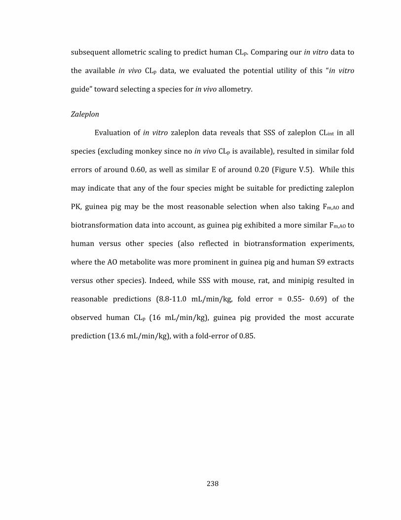
238
subsequent allometric scaling to predict human CLp. Comparing our in vitro data to
the available in vivo CLp data, we evaluated the potential utility of this “in vitro
guide” toward selecting a species for in vivo allometry.
Zaleplon
Evaluation of in vitro zaleplon data reveals that SSS of zaleplon CLint in all
species (excluding monkey since no in vivo CLp is available), resulted in similar fold
errors of around 0.60, as well as similar E of around 0.20 (Figure V.5). While this
may indicate that any of the four species might be suitable for predicting zaleplon
PK, guinea pig may be the most reasonable selection when also taking Fm,AO and
biotransformation data into account, as guinea pig exhibited a more similar Fm,AO to
human versus other species (also reflected in biotransformation experiments,
where the AO metabolite was more prominent in guinea pig and human S9 extracts
versus other species). Indeed, while SSS with mouse, rat, and minipig resulted in
reasonable predictions (8.8-11.0 mL/min/kg, fold error = 0.55- 0.69) of the
observed human CLp (16 mL/min/kg), guinea pig provided the most accurate
prediction (13.6 mL/min/kg), with a fold-error of 0.85.

239
Figure V.5. Evaluation of in vitro data to select a preclinical species for in vivo PK of
zaleplon and subsequent SSS to predict human CLp. Top: In vitro zaleplon biotransformation
experiments reveal similar metabolism between human and guinea pig, while greater
differences were observed between human and mouse, rat, or minipig. Middle: In vitro
zaleplon CLint experiments reveal similar E across all species, as well as a reasonable
prediction of human CLint by SSS; however, guinea pig exhibited a higher Fm,AO versus other
species, which was more similar to human. Bottom: In vivo zaleplon PK studies reveal CLp
as a % of liver blood flow most similar between human and guinea pig, which yielded the
most accurate human CLp prediction by SSS. Although guinea pig provided the most
accurate CLp prediction, SSS with all species generated similar predictions.

240
In addition, according to our in vitro MA studies, either species combination of
minipig/guinea pig/mouse or minipig/rat/mouse may be selected based on the
favorable fold error of 0.68 by both methods (Table V.7). However, as guinea pig
was selected for SSS based on biotransformation and Fm,AO data, guinea pig may also
be a more appropriate selection than rat to include in 3-species allometry along
with minipig and mouse. Indeed, in vitro and in vivo MA with minipig/guinea
pig/mouse and with minipig/rat/mouse both resulted in human predictions within
2-fold of observed CL values, with the inclusion of guinea pig resulting in a slightly
improved in vivo prediction (9.03 mL/min/kg) versus inclusion of rat (7.82
mL/min/kg).
Table V.7. In vitro-to-in vivo comparison of zaleplon human CL (CLint or CLp) predictions by
multispecies allometry (MA) using minipig, guinea pig, and mouse or using minipig, rat, and
mouse.

241
O6-benzylguanine
All in vitro data obtained for O6-benzylguanine indicates guinea pig may be
the most appropriate species selection to predict human CLp (Figure V.6).
Biotransformation experiments (Figure V.6 shows relative formation of M1, which
was the only metabolite observed in S9 extracts) revealed low turnover of O6-
benzylguanine to M1 in mouse, rat, and minipig, while guinea pig demonstrated
similar M1 formation to human. Likewise, guinea pig demonstrated a similar E and
Fm,AO to human while the other three species exhibited lower E, and their Fm,AO could
not be estimated. In vivo, however, the rodent species all exhibited very high O6-
benzylguanine CLp, which exceeded hepatic blood flow (i.e., CLp as % Liver Blood
Flow > 100%), while minipig CLp was high, but below hepatic blood flow. As a result,
SSS with the rodent species generated 3-4 fold over-predictions (53.8-59.8
mL/min/kg) of the observed human CLp (14.5 mL/min/kg), whereas minipig SSS
predicted a human CLp of 13.8 mL/min/kg, with a fold error of 0.95.

242
Figure V.6. Evaluation of in vitro data to select a preclinical species for in vivo PK of O6-
benzlguanine and subsequent SSS to predict human CLp. Top: In vitro O6-benzylguanine
biotransformation experiments reveal low formation of M1 in mouse, rat, and minipig,
while guinea pig demonstrated similar M1 formation to human. Middle: In vitro O6-
benzylguanine CLint experiments reveal similar Fm,AO and E between human and guinea pig,
with low E in mouse, rat, and minipig and low CLint preventing estimation of Fm,AO in these
species. Likewise, guinea pig SSS accurately predicted human CLint, while predictions from
the other three species were low. Bottom: In vivo O6-benzylguanine PK studies reveal CLp as
a % of liver blood flow most similar between human and minipig, which also yielded the
most accurate human CLp prediction by SSS, while mouse, rat and guinea pig over-predicted
human CLp ~3-4 fold.

243
MA with minipig/guinea pig/mouse or minipig/rat/mouse yielded reasonable
human O6-benzylguanine CLp predictions (10.1 and 9.48 mL/min/kg, respectively)
with fold errors of 0.69 and 0.65, respectively (Table V.8). Due to low in vitro CLint in
mouse, rat, and especially minipig, human in vitro CLint predictions by these MA
methods were low, as was the case with in vitro SSS predictions using these species.
Table V.8. In vitro-to-in vivo comparison of O6-benzylguanine human CL (CLint or CLp)
predictions by multispecies allometry (MA) using minipig, guinea pig, and mouse, or
minipig, rat, and mouse.
Zoniporide
When comparing zoniporide in vitro data of mouse, rat, cynomolgus monkey,
and minipig, the species exhibiting the most similarity to human across all data
(with the exception of Fm,AO) is minipig, though cynomolgus monkey also produced
similar in vitro data to human with a SSS CLint prediction fold error of 1.7 (Figure
V.7). Rat and mouse in vitro data indicated that these species may be likely to over-
predict human CLp, and, as anticipated, rat and mouse did over-predict human CLp

244
(21 mL/min/kg), though both over-predictions were within 3-fold (62.7 and 39.2
mL/min/kg, respectively). In addition, cynomolgus monkey did generate a more
accurate prediction (15.2 mL/min/kg) than mouse and rat, with a fold error of 0.72.
SSS with minipig, however, produced a 5-fold over prediction (111 mL/min/kg) of
human CLp. The CLp data employed in the SSS analysis for mouse, rat, and
cynomolgus were obtained from the literature, as plasma concentrations obtained
from our cassette dosing studies in rat and mouse were below our quantitation
limits, therefore preventing PK analysis. Observed concentrations in minipig plasma
were also very low (peak concentrations < 10 ng/mL), which may have limited the
ability to obtain an accurate PK assessment.

245
Figure V.7. Evaluation of in vitro data to select a preclinical species for in vivo PK of
zoniporide and subsequent SSS to predict human CLp. Top: In vitro zoniporide
biotransformation experiments reveal similar metabolism between all species, with rat and
mouse exhibiting much higher turnover of zoniporide relative to human and minipig and
somewhat higher relative to cynomolgus monkey. Middle: In vitro zoniporide CLint
experiments reveal similar Fm,AO across all species, with rat exhibiting a Fm,AO most similar to
human. However, minipig and cynomolgus monkey exhibit an E most similar to human, as
well as more accurate predictions of human CLint by SSS. Bottom: In vivo zoniporide PK
studies reveal CLp as a % of liver blood flow most similar between human and cynomolgus
monkey, which also yielded the most accurate human CLp prediction by SSS.

246
In addition, both in vitro and in vivo 3- or 4-species MA with minipig, cynomolgus,
rat, and/or mouse resulted in zoniporide human CLint or CLp predictions within 2-
fold of observed human values, with the exception of the minipig/rat/mouse
combination, which resulted in a > 7-fold over-prediction of human CLp (Table V.9).
Table V.9. In vitro-to-in vivo comparison of zoniporide human CL (CLint or CLp) predictions
by multispecies allometry (MA) using 3- or 4-species combinations of minipig, cynomolgus
monkey, rat, and/or mouse.

247
BIBX1382
Based on our in vitro analyses of BIBX1382, cynomolgus monkey appears to
be a more appropriate species selection than mouse and rat (Figure V.8). In this
case, all in vitro data pointed toward cynomolgus monkey over the rodent species,
as cynomolgus exhibited similar biotransformation, E, Fm,AO, and most accurately
predicted human CLint by SSS, whereas mouse and rat exhibited dissimilar
biotransformation of BIBX1382, lower E values, lower or unobtainable Fm,AO
estimates, and substantially under-predicted human CLint by SSS. In vivo SSS
resulted in human CLp predictions of 56 mL/min/kg with cynomolgus monkey, 7.2
mL/min/kg with mouse, and 13.4 mL/min/kg with rat, revealing that cynomolgus
monkey indeed was the more appropriate species to predict the observed human
CLp of BIBX1382 (25-55 mL/min/kg). In addition, literature reports note that oral
bioavailability of BIBX1382 in monkey (6%) mirrored that of human (5%) (Hutzler
et al., 2014a), unlike rat and mouse which exhibited 50-100% oral bioavailability
(Dittrich et al., 2002). Hutzler et al. likewise concluded in their report that
cynomolgus monkey accurately represented the rapid clearance of BIBX1382 in
human and that cynomolgus may therefore serve as a suitable animal model for
estimating human AO metabolism and clearance (Hutzler et al., 2014a).

248
Figure V.8. Evaluation of in vitro data to select a preclinical species for in vivo PK of
BIBX1382 and subsequent SSS to predict human CLp. Top: In vitro BIBX1382
biotransformation experiments reveal similar metabolism between human and cynomolgus,
while greater differences were observed between human and rodents. Middle: In vitro
BIBX1382 CLint experiments reveal similar E and Fm,AO between human and cynomolgus, as
well as a reasonable prediction of human CLint by SSS with cynomolgus, while rodents
exhibited lower E, Fm,AO, and predicted human CLint by SSS. Bottom: In vivo BIBX1382 PK
studies reveal similar CLp as a % of liver blood flow and oral bioavailability between human
and cynomolgus, as well as a reasonable human CLp prediction by SSS with cynomolgus,
while rodents under-represented clearance.

249
In addition, allometric scaling of in vitro CLint with cynomolgus/rat/mouse resulted
in a similar fold-error to that obtained from allometric scaling of in vivo CLp using
these species (Table V.10). As was the case in vitro, SSS from cynomolgus monkey
CLp provides the best prediction of human CLp over SSS with mouse and rat or over
MA with the three species.
Table V.10. In vitro-to-in vivo comparison of BIBX1382 human CL (CLint or CLp) predictions
by multispecies allometry (MA) using cynomolgus monkey, rat, and mouse.
SGX523
In vivo allometry could not be evaluated for accuracy of prediction of human
SGX523 CLp, as human CLp data for this compound is not available in the literature.
Evaluation of the in vitro data indicates that mouse, rat, and perhaps guinea pig may
each serve as more suitable species for in vivo SSS to predict human CLp than
cynomolgus monkey or minipig when considering E and SSS CLint predictions
(Figure V.9). Fm,AO estimations were somewhat variable by the two calculation
methods (see Chapter IV), and, therefore, may not serve as a useful parameter for
species selection in this case. In vitro biotransformation of SGX523 was relatively
similar across all species, with some trace metabolites detected in human that were
not detected in mouse and rat, while minipig produced lower levels of M1 relative to

250
other species. SSS of in vivo CLp with mouse, guinea pig, and minipig resulted in
similar human CLp predictions ranging from 9.6-11.0, whereas SSS with rat and
cynomolgus monkey produced lower predictions of 2.2 and 1.9, respectively. CLp as
a percentage of liver blood flow in guinea pig and minipig (57% and 68%,
respectively) were a bit higher (but fairly similar) to the E estimated in vitro (0.41
and 0.48, respectively), while rat CLp as a percentage of liver blood flow was lower
(12%) than E estimated in vitro (0.33). CLp as a percentage of liver blood flow was
substantially higher than E in mouse (92% and 0.30, respectively); the opposite was
true for cynomolgus monkey, where E was much higher in vitro (0.50) than CLp as a
percentage of liver blood flow in vivo (8%).

251
Figure V.9. Evaluation of in vitro data to select a preclinical species for in vivo PK of SGX523
and subsequent SSS to predict human CLp. Top: In vitro SGX523 biotransformation
experiments reveal similar metabolism across all species, with greater turnover of SGX523
by guinea pig and minipig, and some trace metabolites detected in human which were not
detected in mouse and rat. Middle: In vitro SGX523 CLint experiments reveal mouse and rat
E most similar to human and ambiguous Fm,AO values in the low-moderate range for all
species. Prediction of human CLint by SSS were lower using rat and mouse, higher using
cynomolgus and minipig, and in the middle using guinea pig. Bottom: In vivo SGX523 PK
studies reveal similar low human CLp prediction by SSS with cynomolgus and mouse, and
higher predictions by SSS with the other three species.

252
MA of SGX CLp resulted in similar human predictions as those obtained from SSS,
ranging from 0.6-10.5 mL/min/kg (Table V.11). As was observed with SSS, the rank
order (according to predicted CLint) of MA methods in vitro did not mirror the in vivo
rank order—for example, the cyno/rat/mouse combination predicted the highest
human CLint in vitro, but this method predicted the lowest human CLp in vivo.

253
Table V.11. In vitro-to-in vivo comparison of SGX523 human CL (CLint or CLp) predictions by
multispecies allometry (MA) using minipig, cynomolgus monkey, guinea pig, rat, and/or
mouse.

254
As previously noted, the in vivo-in vitro ratios for zaleplon were relatively
consistent across all species, and the same was true of the in vivo-in vitro ratios for
BIBX1382. The in vivo-in vitro ratios for zoniporide, O6-benzylguanine, and SGX523,
alternatively, were more varied across species. This is important to note with
regard to MA, as substantial species differences in IVIVC would be expected to
impact the translation of in vitro MA to in vivo MA with regard to maintaining a
similar fold-error of prediction between in vitro MA and in vivo MA. For example,
MA of zaleplon clearance resulted in similar prediction fold errors both in vitro and
in vivo (average fold error in vitro = 0.68, average fold error in vivo = 0.53, Table
V.7). MA of BIBX1382 clearance likewise resulted in similar fold errors both in vitro
and in vivo (Table V.10). In vivo fold errors obtained for O6-benzylguanine and
zoniporide, however, deviated (sometimes substantially) from in vitro fold errors
(Tables V.8-9). In addition, in cases where the fold errors by MA were similar in
vitro and in vivo (i.e., zaleplon and BIBX1382), the allometric exponent (b) was also
similar in vitro and in vivo (Table V.12). This observation may suggest that a MA
prediction fold error observed in vitro may be expected to translate to a similar fold
error in vivo, when the allometric exponent obtained from in vitro MA is similar to
the allometric exponent obtained from in vivo MA. As noted previously, however,
this will likely be dependent on human exhibiting a similar IVIVC (i.e., in vivo-in vitro
CL ratio) to the species used for MA predictions, which was the case for zaleplon and
BIBX1382.

255
Table V.12 In vitro-to-in vivo comparison of allometric exponents (b) obtained from
multispecies allometry (MA) using minipig, cynomolgus monkey, guinea pig, rat, and/or
mouse. Cyno, cynomolgus monkey; Gpig, guinea pig

256
DISCUSSION
In Chapter IV, we evaluated species differences in the metabolism and
clearance of five AO substrates in vitro, as well as the ability to scale in vitro hepatic
CLint of these substrates by MA and SSS to predict human in vitro hepatic CLint.
Successful prediction of human in vitro hepatic CLint was achieved by one or more of
these methods, albeit in a substrate-dependent manner with regard to the species
employed in the scaling analysis. As traditional in vitro techniques have repeatedly
resulted in under-prediction of in vivo CLp for compounds metabolized by AO
(proposed to potentially be associated with extra-hepatic AO metabolism, instability
of AO in vitro, or interindividual AO variability) (Zientek et al., 2010; Akabane et al.,
2012; Hutzler et al., 2012), we presently sought to evaluate the multispecies IVIVC
(in vitro-in vivo correlation ) of clearance for these five AO substrates. Furthermore,
we evaluated the ability to predict human in vivo CLp of these compounds by MA and
SSS, employing CLp obtained from multiple species. Finally, we assessed the utility of
comparing in vitro metabolism and clearance data from multiple species for the
purpose of selecting suitable species for in vivo PK analysis and subsequent human
CLp prediction by MA or SSS.
Of the compounds/species for which in vivo data was available to evaluate, a
prediction within 3-fold of observed human CLp was successfully obtained by at
least one MA or SSS method for all compounds. In instances where IVIVC was
similar across species (i.e., demonstrated a similar in vivo-in vitro clearance ratio)
employed in MA or SSS analyses , human prediction fold errors obtained from both

257
in vitro and in vivo allometric scaling with these species were also similar.
Consequently, for compounds exhibiting similar IVIVC across all species (e.g.,
zaleplon and BIBX1382) in vitro data successfully indicated which species would be
suitable for employing in vivo CLp in MA or SSS analyses to predict human CLp.
Furthermore, these two cases resulted in similar in vitro and in vivo allometric
exponents obtained from MA, indicating that the interspecies relationship between
in vitro CLint and body weight reflected the interspecies relationship between in vivo
CLp and body weight (i.e., the rate of change in CL with change in body weight was
similar in vitro and in vivo). However, in cases where IVIVC was inconsistent across
species, in vitro data was not always predictive of the most appropriate species for
in vivo allometry. This may indicate that species-specific extra-hepatic mechanisms
may contribute to the clearance of these compounds in vivo, since the in vitro
clearance estimated in our studies is limited to hepatic mechanisms (CLint obtained
using hepatic S9 fractions).
In addition, the IVIVC analyses may offer some possible insight into which of
the previously proposed mechanisms (e.g., extra-hepatic elimination, AO instability
in vitro, interindividual variability, etc.) may be contributing the observed IVIVC
disconnects frequently encountered with AO substrates. For example, for any one
species in our cassette dosing studies, the fold difference between in vitro and in
vivo CL was not always consistent across each compound (e.g., rat demonstrated an
in vivo-in vitro fold-difference of 0.38, 0.04, and 2.6 for zaleplon, O6-benzylguanine,
and SGX523, respectively). Because the same lot of hepatic S9 was used to obtain in

258
vitro CLint estimates for each of the three compounds, the substrate-dependent
variability in IVIVC for a single species cannot be attributed to variable AO activity
in the S9 used to measure the CLint for each compound (e.g., manufacturer
differences in preparation of S9 fractions or lot-to-lot donor variability). Likewise,
because the same animals were used to obtain in vivo CLp of zaleplon, O6-
benzylguanine, and SGX523 (all three compounds dosed together as a cassette to the
same animals), the variability in IVIVC across the three compounds cannot be
attributed to variable AO activity (i.e., interindividual variability) in the animals
receiving each compound. Rather, the differences may likely be ascribed to
substrate-dependent parameters such as red blood cell distribution and/or extra-
hepatic metabolism (tissue or plasma), since the S9 CLHEP data only represents
hepatic clearance. This proposal is also supported by CLp values, either observed in
our studies or reported by others, which exceeded hepatic blood flow (e.g. O6-
bezylguanine, zoniporide, and BIBX1382). Interestingly, the fold-difference was very
similar across all species in the in vitro-in vivo comparison of zaleplon. This might
indicate that the under-estimation is a result of an extra-hepatic mechanism that is
conserved across species. Previous studies in rats receiving a 5 mg/kg oral dose of
[14C]-zaleplon found that highest concentrations of the drug were distributed into
the liver, kidney, gastrointestinal tract, and adrenal glands (Beer et al., 1997).
Human AOX1 mRNA is present in each of these tissues, with the adrenal glands
reported as one of the richest sources of the protein in addition to the liver (Terao et
al., 2016). However, reported tissue expression patterns of AOX1 and other AO

259
isoforms between mouse and human are divergent, and the adrenal gland, for
example, has not been shown to represent a rich source of mRNA for any AO isoform
in mouse (Terao et al., 2016). Unfortunately, until multispecies expression and activity
of extra-hepatic AO are better understood, the mechanism(s) responsible for IVIVC
disconnects will remain unclear.
While the variability in IVIVC for SGX523, O6-benzylguanine, and zaleplon for
any given species could not have resulted from differences in preparation of S9
fractions used to measure CLint for each compound or interindividual AO variability
in the animals used to measure CLp for each compound, this does not preclude in
vitro instability/decreased activity or interindividual variability between the S9
donors and the subjects receiving the test article in vivo from speculation as a
potential contributor to in vitro under-estimation of CLp. Interestingly, however,
SGX523 CLp was over-estimated by rat S9, while zaleplon and O6-benzylguanine
were under-estimated. If in vitro AO instability or interindividual AO variability
between the S9 donor rats and the rats administered substrate in vivo were
responsible for the IVIVC disconnect, the disconnect would be expected to be in the
same direction for all compounds (i.e., all under-estimated or all over-estimated),
Notably, however, we previously reported an Fm,AO (fraction of metabolism mediated
by AO) for SGX523 of 0.28-0.42 in rat S9. This presence of non-AO metabolism
pathways in the clearance of these substrates adds an additional complication to the
interpretation of IVIVC disconnects for AO substrates. For example, in our in vitro
CLint studies, we did not observe measurable substrate turnover of O6-benzylguaine

260
in rat, mouse, and minipig S9 in the absence of NADPH, whereas substrate depletion
was observed in the presence of NADPH, indicating the contribution of a non-AO
pathway. A prior report of O6-benzylguanine metabolism in rat and mouse in vivo
revealed an N-acetyl metabolite in rats and a debenzylated metabolite in both rats
and mice (Dolan et al., 1994). It was proposed that the acetylation may occur in the
kidney since it was only detected in urine and not in the plasma (Dolan et al., 1994).
Neither of these metabolites was detected in our biotransformation experiments in
S9 incubations (Chapter IV), suggesting the possibility that these clearance
pathways were present in vivo, but not in vitro, and contributed to the apparent
extra-hepatic metabolism and IVIVC disconnect observed with O6-benzylguanine.
However, even in the absence of extra-hepatic metabolism, differences in Fm,AO for
each compound could result in variable IVIVC across substrates. Likewise, other
non-AO substrate dependent parameters, such as red blood cell distribution, could
also contribute to inconsistencies between in vitro estimated hepatic clearance
(CLHEP) and CLp, as CLp reflects substrate clearance from plasma as opposed to
whole blood. As additional research is needed to better understand extra-hepatic
AO metabolism, additional research to clarify mechanisms of in vitro AO instability
and interindividual variability (single nucleotide polymorphisms, influence of
disease state, etc.) will be necessary to fully elucidate contributing factors to IVIVC
discrepancies.
Unfortunately, our studies of BIBX1382 were limited due to plasma
concentrations below the limit of quantitation preventing pharmacokinetic

261
parameters from being obtained for this compound. This perhaps is not altogether
surprising, given the exposure in cynomolgus monkeys receiving an IV dose of 1
mg/kg BIBX1382, where peak plasma concentrations only reached approximately
70 nM (~180 ng/mL) (Hutzler et al., 2014a), and animals in our studies only
received one fifth of this dose (0.2 mg/kg). It was determined that BIBX1382
partitioned into red blood cells in cynomolgus monkey with a blood-to-plasma ratio
of 2.1 (Hutzler et al., 2014a). Thus, it is possible that red blood cell partitioning
could account for the low plasma concentrations we observed. In addition,
evaluation of human and cynomolgus plasma stability indicated BIBX1382 was
stable in plasma (for at least 2 hours); however, it is still possible that plasma
instability could contribute to low concentrations in mouse, rat, guinea pig and/or
minipig. In particular, plasma instability of BIBX1382 could result from plasma
xanthine oxidase (XO) mediated oxidation, as Sharma et al. previously demonstrated
that a pyrazine-containing compound was oxidized by XO in plasma of mouse, rat,
and guinea pig plasma but was stable in human, monkey, and dog plasma (Sharma
et al., 2011). In addition, Hutzler et al. noted high AO activity towards BIBX1382 in
cynomolgus monkey lung S9 fractions, suggesting the possibility that pulmonary
first pass metabolism could contribute to the low peak plasma concentrations
observed in our studies (Hutzler et al., 2014a). These mechanisms could have
potentially contributed to low zoniporide plasma concentrations observed in our
studies as well. Zoniporide was reported to partition 1:1 into plasma and red blood

262
cells in rat and human (Tracey et al., 2003), but has not been reported for the other
species in our studies.
In conclusion, we have demonstrated that allometric scaling may be useful to
predict human CLp of AO substrates when the appropriate species are employed, as
predictions within 2-3 fold of observed human CLp values were obtained for
zaleplon, O6-benzylguanine, zoniporide, and BIBX1382 by one or more MA and/or
SSS methods. In addition, MA and SSS resulted in similar prediction fold errors in
vitro and in vivo when IVIVC was consistent across species, suggesting that in vitro
allometry may be useful to guide species selection to conduct in vivo allometric
scaling for human CLp prediction. While additional studies to elucidate mechanisms
behind discrepancies in IVIVC, as well as allometry studies with a larger sample of
AO substrates, would help to better understand the potential to broadly utilize this
approach toward species selection and prediction of human CLp for drug candidates
metabolized by AO, these studies offer direction towards a novel approach to
estimate human clearance of AO substrates, which is of great necessity for the
successful discovery and development of future therapeutics.

263
CHAPTER VI
CONCLUSIONS AND FUTURE DIRECTIONS
Unacceptable pharmacokinetics (PK) once represented a leading cause of
attrition of drug candidates during clinical trials (Kola and Landis, 2004). Today,
with major advances in the understanding of cytochrome P450 function, expression,
and regulation in human and nonclinical species, standardized methods to predict
human PK of drugs metabolized by P450 enzymes are now available, resulting in a
reduction in drug attrition rates associated with unexpectedly poor PK (Kola and
Landis, 2004; Di et al., 2013). Unfortunately, application of these methodologies to
human PK prediction of compounds exhibiting AO-mediated clearance has proven
insufficient and consequently resulted in clinical failures of several promising drug
candidates over the past several years (Kaye et al., 1985; Dittrich et al., 2002;
Diamond et al., 2010a; Akabane et al., 2011; Zhang et al., 2011; Lolkema et al., 2015).
Even so, reliable and standardized methodology to predict the human
pharmacokinetics and drug interaction liability of compounds metabolized by AO
has yet to be firmly established. The research described herein provides a
foundation towards a solution to this challenging problem, offering several
contributions to advance the field surrounding AO drug metabolism and human PK
prediction, which are summarized below along with suggested future directions to
continue improving and developing these methodologies.

264
Drug Interaction Liability Associated with AO-Mediated Drug Clearance
Summary and conclusions
Studies relating to the drug interaction potential associated with inhibition of
AO have been conducted, and several drugs have been identified which exhibit
inhibitory activity towards AO in vitro (Obach, 2004; Barr and Jones, 2011).
However, given the numerous drugs which inhibit P450 enzymes currently on the
market, studies to evaluate the impact of P450 inhibition on compounds
metabolized by both P450 and AO may be of equal importance from a perspective of
clinical significance. Interestingly, we found that administration of the mixed
AO/P450 substrate VU0409106 along with the P450 inhibitor ABT to rats resulted
in a drug interaction, where exposure to not only VU0409106 was elevated, but
exposure to the AO metabolite M1 was also elevated. Notably, while a drug
interaction-mediated elevation in metabolite exposure is typically associated with
induction of a drug metabolizing enzyme (DME), our studies demonstrated an
elevation in metabolite exposure facilitated by DME inhibition. Reports of AO
metabolite-related toxicity associated with methotrexate and the failed clinical drug
candidates SGX523 and JNJ-38877605 highlight an additional concern regarding the
potential clinical significance of this type of drug interaction (Diamond et al., 2010a;
Lolkema et al., 2015). Taken together, these data reveal that mixed AO/P450-
metabolized drugs are susceptible to potentially clinically relevant interactions with
P450-inhibiting drugs, and attention to this liability by the pharmaceutical industry is
warranted.

265
Future directions
The clinical relevance of a pharmacokinetic drug interaction is typically
dependent on the magnitude of change in exposure to the pharmacologically (or
toxicologically) active compound in question, in this case, the AO metabolite.
Therefore, investigations into understanding how to predict the change in AO
metabolite exposure and anticipate the conditions under which this exposure
change is likely to occur will be an important next step in characterizing this type of
drug interaction. Our studies involved the administration of a pan-P450 inhibitor
(ABT), a non-clinical tool which non-selectively inactivates P450 enzymes, resulting
in the likelihood that metabolism of VU0409106 was primarily shunted toward a
single enzyme (AO), thus maximizing the potential for increased exposure to the AO
metabolite. In a clinical setting, the co-administration of a P450-inhibiting drug (or
even multiple drugs) would be unlikely to result in inhibition of all P450 enzymes,
and consequently, shunting may occur toward other uninhibited P450s in addition
to AO, potentially limiting an increase in AO metabolite exposure. For this reason,
future investigations with mixed AO/P450 substrates should focus on modeling
interactions which are likely to occur in a clinical setting. In addition, determination
of the influence of Fm,P450 vs Fm,AO on the magnitude of change in AO metabolite
exposure resulting from P450 inhibition will be an important next step toward
establishing methods to predict drug interactions involving mixed AO/P450-
metabolized drugs. The influence of fraction metabolized via a single P450
isoenzyme (e.g., Fm, 3A4) on drug interaction liability has been established, such that

266
the increase in parent drug exposure under conditions of P450 inhibition can be
predicted when the Fm of that P450 isoenzyme is known (Di et al., 2013). In this
case, a larger Fm, P450 is associated with a greater increase in parent drug exposure
when that P450 isoenzyme is inhibited (Di et al., 2013). Accordingly, a greater
increase in AO metabolite exposure might be expected under conditions of P450
inhibition when the Fm,P450 is large; however, studies to demonstrate this
relationship have yet to be conducted. Furthermore, as the extra-hepatic expression
and activity of AO is poorly understood (Terao et al., 2016), studies examining the
potential influence of intestinal AO-mediated metabolism will be important to
determine the impact of route of administration (e.g., oral versus intravenous) on
the likelihood of a clinically relevant interaction. Many questions remain to be
answered concerning the role of intestinal (as well as other extra-hepatic) AO
expression to drug metabolism and total body clearance of AO-metabolized drugs,
such as endogenous and exogenous factors regulating expression in various tissues
(e.g., influence of the gut microbiome on intestinal expression). Likewise,
investigations into identifying sources of the interindividual variability (hepatic or
extra-hepatic) that has been observed in AO-mediated metabolism (Fu et al., 2013;
Hutzler et al., 2014b) will be of great importance to understand the likelihood of
experiencing a drug interaction. Many possible factors may contribute to variability,
such as disease state, diet, and single-nucleotide polymorphisms (SNPs), and while
efforts have been made to identify these associations, none have been firmly
established (Hartmann et al., 2012; Hutzler et al., 2012; Fu et al., 2013; Hutzler et al.,

267
2014b). In addition, though SNPs of AO are currently poorly understood, SNPs of
P450 enzymes are well-established (Zhou et al., 2009) and also have the potential to
influence the drug interaction liability of a mixed AO/P450 substrate, thus
warranting additional evaluation. These future works will all be essential in
establishing guidelines on metabolite safety testing and drug interaction studies
involving mixed AO/P450 drug substrates, which are likely to increase in number
given the emergence of AO-metabolized drug candidates reaching clinical trials.
Multispecies Allometry to Predict Human Clearance of Drugs Metabolized
by Aldehyde Oxidase
Summary and conclusions
In addition to gaps in our understanding of how to predict drug interactions
associated with AO metabolism, perhaps a more significant challenge remains in
identifying reliable methods to predict the human clearance associated with AO
metabolism. Likely due to the assumption that species differences in clearance
mechanisms precludes the ability to scale clearance by allometry, studies evaluating
allometric scaling in the prediction of human clearance of AO-metabolized drugs are
sparse. While methotrexate, an AO substrate, ironically represents one of the oldest
examples of allometric scaling of clearance, AO-mediated metabolism is a minor
contributor to the clearance of this drug, with renal elimination serving as the major
clearance mechanism (Boxenbaum, 1982). The work described herein not only
provides an evaluation of allometric scaling to predict human plasma clearance

268
(CLp) of AO substrates, but also includes assessments using minipig (as opposed to
dog, which is typically used in allometric scaling), which has been sparsely studied
with regard to AO metabolism and human clearance prediction. Importantly, our
evaluations indicate that multispecies allometry (MA) may be useful in predicting
human CLp of AO substrates, as most human CLp predictions were within 3-fold of
observed values (Table V.5). It is also encouraging to note that in the two instances
where MA did not predict human CLp within 3-fold, these were over-predictions
rather than under-predictions (over-prediction was also more prevalent than
under-prediction in our MA studies using in vitro CLint). While a substantial over-
prediction of human CLp may result in the oversight of a compound that actually
possesses acceptable human PK, a substantial under-prediction could result in the
costly failure of a drug candidate that was inappropriately advanced to clinical
trials. Traditional in vitro methods used to predict human in vivo clearance typically
result in under-prediction for AO substrates (Zientek et al., 2010; Akabane et al.,
2012), and likewise, in vivo PK assessments in traditional preclinical species (mouse,
rat, and dog) also commonly under-represent clearance in human (Kaye et al., 1985;
Dittrich et al., 2002; Akabane et al., 2011; Zhang et al., 2011). Multispecies allometry
utilizing the species evaluated herein may therefore represent a method which can
reduce the risk of encountering unexpectedly rapid human CLp when advancing an AO-
metabolized compound to clinical trials.

269
Future directions
While our data indicate potential utility of MA for human CLp prediction of AO
substrates, understanding the full value of this method will benefit from additional
investigations extended to a larger collection of substrates with reported clinical PK
data (CLp). In general, the ability to study human clearance prediction of AO
substrates is limited by the small number of available compounds for which human
(in vivo) PK have been reported. In addition, most known AO substrates with
reported human PK data are rapidly cleared (Zientek et al., 2010; Akabane et al.,
2011; Zhang et al., 2011), limiting the evaluation of prediction methodologies
primarily to “high clearance” compounds. Importantly, clearance of “high clearance”
compounds are more likely to scale according to an allometric relationship, as the
plasma clearance of these compounds is limited by hepatic blood flow (Wilkinson
and Shand, 1975; Pang and Rowland, 1977), which scales according to an allometric
relationship (Boxenbaum, 1980). Therefore, extension of studies evaluating in vivo
allometric scaling to predict human in vivo CLp to a larger set of compounds,
particularly those exhibiting a wider sampling of low, moderate, and high clearance
AO substrates, would provide a better understanding of the broad applicability of
these methods.

270
Application of In Vitro Intrinsic Clearance to Allometric Scaling
Summary and conclusions
In addition to providing a traditional examination of allometric scaling to
predict human in vivo CLp of AO-metabolized compounds, we also evaluated a novel
approach to allometric scaling with the application of in vitro CLint to these analyses.
Others have proposed that species expressing only the AOX1 isoenzyme in the liver
(e.g., guinea pig, monkey, and pig) may serve as better species to estimate human
AO-mediated metabolism versus species expressing both the AOX1 and AOX3
isoenzymes (e.g., rat and mouse) (Garattini and Terao, 2012). Consistent with this
proposal, our in vitro data indicate that the hepatic CLint of AO substrates may be
scaled from preclinical species to human by SSS with monkey, guinea pig, and
minipig with reasonable accuracy and precision, while this relationship does not
appear to be consistent (highly substrate-dependent) when directly scaling from rat
or mouse. However, similar to our observations in vivo, use of rat and mouse in
combination with guinea pig, monkey, and/or minipig generally enabled allometric
scaling to a reasonable human hepatic CLint prediction. In particular, these in vitro
allometry assessements indicate that 4-species allometry utilizing the species
evaluated herein may provide the greatest utility with regard to minimizing
substrate-dependence in the ability to reasonably predict human clearance. In
addition, comparison of in vitro allometry data to in vivo allometry data suggest that
a fold-error analysis of in vitro allometry predictions could be useful to help
determine which species would be more likely to permit allometric scaling of in vivo

271
CLp (MA or SSS) to predict human CLp. While this approach proved useful for
demonstrating appropriate species to predict human CLp of zaleplon and BIBX1382,
overall the translation of in vitro-to-in vivo allometry (regarding prediction fold-
error) was substrate-dependent, with only those compounds which exhibited a
consistent IVIVC across species (zaleplon and BIBX1382) resulting in successful
species selection. Overall, these data indicate a potential utility of in vitro allometry
in the determination of appropriate species to predict human clearance of AO-
metabolized drugs, while elucidation of discrepancies in interspecies IVIVC will be
important to better understand the full potential of this approach.
Future directions
A factor limiting the interpretation of our current approach to in vitro allometry
is the inconsistent in vitro: in vivo correlation (IVIVC), illustrating the substrate-
dependency of AO mediated metabolism. The assumption that in vitro hepatic CLint
represents in vivo hepatic CLint is hindered by speculation of ex vivo AO instability,
single-nucleotide polymorphisms, and other sources of possible interindividual
variability (e.g., disease state, etc.) (Hartmann et al., 2012; Hutzler et al., 2012; Fu et
al., 2013; Hutzler et al., 2014b). Furthermore, pharmacokinetic properties such as
plasma stability and red blood cell distribution were not determined in each species,
and because these properties could influence in vivo CLp (and consequently the
IVIVC since these components are not present in vitro) the IVIVC across species
could vary if these properties are species-specific. Finally, an additional unknown
factor complicating IVIVC interpretation, is the extra-hepatic expression and activity

272
of AO (Kurosaki et al., 1999; Moriwaki et al., 2001; Nishimura and Naito, 2006;
Terao et al., 2016), which is poorly understood at present, particularly concerning
its contribution to total body clearance of AO substrates. Prior studies indicate that
extra-hepatic AO expression is species-specific (Terao et al., 2016), which could also
influence consistency of IVIVC across species if extra-hepatic metabolism
contributes to CLp. Consequently, each of these factors could have impacted the
interspecies IVIVC obtained in our studies. As mentioned previously, while others
have already initiated research aimed at addressing these questions, a thorough
investigation to build upon their findings will be necessary to elucidate the
mechanisms behind variable AO activity and will be essential to improve current in
vitro techniques to estimate AO-mediated clearance. Likewise, future research to
establish species-specific tissue expression patters, mechanisms regulating AO
expression, and importantly, to develop standardized in vitro scaling factors that can
be used to estimate total organ clearance from in vitro CLint in extra-hepatic tissues
will all be critical steps towards understanding the potential contribution of extra-
hepatic metabolism and establishing confidence in in vitro-to-in vivo extrapolation
where AO metabolism is concerned. In addition, as 4-species allometric scaling of in
vitro CLint appears to exhibit minimal substrate-dependence with regard to
obtaining a reasonable human CLint prediction, evaluation of these species
combinations in vivo will help to validate the proposed use of this method.

273
Influence of Fraction Metabolized by AO (Fm,AO) and Hepatic Extraction (E)
on SSS Prediction Accuracy
Summary and conclusions
Along with our in vitro allometry studies, we estimated interspecies fraction
metabolized by AO (Fm,AO) and hepatic extraction (E) for each of our probe AO
substrates in order to evaluate how these parameters might influence the ability to
predict human hepatic CLint by SSS. Our findings revealed a poor relationship
between the animal:human ratio of Fm,AO and prediction accuracy by SSS, while
further investigation instead revealed a relationship between animal:human ratio of
E and prediction accuracy by SSS. Importantly, these relationships were
recapitulated when the analyses were conducted using in vivo CLp and Fm,UGT data
reported by Deguchi et al. for several UGT substrates (Deguchi et al., 2011),
indicating this is not an isolated observation. In addition, biotransformation
experiments revealed that human and preclinical species may exhibit a similar E
despite divergent Fm,AO values due to compensation via greater NADPH-dependent
metabolism in the preclinical species (e.g., minipig vs. human metabolism of
zaleplon). Consequently, these findings suggest that Fm,AO may be more important to
consider from a metabolite exposure (i.e., toxicology) perspective than a clearance
perspective concerning extrapolation of animal PK to human, whereas consideration
of E should be given priority to Fm,AO when selecting species for human clearance
estimation.

274
Future directions
As four of the five compounds studied herein exhibited human Fm,AO values
≥.0.70., evaluation of additional compounds exhibiting a wider distribution of Fm,AO
values in human would improve our understanding of how important this
parameter may be to accurately predicting human clearance with allometry. In
addition, our understanding of how to appropriately utilize preclinical species for
toxicology studies/metabolite safety testing with regard to AO substrates will
benefit from future investigations focusing on the influence of interspecies Fm, AO. In
addition, while our findings implicated interspecies E as an important factor
associated with SSS prediction accuracy, extension of these analyses to prediction of
oral bioavailability would be a valuable next step to understand the utility of
preclinical species in predicting human PK of AO-metabolized compounds (again
emphasizing the importance of evaluating the role of interspecies
expression/regulation of intestinal AO to oral bioavailability).
Interspecies Evaluation of Metabolism, Clearance, and SSS to Predict
Human Clearance of AO Substrates
Summary and conclusions
Within our in vivo SSS analyses, an apparent substrate-dependency was
observed with regard to successful SSS using a particular species (Table V.3). In
addition, our data (along with prior clinical failures where rat and/or mouse were

275
used for preclinical PK assessments) appear to indicate that MA may be more useful
than SSS with rat or mouse to predict human CLp (even when rat and/or mouse are
included in the MA assessment), which, as previously discussed, was reiterated by
our in vitro SSS assessments. SSS with cynomolgus monkey, on the other hand,
successfully predicted both zoniporide and BIBX1382 CLp in human, and though CLp
data in cynomolgus for zaleplon and O6-benzylguanine was not available for
evaluation, AO-mediated zaleplon metabolite formation in vivo in cynomolgus has
been reported to be similar to human (Kawashima et al., 1999). In addition, while
human CLp of SGX523 was not available for analysis of prediction fold-error by
cynomolgus SSS, Diamond et al. reported that cynomolgus monkey would have
served as a more relevant species for nonclinical toxicological evaluation of SGX523
than rat, which produced low levels of the offending AO metabolite in vivo (Diamond
et al., 2010b). Likewise, comparison of monkey and human in vitro data obtained
from our studies (SSS of CLint, estimated E and Fm,AO, and biotransformation) for each
of the five AO substrates also supports monkey as a suitable species to estimate
human clearance and extent of AO-metabolite formation. Furthermore, a recent
report where unexpectedly rapid AO metabolism resulted in clinical failure of Lu
AF09535 indicated that monkey would have been a suitable species to predict the
poor oral exposure observed in humans (Jensen et al., 2016). However, even
monkey may exhibit some substrate-dependence with regard to predicting human
PK of AO substrates, as Zhang et al. reported that formation of the AO metabolite of
the p38 kinase inhibitor RO1 identified in human was negligible in monkey (Zhang

276
et al., 2011). Overall, monkey appears likely to be an appropriate species for
estimating AO-mediated metabolism and clearance in human and should be
considered more reliable than rat or mouse, particularly when AO-mediated
metabolism is low in rodent and higher in monkey.
Though some substrate dependence was observed, our in vitro data also support
prior speculation that guinea pig could serve as a suitable species for estimation of
human AO-mediated metabolism (Garattini and Terao, 2012). SSS with guinea pig
CLint predicted four of the five compounds within 2-fold of human CLint, and Fm,AO, E,
and biotransformation for these four compounds were similar between human and
guinea pig. However, guinea pig metabolism and clearance of BIBX1382
demonstrated a closer resemblance to the other rodent species than to human, and
consequently, SSS with guinea pig CLint under-predicted human CLint, of BIBX1382
similar to SSS of CLint with rat and mouse. Likewise, though guinea pig SSS
accurately predicted human CLint of O6-benzylguaine in vitro, SSS with guinea pig in
vivo over-predicted human CLp 3.9-fold, similar to over-predictions obtained using
rat and mouse. These data indicate that perhaps guinea pig may be used as an initial
screening tool for metabolic stability, with the recommendation that potential drug
candidates be evaluated in monkey prior to advancing to clinical trials. Notably, when
guinea pig exhibits similar metabolism/clearance to rat and mouse in vitro and/or in
vivo (especially if different from human in vitro), it may be particularly important to
evaluate additional species such as monkey.

277
While minipig successfully predicted human in vivo CLp of zaleplon and O6-
benzylguanine within 2-fold, human zoniporide CLp was over-predicted ~5-fold. In
addition, substrate-dependence of AO metabolism in vitro further indicates that
caution should be advised with regard to broad use of this species to estimate AO-
mediated metabolism in human. Though minipig SSS of in vitro CLint predicted
human CLint within 3-fold for four of the five compounds evaluated, Fm,AO and
biotransformation experiments revealed AO-mediated metabolism was not always
similar between minipig and human (decreased AO-mediated metabolism of
zaleplon and O6-benzylguanine in minipig S9 relative to human). Interestingly,
NADPH-dependent metabolism of zaleplon in minipig S9 apparently compensated
for the decreased AO metabolism, resulting in a similar E between minipig and
human. While this apparent compensation effectively permited prediction of
zaleplon human CLint by minipig SSS, this NADPH-dependent compensatory effect
was not observed for O6-benzylguanine. Furthermore, while an NADPH-dependent
mechanism may compensate for low AO metabolism in a hepatic in vitro system, this
compensatory effect would not be expected in extra-hepatic tissues where AO
metabolism might also occur in vivo. Interestingly, there was essentially no
difference in the IVIVC of zaleplon between human and minipig, though it is not
known if zaleplon is subject to extra-hepatic metabolism in vivo. While minipig is
increasing in popularity as a model species for preclinical toxicology evaluations
(Bode et al., 2010; van der Laan et al., 2010), this apparent substrate-dependence in
AO vs. P450-mediated metabolite formation poses another cause for concern in the

278
utility of this species for toxicology when AO metabolism predominates in human.
However, MA with the inclusion of minipig was generally successful, particularly
with regard to methods employing four species in in vitro allometry assessments
(CLp data was not available for a comprehensive assessment of 4-species allometry
in vivo). Overall, the present data indicate that broad application of a minipig model
for human PK of AO substrates is not advisable, though it still may be useful in
combination with other preclinical species for multispecies allometry.
Future directions
As our in vitro examination of minipig S9 revealed substrate-dependency in
AO-mediated metabolism relative to that in human S9, investigations to study in
vitro versus in vivo metabolism of AO substrates in minipig would shed light on the
potential to use in vitro metabolism data to determine if minipig would be an
appropriate species for toxicology assessments. While we evaluated interspecies in
vivo PK in the present investigations, we did not examine metabolite formation in
vivo. In addition, as was previously reported by Dalvie et al with regard to
metabolism of zoniporide (Dalvie et al., 2013), we observed higher CLint of all five
compounds in hepatic S9 of female minipigs relative to male minipig S9. This
observation leaves open the question as to whether female minipig may serve as a
more useful human PK model than male minipig. Biotransformation experiments
and in vivo PK assmessments in female minipigs will be required to better
understand this possibility. Extension of the proposed future investigations
concerning minipig to investigations in guinea pig and monkey, of course, would

279
also serve well to better inform the drug metabolism and disposition community as
to the general utility of preclinical species in human PK prediction.
Commentary
A comprehensive assessment of all data presented, including in vitro
biotransformation, in vitro estimation of Fm,AO and E, and SSS with both in vitro CLint
and in vivo CLp support prior postulations that guinea pig and monkey would likely
serve as better models of AO-mediated drug clearance in human versus commonly
employed nonclinical models such as rat or mouse (Garattini and Terao, 2012;
Hutzler et al., 2013; Hutzler et al., 2014a); importantly, no single species should be
expected to reflect human clearance of all AO substrates (Choughule et al., 2013b;
Hutzler et al., 2013). Moreover, the minipig represents a species to consider when
investigating AO metabolism, particularly when employed in multispecies allometry.
Collectively, our data support the need for a multiple species assessment when gauging
the intrinsic lability of new chemical entities (NCEs) towards AO metabolism and the
projection of that metabolism-mediated drug clearance in human. With regards to
human pharmacokinetic predictions, our data support a confidence-inspiring
approach towards the scaling of human clearance when nonclinical species
metabolism, single-species scaling, and the corresponding IVIVC assessments are all
similar between the species employed during preclinical investigations (e.g., rat,
guinea pig, and monkey); when these confidence inspriring tenets are not observed,

280
the present data would support use of nonhuman primate for the in vitro and in vivo
investigations of NCEs. Applications of the methodology presented herein, either as a
stand alone or in combination with previously published strategies (Zientek et al.,
2010) would likely reduce the risks associated with AO-mediated clearance in
clinical trials. While mechanisms behind variable AO activity (in vitro and/or in
vivo) and contributions of extra-hepatic metabolism remain important unanswered
questions towards the implementation of standardized methods pertaining to AO-
mediated drug disposition, the work provided here offers new insight to aid in the
appropriate application of preclinical species, thus helping to prevent future clinical
failures resulting from unexpected human AO metabolism. Furthermore, the
present body of research may in fact be applied to other emerging drug
metabolizing enzyme classes by which standardized methodologies (i.e., for P450-
mediated drug clearance) also falls short of predicting hepatic metabolism and drug
clearance.

281
REFERENCES
Adolph EF (1949) Quantitative Relations in the Physiological Constitutions of Mammals. Science 109:579-585.
Akabane T, Gerst N, Masters JN, and Tamura K (2012) A quantitative approach to
hepatic clearance prediction of metabolism by aldehyde oxidase using custom pooled hepatocytes. Xenobiotica 42:863-871.
Akabane T, Tanaka K, Irie M, Terashita S, and Teramura T (2011) Case report of
extensive metabolism by aldehyde oxidase in humans: Pharmacokinetics and metabolite profile of FK3453 in rats, dogs, and humans. Xenobiotica 41:372-384.
Al-Salmy HS (2001) Individual variation in hepatic aldehyde oxidase activity. IUBMB Life
51:249-253. Alfaro JF and Jones JP (2008) Studies on the mechanism of aldehyde oxidase and
xanthine oxidase. J Org Chem 73:9469-9472. Barr JT and Jones JP (2011) Inhibition of human liver aldehyde oxidase: implications for
potential drug-drug interactions. Drug Metab Dispos 39:2381-2386. Barr JT, Jones JP, Joswig-Jones CA, and Rock DA (2013) Absolute quantification of
aldehyde oxidase protein in human liver using liquid chromatography-tandem mass spectrometry. Mol Pharm 10:3842-3849.
Beedham C (1985) Molybdenum hydroxylases as drug-metabolizing enzymes. Drug
Metab Rev 16:119-156. Beedham C, Bruce SE, Critchley DJ, al-Tayib Y, and Rance DJ (1987) Species variation in
hepatic aldehyde oxidase activity. Eur J Drug Metab Pharmacokinet 12:307-310. Beedham C, Critchley DJ, and Rance DJ (1995) Substrate specificity of human liver
aldehyde oxidase toward substituted quinazolines and phthalazines: a comparison with hepatic enzyme from guinea pig, rabbit, and baboon. Arch Biochem Biophys 319:481-490.
Beedham C, Miceli JJ, and Obach RS (2003) Ziprasidone Metabolism, Aldehyde Oxidase,
and Clinical Implications. Journal of Clinical Psychopharmacology 23:229-232.

282
Beer B, Clody DE, Mangano R, Levner M, Mayer P, and Barrett JE (1997) A Review of the Preclinical Development of Zaleplon, a Novel Non-Benzodiazepine Hypnotic for the Treatment of Insomnia. CNS Drug Reviews 3:207-224.
Bode G, Clausing P, Gervais F, Loegsted J, Luft J, Nogues V, and Sims J (2010) The utility
of the minipig as an animal model in regulatory toxicology. J Pharmacol Toxicol Methods 62:196-220.
Bogaards JJ, Bertrand M, Jackson P, Oudshoorn MJ, Weaver RJ, van Bladeren PJ, and
Walther B (2000) Determining the best animal model for human cytochrome P450 activities: a comparison of mouse, rat, rabbit, dog, micropig, monkey and man. Xenobiotica 30:1131-1152.
Boxenbaum H (1980) Interspecies variation in liver weight, hepatic blood flow, and
antipyrine intrinsic clearance: extrapolation of data to benzodiazepines and phenytoin. J Pharmacokinet Biopharm 8:165-176.
Boxenbaum H (1982) Interspecies scaling, allometry, physiological time, and the ground
plan of pharmacokinetics. J Pharmacokinet Biopharm 10:201-227. Cerny MA (2016) Prevalence of Non-Cytochrome P450-Mediated Metabolism in Food
and Drug Administration-Approved Oral and Intravenous Drugs: 2006-2015. Drug Metab Dispos 44:1246-1252.
Choughule KV, Barr JT, and Jones JP (2013a) Evaluation of Rhesus Monkey and Guinea
Pig Hepatic Cytosol Fractions as Models for Human Aldehyde Oxidase. Drug Metabolism and Disposition 41:1852-1858.
Choughule KV, Barr JT, and Jones JP (2013b) Evaluation of rhesus monkey and guinea pig
hepatic cytosol fractions as models for human aldehyde oxidase. Drug Metab Dispos 41:1852-1858.
Clarke SE, Harrell AW, and Chenery RJ (1995) Role of aldehyde oxidase in the in vitro
conversion of famciclovir to penciclovir in human liver. Drug Metab Dispos 23:251-254.
Coelho C, Foti A, Hartmann T, Santos-Silva T, Leimkuhler S, and Romao MJ (2015)
Structural insights into xenobiotic and inhibitor binding to human aldehyde oxidase. Nat Chem Biol 11:779-783.
Coelho C, Mahro M, Trincao J, Carvalho AT, Ramos MJ, Terao M, Garattini E, Leimkuhler
S, and Romao MJ (2012) The first mammalian aldehyde oxidase crystal structure: insights into substrate specificity. J Biol Chem 287:40690-40702.

283
Critchley DJ, Rance DJ, and Beedham C (1994) Biotransformation of carbazeran in guinea
pig: effect of hydralazine pretreatment. Xenobiotica 24:37-47. Dalgaard L (2015) Comparison of minipig, dog, monkey and human drug metabolism and
disposition. J Pharmacol Toxicol Methods 74:80-92. Dalvie D, Sun H, Xiang C, Hu Q, Jiang Y, and Kang P (2012) Effect of structural variation
on aldehyde oxidase-catalyzed oxidation of zoniporide. Drug Metab Dispos 40:1575-1587.
Dalvie D, Xiang C, Kang P, and Zhou S (2013) Interspecies variation in the metabolism of
zoniporide by aldehyde oxidase. Xenobiotica 43:399-408. Dalvie D, Zhang C, Chen W, Smolarek T, Obach RS, and Loi CM (2010) Cross-species
comparison of the metabolism and excretion of zoniporide: contribution of aldehyde oxidase to interspecies differences. Drug Metab Dispos 38:641-654.
Dalvie D and Zientek M (2015) Metabolism of xenobiotics by aldehyde oxidase. Curr
Protoc Toxicol 63:4 41 41-13. Dedrick RL (1973) Animal scale-up. J Pharmacokinet Biopharm 1:435-461. Deguchi T, Watanabe N, Kurihara A, Igeta K, Ikenaga H, Fusegawa K, Suzuki N, Murata S,
Hirouchi M, Furuta Y, Iwasaki M, Okazaki O, and Izumi T (2011) Human Pharmacokinetic Prediction of UDP-Glucuronosyltransferase Substrates with an Animal Scale-Up Approach. Drug Metabolism and Disposition 39:820-829.
Deshmukh SV, Durston J, and Shomer NH (2008) Validation of the use of nonnaive
surgically catheterized rats for pharmacokinetics studies. J Am Assoc Lab Anim Sci 47:41-45.
Di L, Feng B, Goosen TC, Lai Y, Steyn SJ, Varma MV, and Obach RS (2013) A Perspective
on the Prediction of Drug Pharmacokinetics and Disposition in Drug Research and Development. Drug Metabolism and Disposition 41:1975-1993.
Di L and Obach RS (2015) Addressing the challenges of low clearance in drug research.
AAPS J 17:352-357. Diamond S, Boer J, Maduskuie TP, Falahatpisheh N, Li Y, and Yeleswaram S (2010a)
Species-Specific Metabolism of SGX523 by Aldehyde Oxidase and the Toxicological Implications. Drug Metabolism and Disposition 38:1277-1285.

284
Diamond S, Boer J, Maduskuie TP, Jr., Falahatpisheh N, Li Y, and Yeleswaram S (2010b) Species-specific metabolism of SGX523 by aldehyde oxidase and the toxicological implications. Drug Metab Dispos 38:1277-1285.
Dittrich C, Greim G, Borner M, Weigang-Köhler K, Huisman H, Amelsberg A, Ehret A,
Wanders J, Hanauske A, and Fumoleau P (2002) Phase I and pharmacokinetic study of BIBX 1382 BS, an epidermal growth factor receptor (EGFR) inhibitor, given in a continuous daily oral administration. European Journal of Cancer 38:1072-1080.
Dolan ME, Chae MY, Pegg AE, Mullen JH, Friedman HS, and Moschel RC (1994)
Metabolism of O6-benzylguanine, an inactivator of O6-alkylguanine-DNA alkyltransferase. Cancer Res 54:5123-5130.
Felts AS, Rodriguez AL, Morrison RD, Venable DF, Manka JT, Bates BS, Blobaum AL, Byers
FW, Daniels JS, Niswender CM, Jones CK, Conn PJ, Lindsley CW, and Emmitte KA (2013) Discovery of VU0409106: A negative allosteric modulator of mGlu5 with activity in a mouse model of anxiety. Bioorg Med Chem Lett 23:5779-5785.
Foti A, Hartmann T, Coelho C, Santos-Silva T, Romao MJ, and Leimkuhler S (2016)
Optimization of the Expression of Human Aldehyde Oxidase for Investigations of Single-Nucleotide Polymorphisms. Drug Metab Dispos 44:1277-1285.
Fu C, Di L, Han X, Soderstrom C, Snyder M, Troutman MD, Obach RS, and Zhang H (2013)
Aldehyde oxidase 1 (AOX1) in human liver cytosols: quantitative characterization of AOX1 expression level and activity relationship. Drug Metab Dispos 41:1797-1804.
Garattini E, Fratelli M, and Terao M (2008) Mammalian aldehyde oxidases: genetics,
evolution and biochemistry. Cell Mol Life Sci 65:1019-1048. Garattini E and Terao M (2012) The role of aldehyde oxidase in drug metabolism. Expert
Opinion on Drug Metabolism & Toxicology 8:487-503. Guengerich FP (2001) Common and uncommon cytochrome P450 reactions related to
metabolism and chemical toxicity. Chem Res Toxicol 14:611-650. Hartmann T, Terao M, Garattini E, Teutloff C, Alfaro JF, Jones JP, and Leimkuhler S
(2012) The impact of single nucleotide polymorphisms on human aldehyde oxidase. Drug Metab Dispos 40:856-864.

285
Hosea NA, Collard WT, Cole S, Maurer TS, Fang RX, Jones H, Kakar SM, Nakai Y, Smith BJ, Webster R, and Beaumont K (2009) Prediction of Human Pharmacokinetics From Preclinical Information: Comparative Accuracy of Quantitative Prediction Approaches. The Journal of Clinical Pharmacology 49:513-533.
Hoshino K, Itoh K, Masubuchi A, Adachi M, Asakawa T, Watanabe N, Kosaka T, and
Tanaka Y (2007) Cloning, expression, and characterization of male cynomolgus monkey liver aldehyde oxidase. Biol Pharm Bull 30:1191-1198.
Hu R, Xu C, Shen G, Jain MR, Khor TO, Gopalkrishnan A, Lin W, Reddy B, Chan JY, and
Kong AN (2006) Identification of Nrf2-regulated genes induced by chemopreventive isothiocyanate PEITC by oligonucleotide microarray. Life Sci 79:1944-1955.
Huang DY, Furukawa A, and Ichikawa Y (1999) Molecular cloning of retinal
oxidase/aldehyde oxidase cDNAs from rabbit and mouse livers and functional expression of recombinant mouse retinal oxidase cDNA in Escherichia coli. Arch Biochem Biophys 364:264-272.
Huang Y, Li W, Su ZY, and Kong AN (2015) The complexity of the Nrf2 pathway: beyond
the antioxidant response. J Nutr Biochem 26:1401-1413. Hutzler JM, Cerny MA, Yang YS, Asher C, Wong D, Frederick K, and Gilpin K (2014a)
Cynomolgus monkey as a surrogate for human aldehyde oxidase metabolism of the EGFR inhibitor BIBX1382. Drug Metab Dispos 42:1751-1760.
Hutzler JM, Obach RS, Dalvie D, and Zientek MA (2013) Strategies for a comprehensive
understanding of metabolism by aldehyde oxidase. Expert Opinion on Drug Metabolism & Toxicology 9:153-168.
Hutzler JM, Ring BJ, and Anderson SR (2015) Low-Turnover Drug Molecules: A Current
Challenge for Drug Metabolism Scientists. Drug Metab Dispos 43:1917-1928. Hutzler JM, Yang Y-S, Albaugh D, Fullenwider CL, Schmenk J, and Fisher MB (2012)
Characterization of Aldehyde Oxidase Enzyme Activity in Cryopreserved Human Hepatocytes. Drug Metabolism and Disposition 40:267-275.
Hutzler JM, Yang Y-S, Brown C, Heyward S, and Moeller T (2014b) Aldehyde Oxidase
Activity in Donor-Matched Fresh and Cryopreserved Human Hepatocytes and Assessment of Variability in 75 Donors. Drug Metabolism and Disposition 42:1090-1097.

286
Infante JR, Rugg T, Gordon M, Rooney I, Rosen L, Zeh K, Liu R, Burris HA, and Ramanathan RK (2013) Unexpected renal toxicity associated with SGX523, a small molecule inhibitor of MET. Invest New Drugs 31:363-369.
Itoh K, Yamamura M, Takasaki W, Sasaki T, Masubuchi A, and Tanaka Y (2006) Species
differences in enantioselective 2-oxidations of RS-8359, a selective and reversible MAO-A inhibitor, and cinchona alkaloids by aldehyde oxidase. Biopharm Drug Dispos 27:133-139.
Jensen KG, Jacobsen AM, Bundgaard C, Nilausen DO, Thale Z, Chandrasena G, and
Jorgensen M (2016) Lack of exposure in a first-in-man study due to aldehyde oxidase metabolism: Investigated by use of 14C-microdose, humanized mice, monkey pharmacokinetics and in vitro methods. Drug Metab Dispos.
Johnson C, Stubley-Beedham C, and Stell JG (1984) Elevation of molybdenum
hydroxylase levels in rabbit liver after ingestion of phthalazine or its hydroxylated metabolite. Biochem Pharmacol 33:3699-3705.
Johnson C, Stubley-Beedham C, and Stell JG (1985) Hydralazine: a potent inhibitor of
aldehyde oxidase activity in vitro and in vivo. Biochem Pharmacol 34:4251-4256. Kawashima K, Hosoi K, Naruke T, Shiba T, Kitamura M, and Watabe T (1999) Aldehyde
oxidase-dependent marked species difference in hepatic metabolism of the sedative-hypnotic, zaleplon, between monkeys and rats. Drug Metab Dispos 27:422-428.
Kaye B, Rance DJ, and Waring L (1985) Oxidative metabolism of carbazeran in vitro by
liver cytosol of baboon and man. Xenobiotica 15:237-242. Kitamura S, Sugihara K, Nakatani K, Ohta S, Ohhara T, Ninomiya S, Green CE, and Tyson
CA (1999) Variation of hepatic methotrexate 7-hydroxylase activity in animals and humans. IUBMB Life 48:607-611.
Kitamura S, Sugihara K, and Ohta S (2006) Drug-metabolizing ability of molybdenum
hydroxylases. Drug Metab Pharmacokinet 21:83-98. Klecker RW, Cysyk RL, and Collins JM (2006) Zebularine metabolism by aldehyde oxidase
in hepatic cytosol from humans, monkeys, dogs, rats, and mice: influence of sex and inhibitors. Bioorg Med Chem 14:62-66.
Kola I and Landis J (2004) Can the pharmaceutical industry reduce attrition rates? Nat
Rev Drug Discov 3:711-715.

287
Kurosaki M, Demontis S, Barzago MM, Garattini E, and Terao M (1999) Molecular cloning of the cDNA coding for mouse aldehyde oxidase: tissue distribution and regulation in vivo by testosterone. Biochem J 341 ( Pt 1):71-80.
Kurosaki M, Terao M, Barzago MM, Bastone A, Bernardinello D, Salmona M, and
Garattini E (2004) The aldehyde oxidase gene cluster in mice and rats. Aldehyde oxidase homologue 3, a novel member of the molybdo-flavoenzyme family with selective expression in the olfactory mucosa. J Biol Chem 279:50482-50498.
Lake BG, Ball SE, Kao J, Renwick AB, Price RJ, and Scatina JA (2002a) Metabolism of
zaleplon by human liver: evidence for involvement of aldehyde oxidase. Xenobiotica 32:835-847.
Lake BG, Ball SE, Kao J, Renwick AB, Price RJ, and Scatina JA (2002b) Metabolism of
zaleplon by human liver: evidence for involvement of aldehyde oxidase. Xenobiotica 32:835-847.
Li Y, Lai WG, Whitcher-Johnstone A, Busacca CA, Eriksson MC, Lorenz JC, and Tweedie DJ
(2012a) Metabolic switching of BILR 355 in the presence of ritonavir. I. Identifying an unexpected disproportionate human metabolite. Drug Metab Dispos 40:1122-1129.
Li Y, Xu J, Lai WG, Whitcher-Johnstone A, and Tweedie DJ (2012b) Metabolic switching of
BILR 355 in the presence of ritonavir. II. Uncovering novel contributions by gut bacteria and aldehyde oxidase. Drug Metab Dispos 40:1130-1137.
Linder CD, Renaud NA, and Hutzler JM (2009) Is 1-aminobenzotriazole an appropriate in
vitro tool as a nonspecific cytochrome P450 inactivator? Drug Metab Dispos 37:10-13.
Lolkema MP, Bohets HH, Arkenau HT, Lampo A, Barale E, de Jonge MJ, van Doorn L,
Hellemans P, de Bono JS, and Eskens FA (2015) The c-Met Tyrosine Kinase Inhibitor JNJ-38877605 Causes Renal Toxicity through Species-Specific Insoluble Metabolite Formation. Clin Cancer Res 21:2297-2304.
Maeda K, Ohno T, Igarashi S, Yoshimura T, Yamashiro K, and Sakai M (2012) Aldehyde
oxidase 1 gene is regulated by Nrf2 pathway. Gene 505:374-378. Mahmood I (2007) Application of allometric principles for the prediction of
pharmacokinetics in human and veterinary drug development. Adv Drug Deliv Rev 59:1177-1192.
Mahmood I and Balian JD (1996) Interspecies scaling: predicting clearance of drugs in humans. Three different approaches. Xenobiotica 26:887-895.

288
Martignoni M, Groothuis GM, and de Kanter R (2006) Species differences between
mouse, rat, dog, monkey and human CYP-mediated drug metabolism, inhibition and induction. Expert Opin Drug Metab Toxicol 2:875-894.
Moriwaki Y, Yamamoto T, Takahashi S, Tsutsumi Z, and Hada T (2001) Widespread
cellular distribution of aldehyde oxidase in human tissues found by immunohistochemistry staining. Histol Histopathol 16:745-753.
Morrison RD, Blobaum AL, Byers FW, Santomango TS, Bridges TM, Stec D, Brewer KA,
Sanchez-Ponce R, Corlew MM, Rush R, Felts AS, Manka J, Bates BS, Venable DF, Rodriguez AL, Jones CK, Niswender CM, Conn PJ, Lindsley CW, Emmitte KA, and Daniels JS (2012) The Role of Aldehyde Oxidase and Xanthine Oxidase in the Biotransformation of a Novel Negative Allosteric Modulator of Metabotropic Glutamate Receptor Subtype 5. Drug Metabolism and Disposition 40:1834-1845.
Mugford CA and Kedderis GL (1998) Sex-dependent metabolism of xenobiotics. Drug
Metab Rev 30:441-498. Nagilla R and Ward KW (2004) A comprehensive analysis of the role of correction factors
in the allometric predictivity of clearance from rat, dog, and monkey to humans. J Pharm Sci 93:2522-2534.
Nirogi R, Kandikere V, Palacharla RC, Bhyrapuneni G, Kanamarlapudi VB, Ponnamaneni
RK, and Manoharan AK (2014) Identification of a suitable and selective inhibitor towards aldehyde oxidase catalyzed reactions. Xenobiotica 44:197-204.
Nishimura M and Naito S (2006) Tissue-specific mRNA expression profiles of human
phase I metabolizing enzymes except for cytochrome P450 and phase II metabolizing enzymes. Drug Metab Pharmacokinet 21:357-374.
Obach RS (1999) Prediction of human clearance of twenty-nine drugs from hepatic
microsomal intrinsic clearance data: An examination of in vitro half-life approach and nonspecific binding to microsomes. Drug Metab Dispos 27:1350-1359.
Obach RS (2004) Potent inhibition of human liver aldehyde oxidase by raloxifene. Drug
Metab Dispos 32:89-97. Obach RS, Baxter JG, Liston TE, Silber BM, Jones BC, Macintyre F, Rance DJ, and Wastall
P (1997) The Prediction of Human Pharmacokinetic Parameters from Preclinical and In Vitro Metabolism Data. Journal of Pharmacology and Experimental Therapeutics 283:46-58.

289
Obach RS, Huynh P, Allen MC, and Beedham C (2004) Human liver aldehyde oxidase: inhibition by 239 drugs. J Clin Pharmacol 44:7-19.
Ohkubo M, Sakiyama S, and Fujimura S (1983) Increase of nicotinamide
methyltransferase and N1-methyl-nicotinamide oxidase activities in the livers of the rats administered alkylating agents. Cancer Lett 21:175-181.
Otteneder MB, Knutson CG, Daniels JS, Hashim M, Crews BC, Remmel RP, Wang H, Rizzo
C, and Marnett LJ (2006) In vivo oxidative metabolism of a major peroxidation-derived DNA adduct, M1dG. Proc Natl Acad Sci U S A 103:6665-6669.
Pang KS and Rowland M (1977) Hepatic clearance of drugs. I. Theoretical considerations
of a "well-stirred" model and a "parallel tube" model. Influence of hepatic blood flow, plasma and blood cell binding, and the hepatocellular enzymatic activity on hepatic drug clearance. J Pharmacokinet Biopharm 5:625-653.
Prueksaritanont T, Chu X, Gibson C, Cui D, Yee KL, Ballard J, Cabalu T, and Hochman J
(2013) Drug-drug interaction studies: regulatory guidance and an industry perspective. AAPS J 15:629-645.
Prueksaritanont T, Kuo Y, Tang C, Li C, Qiu Y, Lu B, Strong-Basalyga K, Richards K, Carr B,
and Lin JH (2006) In vitro and in vivo CYP3A64 induction and inhibition studies in rhesus monkeys: a preclinical approach for CYP3A-mediated drug interaction studies. Drug Metab Dispos 34:1546-1555.
Pryde DC, Dalvie D, Hu Q, Jones P, Obach RS, and Tran T-D (2010) Aldehyde Oxidase: An
Enzyme of Emerging Importance in Drug Discovery. Journal of Medicinal Chemistry 53:8441-8460.
Ramanathan S, Jin F, Sharma S, and Kearney BP (2016) Clinical Pharmacokinetic and
Pharmacodynamic Profile of Idelalisib. Clin Pharmacokinet 55:33-45. Ramirez J, Kim TW, Liu W, Myers JL, Mirkov S, Owzar K, Watson D, Mulkey F, Gamazon
ER, Stock W, Undevia S, Innocenti F, and Ratain MJ (2014) A pharmacogenetic study of aldehyde oxidase I in patients treated with XK469. Pharmacogenet Genomics 24:129-132.
Rane A, Wilkinson GR, and Shand DG (1977) Prediction of hepatic extraction ratio from
in vitro measurement of intrinsic clearance. J Pharmacol Exp Ther 200:420-424. Rashidi MR, Smith JA, Clarke SE, and Beedham C (1997) In vitro oxidation of famciclovir
and 6-deoxypenciclovir by aldehyde oxidase from human, guinea pig, rabbit, and rat liver. Drug Metab Dispos 25:805-813.

290
Renwick AB, Ball SE, Tredger JM, Price RJ, Walters DG, Kao J, Scatina JA, and Lake BG
(2002) Inhibition of zaleplon metabolism by cimetidine in the human liver: in vitro studies with subcellular fractions and precision-cut liver slices. Xenobiotica 32:849-862.
Rivera SP, Choi HH, Chapman B, Whitekus MJ, Terao M, Garattini E, and Hankinson O
(2005) Identification of aldehyde oxidase 1 and aldehyde oxidase homologue 1 as dioxin-inducible genes. Toxicology 207:401-409.
Roy SK, Gupta E, and Dolan ME (1995a) Pharmacokinetics of O6-benzylguanine in rats
and its metabolism by rat liver microsomes. Drug Metab Dispos 23:1394-1399. Roy SK, Korzekwa KR, Gonzalez FJ, Moschel RC, and Dolan ME (1995b) Human liver
oxidative metabolism of O6-benzylguanine. Biochem Pharmacol 50:1385-1389. Sahi J, Khan KK, and Black CB (2008) Aldehyde oxidase activity and inhibition in
hepatocytes and cytosolic fractions from mouse, rat, monkey and human. Drug Metab Lett 2:176-183.
Sanoh S, Tayama Y, Sugihara K, Kitamura S, and Ohta S (2015) Significance of aldehyde
oxidase during drug development: Effects on drug metabolism, pharmacokinetics, toxicity, and efficacy. Drug Metab Pharmacokinet 30:52-63.
Saretok T, Almersjo O, Biber B, Gustavsson B, and Hasselgren PO (1984) Effects of
hydralazine on liver blood flow in normovolemic dogs. Acta Chir Scand 150:1-4. Sharma R, Eng H, Walker GS, Barreiro G, Stepan AF, McClure KF, Wolford A, Bonin PD,
Cornelius P, and Kalgutkar AS (2011) Oxidative metabolism of a quinoxaline derivative by xanthine oxidase in rodent plasma. Chem Res Toxicol 24:2207-2216.
Shintani Y, Maruoka S, Gon Y, Koyama D, Yoshida A, Kozu Y, Kuroda K, Takeshita I,
Tsuboi E, Soda K, and Hashimoto S (2015) Nuclear factor erythroid 2-related factor 2 (Nrf2) regulates airway epithelial barrier integrity. Allergol Int 64 Suppl:S54-63.
Smeland E, Fuskevag OM, Nymann K, Svendesn JS, Olsen R, Lindal S, Bremnes RM, and
Aarbakke J (1996) High-dose 7-hydromethotrexate: acute toxicity and lethality in a rat model. Cancer Chemother Pharmacol 37:415-422.
Smith DA and Obach RS (2005) Seeing through the mist: abundance versus percentage.
Commentary on metabolites in safety testing. Drug Metab Dispos 33:1409-1417.

291
Smith MA, Marinaki AM, Arenas M, Shobowale-Bakre M, Lewis CM, Ansari A, Duley J,
and Sanderson JD (2009) Novel pharmacogenetic markers for treatment outcome in azathioprine-treated inflammatory bowel disease. Aliment Pharmacol Ther 30:375-384.
Sodhi JK, Wong S, Kirkpatrick DS, Liu L, Khojasteh SC, Hop CE, Barr JT, Jones JP, and
Halladay JS (2015) A novel reaction mediated by human aldehyde oxidase: amide hydrolysis of GDC-0834. Drug Metab Dispos 43:908-915.
Strelevitz TJ, Orozco CC, and Obach RS (2012) Hydralazine As a Selective Probe
Inactivator of Aldehyde Oxidase in Human Hepatocytes: Estimation of the Contribution of Aldehyde Oxidase to Metabolic Clearance. Drug Metabolism and Disposition 40:1441-1448.
Sugihara K, Kitamura S, Yamada T, Ohta S, Yamashita K, Yasuda M, and Fujii-Kuriyama Y
(2001) Aryl hydrocarbon receptor (AhR)-mediated induction of xanthine oxidase/xanthine dehydrogenase activity by 2,3,7,8-tetrachlorodibenzo-p-dioxin. Biochem Biophys Res Commun 281:1093-1099.
Svensson CK, Knowlton PW, and Ware JA (1987) Effect of hydralazine on the elimination
of antipyrine in the rat. Pharm Res 4:515-518. Tang H, Hussain A, Leal M, Mayersohn M, and Fluhler E (2007) Interspecies prediction of
human drug clearance based on scaling data from one or two animal species. Drug Metab Dispos 35:1886-1893.
Tayama Y, Moriyasu A, Sugihara K, Ohta S, and Kitamura S (2007) Developmental
changes of aldehyde oxidase in postnatal rat liver. Drug Metab Pharmacokinet 22:119-124.
Tayama Y, Sugihara K, Sanoh S, Miyake K, Kitamura S, and Ohta S (2012) Developmental
changes of aldehyde oxidase activity and protein expression in human liver cytosol. Drug Metab Pharmacokinet 27:543-547.
Terao M, Kurosaki M, Barzago MM, Fratelli M, Bagnati R, Bastone A, Giudice C, Scanziani
E, Mancuso A, Tiveron C, and Garattini E (2009) Role of the molybdoflavoenzyme aldehyde oxidase homolog 2 in the biosynthesis of retinoic acid: generation and characterization of a knockout mouse. Mol Cell Biol 29:357-377.
Terao M, Kurosaki M, Saltini G, Demontis S, Marini M, Salmona M, and Garattini E
(2000) Cloning of the cDNAs coding for two novel molybdo-flavoproteins

292
showing high similarity with aldehyde oxidase and xanthine oxidoreductase. J Biol Chem 275:30690-30700.
Terao M, Romao MJ, Leimkuhler S, Bolis M, Fratelli M, Coelho C, Santos-Silva T, and
Garattini E (2016) Structure and function of mammalian aldehyde oxidases. Arch Toxicol 90:753-780.
Tomita S, Tsujita M, and Ichikawa Y (1993) Retinal oxidase is identical to aldehyde
oxidase. FEBS Lett 336:272-274. Tracey WR, Allen MC, Frazier DE, Fossa AA, Johnson CG, Marala RB, Knight DR, and
Guzman-Perez A (2003) Zoniporide: a potent and selective inhibitor of the human sodium-hydrogen exchanger isoform 1 (NHE-1). Cardiovasc Drug Rev 21:17-32.
van der Laan JW, Brightwell J, McAnulty P, Ratky J, and Stark C (2010) Regulatory
acceptability of the minipig in the development of pharmaceuticals, chemicals and other products. J Pharmacol Toxicol Methods 62:184-195.
Vila R, Kurosaki M, Barzago MM, Kolek M, Bastone A, Colombo L, Salmona M, Terao M,
and Garattini E (2004) Regulation and biochemistry of mouse molybdo-flavoenzymes. The DBA/2 mouse is selectively deficient in the expression of aldehyde oxidase homologues 1 and 2 and represents a unique source for the purification and characterization of aldehyde oxidase. J Biol Chem 279:8668-8683.
Wienkers LC and Heath TG (2005) Predicting in vivo drug interactions from in vitro drug
discovery data. Nat Rev Drug Discov 4:825-833. Wilkinson GR (1987) Clearance approaches in pharmacology. Pharmacol Rev 39:1-47. Wilkinson GR and Shand DG (1975) Commentary: a physiological approach to hepatic
drug clearance. Clin Pharmacol Ther 18:377-390. Wolf DL, Metzler CM, Froeschke MO, and Luderer JR (1994) Dose-Related Hepatic Blood
Flow Effects Differentiate Nicorandil, Hydralazine, and Isosorbide Dinitrate in Healthy Subjects. Am J Ther 1:150-156.
Yoshihara S and Tatsumi K (1997) Involvement of growth hormone as a regulating factor
in sex differences of mouse hepatic aldehyde oxidase. Biochem Pharmacol 53:1099-1105.

293
Yoshimatsu H, Konno Y, Ishii K, Satsukawa M, and Yamashita S (2016) Usefulness of minipigs for predicting human pharmacokinetics: Prediction of distribution volume and plasma clearance. Drug Metab Pharmacokinet 31:73-81.
Zhang L, Zhang YD, Zhao P, and Huang SM (2009) Predicting drug-drug interactions: an
FDA perspective. AAPS J 11:300-306. Zhang X, Liu HH, Weller P, Zheng M, Tao W, Wang J, Liao G, Monshouwer M, and Peltz G
(2011) In silico and in vitro pharmacogenetics: aldehyde oxidase rapidly metabolizes a p38 kinase inhibitor. Pharmacogenomics J 11:15-24.
Zhou SF, Liu JP, and Chowbay B (2009) Polymorphism of human cytochrome P450
enzymes and its clinical impact. Drug Metab Rev 41:89-295. Zientek M, Jiang Y, Youdim K, and Obach RS (2010) In Vitro-In Vivo Correlation for
Intrinsic Clearance for Drugs Metabolized by Human Aldehyde Oxidase. Drug Metabolism and Disposition 38:1322-1327.
Zientek MA and Youdim K (2015) Reaction phenotyping: advances in the experimental
strategies used to characterize the contribution of drug-metabolizing enzymes. Drug Metab Dispos 43:163-181.
Related Documents