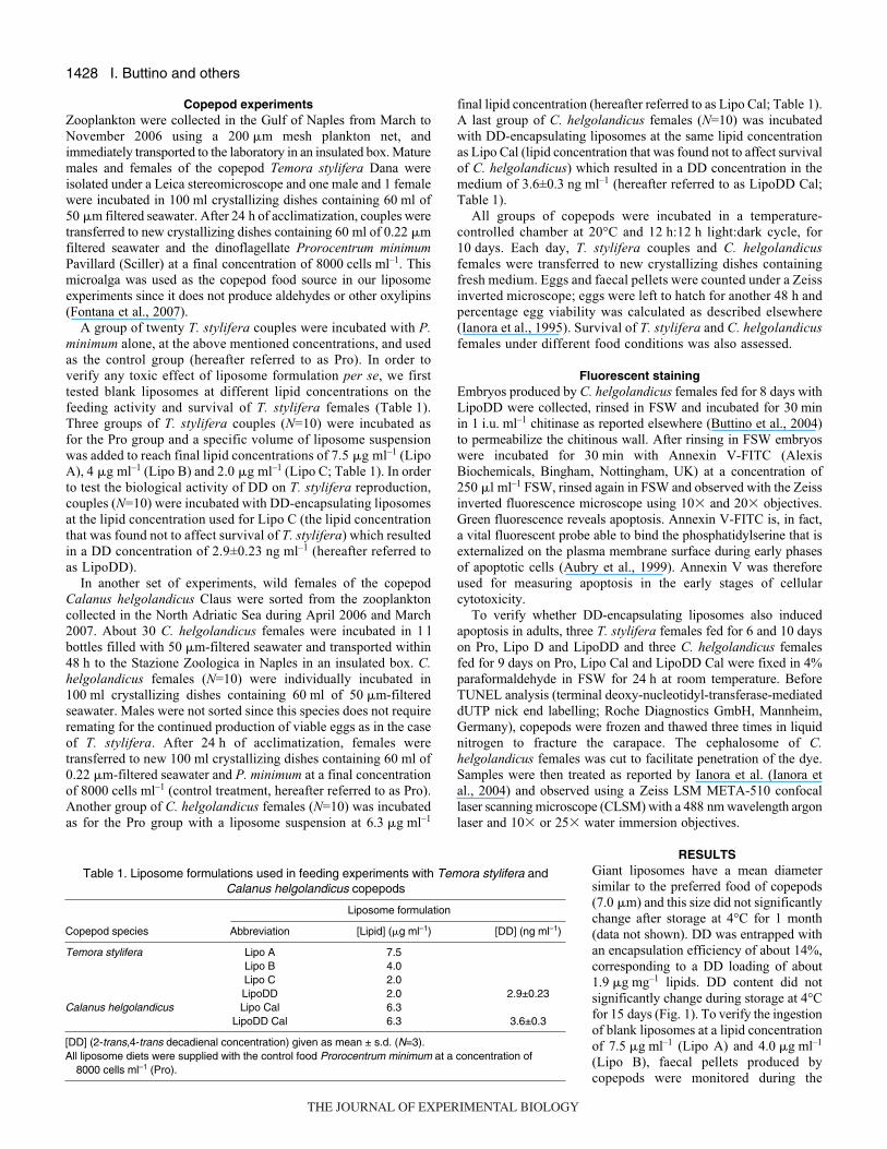
1426 INTRODUCTION In the last decade, there has been an increased awareness that secondary metabolites produced by diatoms, a major class of unicellular algae contributing to over 45% of the oceanic primary production, negatively impact the reproductive success of their principal zooplankton predators such as copepods (Ianora et al., 2003; Pohnert, 2005) with possible consequences on the transfer of energy through the marine food chain to top carnivores (Miralto et al., 1999; Ianora et al., 2004). This insidious mechanism, which does not deter the herbivore from feeding but impairs its recruitment, will restrain the cohort size of the next generation. The compounds responsible for reduced hatching success and abnormal larval development in these small crustaceans are several oxylipins, products of the enzymatic oxidation of fatty acids, which include polyunsaturated aldehydes (PUAs) as well as fatty acid hydroxides, epoxy alcohols, hydroperoxides and reactive oxygen species (ROS) (Fontana et al., 2007). These compounds are not constitutively present in the cells but are only produced when the cell is damaged as would occur during grazing (Pohnert, 2000). Owing to the teratogenic nature of diatom oxylipins, the mechanism of chemical defense in diatoms functions through induction of apoptosis which can occur in maturing oocytes (Poulet et al., 2007) or during embryo development (Romano et al., 2003) and in newly hatched nauplii (Ianora et al., 2004; Poulet et al., 2003). The toxicity of oxylipins, and more specifically of PUAs, has been demonstrated in classical feeding experiments using diatoms as copepod food (Ianora et al., 2003; Poulet et al., 2007) or in in vitro tests by incubating embryos and adults in known concentrations of pure molecules dissolved into seawater (Ianora et al., 1999; Romano et al., 2003; Adolph et al., 2004; Caldwell et al., 2005; Taylor et al., 2007). Recently, Ianora et al. used the dinoflagellate Prorocentrum minimum, which does not produce oxylipins, as a live carrier of 2-trans,4-trans-decadienal (henceforth called decadienal or DD), which has been widely used as a model aldehyde to study the deleterious effects of these compounds on marine invertebrates (Ianora et al., 2004). The daily ingestion rate of DD by females of the copepod Calanus helgolandicus was indirectly calculated from the filtration rate of P. minimum, considering the amount of DD adsorbed onto the algal cells. However, since the interaction between PUAs and live carriers is unknown, the question as to how much and for how long ingestion of oxylipins affects copepod reproduction remains a critical point to understanding the functional role of such compounds in the marine system. Until now, the lack of an efficient method to deliver realistic amounts of these compounds into the copepod body has represented a major limitation to a deeper investigation of the toxicological impact of oxylipins in copepods. Recently, several authors have discussed the need to develop inert carriers delivering known concentrations of oxylipins into copepods, and able to mimic their release as occurs during diatom feeding under natural conditions (Caldwell et al., 2004; Paffenhofer et al., 2005), in order to better understand the impact of these compounds on The Journal of Experimental Biology 211, 1426-1433 Published by The Company of Biologists 2008 doi:10.1242/jeb.015859 Aldehyde-encapsulating liposomes impair marine grazer survivorship Isabella Buttino 1, *, Giuseppe De Rosa 2 , Ylenia Carotenuto 1 , Marialuisa Mazzella 2 , Adrianna Ianora 1 , Francesco Esposito 1 , Valentina Vitiello 1 , Fabiana Quaglia 2 , Maria Immacolata La Rotonda 2 and Antonio Miralto 1 1 Stazione Zoologica Anton Dohrn, Villa Comunale, 80121 Napoli, Italy and 2 Dipartimento di Chimica Farmaceutica e Tossicologica, Università degli Studi di Napoli Federico II, Via D. Montesano 49, 80131 Napoli, Italy *Author for correspondence (e-mail: [email protected]) Accepted 2 March 2008 SUMMARY In the last decade, there has been an increased awareness that secondary metabolites produced by marine diatoms negatively impact the reproductive success of their principal predators, the copepods. Several oxylipins, products of the enzymatic oxidation of fatty acids, are produced when these unicellular algae are damaged, as occurs during grazing. In the past, the dinoflagellate Prorocentrum minimum, which does not produce the oxylipin 2-trans,4-trans-decadienal (DD), has been used as a live carrier to calculate daily ingestion rates of this molecule by copepod crustaceans. However, since the interaction between oxylipins and live carriers is unknown, the question as to how much and for how long ingestion of these molecules affects copepod reproduction remains a critical point to understanding the functional role of such compounds at sea. In the investigation presented here we used giant liposomes (~7·m) as a delivery system for the oxylipin DD, prepared in the same size range as copepod food and containing known amounts of DD. The aim of this work was to relate the ingestion of DD to the reproductive failure of the copepods Temora stylifera and Calanus helgolandicus. Liposomes were very stable over time and after 10·days of feeding, liposomes encapsulating DD reduced egg hatching success and female survival with a concomitant appearance of apoptosis in both copepod embryos and female tissues. Concentrations of DD inducing blockage were one order of magnitude lower that those used in classical feeding experiments demonstrating that liposomes are a useful tool to quantitatively analyze the impact of toxins on copepods. Key words: copepod, diatom, decadienal, reproduction, egg viability, apoptosis. THE JOURNAL OF EXPERIMENTAL BIOLOGY

Welcome message from author
This document is posted to help you gain knowledge. Please leave a comment to let me know what you think about it! Share it to your friends and learn new things together.
Transcript

1426
INTRODUCTIONIn the last decade, there has been an increased awareness that
secondary metabolites produced by diatoms, a major class of
unicellular algae contributing to over 45% of the oceanic primary
production, negatively impact the reproductive success of their
principal zooplankton predators such as copepods (Ianora et al.,
2003; Pohnert, 2005) with possible consequences on the transfer of
energy through the marine food chain to top carnivores (Miralto et
al., 1999; Ianora et al., 2004). This insidious mechanism, which
does not deter the herbivore from feeding but impairs its recruitment,
will restrain the cohort size of the next generation. The compounds
responsible for reduced hatching success and abnormal larval
development in these small crustaceans are several oxylipins,
products of the enzymatic oxidation of fatty acids, which include
polyunsaturated aldehydes (PUAs) as well as fatty acid hydroxides,
epoxy alcohols, hydroperoxides and reactive oxygen species (ROS)
(Fontana et al., 2007). These compounds are not constitutively
present in the cells but are only produced when the cell is damaged
as would occur during grazing (Pohnert, 2000). Owing to the
teratogenic nature of diatom oxylipins, the mechanism of chemical
defense in diatoms functions through induction of apoptosis which
can occur in maturing oocytes (Poulet et al., 2007) or during embryo
development (Romano et al., 2003) and in newly hatched nauplii
(Ianora et al., 2004; Poulet et al., 2003). The toxicity of oxylipins,
and more specifically of PUAs, has been demonstrated in classical
feeding experiments using diatoms as copepod food (Ianora et al.,
2003; Poulet et al., 2007) or in in vitro tests by incubating embryos
and adults in known concentrations of pure molecules dissolved
into seawater (Ianora et al., 1999; Romano et al., 2003; Adolph et
al., 2004; Caldwell et al., 2005; Taylor et al., 2007).
Recently, Ianora et al. used the dinoflagellate Prorocentrumminimum, which does not produce oxylipins, as a live carrier of
2-trans,4-trans-decadienal (henceforth called decadienal or DD),
which has been widely used as a model aldehyde to study the
deleterious effects of these compounds on marine invertebrates
(Ianora et al., 2004). The daily ingestion rate of DD by females of
the copepod Calanus helgolandicus was indirectly calculated from
the filtration rate of P. minimum, considering the amount of DD
adsorbed onto the algal cells. However, since the interaction
between PUAs and live carriers is unknown, the question as to how
much and for how long ingestion of oxylipins affects copepod
reproduction remains a critical point to understanding the functional
role of such compounds in the marine system.
Until now, the lack of an efficient method to deliver realistic
amounts of these compounds into the copepod body has
represented a major limitation to a deeper investigation of the
toxicological impact of oxylipins in copepods. Recently, several
authors have discussed the need to develop inert carriers
delivering known concentrations of oxylipins into copepods, and
able to mimic their release as occurs during diatom feeding under
natural conditions (Caldwell et al., 2004; Paffenhofer et al., 2005),
in order to better understand the impact of these compounds on
The Journal of Experimental Biology 211, 1426-1433Published by The Company of Biologists 2008doi:10.1242/jeb.015859
Aldehyde-encapsulating liposomes impair marine grazer survivorship
Isabella Buttino1,*, Giuseppe De Rosa2, Ylenia Carotenuto1, Marialuisa Mazzella2, Adrianna Ianora1,Francesco Esposito1, Valentina Vitiello1, Fabiana Quaglia2, Maria Immacolata La Rotonda2
and Antonio Miralto1
1Stazione Zoologica Anton Dohrn, Villa Comunale, 80121 Napoli, Italy and 2Dipartimento di Chimica Farmaceutica e Tossicologica,Università degli Studi di Napoli Federico II, Via D. Montesano 49, 80131 Napoli, Italy
*Author for correspondence (e-mail: [email protected])
Accepted 2 March 2008
SUMMARYIn the last decade, there has been an increased awareness that secondary metabolites produced by marine diatoms negativelyimpact the reproductive success of their principal predators, the copepods. Several oxylipins, products of the enzymatic oxidationof fatty acids, are produced when these unicellular algae are damaged, as occurs during grazing. In the past, the dinoflagellateProrocentrum minimum, which does not produce the oxylipin 2-trans,4-trans-decadienal (DD), has been used as a live carrier tocalculate daily ingestion rates of this molecule by copepod crustaceans. However, since the interaction between oxylipins and livecarriers is unknown, the question as to how much and for how long ingestion of these molecules affects copepod reproductionremains a critical point to understanding the functional role of such compounds at sea. In the investigation presented here weused giant liposomes (~7·�m) as a delivery system for the oxylipin DD, prepared in the same size range as copepod food andcontaining known amounts of DD. The aim of this work was to relate the ingestion of DD to the reproductive failure of thecopepods Temora stylifera and Calanus helgolandicus. Liposomes were very stable over time and after 10·days of feeding,liposomes encapsulating DD reduced egg hatching success and female survival with a concomitant appearance of apoptosis inboth copepod embryos and female tissues. Concentrations of DD inducing blockage were one order of magnitude lower thatthose used in classical feeding experiments demonstrating that liposomes are a useful tool to quantitatively analyze the impactof toxins on copepods.
Key words: copepod, diatom, decadienal, reproduction, egg viability, apoptosis.
THE JOURNAL OF EXPERIMENTAL BIOLOGY
1427PUA-liposomes affect copepod survival
copepod population control at sea. Although the use of liposomes
as a delivery system for drugs and chemicals in zooplankton and
in aquaculture is not new (Hontoria et al., 1994; Ozkizilcik and
Chu, 1994; Koven et al., 1999; Touraki et al., 1995), this
technique has only recently been proposed in marine copepods
(Buttino et al., 2006). Buttino and co-workers demonstrated that
giant liposomes of about 7·�m diameter, which corresponds to
the same dimensional range as phytoplankton cells ingested by
copepods, were actively ingested by the copepod Temora stylifera.
Liposome uptake and palatability was confirmed using liposomes
containing a fluorescent marker (fluorescein isothiocyanate–
dextran), and 3H-labelled liposomes were used to calculate the
rate of liposome ingestion. These authors also showed that when
administered with the dinoflagellate algae P. minimum, liposomes
were actively ingested over a 48·h period, and copepods grazed
twice as much as with a diet of liposomes alone. Moreover, a
diet of liposomes had no supplementary effects on copepod egg
production and egg viability, making them a good candidate as
a delivery system for chemicals in copepods.
In the present work, we use giant liposomes as a delivery system
for the PUA 2-trans,4-trans decadienal (DD), to study the effect of
this molecule on egg production, egg hatching success, faecal pellet
production and adult survival in the copepods Temora styliferaand Calanus helgolandicus. Giant liposomes containing known
quantities of DD were prepared and fully characterized. Blank or
DD-encapsulating liposomes and the control diet P. minimum were
supplied for 10·days to both copepod species, which represent target
species used in numerous aldehyde–copepod reproduction studies.
In addition, the induction of apoptosis in embryos and females were
also analyzed using specific fluorescent markers. The aim of this
work was to relate the ingestion of DD, via a liposome-based
delivery system, to the reproductive failure of copepods in order to
better understand the ecological relevance of diatom-derived
oxylipins for copepod recruitment at sea.
MATERIALS AND METHODSMaterials
Cholesterol, sea salts and 2-trans,4-trans decadienal (DD) were
purchased from Sigma-Aldrich (Milan, Italy). Phosphatidylcholine
from soybean lecithin (SPC) was kindly provided by Lipoid GmbH
(Cam, Switzerland). Analytical grade chloroform, methanol, ethanol,
96% sulfuric acid and 2,4-dinitrophenylhydrazine were supplied by
Carlo Erba Reagenti (Milan, Italy).
Liposome preparationLiposomes were prepared by a modified hand-shaking method
(Bangham et al., 1965). Briefly, a lipid mixture containing 110·mg
SPC and 40·mg cholesterol in 5·ml of a chloroform–methanol
solvent mixture (2:1 v/v) was introduced into a 250·ml round-
bottomed flask. The solvent was removed in a rotary evaporator
(Laborota 4010 Digital, Heidolph, Schwabach, Germany) until
formation of a lipid film on the wall of the flask. To prepare blank
liposomes, the lipid film was hydrated with 5·ml of 0.22·�m-
filtered seawater (FSW). The resulting suspension was gently
mixed in the presence of 0.5·g glass beads until the lipid layer
was removed from the glass wall. The flask was then attached to
the evaporator again, rotated at room temperature for about
30·min, and left at room temperature for 2·h. DD-encapsulating
liposomes (LipoDD) were prepared by adding 500·�l of an
ethanol solution containing 0.4·mg·ml–1 DD to the organic solution
containing lipids. After preparation, LipoDD was washed twice
by centrifugation at 4470·g for 30·min at 4°C to remove un-
encapsulated DD. All liposome formulations were stored at 4°C
under nitrogen.
Liposome size analysis and lipid dosageThe mean diameter and size distribution of liposomes were analyzed
by laser light scattering (Coulter LS, 100Q, Beckman Coulter,
Miami, USA) on a dispersion of liposomes in FSW. Particle size
was expressed as mean volume diameter ± standard deviation (s.d.)
calculated on three different batches (N=3). The amount of lipids
present in the liposome suspension after preparation was determined
using the Stewart assay (Stewart, 1980). This test allows the
quantification of SPC concentration in the suspension; total lipid
content was calculated by assuming the same SPC:cholesterol ratio
before and after preparation. Briefly, 0.1·ml of liposome suspension
(approximately at a concentration of 0.1·mg·ml–1) was added to
1.9·ml of an aqueous 0.1·mol·l–1 ammonium ferrothiocyanate
solution in a test tube. The resulting suspension was mixed with
2·ml of chloroform for 15·s using a vortex, and then centrifuged for
5·min at 894·g The upper layer was recovered and analyzed at
485·nm by an UV-vis spectrophotometer (Shimadzu, Milano, Italy;
model 1204), by comparison with a standard curve.
Dosage of 2-trans,4-trans decadienalTo quantify DD in water and liposomes, the aldehyde was
previously transformed into the corresponding hydrazone by a
reaction with 2,4-dinitrophenihydrazine (DNPH). DNPH solution
was prepared as follows: 50·mg DNPH were dissolved in 0.5·ml
of sulfuric acid and 4.5·ml of ethanol. DD was quantified by mixing
100·�l of the solution containing DD with 100·�l of the DNPH
solution; the resulting solution was made up to 10·ml with ethanol.
DD analysis was carried out by high-performance liquid
chromatography (HPLC) using a Luna C18 (250�4.6·mm, 5·�m)
column (Phenomenex, Klwid, Torrance, CA, USA), an HPLC LC-
10AD pump (Shimadzu), a 7725i injection valve (Rheodyne,
Rohnert Park, CA, USA) and a SPV-10A UV-Vis detector
(Shimadzu) set at 360·nm. The system was controlled by a SCL-
10A VP System Controller (Shimadzu) connected to a computer.
Chromatograms were acquired and analysed by a Class VP
Client/Server 7.2.1 program (Shimadzu). The analysis was
performed with a mobile phase acetonitrile:water 15:85 (v/v) in
isocratic conditions at a flow rate of 1·ml·min–1.
To determine the amount of DD loaded into liposomes, 100·�l
of LipoDD was transferred to a 1.5·ml Eppendorf vial and
centrifuged at 9615 g for 15·min (Mikro 20, Hettich, Town,
Tuttlingen, Germany). The pellet and supernatant were separated
and analyzed to determine DD content in liposomes and in the
suspending medium, respectively. Briefly, 50·�l of supernatant was
mixed with 50·�l of DNPH solution and, after vigorous mixing by
vortexing, diluted to 5·ml with ethanol; the resulting solution was
analyzed by HPLC. The pellet obtained by centrifugation was
washed and re-suspended in FSW to a volume of 100·�l; the
suspension was mixed with 100·�l of DNPH and, after complete
liposome dissolution, diluted to 1·ml with ethanol. The samples were
then centrifuged at 9615 g for 15·min at 4°C, the supernatant was
withdrawn, diluted 1:10 with ethanol and analyzed by HPLC. Results
were expressed as DD actually loaded (�g·DD·mg–1·total lipids)
and encapsulation efficiency calculated as the ratio between DD
actually loaded and DD theoretically loaded �100. For each
liposome formulation, DD actually loaded was determined soon after
the preparation (t=0) and at 1, 3, 7, 9, 12 and 15 days, in order to
follow the amount of DD administered during copepod feeding
experiments.
THE JOURNAL OF EXPERIMENTAL BIOLOGY
1428
Copepod experimentsZooplankton were collected in the Gulf of Naples from March to
November 2006 using a 200·�m mesh plankton net, and
immediately transported to the laboratory in an insulated box. Mature
males and females of the copepod Temora stylifera Dana were
isolated under a Leica stereomicroscope and one male and 1 female
were incubated in 100·ml crystallizing dishes containing 60·ml of
50·�m filtered seawater. After 24·h of acclimatization, couples were
transferred to new crystallizing dishes containing 60·ml of 0.22·�m
filtered seawater and the dinoflagellate Prorocentrum minimumPavillard (Sciller) at a final concentration of 8000·cells·ml–1. This
microalga was used as the copepod food source in our liposome
experiments since it does not produce aldehydes or other oxylipins
(Fontana et al., 2007).
A group of twenty T. stylifera couples were incubated with P.minimum alone, at the above mentioned concentrations, and used
as the control group (hereafter referred to as Pro). In order to
verify any toxic effect of liposome formulation per se, we first
tested blank liposomes at different lipid concentrations on the
feeding activity and survival of T. stylifera females (Table·1).
Three groups of T. stylifera couples (N=10) were incubated as
for the Pro group and a specific volume of liposome suspension
was added to reach final lipid concentrations of 7.5·�g·ml–1 (Lipo
A), 4·�g·ml–1 (Lipo B) and 2.0·�g·ml–1 (Lipo C; Table·1). In order
to test the biological activity of DD on T. stylifera reproduction,
couples (N=10) were incubated with DD-encapsulating liposomes
at the lipid concentration used for Lipo C (the lipid concentration
that was found not to affect survival of T. stylifera) which resulted
in a DD concentration of 2.9±0.23·ng·ml–1 (hereafter referred to
as LipoDD).
In another set of experiments, wild females of the copepod
Calanus helgolandicus Claus were sorted from the zooplankton
collected in the North Adriatic Sea during April 2006 and March
2007. About 30 C. helgolandicus females were incubated in 1·l
bottles filled with 50·�m-filtered seawater and transported within
48·h to the Stazione Zoologica in Naples in an insulated box. C.helgolandicus females (N=10) were individually incubated in
100·ml crystallizing dishes containing 60·ml of 50·�m-filtered
seawater. Males were not sorted since this species does not require
remating for the continued production of viable eggs as in the case
of T. stylifera. After 24·h of acclimatization, females were
transferred to new 100·ml crystallizing dishes containing 60·ml of
0.22·�m-filtered seawater and P. minimum at a final concentration
of 8000·cells·ml–1 (control treatment, hereafter referred to as Pro).
Another group of C. helgolandicus females (N=10) was incubated
as for the Pro group with a liposome suspension at 6.3·�g·ml–1
I. Buttino and others
final lipid concentration (hereafter referred to as Lipo Cal; Table·1).
A last group of C. helgolandicus females (N=10) was incubated
with DD-encapsulating liposomes at the same lipid concentration
as Lipo Cal (lipid concentration that was found not to affect survival
of C. helgolandicus) which resulted in a DD concentration in the
medium of 3.6±0.3·ng·ml–1 (hereafter referred to as LipoDD Cal;
Table·1).
All groups of copepods were incubated in a temperature-
controlled chamber at 20°C and 12·h:12·h light:dark cycle, for
10·days. Each day, T. stylifera couples and C. helgolandicusfemales were transferred to new crystallizing dishes containing
fresh medium. Eggs and faecal pellets were counted under a Zeiss
inverted microscope; eggs were left to hatch for another 48·h and
percentage egg viability was calculated as described elsewhere
(Ianora et al., 1995). Survival of T. stylifera and C. helgolandicusfemales under different food conditions was also assessed.
Fluorescent stainingEmbryos produced by C. helgolandicus females fed for 8·days with
LipoDD were collected, rinsed in FSW and incubated for 30·min
in 1·i.u.·ml–1 chitinase as reported elsewhere (Buttino et al., 2004)
to permeabilize the chitinous wall. After rinsing in FSW embryos
were incubated for 30·min with Annexin V-FITC (Alexis
Biochemicals, Bingham, Nottingham, UK) at a concentration of
250·�l·ml–1 FSW, rinsed again in FSW and observed with the Zeiss
inverted fluorescence microscope using 10� and 20� objectives.
Green fluorescence reveals apoptosis. Annexin V-FITC is, in fact,
a vital fluorescent probe able to bind the phosphatidylserine that is
externalized on the plasma membrane surface during early phases
of apoptotic cells (Aubry et al., 1999). Annexin V was therefore
used for measuring apoptosis in the early stages of cellular
cytotoxicity.
To verify whether DD-encapsulating liposomes also induced
apoptosis in adults, three T. stylifera females fed for 6 and 10·days
on Pro, Lipo D and LipoDD and three C. helgolandicus females
fed for 9·days on Pro, Lipo Cal and LipoDD Cal were fixed in 4%
paraformaldehyde in FSW for 24·h at room temperature. Before
TUNEL analysis (terminal deoxy-nucleotidyl-transferase-mediated
dUTP nick end labelling; Roche Diagnostics GmbH, Mannheim,
Germany), copepods were frozen and thawed three times in liquid
nitrogen to fracture the carapace. The cephalosome of C.helgolandicus females was cut to facilitate penetration of the dye.
Samples were then treated as reported by Ianora et al. (Ianora et
al., 2004) and observed using a Zeiss LSM META-510 confocal
laser scanning microscope (CLSM) with a 488·nm wavelength argon
laser and 10� or 25� water immersion objectives.
RESULTSGiant liposomes have a mean diameter
similar to the preferred food of copepods
(7.0·�m) and this size did not significantly
change after storage at 4°C for 1 month
(data not shown). DD was entrapped with
an encapsulation efficiency of about 14%,
corresponding to a DD loading of about
1.9·�g·mg–1 lipids. DD content did not
significantly change during storage at 4°C
for 15·days (Fig.·1). To verify the ingestion
of blank liposomes at a lipid concentration
of 7.5·�g·ml–1 (Lipo A) and 4.0·�g·ml–1
(Lipo B), faecal pellets produced by
copepods were monitored during the
Table·1. Liposome formulations used in feeding experiments with Temora stylifera andCalanus helgolandicus copepods
Liposome formulation
Copepod species Abbreviation [Lipid] (�g·ml–1) [DD] (ng·ml–1)
Temora stylifera Lipo A 7.5Lipo B 4.0Lipo C 2.0LipoDD 2.0 2.9±0.23
Calanus helgolandicus Lipo Cal 6.3 LipoDD Cal 6.3 3.6±0.3
[DD] (2-trans,4-trans decadienal concentration) given as mean ± s.d. (N=3).All liposome diets were supplied with the control food Prorocentrum minimum at a concentration of
8000·cells·ml–1 (Pro).
THE JOURNAL OF EXPERIMENTAL BIOLOGY

1429PUA-liposomes affect copepod survival
10·days of experiments (Fig.·2A). In the first 3·days of feeding, the
mean faecal pellets produced by couples fed Lipo A and Lipo B
were double the control group fed Pro (82.6; 91.9; 46.5,
respectively). From day·4 to 10, faecal pellets produced by couples
fed Lipo A and B decreased to <50·couple–1·day–1. With the control
food Pro, faecal pellet production was comparatively more stable
than with Lipo A and B even if, on average, all groups had similar
rates with a total mean production of 60.1, 58.1 and 54.1·faecal
pellets·couple–1·day–1 for Lipo A, B, and Pro, respectively.
Experiments with Lipo A terminated on day·8 because no females
survived beyond this day.
Fig.·2B shows the percentage survival of females incubated with
Lipo A and B and the control Pro. Treatment with Lipo A strongly
reduced T. stylifera female survival during the experiment: survival
dropped to 45% after 7·days and all females died on day·9. Less
than 30% of females survived with Lipo B at the end of the
experiment whereas females fed Pro had the highest survival rate
over 10·days (90.7±10.6). Since Lipo A and B affected female
survival, we used a lower liposome formulation with a concentration
of 2·�g·ml–1 lipids (Lipo C) as a carrier for decadienal.
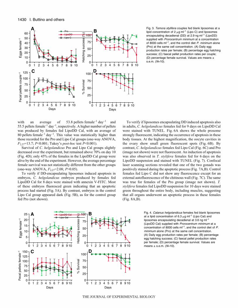
Fig.·3 shows egg and faecal pellet production rates and egg
hatching success of T. stylifera females fed blank liposomes (Lipo
C), DD-encapsulating liposomes (LipoDD) and the control food Pro.
All three groups had the highest egg production rates at the
beginning of the experiment (day·1) with a mean of
48.4·eggs·female–1·day–1 (Fig.·3A). Egg production decreased
on day·2 to the end of the experiment in each treatment,
more dramatically in the Lipo C group, with an egg production
rate of <1·egg·female–1·day–1 compared to 10·eggs·female–1·day–1
produced by females fed Pro and LipoDD on day·10. On average,
T. stylifera females fed Pro, LipoDD and Lipo C produced
17.1·eggs·female–1·day–1, 16.9·eggs·female–1·day–1, and
9.2·eggs·female–1·day–1, respectively, during the experiment. These
values were not statistically different from each other (one-way
ANOVA, F2,27=1.08; P>0.05).
Hatching success for T. stylifera females fed Pro and Lipo C was
very high for the duration of the experiment, ranging from 100%
to 66.7%, and with a mean hatching success of 87.7% and 79.7%,
respectively (Fig.·3B). By contrast, hatching success decreased in
the group fed LipoDD to about 40% after 8 days of feeding.
Statistical analysis showed that, on average, hatching success for
females fed LipoDD (49.7%) was significantly lower than that for
females fed Pro and Lipo C (one-way ANOVA, F2,27=21.0;
P<0.001; Tukey’s post-hoc test P<0.001).
The average number of faecal pellets produced by T. styliferacouples fed LipoDD was 65.5·pellets·couple–1·day–1 similar to those
of females fed Pro (54.0·pellets·couple–1·day–1; Fig.·3C). Higher
pellet production was initially recorded for couples fed Lipo C during
the first 6·days of the experiment, with a mean of 97.2, but by day·7,
the value resembled those of the Pro and LipoDD groups. On
average, the Lipo C group produced significantly more pellets than
the Pro and LipoDD groups (70.5·pellets·couple–1·day–1, one-way
variance ANOVA, F2,27=3.8; P<0.05; Tukey’s post-hoc test
P<0.05).
Fig.·3D shows the percentage survival of T. stylifera females after
10·days of feeding with Pro, Lipo C and LipoDD. The percentage
survival was very high for both Pro and Lipo C diets during the
whole experiment, with a mean of 92.4% and 95%, respectively.
By contrast, the survivorship of females fed LipoDD declined
steadily throughout the experiment, with only 47% live females by
day 10. The average percentage survival was 76.0% in the LipoDD
group, which was statistically lower than the Lipo C and Pro groups
(one-way ANOVA, F2,27=6.1; P<0.01; Tukey’s post-hoc test
P<0.05).
Egg production rate, hatching success and faecal pellet production
of C. helgolandicus females fed Lipo Cal, LipoDD Cal and Pro are
reported in Fig.·4. The daily pattern with a LipoDD diet was very
stable during the experiment and similar to that of Lipo Cal and
Pro groups, with an average of 8.9·eggs·female–1·day–1,
8.9·eggs·female–1·day–1 and 11.7·eggs·female–1·day–1, respectively
(Fig.·4A). Hatching success for the three treatments remained high
until day·7 after which hatching success for females fed LipoDD
Cal was reduced to <50% and fell to 0% by the end of the experiment
(Fig.·4B). By contrast, Pro and Lipo Cal diets did not reduce hatching
success, which was on average 76.7% and 71.6%, respectively.
These values were statistically higher than those recorded for females
fed LipoDD Cal (49.8%; one-way ANOVA, F2,27=9.2; P<0.001;
Tukey’s post-hoc test P<0.01).
C. helgolandicus females had similar daily patterns for faecal
pellet production when feeding on Pro and Lipo Cal (Fig.·4C),
0
0.5
1.0
1.5
2.0
2.5
3.0
0 2 4 6Days
DD
load
ing
into
lipo
som
es(µ
g D
D m
g–1 li
pids
)
8 10 12 14 16
Fig.·1. 2-trans,4-trans decadienal loaded into liposomes, expressed as�g·mg–1 (means ± s.d., N=3) of the total lipids during storage at 4°C for15·days.
0 1 2 3 4 5 6 7 8 9 100
25
50
75
100
125 Lipo ALipo B
PRO
Fec
al p
elle
ts c
oupl
e–1
0 1 2 3 4 5 6 7 8 9 100
25
50
75
100
Days
% S
urvi
val
A
B
Fig.·2. Temora stylifera daily faecal pellet production (A) and percentagefemale survival (B) for couples fed liposomes at a lipid concentration ofeither 7.5·�g·ml–1·lipids (Lipo A) or at 4.0·�g·ml–1·lipids (Lipo B) suppliedwith Prorocentrum minimum at a concentration of 8000·cells·ml–1 andcompared with a control diet of P. minimum alone (Pro) at the same cellconcentration. Values are means ± s.e.m. (N=10).
THE JOURNAL OF EXPERIMENTAL BIOLOGY
1430
with an average of 53.8·pellets·female–1·day–1 and
55.5·pellets·female–1·day–1, respectively. A higher number of pellets
was produced by females fed LipoDD Cal, with an average of
80·pellets·female–1·day–1. This value was statistically higher than
those recorded for the Pro and Lipo Cal groups (one-way ANOVA,
F2,27=13.7; P<0.001; Tukey’s post-hoc test P<0.001).
Survival of C. helgolandicus Pro and Lipo Cal groups slightly
decreased over the experiment, but remained above 70% on day·10
(Fig.·4D); only 45% of the females in the LipoDD Cal group were
alive by the end of the experiment. However, the average percentage
female survival was not statistically different from the other groups
(one-way ANOVA, F2,27=2.08; P>0.05).
To verify if DD-encapsulating liposomes induced apoptosis in
embryos, C. helgolandicus embryos produced by females fed
LipoDD Cal for 8·days were stained with annexin V-FITC. Most
of these embryos fluoresced green indicating that an apoptotic
process had started (Fig.·5A). By contrast, embryos in the control
Lipo Cal group appeared dark (Fig.·5B), as for the control group
fed Pro (not shown).
I. Buttino and others
To verify if liposomes encapsulating DD induced apoptosis also
in adults, C. helgolandicus females fed for 9·days on LipoDD Cal
were stained with TUNEL. Fig.·6A shows the whole prosome
strongly fluorescent, indicating the occurrence of apoptosis in these
body tissues. At the highest magnification, the oocyte cavities in
the ovary show small green fluorescent spots (Fig.·6B). By
contrast, C. helgolandicus females fed Lipo Cal (Fig.·6C) and Pro
(image not shown) were not fluorescent. An induction of apoptosis
was also observed in T. stylifera females fed for 6·days on the
LipoDD suspension and stained with TUNEL (Fig.·7). Confocal
laser scanning sections revealed that one of the two gonads was
positively stained during the apoptotic process (Fig.·7A,B). Control
females fed Lipo C did not show any fluorescence except for an
external autofluorescence of the chitinous wall (Fig.·7C). The same
was true for females of the Pro group (image not shown). T.stylifera females fed LipoDD suspension for 10·days were stained
green throughout the entire body, including muscles, suggesting
that all organs underwent an apoptotic process in these females
(Fig.·8A,B).
0 1 2 3 4 5 6 7 8 9 100
10
20
30
40
50
60ProLipoDDLipo C
Egg
s fe
mal
e–1
0 1 2 3 4 5 6 7 8 9 100
25
50
75
100
Hat
chin
g su
cces
s (%
)
0 1 2 3 4 5 6 7 8 9 100
25
50
75
100
125
Days
Fec
al p
elle
ts c
oupl
e–1
A B
C D
0 1 2 3 4 5 6 7 8 9 100
25
50
75
100
Days
% S
urvi
val
Fig.·3. Temora stylifera couples fed blank liposomes at alipid concentration of 2·�g·ml–1 (Lipo C) and liposomesencapsulating decadienal (DD) at 2.9·ng·ml–1 (LipoDD)supplied with Prorocentrum minimum at a concentrationof 8000·cells·ml–1, and the control diet P. minimum alone(Pro) at the same cell concentration. (A) Daily eggproduction rates per female; (B) percentage egg hatchingsuccess; (C) faecal pellet production rates per couple;(D) percentage female survival. Values are means ±s.e.m. (N=10).
0 1 2 3 4 5 6 7 8 9 100
5
10
15
20
25ProLipoDD CalLipo Cal
Egg
s fe
mal
e–1
0 1 2 3 4 5 6 7 8 9 100
25
50
75
100
125
150
Days
Fec
al p
elle
ts fe
mal
e–1
0 1 2 3 4 5 6 7 8 9 100
25
50
75
100
Hat
chin
g su
cces
s (%
)A B
C D
0 1 2 3 4 5 6 7 8 9 100
25
50
75
100
Days
% S
urvi
val
Fig.·4. Calanus helgolandicus females fed blank liposomesat a lipid concentration of 6.3·�g·ml–1 (Lipo Cal) andliposomes encapsulating decadienal at 3.6·ng·ml–1
(LipoDD Cal) supplied with Prorocentrum minimum at aconcentration of 8000·cells·ml–1, and the control diet of P.minimum alone (Pro) at the same cell concentration.(A) Daily egg production rates per female; (B) percentageegg hatching success; (C) faecal pellet production ratesper female; (D) percentage female survival. Values aremeans ± s.e.m. (N=10).
THE JOURNAL OF EXPERIMENTAL BIOLOGY
1431PUA-liposomes affect copepod survival
DISCUSSIONIn a previous study, we demonstrated that giant liposomes were
efficiently ingested by the copepod Temora stylifera (Buttino et al.,
2006). These results suggested that liposomes could be a very useful
tool to deliver bioactive molecules into the gastrointestinal tract of
aquatic organisms. Here we prepared giant liposomes encapsulating
DD to unequivocally clarify the role of polyunsaturated diatom-
derived aldehydes in copepod reproduction. Experiments in which
copepods had been incubated in the presence of DD had already
been carried out (Miralto et al., 1999; Ianora et al., 2004). However,
in these studies the relationship between the observed effect and
the amount of DD ingested by copepods was probably overestimated
due to the high volatility of this aldehyde in the incubation medium
(Miralto et al., 1999). Moreover, DD may have been transformed
prior to delivery when live carriers were used (Ianora et al., 2004).
By contrast, liposomes are inert carriers and able to entrap molecules
with different chemical-physical characteristics. In particular,
hydrophilic molecules can be entrapped into the aqueous internal
cavity, whereas lipophilic compounds can enter into the lipid bilayer.
This is what occurs with DD, a very lipophilic molecule that can
be dissolved in the organic solution containing the lipid mix, before
liposome preparation. We found that DD loading into liposomes
did not significantly change during the time frame considered,
probably due to a hydrophobic interaction between DD and the
lipids. Therefore, we performed long-term incubation experiments
with DD-encapsulating liposomes and tested the effects of these on
the reproduction of two copepod species. Blank liposomes were also
tested to evaluate lipid toxicity on copepods, with faecal pellet
production used as a proxy for copepod ingestion.
Interestingly, our results show that the production of faecal pellets
increased when T. stylifera females were fed a mixed diet of
liposomes and Pro with respect to the single diet Pro. Previous results
(Buttino et al., 2006) demonstrated that copepod ingestion rate,
calculated using radiolabelled cholesterol, was double when
liposomes where supplied together with Pro. An increase in the
number of faecal pellets was also recorded during the experiments
using lipo DD. However, whereas the number of pellets produced
by T. stylifera was significantly higher with a diet of blank
liposomes than with LipoDD, for C. helgolandicus the opposite was
true. Much more LipoDD was ingested by C. helgolandicus than
liposomes without DD. These results suggest different behaviors
for the two copepod species, with one presumably more attracted
by the ‘flavor’ of decadienal than the other. Odorous compounds
such as the aldehyde decatrienal were liberated into water after cell
disruption of some benthic diatoms, acting as a repellent to pelagic
freshwater crustaceans (Jüttner, 2005). Such repellence consisted
of reduced swimming movements and grazing activity. In our
experiments, a similar repellent reaction was not evident, at least
in one of the two species.
An increase in liposomes ingested was not matched by increased
egg production rates for either copepod species, confirming that
liposomes had no supplementary effect on egg production as also
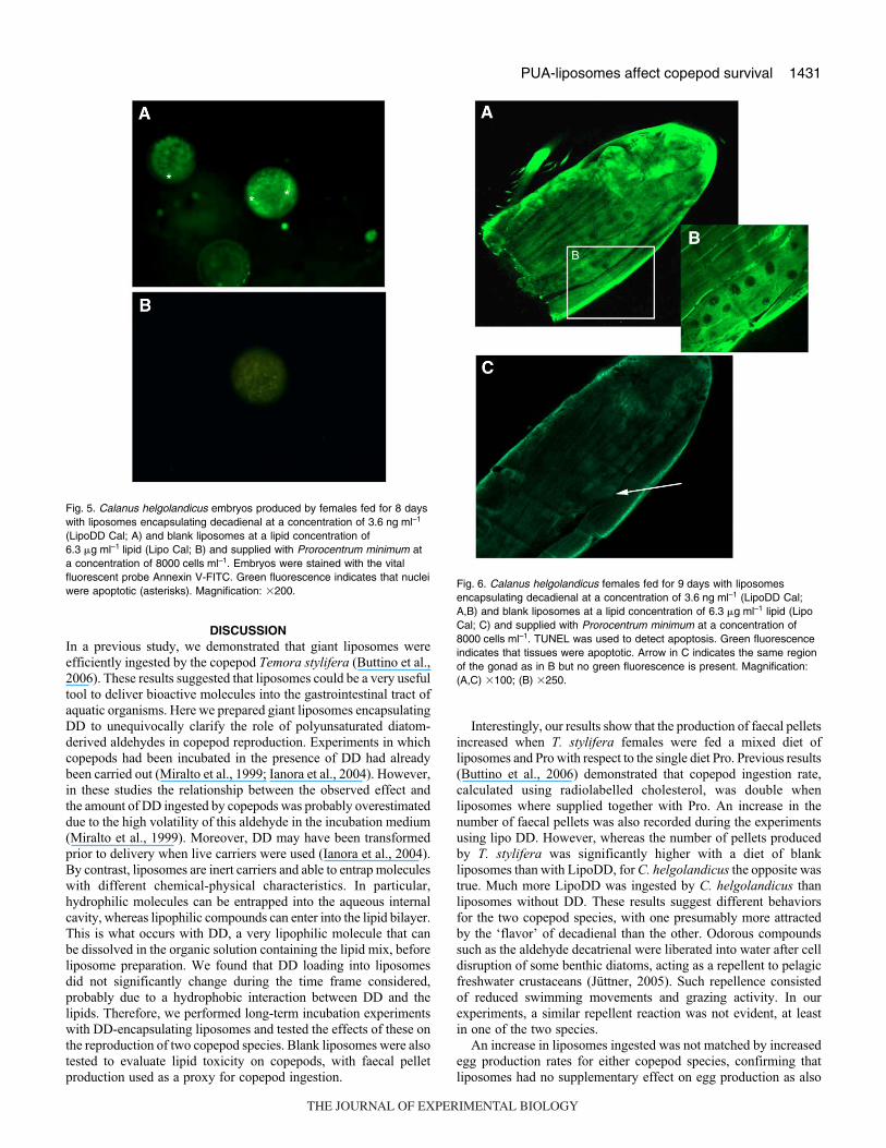
Fig.·5. Calanus helgolandicus embryos produced by females fed for 8 dayswith liposomes encapsulating decadienal at a concentration of 3.6·ng·ml–1
(LipoDD Cal; A) and blank liposomes at a lipid concentration of6.3·�g·ml–1·lipid (Lipo Cal; B) and supplied with Prorocentrum minimum ata concentration of 8000·cells·ml–1. Embryos were stained with the vitalfluorescent probe Annexin V-FITC. Green fluorescence indicates that nucleiwere apoptotic (asterisks). Magnification: �200.
Fig.·6. Calanus helgolandicus females fed for 9·days with liposomesencapsulating decadienal at a concentration of 3.6·ng·ml–1 (LipoDD Cal;A,B) and blank liposomes at a lipid concentration of 6.3·�g·ml–1 lipid (LipoCal; C) and supplied with Prorocentrum minimum at a concentration of8000·cells·ml–1. TUNEL was used to detect apoptosis. Green fluorescenceindicates that tissues were apoptotic. Arrow in C indicates the same regionof the gonad as in B but no green fluorescence is present. Magnification:(A,C) �100; (B) �250.
THE JOURNAL OF EXPERIMENTAL BIOLOGY
1432
reported by Buttino et al. (Buttino et al., 2006). Our results showed
that when liposomes were supplied at lipid concentrations ranging
from 7.5 to 4.0·�g·lipids·ml–1 (Lipo A and B) most T. styliferafemales were dead a few days later and no adults survived more
I. Buttino and others
than 8·days with a Lipo B formulation. This effect was not attributed
to lipid toxicity but rather to the obstruction of copepod mouthparts
by highly concentrated liposome particles that were observed in
some females (I.B., personal observations). This is why we used
the lowest lipid concentration to incubate DD into liposomes for
this species. By contrast, C. helgolandicus did not show a reduction
in adult survival at the liposome formulation used. Different
responses among the two species suggest that blank liposome
incubations must be assessed for each copepod species before using
them in toxicological experiments.
Until now, the toxicity of PUAs in an aqueous medium has only
been tested by exposing organisms in vitro to dissolved PUAs in
water or by using PUA-producing diatoms as food. Our results
indicate that the concentration affecting hatching success in both
copepod species was one order of magnitude lower than those
recorded in previous incubation experiments [e.g. 1·�g·ml–1 in
incubation experiments (Miralto et al., 1999; Ceballos and Ianora,
2003; Taylor et al., 2007)]. Concentrations blocking hatching
success in our experiments were much lower in both T. stylifera(97·ng DD) and C. helgolandicus (121·ng DD) considering filtration
rates of 0.14·ml·h–1 per copepod (Buttino et al., 2006) and incubation
concentrations of 2.9·ng·ml–1 and 3.6·ng·ml–1 DD, respectively, for
10·days. Adolph et al. (Adolph et al., 2004) calculated similar values
for feeding experiments where 30·ng·ml–1 potential oxylipins
equivalent concentrations reduced hatching success to 30% after
5·days of feeding on a diatom diet.
Another interesting finding here is that when the percentage of
egg viability for C. helgolandicus was below 25% and adult survival
was less than 50% (Fig.·4B,D), gonad tissues of some females
appeared positively stained by TUNEL (Fig.·6A), suggesting that
they were undergoing apoptosis. The apoptotic region corresponded
to oocyte 3-stage producers (OS3) (Poulet et al., 2007). Follicle
chambers with apoptotic body-like spots were similar to structures
found in histological sections by these authors (Fig.·6B). Poulet and
co-workers reported a concomitant arrest of OS3 maturation,
characterized by cell fragmentation and by the presence of apoptotic
bodies, when C. helgolandicus females were fed some diatom diets
that reduced both egg production and hatching. Our results also
indicate a concomitance between a reduction in egg hatching success
and induction of apoptosis even if egg production was similar to the
control. Because TUNEL detects early apoptotic events, it is probable
that eggs are released but they die later, and the observed effect
indicates a reduction in hatching success rather than fecundity.
Similarly, a concomitance between TUNEL positivity and a reduction
in hatching success (�50%) occurred for T. stylifera females fed
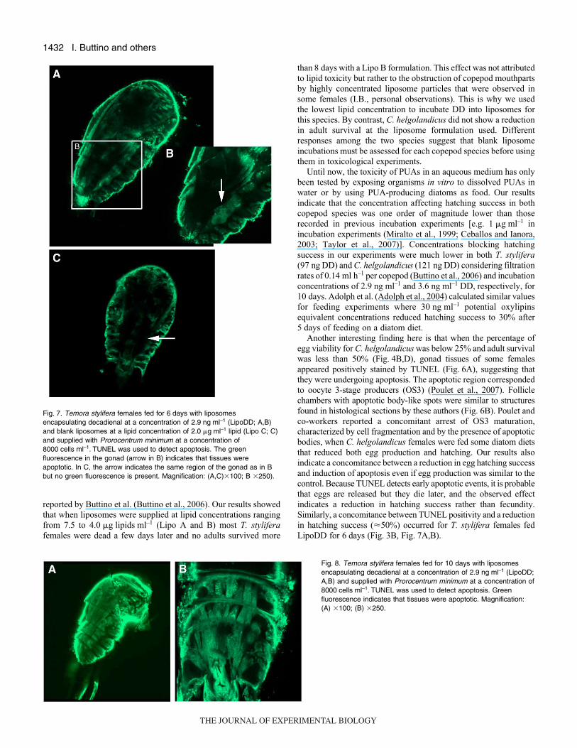
LipoDD for 6·days (Fig.·3B, Fig.·7A,B).
Fig.·7. Temora stylifera females fed for 6·days with liposomesencapsulating decadienal at a concentration of 2.9·ng·ml–1 (LipoDD; A,B)and blank liposomes at a lipid concentration of 2.0·�g·ml–1 lipid (Lipo C; C)and supplied with Prorocentrum minimum at a concentration of8000·cells·ml–1. TUNEL was used to detect apoptosis. The greenfluorescence in the gonad (arrow in B) indicates that tissues wereapoptotic. In C, the arrow indicates the same region of the gonad as in Bbut no green fluorescence is present. Magnification: (A,C)�100; B �250).
Fig.·8. Temora stylifera females fed for 10 days with liposomesencapsulating decadienal at a concentration of 2.9·ng·ml–1 (LipoDD;A,B) and supplied with Prorocentrum minimum at a concentration of8000·cells·ml–1. TUNEL was used to detect apoptosis. Greenfluorescence indicates that tissues were apoptotic. Magnification:(A) �100; (B) �250.
THE JOURNAL OF EXPERIMENTAL BIOLOGY
1433PUA-liposomes affect copepod survival
Moreover, our results also show, for the first time, that DD
affected adult survivorship. Ceballos and Ianora (Ceballos and
Ianora, 2003) reported a similar reduction in adult survival when
T. stylifera was fed the diatom Skeletonema costatum for more
than 10·days. Presumably this decrease in adult survival was due
to DD and not to poor food quality. Females of both species
appeared strongly positive for apoptosis coincidentally with
reduced survival (�50%; Fig.·8A,B, Fig.·3D). Hence the
mechanism of chemical defense in diatoms not only functions by
reducing grazing effects of subsequent generations of copepods,
as hitherto believed, but also targets the direct predator. These
compounds are of lower acute toxicity to adult predators compared
to other feeding deterrents such as dinoflagellate toxins even
though they eventually induce death if ingested for a sufficient
length of time, and lead to post-digestive reduction in fecundity
or depressed viability of the offspring. Grazing pressure is thus
reduced, allowing diatom blooms to persist when grazing pressure
would otherwise have caused them to crash.
An important application of our work is the possibility of
delivering a known quantity of toxin and being able to calculate the
efficiency of adsorption and longevity of the toxins once
encapsulated. Liposomes have been extensively used in the
pharmaceutical and chemical industries for the past 30 years and
in aquaculture to deliver food supplements in the diet [Buttino et
al. (Buttino et al., 2006) and references therein]. However, until
recently liposomes >6·�m were unstable over multiday experiments
(Ravet et al., 2003) and were only used for short-term incubations.
Here we were able to produce giant (�7·�m diameter) DD-
encapsulating liposomes that were very stable over time allowing
us to carry out long-term incubations necessary to test the effects
of PUAs on copepod reproductive fitness. Further studies using
liposomes are in progress to test synergistic or antagonistic effects
of different chemicals and varied nutritional content of copepods,
providing a tool to qualitatively and quantitatively analyze the impact
of toxins and nutrient supplements on copepod grazers.
LIST OF ABBREVIATIONSDD 2-trans,4-trans decadienal
DNPH 2,4-dinitrophenihydrazine
FSW filtered seawater
HPLC high-performance liquid chromatography
LipoDD DD-encapsulating liposome
PUA polyunsaturated aldehyde
ROS reactive oxygen species
SPC soybean lecithin
Funding for this research was partly provided by the project founded by theRegione Campania (D.G.R. no. 889; 30/06/2006) to I.B., and by the EU Networkof Excellence Marine Biodiversity and Ecosystem Functioning (contract numberGOCE-CT-2003-505446).
REFERENCESAdolph, S., Bach, S., Blondel, M., Cueff, A., Moreau, M., Pohnert, G., Poulet, S. A.,
Wichard, T. and Zuccaro, A. (2004). Cytotoxicity of diatom-derived oxylipins inorganisms belonging to different phyla. J. Exp. Biol. 207, 2935-2946.
Aubry, J. P., Blaeckem, A., Lecoanet-Henchoz, S., Jeannin, P., Herbault, N.,Caron, G., Moine, V. and Bonnefoy, J. Y. (1999). Annexin-V used for measuringapoptosis in the early events of cellular cytotoxicity. Cytometry 37, 197-204.
Bangham, A. D., Standish, M. M. and Watkins, J. C. (1965). Diffusion of univalentions across the lamellae of swollen phospholipids. J. Mol. Biol. 13, 238-252.
Buttino, I., Do Espirito Santo, M., Ianora, A. and Miralto, A. (2004). Rapidassessment of copepod (Calanus helgolandicus) embryo viability using fluorescentprobes. Mar. Biol. 145, 393-399.
Buttino, I., De Rosa, G., Carotenuto, Y., Ianora, A., Fontana, A., Quaglia, F., LaRotonda, M. I. and Miralto, A. (2006). Giant liposomes as delivery system forecophysiological studies in copepods. J. Exp. Biol. 209, 801-809.
Caldwell, G. S., Watson, S. B. and Bentley, M. G. (2004). How to assess toxiningestion and post-ingestion partitioning in zooplankton? J. Plankton Res. 26, 1369-1377.
Caldwell, G. S., Lewis, C., Olive, P. J. and Bentley, M. G. (2005). Exposure to 2,4-decadienal negatively impacts upon marine invertebrate larval fitness. Mar. Environ.Res. 59, 405-417.
Ceballos, S. and Ianora, A. (2003). Different diatoms induce contrasting effects on thereproductive success of the copepod Temora stylifera. J. Exp. Mar. Biol. Ecol. 294,189-202.
Fontana, A., d’Ippolito, G., Cutignano, A., Romano, G., Lamari, N., MassaGallucci, A., Cimino, G., Miralto, A. and Ianora, A. (2007). LOX-induced lipidperoxidation mechanism responsible for the detrimental effect of marine diatoms onzooplankton grazers. Chembiochem 8, 1810-1818.
Hontoria, F., Crowe, J. H., Crowe, L. M. and Amat, F. (1994). Potential use ofliposomes in larviculture as a delivery system through Artemia nauplii. Aquaculture127, 255-264.
Ianora, A., Poulet, S. A. and Miralto, A. (1995). A comparative study of the inhibitoryeffect of diatoms on the reproductive biology of the copepod Temora stylifera. Mar.Biol. 121, 533-539.
Ianora, A., Miralto, A. and Poulet, S. A. (1999). Are diatoms good or toxic forcopepods? Reply to comment by Jonasdottir et al. Mar. Ecol. Prog. Ser. 177, 305-308.
Ianora, A., Poulet, S. and Miralto, A. (2003). The effects of diatoms on copepodreproduction: a review. Phycologia 42, 351-363.
Ianora, A., Miralto, A., Poulet, S. A., Carotenuto, Y., Buttino, I., Romano, G.,Casotti, R., Pohnert, G., Wichard, T., Colucci-D’Amato, L. et al. (2004). Aldehydesuppression of copepod recruitment in blooms of a ubiquitous planktonic diatom.Nature 429, 403-407.
Jüttner, F. (2005). Evidence that polyunsaturated aldehydes of diatoms are repellentsfor pelagic crustacean grazers. Aquat. Ecol. 39, 271-282.
Koven, W., Barr, Y., Hadas, E., Ben-Atia, I., Chen, Y., Weiss, R. and Tandler, A.(1999). The potential of liposomes as a nutrient supplement in first-feeding marinefish larvae. Aquac. Nutr. 5, 251-256.
Miralto, A., Barone, G., Romano, G., Poulet, S. A., Ianora, A., Russo, G. L.,Buttino, I., Mazzarella, G., Laabir, M., Cabrini, M. et al. (1999). The insidiouseffect of diatoms on copepod reproduction. Nature 402, 173-176.
Ozkizilcik, S. and Chu, F. L. E. (1994). Uptake and metabolism of liposomes byArtemia nauplii. Aquaculture 128, 131-141.
Paffenhofer, G. A., Ianora, A., Miralto, A., Turner, J. T., Kleppel, G. S., Riberad’Alcalà, M., Casotti, R., Caldwell, G. S., Pohnert, G., Fontana, A. et al. (2005).Colloquium on diatom-copepod interactions. Mar. Ecol. Prog. Ser. 286, 293-305.
Pohnert, G. (2000). Wound-activated chemical defense in unicellular planktonic algae.Angew. Chem. Int. Ed. 39, 4352-4354.
Pohnert, G. (2005). Diatom/copepod interactions in plankton: the indirect chemicaldefense of unicellular algae. Chembiochem 6, 946-959.
Poulet, S. A., Richer de Forges, M., Cueff, A. and Lennon, J. F. (2003). Double-labelling methods used to diagnose apoptotic and necrotic cell degradations incopepod nauplii. Mar. Biol. 143, 889-895.
Poulet, S. A., Cueff, A., Wichard, T., Marchetti, J., Dancie, C. and Pohnert, G.(2007). Influence of diatoms on copepod reproduction. III. Consequences ofabnormal oocyte maturation on reproductive factors in Calanus helgolandicus. Mar.Biol. 152, 415-428.
Ravet, J. L., Brett, M. T. and Muller-Navarra, D. C. (2003). A test of the role ofpolyuasturated fatty acids in phytoplankton food quality for Daphnia using liposomesupplementation. Limnol. Oceanogr. 48, 1938-1947.
Romano, G., Russo, G. L., Buttino, I., Ianora, A. and Miralto, A. (2003). A marinediatom-derived aldehyde induces apoptois in copepod and sea urchin embryos. J.Exp. Biol. 206, 3487-3494.
Stewart, J. C. M. (1980). Colorimetric determination of phospholipids with ammoniumferrothiocyanate. Anal. Biochem. 104, 10-14.
Taylor, R. L., Caldwell, G. S., Dunstan, H. J. and Bentley, M. G. (2007). Short-termimpacts of polyunsaturated aldehyde-producing diatoms on the harpacticoidcopepod, Tisbe holothuriae. J. Exp. Mar. Biol. Ecol. 1, 60-69.
Touraki, M., Rigas, P. and Kastritsis, C. (1995). Liposome mediated delivery of watersoluble antibiotics to the larvae of aquatic animals. Aquaculture 136, 1-10.
THE JOURNAL OF EXPERIMENTAL BIOLOGY
Related Documents