Alcohol Dependence Is Associated with Blunted Dopamine Transmission in the Ventral Striatum Diana Martinez, Roberto Gil, Mark Slifstein, Dah-Ren Hwang, Yiyun Huang, Audrey Perez, Lawrence Kegeles, Peter Talbot, Suzette Evans, John Krystal, Marc Laruelle, and Anissa Abi-Dargham Background: A decrease in dopamine type 2 receptors (D 2 ) and mesolimbic dopamine transmission predisposes animals to consume alcohol. This study measured D 2 receptors and dopamine transmission in human alcohol-dependent (AD) subjects using positron emission tomography (PET) and [ 11 C]raclopride. Methods: Fifteen AD and 15 healthy control (HC) subjects were scanned before and after a psychostimulant challenge (amphetamine .3 mg/kg intravenous). The outcome measures for baseline D 2 receptor availability were binding potential (BP) and the equilibrium partition coefficient (V 3 ). Amphetamine-induced [ 11 C]raclopride displacement was measured as the difference in V 3 between the two scans. Results: [ 11 C]raclopride BP was significantly reduced by 16.6% in the limbic striatum, 19.2% in the associative striatum, and 21.3% in the sensorimotor striatum in AD subjects compared with HC. The alcohol-dependent subjects showed a blunting of amphetamine- induced dopamine release in the limbic striatum: [ 11 C]raclopride displacement was 5.2% 3.6% in AD subjects compared with 13.0% 8.8% in HC. However, no significant difference in [ 11 C]raclopride displacement was seen in the associative (4.6% 5.8% in AD subjects vs. 6.7 5.4% in HC) and sensorimotor (12.3% 7.3% in AD subjects vs. 13.7 7.5% in HC) subdivisions of the striatum between the two groups. Conclusions: Alcohol dependence was associated with a decrease in D 2 receptors in each striatal subdivision, whereas amphetamine- induced dopamine release was reduced in the limbic striatum only. Key Words: Alcohol dependence, amphetamine, dopamine, me- solimbic, positron emission tomography (PET), ventral striatum S tudies in rodents have reported a decrease in D 2 receptor density in the caudate–putamen and nucleus accumbens of alcohol-preferring rats compared with non-alcohol-prefer- ring rats (McBride et al 1993). Furthermore, lower dopamine concentrations in the mesolimbic terminals have also been measured in alcohol-preferring compared with alcohol-nonpre- ferring rodents (Murphy et al 1982). These studies suggest that a deficit in mesolimbic dopamine function, either presynaptic (low dopamine levels) or postsynaptic (low D 2 receptor density), may be associated with alcohol dependence. Previous imaging stud- ies in alcohol-dependent human subjects have reported a de- crease in D 2 receptor availability (Heinz et al 2004; Hietala et al 1994; Volkow et al 1996, 2002). However, alterations in presyn- aptic dopamine release have not yet been reported in alcohol- dependent subjects. The goal of this study was to investigate D 2 receptor avail- ability and presynaptic dopamine function in alcohol depen- dence. D 2 receptor availability was measured with positron emission tomography (PET) and the D 2 receptor radiotracer [ 11 C]raclopride. Presynaptic function was measured as the change in D 2 receptor availability induced by amphetamine. Amphetamine administration results in an acute reduction in [ 11 C]raclopride binding, and the magnitude of the decrease correlates with the change in extracellular dopamine (Breier et al 1997). The comparison of the pre- and post-amphetamine [ 11 C]raclopride scans provides a noninvasive measure of changes in dopamine concentration in the human brain. Subjects underwent two scans with [ 11 C]raclopride: baseline and following amphetamine (.3 mg/kg, intravenous). The scans were obtained with a high-resolution PET camera (ECAT EXACT HR) to measure [ 11 C]raclopride binding in the functional sub- divisions of the striatum (the limbic striatum (LST), associative striatum (AST), and sensorimotor striatum (SMST; Martinez et al 2003). The hypotheses were that alcohol dependence would be associated with reduced D 2 receptor availability and a deficit in presynaptic dopamine function in the limbic striatum. Methods and Materials Subjects The study was approved by the Institutional Review Boards of the New York State Psychiatric Institute and Columbia Presbyte- rian Medical Center. All subjects provided written informed consent. Inclusion criteria for the alcohol-dependent (AD) sub- jects were as follows: 1) aged 25– 45 years; 2) DSM-IV criteria for alcohol dependence and no other current Axis I disorders (a history of major depression was allowed); 3) no past or current abuse or dependence on other drugs except for nicotine, with a negative urine toxicology (a history of marijuana use was al- lowed, but no use within the last 6 months); and 4) no significant medical illnesses. Study criteria for healthy control subjects (HC) included 1) aged 25– 45 years; 2) no current or past DSM-IV Axis I disorder (except nicotine dependence); 3) no significant med- ical illness; 4) social drinking not exceeding 7 drinks per week. The alcohol-dependent subjects were recruited from advertise- ments and the emergency department at Columbia Presbyterian Medical Center. Healthy control subjects were recruited through advertisements. The timeline follow-back interview (Maisto et al 1982) was used to estimate daily drinking over the 30 days before study entry. Severity of alcoholism was also assessed with the Alcohol Dependence Scale (ADS; Skinner and Allen 1982) and the Michigan Alcoholism Screening Test (MAST; Selzer 1971). Crav- From the Departments of Psychiatry (DM, RG, MS, D-RH, YH, AP, LK, PT, SE, ML, AA-D) and Radiology (YH, ML, AA-D), Columbia University College of Physicians and Surgeons, New York, New York; and Department of Psy- chiatry (JK), Yale University, New Haven, Connecticut. Address reprint requests to Diana Martinez, M.D., 1051 Riverside Drive #31, New York, NY 10032; E-mail: [email protected]. Received December 4, 2004; revised March 1, 2005; accepted April 23, 2005. BIOL PSYCHIATRY 2005;58:779 –786 0006-3223/05/$30.00 doi:10.1016/j.biopsych.2005.04.044 © 2005 Society of Biological Psychiatry

Welcome message from author
This document is posted to help you gain knowledge. Please leave a comment to let me know what you think about it! Share it to your friends and learn new things together.
Transcript
ADDL
BaeM.psRii�itCi
Ks
Srcmfddbic1ad
ade[cA[c1
F
A
R
0d
lcohol Dependence Is Associated with Bluntedopamine Transmission in the Ventral Striatum
iana Martinez, Roberto Gil, Mark Slifstein, Dah-Ren Hwang, Yiyun Huang, Audrey Perez,awrence Kegeles, Peter Talbot, Suzette Evans, John Krystal, Marc Laruelle, and Anissa Abi-Dargham
ackground: A decrease in dopamine type 2 receptors (D2) and mesolimbic dopamine transmission predisposes animals to consumelcohol. This study measured D2 receptors and dopamine transmission in human alcohol-dependent (AD) subjects using positronmission tomography (PET) and [11C]raclopride.ethods: Fifteen AD and 15 healthy control (HC) subjects were scanned before and after a psychostimulant challenge (amphetamine
3 mg/kg intravenous). The outcome measures for baseline D2 receptor availability were binding potential (BP) and the equilibriumartition coefficient (V3�). Amphetamine-induced [11C]raclopride displacement was measured as the difference in V3� between the twocans.esults: [11C]raclopride BP was significantly reduced by 16.6% in the limbic striatum, 19.2% in the associative striatum, and 21.3%
n the sensorimotor striatum in AD subjects compared with HC. The alcohol-dependent subjects showed a blunting of amphetamine-nduced dopamine release in the limbic striatum: [11C]raclopride displacement was �5.2% � 3.6% in AD subjects compared with13.0% � 8.8% in HC. However, no significant difference in [11C]raclopride displacement was seen in the associative (�4.6% � 5.8%
n AD subjects vs. �6.7 � 5.4% in HC) and sensorimotor (�12.3% � 7.3% in AD subjects vs. �13.7 � 7.5% in HC) subdivisions ofhe striatum between the two groups.onclusions: Alcohol dependence was associated with a decrease in D2 receptors in each striatal subdivision, whereas amphetamine-
nduced dopamine release was reduced in the limbic striatum only.
ey Words: Alcohol dependence, amphetamine, dopamine, me-olimbic, positron emission tomography (PET), ventral striatum
tudies in rodents have reported a decrease in D2 receptordensity in the caudate–putamen and nucleus accumbens ofalcohol-preferring rats compared with non-alcohol-prefer-
ing rats (McBride et al 1993). Furthermore, lower dopamineoncentrations in the mesolimbic terminals have also beeneasured in alcohol-preferring compared with alcohol-nonpre-
erring rodents (Murphy et al 1982). These studies suggest that aeficit in mesolimbic dopamine function, either presynaptic (lowopamine levels) or postsynaptic (low D2 receptor density), maye associated with alcohol dependence. Previous imaging stud-es in alcohol-dependent human subjects have reported a de-rease in D2 receptor availability (Heinz et al 2004; Hietala et al994; Volkow et al 1996, 2002). However, alterations in presyn-ptic dopamine release have not yet been reported in alcohol-ependent subjects.
The goal of this study was to investigate D2 receptor avail-bility and presynaptic dopamine function in alcohol depen-ence. D2 receptor availability was measured with positronmission tomography (PET) and the D2 receptor radiotracer
11C]raclopride. Presynaptic function was measured as thehange in D2 receptor availability induced by amphetamine.mphetamine administration results in an acute reduction in
11C]raclopride binding, and the magnitude of the decreaseorrelates with the change in extracellular dopamine (Breier et al997). The comparison of the pre- and post-amphetamine
rom the Departments of Psychiatry (DM, RG, MS, D-RH, YH, AP, LK, PT, SE,ML, AA-D) and Radiology (YH, ML, AA-D), Columbia University College ofPhysicians and Surgeons, New York, New York; and Department of Psy-chiatry (JK), Yale University, New Haven, Connecticut.
ddress reprint requests to Diana Martinez, M.D., 1051 Riverside Drive #31,New York, NY 10032; E-mail: [email protected].
eceived December 4, 2004; revised March 1, 2005; accepted April 23, 2005.
006-3223/05/$30.00oi:10.1016/j.biopsych.2005.04.044
[11C]raclopride scans provides a noninvasive measure of changesin dopamine concentration in the human brain.
Subjects underwent two scans with [11C]raclopride: baselineand following amphetamine (.3 mg/kg, intravenous). The scanswere obtained with a high-resolution PET camera (ECAT EXACTHR�) to measure [11C]raclopride binding in the functional sub-divisions of the striatum (the limbic striatum (LST), associativestriatum (AST), and sensorimotor striatum (SMST; Martinez et al2003). The hypotheses were that alcohol dependence would beassociated with reduced D2 receptor availability and a deficit inpresynaptic dopamine function in the limbic striatum.
Methods and Materials
SubjectsThe study was approved by the Institutional Review Boards of
the New York State Psychiatric Institute and Columbia Presbyte-rian Medical Center. All subjects provided written informedconsent. Inclusion criteria for the alcohol-dependent (AD) sub-jects were as follows: 1) aged 25–45 years; 2) DSM-IV criteria foralcohol dependence and no other current Axis I disorders (ahistory of major depression was allowed); 3) no past or currentabuse or dependence on other drugs except for nicotine, with anegative urine toxicology (a history of marijuana use was al-lowed, but no use within the last 6 months); and 4) no significantmedical illnesses. Study criteria for healthy control subjects (HC)included 1) aged 25–45 years; 2) no current or past DSM-IV AxisI disorder (except nicotine dependence); 3) no significant med-ical illness; 4) social drinking not exceeding 7 drinks per week.The alcohol-dependent subjects were recruited from advertise-ments and the emergency department at Columbia PresbyterianMedical Center. Healthy control subjects were recruited throughadvertisements.
The timeline follow-back interview (Maisto et al 1982) wasused to estimate daily drinking over the 30 days before studyentry. Severity of alcoholism was also assessed with the AlcoholDependence Scale (ADS; Skinner and Allen 1982) and the
Michigan Alcoholism Screening Test (MAST; Selzer 1971). Crav-BIOL PSYCHIATRY 2005;58:779–786© 2005 Society of Biological Psychiatry
i(dipocoa
I
i2fuMic5toa
lraiao(ptctBcLr
D
[esamaoRa
at(
bw(V
780 BIOL PSYCHIATRY 2005;58:779–786 D. Martinez et al
w
ng was assessed with the Obsessive Compulsive Drinking ScaleOCDS; Anton et al 1995), administered at admission and on theay of the PET scans. All AD subjects underwent a 3-weeknpatient stay, with a 3- to 5-day detoxification with chlordiaze-oxide. The PET scans were performed 2 weeks after completionf detoxification. The AD subjects were allowed to smokeigarettes and drink caffeine during their hospitalization, exceptn scan days. The HC subjects participated as outpatients andbstained from cigarette smoking and caffeine on PET scan days.
maging MethodsThe [11C]raclopride was delivered as a bolus plus constant
nfusion (Kbol of 105 min) as described previously (Martinez et al003; Mawlawi et al 2001). Emission data were collected as eightrames of 5 min each obtained over 40–80 min. Each subjectnderwent two scans: before and after amphetamine (.3 mg/kg;artinez et al 2003). The second [11C]raclopride administration was
nitiated 2 min following the amphetamine, and subjects were underardiovascular monitoring. Four venous samples (collected at 40,0, 60, and 70 min) were obtained to measure the plasma concen-ration of [11C]raclopride and the free fraction (f1). A sample wasbtained at 40 min to measure amphetamine levels. An MRI wascquired on a GE 1.5-T Signa Advantage system.
Image analysis was performed in MEDx (Sensor Systems, Ster-ing, Virginia) as described previously (Mawlawi et al 2001). Theegions of interest (ROIs) were drawn on each individual’s MRI andpplied to the coregistered PET images. The striatum was dividednto five anatomic ROIs and three functional subdivisions (Figure 1)nd included the LST, AST, and SMST striatum as described previ-usly (Martinez et al 2003). The ROIs included the ventral striatumVST), the dorsal caudate rostral to the anterior commissure (AC;recommissural dorsal caudate, preDCA), the dorsal putamen ros-ral to the AC (precommissural dorsal putamen, preDPU), theaudate caudal to the AC (postcommissural caudate, postCA), andhe putamen caudal to the AC (postcommissural putamen, postPU).ased on their cortical and subcortical connections, the ROIs werelassified as belonging to the AST (preDCA, preDPU, postCA), theST (VST), or the SMST (postPU). Activities from left and rightegions were averaged.
erivation of Outcome MeasuresBaseline D2 receptor availability was estimated using the
11C]raclopride binding potential (BP) and specific to nonspecificquilibrium partition coefficient (V3�). [11C]Raclopride has aimilar affinity for the D2 and D3 receptors (Sokoloff et al 1990),nd D2 receptors is used to denote both receptors. Both outcomeeasures were obtained using an equilibrium analysis. The BP
nd V3� for the AST were calculated from the weighted averagef the ROI that make up this subdivision (weighted by size of theOI) and BP and V3� for the STR were calculated as the weightedverage of all 5 ROIs.
The regional tissue distribution volume (VT, mLg–1) is defineds the ratio of the ligand concentration in the ROI (CT, �Ci g�1)o the concentration of unmetabolized ligand in venous plasmaCP, �C mL�1) at equilibrium:
VT �CT
CP
The concentration of D2 receptors is negligible in the cere-ellum (Hall et al 1994), so that VT in the cerebellum (VT CER)as assumed to be equal to the nonspecific distribution volume
V ). BP can be derived from the difference between VT and
2 ROIT CER:
ww.sobp.org/journal
VT ROI � VT CER � BP � f1 �BMAX
KD′
where f1 is the plasma free fraction, Bmax is the concentration ofD2 receptors (nmol/g of tissue), and KD= is the in vivo equilib-rium dissociation constant of the radiotracer (nmol/mL of brainwater) in the presence of dopamine (Slifstein and Laruelle 2001).
V3� is the ratio of BP to VT CER and is related to receptorparameters by:
VT STR � VT CER
VT CER
� V3′′ � f2 �
BMAX
KD′
where f2 is the free fraction of free plus nonspecifically boundligand in brain (f2 � f1/V2).
The use of BP for between group comparisons assumes thatf1 is not significantly different between groups, whereas V3�assumes that f2 is not significantly different between groups. Inthis study, f1, BP, and V3� were measured to assess the validity ofthese assumptions.
The reduction in D2 receptor availability following amphet-amine (�V3�) was calculated as the relative reduction in V3�:
� V3′′ � (V3 baseline � V3
′′ postamphetamine) ⁄ V3′′ baseline
Partial Volume Effects AnalysisBecause of limitations in PET camera resolution, the activity
Figure 1. Striatal subregions drawn on a representative subject’s magneticresonance image. (Top) A coronal plane 6 mm anterior to the plane of theanterior commissure. The regions of interest on this plane include the ven-tral striatum (VST), the precommissural dorsal caudate (preDCA), and pre-commissural dorsal putamen (preDPU). (Bottom) A coronal plane 3 mmposterior to the plane of the anterior commissure. The regions of interest onthis plane include postcommissural caudate (postCA) and postcommissuralputamen (postPU). The regions are then categorized into the limbic (VST),associative (preDCA, preDPU, and postCA), and sensorimotor (postPU) sub-divisions.
measured in a given ROI includes activity from adjacent regions,
wcwv(iiitwbf
S
OVvptwca
R
G
fisNCseicftsaa
I
i1
T
FS
cpdt
D. Martinez et al BIOL PSYCHIATRY 2005;58:779–786 781
hich result in error due to partial volume effects (PVE). The PVEorrection was performed as previously described using a full-idth at half maximum of 5.1 mm at the center of the field ofiew (Mawlawi et al 2001). Briefly, the geometric transfer matrixGTM; Rousset et al 1998) was formed by generating binarymage sets of the ROI from each subject’s magnetic resonancemage, which were realigned to the location of the original PETn the camera field of view. Using this model to correct for PVE,he measured activity in the ROIs can be represented as aeighted average of the true activity in the ROIs and theackground. The true activity in each ROI can then be estimatedrom the measured activity and the GTM.
tatistical AnalysisGroup comparisons were performed with unpaired t test.
utcomes related to D2 receptor availability ([11C]raclopride BP,
3�, and �V3� were analyzed by repeated-measures analysis ofariance (ANOVA), with the region or subdivision as the re-eated measure and group as the cofactor. Relationships be-ween PET data and the clinical characteristics of the AD subjectsere analyzed with the Pearson Product–Moment correlation
oefficient. A two-tailed probability value of p � .05 was chosens significant.
esults
roup CompositionFifteen AD subjects (13 men/2 women, 34 � 6 years) and
ifteen HC subjects (12 men/3 women, 35 � 6 years) werencluded in this study. Subjects were matched for ethnicity (ADubjects: 4 African American, 4 Caucasian, 5 Hispanic, and 2ative American/Asian; HC subjects: 3 African American, 6aucasian, 4 Hispanic, and 2 Asian) and cigarette smoking (ADubjects smoked 13 � 6 cigarettes/day and included 5 nonsmok-rs and 1 ex-smoker; HC smoked 11 � 7 cigarettes/day andncluded 7 nonsmokers and 1 ex-smoker). The AD subjectsonsumed 20 � 8 standard drinks per day and had been drinkingor 18 � 7 years. All met criteria for alcohol dependence beforehe age of 25. At screening, the AD subjects had an average MASTcore of 25.7 � 7.8 and average ADS score of 20.3 � 8.7. Theirverage OCDS score was 22.2 � 7.7 at screening and 11.4 � 8.8t scanning.
maging ResultsThere was no significant difference in the [11C]raclopride
njected dose between the HC and AD subjects at baseline (HC:
able 1. Region of Interest Volumes (mm3)
unctionalubdivision
AnatomicSubdivision HC AD pa
LST VST 2188 � 853 2403 � 755 .47AST preDPU 4836 � 729 4661 � 559 .47
preDCA 5609 � 819 5415 � 858 .53postCA 1744 � 416 1887 � 555 .43
SMST postPU 5685 � 915 6048 � 944 .29
Values are mean � SD, n � 15 per groups.AD, alcohol-dependent subjects; AST, associative striatum; HC, healthy
ontrol subjects; LST, limbic striatum; postCA, postcommissural caudate;ostPU, postcommissural putamen; preDCA, precommissural dorsal cau-ate; preDPU, precommissural dorsal putamen; SMST, sensorimotor stria-
um; VST, ventral striatum.aUnpaired t test.
4.3 � 3.0 mCi; AD: 14.5 � 2.5 mCi, p � .8) or post-amphetamine
(HC: 13.0 � 2.8 mCi; AD: 12.9 � 3.2 mCi, p � .9). The averagespecific activity was 1679 � 748 Ci/mmol for HC and 1561 � 479Ci/mmol for the AD (p � .6) at baseline and 1482 � 708 Ci/mmolfor HC and 1528 � 474 Ci/mmol for the AD (p � .8) post-amphetamine. The injected mass of raclopride also did not differbetween the groups in each condition (baseline HC: 3.4 � 1.3 �gand AD: 3.4 � .8 �g, p � .9; post-amphetamine HC: 3.4 � 1.0 �gand AD 3.1 � .8 �g, p � .3).
[11C]raclopride plasma clearance did not differ betweengroups both at baseline (HC: 12.9 � 4.0 L h�1; AD: 13.5 � 2.7 Lh�1; p � .6) or post-amphetamine (HC: 12.4 � 4.0 L h�1; AD:13.1 � 2.8 L h�1; p � .6). Likewise, plasma free fraction (f1) didnot differ between groups at baseline (HC: 3.6 � .5%; AD: 3.4 �.6%; p � .31) or post-amphetamine (HC: 3.5 � .4%; AD: 3.3 �.5%; p � .2). No difference was seen in the plasma amphetaminelevels between the two groups (HC: 45.0 � 16.1 ng/mL, AD: 47.2� 12.7 ng/mL, p � .7). The volumes of the ROIs did not differbetween the two groups (Table 1).
Cerebellum V2. The volume of distribution of the cerebellum(V2) was .40 � .08 mL g�1 in HC subjects and .39 � .06 mL g�1
in AD subjects (p � .7) at baseline and .36 � .08 mL g�1 in HCsubjects and .36 � .06 mL g�1 in AD subjects (p � .8) post-amphetamine. The free fraction of the cerebellum (f2) was 9.2 �1.8% in HC subjects and 8.8 � 1.7% in AD subjects (p � .5) and9.9 � 2.2% in HC subjects at baseline and 9.3 � 1.7% in ADsubjects (p � .4) post-amphetamine.
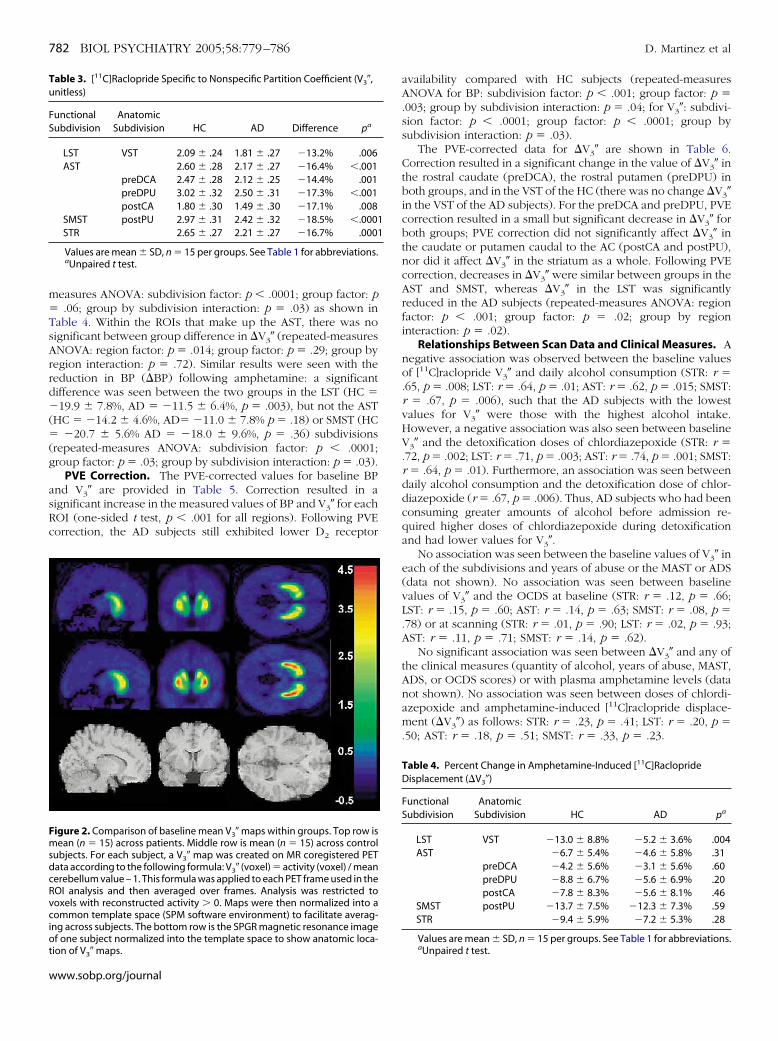
Baseline D2 Receptor Availability. Two outcome measureswere derived to assess D2 receptor availability: [11C]raclopridebinding potential (BP) and the specific to nonspecific partitioncoefficient (V3�), as shown in Tables 2 and 3. Significant groupdifferences in D2 receptor availability for each subdivision of thestriatum were found with both BP and V3� (repeated-measuresANOVA for BP: subdivision factor: p � .001; group factor: p �.007; group by subdivision interaction: p � .0024; for V3�:subdivision factor: p � .0001; group factor: p � .001; group bysubdivision interaction: p � .0015). Within the associative stria-tum (AST), a significant decrease was found in all regions for BP(Table 2) and V3� (Table 3; repeated-measures ANOVA for BP:region factor: p � .001; group factor: p � .008; group by regioninteraction: p � .04; for V3�: region factor: p � .0001; groupfactor: p � .0003; group by region interaction: p � .02). Thus, ineach subdivision, AD subjects had a lower D2 receptor availabil-ity compared with HC subjects. The reduction in D2 receptoravailability was also observed at the voxel level, as depicted bythe V3� comparison map (Figure 2).
Amphetamine Effect on [11C]Raclopride V3�. The effect ofamphetamine on [11C]raclopride V3� (denoted �V3�) was similarbetween the HC and AD in the AST, SMST, but the AD subjectsshowed a significant decrease in �V3� in the LST (repeated-
Table 2. [11C] Raclopride Binding Potential (Binding Potential, mL g�1)
FunctionalSubdivision
AnatomicSubdivision HC AD Difference pa
LST VST .84 � .21 .70 � .11 �16.6% .03AST 1.04 � .25 .84 � .13 �19.2% .009
preDCA .99 � .23 .82 � .12 �22.2% .02preDPU 1.21 � .28 .97 � .14 �20.1% .006postCA .72 � .19 .57 � .11 �22.2% .02
SMST postPU 1.19 � .27 .93 � .12 �21.3% .003STR 1.06 � .24 .85 � .12 �20.0% .01
Values are mean � SD, n � 15 per groups. See Table 1 for abbreviations.
aUnpaired t test.www.sobp.org/journal
m�TsArrd�(�(g
asRc
Tu
FS
FmsdcRvciot
782 BIOL PSYCHIATRY 2005;58:779–786 D. Martinez et al
w
easures ANOVA: subdivision factor: p � .0001; group factor: p.06; group by subdivision interaction: p � .03) as shown in
able 4. Within the ROIs that make up the AST, there was noignificant between group difference in �V3� (repeated-measuresNOVA: region factor: p � .014; group factor: p � .29; group byegion interaction: p � .72). Similar results were seen with theeduction in BP (�BP) following amphetamine: a significantifference was seen between the two groups in the LST (HC �19.9 � 7.8%, AD � �11.5 � 6.4%, p � .003), but not the AST
HC � �14.2 � 4.6%, AD� �11.0 � 7.8% p � .18) or SMST (HC�20.7 � 5.6% AD � �18.0 � 9.6%, p � .36) subdivisions
repeated-measures ANOVA: subdivision factor: p � .0001;roup factor: p � .03; group by subdivision interaction: p � .03).
PVE Correction. The PVE-corrected values for baseline BPnd V3� are provided in Table 5. Correction resulted in aignificant increase in the measured values of BP and V3� for eachOI (one-sided t test, p � .001 for all regions). Following PVEorrection, the AD subjects still exhibited lower D2 receptor
able 3. [11C]Raclopride Specific to Nonspecific Partition Coefficient (V3�,nitless)
unctionalubdivision
AnatomicSubdivision HC AD Difference pa
LST VST 2.09 � .24 1.81 � .27 �13.2% .006AST 2.60 � .28 2.17 � .27 �16.4% �.001
preDCA 2.47 � .28 2.12 � .25 �14.4% .001preDPU 3.02 � .32 2.50 � .31 �17.3% �.001postCA 1.80 � .30 1.49 � .30 �17.1% .008
SMST postPU 2.97 � .31 2.42 � .32 �18.5% �.0001STR 2.65 � .27 2.21 � .27 �16.7% .0001
Values are mean � SD, n � 15 per groups. See Table 1 for abbreviations.aUnpaired t test.
igure 2. Comparison of baseline mean V3� maps within groups. Top row isean (n � 15) across patients. Middle row is mean (n � 15) across control
ubjects. For each subject, a V3� map was created on MR coregistered PETata according to the following formula: V3� (voxel) � activity (voxel) / meanerebellum value – 1. This formula was applied to each PET frame used in theOI analysis and then averaged over frames. Analysis was restricted tooxels with reconstructed activity 0. Maps were then normalized into aommon template space (SPM software environment) to facilitate averag-
ng across subjects. The bottom row is the SPGR magnetic resonance imagef one subject normalized into the template space to show anatomic loca-
ion of V3� maps.
ww.sobp.org/journal
availability compared with HC subjects (repeated-measuresANOVA for BP: subdivision factor: p � .001; group factor: p �.003; group by subdivision interaction: p � .04; for V3�: subdivi-sion factor: p � .0001; group factor: p � .0001; group bysubdivision interaction: p � .03).
The PVE-corrected data for �V3� are shown in Table 6.Correction resulted in a significant change in the value of �V3� inthe rostral caudate (preDCA), the rostral putamen (preDPU) inboth groups, and in the VST of the HC (there was no change �V3�in the VST of the AD subjects). For the preDCA and preDPU, PVEcorrection resulted in a small but significant decrease in �V3� forboth groups; PVE correction did not significantly affect �V3� inthe caudate or putamen caudal to the AC (postCA and postPU),nor did it affect �V3� in the striatum as a whole. Following PVEcorrection, decreases in �V3� were similar between groups in theAST and SMST, whereas �V3� in the LST was significantlyreduced in the AD subjects (repeated-measures ANOVA: regionfactor: p � .001; group factor: p � .02; group by regioninteraction: p � .02).
Relationships Between Scan Data and Clinical Measures. Anegative association was observed between the baseline valuesof [11C]raclopride V3� and daily alcohol consumption (STR: r �.65, p � .008; LST: r � .64, p � .01; AST: r � .62, p � .015; SMST:r � .67, p � .006), such that the AD subjects with the lowestvalues for V3� were those with the highest alcohol intake.However, a negative association was also seen between baselineV3� and the detoxification doses of chlordiazepoxide (STR: r �.72, p � .002; LST: r � .71, p � .003; AST: r � .74, p � .001; SMST:r � .64, p � .01). Furthermore, an association was seen betweendaily alcohol consumption and the detoxification dose of chlor-diazepoxide (r � .67, p � .006). Thus, AD subjects who had beenconsuming greater amounts of alcohol before admission re-quired higher doses of chlordiazepoxide during detoxificationand had lower values for V3�.
No association was seen between the baseline values of V3� ineach of the subdivisions and years of abuse or the MAST or ADS(data not shown). No association was seen between baselinevalues of V3� and the OCDS at baseline (STR: r � .12, p � .66;LST: r � .15, p � .60; AST: r � .14, p � .63; SMST: r � .08, p �.78) or at scanning (STR: r � .01, p � .90; LST: r � .02, p � .93;AST: r � .11, p � .71; SMST: r � .14, p � .62).
No significant association was seen between �V3� and any ofthe clinical measures (quantity of alcohol, years of abuse, MAST,ADS, or OCDS scores) or with plasma amphetamine levels (datanot shown). No association was seen between doses of chlordi-azepoxide and amphetamine-induced [11C]raclopride displace-ment (�V3�) as follows: STR: r � .23, p � .41; LST: r � .20, p �.50; AST: r � .18, p � .51; SMST: r � .33, p � .23.
Table 4. Percent Change in Amphetamine-Induced [11C]RacloprideDisplacement (�V3�)
FunctionalSubdivision
AnatomicSubdivision HC AD pa
LST VST �13.0 � 8.8% �5.2 � 3.6% .004AST �6.7 � 5.4% �4.6 � 5.8% .31
preDCA �4.2 � 5.6% �3.1 � 5.6% .60preDPU �8.8 � 6.7% �5.6 � 6.9% .20postCA �7.8 � 8.3% �5.6 � 8.1% .46
SMST postPU �13.7 � 7.5% �12.3 � 7.3% .59STR �9.4 � 5.9% �7.2 � 5.3% .28
Values are mean � SD, n � 15 per groups. See Table 1 for abbreviations.
aUnpaired t test.
D
amwsap
B
rVcipBdta
ra(rtwrcPtl(a
T
FS
TA
FS
D. Martinez et al BIOL PSYCHIATRY 2005;58:779–786 783
iscussion
The results of this study show that striatal D2 receptorvailability is decreased in the limbic, associative, and sensori-otor regions of the striatum in recently detoxified AD comparedith HC subjects. Following an amphetamine challenge, the AD
ubjects were found to have a selective blunting of amphet-mine-induced dopamine release in the limbic striatum com-ared with HC subjects.
aseline D2 Receptor Availability and Alcohol DependenceIn this data set, alcohol dependence was associated with a
eduction in D2 receptor availability measured with both BP and3�. No between-group differences were observed in nonspe-ific binding (V2), free fraction in the plasma (f1), or free fractionn the brain (f2), such that the decrease in these bindingarameters can be attributed to a decrease in the D2 receptormax/KD’ ratio (Slifstein and Laruelle 2001). Furthermore, noifference in the size of the regions of interest was seen betweenhe two groups, and the decrease in D2 receptors could not bettributed to partial volume effects.
Previous PET and single photon emission computed tomog-aphy (SPECT) studies in alcohol dependence have shown bothdecrease (Hietala et al 1994; Volkow et al 1996) and no change
Guardia et al 2000; Kuikka et al 2000; Repo et al 1999) in D2
eceptor availability in the striatum. Because of camera resolu-ion, these studies were performed measuring the striatum as ahole rather than its subdivisions. Volkow et al (2002) recently
eported a decrease in D2 receptor availability in both theaudate and putamen using [11C]raclopride and a high-resolutionET camera. Alcohol-dependent subjects were scanned at twoime points: within 6 weeks of detoxification and at 1–4 monthsater, with no recovery of D2 receptors within this time frameVolkow et al 2002). This study did not measure D2 receptorvailability in the ventral striatum, however. On the other hand,
able 5. Partial Volume Effects Corrected Baseline [11C]Raclopride Binding
unctionalubdivision
AnatomicSubdivision
Binding Potential (BP, mL g
HC AD Differ
LST VST 1.34 � .36 1.07 � .16 �20AST 1.57 � .37 1.24 � .17 �20
preDCA 1.48 � .34 1.20 � .18 �19preDPU 1.71 � .40 1.33 � .18 �21postCA 1.50 � .42 1.17 � .23 �21
SMST postPU 1.97 � .50 1.51 � .20 �23STR 1.66 � .38 1.30 � .17 �21
See Table 1 for abbreviations.
able 6. Partial Volume Effects–Corrected Percent Change inmphetamine-Induced [11C]Raclopride Displacement (�V3�)
unctionalubdivision
AnatomicSubdivision HC AD pa
LST VST �15.2 � 10.4% �4.9 � 5.3% .002AST �6.2 � 5.3% �4.1 � 6.2% .31
preDCA �3.5 � 5.9% �2.4 � 5.8% .60preDPU �8.2 � 6.6% �4.7 � 7.7% .20postCA �8.1 � 9.5% �5.7 � 9.0% .48
SMST postPU �13.9 � 7.8% �12.3 � 7.7% .59STR �9.5 � 5.9% �7.0 � 5.4% .24
Values are mean � SD, n � 15 per groups. See Table 1 for abbreviations.
aUnpaired t test.Heinz et al reported a decrease in D2 receptor availabilitymeasured with [18F]desmethoxyfallypride in the ventral striatumand putamen but reported no difference in the caudate (Heinz etal 2004). Thus, our results support the previous reports of a15%–20% reduction in D2 receptor availability in alcohol depen-dence and show that this reduction involves the caudate, puta-men, and ventral striatum.
Human autoradiography studies have shown that alcoholdependence is associated with a reduction in striatal D2 receptordensity (Tupala et al 2003, 2001). In type 1 alcoholics, D2
receptors were reduced 22%–32% in the dorsal striatum (caudateand putamen) and 20% in the nucleus accumbens comparedwith control subjects (Tupala et al 2001, 2003). Decreases in D2
receptor availability were also seen in the dorsal striatum andnucleus accumbens in type 2 alcoholics, but these differences didnot reach significance (Tupala et al 2003; Tupala et al 2001).Although the alcohol-dependent subjects in our study reportedonset of alcohol dependence before the age 25, they could notbe clearly categorized as type 2 alcoholics based on their pastbehavior. Nevertheless, the results from this PET study are largelyconsistent with the human autoradiography data showing amoderate decrease in striatal D2 receptors.
Studies in animals show that the D2 receptors appear to beimportant in mediating the reinforcing effects of alcohol. Alco-hol-preferring rats have decreased D2 receptor density in boththe caudate-putamen and nucleus accumbens compared withnon-alcohol-preferring rats, even before exposure to alcohol(McBride et al 1993). Overexpression of the D2 receptor in ratstrained to self-administer alcohol reduced both their preferenceand intake of alcohol (Thanos et al 2001). Decreases in D2
receptor availability of a similar magnitude have been shown inPET studies of other addictive behaviors, such as heroin (Wanget al 1997) and cocaine dependence (Volkow et al 1990),methamphetamine abuse (Volkow et al 2001), and even obesity(Wang et al 2001), showing that this finding is not specific to aparticular substance of abuse. Thus, the findings from this studyadd to a growing body of evidence demonstrating that substanceabuse is associated with abnormal transmission at the striatal D2
receptor.
Baseline D2 Receptor Availability and Behavioral DataNo correlation was seen between baseline D2 receptor V3�
and the OCDS, which has been shown to reliably measurecraving (Anton 2000). This is in contrast to the finding of Heinzet al (2004), who showed that low D2 receptor availabilitycorrelated with craving for alcohol. The reason behind thisdiscrepancy is not clear because both studies imaged a similarnumber of subjects who were of comparable severity, and
ntial (BP) and Specific to Nonspecific Partition Coefficient (V3�)
Specific to Nonspecific Partition Coefficient (V3�, unitless)
p HC AD Difference p
.01 3.34 � .49 2.79 � .47 �16.7% .003
.004 3.93 � .41 3.22 � .40 �18.0% �.001
.01 3.70 � .40 3.09 � .38 �13.2% .0002
.003 4.27 � .45 3.46 � .41 �19.1% �.001
.01 3.75 � .68 3.06 � .74 �18.3% .014
.002 4.94 � .64 3.93 � .56 �20.4% �.001
.003 4.14 � .41 3.38 � .42 �18.4% �.001
Pote
�1)
ence
.1%
.8%
.1%
.9%
.9%
.4%
.3%
imaging occurred after a similar period of abstinence. Both
www.sobp.org/journal

sDmtArhspbb
[crdjncrl[Dneectbc
AD
mdosrsssnra
aaisiArt(rtmcpswr
784 BIOL PSYCHIATRY 2005;58:779–786 D. Martinez et al
w
tudies used radiotracers and methods shown to measure striatal
2 receptors reliably, and both studies used V3� as the outcomeeasure. Thus, the main difference between these studies were
he scales to measure craving (the OCDS in this study and thelcohol Craving Questionnaire in the study of Heinz et al). Aecent study showed a significant correlation between the Alco-ol Craving Questionnaire and the obsessive and compulsiveubscales of the OCDS (Raabe et al 2005). Thus, we performed aost hoc analysis comparing these subscales of the OCDS andaseline D2 receptor V3� but found no significant associationetween these subscales and V3� in any region (all p .6).
In this study, we found a negative correlation between11C]raclopride binding and the average daily quantity of alcoholonsumed. We also found a similar correlation between D2
eceptor availability and the doses of chlordiazepoxide used foretoxification. Not surprisingly, the magnitude of the AD sub-
ects’ alcohol intake correlated with the dose of chlordiazepoxideeeded for detoxification. Based on previous imaging studies,hlordiazepoxide is not expected to decrease measures of D2
eceptor availability. Hietala et al (1997) demonstrated thatorazepam (2 mg taken orally for 1 week) did not affect11C]raclopride binding in healthy human subjects, whereasewey et al demonstrated an increase [11C]raclopride BP inonhuman primates following gamma-vinyl-GABA and loraz-pam (Dewey et al 1992). Thus, the administration of GABA-nhancing agents should result in either an increase or nohange in the [11C]raclopride BP, rather than a decrease. Never-heless, it cannot be stated with certainty that the correlationetween D2 receptor availability and quantity of alcohol is notonfounded by the detoxification with chlordiazepoxide.
mphetamine-Induced Dopamine Release and AlcoholependenceTo our knowledge, this study is the first published report
easuring dopamine release in the striatum in alcohol depen-ence. Blunted amphetamine-induced dopamine release wasbserved in the limbic striatum in AD compared with HCubjects. Amphetamine-induced dopamine release was slightlyeduced in the associative and sensorimotor striatum of ADubjects compared with HC, but these differences did not reachignificance. This finding is in agreement with rodent studieshowing a reduction in dopamine and its metabolites in theucleus accumbens (Murphy et al 1982) and studies showing aeduction in midbrain dopamine projections to the nucleusccumbens (Casu et al 2002; Zhou et al 1995).
In human subjects, the etiology behind the reduction inmphetamine-induced dopamine release is not clear. Tiihonen etl (1998) demonstrated an increase in 6-[18F]-fluoroDOPA uptaken the putamen and caudate in AD subjects compared with HCubjects, a finding that suggests that alcoholics should havencreased presynaptic dopamine function rather than a decrease.lternatively, using PET and the radioligand (�)[18F]dihydrotet-abenazine, Gilman et al (1998) reported a decrease in striatalype 2 vesicular monoamine transporters (VMAT2) in the caudate6%) and putamen (13%) of AD subjects, although this onlyeached significance in the putamen. Levels of VMAT2 may affecthe magnitude of the amphetamine effect on extracellular dopa-ine (Patel et al 2003) such that a loss of VMAT2 would be
onsistent with a decrease in amphetamine-induced [11C]raclo-ride displacement. In the study of Gilman et al, VMAT2 was notpecifically measured in the ventral striatum, so it is unclearhether this factor could explain the reduction in dopamine
elease seen in this study.
ww.sobp.org/journal
Mesolimbic dopamine has been shown to play a crucial rolein mediating the reinforcing effects of alcohol. Ethanol has beenshown to increase mesolimbic dopamine release in rodents(Imperato and Di Chiara 1986), nonhuman primates (Bradberry2002), and human healthy control subjects (Boileau et al 2003).Alcohol-preferring rodents have a greater dopamine release inthe nucleus accumbens following their initial exposure to alcoholcompared with non-alcohol-preferring rats (Katner and Weiss2001), and a reduced response to ethanol following repeatedexposure (Murphy et al 1988). Acute alcohol withdrawal hasbeen shown to reduce mesolimbic dopamine function (Diana etal 1993), and it has been postulated that a deficit in dopamineactivity may play a critical role in relapse (Wise 1988). Thus, theresults of these animal studies suggest that individuals whobecome alcohol dependent may initially have an exaggerateddopamine response to ethanol that is reinforcing. With time andthe onset of dependence, however, alcohol-dependent subjectsmay loose dopamine tone in the ventral striatum. Insofar asdopamine in the nucleus accumbens is thought to serve as abehavioral switching device (Pennartz et al 1994), this deficit indopamine release may represent an impaired ability of alcohol-dependent individuals to shift from the compulsive, maladaptivepatterns of behavior that are indicative of addiction.
Partial Volume Effects CorrectionThe PVE correction of the baseline [11C]raclopride images
resulted in a significant increase in the values of BP and V3�, from40% in the rostral putamen (an area with less contact with thebackground) to 106% in the caudal caudate (a long, thin regionthat is largely surrounded by background). These results areconsistent with previous reports (Martinez et al 2003; Mawlawi etal 2001) and reflect the dilution of signal experienced by theseregions due to low activity in the surrounding background tissue.On the other hand, PVE correction resulted in little or no effecton �V3�, even when correcting for adjacent regions with largedifferences in activity, consistent with previous studies (Martinezet al 2003; Slifstein et al 2004). The effect of PVE correction on�V3� in a given ROI can be understood by looking at thegeometry of the striatum and �V3� of the adjacent ROIs. The PVEcorrection had no effect on �V3� in the caudate or putamencaudal to the AC; (postCA and postPU) or the striatum as awhole. These are regions that are largely surrounded by back-ground, and we have previously shown that PVE correction hasa negligible effect in this setting (Martinez et al 2003; Slifstein etal 2004).
The subdivisions of the striatum can be distinguished fromeach other based on their response to amphetamine: the limbicand sensorimotor subdivisions have a greater [11C]raclopridedisplacement compared with the associative subdivision (seeMartinez et al 2003, and Table 4). Thus, PVE correction slightlydecreased �V3� in the caudate and putamen anterior to the AC(preDCA and preDPU) because these regions displace less[11C]raclopride following amphetamine and are adjacent to re-gions that have a greater amphetamine effect (VST and postPU).Interestingly, PVE correction increased �V3� in the VST of the HCsubjects but did not affect �V3� in the LST of the AD subjects.This is because �V3� in the VST of the HC was affected by thelower �V3� of the adjacent preDCA and preDPU. However, �V3�in the VST of the AD subjects was blunted and of a similarmagnitude to the �V3� of the preDCA and preDPU.
Overall, PVE correction did not alter the primary findings inthis study: AD subjects still had a lower D receptor availability
2compared with HC subjects, and �V3� was reduced in the limbic
snob
L
nbdrapStmcAstt
RESt
A
A
B
B
B
C
D
D
G
G
H
H
D. Martinez et al BIOL PSYCHIATRY 2005;58:779–786 785
triatum only. These findings suggest that PVE correction mayot be necessary when comparing between-subject differencesf this magnitude when no difference in ROI size is seenetween groups.
imitationsSeveral limitations of this study should be recognized. 1) The
umber of subjects per group is relatively small. 2) Changes inaseline D2 receptor availability could result from either aecrease in receptor concentration or in receptor affinity for theadiotracer. 3) The amphetamine challenge provides informationbout the increase in dopamine transmission induced by am-hetamine, but not about the baseline synaptic dopamine levels.tudies using rapid dopamine depletion techniques are requiredo assess this (Laruelle et al 1997). 4) The amphetamine challengeeasures changes in D2 receptor availability secondary to
hanges in endogenous dopamine. Thus, the difference betweenD and HC subjects could be due to a smaller increase inynaptic dopamine or lower affinity of D2 receptors. Studies withhe radiolabeled agonist [11C]N-propyl-apomorphine are neededo clarify this issue (Hwang et al 2004).
Supported by the Public Health Service (Grant Nos. NIAAA01 AA12759-0 and RR00645). The authors thank Jennifer Bae,lizabeth Hackett, Kimchung Ngo, Chaka Peters, Lyudmilaavenkova , and Kris Wolff for their excellent technical assis-ance.
nton RF (2000): Obsessive–compulsive aspects of craving: Development ofthe Obsessive Compulsive Drinking Scale. Addiction 95(Suppl 2):S211–217.
nton RF, Moak DH, Latham P (1995): The Obsessive Compulsive DrinkingScale: A self-rated instrument for the quantification of thoughts aboutalcohol and drinking behavior. Alcohol Clin Exp Res 19:92–99.
oileau I, Assaad JM, Pihl RO, Benkelfat C, Leyton M, Diksic M, et al (2003):Alcohol promotes dopamine release in the human nucleus accumbens.Synapse 49:226 –231.
radberry CW (2002): Dose-dependent effect of ethanol on extracellular dopa-mine in mesolimbic striatum of awake rhesus monkeys: comparison withcocaine across individuals. Psychopharmacology (Berl) 165:67–76.
reier A, Su TP, Saunders R, Carson RE, Kolachana BS, deBartolomeis A, et al(1997): Schizophrenia is associated with elevated amphetamine-in-duced synaptic dopamine concentrations: Evidence from a novelpositron emission tomography method. Proc Natl Acad Sci U S A 94:2569 –2574.
asu MA, Dinucci D, Colombo G, Gessa GL, Pani L (2002): Reduced DAT- andDBH-immunostaining in the limbic system of Sardinian alcohol-prefer-ring rats. Brain Res 948:192–202.
ewey SL, Smith GS, Logan J, Brodie JD, Yu DW, Ferrieri RA, et al (1992):GABAergic inhibition of endogenous dopamine release measured invivo with 11C-raclopride and positron emission tomography. J Neurosci12:3773–3780.
iana M, Pistis M, Carboni S, Gessa GL, Rossetti ZL (1993): Profound decre-ment of mesolimbic dopaminergic neuronal activity during ethanolwithdrawal syndrome in rats: Electrophysiological and biochemical evi-dence. Proc Natl Acad Sci U S A 90:7966 –7969.
ilman S, Koeppe RA, Adams KM, Junck L, Kluin KJ, Johnson-Greene D, et al(1998): Decreased striatal monoaminergic terminals in severe chronicalcoholism demonstrated with (�)[11C]dihydrotetrabenazine andpositron emission tomography. Ann Neurol 44:326 –333.
uardia J, Catafau AM, Batlle F, Martin JC, Segura L, Gonzalvo B, et al (2000):Striatal dopaminergic D(2) receptor density measured by [(123)I]iodo-benzamide SPECT in the prediction of treatment outcome of alcohol-dependent patients. Am J Psychiatry 157:127–129.
all H, Sedvall G, Magnusson O, Kopp J, Halldin C, Farde L (1994): Distribu-tion of D1- and D2-dopamine receptors, and dopamine and its metabo-lites in the human brain. Neuropsychopharmacology 11:245–256.
einz A, Siessmeier T, Wrase J, Hermann D, Klein S, Grusser SM, et al (2004):
Correlation between dopamine D(2) receptors in the ventral striatumand central processing of alcohol cues and craving. Am J Psychiatry161:1783–1789.
Hietala J, Kuoppamaki M, Nagren K, Lehikoinen P, Syvalahti E (1997): Effectsof lorazepam administration on striatal dopamine D–2 receptor bindingcharacteristics in man—a positron emission tomography study. Psycho-pharmacology 132:361–365.
Hietala J, West C, Syvalahti E, Nagren K, Lehikoinen P, Sonninen P, Ruot-salainen U (1994): Striatal D2 dopamine receptor binding characteristicsin vivo in patients with alcohol dependence. Psychopharmacology (Berl)116:285–290.
Hwang DR, Narendran R, Huang Y, Slifstein M, Talbot PS, Sudo Y, et al (2004):Quantitative analysis of (-)-N-(11)C-propyl-norapomorphine in vivobinding in nonhuman primates. J Nucl Med 45:338 –346.
Imperato A, Di Chiara G (1986): Preferential stimulation of dopamine releasein the nucleus accumbens of freely moving rats by ethanol. J PharmacolExp Ther 239:219 –228.
Katner SN, Weiss F (2001): Neurochemical characteristics associated withethanol preference in selected alcohol-preferring and -nonpreferringrats: A quantitative microdialysis study. Alcohol Clin Exp Res 25:198 –205.
Kuikka JT, Repo E, Bergstrom KA, Tupala E, Tiihonen J (2000): Specific bindingand laterality of human extrastriatal dopamine D2/D3 receptors in lateonset type 1 alcoholic patients. Neurosci Lett 292:57–59.
Laruelle M, DSouza CD, Baldwin RM, AbiDargham A, Kanes SJ, Fingado CL, etal (1997): Imaging D–2 receptor occupancy by endogenous dopamine inhumans. Neuropsychopharmacology 17:162–174.
Maisto SA, Sobell LC, Cooper AM, Sobell MB (1982): Comparison of twotechniques to obtain retrospective reports of drinking behavior fromalcohol abusers. Addict Behav 7:33–38.
Martinez D, Slifstein M, Broft A, Mawlawi O, Hwang DR, Huang Y, et al (2003):Imaging human mesolimbic dopamine transmission with positron emis-sion tomography. Part II: amphetamine-induced dopamine release inthe functional subdivisions of the striatum. J Cereb Blood Flow Metab23:285–300.
Mawlawi O, Martinez D, Slifstein M, Broft A, Chatterjee R, Hwang DR, et al(2001): Imaging human mesolimbic dopamine transmission with PET: IAccuracy and precision of D2 parameter measurements in the ventralstriatum. J Cereb Blood Flow Metab 21:1034 –1057.
McBride WJ, Chernet E, Dyr W, Lumeng L, Li TK (1993): Densities of dopamineD2 receptors are reduced in CNS regions of alcohol-preferring P rats.Alcohol 10:387–390.
Murphy JM, McBride WJ, Gatto GJ, Lumeng L, Li TK (1988): Effects of acuteethanol administration on monoamine and metabolite content in fore-brain regions of ethanol-tolerant and -nontolerant alcohol-preferring (P)rats. Pharmacol Biochem Behav 29:169 –174.
Murphy JM, McBride WJ, Lumeng L, Li TK (1982): Regional brain levels ofmonoamines in alcohol-preferring and -nonpreferring lines of rats. Phar-macol Biochem Behav 16:145–149.
Patel J, Mooslehner KA, Chan PM, Emson PC, Stamford JA (2003): Presynapticcontrol of striatal dopamine neurotransmission in adult vesicular mono-amine transporter 2 (VMAT2) mutant mice. J Neurochem 85:898 –910.
Pennartz CM, Groenewegen HJ, Lopes da Silva FH (1994): The nucleus ac-cumbens as a complex of functionally distinct neuronal ensembles: anintegration of behavioural, electrophysiological and anatomical data.Prog Neurobiol 42:719 –761.
Raabe A, Grusser SM, Wessa M, Podschus J, Flor H (2005): The assessment ofcraving: psychometric properties, factor structure and a revised versionof the Alcohol Craving Questionnaire (ACQ). Addiction 100:227–234.
Repo E, Kuikka JT, Bergstrom KA, Karhu J, Hiltunen J, Tiihonen J (1999):Dopamine transporter and D2-receptor density in late-onset alcoholism.Psychopharmacology (Berl) 147:314 –318.
Rousset OG, Ma Y, Evans AC (1998): Correction for partial volume effects inPET: Principle and validation. J Nucl Med 39:904 –911.
Selzer ML (1971): The Michigan alcoholism screening test: the quest for anew diagnostic instrument. Am J Psychiatry 127:1653–1658.
Skinner HA, Allen BA (1982): Alcohol dependence syndrome: measurementand validation. J Abnorm Psychol 91:199 –209.
Slifstein M, Laruelle M (2001): Models and methods for derivation of in vivoneuroreceptor parameters with PET and SPECT reversible radiotracers.Nucl Med Biol 28:595– 608.
Slifstein M, Narendran R, Hwang DR, Sudo Y, Talbot PS, Huang Y, Laruelle M(2004): Effect of amphetamine on [(18)F]fallypride in vivo binding to D(2)receptors in striatal and extrastriatal regions of the primate brain: Single
bolus and bolus plus constant infusion studies. Synapse 54:46 – 63.www.sobp.org/journal
S
T
T
T
T
V
786 BIOL PSYCHIATRY 2005;58:779–786 D. Martinez et al
w
okoloff P, Giros B, Martres M-P, Bouthenet M-L, Schwartz J-C (1990): Molec-ular cloning and characterization of a novel dopamine receptor D3 as atarget for neuroleptics. Nature 347:146 –151.
hanos PK, Volkow ND, Freimuth P, Umegaki H, Ikari H, Roth G, et al (2001):Overexpression of dopamine D2 receptors reduces alcohol self-adminis-tration. J Neurochem 78:1094 –1103.
iihonen J, Vilkman H, Rasanen P, Ryynanen OP, Hakko H, Bergman J, et al(1998): Striatal presynaptic dopamine function in type 1 alcoholics mea-sured with positron emission tomography. Mol Psychiatry 3:156 –161.
upala E, Hall H, Bergstrom K, Mantere T, Rasanen P, Sarkioja T, Tiihonen J(2003): Dopamine D2 receptors and transporters in type 1 and 2 alcohol-ics measured with human whole hemisphere autoradiography. HumBrain Mapp 20:91–102.
upala E, Hall H, Bergstrom K, Sarkioja T, Rasanen P, Mantere T, et al (2001):Dopamine D(2)/D(3)-receptor and transporter densities in nucleus accum-bens and amygdala of type 1 and 2 alcoholics. Mol Psychiatry 6:261–267.
olkow ND, Chang L, Wang GJ, Fowler JS, Ding YS, Sedler M, et al (2001): Lowlevel of brain dopamine D2 receptors in methamphetamine abusers:
Association with metabolism in the orbitofrontal cortex. Am J Psychiatry158:2015–2021.ww.sobp.org/journal
Volkow ND, Fowler JS, Wolf AP, Schlyer D, Shiue CY, Alpert R, et al (1990):Effects of chronic cocaine abuse on postsynaptic dopamine receptors.Am J Psychiatry 147:719 –724.
Volkow ND, Wang GJ, Fowler JS, Logan J, Hitzemann R, Ding YS, et al (1996):Decreases in dopamine receptors but not in dopamine transporters inalcoholics. J Nucl Med 37:33P.
Volkow ND, Wang GJ, Maynard L, Fowler JS, Jayne B, Telang F, et al (2002):Effects of alcohol detoxification on dopamine D2 receptors in alcoholics:a preliminary study. Psychiatry Res 116:163–172.
Wang GJ, Volkow ND, Fowler JS, Logan J, Abumrad NN, Hitzemann RJ, et al(1997): Dopamine D2 receptor availability in opiate-dependent subjectsbefore and after naloxone-precipitated withdrawal. Neuropsychophar-macology 16:174 –182.
Wang GJ, Volkow ND, Logan J, Pappas NR, Wong CT, Zhu W, et al (2001):Brain dopamine and obesity. Lancet 357:354 –357.
Wise RA (1988): The neurobiology of craving: Implication for the un-derstanding of treatment of addiction. J Abnorm Psychol 2:118 –132.
Zhou FC, Zhang JK, Lumeng L, Li TK (1995): Mesolimbic dopamine system inalcohol-preferring rats. Alcohol 12:403– 412.
Related Documents