Accepted Article Clinical Characteristics and Whole Exome/Transcriptome Sequencing of Coexisting Chronic Myeloid Leukemia and Myelofibrosis Malathi Kandarpa 1 , Yi-Mi Wu 2 , Dan Robinson 2 , Patrick William Burke 1 , Arul M. Chinnaiyan 2 and Moshe Talpaz 1 1 Department of Internal Medicine, Division of Hematology/Oncology, University of Michigan Comprehensive Cancer Center, Ann Arbor, MI 48109, USA. 2 Michigan Center for Translational Pathology, University of Michigan Medical School, Ann Arbor, MI 48109, USA. Corresponding Author: Moshe Talpaz, MD Department of Internal Medicine, Division of Hematology Oncology 1500 East Medical Center Drive 4302 CCC-SPC 5936 Ann Arbor, MI 48109-5936 Phone: 734-764-8195 Fax: 734-647-9654 Email: [email protected] Abstract Word Count: 248 Text Word Count: 2440 Number of Tables: 2 Number of Figures: 2 Supplemental Tables: 2 Short Running Title: Molecular characteristics of dual-MPN Keywords: myelofibrosis, CML, myeloproliferative neoplasm Page 1 of 16 American Journal of Hematology This is the author manuscript accepted for publication and has undergone full peer review but has not been through the copyediting, typesetting, pagination and proofreading process, which may lead to differences between this version and the Version record. Please cite this article as doi:10.1002/ajh.24728. This article is protected by copyright. All rights reserved.

Welcome message from author
This document is posted to help you gain knowledge. Please leave a comment to let me know what you think about it! Share it to your friends and learn new things together.
Transcript

Acc
epte
d A
rtic
leClinical Characteristics and Whole Exome/Transcriptome Sequencing of
Coexisting Chronic Myeloid Leukemia and Myelofibrosis
Malathi Kandarpa1, Yi-Mi Wu
2, Dan Robinson
2, Patrick William Burke
1, Arul M. Chinnaiyan
2
and Moshe Talpaz1
1Department of Internal Medicine, Division of Hematology/Oncology, University of Michigan
Comprehensive Cancer Center, Ann Arbor, MI 48109, USA.
2Michigan Center for Translational Pathology, University of Michigan Medical School, Ann
Arbor, MI 48109, USA.
Corresponding Author:
Moshe Talpaz, MD
Department of Internal Medicine, Division of Hematology Oncology
1500 East Medical Center Drive
4302 CCC-SPC 5936
Ann Arbor, MI 48109-5936
Phone: 734-764-8195
Fax: 734-647-9654
Email: [email protected]
Abstract Word Count: 248
Text Word Count: 2440
Number of Tables: 2
Number of Figures: 2
Supplemental Tables: 2
Short Running Title: Molecular characteristics of dual-MPN
Keywords: myelofibrosis, CML, myeloproliferative neoplasm
Page 1 of 16 American Journal of Hematology
This is the author manuscript accepted for publication and has undergone full peer review but has not beenthrough the copyediting, typesetting, pagination and proofreading process, which may lead to differencesbetween this version and the Version record. Please cite this article as doi:10.1002/ajh.24728.
This article is protected by copyright. All rights reserved.

Acc
epte
d A
rtic
le
ABSTRACT
Myeloproliferative neoplasms (MPNs) are clonal hematopoietic stem cell (HSC) disorders that
can be classified on the basis of genetic, clinical, phenotypic features. Genetic lesions such as
JAK2 mutations and BCR-ABL translocation are often mutually exclusive in MPN patients and
lead to essential thrombocythemia, polycythemia vera or myelofibrosis (ET/PV/MF) or chronic
myeloid leukemia, respectively. Nevertheless, coexistence of these genetic aberrations in the
same patient has been reported. Whether these aberrations occur in the same stem cell or a
different cell is unclear, but an unstable genome in the HSCs seems to be the common
antecedent. In an effort to characterize the underlying genetic events that might contribute to the
appearance of more than one MPN in a patient, we studied neoplastic cells from patients with
dual MPNs by next-generation sequencing. We observed that most patients with two MPNs
harbored mutations in genes known to contribute to clonal hematopoiesis through altered
epigenetic regulation such as TET2, ASXL1/2, SRSF2, and IDH2 at varying frequencies (1-
47%). In addition, we found that some patients also harbored oncogenic mutations in N/KRAS,
TP53, BRAF, EZH2, GNAS at low frequencies, which probably represent clonal evolution.
These findings support the hypothesis that hematopoietic cells from MPN patients harbor
multiple genetic aberrations, some of which can contribute to clonal dominance. Acquiring
mutations in JAK2/CALR/MPL or the BCR-ABL translocation probably drive the oncogenic
phenotype towards a specific MPN. Further, we propose that the acquisition of BCR-ABL in
these patients is frequently a secondary event resulting from an unstable genome.
Page 2 of 16
John Wiley & Sons
American Journal of Hematology
This article is protected by copyright. All rights reserved.

Acc
epte
d A
rtic
le
INTRODUCTION
Myeloproliferative neoplasms (MPNs) are characterized by an expansion of one or more lineages
of myeloid stem cells. According to the WHO classification, there are 8 different phenotypes of
MPNs [1]. Several lines of evidence suggest that genomic instability in the HSC leads to
development of molecular lesions that generate the myeloproliferative phenotype [2]. However,
the underlying cause of this genomic instability is not well understood. The presence of the
BCR-ABL1 fusion gene leads to a chronic myeloid leukemia (CML) phenotype, while mutations
in the JAK2 gene are linked to essential thrombocythemia (ET), polycythemia vera (PV) and
myelofibrosis (MF). In addition exclusive of JAK2, ET and MF patients can harbor CALR or
MPL mutations [3]. These genetic aberrations result in dysregulated tyrosine kinases that
generate proliferative signals in the disease-initiating cells. Since these molecular aberrations are
usually perceived as mutually exclusive [4], once one MPN is diagnosed, tests for the other
mutation/fusion are rarely performed.
Recently, several case studies have described either concomitant CML and ET/PV/MF or
emergence of one of these diseases in patients previously diagnosed with another MPN [5-9]. In
two cases where CML emerged after PV, the BCR-ABL translocation was suggested to be a
secondary event in the JAK2-mutated clone [7, 8]. However, others have suggested the
mutations may arise in two independent clones [10]. Nevertheless, both these scenarios
presuppose an unstable genome that induces multiple changes in a stem cell or favors emergence
of other competing clones. In this study, we evaluated the molecular landscape of hematopoietic
stem and progenitor cells from patients with coexistent CML and MF using next-generation
sequencing (NGS) methods.
METHODS
Patients and samples
Patients were diagnosed and treated at the University of Michigan Health system. Criteria for
diagnosis of CML and post-ET and post-PV MF were based on the WHO classification [1].
Samples from consenting patients were obtained with approval from the institutional review
board of the University of Michigan. Bone marrow and peripheral blood mononuclear cells were
prepared by Ficoll density gradient centrifugation. For samples that were sequenced, bone
marrow mononuclear cells were CD34 enriched using CD34 magnetic microbeads (Miltenyi
Biotec, San Diego CA), or total bone marrow or peripheral blood mononuclear cells were used
Page 3 of 16
John Wiley & Sons
American Journal of Hematology
This article is protected by copyright. All rights reserved.

Acc
epte
d A
rtic
le
for isolation of nucleic acids. Matched buccal swabs were used as the source of normal control
DNA for each patient. Patients were identified throughout the study using their study ID
assigned during enrollment.
Clinical data
Clinical characteristics of each consenting patient were extracted from patient charts. Laboratory
results (e.g., diagnostic molecular testing), treatment timelines and drug regimens were compiled
from patient charts. Most cases of CML were diagnosed based on classical karyotype and/or
FISH. Quantitative molecular diagnosis of BCR-ABL-positive CML to determine response was
based on PCR-based testing and was available from some patients for a few time points and the
data is presented in International Scale (IS) or as otherwise indicated. Spleen size measurements
were based on palpation unless otherwise stated.
Integrative high-throughput sequencing and mutation calls
Nucleic acid preparation, sequencing library construction and high-throughput sequencing were
performed using standard protocols in our sequencing laboratory, which adheres to the Clinical
Laboratory Improvement Amendments. Paired-end whole-exome libraries from tumor and
matched normal DNA were prepared using the Agilent SureSelect human all exon v4 probes
(Agilent Technologies, Santa Clara CA). Transcriptome libraries were prepared from total RNA
and captured by the Agilent SureSelect human all exon v4 probes [11]. All the libraries were
sequenced using the Illumina HiSeq2500 (Illumina Inc., San Diego CA). Aligned exome and
transcriptome sequences were analyzed to detect putative somatic mutations, insertions and
deletions (indels), copy-number alterations, gene fusions, and gene expression as described
previously [12, 13].
COSMIC v 79 was interrogated using the Cancer Browser tool on the COSMIC web application,
http://cancer.sanger.ac.uk/cosmic/browse/tissue.
RESULTS
Concomitant vs sequential diagnosis of dual MPN phenotype
We identified eight patients with diagnosis of two different MPNs (one being CML) during the
course of their treatment or at initial diagnosis. Clinical characteristics of the patients and their
diagnosis criteria for each disease are summarized in Table I. In two of the patients, both
Page 4 of 16
John Wiley & Sons
American Journal of Hematology
This article is protected by copyright. All rights reserved.

Acc
epte
d A
rtic
le
diseases were diagnosed concomitantly; in the other patients, the second condition was
diagnosed during treatment due to ongoing and complex physical findings (splenomegaly),
cytogenetic findings of the Philadelphia chromosome (Ph), and pathological findings in the bone
marrow, such as CML patient with dysmegakaryopoiesis indicating more than one
myeloproliferative disease. The time between the establishments of the two diagnoses varied
from 0-15 years. In addition to the patients presented, we identified three other MF patients who
had minimal levels of BCR-ABL by PCR without hematological disease (data not shown). The
predominant diagnosis at presentation varied among the eight patients. Patient (Pt) 1471 was
under observation for ET, which progressed to MF and exhibited a BCR-ABL translocation
along with a JAK2V617F mutation. Similarly, Pt2105 with post-ET MF was found to have BCR-
ABL translocation during testing prior to enrollment onto a clinical trial. On the other hand,
Pt1191 was Ph+ve
and did not have a JAK2V617F mutation at the time of CML diagnosis;
however, after achieving a major molecular response, the patient’s bone marrow showed a
fibrotic myeloid neoplasm with prominent large megakaryocytes in clusters with sinusoidal
dilation containing hematopoietic elements. The bone marrow findings, positive reticulin
staining and persistent splenic enlargement implicated an overlap diagnosis of MF, which was
eventually confirmed by the presence of the CALR mutation. Overall, CML was the first MPN
diagnosis in two patients, second diagnosis in four patients, and concurrent diagnosis in five
patients (data not shown for three) following low level BCR-ABL detection.
Treatment paradigms for the dual disease patients
Treatment was tailored to the clinically dominant disease in cases of concomitant diagnosis; in
the other cases treatment was adjusted to address the second condition. The patients’ treatment
timelines are summarized in Figure 1A. Each patient’s response to the treatment, as measured by
spleen size, WBC count and BCR-ABL levels, is summarized in Figure 1B. An addition or
change in therapy was warranted when a patient did not respond to a treatment as indicated by an
increase in BCR-ABL transcripts, increase in WBCs, or persistence or increase in splenomegaly.
A few of the patients were switched to TKI therapy for CML combined with a JAK inhibitor or
another therapy for MF, either given together (Pt2105, dosed with ruxolitinib and
imatinib/dasatinib every day) or in an alternating schedule (Pt1137, alternating schedule of
nilotinib 4 days on/1 day off, then ruxolitinib 8 days on/1 day off). Most of these patients
demonstrated improved response and safe tolerability with this regimen, potentially endorsing a
Page 5 of 16
John Wiley & Sons
American Journal of Hematology
This article is protected by copyright. All rights reserved.
Acc
epte
d A
rtic
le
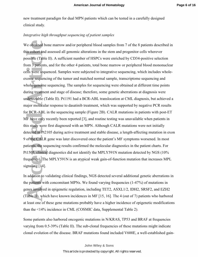
new treatment paradigm for dual MPN patients which can be tested in a carefully designed
clinical study.
Integrative high throughput sequencing of patient samples
We obtained bone marrow and/or peripheral blood samples from 7 of the 8 patients described in
this cohort and assessed all genomic alterations in the stem and progenitor cells wherever
possible (Table II). A sufficient number of HSPCs were enriched by CD34-positive selection
from 3 patients, and for the other 4 patients, total bone marrow or peripheral blood mononuclear
cells were sequenced. Samples were subjected to integrative sequencing, which includes whole-
exome sequencing of the tumor and matched normal sample, transcriptome sequencing and
whole-genome sequencing. The samples for sequencing were obtained at different time points
during treatment and stage of disease; therefore, some genetic aberrations at diagnosis were
undetectable (Table II). Pt1191 had a BCR-ABL translocation at CML diagnosis, but achieved a
major molecular response to dasatinib treatment, which was supported by negative PCR results
for BCR-ABL in the sequencing sample (Figure 2B). CALR mutations in patients with post-ET
MF have only recently been reported [3], and routine testing was unavailable when patients in
this study were first diagnosed with an MPN. Although CALR mutations were not initially
detected in Pt2105 during active treatment and stable disease, a length-affecting mutation in exon
9 of the CALR gene was later discovered once the patient’s MF symptoms worsened. In most
patients, the sequencing results confirmed the molecular diagnostics in the patient charts. For
Pt1505, clinical diagnostics did not identify the MPLY591N mutation detected by NGS (10%
frequency). The MPLY591N is an atypical weak gain-of-function mutation that increases MPL
signaling [14].
In addition to validating clinical findings, NGS detected several additional genetic aberrations in
the patients with concomitant MPNs. We found varying frequencies (1-47%) of mutations in
genes involved in epigenetic regulation, including TET2, ASXL1/2, IDH2, SRSF2, and EZH2
(Table II), which have known incidences in MF [15, 16]. The 4 (out of 7) patients who harbored
at least one of these gene mutations probably have a higher incidence of epigenetic modifications
than the <14% incidence in CML (COSMIC data, Supplemental Table 2).
Some patients also harbored oncogenic mutations in N/KRAS, TP53 and BRAF at frequencies
varying from 0.5-39% (Table II). The sub-clonal frequencies of these mutations might indicate
clonal evolution of the disease. BRAF mutations found included V600E, a well-established gain-
Page 6 of 16
John Wiley & Sons
American Journal of Hematology
This article is protected by copyright. All rights reserved.

Acc
epte
d A
rtic
le
of-function mutation, as well as G469V and D594E, which are two atypical mutations that like
V600E, occur within the kinase domain. BRAF D594E has been previously reported in CMML
cases [17].
We interrogated the COSMIC database to determine the genes that have been previously
reported in CML (including blastic phase CML), ET, PV and MF. The top 20 genes in CML and
blastic phase CML were compared to each of the top 20 genes from ET/PV/MF (Figure 2). The
common genes found to be mutated in these diseases included the same epigenetic regulators we
found mutated in our patient set, namely TET2, ASXL1, IDH1/2, SRSF2 and EZH2. Outside of
ABL1 (47-28%), genetic variations in CML occur infrequently. Frequency of TP53 mutations
increased from 4% in chronic-phase CML to 26% in blast-phase CML. In Ph+ve
MPNs, TP53
variant frequency was only 2-6%, suggesting that TP53 variants are associated with advanced
disease and are not driver mutations in these diseases.
DISCUSSION
This report of eight patients with coexistent ET/PV/MF and CML, along with other reports in the
literature underscore that the frequency of this phenomenon is significant. Most often one MPN
is diagnosed, precluding further testing for other diseases. Lack of response to therapy is often
the reason for alternate/additional diagnoses. Here we describe the diagnosis and management of
patients with coexistent MPNs. The treatment was tailored to the patient’s clinical presentation
and tolerance to therapy, and guided by our experience.
We used a whole-genome sequencing approach to understand the pathophysiology of the dual
disease phenotype. Our observations suggest that patients with concurrent MPNs acquire either
simultaneous or sequential mutations in the HSPC. We therefore propose two models to explain
the development of concurrent MPNs at the cellular level: 1) two independent clones arise from
genetically unstable HSPCs and compete with each other, and 2) an already mutated HSPC
acquires a “second hit”. Previous reports from patients with two MPNs suggest that BCR-ABL1
and JAK2V617F
can be present in the same clone of cells or in distinct clones [6, 7, 9, 10, 18-21].
In some patients (n=4) from our cohort, the NGS analysis did not detect a previously identified
BCR-ABL1 translocation because patients were in molecular remission at the time of sampling.
JAK2 or MPL mutations were detected in these samples after the CML sub-clone was
suppressed. The other interpretation is that the JAK2 mutation precedes BCR-ABL1
translocation and therefore JAK2 persisted after BCR-ABL1 eradication. Therefore, our data
Page 7 of 16
John Wiley & Sons
American Journal of Hematology
This article is protected by copyright. All rights reserved.

Acc
epte
d A
rtic
le
suggest that genetic events that lead to ET/PV/MF arise in the HSPC and CML is a result of a
“second hit”.
The literature supports that genetic instability in HSPCs is a precondition for MPN initiation.
HSPCs from “healthy” individuals, who have no overt disease, can acquire mutations in several
genes that might contribute to a pre-leukemic state, which over time could further destabilize the
genome and result in the disease phenotype [22, 23]. These pre-leukemic genes in healthy
individuals could provide a competitive advantage to the HSPCs, thus resulting in clonal
hematopoiesis. Mutations in epigenetic regulators such as DNMT3A, TET2 and ASXL1 genes
occur frequently in these individuals and in patients with MPNs including MF/ET/PV [16, 24,
25]. These epigenetic regulators might contribute to the instability of the HSPC genome [26, 27].
In our studies, mutations in TET2 (n=2) and ASXL1 (n=3) were found in some patients who
were stable or in remission at the time of sampling. We did not detect any DNMT3A mutations,
which is consistent with the low frequencies previously reported in MPNs [15]. TET2 loss-of-
function mutations in MPNs can worsen the course of JAK2V617F-induced disease and increase
the proliferative state of HSCs [27]. ASXL1 was found to be mutated frequently in MF (36%)
and rarely in ET and PV [25]. The pathophysiology of loss-of-function ASXL1 mutations is not
clear, though some reports have suggested that mutant ASXL1 can collaborate with NRASG12D
in promoting myeloid leukemogenesis in mice [26]. Interestingly, ASXL1 mutations along with
KRAS or NRAS mutation were found in 2 of the 7 patients with concomitant MPNs. Based on
COSMIC data, TET2 loss-of-function (4%) and ASXL1 loss-of-function (10%) mutations are
rarely found in CML, and the biological significance of these mutants in a Ph+ setting is not yet
understood [24]. The relative high frequency of these mutations in our series suggests either
dominance of MF with later development of CML, or may explain the presence of two diseases
in the same patient.
Novel findings from this small data set include GNAS and BRAF mutations. Although BRAF
mutations are frequent in Langerhans cell histiocytosis and hairy cell leukemia (HCL), they are
not considered driver mutations in any myeloid neoplasms [28]. Expression of BRAFV600E in
murine HSPCs resulted in features consistent with HCL in mice [29]. In our study cohort, 2 of 7
patients had BRAF mutations albeit at low frequency and probably due to clonal evolution. One
patient harbored a GNAS hotspot mutation at a relatively high frequency (21%), and this
mutation is reported in several gastrointestinal tumors and endocrine tumors [30]. The small
Page 8 of 16
John Wiley & Sons
American Journal of Hematology
This article is protected by copyright. All rights reserved.

Acc
epte
d A
rtic
le
sample size of this study does not allow us to conclude whether these mutations are of
significance to the dual-disease phenotype.
In conclusion, this is the first report of whole-genome sequencing to determine genomic changes
that might contribute to the manifestation of more than one myeloid neoplasm in the same
patient. Our data affirms that HSCs accumulate multiple genetic variants, which is a hallmark of
patients with hematological malignancies. Identification of one genetic variant did not preclude
the presence of another that could drive a phenotypically distinct disease. The data also suggest
that the CML in these patients might be a secondary disease arising from underlying genetic
instability. Therefore, treatment paradigms for these unusual cases should be tailored to target
more than one signaling pathway to sustain remission and improve outcomes of patients.
ACKNOWLEDGEMENTS
We thank the patients and their families for their participation in this study. We thank Jessica
Mercer for her editorial assistance. This study was supported by the Dan and Betty Kahn
Foundation Grant to MT.
REFERENCES
1. Arber DA, Orazi A, Hasserjian R, et al. The 2016 revision to the World Health Organization
classification of myeloid neoplasms and acute leukemia. Blood 2016;127:2391-2405.
2. Jamieson CH. Chronic Myeloid Leukemia Stem Cells. ASH Education Program Book
2008;2008:436-442.
3. Klampfl T, Gisslinger H, Harutyunyan AS, et al. Somatic mutations of calreticulin in
myeloproliferative neoplasms. N Engl J Med 2013;369:2379-2390.
4. Tefferi A, Vardiman JW. Classification and diagnosis of myeloproliferative neoplasms: The
2008 World Health Organization criteria and point-of-care diagnostic algorithms. Leukemia
2007;22:14-22.
5. Hassan A, Dogara LG, Babadoko AA, et al. Coexistence of JAK2 and BCR-ABL mutation in
patient with myeloproliferative neoplasm. Niger Med J 2015;56:74-76.
6. Hussein K, Bock O, Theophile K, et al. Chronic myeloproliferative diseases with concurrent
BCR-ABL junction and JAK2V617F mutation. Leukemia 2008;22:1059-1062.
7. Wang X, Tripodi J, Kremyanskaya M, et al. BCR-ABL1 is a secondary event after
JAK2V617F in patients with polycythemia vera who develop chronic myeloid leukemia.
Blood 2013;121:1238-1239.
Page 9 of 16
John Wiley & Sons
American Journal of Hematology
This article is protected by copyright. All rights reserved.

Acc
epte
d A
rtic
le
. Yamada O, Mahfoudhi E, Plo I, et al. Emergence of a BCR-ABL translocation in a patient
with the JAK2V617F mutation: evidence for secondary acquisition of BCR-ABL in the
JAK2V617F clone. J Clin Oncol 2014;32:e76-79.
. Martin-Cabrera P, Haferlach C, Kern W, et al. BCR-ABL1-positive and JAK2 V617F-
positive clones in 23 patients with both aberrations reveal biologic and clinical importance.
Br J Haematol 2017;176:135-139.
0. Cambier N, Renneville A, Cazaentre T, et al. JAK2V617F-positive polycythemia vera and
Philadelphia chromosome-positive chronic myeloid leukemia: one patient with two distinct
myeloproliferative disorders. Leukemia 2008;22:1454-1455.
1. Cieslik M, Chugh R, Wu YM, et al. The use of exome capture RNA-seq for highly degraded
RNA with application to clinical cancer sequencing. Genome Res 2015;25:1372-1381.
2. Mody RJ, Wu YM, Lonigro RJ, et al. Integrative Clinical Sequencing in the Management of
Refractory or Relapsed Cancer in Youth. JAMA 2015;314:913-925.
3. Robinson D, Van Allen EM, Wu YM, et al. Integrative clinical genomics of advanced
prostate cancer. Cell 2015;161:1215-1228.
4. Cabagnols X, Favale F, Pasquier F, et al. Presence of atypical thrombopoietin receptor
(MPL) mutations in triple-negative essential thrombocythemia patients. Blood 2016;127:333-
342.
5. Lundberg P, Karow A, Nienhold R, et al. Clonal evolution and clinical correlates of somatic
mutations in myeloproliferative neoplasms. Blood 2014;123:2220-2228.
6. Magor GW, Tallack MR, Klose NM, et al. Rapid Molecular Profiling of Myeloproliferative
Neoplasms Using Targeted Exon Resequencing of 86 Genes Involved in JAK-STAT
Signaling and Epigenetic Regulation. J Mol Diagn 2016;18:707-718.
7. Zhang L, Singh RR, Patel KP, et al. BRAF kinase domain mutations are present in a subset
of chronic myelomonocytic leukemia with wild-type RAS. Am J Hematol 2014;89:499-504.
8. Grisouard J, Ojeda-Uribe M, Looser R, et al. Complex subclone structure that responds
differentially to therapy in a patient with essential thrombocythemia and chronic myeloid
leukemia. Blood 2013;122:3694-3696.
9. Pieri L, Spolverini A, Scappini B, et al. Concomitant occurrence of BCR-ABL and
JAK2V617F mutation. Blood 2011;118:3445-3446.
0. Xu N, Ding L, Yin C, et al. A report on the co-occurrence of JAK2V617F and CALR
mutations in myeloproliferative neoplasm patients. Ann Hematol 2015;94:865-867.
Page 10 of 16
John Wiley & Sons
American Journal of Hematology
This article is protected by copyright. All rights reserved.

Acc
epte
d A
rtic
le
1. Zhou A, Knoche EM, Engle EK, et al. Concomitant JAK2 V617F-positive polycythemia vera
and BCR-ABL-positive chronic myelogenous leukemia treated with ruxolitinib and
dasatinib. Blood Cancer J 2015;5:e351.
2. Jaiswal S, Fontanillas P, Flannick J, et al. Age-Related Clonal Hematopoiesis Associated
with Adverse Outcomes. New England Journal of Medicine 2014;371:2488-2498.
3. Link DC, Walter MJ. 'CHIP'ping away at clonal hematopoiesis. Leukemia 2016;30:1633-
1635.
4. Soverini S, de Benedittis C, Mancini M, et al. Mutations in the BCR-ABL1 Kinase Domain
and Elsewhere in Chronic Myeloid Leukemia. Clin Lymphoma Myeloma Leuk 2015;15
Suppl:S120-128.
5. Stein BL, Williams DM, O’Keefe C, et al. Disruption of the ASXL1 gene is frequent in
primary, post-essential thrombocytosis and post-polycythemia vera myelofibrosis, but not
essential thrombocytosis or polycythemia vera: analysis of molecular genetics and clinical
phenotypes. Haematologica 2011;96:1462-1469.
6. Abdel-Wahab O, Adli M, LaFave Lindsay M, et al. ASXL1 Mutations Promote Myeloid
Transformation through Loss of PRC2-Mediated Gene Repression. Cancer Cell 2012;22:180-
193.
7. Kameda T, Shide K, Yamaji T, et al. Loss of TET2 has dual roles in murine
myeloproliferative neoplasms: disease sustainer and disease accelerator. Blood
2015;125:304-315.
8. Trifa AP, Popp RA, Cucuianu A, et al. Absence of BRAF V600E mutation in a cohort of 402
patients with various chronic and acute myeloid neoplasms. Leukemia & Lymphoma
2012;53:2496-2497.
9. Chung SS, Kim E, Park JH, et al. Hematopoietic Stem Cell Origin of BRAFV600E
Mutations in Hairy Cell Leukemia. Science Translational Medicine 2014;6:238ra271-
238ra271.
0. Innamorati G, Valenti MT, Giacomello L, et al. GNAS Mutations: Drivers or Co-Pilots? Yet,
Promising Diagnostic Biomarkers. Trends in Cancer;2:282-285.
Page 11 of 16
John Wiley & Sons
American Journal of Hematology
This article is protected by copyright. All rights reserved.

Acc
epte
d A
rtic
le
Figure Legends
Figure 1: Clinical timeline of dual MPN diagnosis, treatment, response and NGS sampling. (A)
Each line depicts the clinical course of one patient. The bars below show the timeframe of
treatment directed towards MF or CML. The diagnosis of MF or CML is shown by a vertical
arrow on the top of the timeline. The time point of NGS sample acquisition is also shown as a
vertical arrow. (B) Each line represents one patient as in A, with the same time intervals. The
treatment changes are shown as vertical arrows. Spleen size measurements as based on palpation
in cm below the right costal margin or CT scans where indicated by a CT superscript. WBC
counts are represented as 1000s of WBC/microL of blood. BCR-ABL was measured by PCR
and is presented in IS (superscript % indicates % BCR-ABL1/ABL1, where IS units were
unavailable).
Figure 2: COSMIC query of the top 20 genes in CML and ET/PV/MF. (A) Venn diagram
showing overlap of genes in both disease subtypes (B) List of genes represented in the circles in
A.
Table I: Clinical characteristics of dual MPN patients
Table II: Driver gene mutations in dual disease patients identified by integrative high-
throughput sequencing
Page 12 of 16
John Wiley & Sons
American Journal of Hematology
This article is protected by copyright. All rights reserved.

Acc
epte
d A
rtic
le
Table I: Clinical characteristics of dual MPN patients
Patient
IDSex Race
Age at
1st Dx
Time to 2nd
DxDiagnosis* Splenomegaly Size of Spleen WBC PLT Hgb
% PB
Blasts
Bcr-Abl
quantitation
Jak2, Mpl,
CRT mutation
Bone
marrow
reticulin
1471 Male Caucasian Post-ET MF Yes 3 cm 5.4 496 13.3 0 negative JAK2 V617F Moderate
CML Yes 18 cm 24 575 13.3 0.5 50% by FISH & 4.4
by PCR
JAK2 V617F MF-3
1191 Male Caucasian 54 2 yrs CML Yes 7cm 55.2 255 13.9 2 17.44 %
p210(b2a2/b3a2)
negative for
JAK2V617F
ND (Mild MF)
MF Yes 10 cm 5.6 270 12.5 0 0.062 % (IS) CALR Positive Diffuse MF-2
2105 Male Caucasian 70 4 yrs Post-ET MF Yes nd 3.8 589 14.3 nd nd nd Moderate
CML Yes 10 cm 15.2 287 10.2 1 93.3 % (IS) CALR Positive MF-3
1505 Female Caucasian 59 13 yrs ET No None 48.2 380 11.7 0 Negative JAK2 V617F Mild to none
CML No None 9.7 383 8 0 3.291 % (IS) JAK2 V617F MF-3
1137 Male Caucasian 61 3 yrs CML Yes Not palpable
(18.4 cm by CT)
15.7 327 12.2 3 99.25% by FISH
20 m Post-Dx
negative Mild to
moderate
PMF Yes 19 cm 46.8 275 10.5 2 0.167% (IS) MPLW515L
Pos
Mild to
moderate
2158 Female Caucasian 68 15 yrs Post-PV MF Yes 18.1 by
Ulttrasound
9.3 334 10.9 Rare Negative JAK2 V617F Moderate
CML Yes 16 cm 4 41 9.5 1 11% by FISH JAK2 V617F High
1565 Female Unknown Post-PV MF Yes 14.5 cm 10.5 161 11.2 JAK2 exon 12 MF 1-2
CML Yes 0.02% by PCR
6281 Male Caucasian 70 concomitant CML Yes 17 cm 98.7 179 15.3 0 JAK2 V617F Moderate
Post-PV MF p210(b3a2) +ve
* The disease diagnosed 1st or is more prominent is listed first
2 yrs
concomitant
63
56
Page 13 of 16
John Wiley & Sons
American Journal of Hematology
57585960
This article is protected by copyright. All rights reserved.

Acc
epte
d A
rtic
le
Table II: Driver gene mutations in dual disease patients identified by integrative high throughput sequencing
PatientTime point
of sampleSample type
BCR-ABL1
Gene fusion
JAK2
(% Allele
frequency)
CALR
(% Allele
frequency)
MPL
(% Allele
frequency)
Other mutations (variant, %
Allele frequency)
1471 BM (CD33/34+) Positive V617F (87%) Negative Negative BRAF (V600E, 0.5%)
BRAF (G469V, 3%)
KRAS (A146V, 1.5%)
TP53 (C238Y, 1%)
NF1 (R2258*, 4%)
PIK3R3 (R105W, 6%)
ASXL1 (G645V, 4%)
KMT2C (N729D, 7%)
1191 BMMCs Negative* Negative Negative Negative KMT2D (A1740T, 37%)
FLG (S2366T, 20%)
2105 2.5 yrs post
2nd Dx
BMMCs and
PBMCs
Positive Negative E364fs (32%) Negative -
1505 BM (CD33/34+ Negative* V617F (94%) Negative Y591N (10%) TET2 (Splice donor, E1268, 34%)
TET2 (S217fs, 23%)
SH2B3 (Y572fs, 3%)
1137 At 2nd Dx PBMCs Negative* Negative Negative W515L (95%) NRAS (G12V, 39%)
SRSF2 (P95H, 47%)
IDH2 (R140W, 43%)
EZH2 (S695L, 22%)
ASXL1 (D457fs, 44%)
2158 At 2nd Dx PBMCs Positive (low level) V617F (4%) Negative Negative BRAF (D594E, 1%)
ASXL1 (G658*, 1%)
ASXL2 (R614*, 2%)
1565 5mos post
2nd Dx
BM (CD33/34+) Negative* I540_E543
delinsMK
Negative Negative GNAS (R202H, 21%)
* denotes negative finding for BCR-ABL1 fusion gene due to sample timing was when patient was in major molecular repsonse.
5.5 yrs post
2nd Dx
1 yr post
2nd Dx
2 yrs post
2nd Dx
Driver Gene Mutations
Page 14 of 16
John Wiley & Sons
American Journal of Hematology
57585960
This article is protected by copyright. All rights reserved.
Acc
epte
d A
rtic
le
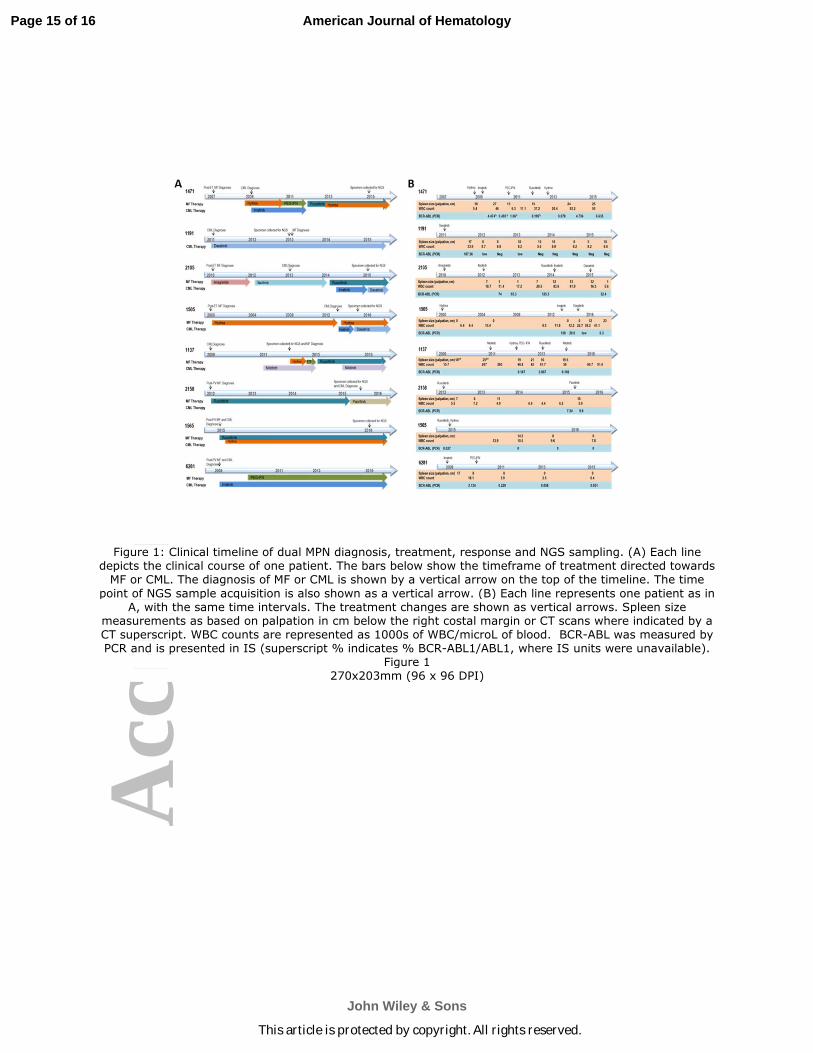
Figure 1: Clinical timeline of dual MPN diagnosis, treatment, response and NGS sampling. (A) Each line depicts the clinical course of one patient. The bars below show the timeframe of treatment directed towards MF or CML. The diagnosis of MF or CML is shown by a vertical arrow on the top of the timeline. The time
point of NGS sample acquisition is also shown as a vertical arrow. (B) Each line represents one patient as in A, with the same time intervals. The treatment changes are shown as vertical arrows. Spleen size
measurements as based on palpation in cm below the right costal margin or CT scans where indicated by a CT superscript. WBC counts are represented as 1000s of WBC/microL of blood. BCR-ABL was measured by PCR and is presented in IS (superscript % indicates % BCR-ABL1/ABL1, where IS units were unavailable).
Figure 1 270x203mm (96 x 96 DPI)
Page 15 of 16
John Wiley & Sons
American Journal of Hematology
This article is protected by copyright. All rights reserved.

Acc
epte
d A
rtic
le
Figure 2: COSMIC query of the top 20 genes in CML and ET/PV/MF. (A) Venn diagram showing overlap of genes in both disease subtypes (B) List of genes represented in the circles in A.
Figure 2
254x190mm (96 x 96 DPI)
Page 16 of 16
John Wiley & Sons
American Journal of Hematology
This article is protected by copyright. All rights reserved.
Related Documents