ELSEVIER SAUNDERS EMERGENCY MEDIClNE CUNICSDF NORTHAMERICA Ernerg Med Clin N Arn 25 (2007) 603-{í22 Airway Management in Trauma: An Update John McGill MDa,b,* , aDepartment of Emergency Medicine, Hennepin County Medical Center, 701 Park Avenue North, Minneapolis, MN 55415, USA bDepartment of Emergency Medicine, University of Minnesota, Minneapolis, MN, USA The past 10 years has seen a proliferation in the number of devices that are designed specifically for the difficult airway. The advantage of most of these devices is that the laryngeal opening can be visualized indirectly; they do not require the head and neck to be positioned so that the oral, pha- ryngeal, and laryngeal axes line up. This is particularly advantageous in the setting of trauma where movement of the neck can be dangerous. Another major advancement in the management of the airway in trauma has been the development and increasingly widespread use of the laryngeal mask airways, particularly the intubating laryngeal mask airway. These devices have be- come critical airway adjuncts in the management of patients who are diffi- cult to ventilate or to intubate. This article reviews the more recent theoretic and practical information that pertains to airway management in trauma. This includes newer airway devices, techniques, or maneuvers that are usefui in the trauma setting, though potentially are underused. The algorithmic approach to the difficult airway, which aptly describes the trauma airway, is not discussed because several well-informed algorithms are readily available (www.asahq.org, www.das.uk.com). Each clinician needs to be knowledgeable about the var- ious airway options and then, based on the physician's particular skills and resources, construct an algorithm that works best. Intubation in the setting of potential cervical spine injury The safety of inline immobilization of the cervical spine and emergency orotracheal intubation in multiple trauma is well established. Since the * Departrnent of Ernergency Medicine, Hennepin County Medical Center, 701 Park Avenue North, Minneapolis, MN 55415. 0733-8627/07/$ - see front rnatter @ 2007 EIsevier lnc. Ali rights reserved. doi: 10.1 016/j.ernc.2007 .06.007 emed.theclinics.com

Welcome message from author
This document is posted to help you gain knowledge. Please leave a comment to let me know what you think about it! Share it to your friends and learn new things together.
Transcript

-
ELSEVIERSAUNDERS
EMERGENCY
MEDIClNE
CUNICSDF
NORTHAMERICAErnerg Med Clin N Arn 25 (2007) 603-{í22
Airway Management in Trauma:An Update
John McGill MDa,b,*,aDepartment of Emergency Medicine, Hennepin County Medical Center,
701 Park Avenue North, Minneapolis, MN 55415, USA
bDepartment of Emergency Medicine, University of Minnesota, Minneapolis, MN, USA
The past 10 years has seen a proliferation in the number of devices thatare designed specifically for the difficult airway. The advantage of most ofthese devices is that the laryngeal opening can be visualized indirectly;they do not require the head and neck to be positioned so that the oral, pha-ryngeal, and laryngeal axes line up. This is particularly advantageous in thesetting of trauma where movement of the neck can be dangerous. Anothermajor advancement in the management of the airway in trauma has been thedevelopment and increasingly widespread use of the laryngeal mask airways,particularly the intubating laryngeal mask airway. These devices have be-come critical airway adjuncts in the management of patients who are diffi-cult to ventilate or to intubate.
This article reviews the more recent theoretic and practical informationthat pertains to airway management in trauma. This includes newer airwaydevices, techniques, or maneuvers that are usefui in the trauma setting,though potentially are underused. The algorithmic approach to the difficultairway, which aptly describes the trauma airway, is not discussed becauseseveral well-informed algorithms are readily available (www.asahq.org,www.das.uk.com). Each clinician needs to be knowledgeable about the var-ious airway options and then, based on the physician's particular skills andresources, construct an algorithm that works best.
Intubation in the setting of potential cervical spine injury
The safety of inline immobilization of the cervical spine and emergencyorotracheal intubation in multiple trauma is well established. Since the
* Departrnent of Ernergency Medicine, Hennepin County Medical Center, 701 ParkAvenue North, Minneapolis, MN 55415.
0733-8627/07/$ - see front rnatter @ 2007 EIsevier lnc. Ali rights reserved.
doi: 10.1 016/j.ernc.2007 .06.007 emed.theclinics.com
27

604 MCGILL
mid-1980s, when this became an accepted practice, there have been onlya handful of reports of intubation causing spinal cord injury and, in eachcase, the head and neck were not immobilized [1-3].
Despite this exceptional safety record, during which time most intuba-tions were performed using direct laryngoscopy, studies continue to be pub-lished comparing cervical spine movement during direct laryngoscopy withthat observed using other airway interventions. There are two models: thepatient with normal airway anatomy undergoing elective intubation in theoperating room and cadavers in whom unstable cervical spines were createdsurgically. Spine movement is documented fluoroscopically or with radio-graphs. In the normal patients, in whom angulation of the cervical bodiesis the sole measurement, most cervical spine movement during intubationoccurs in the upper cervical spine, especially the occiput-Cl and atlantoax-ial segments, regardless of which device is used [4--9].Although these studiesproduce results that are statistically significant, their clinical significance isquestionable. Specifically, a clinically significant degree of angulation hasnot been described (the largest reported angle was 15° at the occiput-Cljunction) [9]; displacement, not angulation, accounts for most spinal cordinjuries and; several studies were conducted without immobilization. Inthe cadaveric model of the unstable cervical spine, there are data to suggestthat the lighted stylet and fiber-optically guided nasotracheal intubationcause the least amount of subluxation, but, again, the differences were smalland of unknown clinical significance [10-15]. Crosby [16], in a comprehen-sive review of airway management after cervical spine trauma, concludedthat, as of 2006, there was no clearly superior modality for intubation inthis setting.
Although the clinical relevance of much of the research on the movementof the C-spine during intubation is unclear, the cadaveric studies served tovalidate the practice of removing the C-collar and immobilizing the C-spineduring intubation. Gerling and colleagues [14], using a C5-6 transectionmodel, reported more subluxation during direct laryngoscopy with the col-lar than with inline immobilization. Laryngeal visualization also was im-proved after collar removal. Immediately following intubation, the collaris reapplied. Studies of unstable c-spines also demonstrated that there isas much subluxation with facemask ventilation, the chin lift, and the jawthrust as with intubation itself [11,12], thus emphasizing the need for immo-bilization during alI airway maneuvers.
Before leaving the topic of inline immobilization, a final comment shouldbe made regarding the concept of "neutral" positioning of the head andneck in adult trauma. Although this term is used commonly, there is no ac-cepted definition. Some studies defined it clinically [9,17], but most majortrauma references do not address the subject [18-20). Schriger and col-leagues [17]defined neutral as the position that one assumes when standingand looking straight ahead; they found that a mean of 4 cm of occiput ele-vation was required to achieve this position. Data from De Lorenzo and
28-
AIRWAY MANAGEMENT IN TRAUMA 605
colleagues [21],using MRI to determine the optimal elevation of the occiputfor maximizing the spinal canaljspinal cord ratio at C5 and C6 (a commonleveI of injury), suggested that at least 2 em of elevation is desirable in nor-mal individuaIs. Regardless of how neutral is defined, it is c1ear that thecommon practice of leaving the head immobilized on the backboard resultsin a position that may be far from neutral. This is especially evident in theprofile of an obese patient whose thick back puts the cervical spine in hyper-extension when the head rests on the backboard. Head elevation needs to beindividualized in the morbidly obese and the elderly who may have preexis-tent lordotic deformities. Optimizing head and neck position, within theconstraints of the trauma setting, will improve positioning for intubationwhile maintaining the integrity of the spinal cord.
Anterior neck maneuvers during intubation
There are two manipulations of the anterior neck that often improve la-ryngeal visualization during intubation. "BURP" (backward upward rightpressure) is applied by an assistant over the lower thyroid cartilage to im-prove the glottic view when it is found to be suboptimal. The second maneu-ver, optimal external laryngeal manipulation (OELM), differs primarily inthat it is the laryngoscopist who, by reaching around with the right hand,manipulates the anterior neck and identifies the position of the larynxthat best exposes the laryngeal inlet. Once identified, the assistant assumesthe position of the laryngoscopist's hand so that intubation can beperformed.
Although both manipulations improve the laryngeal view [22-24],OELM seems to be superior [25]. Given the frequency of suboptimallaryn-geal views during inline immobilization [26], these maneuvers, preferablyOELM, should be used whenever visualization is poor. Cricoid pressure,the "Sellick's maneuver," is used to prevent stomach insufllation and regur-gitation, yet it may make intubation more difficult [27-29]. Although it isstill recommended, cricoid pressure should be relaxed, or even removed, ifit is believed to impede intubation significant1y.
Tracheal tube introducers
The tracheal tube introducer, originally the gum elastic bougie, was de-veloped decades ago to assist in difficult intubations (Fig. 1). It is a long,thin, semirigid adjunct that, with the aid of a laryngoscope, is passedthrough the laryngeal inlet and over which an endotracheal (ET) tube is ad-vanced into the trachea. Although ideally used when a portion of the laryn-geal inlet is visible, it can be effective when only the epiglottis is seen [30].This attribute, coupled with the fact that it is not affected by the presenceof blood and secretions, makes the bougie well suited in the trauma setting.
29

AIRWAY MANAGEMENT IN TRAUMA 605
colleagues [21), using MR1 to determine the opilinaJ eJevarion ofthe ocdplJtfor maxÚmzIÍ1g{ltespinal canal(spinal corá ratío at C5 aná C6 (a commonleveI of injury), suggested that at least 2 em of elevation is desirable in nor-mal individuaIs. Regardless of how neutral is defined, it is c1ear that thecommon practice of leaving the head immobilized on the backboard resultsin a position that may be far from neutral. This is especially evident in theprofile of an obese patient whose thick back puts the cervical spine in hyper-extension when the head rests on the backboard. Head elevation needs to beindividualized in the morbidly obese and the elder1y who may have preexis-tent lordotic deformities. Optimizing head and neck position, within theconstraints of the trauma setting, will improve positioning for intubationwhile maintaining the integrity of the spinal cord.
Anterior neck maneuvers during intubation
There are two manipulations of the anterior neck that often improve la-ryngeal visualization during intubation. "BURP" (backward upward rightpressure) is applied by an assistant over the lower thyroid cartilage to im-prove the glottic view when it is found to be suboptimal. The second maneu-ver, optimal externallaryngeal manipulation (OELM), ditfers primarily inthat it is the laryngoscopist who, by reaching around with the right hand,manipulates the anterior neck and identifies the position of the larynxthat best exposes the laryngeal inlet. Once identified, the assistant assumesthe position of the laryngoscopist's hand so that intubation can beperformed.
Although both manipulations improve the laryngeal view [22-24],OELM seems to be superior [25]. Given the frequency of suboptimallaryn-geal views during inline immobilization [26], these maneuvers, preferablyOELM, should be used whenever visualization is poor. Cricoid pressure,the "Sellick's maneuver," is used to prevent stomach insuffiation and regur-gitation, yet it may make intubation more difficult [27-29]. Although it isstill recommended, cricoid pressure should be relaxed, or even removed, ifit is believed to impede intubation significantly.
Tracheal tube introducers
The tracheal tube introducer, originally the gum elastic bougie, was de-veloped decades ago to assist in difficult intubations (Fig. I). It is a long,thin, semirigid adjunct that, with the aid of a laryngoscope, is passedthrough the laryngeal inlet and over which an endotracheal (ET) tube is ad-vanced into the trachea. Although ideally used when a portion of the laryn-geal inlet is visible, it can be etfective when only the epiglottis is seen (30).This attribute, coupled with the fact that it is not atfected by the presenceof blood and secretions, makes the bougie well suited in the trauma setting.
29
606 MCGILL
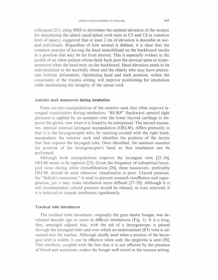
Fig. I. Several types of tracheal tube introducers. The gum elastic bougie (dark yeIlow) is reus-
able and comes in curved- and straight-tip adult forms and a straight pediatric formo The
straight bougies are 70 em, and the curved-tipped bougies are 60 em. The blue introducer (Flex-
trach ETTube Guide) is polyethylene, designed for single use, and comes only in a curved-tipadult form (60 em). The inset demonstrates the 60° optimum curve ifnone ofthe laryngeal inlet
can be seen on laryngoscopy.
Its efticacy was demonstrated prospectively in difticult intubations in the Op-erating room (OR), as a key component of a difticult airway algorithm in theOR, and it has been compared favorably to conventionallaryngoscopy inthe emergency department (ED) [31-33]. Despite this track record, the tra-cheal tube introducer has seen limited use in the United States.
The original adjunct, called the gum elastic bougie or simply "the bou-gie," is available in reusable form for adults and pediatrics (EschmannTracheal Tube Introducer, Portex Sims, Kent, UK). The adult version ac-commodates a 5.5-mm ET tube, whereas the pediatric version can accom-modate a 4.0-mm tube. An adult polyethylene introducer, designed forsingle use, also is available (Flextrach ETTube Guide, Greenfield MedicalSourcing, Inc., Austin, Texas).
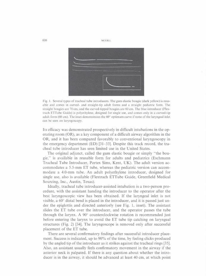
Ideally, tracheal tube introducer-assisted intubation is a two-person pro-cedure, with the assistant handing the introducer to the opera to r after thebest laryngoscopic view has been obtained. If the laryngeal inlet is notvisible, a 60° distal bend is placed in the introducer, and it is passed just un-der the epiglottis and directed anteriorly (see Fig. 1, inset). The assistantslides the ET tube over the introducer, and the operator passes the tubethrough the larynx. A 90° counterc1ockwise rotation is recommended justbefore entering the larynx to avoid the ET tube tip catching on laryngealstructures (Fig. 2) [34]. The laryngoscope is removed only after successfulplacement of the ET tube.
There are several confirmatory findings after successful introducer place-ment. Success is indicated, up to 90% of the time, by feeling clicks producedby the angled tip of the introducer as it strikes against the tracheal rings [35].Also, an assistant usually feels confirmatory movement in the airway if theanterior neck is palpated. If there is any question about whether the intro-ducer is in the airway, it should be advanced at least 40 cm, at which point
I
30......
AIRWAY MANAGEMENT IN TRAUMA 607
Fig. 2. A common cause of difficulty when railroading an ET tube over a tracheal tube intro-
ducer. (A) ET tube tip caught on the right arytenoid as it is being railroaded over the introducer.
Corrective maneuvers: (B) ET tube is withdrawn 2 cm to disengage the arytenoids and counter-cIockwise rotation of ET tube 90° to orient the beve\ posteriorly. (C) The bevel ofthe ET tube is
facing posteriorly and allows for smooth passage through the glottis. (Courtesy ofDepartment
of Emergency Medicine, Hennepin County Medical Center, Minneapolis, MN.)
resistance should be felt as the introducer passes the carina into amambronchus.
In the prehospital setting, where assistance may not be available, removalof the laryngoscope may be required so the operator can mount the ET tubeonto the introducer. Once the tube is on the introducer, the laryngoscopemay be reinserted and the tube rotated 90° counterclockwise to ensure suc-cessful passage of the tube. Mounting the tube onto the introducer and I
I
I
'I31

608 MCGILL
introducing them as a unit is not advised because it often is difficult to directthe introducer into the laryngeal inlet as it moves within the ET tube.
In addition to its usefulness in multiple trauma, the tracheal tube intro-ducer can be useful when intubating patients who have blunt and penetrat-ing upper airway injury. In this setting, a carefully passed introducer thatadvances into the trachea without resistance is added assurance that anET tube will be able to be placed without causing further damage. It alsocan be used as an introducer for an ET tube being placed through anopen neck wound [36] or, as in the author's institution, when performinga cricothyrotomy.
Laryngeal mask airways
The laryngeal mask airway (LMA) and the intubating LMA (ILMA) areexcellent rescue devices for the "cannot intubateJcannot ventila te" situation.Both devices are valuable for rescue ventilation, but the ILMA is superior asa conduit for intubation [37]. As ventilatory devices, their reliability hasbeen excellent. The ILMA also is successful when intubating patients withknown difficult airways [38,39]. In the OR, blind intubation through theILMA has an overall success rate of 90%; when aided by fiberoscopy, thesuccess rate approaches 100% [37]. LMAs are contraindicated in patientswith less than 2 cm of mouth opening and are unlikely to be successful inpatients with grossly distorted supraglottic anatomy from disease processesor postradiation scarring or in patients with an intact gag reflexo
Most intubations through the ILMA are performed blindly, using thedesignated LMA ET tube or a standard ET tube. If a standard ET tube isused, it should be inserted so that the curve is opposite the normal orienta-tion (Fig. 3A). This results in the ET tube exiting the ILMA at a less acuteangle and allows it to pass into the trachea more easily (Fig. 3B) [40]. If dif-ficulty is encountered, a fiber-optic scope can be helpful in guidingintubation.
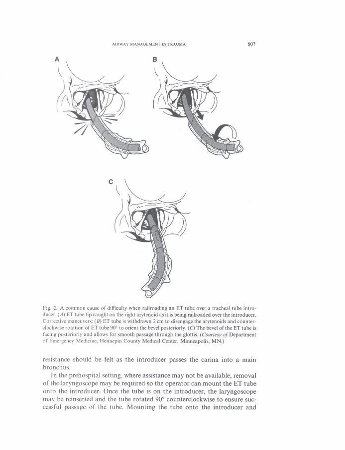
If an LMA has been placed, blind ET tube placement is not an option. Afiber-optic scope must be used or, better yet, a hollow introducer, such as anAintree Intubation Catheter (Cook Critical Care, Bloomington, Indiana;www.cookmedical.com) in conjunction with a fiber-optic scope, is placedthough the LMA into the trachea (Fig. 4). Once placed, the LMA is re-moved, and an ET tube is railroaded over the hollow introducer into the tra-chea. Use of a regular tracheal tube introducer is not recommended becauseof a high failure rate [41]. If intubation fails, despite the recommendedadjustment maneuvers and fiber-optic assistance, definitive airway control-using a retrograde wire or a surgical airway-can be accomplished in a con-trolled fashion while the patient continues to be ventilated with the laryngealmask [42].An excellent resource for troubleshooting LMA difficulties can befound on the company's website (www.lmana.com) under "Insertion Tech-nique and Maneuvers Guide."
32

AIRWAY MANAGEMENT IN TRAUMA 609
.1
Fig. 3. Intubating through an ILMA using a standard tracheal tube. (A) The tracheal tube isinserted so the curve of the tube is opposite the curve of the ILMA. This allows the tube tipto exit the ILMA at an angle more conducive to smooth passage into the trachea. (B) Angleof the tracheal tube tip after normal orientation of the ET tube (lower) versus the correct "re-verse curve" orientation (upper).
LMAs provide an excellent means of oxygenating and ventilating whenfacemask ventilation is difticult or impossible. In addition, the ILMA is par-ticularly useful in managing patients who are difticult to intubate with directlaryngoscopy. Thus, by facilitating the management of difticultjfailed venti-lation and difticultjfailed intubation, the ILMA has become an indispens-able component of any difticult airway algorithm.
The Combitube and laryngeal tube
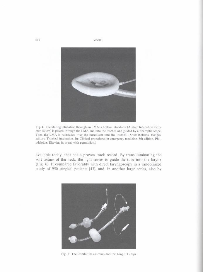
The Esophageal- Tracheal Combitube remains an etfective intermediateairway that is used primarily in the prehospital setting. Its popularity has de-creased as the use of laryngeal masks has increased. Its biggest drawback isthe difticulty establishing a definitive airway after the Combitube has beenplaced. The laryngeal tube, also known as the King LT, is a new intermedi-ate airway that is replacing the Combitube in some prehospital systems be-cause of its simpler design and availability in a disposable version (Fig. 5).A major ditference between the King LT and the Combitube is that it is de-signed specifically not to go into the trachea.
Lighted stylet intubation
The Trachlight (Laerdal Medical Corp., Wappingers Falls, New York)was introduced in 1995 and is the only lighted stylet, among several
33
610 MCGlLL
Fig.4. Faeilitating intubation through an LMA: a hollow introdueer (Aintree Intubation Cath-eter, 60 em) is plaeed through the LMA and into the traehea and guided by a fiberoptie seope.Then the LMA is railroaded over the introducer into the traehea. (Fram Roberts, Hedges,editors. Traeheal intubation. In: Clinieal proeedures in emergeney medieine. 5th edition. Phil-adelphia: Elsevier; in press; with permission.)
available today, that has a proven track recordo By transilluminating thesoft tissues of the neck, the light serves to guide the tube into the larynx(Fig. 6). It compared favorably with direct laryngoscopy in a randomizedstudy of 950 surgical patients [43], and, in another large series, also by
Fig. 5. The Combitube (battam) and the King LT (tap).
34.....

AIRWAY MANAGEMENT IN TRAUMA 611
Fig. 6. Lighted stylet intubation. Use of the Trachlight for ET intubation using transillumina-tion of soft tissues as a guide to placement. (Courtesy olDepartment of Emergency Medicine,Hennepin County Medical Center, Minneapolis, MN.)
Hung and colJeagues [44], the device was 99% successful in intubating pa-tients who had difficult airways. The stylets come in adult, pediatric, and in-fant sizes and can accommodate down to a 2.5-mm internal diameter (ID)tube (Fig. 7).
The patient's head and neck are optimalJy placed in the neutral or, if pos-sible, slightly extended position. This makes the Trachlight welJ suited forthe patient who has experienced trauma, especialJy given the fact that itssuccess is not impacted by the presence of blood or secretions. Becauselighted stylet intubation is a blind approach, it should be avoided in patientswith expanding neck masses or laryngopharyngeal trauma. Morbid obesityis the most common cause for failure because ofthe difficulty transilluminat-ing through generous soft tissue.
Fig. 7. Trachlight and related stylets. The malleable stylet is bent at a 90° angle, 6.5 to 8.5 cmfrom the tip (marked ou stylet). The smallest stylet accommodates a 2.5-mm 10 tube.
35

612 MCGlLL
The clinician stands at the head of the patient. When this is not possible,the patient can be approached from either side. With the patient's head andneck in the neutral position or slightly extended, the jaw is grasped near thecorner of the mouth, between the thumb, index, and middle fingers, andlifted to elevate the tongue and epiglottis (see Fig. 6). A transilluminatingglow indicates the location of the tube tipo Application of cricoid pressuremay enhance the transillumination [45]. The overhead light often must bedimmed in the obese patient. Positioning is optimal when a light bulb-likeglow emanates from the midline, just below the leveI of the thyroid promi-nence. In extremely thin patients, it is possible to observe transilluminationand still be in the esophagus. The clue is that the glow is diffuse if the tip is inthe esophagus, as opposed to the well-circumscribed glow of intralaryngealplacement.
The Trachlight is a safe, effective, rapid, and inexpensive intubating devicethat is placed with the patient in neutral position and is not affected adverselyby blood. These features make it especially amenable to the trauma setting.
Semirigid fiber-opticstylets
A class of devices that incorporate fiber optics into a semirigid metal ormetal-reinforced stylet was introduced in the late 1990s. They can be used inconjunction with direct laryngoscopy or alone. They require direct laryngo-scopic assistance in 8% to 20% of cases and, in general, are more successfulwhen used in this way [46,47]. In either case, the tip of the fiber-optic stylet,with its overlying ET tube, is passed under the epiglottis and directedthrough the glottis into the trachea. Visibility can be hampered by bloodand secretions, but less so than with flexible fiberoscopy [48]. They alsoare easier to learn how to use. The Shikani Optical Stylet and LevitanFPS Scope (Clarus Medical, Minneapolis, Minnesota) are examples of thesemalleable optical stylets (Fig. 8). Most often, the Shikani Stylet is used sim-ilarly to the Trachlight, with the added feature of being able to see fiber-optically what is just beyond the tube tipo The Levitan Scope is designedto be used with a laryngoscope, serving as a stylet for the tracheal tube whileproviding fiber-optic visualization if required. The Levitan stylet can accom-modate a minimum tube size of 5.5-mm ID, whereas the Shikani comes intwo sizes: the adult, accommodating a 5.5-mm ID tube, and a pediatric ver-sion that accommodates a 3.5-mm ID tube.
The semirigid fiber-optic stylets are useful in patients in whom the glottiscannot be seen readily. Blood and secretions may complicate their use, butless so than with flexible fiberoscopy. The Shikani scope was more successfulthan the gum elastic boogie in simulated Cormack and Lehane Grade 3views [49]. There are no absolute contraindications and no reported com-plications with semirigid fiber-optic stylets, other than the failures orprolonged attempts usually related to poor visibility from blood andsecretions [46].
36--

AIRWAY MANAGEMENT IN TRAUMA 613
Fig. 8. Examples of semirigid fiber-optic stylets: the Shikani (top) and Levitan (bottom). The
recommended bends when used alone (top) and when used with a direct laryngoscope (bottom)
are demonstrated. The sliding adapter on the Shikani stylet allows for various lengths of tra-cheal tubes.
Semirigid fiber-optic stylets combine the features of direct and indirectlaryngoscopy and provide a valuable tool when one is faced with a difficultairway. They can be used alone or in conjunction with a laryngoscope. Theyare considerably less expensive than rigid fiber-optic devices and are easierto use than ftexible scopes. There are no head-to-head clinical trials withother devices; however, early reports suggest that the semirigid fiber-opticstylets have a role to play in managing the airway in trauma.
Nasal and orotrachealintubationover a flexible fiber-opticbronchoscope
Flexible fiber-optic intubation is cited often in the anesthesia literature asthe preferred method for intubating a patient with an unstable C-spine. Italso is recommended in patients with suspected or known injury to the up-per airway. In a survey at the 1999 annua1 meeting of the American Societyof Anesthesio10gists, 75% of respondents said they wou1d use awake fiber-optic intubation in the setting of cervica1 spine disease, yet only 59% feltcompetent with this approach [50]. In the ED, the success rate is 50% to90% [51-53]. Failure is attributed most often to poor visibility from blood,vomitus, and other secretions. M1inek and coworkers [53] found that suc-cessful ED fiber-optic intubations averaged 2 minutes, whereas failures av-eraged 8 minutes; they recommended considering alternative approaches ifintubation attempts take more than 3 minutes.
Fiber-optically guided intubation can be accomp1ished using the nasal ororal route. The nasal approach is technically easier because the angle of in-sertion allows for easier visualization of the larynx, with minima1 manipu1a-tion of the fiberscope, and because patient cooperation is not as critica!.Insertion using the oral route can be facilitated by using an oral-intubating
37 I'I

614 MCGILL
airway (Fig. 9). Contraindications to the nasal approach are severe midfacetrauma and coagulopathy. Active airway bleeding and vomiting are relativecontraindications for both approaches because successful intubation is un-likely in these settings. If the c1inician is inexperienced in fiber-optic intuba-tion, significant hypoxia is another relative contraindication to its use.
The primary advantages of fiber-optic intubation are the ability to visu-alize upper airway abnormalities, to negotiate difficult airway anatomy, andto carefully perform tracheal intubation with visual guidance. A major lim-itation in trauma is the lack of visibility in the presence of blood and secre-tions. If blood is present, it is best to avoid the fiber-optic scope.
Rigid fiber-optic laryngoscopy
Rigid fiber-optic laryngoscopes are designed to approximate the anatomyof the upper airway and provide indirect fiber-optic visualization of thelaryngeal inlet. Examples inc1ude the Bullard laryngoscope (Circon Corp.,Stamford, Connecticut), the UpsherScope (Mercury Medical, Clearwater,Florida), and the WuScope (Achi Corp., San Jose, California). These de-vices offer the advantage of a conventional fiber-optic scope but requireless training [54]. They are well suited for patients who have potential cervi-cal spine injury because no movement of the neck is necessary. Case reportsalso suggested their usefulness in patients who had upper airway distortionfrom mass or hematoma [55,56]. One potential advantage of the WuScope isthat its fiber-optic element is relatively protected from blood and secretionsby its tubular blade.
The Bullard laryngoscope comes in two sizes: pediatric (newborn to 10years) and adult. It can be used in awake or unresponsive patients [57].At least 2 cm of occ1usal opening is necessary for the introduction of thescope and an ET tube. The technique for introducing the Bullard
Fig. 9. Examples of oral intubating airways. The WilIiams Airway lntubator (lelt) cradles theET tube in an open, curved guide, whereas the Ovassapian Fiberoptic lntubating Airway (right)positions the ET tube on the posterior surface of the intubation airway.
38--

1
AIRWAY MANAGEMENT IN TRAUMA 615
laryngoscope blade is similar to that for direct laryngoscopy (Fig. 10). Withthe glottis in view, the tube is advanced off the attached stylet and into thelarynx. Cricoid pressure does not seem to interfere with Bullard scope suc-cess [58]. A newer, multifunctional stylet has a hollow lumen that allows anintroducer to be passed through it and into the laryngeal inlet and overwhich the tube can be advanced. Plastic tip extensions are available forthe larger patient in whom the epiglottis may be difficult to elevate.
The major difficulty in using rigid fiber-optic laryngoscopes is the inabil-ity to visualize the larynx because of blood, emesis, or secretions; a possibleexception is the WuScope, with its relatively protected optical element, but ithas yet to be studied in this setting. Once visualized, the glottis usually canbe intubated, although accessing the inlet and passing the tube occasionallymay be difficult.
Video-assisted laryngoscopy
Video-assisted laryngoscopy transmits an image from an optical elementlocated on the laryngoscope blade to a monitor. In addition to providing in-direct images that may be unobtainable with direct laryngoscopy, the imageis enhanced by magnification and a wide-angle view. The newer devices in-corporate miniature video cameras on their blades. The ability to view theimages on a monitor provides immediate feedback to an assistant providing
._----..
Fig. 10. Bullard laryngoscope. Anatomically shaped laryngoscope visualizing the glottis; tra-cheal tube is mounted on the attached stylet to facilitate laryngeal placement. (Courtesy ofDepartment of Emergency Medicine, Hennepin County Medical Center, Minneapolis, MN.)
39

.
616 MCGILL
externallaryngeal manipulation. Video-assisted laryngoscopy also has greatpotential as a teaching aid.
The GlideScope videolaryngoscope (Verathon, Bothell, Washington),first introduced in 200 I, is the first of a new class of video-assisted rigidscopes that provide enhanced video views of the laryngeal inlet while beingless likely to be atTected by the presence of blood and secretions. There arethree sizes: small for neonates and infants, medium for children and smalladults, and large for adults. It comes with a small monitor on a stand ora smaller, more portable monitor. The McGrath Portable Video Laryngo-scope (Aircraft Medical Ltd., Edinburgh, UK) is a device with similar char-acteristics, but it is more compact, has an adjustable blade length, and usesdisposable blade covers (Fig. 11). It is new, and no studies have been re-ported on its use.
The GlideScope has a miniature camera embedded midway along the un-dersurface of the blade that has a 60° midblade angulation (Fig. 12). Bloodand secretions are unlikely to compromise laryngeal visualization because ofthe camera's orientation, its location well away from the blade tip, and itsantifogging technology. Visualization of the laryngeal inlet is excellentand is usually less of a problem than placing a tube through it. Difficult lar-yngoscopic cases are intubated more easily with this scope than with directlaryngoscopy, but easy cases are more time consuming because of difficultiesin advancing the tube [59]. A contraindication is a small mouth throughwhich the blade cannot be passed safely (opening less than 2 cm). Learningto use the GlideScope is easy because the overall feel of the device is similarto that of a conventionallaryngoscope. The absence of fiber-optic elementsincreases its durability and decreases inevitable breakage expenses.
The styletted tracheal tube is usually bent to conform to the 60° angle ofthe GlideScope blade (see Fig. 12). The GlideScope blade is placed into themouth and, under direct vision, is advanced in the midline. Verifying mid-line position on the monitor, the uvula is passed as the GlideScope blade
Fig. 11. The McGrath Portable Video Laryngoscope.
40.....

AIRWAY MANAGEMENT IN TRAUMA 617
Fig. 12. The GlideScope. The ET tube has the 60° bend recommended by the manufacturer.
is slipped behind the tongue, hugging the tongue base. With a gentle liftingmotion, the tip of the blade enters the valIecula, and the epiglottis is seenand elevated out of the way to expose the laryngeal inlet. The ET tube ispassed blindly along the side of the blade and into the hypopharynx.When the tip of the ET tube comes into view, it is directed into the glottisand advanced to the appropriate depth. If difficulty is encountered tryingto access the laryngeal inlet, success may be achieved by a variety of means,induding application of externallaryngeal pressure, increasing the bend ofthe stylet, using a tracheal tube introducer, or using a Parker Flex-It Stylet(Parker Medical, Englewood, Colorado) [60]. This is a new device, but sev-eral minor injuries have been reported: two cases of puncture of the rightpalatopharyngeal arch, with one requiring surgical repair [61].
Video-assisted laryngoscopy is a major advancement in the visualizationof the laryngeal inlet. The GlideScope, the first device that incorporates mi-cro video camera technology, provides an improved laryngeal view com-pared with difficult direct laryngoscopy while avoiding some of theproblems associated with fiber-optic bronchoscopy. It has yet to be studiedextensively in the setting of difficult intubation, although preliminary reportsare promising [62]. Most intubation failures result from the inability to passthe tube through the larynx, despite adequa te glottic views [60]. Improve-ments in the configuration of the tracheal stylet should decrease thisproblem.
Blind nasotracheal intubation
There are few indications for blind nasotracheal intubation in trauma.One potential circumstance is in the patient with minimal mouth opening,because alI of the newer devices that provide an indirect view of the laryn-geal inlet require at least 2 cm of mouth opening. In this situation,
II
41 1ti

618 MCGILL
nasotracheal intubation may be a reasonable option if a surgical airway isnot immediately indicated.
Surgical airways
Cricothyrotomy remains the preferred procedure for establishing a defin-itive emergency surgical airway. The four-step approach, first described in1996 [63], has gained popularity; in a series of 50 patients [64], the compli-cation rate compared favorably with that found when the more traditionalapproach was used [65]. This approach begins with a smalIer, horizontalstab wound that is carried down through the cricothyroid membrane. Thetracheal hook is placed caudalIy under the cricoid cartilage as opposed tosuperiorly, under the inferior edge of the thyroid cartilage, as with the tra-ditional approach. Regardless of the approach, a generous vertical incisionshould be made if there is difficulty identifying the cricothyroid membrane[65,66]. In addition to gaining exposure in the patient with a large neck,this incision permits definitive, digital identification ofthe membrane and re-duces the chances of suprathyroid tube placement. If difficulty is encoun-tered placing a Shiley tracheostomy tube, a tracheal tube introducer canbe inserted and a #6 ET tube passed over it into the trachea.
Percutaneous cricothyrotomy is another potential approach for establish-ing a surgical airway. Schaumann and colIeagues [67], in a large, cadavericstudy comparing the Seldinger technique (Arndt Emergency Cricothyrot-omy Catheter Set, Cook Critical Care, Bloomington, Illinois) to the stan-dard surgical approach, concluded that the Seldinger technique had theadvantage of being quicker. They failed to mention, however, that 4 of100 catheters placed with the Seldinger technique ended up in the subcuta-neous tissue. This serious complication results from the insufficient catheterlength (6 cm) used in the study; this study, using normal-sized cadavers, un-doubtedly underestimates the likelihood of this complication occurring ina patient with a large neck. The Melker Cricothyrotomy Catheter Set(Cook Medical, Inc., Bloomington, Indiana) (9 cm) is long enough formost patients; however, even with a vertical skin incision, sets of this varietycan be difficult to place through the tissues of the anterior neck [68]. Thus,although the percutaneous approach is far better than no surgical attempt atalI, the approaches that ultimately permit digital identification of the crico-thyroid membrane and incorpora te a scalpel incision through the membraneremain the preferred surgical approaches for the emergency trauma airway.
Summary
ConceptualIy, the management of the airway in trauma has not changedsignificantly in recent years. The safety of inline immobilization and rapidsequence intubation has been reaffirmed clinicalIy. Although the general
42
AIRWAY MANAGEMENT IN TRAUMA 619
management has not changed, the choice of airway devices has markedly in-creased. The most important among these is the ILMA because it can pro-vide effective rescue ventilation and intubation. The newer devices thatallow for indirect visualization of the laryngeal inlet also will contributeto effective trauma airway management.
Despite the advances in intubating devices, direct laryngoscopy likely willcontinue to be the initial approach to intubating the patient after trauma. Aswith any intubation, preoxygenation is essential and, by extending the apneatime before desaturation, it can make the difference between a successfuland a failed intubation. Positioning, another key element in successful intu-bation, is more complicated in the setting of trauma; however, it can be im-proved upon, from a neurosurgical and an airway perspective, by elevatingthe head off the backboard. If the laryngoscopic view is suboptimal, intuba-tion often can be accomplished rapidly using a tracheal tube introducer. Ex-ternal laryngeal manipulation also may be useful at this juncture. Thesesteps should be accomplished rapidly and usually result in successful intuba-tion. If unsuccessful, a device that provides indirect glottic visualizationshould be used. Alternatively, especially in the setting of oxygen desatura-tion, an ILMA can be placed to ventilate and intubate the patient. Crico-thyrotomy remains the ultimate back-up airway and should be performedusing the technique that corresponds best to the patient's body habitusand operator experience.
References
[I] Hastings RH, Kelley SD. Neurologic deterioration associated with airway management in
a cervical spine-injured patient. Anesthesiology 1993;78(3):580--3.
[2] Muckart DJ, Bhagwanjee S, van der Merwe R. Spinal cord injury as a result of endotracheal
intubation in patients with undiagnosed cervical spine fractures. Anesthesiology 1997;87(2):418-20.
[3] Liang BA, Cheng MA, Tempelhoff R. Efforts at intubation: cervical injury in an emergencycircumstance? J Clin Anesth 1999;11:349-52.
[4] Sawin PD, Todd MM, Traynelis VC, et aI. Cervical spine motion with direct laryngoscopyand orotracheal intubation. Anesthesiology 1999;85(1):26-36.
[5] Watts ADJ, Gelb A W, Bach DB, et aI. Comparison ofthe Bullard and Macintosh laryngo-
scopes for endotracheal intubation ofpatients with a potential cervical spine injury. Anesthe-
siology 1997;87(6): 1335-42.[6] Shulman GB, Connelly NR. A comparison of the Bullard laryngoscope versus the flexible
fiberoptic bronchoscope during intubation in patients afforded inline stabilization. J Clin
Anesth 2001;13(3):182-5.
[7] Waltl B, Melischek M, Schuschnig C, et aI. Tracheal intubation and cervical spine excursion:direct laryngoscopy vs. intubating laryngeal mask. Anaesthesia 2001;56:221--{).
[8] Sahin A, Salman MA, Erden IA, et aI. Upper cervical vertebrae movement during the intu-
bating laryngeal mask, fiberoptic and direct laryngoscopy: a video-fluoroscopic study. Eur JAnaesthesioI2004;21:819-23.
[9] Turkstra TP, Eng M, Eng P, et aI. Cervical spine motion: a fluoroscopic comparison during
intubation with lighted stylet, GlideScope, and Macintosh laryngoscope. Anesth Analg
2005; I 01 (3):910--5.
43
620 MCGILL
[10] Bilgin H, Bozkurt M. Tracheal intubation using the ILMA, C-Trach or McCoy laryngo-scope in patients with simulated cervical spine injury. Anaesthesia 2006;61:685-91.
[11] Brimacombe I, Keller C, Kunzel KH, et aI. Cervical spine motion during airway manage-ment: a cinefluoroscopic study of the posteriorly destabilized third cervical vertebrae in hu-man cadavers. Anesth Analg 2000;1(5):1274-8.
[12] Donaldson WF 11I, Heil BV, Donaldson VP, et aI. The effect of airway maneuvers on theunstable CI-C2 segment: a cadaver study. Spine 1997;22:1215-8.
[13] Lennarson PI, Smith DW, Sawin PD, et aI. Cervical spine motion during intubation: efficacy
of stabilization maneuvers in the setting of complete segmental instability. I Neurosurg 2001;94:265-70.
[14] Gerling MC, Davis DP, Hamilton RS, et aI. Effects of cervical spine immobilization tech-
nique and laryngoscope blade selection on an unstable cervical spine in a cadaver modelof intubation. Ann Emerg Med 2006;36(4):293-300.
[15] Gerling MC, Davis DP, Hamilton RS, et aI. Effect ofsurgical cricothyrotomy on the uns ta-
ble cervical spine in a cadaver model ofintubation. I Emerg Med 2001;20(1):1-5.
[16] Crosby ET. Airway management in adults after cervical spine trauma. Anesthesiology 2006;104(6): 1293-318.
[17] Schriger DL, Lamon B, LeGassick T, et aI. Spinal immobilization on a flat backboard: does
it result in neutral position ofthe cervical spine? Ann Emerg Med 1991;20(8):878-81.
[18] Wilmore DW, Cheung L Y, Harken AH, et ai, editors. ACS surgery: principies and practice.3rd edition. New York: WebMD Corp; 2003.
[19] Danne PD, Hunter M, Mackillop AD. Airway control. In: Moore EE, Feliciano DV, Mat-
tox, editors. Trauma. 5th edition. New York: McGraw Hill; 2004. p. 181-2.[20] Head trauma. In: American College of Surgeons Committee on Trauma. Advanced trauma
life support for doctors: student course manual. 7th edition. Chicago: American College ofSurgeons; 2004. p. 151-67.
[21] De Lorenzo RA, Olson IE, Boska M, et aI. Optimal positioning for cervical immobilization.Ann Emerg Med 1996;28(3):301-8.
[22] Takahata O, Kubota M, Mamiya K, et aI. The efficacy of the "BURP" maneuver duringa difficult laryngoscopy. Anesth Analg 1997;84:419-21.
[23] Benumof IL, Cooper SD. Quantitative improvement in laryngoscopic view by optimal exter-nallaryngeal manipulation. I Clin Anesth 1996;8(2):136-40.
[24] Krill RL. Difficult laryngoscopy made easy with a "BURP". Can I Anaesth 1993;40:798-9.
[25] Levitan RM, Everett WW, Kinkle WC, et aI. Pressing on the neck during laryngoscopy:
a comparison of cricoid pressure, backward upward rightward pressure, and externallaryn-geal manipulation. Acad Emerg Med 2005; 12(Suppl 1):92.
[26] Nolan IP, Wilson ME. Orotracheal intubation in patients with potential cervical spine in-juries. Anaesthesia 1993;48:630-3.
[27] Ho AMH, Wong W, Ling E, et aI. Airway difficulties caused by improperly applied cricoidpressure. I Emerg Med 2001;20:29-31.
[28] Brimacombe I, White A, Berry A. Effect of cricoid pressure on the ease of insertion of thelaryngeal mask airway. Br I Anaesth 1993;71(6):503-7.
[29] Smith CE, Boyer D. Cricoid pressure decreases ease of tracheal intubation using fiberopticlaryngoscopy (WuScope System). Can I Anaesth 2002;49(6):614-9.
[30] Nolan IP, Wilson ME. An evaluation of the gum eIastic bougie. Intubation times and inci-dence of sore throat. Anaesthesia 1992;47(10):878-81.
[31] Combes X, Le Roux B, Suen P, et aI. Unanticipated difficult airway in anesthetized patients:
prospective validation of a management algorithm. Anesthesiology 2004;100(5): 1146-50.
[32] Latto IP, Stacey M, Mecklenburgh I, et aI. Survey ofthe use ofthe gum elastic bougie in cIin-ical practice. Anaesthesia 2002;57(4):379-84.
[33] McGiII I, Vogel EC, Rodgerson ID. Use ofthe gum elastic bougie as an adjunct for orotra-
cheal intubation in the emergency department. Acad Emerg Med 2000;7(5):526.
44-

AIRWA Y MANAGEMENT IN TRAUMA 621
[34] Dogra S, Falconer R, Latto IP. Successful difficult intubation. Tracheal tube placement overa gum-elastic bougie. Anaesthesia 1990;45(9):774-6.
[35] Kidd JF, Dyson A, Latto IP. Successful difficult intubation. Use ofthe gum eIastic bougie.Anaesthesia 1988;43(6):437-8.
[36] Steinfeldt J, Bey TA, Rich JM. Use of a gum elastic bougie (GEB) in a zone 11penetratingneck trauma: a case report. J Emerg Med 2003;24(3):267-70.
[37] Brimacombe J. Laryngeal mask anesthesia: principies and practice. 2nd edition. Philadel-phia: Saunders; 2005.
[38] Joo H, Rose K. Fastrach-a new intubating laryngeal mask airway: successful use in patientswith difficult airways. Can J Anaesth 1998;45(3):253--6.
[39] Langeron O, Semjen F, Bourgain JL, et aI. Comparison ofthe intubating laryngeal mask air-
way with the fiberoptic intubation in anticipated difficult airway management. Anesthesiol-ogy 2001;94(6):968-72.
[40] Joo HS, Rose DK. The intubating laryngeal mask airway with and without fiberoptic guid-ance. Anesth Analg 1999;88(3):662--6.
[41] Gabbott DA, Sasada MP. Tracheal intubation through the laryngeal mask using a gum elas-tic bougie in the presence of cricoid pressure and manual in line stabilisation of the neck. An-aesthesia 1996;51(4):389-90.
[42] Harvey SC, Fishman RL, Edwards SM. Retrograde intubation through a laryngeal maskairway. Anesthesiology 1996;85(6):1503-4.
[43] Hung OR, Pytka S, Morris I, et aI. Clinical trial of a new lightwand device (Trachlight) tointubate the trachea. Anesthesiology 1995;83(3):509-14.
[44] Hung OR, Pytka S, Morris I, et aI. Lightwand intubation: II-clinical trial ofa new lightwandfor tracheal intubation in patients with difficult airways. Can J Anaesth 1995;42(9):826-30.
[45] Mehta S. Transtracheal illumination for optimal tracheal tube placement. A clinical study.Anaesthesia 1989;44(12):970-2.
[46] Rudolph C, Schlender M. [Clinical experiences with fiberoptic intubation with the Bonfils
intubation fiberscope]. Anaesthesiol Reanim 1996;21:127-30 [in German].[47] Shikani AH. New "seeing" stylet-scope and method for the management ofthe difficult air-
way. Otolaryngol Head Neck Surg 1999;120(1):113--6.
[48] Agro F, Cataldo R, Carassiti M, et aI. The seeing stylet: a new device for tracheal intubation.Resuscitation 2000;44(3): 177-80.
[49] Evans A, Morris S, Petterson J, et aI. A comparison ofthe Seeing Optical Stylet and the gumelastic bougie in simulated difficult tracheal intubation: a manikin study. Anaesthesia 2006;61(5):478-81.
[50] Ezri T, Szmuk P, Warters RD, et aI. Difficult airway management practice patterns amonganesthesiologists practicing in the United States: have we made any progress? J Clin Anesth2003; 15:418-22.
[51] Afilalo M, Guttman A, Stern E, et aI. Fiberoptic intubation in the emergency department:a case series. J Emerg Med 1993;11(4):387-9.
[52] Delaney KA, Hessler R. Emergency flexible fiberoptic nasotracheal intubation: a report of60 cases. Ann Emerg Med 1988;17(9):919-26.
[53] Mlinek EJ Jr, Clinton JE, Plummer D, et aI. Fiberoptic intubation in the emergency depart-ment. Ann Emerg Med 1990;19(4):359--62.
[54] Borland LM, Casselbrant M. The Bullard laryngoscope. A new indirect orallaryngoscope(pediatric version). Anesth Analg I990;70( I): 105-8.
[55] Liem EB, Bjoraker DG, Gravenstein D. New options for airway management: intubatingfibreoptic stylets. Br J Anaesth 2003;91(3):408-18.
[56] Sprung J, Weingarten T, Dilger J. The use ofWuScope fiberoptic laryngoscopy for tracheal
intubation in complex clinical situations. Anesthesiology 2003;98(1):263-5.
[57] Cohn AI, McGraw SR, King WH. Awake intubation of the adult trachea using the Bullardlaryngoscope. Can J Anaesth 1995;42(3):246--8.
45

622 MCGILL
[58] Shulman GB, Connelly NR. A comparison of the Bullard Laryngoscope versus the flexible
fiberoptic broncho scope during intubation in patients afforded inline stabilization. J Clin
Anesth 2001;13(3):182-5.
[59] Lim TJ, Lim Y, Liu EH. Evaluation ofease ofintubation with the GlideScope or Macintosh
laryngoscope by anaesthetists in simulated easy and difficult laryngoscopy. Anaesthesia2005;60(2): 180-3.
[60] Cooper RM, Pacey JA, Bishop MJ, et aI. Early clinical experience with a new videolaryngo-
scope (GlideScope) in 728 patients. Can J Anaesth 2005;52(2):191-8.
[61] Cooper RM. Complications associated with the use of the GlideScope videolaryngoscope.Can J Anaesth 2007;54(1):54-7.
[62] Lai HY, Chen lH, Chen A, et aI. The use ofthe GlideScope for tracheal intubation in patients
with ankylosing spondylitis. Br J Anaesth 2006;97(3):419-22.
[63] Brofeldt BT, Panacek EA, Richards JR. An easy cricothyrotomy approach: the rapid four-
step technique. Acad Emerg Med 1996;3:1060-3.[64] Bair AE, Panacek EA, Wisner DH, et aI. Cricothyrotomy: a 5-year experience at one insti-
tution. J Emerg Med 2003;24(2):151-6.
[65] Erlandson MJ, Clinton JE, Ruiz E, et aI. Cricothyrotomy in the emergency department re-
visited. J Emerg Med 1989;7:115-8.[66] Holmes JF, Panacek EA, Sakles JC, et aI. Comparison of2 cricothyrotomy techniques: stan-
dard method versus rapid 4-step technique. Ann Emerg Med 1998;32(4):442-6.
[67] Schaumann N, Lorenz V, Schellongowski P, et aI. Evaluation ofSeldinger technique emer-
gency cricothyroidotomy versus standard surgical cricothyroidotomy in 200 cadavers. An-
esthesiology 2005; 102:7-11.[68] Abbrecht PH, Kyle RR, Reams WH, et aI. lnsertion forces and risk of complications during
cricothyroid cannulation. J Emerg Med 1992;10:417-26.
Reprinted with permission from Emergency Medicine Clinics ofNorth America 2007; 25(3):603-22, vii. McGill 1.Airway management'trauma: an update. Copyright 2007 @Elsevier, Inc.
46
Related Documents