The EMBO Journal Vol.16 No.22 pp.6737–6747, 1997 Agonists induce conformational changes in transmembrane domains III and VI of the β 2 adrenoceptor Ulrik Gether 1,4,5 , Sansan Lin 1 , Pejman Ghanouni 1 , Juan A.Ballesteros 3 , Harel Weinstein 3 and Brian K.Kobilka 1,2,5 1 Howard Hughes Medical Institute, 2 Division of Cardiovascular Medicine, Stanford University Medical School, Stanford, CA 94305 and 3 Department of Physiology and Biophysics, Mount Sinai School of Medicine, New York, NY 10029, USA 4 Present address: Department of Medical Physiology, The Panum Institute, University of Copenhagen, DK-2200 Copenhagen, Denmark 5 Corresponding authors e-mail: [email protected] or [email protected] Agonist binding to G protein-coupled receptors is believed to promote a conformational change that leads to the formation of the active receptor state. However, the character of this conformational change which provides the important link between agonist binding and G protein coupling is not known. Here we report evidence that agonist binding to the β 2 adrenoceptor induces a conformational change around 125 Cys in transmembrane domain (TM) III and around 285 Cys in TM VI. A series of mutant β 2 adrenoceptors with a limited number of cysteines available for chemical derivatization were purified, site-selectively labeled with the conformationally sensitive, cysteine-reactive fluorophore IANBD and analyzed by fluorescence spectroscopy. Like the wild-type receptor, mutant receptors containing 125 Cys and/or 285 Cys showed an agonist-induced decrease in fluorescence, while no agonist-induced response was observed in a receptor where these two cysteines were mutated. These data suggest that IANBD bound to 125 Cys and 285 Cys are exposed to a more polar environment upon agonist binding, and indicate that movements of transmem- brane segments III and VI are involved in activation of G protein-coupled receptors. Keywords: fluorescence spectroscopy/G protein-coupled receptors/molecular modeling/signal transduction Introduction The majority of hormones and neurotransmitters exert their physiological response by binding to cell-surface receptors belonging to the large family of G protein- coupled receptors (GPCRs) (Probst et al., 1992; Schwartz et al., 1996). The chemical variety among the endogenous ligands for this superfamily of seven transmembrane segment receptors is exceptional, ranging from mono- amines, amino acids and purines to lipids, peptides and glycoproteins (Probst et al., 1992; Schwartz et al., 1996). GPCRs also act as important sensors of exogenous stimuli, such as light and odors (Probst et al., 1992; Schwartz et al., 1996). Moreover, the receptors for certain chemo- © Oxford University Press 6737 kines have recently been shown to play an essential role in the cellular entry of human immunodeficiency virus (HIV) (Choe et al., 1996; Feng et al., 1996). In spite of this impressive functional variability among GPCRs, it is believed that the receptors share both a common topology and a common mechanism of activation; however, the molecular processes underlying activation of this class of receptors remain unknown. Our current knowledge about structure–function rela- tionships in GPCRs is primarily based on the results from mutagenesis studies performed over the last decade on many members of the receptor family (reviewed in Kobilka, 1992; Savarese and Fraser, 1992; Schwartz, 1994; Strader et al., 1994). These studies have led to identification of domains involved in ligand binding and G protein coupling (Kobilka, 1992; Savarese and Fraser, 1992; Schwartz, 1994; Strader et al., 1994). In addition, mutagenesis techniques have provided evidence for dis- tinct intramolecular interactions supporting an anticlock- wise orientation of the seven transmembrane helices in the plasma membrane (Suryanarayana et al., 1992; Zhou et al., 1994; Liu et al., 1995; Elling and Schwartz, 1996; Mizobe et al., 1996). An increasing number of three- dimensional models of the receptors have also been developed (MaloneyHuss and Lybrand, 1992; Trumpp- Kallmeyer et al., 1992; Baldwin et al., 1993; Cronet et al., 1993; Roper et al., 1994; Ballesteros and Weinstein, 1995; Fanelli et al., 1995; Scheer et al., 1996). These models were initially developed based on the known tertiary structure of bacteriorhodopsin, but later models have incorporated the structural information obtained from the 9 Å electron density projection map of rhodopsin (Schertler et al., 1993). Unfortunately, the models remain only quali- fied predictions of the actual tertiary structure as long as no accurate structural information at the atomic level is available for this family of receptor proteins. It is generally assumed that binding of the agonist to the receptor induces a set of finely orchestrated changes in the tertiary structure of the receptor which are recognized by the associated G protein alpha subunit. However, mutagenesis studies have not been able to elucidate the conformational changes that are critical for transferring information across the plasma membrane. Current models for activation of GPCRs are based on studies in which the conformation of the receptor was inferred from activ- ation of messenger systems and/or from computational simulations (Samama et al., 1993; Luo et al., 1994; Ballesteros and Weinstein, 1995; Fanelli et al., 1995; Scheer et al., 1996). Techniques that can directly measure conformational changes have only recently been applied to this family of receptor proteins (Gether et al., 1995; Farrens et al., 1996; Turcatti et al., 1996). Hubbell, Khorana and co-workers have used electron paramagnetic resonance (EPR) spectroscopy to study conformational

Welcome message from author
This document is posted to help you gain knowledge. Please leave a comment to let me know what you think about it! Share it to your friends and learn new things together.
Transcript
The EMBO Journal Vol.16 No.22 pp.6737–6747, 1997
Agonists induce conformational changes intransmembrane domains III and VI of theβ2 adrenoceptor
Ulrik Gether1,4,5, Sansan Lin1,Pejman Ghanouni1, Juan A.Ballesteros3,Harel Weinstein3 and Brian K.Kobilka1,2,5
1Howard Hughes Medical Institute,2Division of CardiovascularMedicine, Stanford University Medical School, Stanford, CA 94305and3Department of Physiology and Biophysics, Mount Sinai Schoolof Medicine, New York, NY 10029, USA4Present address: Department of Medical Physiology, The PanumInstitute, University of Copenhagen, DK-2200 Copenhagen, Denmark5Corresponding authorse-mail: [email protected] or [email protected]
Agonist binding to G protein-coupled receptors isbelieved to promote a conformational change that leadsto the formation of the active receptor state. However,the character of this conformational change whichprovides the important link between agonist bindingand G protein coupling is not known. Here we reportevidence that agonist binding to the β2 adrenoceptorinduces a conformational change around 125Cys intransmembrane domain (TM) III and around 285Cysin TM VI. A series of mutant β2 adrenoceptors with alimited number of cysteines available for chemicalderivatization were purified, site-selectively labeledwith the conformationally sensitive, cysteine-reactivefluorophore IANBD and analyzed by fluorescencespectroscopy. Like the wild-type receptor, mutantreceptors containing 125Cys and/or 285Cys showed anagonist-induced decrease in fluorescence, while noagonist-induced response was observed in a receptorwhere these two cysteines were mutated. These datasuggest that IANBD bound to 125Cys and 285Cys areexposed to a more polar environment upon agonistbinding, and indicate that movements of transmem-brane segments III and VI are involved in activationof G protein-coupled receptors.Keywords: fluorescence spectroscopy/G protein-coupledreceptors/molecular modeling/signal transduction
Introduction
The majority of hormones and neurotransmitters exerttheir physiological response by binding to cell-surfacereceptors belonging to the large family of G protein-coupled receptors (GPCRs) (Probstet al., 1992; Schwartzet al., 1996). The chemical variety among the endogenousligands for this superfamily of seven transmembranesegment receptors is exceptional, ranging from mono-amines, amino acids and purines to lipids, peptides andglycoproteins (Probstet al., 1992; Schwartzet al., 1996).GPCRs also act as important sensors of exogenous stimuli,such as light and odors (Probstet al., 1992; Schwartzet al., 1996). Moreover, the receptors for certain chemo-
© Oxford University Press 6737
kines have recently been shown to play an essential rolein the cellular entry of human immunodeficiency virus(HIV) (Choe et al., 1996; Fenget al., 1996). In spite ofthis impressive functional variability among GPCRs, it isbelieved that the receptors share both a common topologyand a common mechanism of activation; however, themolecular processes underlying activation of this class ofreceptors remain unknown.
Our current knowledge about structure–function rela-tionships in GPCRs is primarily based on the results frommutagenesis studies performed over the last decade onmany members of the receptor family (reviewed inKobilka, 1992; Savarese and Fraser, 1992; Schwartz,1994; Straderet al., 1994). These studies have led toidentification of domains involved in ligand binding andG protein coupling (Kobilka, 1992; Savarese and Fraser,1992; Schwartz, 1994; Straderet al., 1994). In addition,mutagenesis techniques have provided evidence for dis-tinct intramolecular interactions supporting an anticlock-wise orientation of the seven transmembrane helices inthe plasma membrane (Suryanarayanaet al., 1992; Zhouet al., 1994; Liu et al., 1995; Elling and Schwartz, 1996;Mizobe et al., 1996). An increasing number of three-dimensional models of the receptors have also beendeveloped (MaloneyHuss and Lybrand, 1992; Trumpp-Kallmeyeret al., 1992; Baldwinet al., 1993; Cronetet al.,1993; Roperet al., 1994; Ballesteros and Weinstein, 1995;Fanelli et al., 1995; Scheeret al., 1996). These modelswere initially developed based on the known tertiarystructure of bacteriorhodopsin, but later models haveincorporated the structural information obtained from the9 Å electron density projection map of rhodopsin (Schertleret al., 1993). Unfortunately, the models remain only quali-fied predictions of the actual tertiary structure as long asno accurate structural information at the atomic level isavailable for this family of receptor proteins.
It is generally assumed that binding of the agonist tothe receptor induces a set of finely orchestrated changesin the tertiary structure of the receptor which are recognizedby the associated G protein alpha subunit. However,mutagenesis studies have not been able to elucidate theconformational changes that are critical for transferringinformation across the plasma membrane. Current modelsfor activation of GPCRs are based on studies in whichthe conformation of the receptor was inferred from activ-ation of messenger systems and/or from computationalsimulations (Samamaet al., 1993; Luo et al., 1994;Ballesteros and Weinstein, 1995; Fanelliet al., 1995;Scheeret al., 1996). Techniques that can directly measureconformational changes have only recently been appliedto this family of receptor proteins (Getheret al., 1995;Farrens et al., 1996; Turcatti et al., 1996). Hubbell,Khorana and co-workers have used electron paramagneticresonance (EPR) spectroscopy to study conformational
U.Gether et al.
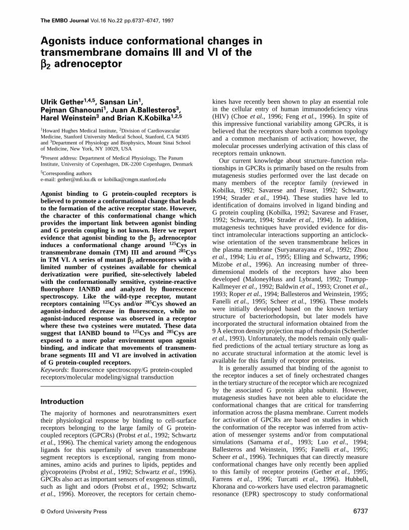
Fig. 1. ‘Helical net’ representation of the humanβ2 adrenoceptor. The receptor contains 13 Cys residues of which five (77Cys, 116Cys, 125Cys,285Cys and327Cys) (large black circles) are predicted to be in the transmembrane domain. Three Cys residues are predicted to be in the cytoplasmicregions (265Cys, 378Cys and406Cys) (large black circles). Five Cys residues are not expected to be available for chemical derivatization (small blackcircles); four residues (106Cys, 184Cys, 190Cys and191Cys) form two disulfide bridges (Fraser, 1989; Dohlmanet al., 1990; Nodaet al., 1994) and341Cys in the intracellular C-terminal tail has been shown to be palmitoylated (O’Dowdet al., 1989; Mouillacet al., 1992). Residues shown shadeddelineate the faces of TM III and TM VI segments that are oriented towards other transmembrane domains and not towards the lipid. Note thatresidues125Cys (TM III) and 285Cys (TM VI) are seen on the edge of the patch of shaded residues oriented towards the interior of the protein, i.e.the fluorophores would be predicted to lie at the interface between the protein and lipid environments. The specific residues shown shaded have beenidentified by: (i) the substituted Cys accessibility method on TM III of the D2 receptor and (ii) from mutations that significantly affect ligandbinding and/or activation of neurotransmitter receptors (Javitchet al., 1995; VanRhee, 1996). Residues known to be involved in agonist binding inβAR (113Asp in TM III, 203Ser and207Ser in TM V, 290Phe and293Asn in TM VI) are indicated by white letters in dark shaded circles (Totaet al.,1990; Straderet al., 1991, 1994; Wielandet al., 1996).
changes in rhodopsin accompanying photoisomerizationof retinal (Farrenset al., 1996). Their data providedevidence that light activation of rhodopsin may involvechanges in the arrangement of hydrophobic segments Cand F [transmembrane segments (TM) III and VI in thenomenclature of GPCRs] (Farrenset al., 1996). In theβ2adrenoceptor (β2AR), we have reported the use of theenvironmentally sensitive and cysteine-reactive fluorescentprobe IANBD to monitor agonist-induced structuralchanges in the receptor molecule (Getheret al., 1995).We found that agonists caused a dose-dependent andreversible decrease in fluorescence from the purifiedIANBD-labeled β2AR (Gether et al., 1995). This sug-gested that agonists promote a conformational change inthe receptor that leads to an increase in the polarity of theenvironment around one or more IANBD-labeled cysteines(Getheret al., 1995). The wild-type receptor contains eightfree cysteines, and mutagenesis and peptide mappingexperiments have indicated that several of these sitesare accessible for chemical derivatization (U.Gether andB.K.Kobilka, unpublished observation). To identify thecysteine(s) involved in the agonist-induced change influorescence and thereby map agonist-induced conform-ational changes in theβ2AR, we generated a series ofmutant receptors having limited numbers of cysteinesavailable for fluorescent labeling. The fluorescencespectroscopy analysis of the purified and site-selectively
6738
labeled mutants showed that IANBD-labeled125Cys and285Cys are responsible for the observed changes in fluores-cence consistent with movements of TM III and VI inresponse to agonist binding.
Results
Cysteines in the β2 adrenoceptor can be mutatedwith minimal effect on receptor functionIANBD is a sulfhydryl-reactive, environmentally sensitivefluorophore. Covalent incorporation of IANBD into theβ2AR receptor is associated with an increase in fluores-cence intensity and a 15–20 nm decrease inλmax (wave-length at which maximal fluorescence occurs) relative tofree IANBD in aqueous solution (Getheret al., 1995).This suggests that the observed fluorescence is derivedfrom IANBD-labeled cysteines in the hydrophobic coreof the receptor. This was further supported by quenchingexperiments with the hydrophilic quencher, potassiumiodide, demonstrating a substantially lower accessibilityto IANBD in the receptor as compared with IANBD freein solution (data not shown). Theβ2AR contains 13cysteines of which five (77Cys, 116Cys, 125Cys 285Cys and327Cys) are predicted to be in the hydrophobic transmem-brane core of the receptor (Figure 1). One cysteine,265Cys,is predicted to be in the third intracellular loop and twocysteines,378Cys and 406Cys, are located close to the
Conformational changes in the β2 adrenoceptor
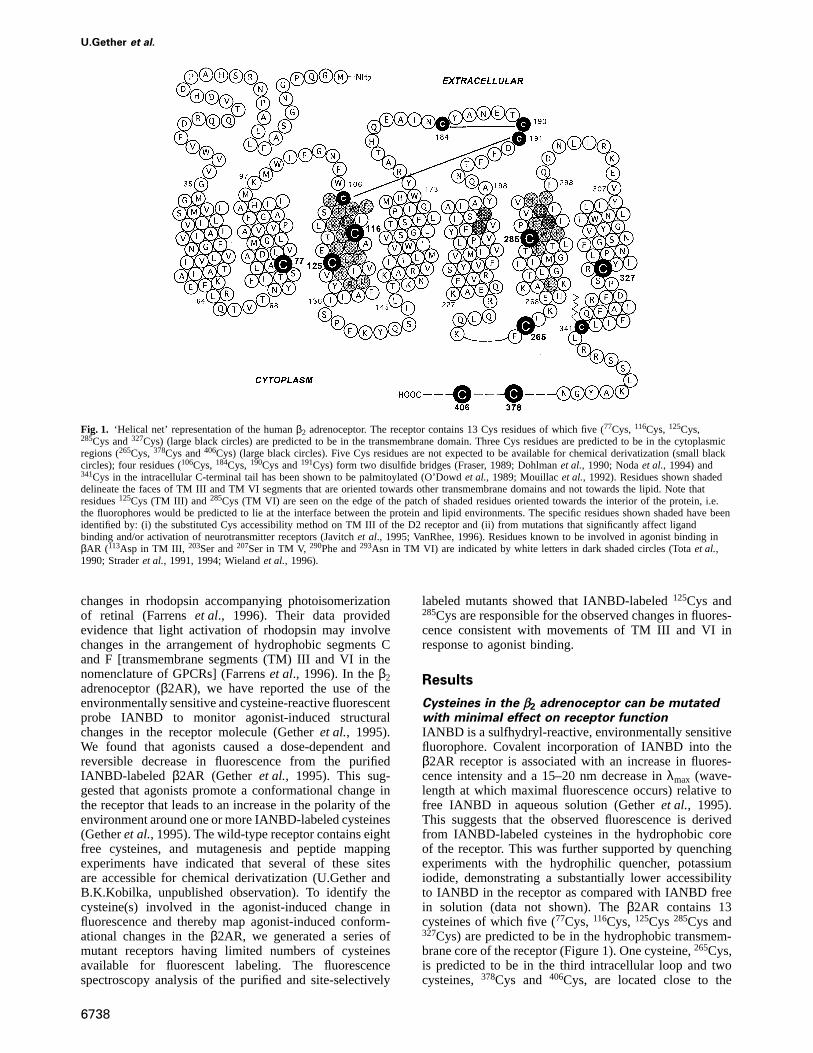
Table I. Binding properties and functional coupling of the wild-typeβ2AR and mutant receptors
Receptor Bmax KD, [3H]DHA Maximum cyclase EC50, ISO, cyclase IC50, ISO(pmol/mg protein) (nM) (% of basal) (nM) (µM, purified
receptor)
Wild-type β2AR (high expression) 9.0 0.29 (4) 259 (3) 16 (3) 1.6 (3)Wild-type β2AR (low expression) ~1.2 – 199 (2) 85 (2) –Cys(285) 2.2 0.23 (4) 347 (3) 26 (3) 1.9 (3)Cys(116, 285) 5.7 0.33 (4) 331 (2) 21 (2) 3 (3)Cys(116, 125) 4.1 0.46 (2) 471 (3) 17 (3) 19 (2)Cys(116, 125, 285) 11.1 0.31 (4) 421 (2) 10 (2) 1.6 (3)Cys(77, 116, 265, 327, 378, 406) 2.7 0.51 (2) 346 (3) 103 (3) 17 (2)
Bmax andKD values for [3H]DHA were determined from saturation binding experiments by non-linear regression analysis using Inplot 4.0 (GraphPadSoftware, San Diego, CA). Sf-9 cells expressing high and low levels of wild-type receptor were obtained by infecting cells for 48 and 24 h,respectively. Note that theBmax value for the low expressing wild-type membranes was determined using a single saturating concentration of[3H]DHA. Adenylyl cyclase activity was measured in Sf-9 cell membranes according to Materials and methods. Maximum cyclase activity and EC50values were determined from dose-response curves with isoproterenol (ISO) by non-linear regression analysis. IC50 values for inhibition of [3H]DHAbinding by ISO to the purified receptor were determined from competition binding experiments by non-linear regression analysis. All data are meansof the number of experiments performed in duplicate shown in parentheses.
C-terminus of the β2AR (Figure 1). There are fivecysteines in the receptor which are not expected to beavailable for chemical derivatization. In the extracellularloops, four cysteines (106Cys, 184Cys, 190Cys and191Cys)form two disulfide bridges (Figure 1) (Vauquelinet al.,1979; Fraser, 1989; Dohlmanet al., 1990; Nodaet al.,1994; Mejean-Galziet al., 1995) and in the intracellularC-terminal tail,341Cys has been shown to be palmitoylated(O’Dowd et al., 1989; Mouillacet al., 1992).
To identify the cysteine(s) that are responsible for theagonist-induced change in fluorescence from the IANBDlabeledβ2AR, we mutated cysteines in the receptor andgenerated a series of mutant receptors with one, two orthree cysteines in the TM regions of the receptor (Table Iand Figure 1). Cysteine residues77Cys,116Cys and125Cyswere all changed to valines, and327Cys was changed toserine based on previous experience with single substitu-tion of these residues in theβ2AR (Fraser, 1989; Dohlmanet al., 1990).285Cys is highly conserved among G protein-coupled receptors and was mutated to serine, since this isthe most frequently observed substitution in this position(Probstet al., 1992). The combined cysteine substitutionsin the β2AR had little effect on the general pharmaco-logical properties of the receptor (Table I). Antagonistbinding was unchanged in all mutants as assessed bysaturation binding experiments with [3H]dihydro-alpreno-lol ([3H]DHA) (Table I). However, mutation of severalcysteine residues led to a reduction in receptor expression(Table I). Notably, a mutant receptor with all free cysteinessubstituted was expressed so poorly that purification insufficient quantities for fluorescence spectroscopy analysiswas impossible (data not shown). The reduced expressionof cysteine mutants may be due to inefficient post-translational processing of these proteins so that a largerfraction of the mutant receptor protein is incorrectlyfolded. However, this should not affect our spectroscopydata since the last step of the purification procedureinvolves a ligand affinity chromatography which separatesfunctional from non-functional receptors.
The mutants were also analyzed for their ability toactivate adenylyl cyclase. As shown in Table I, all mutantsdemonstrated efficient coupling to adenylyl cyclase with3- to 4-fold increases in basal activity in response to
6739
stimulation with the agonist, isoproterenol (Table I). Theobserved EC50 values were found to be highly dependenton receptor expression. Two different expression levelsof the wild-type receptor, 1.2 pmol/mg protein and9.0 pmol/mg protein, resulted in a 5-fold difference in theEC50 value (85 nM at the low expression level and 16nM at the high expression level; Table I). Taking thedifferent levels of expression into account, the EC50 valuesfor the mutants were all comparable with the EC50 of thewild-type (Table I). Overall these data show that cysteineresidues77Cys, 116Cys, 125Cys, 265Cys, 285Cys, 327Cys,378Cys and406Cys are not essential for antagonist binding,nor are they required for the functional coupling ofthe β2AR.
Following purification, the mutant receptors were ana-lyzed for their ability to bind the agonist, isoproterenol(Table I). We found that the IC50 values for inhibition of[3H]DHA binding to the Cys(285) and Cys(116, 125, 285)mutants were identical to the wild-type receptor but thatthe IC50 values for the mutants, Cys(116, 285), Cys(116,125) and Cys(77, 116, 265, 327, 378, 406), were ~10-foldhigher than that for the wild-type (Table I). Nevertheless,the efficient coupling of these mutants to adenylyl cyclasedemonstrates that they are able to undergo agonist-inducedconformational changes similar to the wild-type receptor(Table I). We should also mention that the binding andfunctional coupling properties of theβ2AR expressed ininsect cells are comparable with those of the receptorexpressed in mammalian cells (Benovicet al., 1984;Bouvier et al., 1989).
Fluorescence spectroscopy analysis ofsite-selectively labeled mutant β2 adrenoceptorsThe mutant receptors described in Table I were all purifiedand fluorescently labeled with the IANBD fluorophore.As expected, the IANBD-labeled mutants all demonstratedemission maxima around 525 nm (data not shown). Inaddition, the fluorescence intensity of the labeled mutantswas reduced relative to the wild-type receptor consistentwith the reduced number of derivatizable cysteines (datanot shown). We have previously demonstrated that agoniststimulation of the purified IANBD-labeled wild-typereceptor causes a reversible decrease in fluorescence
U.Gether et al.
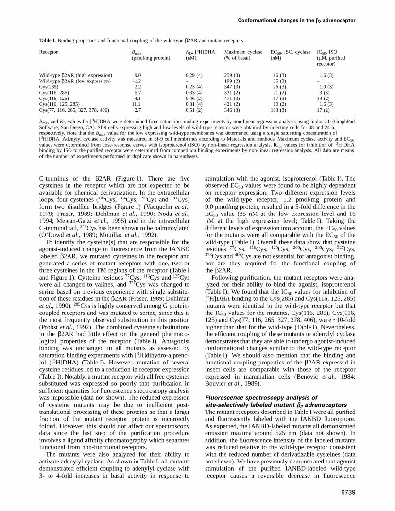
Fig. 2. Time course experiments with purified, IANBD-labeled wild-type and mutantβ2 adrenoceptors. Emission from IANBD-labeled wild-typeβ2adrenoceptor (A), mutant Cys(285) (B), mutant Cys(116, 125) (C), mutant Cys(116, 285) (D), mutant Cys(116, 125, 285) (E) and mutant Cys(77,116, 265, 327, 378, 406) (F) measured over time in response to stimulation with the full agonist isoproterenol (10–3 M). The responses werereversed by either the antagonist (–)alprenolol (10–4 M) or by (–)propranolol (10–4 M) both having a more than 100-fold higher affinity for thereceptors than isoproterenol (data not shown). By themselves, alprenolol and propranolol caused a relative increase in baseline fluorescence overtime but with a slower time-course than the reversal of the agonist response (data not shown and Getheret al., 1995). The ligand concentrations usedwere chosen to ensure saturation of the receptors eliminating any influence from different agonist affinities. Excitation was set at 481 nm andemission was measured at 525 nm. Fluorescence in the individual traces was normalized to the fluorescence observed immediately before addition ofligand. The experiments shown are representative of at least three identical experiments. (G) Bar diagram of changes in fluorescence in response tothe full agonist isoproterenol for the wild-type receptor and indicated mutants. Data are given as percent change in fluorescence (mean6 SE,n 5 3–6). The percent change was calculated as the change in fluorescence relative to the extrapolated baseline att 5 15 min after addition ofligand.
(Getheret al., 1995). A typical experiment with wild-typereceptor is shown in Figure 2A. Analysis of the mutantsrevealed that agonist-induced changes in fluorescence areobserved only in receptors in which285Cys or 125Cys arepresent (Figure 2). A mutant lacking only these twocysteines (Cys77, 116, 265, 327, 378, 406) showed noresponse to agonist binding (Figure 2F).
Both 285Cys–NBD (285Cys labeled with IANBD) and125Cys–NBD appeared to contribute to the agonist-inducedchange in fluorescence (Figure 2G). A decrease in fluores-
6740
cence of similar amplitude was observed in IANBD-labeled Cys(116, 125) and in IANBD-labeled Cys(116,285). The change in fluorescence in IANBD-labeledCys(116, 285) was similar to that in IANBD-labeledCys(285), confirming the lack of contribution of116Cys–NBD to the change in fluorescence. Moreover, the changein fluorescence from IANBD-labeled Cys(116, 125, 285)was nearly twice that of IANBD-labeled Cys(116, 125)or IANBD-labeled Cys(116, 285), further confirming thatthe molecular environment around both285Cys–NBD and
Conformational changes in the β2 adrenoceptor
125Cys–NBD is altered following agonist binding. Inconclusion, the data suggest that agonist binding to theβ2AR promotes a conformational change in the receptorthat exposes NBD attached to125Cys in TM III and to285Cys in TM VI to a more polar environment.
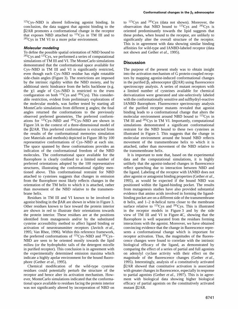
Molecular modelingTo define the possible spatial orientation of NBD bound to285Cys and125Cys, we performed a series of computationalsimulations of TM III and VI. The MonteCarlo simulationsdemonstrated that the conformational space available forCys–NBD in TM III and VI is significantly restricted,even though each Cys–NBD residue has eight rotatableside-chain angles (Figure 3). The restrictions are imposedby the intrinsic rigidity within the NBD moiety, and byadditional steric hindrance from the helix backbone (e.g.the χ1 angle of Cys–NBD is restricted to thetransconfiguration on both125Cys and285Cys). This consider-able restriction, evident from preliminary explorations ofthe molecular models, was further tested by starting allMonteCarlo simulations from differentχ angles; the finalangles returned the conformations to the previouslyobserved preferred geometries. The preferred conform-ations for 125Cys–NBD and285Cys–NBD are shown inFigure 3A in the context of a three-dimensional model ofthe β2AR. This preferred conformation is extracted fromthe results of the conformational memories simulation(see Materials and methods) depicted in Figure 3B by 100representative conformations of Cys–NBD at each site.The space spanned by these conformations provides anindication of the conformational freedom of the NBDmolecules. The conformational space available for thefluorophore is clearly confined to a limited number ofpreferred orientations adopted by the 100 representativestructures, illustrating the conformational rigidity men-tioned above. This conformational restraint for NBDattached to cysteines suggests that changes in emissionfrom the fluorophore most likely reflects changes in theorientation of the TM helix to which it is attached, ratherthan movement of the NBD relative to the transmem-brane helix.
Residues in TM III and VI known to be involved inagonist binding in theβAR are shown in white in Figure 3.Other residues known to face toward the protein interiorare shown in red to illustrate their orientations towardsthe protein interior. These residues are at the positionsidentified from mutagenesis and/or by the substitutedcysteine accessibility method to affect ligand binding oractivation of neurotransmitter receptors (Javitchet al.,1995; Van Rhee, 1996). Within this reference framework,the preferred conformations of125Cys–NBD and285Cys–NBD are seen to be oriented mostly towards the lipidmilieu (or the hydrophobic tails of the detergent micellein purified receptor). This conclusion is in agreement withthe experimentally determined emission maxima whichindicate a highly apolar environment for the bound fluoro-phore (Getheret al., 1995).
Chemical modification of the wild-type cysteineresidues could potentially perturb the structure of thereceptor and hence alter its activation mechanism. How-ever, MonteCarlo simulations indicated that the conforma-tional space available to residues facing the protein interiorwas not significantly altered by incorporation of NBD on
6741
to 125Cys and 285Cys (data not shown). Moreover, theobservation that NBD bound to125Cys and 285Cys isoriented predominantly towards the lipid suggests thatthese probes, when bound to the receptor, are unlikely tosignificantly alter the overall structure of the receptor.This is in agreement with data showing similar bindingaffinities for wild-type and IANBD-labeled receptor (datanot shown and Getheret al., 1995).
Discussion
The purpose of the present study was to obtain insightinto the activation mechanism of G protein-coupled recep-tors by mapping agonist-induced conformational changesin the purifiedβ2 adrenoceptor (β2AR) using fluorescencespectroscopy analysis. A series of mutant receptors witha limited number of cysteines available for chemicalderivatization were generated and site-selectively labeledwith the conformationally sensitive and sulfhydryl-reactiveIANBD fluorophore. Fluorescence spectroscopy analysisof the purified receptor mutants revealed that agonistbinding leads to a conformational change that alters themolecular environment around NBD bound to125Cys inTM III and 285Cys in TM VI. Importantly, computationalsimulations demonstrated a significant conformationalrestraint for the NBD bound to these two cysteines asillustrated in Figure 3. This suggests that the change inmolecular environment around the bound NBD reflectsmovement of the transmembrane helix to which it isattached, rather than movement of the NBD relative tothe transmembrane helix.
It is important to note that, based on the experimentaldata and the computational simulations, it is highlyunlikely that the agonist-induced changes in fluorescencereflect quenching due to interaction between NBD andthe ligand. Labeling of the receptor with IANBD does notalter agonist or antagonist binding properties (Getheret al.,1995), as would be expected if the bound NBD waspositioned within the ligand-binding pocket. The resultsfrom mutagenesis studies have also provided substantialevidence that amino acids involved in forming the ligand-binding pocket are on a different side of the transmembraneα helix, and 1–2α-helical turns closer to the membranesurface relative to125Cys and285Cys. This is illustratedin the receptor models in Figure 3 and by the sideview of TM III and VI in Figure 4C, showing that thefluorophore is well separated from the residues forminginteractions with the agonist. Moreover, we have obtainedconvincing evidence that the change in fluorescence repre-sents a conformational change which is important forreceptor activation. Thus, the magnitudes of the fluores-cence changes were found to correlate with the intrinsicbiological efficacy of the ligand, as demonstrated bycomparing the effect of a series of partial and full agonistson adenylyl cyclase activity with their effect on themagnitude of the fluorescence changes (Getheret al.,1995). Interestingly, analysis of a constitutively activatedβ2AR showed that constitutive activation is associatedwith greater changes in fluorescence, especially in responseto partial agonists (Getheret al., 1997). This is in agree-ment with biological data showing higher biologicalefficacy of partial agonists on the constitutively activatedmutantβ2AR.
U.Gether et al.
Fig. 3. Orientation of125Cys–NBD and285Cys–NBD in the context of a model of the transmembrane domains of theβ2 adrenoceptor, seen from theextracellular side. (A) In the preferred conformation,125Cys–NBD and285Cys–NBD lie at the protein–lipid interface, oriented predominantly towardsthe lipid. This orientation is substantiated by comparison with the residues that are known to be oriented towards the interior of the protein (shownin red and identified in Figure 1), and by residues in TM III and VI known to be involved in agonist binding inβAR (113Asp, 290Phe and293Asn,shown in white) (Totaet al., 1990; Straderet al., 1991, 1994; Wielandet al., 1996). The seven transmembrane helical domains are shownschematically by helical ribbons (yellow) following the electron density footprint of rhodopsin (Schertleret al., 1993). Note the highly conserved Proresidue in TM VI and the kink that it induces in this helix. (B) Superposition of 100 representative conformations of125Cys–NBD and285Cys–NBDobtained from the conformational memories calculations, shown in the same structural context as in (A). The close clustering of these conformationsthat identify the conformational space available to the Cys-bound fluorophores indicates that the preferred conformation of the fluorophores isrestricted to a well defined region.
The most likely interpretation of our results is thereforethat agonist binding promotes conformational changesinvolving movements of helix III and VI. As shown inFigure 3, 285Cys is predicted to be at the helix VI–VII
6742
interface in a boundary zone between the lipid bilayer (orthe hydrophobic tails of the detergent micelle) and themore polar interior of the protein. Thus, an agonist-induced movement of helix VI could lead to movement
Conformational changes in the β2 adrenoceptor
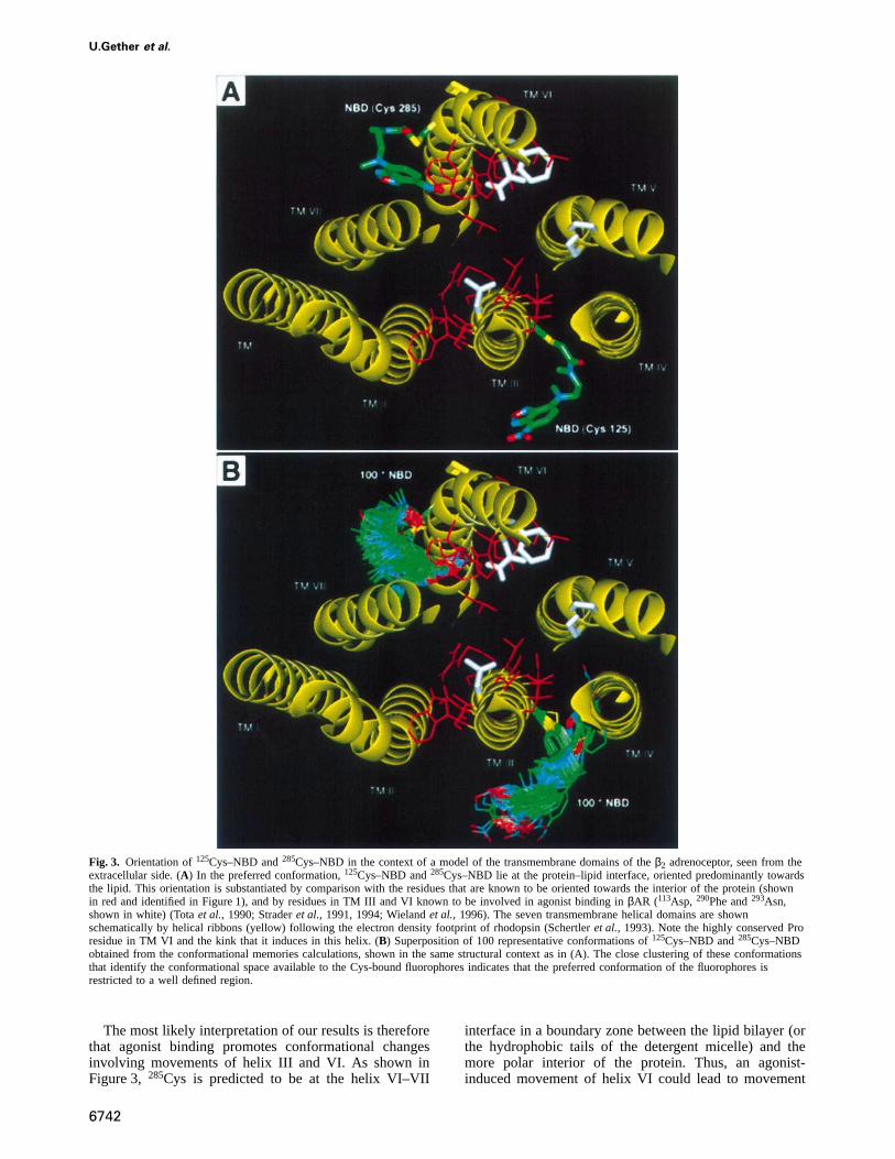
Fig. 4. The relationship of285Cys–NBD to the Pro-kink of TM VI. (A) The fluorophore is positioned within the region of TM VI (yellow ribbon)that contains the kink induced by288Pro, which spans from284Leu to 288Pro. Note the position of the preferred conformation of285Cys–NBD at theprotein–lipid interface, defined by the residues that are known to be oriented inwards towards the protein (shown in red and identified in Figure 1),and by residues in TM VI known to be involved in agonist binding inβAR (290Phe and293Asn, shown in white) (Totaet al., 1990; Straderet al.,1994; Wielandet al., 1996). The location of residues involved in G protein coupling is illustrated by the position of285Leu shown in red at thecytoplasmic end of TM VI. (B) Superposition of 100 representative conformations of285Cys–NBD and of the corresponding helix backbones(identified by their helix axes, in blue), obtained from the exploration of conformational space with the conformational memories method. Note thatthe kink induced by288Pro provides a flexible hinge connecting the binding site residues (in white, at the top of the figure) with the putative Gprotein-coupling domain at the cytoplasmic end of the helix. Varying degrees of kinking indicated by the helix axis (blue) have been implicated inthe activation mechanism (Zhang and Weinstein, 1993). Thus, the positioning of285Cys–NBD at the Pro-kink of TM VI makes it particularlysensitive to agonist-induced changes in the orientation of this TM helix. (C) Side view of the seven TM model including 100 representativeconformations of285Cys–NBD and125Cys–NBD, highlighting TM VI and III. Note that the NBD moieties are well within the space representing themembrane environment, at the lipid–protein interface, but not in contact with theβAR-binding site residues (shown in white). The flexible hingeprovided by the Pro-kink is illustrated by the wide margin span of the 100 representative TM VI helix axes (in blue).
6743

U.Gether et al.
of NBD bound to285Cys from the non-polar environmentof the lipid bilayer (or detergent micelle) to the morepolar environment of the interior of the protein, explainingthe observed changes in fluorescence. Notably,285Cys issituated oneα-helical turn below288Pro, which is highlyconserved among GPCRs and provides a flexible hinge inTM VI. Figure 4 shows how varying degrees of kinkingof TM VI around the proline hinge affect the position ofthe fluorophore. Consequently, the movement of285Cys–NBD to a more polar environment in the protein interiorwould be directly facilitated by this flexible hinge con-necting the binding site residues (in white, at the top ofFigure 4A, B and C) with the putative G protein-couplingdomain at the cytoplasmic end of the helix. Involvementof the proline in receptor activation has been suggestedfrom modeling studies (Ballesteros and Weinstein, 1995).Like 285Cys–NBD,125Cys–NBD is predominantly exposedto the lipid bilayer (or detergent micelle) (Figure 3). Ourdata indicate that the extent of lipid exposure is changedin response to agonist binding. This suggests that theagonist induces a movement of TM III, causing thefluorophore to be exposed to a more polar face of TM IVand/or the more polar interior of the receptor (Figure 3).It is however important to emphasize that the amplitudeof the fluorescent change is only a rough indicator of themagnitude of conformational change. For example, wecannot assume that there is a linear correlation betweenchange in fluorescence and magnitude of movements.Therefore, the movement of TM III may not be of thesame magnitude as movement of TM VI.
The present data with theβ2AR demonstrate strikingparallels to recent studies in rhodopsin where EPR spectro-scopy was used to delineate structural changes associatedwith activation (Farahbakhshet al., 1995; Altenbachet al.,1996; Farrenset al., 1996). The studies suggested thatrhodopsin activation entails a rigid-body movement ofTM VI relative to TM III. Specifically, Farrenset al.(1996) propose an anticlockwise rotation (viewed fromthe extracellular side as in Figure 3) of the TM VI segmentand a translation away from the TM III segment at thecytoplasmic side. This rotation of TM VI proposed forrhodopsin activation, if applied to theβ2AR model shownin Figure 3, would rotate285Cys–NBD in an anticlockwisedirection and thus reorient the fluorophore towards theinterior of the protein in full agreement with our experi-mental observations. Rearrangement of TM III and VI asa key element in receptor activation has also been sup-ported by the observation that binding of Zn21 to a bis-His metal ion-binding site constructed between TM IIIand VI in rhodopsin can block transducin activation (Sheiket al., 1996).
The binding of catecholamine agonists to theβ2ARalso involves important interactions in both TM III andTM VI (Figure 4C). In TM III the highly conserved asparticacid (113Asp) is believed to form an ionic interaction withthe positively charged nitrogen of epinephrine (Straderet al., 1991, 1994). In TM VI the agonist is believed toform hydrophobic interactions with290Phe (Totaet al.,1990), and evidence suggests the presence of a hydrogenbond between theβ-OH-group of epinephrine and293Asn(Wielandet al., 1996). Critical agonist interactions in TMVI have also been described for other G protein-coupledreceptors (Wesset al., 1991; Choudaryet al., 1995) and,
6744
in rhodopsin, retinal has been shown to interact with thehighly conserved265Trp (286Trp in β2AR) (Nakayama andKhorana, 1991). An essential role of TM VI in receptoractivation is further indicated by several studies demon-strating that discrete mutations at the bottom of TM VIcan constitutively activate many GPCRs including theβ2AR (Allen et al., 1991; Lefkowitzet al., 1993; Samamaet al., 1993). These mutations may disrupt constrainingintramolecular interactions, thereby leading to agonist-independent movements of TM VI. Importantly, we haveobserved a substantially enhanced fluorescence changefollowing agonist stimulation of an IANBD-labeled, con-stitutively active mutant of theβAR as compared withthe wild-type receptor (Getheret al., 1995).
In light of the suggested movements of TM III as apart of receptor activation (Farrenset al., 1996; Sheiket al., 1996; this study), it is noteworthy that protonationof the aspartic acid in the highly conserved DRY-motif atthe cytoplasmic border of TM III has been proposed tobe essential for receptor activation (Arniset al., 1994;Scheeret al., 1997). Thus, molecular dynamics simulationsin the adrenergicα1B receptor comparing the protonatedstate of the aspartic acid with the deprotonated stateindicated that protonation caused significant movementsof TM III due to a shift of the arginine in the DRY-motifout of a ‘polar pocket’ formed by residues in TM I, IIand VII (Scheeret al., 1996).
In conclusion, the present data represent the firstmapping of agonist-induced conformational changes in ahormone-activated G protein-coupled receptor. Comput-ational simulation demonstrated that the changes areconsistent with significant movements of TM III and VIin agreement with recent spin-labeling studies in rhodop-sin. This strongly suggests that our observations can begeneralized to other members of the superfamily of Gprotein-coupled receptors. Nevertheless, the fact that TMIII and VI may undergo significant conformational changesduring receptor activation does not exclude the possibilitythat other domains also move in response to agonistbinding. For example, movements of TM V and VII havebeen indicated from computational simulations comparingthe agonist bound and unliganded form of the 5-HT2Areceptor (Zhang and Weinstein, 1993). Hence, additionalexperiments based on biophysical techniques are requiredin the future to clarify further the changes involved inactivation of G protein-coupled receptors.
Materials and methods
NomenclatureThe cysteine mutants constructed in the present study were namedaccording to the cysteines still present in the receptor and available forchemical derivatization. Thus, Cys(285) describes a construct where285Cys is present but where77Cys,116Cys,125Cys,265Cys,327Cys, 378Cysand406Cys have been mutated.106Cys,184Cys,190Cys and191Cys, whichform two-disulfide bridges, and341Cys, which is palmitoylated, wereexcluded from the nomenclature, since they are not available for chemicalderivatization (Fraser, 1989; Dohlmanet al., 1990; Nodaet al., 1994).
MutagenesisThe cDNA encoding the humanβ2 adrenoceptor, epitope-tagged at theN-terminus with the cleavable influenza hemagglutinin signal sequencefollowed by the ‘FLAG’-epitope (IBI, New Haven, CT) and tagged atthe C-terminus with six histidines (SF-hβ2-6H), was used as a templatefor the mutagenesis (Guanet al., 1992). To facilitate the cloning
Conformational changes in the β2 adrenoceptor
procedures, anNheI site at bp 426 (as counted from the initiator codon)and aSacI site at bp 1083 were introduced in SF-hβ2-6H by mutagenesisbefore constructing the cysteine mutants. TheNheI and SacI sites andcysteine mutations C77V, C116V, C125V, C265A, C285S, C327S, C378Aand C406A were all generated by PCR-mediated mutagenesis usingPfupolymerase according to the manufacturer’s instructions (Stratagene, LaJolla, CA). The generated PCR fragments were digested with theappropiate enzymes, purified by agarose gel electrophoresis and clonedinto the baculovirus expression vetor pVL1392 containing SF-hβ2-6H(Guan et al., 1992). The constructs, Cys(285), Cys(116, 125, 285),Cys(116, 125), Cys(116, 285) and Cys(77, 116, 265, 327, 378, 406),were obtained by combining selected restriction enzyme fragments. Allmutations were confirmed by restriction enzyme analysis and sequenced.
Expression of receptors in Sf-9 insect cellsSF-hβ2-6H and mutant constructs in the baculovirus expression vectorpVL1392 were co-transfected with linerarized BaculoGold DNA intoSf-9 insect cells using the BaculoGold transfection kit (Pharmingen, SanDiego, CA). The resulting viruses were harvested after 4–5 days andamplified once before plaque purification. The plaque-purified viruseswere amplified several times to obtain 500 ml of a high titer virus stock(~13109 p.f.u.). Sf-9 insect cells were grown in suspension culture inSF 900 II medium (Gibco, Grand Island, NY) containing 5% fetal calfserum (Gemini, Calabasas, CA) and 0.1 mg/ml gentamicin (Gibco). Forpurification, the cells were grown in 1000–1200 ml cultures. Cells wereinfected with a 1:30–40 dilution of a high titer virus stock at a densityof 3.5–5.53106 cells/ml and harvested after 48 h by centrifugation for10 min at 5000g. The resulting cell pellets were kept at –70°C untilused for purification.
PCR analysis of virus stocksThe virus stocks were routinely checked for expression of the correctmutant by PCR analysis. Briefly, virus was isolated by centrifugation of5–10 ml virus stock for 20 min at 40 000g. The pelleted virus wasresuspended in 400µl TE-buffer (10 mM Tris–HCl plus 0.1 mM EDTA)and incubated for 30 min at 37°C with 10µg/ml RNase followed byincubation for 30 min at 37°C in the presence of 10µg/ml proteinaseK and 0.5% SDS. The viral DNA was isolated from the lysate byphenol–chloroform extraction and ethanol precipitation. The entire codingsequence of the receptor was amplified by PCR usingTaq polymerase,and the resulting fragments analyzed by restriction enzyme analysis andagarose gel electrophoresis.
Membrane preparation and adenylyl cyclase assayMembranes were prepared as described (Getheret al., 1995) from Sf-9cells (30 ml cultures in 125 ml disposable Erlenmeyer flasks at a densityof 33106 cells/ml) that were infected with baculovirus encoding thedifferent receptor constructs for 24 or 48 h. Protein was determinedusing the Bio-RadDC protein assay kit (Bio-Rad, Hercules, CA).Adenylyl cyclase in membranes was measured as described(Suryanarayanaet al., 1991).
Binding assaysSaturation binding assays and competition binding assays on membranepreparations from cells expressing the different receptors were carriedout as previously described using [3H]dihydro-alprenolol (Amersham,Arlington Heights, IL) as radioligand (Suryanarayanaet al., 1991).Binding assays on solubilized and purifiedβ2 receptor were alsoperformed as described using [3H]dihydro-alprenolol as radioligand(Getheret al., 1995). Binding data were analyzed by non-linear regressionanalysis using Inplot 4.0 from GraphPad Software, San Diego, CA.
Purification proceduresThe wild-type and mutant receptors were purified using a two-steppurification procedure (Getheret al., 1995, 1997; Kobilka, 1995). Oneor two pellets of cells from 1000 ml infected cultures were lysed in a10 mM Tris-HCl buffer, pH 7.5, containing 1 mM EDTA, 10µg/mlleupeptin (Boehringer, Mannheim, Germany), 10µg/ml benzamidine(Sigma) and 0.2 mM phenylmethylsulfonyl fluoride (Sigma). Followingcentrifugation (30 000g for 20 min), the lysed cells were resuspendedand subsequently solubilized under constant stirring for 2 h at 4°C in a20 mM Tris-HCl buffer, pH 7.5, containing 1.0%n-dodecyl-β-D-maltoside (DβM), 500 mM NaCl, 10 µg/ml leupeptin (Boehringer,Mannheim, Germany), 10µg/ml benzamidine (Sigma), 0.2 mM phenyl-methylsulfonyl fluoride (Sigma) and 10–6 M alprenolol (Sigma). Thesolubilized receptor was purified by nickel-column chromatographyusing chelating Sepharose (Pharmacia, Uppsala, Sweden) as described
6745
(Getheret al., 1995; Kobilka, 1995). The eluate from the Ni-columnwas further purified by alprenolol-affinity chromatography as described(Getheret al., 1995; Kobilka, 1995). Approximately 5 nmol of purifiedprotein could generally be obtained from a 1000 ml culture. The specificactivity of the purified receptors varied between 4 and 12 nmol/mgprotein. Protein was determined using the detergent-insensitive Bio-RadDC protein assay kit (Bio-Rad, Hercules, CA). Purified receptors wereanalyzed by classical 10% SDS–PAGE. The protein was visualized bystandard Coomassie staining. The results obtained with wild-type purifiedby the two-step purification procedure described here were indistinguish-able from results obtained using our previously described three-steppurification procedure (Getheret al., 1995; Kobilka, 1995).
Fluorescence labelingPurified receptors (1–1.5 nmol) were labeled according to previouslydescribed methods with 15-to 20-fold molar excess of IANBD [N,N9-dimethyl-N (iodoacetyl)-N9-(7-nitrobenz-2-oxa-1,3-diazol-4-yl) ethy-lene-diamine] (Molecular Probes, Eugene, OR) in a total volume of100 µl buffer (20 mM Tris-buffer, pH 7.4, containing 100 mM NaCland 0.08% DβM). The reaction was allowed to proceed for 1 h at roomtemperature in the dark and was quenched by addition of 1 mM cysteine.Cysteine-reacted dye was removed by desalting on a Sephadex G50 gelfiltration column (0.539 cm) followed by concentrating the resultingsample to 100µl using a Centricon-30 concentrator (Amicon, Beverly,MA). Alternatively, purified receptor was bound to a 150µl nickelcolumn (chelating Sepharose) and IANBD labeling achieved by recycling1.0 ml of 0.5 mM IANBD in buffer (Tris–HCl, pH 7.5, containing500 mM NaCl and 0.08% DβM) over the nickel column for 20 min.Excess dye was removed by extensive washing of the column withapproximately 50 column volumes of buffer. Labeled receptor was elutedwith 200 mM imidazole in buffer. Both labeling procedures resulted,depending on the mutant, in incorporation of 0.5–1.5 mol IANBD permol receptor, as determined by measuring absorption at 481 nm andusing an extinction coefficient of 21 000 M/cm for IANBD and a mol.wt of 50 000 Da for the receptor. Identical results were obtained withboth labeling procedures. Derivatization of the receptor with IANBDdid not perturb receptor pharmacology and labeling could be selectivelyblocked by sulfhydryl-reactive agents (Getheret al., 1995).
Fluorescence spectroscopyFluorescence spectroscopy was performed at room temperature on aSPEX Fluoromax spectrofluorometer with photon counting mode usingan excitation emission bandpass of 4.2 nm as described previously(Gether et al., 1995). Briefly, both emission scans and time courseexperiments were done with 50 pmol IANBD-labeled receptor in 500µlbuffer (20 mM Tris-buffer, pH 7.4, containing 100 mM NaCl and 0.08%DβM) under constant stirring. In emission scan experiments the excitationwavelength was 481 nm and emission measured from 490 to 625 nmwith an integration time at 0.3 s/nm. During time scan experiments theexcitation wavelength was 481 nm and emission was measured at awavelength of 525 nm. The volume of the added ligands was 1% oftotal volume and fluorescence was corrected for this dilution. Thecompounds tested in the fluorescence experiments had an absorbance ofless than 0.01 at 481 and 523 nm in the concentrations used excludinginner filter effects.
Molecular modelingThe TM segments III (residues 107–136) and VI (residues 227–197)containing the Cys–NBD labels were modeled asα-helices, except forthe Pro-kink in TM VI, as described (Ballesteros and Weinstein, 1995),and subjected to MonteCarlo simulations. The simulations explored aregion of the TM domains composed of 11 residues centered onthe Cys–IANBD. The novel technique of conformational memories(Guarnieri and Wilson, 1995, 1996) was used to perform a search of theconformational space by variation of torsional angles. The variation ofbackbone dihedral anglesφ and ψ were restrained to 20° from theirinitial values, except for the Pro-kink region (residues284Leu to288Pro),whereφ andψ angles were restrained within 50° of their initial valuesdue to the known flexibility of Pro-kinks relative toα-helical domains(Ballesteros and Weinstein, 1995). Side chain dihedral angles wererotated freely. More than 100 rounds of independent random simulationswere performed for each TM segment. In each round, repeated runs ofMonteCarlo simulated annealing (Kirkpatrick, 1983) were performedfrom a starting temperature of T1 5 2070 K, with a cooling scheduleof Tn11 5 0.9*Tn, and 10 000 steps per temperature to reach 310 K.This first stage identifies the regions of the torsional angle space thatare populated at 310 K for each dihedral angle, i.e. the conformational

U.Gether et al.
memories. In the second stage the conformational memories are used ina biased MonteCarlo procedure to sample only from these populatedregions. This step yields the ensemble of structures accessible at 310 K.The conformational space of each NBD–Cys was analyzed in thecontext of a model of theβ2AR constructed using previously describedapproaches (Ballesteros and Weinstein, 1995), that follows the electrondensity footprint of rhodopsin (Schertleret al., 1993).
Acknowledgements
The study was supported in part by National Institute of Health GrantRO 1 NS28471, DA-09083 and DA-00060, and by the Harold G. andLeila Y.Mathers Charitable Foundation. Pejman Ghanouni is a MedicalScientist Training Program trainee and receives support from NIGMStraining grant GM07365.
References
Allen,L.F., Lefkowitz,R.J., Caron,M.G. and Cotecchia,S. (1991) G-protein-coupled receptor genes as protooncogenes: constitutivelyactivating mutation of the α1B-adrenergic receptor enhancesmitogenesis and tumorigenicity.Proc. Natl Acad. Sci. USA, 88,11354–11358.
Altenbach,C., Yang,K., Farrens,D.L., Farahbakhsh,Z.T., Khorana,H.G.and Hubbell,W.L. (1996) Structural features and light-dependentchanges in the cytoplasmic interhelical E–F loop region of rhodopsin:a site-directed spin-labeling study.Biochemistry, 35, 12470–12478.
Arnis,S., Fahmy,K., Hofmann,K.P. and Sakmar,T.P. (1994) A conservedcarboxylic acid group mediates light-dependent proton uptake andsignaling by rhodopsin.J. Biol. Chem., 269, 23879–23881.
Baldwin,J.M. (1993) The probable arangement of the helices in G proteincoupled receptors.EMBO J., 12, 1693–1703.
Ballesteros,J.A. and Weinstein,H. (1995) Integrated methods for theconstruction of three-dimensional models and computational probingof structure–function relations in G protein coupled receptors.MethodsNeurosci., 25, 366–428.
Benovic,J.L., Shorr,R.G.L., Caron,M.C. and Lefkowitz,R.J. (1984) Themammalian beta2-adrenergic receptor: purification andcharacterization.Biochemistry, 23, 4510–4518.
Bouvier,M. et al. (1989) Two distinct pathways for cAMP-mediateddown-regulation of theβ2-adrenergic receptor.J. Biol. Chem., 264,16786–16792.
Choe,H.et al. (1996) The beta-chemokine receptors CCR3 and CCR5facilitate infection by primary HIV-1 isolates.Cell, 85, 1135–1148.
Choudhary,M.S., Sachs,N., Uluer,A., Glennon,R.A., Westkaemper,R.B.and Roth,B.L. (1995) Differential ergoline and ergopeptine bindingto 5-hydroxytryptamine2A receptors: ergolines require an aromaticresidue at position 340 for high affinity binding.Mol. Pharmacol.,47, 450–457.
Cronet,P., Sander,C. and Vriend,G. (1993) Modeling of transmembraneseven helix bundles.Protein Eng., 6, 59–64.
Dohlman,H.G., Caron,M.G., DeBlasi,A., Frielle,T. and Lefkowitz,R.J.(1990) Role of extracellular disulfide-bonded cysteines in the ligandbinding function of theβ2-adrenergic receptor.Biochemistry, 29,2335–2342.
Elling,C.E. and Schwartz,T.W. (1996) Connectivity and orientation ofthe seven helical bundle in the tachykinin NK-1 receptor probed byzinc site engineering.EMBO J., 15, 6213–6219.
Fanelli,F., Menziani,M.C. and De Benedetti,P.G. (1995) Moleculardynamics simulations of m3-muscarinic receptor activation and QSARanalysis.Bioorg. Med. Chem., 3, 1465–1477.
Farahbakhsh,Z.T., Ridge,K.D., Khorana,H.G. and Hubbell,W.L. (1995)Mapping light-dependent structural changes in the cytoplasmic loopconnecting helices C and D in rhodopsin: a site-directed spin labelingstudy.Biochemistry, 34, 8812–8819.
Farrens,D.L., Altenbach,C., Yang,K., Hubbell,W.L. and Khorana,H.G.(1996) Requirement of rigid-body motion of transmembrane helicesfor light activation of rhodopsin.Science, 274, 768–770.
Feng,Y., Broder,C.C., Kennedy,P.E. and Berger,E.A. (1996) HIV-1 entrycofactor: functional cDNA cloning of a seven-transmembrane, Gprotein-coupled receptor.Science, 272, 872–877.
Fraser,C.M. (1989) Site-directed mutagenesis of beta-adrenergicreceptors. Identification of conserved cysteine residues thatindependently affect ligand binding and receptor activation.J. Biol.Chem., 264, 9266–9270.
Gether,U., Lin,S. and Kobilka,B.K. (1995) Fluorescent labeling of
6746
purified beta2-adrenergic receptor: evidence for ligand-specificconformational changes.J. Biol. Chem., 270, 28268–28275.
Gether,U., Ballesteros,J.A., Seifert,R., Sanders-Bush,E., Weinstein,H.and Kobilka,B.K. (1997) Structural instability of a constitutivelyactive G protein-coupled receptor. Agonist-independent activation dueto conformational flexibility.J. Biol. Chem., 272, 2587–2590.
Guan,X.M., Kobilka,T.S. and Kobilka,B.K. (1992) Enhancement ofmembrane insertion and function in a type IIIb membrane proteinfollowing introduction of a cleaveable signal peptide.J. Biol. Chem.,267, 21995–21998.
Guarnieri,F. and Weinstein,H. (1996) Conformational memories and theexploration of biologically relevant conformations: an illustrationfor the gonadoropin-releasing hormone.J. Amer. Chem. Soc., 118,5580–5589.
Guarnieri,F. and Wilson,S.R. (1995) Conformational memories and asimulated annealing program that learns: application to LTB4.J. Comput. Chem., 16, 648–653.
Javitch,J.A., Fu,D., Chen,J. and Karlin,A. (1995) Mapping the binding-site crevice of the dopamine D2 receptor by the substituted-cysteineaccessibility method.Neuron, 14, 825–831.
Kirkpatrick,S., Gelatt,C.D. and Vecchi,M.P. (1983) Simulated annealing.Science, 220, 671.
Kobilka,B.K. (1992) Adrenergic receptors as models for G protein-coupled receptors.Annu. Rev. Neurosci., 15, 87–114.
Kobilka,B.K. (1995) Amino and carboxyterminal modifications tofacilitate the production and purification of a G protein coupledreceptor.Anal. Biochem., 231, 269–271.
Lefkowitz,R.J., Cotecchia,S., Samama,P. and Costa,T. (1993)Constitutive activity of receptors coupled to guanine nucleotideregulatory proteins.Trends Pharmacol. Sci., 14, 303–307.
Liu,J., Schoneberg,T., Van Rhee,M. and Wess,J. (1995) Mutationalanalysis of the relative orientation of transmembrane helices I andVII in G protein-coupled receptors.J. Biol. Chem., 270, 19532–19539.
Luo,X., Zhang,D. and Weinstein,H. (1994) Ligand-induced domainmotion in the activation mechanism of a G-protein-coupled receptor.Protein Eng., 7, 1441–1448.
MaloneyHuss,K. and Lybrand,T.P. (1992) Three-dimensional structurefor the beta 2 adrenergic receptor protein based on computer modelingstudies.J. Mol. Biol., 225, 859–871.
Mejean-Galzi,A., Guillaume,J.L. and Strosberg,A.D. (1995) Carazolol:a potent, selectiveβ3-adrenoceptor agonist.Eur. J. Pharmacol., 291,359–366.
Mizobe,T., Maze,M., Lam,V., Suryanarayana,S. and Kobilka,B.K. (1996)Arrangement of transmembrane domains in adrenergic receptors.Similarity to bacteriorhodopsin.J. Biol. Chem., 271, 2387–2389.
Mouillac,B., Caron,M., Bonin,H., Dennis,M. and Bouvier,M. (1992)Agonist-modulated palmitoylation ofβ2-adrenergic receptor in Sf9cells. J. Biol. Chem., 267, 21733–21737.
Nakayama,T.A. and Khorana,H.G. (1991) Mapping of the amino acidsin membrane-embedded helices that interact with the retinalchromophore in bovine rhodopsin.J. Biol. Chem., 266, 4269–4275.
Noda,K., Saad,Y., Graham,R.M. and Karnik,S.S. (1994) The high affinitystate of theβ2-adrenergic receptor requires unique interaction betweenconserved and non-conserved extracellular loop cysteines.J. Biol.Chem., 269, 6743–6752.
O’Dowd,B.F., Hnatowich,M., Caron,M.G., Lefkowitz,R.J. andBouvier,M. (1989) Palmitoylation of the humanβ2-adrenergic receptor.Mutation of Cys341 in the carboxyl tail leads to an uncouplednonpalmitoylated form of the receptor.J. Biol. Chem., 264, 7564–7569.
Probst,W.C., Snyder,L.A., Schuster,D.I., Brosius,J. and Sealfon,S.C.(1992) Sequence alignment of the G-protein coupled receptorsuperfamily.DNA Cell. Biol., 11, 1–20.
Roper,D., Jacoby,E., Kruger,P., Engels,M., Grotzinger,J., Wollmer,A. andStrassburger,W. (1994) Modeling of G-protein coupled receptors withbacteriorhodopsin as a template. A novel approach based on interactionenergy differences.J. Recept. Res., 14, 167–186.
Samama,P., Cotecchia,S., Costa,T. and Lefkowitz,R.J. (1993) A mutation-induced activated state of theβ2-adrenergic receptor: extending theternary complex model.J .Biol. Chem., 268, 4625–4636.
Savarese,T.M. and Fraser,C.M. (1992)In vitro mutagenesis and thesearch for structure–function relationships among G protein-coupledreceptors.Biochem. J., 283, 1–19.
Scheer,A., Fanelli,F., Costa,T., De Benedetti,P.G. and Cotecchia,S. (1996)Constitutively active mutants of theα1B-adrenergic receptor: role ofhighly conserved polar amino acids in receptor activation.EMBO J.,15, 3566–3578.
Conformational changes in the β2 adrenoceptor
Scheer,A., Fanelli,F., Costa,T., De Benedetti,P.G. and Cotecchia,S. (1997)The activation process of theα1B-adrenergic receptor: potential roleof protonation and hydrophobicity of a highly conserved aspartate.Proc. Natl Acad. Sci. USA, 94, 808–813.
Schertler,G., Villa,C. and Henderson,R. (1993) Projection structure ofrhodopsin.Nature, 362, 770–772.
Schwartz,T.W. (1994) Locating ligand-binding sites in 7TM receptorsby protein engineering.Current Opinion Biotech., 5, 434–444.
Schwartz,T.W. (1996) Molecular Structure of G protein coupled receptors.In Foreman,J.C. and Johansen,T. (eds),Molecular Structure of Gprotein coupled receptors. CRC Press, Boca Raton, pp. 65–84.
Sheikh,S.P., Zvyaga,T.A., Lichtarge,O., Sakmar,T.P. and Bourne,H.R.(1996) Rhodopsin activation blocked by metal-ion-binding sites linkingtransmembrane helices C and F.Nature, 383, 347–350.
Strader,C.D., Gaffney,T., Sugg,E.E., Candelore,M.R., Keys,R.,Patchett,A.A. and Dixon,R.A.F. (1991) Allele-specific activation ofgenetically engineered receptors.J. Biol. Chem., 266, 5–8.
Strader,C.D., Fong,T.M., Tota,M.R., Underwood,D. and Dixon,R.A.F.(1994) Structure and function of G protein-coupled receptors.Annu.Rev. Biochem., 63, 101–132.
Suryanarayana,S. and Kobilka,B.K. (1991) Construction and expressionof chimeric receptors to understand the structure–function relationshipsin adrenergic receptors.Methods, 3, 193–204.
Suryanarayana,S., Von Zastrow,M. and Kobilka,B.K. (1992)Identification of intramolecular interaction in adrenergic receptors.J. Biol. Chem., 267, 21991–21994.
Tota,R.T., Candelore,M.R., Dixon,R.A.F. and Strader,C.D. (1990)Biophysical and genetic analysis of the ligand-binding site of the beta-adrenoceptor.Trends Pharmacol. Sci., 12, 4–6.
Turcatti,G., Nemeth,K., Edgerton,M.D., Meseth,U., Talabot,F.,Peitsch,M., Knowles,J., Vogel,H.and Chollet,A. (1996) Probing thestructure and function of the tachykinin neurokinin-2 receptor throughbiosynthetic incorporation of fluorescent amino acids at specific sites.J. Biol. Chem., 271, 19991–19998.
Trumpp-Kallmeyer,S., Hoflack,J., Bruinvels,A. and Hibert,M. (1992)Modeling of G-protein-coupled receptors: application to dopamine,adrenaline, serotonin, acetylcholine, and mammalian opsin receptors.J. Med. Chem., 35, 3448–3462.
Van Rhee,A.M. and Jacobson,K.A. (1996) Molecular architecture of Gprotein-coupled receptors.Drug Dev. Res., 37, 1–38.
Vauquelin,G., Bottari,S., Kanarek,L. and Strosberg,A.D. (1979) Evidencefor essential disulfide bonds inβ1-adrenergic receptors of turkeyerythrocyte membranes.J. Biol. Chem., 254, 4462–4469.
Wess,J., Gdula,D. and Brann,M.R. (1991) Site-directed mutagenesis ofthe m3 muscarinic receptor: identification of a series of threonine andtyrosine residues involved in agonist but not antagonist binding.EMBO J., 10, 3729–3734.
Wieland,K., Zuurmond,H.M., Krasel,C., Ijzerman,A.P. andLohse,M.J. (1996) Involvement of Asn-293 in stereospecific agonistrecognition and in activation of theβ2-adrenergic receptor.Proc. NatlAcad. Sci. USA, 93, 9276–9281.
Zhang,D. and Weinstein,H. (1993) Signal transduction by a 5-HT2receptor: a mechanistic hypothesis from molecular dynamicssimulations of the three-dimensional model of the receptor complexedto ligands.J. Med. Chem., 36, 934–8.
Zhou,W., Flanagan,C., Ballesteros,J.A., Konvicka,K., Davidson,J.S.,Weinstein,H., Millar,R.P. and Sealfon,S.C. (1994) A reciprocalmutation supports helix 2 and helix 7 proximity in the gonadotropin-releasing hormone receptor.Mol. Pharmacol., 45, 165–170.
Received on April 18, 1997; revised on July 28, 1997
6747
Related Documents