Stem Cell Reports Ar ticle Aging-like Phenotype and Defective Lineage Specification in SIRT1-Deleted Hematopoietic Stem and Progenitor Cells Pauline Rimmele ´, 1 Carolina L. Bigarella, 1 Raymond Liang, 1,2 Brigitte Izac, 1 Rebeca Dieguez-Gonzalez, 1 Gaetan Barbet, 3 Michael Donovan, 4 Carlo Brugnara, 5 Julie M. Blander, 3,6 David A. Sinclair, 7 and Saghi Ghaffari 1,2,6,8,9, * 1 Department of Developmental & Regenerative Biology 2 Developmental and Stem Cell Biology Multidisciplinary Training Area 3 Division of Clinical Immunology, Department of Medicine 4 Department of Experimental Pathology Icahn School of Medicine at Mount Sinai, New York, NY 10029, USA 5 Department of Lab Medicine, Children’s Hospital, Boston, MA 02115, USA 6 Tisch Cancer Institute, Icahn School of Medicine at Mount Sinai, New York, NY 10029, USA 7 Paul F. Glenn Laboratories for the Biological Mechanisms of Aging, Department of Genetics, Harvard Medical School, Boston, MA 02115, USA 8 Division of Hematology and Oncology, Department of Medicine 9 Black Family Stem Cell Institute Icahn School of Medicine at Mount Sinai, New York, NY 10029, USA *Correspondence: [email protected] http://dx.doi.org/10.1016/j.stemcr.2014.04.015 This is an open access article under the CC BY-NC-ND license (http://creativecommons.org/licenses/by-nc-nd/3.0/). SUMMARY Aging hematopoietic stem cells (HSCs) exhibit defective lineage specification that is thought to be central to increased incidence of myeloid malignancies and compromised immune competence in the elderly. Mechanisms underlying these age-related defects remain largely unknown. We show that the deacetylase Sirtuin (SIRT)1 is required for homeostatic HSC maintenance. Differentiation of young SIRT1-deleted HSCs is skewed toward myeloid lineage associated with a significant decline in the lymphoid compartment, anemia, and altered expression of associated genes. Combined with HSC accumulation of damaged DNA and expression patterns of age-linked mol- ecules, these have striking overlaps with aged HSCs. We further show that SIRT1 controls HSC homeostasis via the longevity transcription factor FOXO3. These findings suggest that SIRT1 is essential for HSC homeostasis and lineage specification. They also indicate that SIRT1 might contribute to delaying HSC aging. INTRODUCTION Adult stem cells maintain tissue homeostasis by regenerat- ing damaged or lost cells during their lifetime. The decline of the regenerative capacity of stem cells with age compro- mises tissue integrity and may promote organ failure and diseases of aging (Liu and Rando, 2011). This age-related decline in tissue function is considered to be at the root of overall organismal aging. Whether mechanisms that control aging of stem cells influence organismal longevity is unknown. Identifying regulators of stem cell aging is of major significance for public health because such regula- tors may contribute to promote healthy aging and be valu- able therapeutic targets to combat disorders of aging like cancer and Parkinson’s disease. Hematopoietic stem cells (HSCs) are the most extensively studied model of stem cell aging. Although it has been known for decades that HSC age (Harrison, 1983), and the properties of aged HSCs have been greatly character- ized, the mechanisms that govern HSC aging have only begun to be defined. HSC aging leads to a paradoxical in- crease in the stem cell pool and decline in stem cell func- tion (Morrison et al., 1996; Sudo et al., 2000). One of the prominent modifications of HSC properties with age is their biased differentiation toward myeloid lineage at the expense of their lymphoid potential (Challen et al., 2010; Dykstra et al., 2011; Rossi et al., 2005). These age-associated modulations of the composition of HSC progenies lead to defective adaptive immune response. Similarly, the age- related increased incidence of myeloid malignancies, including acute myeloid leukemias, myelodysplasias, and myeloproliferative neoplasms, may be related to the enhanced generation of myeloid skewed HSC progenies. Aging of HSCs is also associated with increased onset of anemia. Although defects in the DNA damage repair pro- gram, increased tumor suppressor function, loss of polarity, and epigenetic deregulation have all been implicated in HSC aging, the mechanisms underpinning the age-associ- ated alterations of HSC lineage specification remain largely unknown (Chambers et al., 2007; Dykstra and de Haan, 2008; Florian et al., 2012; Rossi et al., 2005). The NAD-dependent protein silent information regu- lator 2 (Sir2) is a deacetylase for histones and other proteins and a key regulator of life span in several organisms. Sirtuin (SIRT)1 of the Sirtuin family is the closest homolog of yeast Sir2 in mammals and has critical functions in the 44 Stem Cell Reports j Vol. 3 j 44–59 j July 8, 2014 j ª2014 The Authors

Welcome message from author
This document is posted to help you gain knowledge. Please leave a comment to let me know what you think about it! Share it to your friends and learn new things together.
Transcript

Stem Cell Reports
ArticleAging-like Phenotype and Defective Lineage Specification in SIRT1-DeletedHematopoietic Stem and Progenitor Cells
Pauline Rimmele,1 Carolina L. Bigarella,1 Raymond Liang,1,2 Brigitte Izac,1 Rebeca Dieguez-Gonzalez,1
Gaetan Barbet,3 Michael Donovan,4 Carlo Brugnara,5 Julie M. Blander,3,6 David A. Sinclair,7
and Saghi Ghaffari1,2,6,8,9,*1Department of Developmental & Regenerative Biology2Developmental and Stem Cell Biology Multidisciplinary Training Area3Division of Clinical Immunology, Department of Medicine4Department of Experimental Pathology
Icahn School of Medicine at Mount Sinai, New York, NY 10029, USA5Department of Lab Medicine, Children’s Hospital, Boston, MA 02115, USA6Tisch Cancer Institute, Icahn School of Medicine at Mount Sinai, New York, NY 10029, USA7Paul F. Glenn Laboratories for the Biological Mechanisms of Aging, Department of Genetics, Harvard Medical School, Boston, MA 02115, USA8Division of Hematology and Oncology, Department of Medicine9Black Family Stem Cell Institute
Icahn School of Medicine at Mount Sinai, New York, NY 10029, USA
*Correspondence: [email protected]
http://dx.doi.org/10.1016/j.stemcr.2014.04.015
This is an open access article under the CC BY-NC-ND license (http://creativecommons.org/licenses/by-nc-nd/3.0/).
SUMMARY
Aging hematopoietic stem cells (HSCs) exhibit defective lineage specification that is thought to be central to increased incidence of
myeloid malignancies and compromised immune competence in the elderly. Mechanisms underlying these age-related defects remain
largely unknown. We show that the deacetylase Sirtuin (SIRT)1 is required for homeostatic HSC maintenance. Differentiation of young
SIRT1-deleted HSCs is skewed toward myeloid lineage associated with a significant decline in the lymphoid compartment, anemia, and
altered expression of associated genes. Combined with HSC accumulation of damaged DNA and expression patterns of age-linked mol-
ecules, these have striking overlapswith agedHSCs.We further show that SIRT1 controlsHSChomeostasis via the longevity transcription
factor FOXO3. These findings suggest that SIRT1 is essential for HSC homeostasis and lineage specification. They also indicate that SIRT1
might contribute to delaying HSC aging.
INTRODUCTION
Adult stem cells maintain tissue homeostasis by regenerat-
ing damaged or lost cells during their lifetime. The decline
of the regenerative capacity of stem cells with age compro-
mises tissue integrity and may promote organ failure and
diseases of aging (Liu and Rando, 2011). This age-related
decline in tissue function is considered to be at the root
of overall organismal aging. Whether mechanisms that
control aging of stem cells influence organismal longevity
is unknown. Identifying regulators of stem cell aging is of
major significance for public health because such regula-
tors may contribute to promote healthy aging and be valu-
able therapeutic targets to combat disorders of aging like
cancer and Parkinson’s disease.
Hematopoietic stem cells (HSCs) are themost extensively
studied model of stem cell aging. Although it has been
known for decades that HSC age (Harrison, 1983), and
the properties of aged HSCs have been greatly character-
ized, the mechanisms that govern HSC aging have only
begun to be defined. HSC aging leads to a paradoxical in-
crease in the stem cell pool and decline in stem cell func-
tion (Morrison et al., 1996; Sudo et al., 2000). One of the
44 Stem Cell Reports j Vol. 3 j 44–59 j July 8, 2014 j ª2014 The Authors
prominent modifications of HSC properties with age is
their biased differentiation toward myeloid lineage at the
expense of their lymphoid potential (Challen et al., 2010;
Dykstra et al., 2011; Rossi et al., 2005). These age-associated
modulations of the composition of HSC progenies lead to
defective adaptive immune response. Similarly, the age-
related increased incidence of myeloid malignancies,
including acute myeloid leukemias, myelodysplasias, and
myeloproliferative neoplasms, may be related to the
enhanced generation of myeloid skewed HSC progenies.
Aging of HSCs is also associated with increased onset of
anemia. Although defects in the DNA damage repair pro-
gram, increased tumor suppressor function, loss of polarity,
and epigenetic deregulation have all been implicated in
HSC aging, the mechanisms underpinning the age-associ-
ated alterations of HSC lineage specification remain largely
unknown (Chambers et al., 2007; Dykstra and de Haan,
2008; Florian et al., 2012; Rossi et al., 2005).
The NAD-dependent protein silent information regu-
lator 2 (Sir2) is a deacetylase for histones and other proteins
and a key regulator of life span in several organisms. Sirtuin
(SIRT)1 of the Sirtuin family is the closest homolog of yeast
Sir2 in mammals and has critical functions in the

Stem Cell ReportsAging-like Phenotype in SIRT1-Deleted HSCs
regulation of metabolism, genome stability, DNA repair,
chromatin remodeling, and stress response (Guarente,
2011; Haigis and Sinclair, 2010). SIRT1 coordinates plurip-
otency, differentiation, and stress response in mouse
embryonic stem cells (ESCs) (Han et al., 2008). Whether
SIRT1 regulates adult stem cells particularly in the hemato-
poietic system has been a matter of debate (Leko et al.,
2012; Li et al., 2012; Narala et al., 2008; Singh et al.,
2013; Yuan et al., 2012). Despite recent advances in under-
standing SIRT1 regulation of malignant and stressed hema-
topoiesis, whether SIRT1 has any function in the control of
adult HSC homeostasis or aging remains unknown.
The study of SIRT1 in adult mice and during aging has
been hampered by the developmental defects and perinatal
death of germline SIRT1 knockoutmice (Cheng et al., 2003;
McBurney et al., 2003). Using a recently developed adult
tamoxifen-inducible SIRT1 knockout mouse model (Price
et al., 2012), we show that SIRT1 is essential for the self-
renewal and homeostatic maintenance of the HSC pool.
Importantly, we show that loss of SIRT1 is associated with
anemia and a significant expansion of the myeloid
compartment, specifically granulocyte-monocyte progeni-
tors (GMPs), at the expense of the lymphoid compartment.
These phenotypic alterations are concomitant with signif-
icant modulations of expression of transcription factors
implicated in the generation of GMPs and common
lymphoid progenitors (CLPs). Notably, we show that the
longevity transcription factor FOXO3 mediates SIRT1
homeostatic effects in HSCs. These unexpected results
indicate that young SIRT1-deleted HSCs have several over-
lapping features with normal aged HSCs. Altogether, our
studies identify SIRT1 as a key regulator of HSC mainte-
nance under homeostasis. In addition, the evidence sup-
ports an essential function for SIRT1 in the regulation of
HSC lineage specification. Overall, our findings suggest
that SIRT1 might be implicated in delaying HSC aging.
RESULTS
Loss of SIRT1 Compromises Hematopoietic Stem Cell
Function at the Steady State
To address the potential function of SIRT1 in hemato-
poietic stem and progenitor cells (HSPCs), we first analyzed
SIRT1 expression. As predicted by previous studies (De-
neault et al., 2009), SIRT1 transcript was increased in
Lin�SCA-1+C-KIT+ (LSK) cells enriched for HSCs and in
Lin�SCA�1+C-KIT+ (c-Kit+) hematopoietic multipotential
progenitors as compared to total bone marrow (BM) cells
(Figure S1A available online). However, the level of SIRT1
transcript in LSK CD48�CD150+ that is highly enriched
for long-term HSCs (LT-HSCs), and in all hematopoietic
progenitor cells surveyed, was relatively similar (Fig-
ure S1B). SIRT1 protein was also readily detected in the
nucleus of HSPCs (Figure S1C). Next, we used sirtinol, a
pharmacological inhibitor of SIRT1 (Grozinger et al.,
2001), to evaluate whether SIRT1 has any functions in
HSCs. A 3 week in vivo injection of sirtinol led to a signifi-
cant decrease of the frequency and the total number of LSK
cells (Figures S1D and S1E) in the treated animals, including
the LSK CD48�CD150+ cell subset (Figures S1F and S1G).
To circumvent potential off-target effects of pharma-
cological inhibitors of SIRT1 and investigate the function
of SIRT1 more directly, we used a tamoxifen-inducible
SIRT1 deletion mouse model (Price et al., 2012). The
SIRT1 catalytic domain was conditionally deleted from 6-
to 8-week-old floxed SIRT1Dex4; Cre-ERT2mice by intraper-
itoneal tamoxifen injection over 5 days. The regimen led to
the expression of a truncated protein that was detected as a
lower molecular weight SIRT1 4 weeks after tamoxifen
treatment in the BM and spleen (Figure S2A). At the same
time, the truncated SIRT1 protein was detected in highly
purified HSPCs (Figure S2B). Notably, and in contrast to
Singh et al. (2013), this regimen did not significantly
modulate the BM cellularity (Figure S2C) overtimewhether
or not Crewas present (Figure S2D), enabling us to examine
the effects of loss of SIRT1 function on HSPCs under ho-
meostatic conditions.
The effect of SIRT1 deletion on the HSC compartment of
Sirt1fl/fl;Cre+ (D/D) mice was monitored at different time
points after tamoxifen treatment using both Sirt1fl/fl;Cre�
(fl/fl) and Sirt1WT/WT;Cre+ (WT/WT) mice as controls.
Loss of SIRT1 function in young adult mice resulted in a
gradual increase in the total number and the frequency of
both LSK cells (Figure 1A, top panel; Figure S2E) and LT-
HSC (LSK CD48�CD150+) (Figure 1A, bottom panel; Fig-
ure S2F) in the BM. In addition, the total number and
frequency of multipotent progenitors (MPPs; LSK FLK2+
CD34+) and lymphoid multipotent progenitor (LMPP; LSK
FLK2highCD34+) cells also increased in SIRT1-deleted mice
without significant effects on the short-term HSC (ST-
HSC; LSK FLK2�CD34+) (Figures 1B and S2G–S2H). These
results were intriguing and suggested that SIRT1-deficiency
compromised the HSC compartment at the steady state.
To evaluate whether SIRT1 has cell-autonomous func-
tions, we injected 100 highly purified SIRT1-deficient LT-
HSCs into lethally irradiated congenic-recipientmice along
with 200,000 recipient BM cells in an in vivo competitive
repopulation assay. Whereas control cells gave rise to
strong chimerism and multilineage reconstitution in all
12 transplanted recipients, the long-term repopulation
ability of SIRT1-deficient HSCs was 2.5-fold reduced as
compared to controls (Figure 1C). The analysis of recipient
mice 16 weeks after the primary transplantation showed a
significant decrease in the frequency and total number of
donor-derived LSK cells (Figure 1D), suggesting that
Stem Cell Reports j Vol. 3 j 44–59 j July 8, 2014 j ª2014 The Authors 45

Figure 1. Conditional Deletion of SIRT1 Compromises Homeostatic HSC Function(A) Representative fluorescence-activated cell sorting (FACS) plots of LSK cells (left upper panel) and LSK CD48�CD150+ (LT-HSC) (leftlower panel) frequencies from Sirt1WT/WT (WT/WT; cre+), Sirt1fl/fl (fl/fl; cre�), and Sirt1D/D (D/D, cre+) BM (see Figures S2E and, S2F).Fold change of total BM LSK (right upper panel) and LT-HSC (right lower panel) numbers normalized to Sirt1WT/WT controls set at one(= 25,000 LSK and 4,000 LT-HSC; n = 6 mice for each group and time points; d, days; w, weeks after tamoxifen [Tamox] treatment).(B) CD34 versus FLK2 expression analysis of LSK cells. Representative FACS plots of frequency of LT-HSC (LT) and ST-HSC (ST), MPP, andLMPP within LSK cells (left panel) are shown. Total numbers of each population in the BM (right panel) 8 weeks after tamox treatment areshown (n = 3 mice).
(legend continued on next page)
46 Stem Cell Reports j Vol. 3 j 44–59 j July 8, 2014 j ª2014 The Authors
Stem Cell ReportsAging-like Phenotype in SIRT1-Deleted HSCs
Stem Cell ReportsAging-like Phenotype in SIRT1-Deleted HSCs
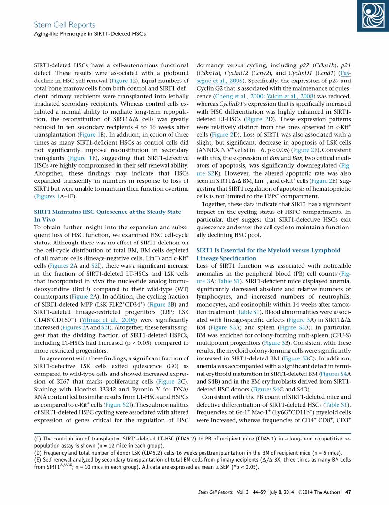
SIRT1-deleted HSCs have a cell-autonomous functional
defect. These results were associated with a profound
decline in HSC self-renewal (Figure 1E). Equal numbers of
total bone marrow cells from both control and SIRT1-defi-
cient primary recipients were transplanted into lethally
irradiated secondary recipients. Whereas control cells ex-
hibited a normal ability to mediate long-term repopula-
tion, the reconstitution of SIRT1D/D cells was greatly
reduced in ten secondary recipients 4 to 16 weeks after
transplantation (Figure 1E). In addition, injection of three
times as many SIRT1-deficient HSCs as control cells did
not significantly improve reconstitution in secondary
transplants (Figure 1E), suggesting that SIRT1-defective
HSCs are highly compromised in their self-renewal ability.
Altogether, these findings may indicate that HSCs
expanded transiently in numbers in response to loss of
SIRT1 but were unable to maintain their function overtime
(Figures 1A–1E).
SIRT1 Maintains HSC Quiescence at the Steady State
In Vivo
To obtain further insight into the expansion and subse-
quent loss of HSC function, we examined HSC cell-cycle
status. Although there was no effect of SIRT1 deletion on
the cell-cycle distribution of total BM, BM cells depleted
of all mature cells (lineage-negative cells, Lin�) and c-Kit+
cells (Figures 2A and S2I), there was a significant increase
in the fraction of SIRT1-deleted LT-HSCs and LSK cells
that incorporated in vivo the nucleotide analog bromo-
deoxyuridine (BrdU) compared to their wild-type (WT)
counterparts (Figure 2A). In addition, the cycling fraction
of SIRT1-deleted MPP (LSK FLK2+CD34+) (Figure 2B) and
SIRT1-deleted lineage-restricted progenitors (LRP; LSK
CD48+CD150�) (Yilmaz et al., 2006) were significantly
increased (Figures 2A and S2I). Altogether, these results sug-
gest that the dividing fraction of SIRT1-deleted HSPCs,
including LT-HSCs had increased (p < 0.05), compared to
more restricted progenitors.
In agreementwith these findings, a significant fraction of
SIRT1-defective LSK cells exited quiescence (G0) as
compared to wild-type cells and showed increased expres-
sion of KI67 that marks proliferating cells (Figure 2C).
Staining with Hoechst 33342 and Pyronin Y for DNA/
RNA content led to similar results from LT-HSCs andHSPCs
as compared to c-Kit+ cells (Figure S2J). These abnormalities
of SIRT1-deleted HSPC cycling were associated with altered
expression of genes critical for the regulation of HSC
(C) The contribution of transplanted SIRT1-deleted LT-HSC (CD45.2)population assay is shown (n = 12 mice in each group).(D) Frequency and total number of donor LSK (CD45.2) cells 16 week(E) Self-renewal analyzed by secondary transplantation of total BM cfrom SIRT1D/D3X; n = 10 mice in each group). All data are expressed
dormancy versus cycling, including p27 (Cdkn1b), p21
(Cdkn1a), CyclinG2 (Ccng2), and CyclinD1 (Ccnd1) (Pas-
segue et al., 2005). Specifically, the expression of p27 and
CyclinG2 that is associatedwith themaintenance of quies-
cence (Cheng et al., 2000; Yalcin et al., 2008) was reduced,
whereas CyclinD1’s expression that is specifically increased
with HSC differentiation was highly enhanced in SIRT1-
deleted LT-HSCs (Figure 2D). These expression patterns
were relatively distinct from the ones observed in c-Kit+
cells (Figure 2D). Loss of SIRT1 was also associated with a
slight, but significant, decrease in apoptosis of LSK cells
(ANNEXIN V+ cells) (n = 6, p < 0.05) (Figure 2E). Consistent
with this, the expression of Bim and Bax, two critical medi-
ators of apoptosis, was significantly downregulated (Fig-
ure S2K). However, the altered apoptotic rate was also
seen in SIRT1D/D BM, Lin�, and c-Kit+ cells (Figure 2E), sug-
gesting that SIRT1 regulation of apoptosis of hematopoietic
cells is not limited to the HSPC compartment.
Together, these data indicate that SIRT1 has a significant
impact on the cycling status of HSPC compartments. In
particular, they suggest that SIRT1-defective HSCs exit
quiescence and enter the cell cycle to maintain a function-
ally declining HSC pool.
SIRT1 Is Essential for the Myeloid versus Lymphoid
Lineage Specification
Loss of SIRT1 function was associated with noticeable
anomalies in the peripheral blood (PB) cell counts (Fig-
ure 3A; Table S1). SIRT1-deficient mice displayed anemia,
significantly decreased absolute and relative numbers of
lymphocytes, and increased numbers of neutrophils,
monocytes, and eosinophils within 14 weeks after tamox-
ifen treatment (Table S1). Blood abnormalities were associ-
ated with lineage-specific defects (Figure 3A) in SIRT1D/D
BM (Figure S3A) and spleen (Figure S3B). In particular,
BM was enriched for colony-forming unit-spleen (CFU-S)
multipotent progenitors (Figure 3B). Consistent with these
results, themyeloid colony-forming cells were significantly
increased in SIRT1-deleted BM (Figure S3C). In addition,
anemiawas accompaniedwith a significant defect in termi-
nal erythroid maturation in SIRT1-deleted BM (Figures S4A
and S4B) and in the BM erythroblasts derived from SIRT1-
deleted HSC donors (Figures S4C and S4D).
Consistent with the PB count of SIRT1-deleted mice and
defective differentiation of SIRT1-deleted HSCs (Table S1),
frequencies of Gr-1+ Mac-1+ (Ly6G+CD11b+) myeloid cells
were increased, whereas frequencies of CD4+ CD8+, CD3+
to PB of recipient mice (CD45.1) in a long-term competitive re-
s posttransplantation in the BM of recipient mice (n = 6 mice).ells from primary recipients (D/D 3X, three times as many BM cellsas mean ± SEM (*p < 0.05).
Stem Cell Reports j Vol. 3 j 44–59 j July 8, 2014 j ª2014 The Authors 47

Figure 2. Conditional Deletion of SIRT1 Compromises HSC Quiescence(A and B) Representative FACS plots (upper panel) and mean values (lower panel) of cell-cycle distribution of BrdU incorporation of LT-HSC, LSK, LSK CD48+CD150� (lineage-restricted progenitors, LRP; n = 3 mice) and c-Kit+ (n = 6 mice per genotype) (A) and of LSKFLK2+CD34+ MPP cells (n = 3 mice per genotype) (B).(C) Representative FACS plots (upper panel) and mean values (lower panel) of cell-cycle distribution by KI67/DAPI staining of LSK cells(n = 6 mice per genotype).
(legend continued on next page)
48 Stem Cell Reports j Vol. 3 j 44–59 j July 8, 2014 j ª2014 The Authors
Stem Cell ReportsAging-like Phenotype in SIRT1-Deleted HSCs

Stem Cell ReportsAging-like Phenotype in SIRT1-Deleted HSCs
T lymphocytes were reduced in the PB of both primary
recipients of SIRT1-deleted HSC donors (Figures 3C and
S5A) and of secondary recipients at 16 weeks posttrans-
plantation (Figures 3D and S5B). These results altogether
reflect cell-autonomous defects of HSC differentiation.
Similar abnormalities were found in the BM (Figure S5C)
and spleen (Figure S5D) of the primary recipients. To
further delineate whether the SIRT1-deleted T and B
lymphoid abnormalities were HSPC driven, we isolated
SIRT1-deficient LSK cells 4 weeks after tamoxifen injections
and measured their potential to produce lymphoid cells
when cultured on stromal OP9-DL1 and OP9 monolayers
that support T and B cell growth, respectively (Schmitt
and Zuniga-Pflucker, 2002; Vieira and Cumano, 2004).
The OP9 stromal cell line supports B cell growth under
defined in vitro condition (Vieira and Cumano, 2004),
whereas the OP9 line ectopically expressing the NOTCH
ligand Delta-like-1 protein supports the differentiation of
hematopoietic progenitors into T cells (Schmitt and Zu-
niga-Pflucker, 2002). In agreement with the in vivo find-
ings, the potential of SIRT1-deficient HSPCs to generate
T cells in vitro was significantly compromised without
noticeable effect on cell viability (Figures 3E, S6A, and
S6B; data not shown). Similarly, the B cell differentiation
potential was also impaired, albeit more mildly and with
a slightly different kinetic (Figures 3F, S6C, and S6D). These
combined findings strongly suggest that SIRT1-deleted
HSC differentiation is altered generating increasedmyeloid
and decreased lymphoid cells both in vivo and in vitro.
In support of this notion, SIRT1-deficient mice exhibited
splenomegaly (Figure 4A). The splenic architecture was
noticeably disorganized with regional disruption of the
white pulp and a concomitant increase in hematopoietic
elements in the red pulp that consisted mostly of myeloid
cells (Figure 4B). In contrast, the white pulp was composed
of a large number of pale, poorly defined lymphoid nodules
(Figure 4B). The total number of LSK cells in the spleen was
also increased (Figure 4C). Consistent with these results,
spleen histology revealed abundant extramedullary hema-
topoiesis associatedwith hemosiderin deposition in the red
pulp (Figure 4B).
In search for the source of lineage abnormalities, we
examined the hematopoietic progenitor compartment.
Unexpectedly, the myeloid-biased HSC differentiation
observed in SIRT1-deficient hematopoietic organs, was
associated with a significant and specific increase in the
GMP compartment in the BM detectable 10 weeks after
(D) qRT-PCR analysis of cell-cycle regulators. Results are relative tindependent experiments each based on three replicates of one pool(E) Representative FACS plots (left panel, LSK) and frequency (right pper genotype). In all experiments, SIRT1-deleted cells were isolated 4(*p < 0.05).
tamoxifen treatment, whereas the size of commonmyeloid
progenitor (CMP) and megakaryocyte/erythroid progeni-
tor (MEP) compartments did not change (Figures 5A and
5B). These surprising alterations were further associated
with a progressive 2-fold decrease in the frequency of CLP
overtime (Figure 5B). Remarkably, the mild but specific
enhanced production of GMP associated with anemia
and decreased CLP characterizes the age-associated alter-
ations of HSC differentiation (Rossi et al., 2005, 2007a).
Hematopoietic lineage abnormalities of SIRT1-deleted
HSCs (Figures 3, 4, and 5; Figures S3–S6; Table S1)were asso-
ciated with alterations in the expression of several tran-
scription factors implicated in the generation of GMPs,
including C/EBPa, PU.1, GATA-1, and GATA-2 (Iwasaki
et al., 2006) (Figure 5C). Similarly, expression of lymphoid
specification transcription factors IKAROS andGATA-3 was
significantly reduced in SIRT1-deleted c-Kit+ multipotent
progenitors (Figure 5C). Key genes (Rothenberg and
Hogan, 2006) associated with eosinophil production,
including Gata2, Interleukin 5 receptor a (Il5ra), andmajor
basic protein 1 (Mbp1), were abnormally expressed in BM
myeloid progenitors (Figure 5D). Interestingly, Il5ra and
Mbp1 were highly upregulated in MEPs, where these genes
are normally undetectable (Figure 5D). Collectively, these
findings suggest that SIRT1 has a critical function in HSC
lineage specification, in particular in the control ofmyeloid
versus lymphoid lineage commitment.
Young SIRT1-Deleted HSCs Recapitulate the Main
Features of Aged HSCs
We further examined whether SIRT1-deleted HSCs exhibit
age-associated features. DNA damage is chief among mo-
lecular processes leading to cellular aging, including aging
of HSCs (Rossi et al., 2007b). Freshly isolated SIRT1-defi-
cient HSPCs exhibited accumulation of damaged DNA
overtime as indicated by increased phosphorylation of
histone H2AX that marks the response to DNA damage
(Figure 6A). These abnormalities were associated with
increased length of comets generated by DNA breaks in
gel electrophoresis (Figure 6B), suggesting that loss of
SIRT1 results in relative loss of DNA repair potential of
HSPCs. Accumulation of reactive oxygen species (ROS)
has been associated with an aging phenotype in many
cell types (Dykstra and de Haan, 2008). SIRT1-deficient
HSPCs exhibited increased levels of ROS measured by
flow cytometry in freshly isolated cells using the oxidative
stress-sensitive CM-H2DCFDA probe (Figure 6C). ROS
o Sirt1fl/fl set at one in each population (results are from threeof three mice).anel) of apoptotic cells (ANNEXIN V+ 7AAD-) in the BM (n = 6 miceweeks after tamox treatment. All data are expressed as mean ± SEM
Stem Cell Reports j Vol. 3 j 44–59 j July 8, 2014 j ª2014 The Authors 49

Figure 3. SIRT1-Deleted HSCs Generate In Vivo Myeloid-Biased and Lymphoid-Defective Progenies(A) Impact of SIRT1 deficiency on the hematopoietic lineages is shown. Green and red arrows indicate increased and decreased cellpopulations, respectively.(B) Representative spleens with CFU-S-derived colonies (upper panel) and mean values of BM CFU-Sd12 frequency (lower panel). Onerepresentative of three independent experiments (n = 5 mice in each group) is shown.(C) Frequency of donor (CD45.2+)-derived multilineage mature cells in the PB of primary transplants (n = 4), at indicated time pointsposttransplantation.
(legend continued on next page)
50 Stem Cell Reports j Vol. 3 j 44–59 j July 8, 2014 j ª2014 The Authors
Stem Cell ReportsAging-like Phenotype in SIRT1-Deleted HSCs

Figure 4. Extramedullary Hematopoiesis in SIRT1-Deleted Mice(A) Representative spleens (left panel) and mean values of cellularity (mid panel) and weight (right panel), 4 weeks after tamox treatmentare shown (n = 6 mice per genotype).(B) Representative hematoxylin and eosin (H&E) staining of paraffin-embedded sections of spleen 8 weeks after tamox treatment atdistinct magnifications. Stars and arrows indicate the red and white pulps, respectively. Scale bar, 200 mm (upper panel). Stars and arrowsshow myeloid cell increase and hemosiderin deposition in the red pulp, respectively. Scale bar, 20 mm (lower panel).(C) Total number of LSK cells in the spleen 4 weeks after tamox treatment (n = 6 mice in each group). All data are expressed as mean ± SEM(*p < 0.05).
Stem Cell ReportsAging-like Phenotype in SIRT1-Deleted HSCs
accumulation was associated with decrease of several anti-
oxidant transcripts (Figure 6D) 4 weeks after tamoxifen
treatment, indicating that SIRT1 is required for themainte-
nance of HSPC redox homeostasis. We also examined the
expression of a number of molecules that are modulated
with age in HSCs. Among these, the levels of LT-HSC
expression of integrin aIIb (itga2b, CD41), a classic platelet
marker, and the CD150 Slam protein that marks LT-HSCs
increase with age (Beerman et al., 2010; Gekas and Graf,
2013). Similarly, CD41 was highly upregulated on SIRT1-
deleted LT-HSCs (Figure 6E). The frequency of SIRT1-
deleted LT-HSC subset expressing CD41 also increased to
(D) Frequency of donor (CD45.2+)-derived multilineage mature cells i(E and F) Representative FACS plots of T (E) and B (F) cell differentiastromal lines. Frequencies of T cells within the CD4� and CD8� doublwithin CD45-positive cells (F, right panel) are shown; one representatischematic progression of the T and B cell differentiation is shown (sa(*p < 0.05).
79.4% ± 2.3% from 64.4% ± 3.2% on wild-type controls
(p < 0.011, n = 3 mice) (Figure 6E). Notably, the increase
of CD41 levels was the highest on CD150high-expressing
LSK cells (Figure S7A), further supporting the aging-like
phenotype of SIRT1-deleted HSCs. Like on old (16months)
wild-type LSK cells, the expression of P-SELECTIN protein
(Chambers et al., 2007; Rossi et al., 2005) was increased
on the surface of young SIRT1D/D LSK cells (Figure 6F).
Expression of several other genes also highly modulated
with age in HSCs, including Sox4, Fos, and Pml (Rossi
et al., 2005), was similarly altered in young SIRT1-deficient
HSCs and old controls (Figure 6G). In line with a potential
n the PB of secondary transplants (n = 5).tion of LSK cells cultured in vitro for 16 days on OP9-DL1 and OP9e-negative (DN) cells positive for CD45 (E, right panel) and B cellsve of two independent experiments (n = 3 technical replicates). Theme as the third panel of Figure S6). Data expressed as mean ± SEM
Stem Cell Reports j Vol. 3 j 44–59 j July 8, 2014 j ª2014 The Authors 51

Figure 5. SIRT1-Deleted HSPCs Are Altered in Their Lineage Specification(A) Representative FACS plots of GMP, CMP, and MEP 10 weeks after tamox treatment.(B) GMP, CMP, MEP, and CLP frequencies in the BM after tamox treatment (n = 6 mice per group).(C) qRT-PCR analysis 10 weeks (20 weeks in the case of IKAROS and GATA-3) after tamox treatment. Results are relative to Sirt1fl/fl (n = 6from two independent experiments). nd, not done.(D) Gene expression analysis of eosinophil markers in GMP, CMP, and MEP by qRT-PCR (normalized to b-actin) 14 weeks aftertamox treatment. Results are relative to Sirt1fl/fl c-Kit+ (n = 3 replicates from one pool of three mice). All data are expressed as mean ± SEM(*p < 0.05).
Stem Cell ReportsAging-like Phenotype in SIRT1-Deleted HSCs
contribution of defective SIRT1 to HSC aging, expression of
SIRT1 in the old versus young HSPCs was reduced 2-fold
(Figure 6H).
One of the main features of aging is a declining immune
system associated with impairment in T cell production
52 Stem Cell Reports j Vol. 3 j 44–59 j July 8, 2014 j ª2014 The Authors
and function (Linton and Dorshkind, 2004; Rossi et al.,
2007a). We found that despite the significant decrease in
the CD4+ T cell population (Figures 2 and 3; Figures S3,
S5, and S7A), the total number of CD4+ CD25+ FOXP3+ reg-
ulatory T cells (Treg cells) remained unaltered by the loss of
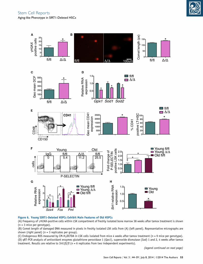
Figure 6. Young SIRT1-Deleted HSPCs Exhibit Main Features of Old HSPCs(A) Frequency of gH2AX-positive cells within LSK compartment of freshly isolated bone marrow 38 weeks after tamox treatment is shown(n = 3 mice per genotype).(B) Comet length of damaged DNA measured in pixels in freshly isolated LSK cells from (A) (left panel). Representative micrographs areshown (right panel) (n = 3 replicates per group).(C) Endogenous ROS measured by CM-H2DCFDA in LSK cells isolated from mice 4 weeks after tamox treatment (n = 9 mice per genotype).(D) qRT-PCR analysis of antioxidant enzymes glutathione peroxidase 1 (Gpx1), superoxide dismutase (Sod) 1 and 2, 4 weeks after tamoxtreatment. Results are relative to Sirt1fl/fl (n = 6 replicates from two independent experiments).
(legend continued on next page)
Stem Cell Reports j Vol. 3 j 44–59 j July 8, 2014 j ª2014 The Authors 53
Stem Cell ReportsAging-like Phenotype in SIRT1-Deleted HSCs

Stem Cell ReportsAging-like Phenotype in SIRT1-Deleted HSCs
SIRT1 (Figure S7B), indicating that the frequency of T regs
was increased in the peripheral blood of SIRT1-deleted
mice (Figures S7C and S7D). Similar alterations are
observed in some of the disorders of aging (Kordasti et al.,
2007).
Collectively, these findings suggest that loss of SIRT1
leads to disparate and considerable changes inHSCmainte-
nance and lineage specification and may promote an
aging-like phenotype in young HSPCs.
FOXO3 Is Required for SIRT1 Regulation of HSC
Activity
SIRT1’s impact on HSCs may be via several mediators. One
of the potential mechanisms through which SIRT1 may
contribute to the HSC phenotype is by deacetylation of a
number of targets, including FOXO3, p53, and HIF1a
(Haigis and Sinclair, 2010). Among these, we focused on
FOXO3 as the hematopoietic phenotypes of SIRT1D/D
and Foxo3�/� mice are similar in many respects (Figures 1,
2, 3, 4, 5, and 6; Figures S3, S5, S7E, and S7F), specifically
in the HSC and myeloid compartments, despite condi-
tional versus germline deletion of Sirt1 versus Foxo3, respec-
tively (Hedrick et al., 2012; Marinkovic et al., 2007; Miya-
moto et al., 2007; Yalcin et al., 2008, 2010). Furthermore,
although FOXO3 is a known SIRT1 substrate (Brunet
et al., 2004; Motta et al., 2004), the outcome of SIRT1
deacetylation of FOXO3 is likely context dependent and
unknown in HSCs. Lastly, FOXO3, like SIRT1, is an evolu-
tionarily conserved regulator of organismal longevity (Eij-
kelenboom and Burgering, 2013; Zhang et al., 2011a).
We reasoned that if FOXO3 was the mediator of SIRT1’s
impact on HSCs, then inhibition of SIRT1 would not affect
significantly the Foxo3�/� HSC compartment. Whereas
sirtinol inhibition of SIRT1 in vivo compromised the fre-
quency of LSK, LT-HSCs, and the capacity for long-term
competitive repopulation in WT mice (Figures 7A and 7B)
as we had previously observed (Figures S1D–S1G), adminis-
tration of sirtinol to Foxo3�/� mice did not affect their LSK
or LT-HSC compartments (Figure 7A) or the competitive
repopulation ability of Foxo3�/� HSCs (Figure 7B), suggest-
ing that FOXO3 is required for the inhibitory effects of
sirtinol on HSCs.
Similarly, we hypothesized that if FOXO3 is a SIRT1 sub-
strate in HSPCs, then mice lacking both FOXO3 and SIRT1
in HSPCs should exhibit a phenotype similar to that
(E) Representative FACS plots (left panel) of levels of CD41 on LSK CDexpressing CD41 (right panel) measured 4 weeks after tamox treatme(F) Representative FACS plots (left panel) of P-SELECTIN expression inFold change of P-SELECTIN-positive LSK cells (right panel) normalize(G) qRT-PCR analysis 14 weeks after tamox treatment. Results are relati(H) qRT-PCR analysis of young (12-week-old) and old (16-months-old)three independent experiments). All data are expressed as mean ± SE
54 Stem Cell Reports j Vol. 3 j 44–59 j July 8, 2014 j ª2014 The Authors
observed in FOXO3-deficient HSPCs. Indeed, Foxo3�/�/Sirt1D/D and Foxo3�/� HSPC phenotypes were remarkably
similar in that they were both reduced in numbers (Fig-
ure 7C) and in the quiescent fraction (Figure 7D), further
suggesting that SIRT1 is an upstream activator of FOXO3
in HSPCs. We next asked whether SIRT1 deacetylation
directly modulates FOXO3’s activity in HSPCs. In addition
to acetylation/deacetylation, phosphorylation by AKT
kinase represses FOXO3’s function by promoting its nu-
clear exit (Eijkelenboom and Burgering, 2013; Zhang
et al., 2011a). However in HSPCs, FOXO3 is mostly in the
nucleus even when pAKT is constitutively active (Lee
et al., 2010; Yalcin et al., 2008) (data not shown), suggest-
ing that additional mechanisms are involved. In contrast
to wild-type cells, FOXO3’s localization in SIRT1-deficient
HSPC nuclei was significantly reduced (Figure 7E).
FOXO3’s transcriptional activity was also decreased in
SIRT1-deficient HSPCs as illustrated by reduced expression
of a number of FOXO3’s direct targets, including p27
(Cdkn1b), Bnip3, CyclinG2 (Ccng2), Sod2, and Bim, and by
modulation of CyclinD1 (CCND1) (Figures 2D, 6D, and
S7F). In agreement with these results, the nuclear localiza-
tion of an ectopically expressed FOXO3 that is mutated to
mimic constitutive acetylation (FOXO3 5KQ, 5 lysine resi-
dues mutated to glutamine) was significantly reduced in
BM mononuclear cells (Figure 7F).
These combined results strongly suggest that FOXO3 is a
substrate of SIRT1 in HSCs and that SIRT1 deacetylation is
key in promoting FOXO3’s retention in HSC nuclei and
maintaining FOXO3 in an active form in these cells. They
also support the notion that reduced FOXO3 proapoptotic
function (Figures 2E and S2K) may protect SIRT1-deleted
HSCs from apoptosis despite elevated ROS (Figures 2E,
6C, and 6D). Collectively, these findings indicate that
SIRT1-FOXO3 constitutes a regulatory pathway controlling
HSC maintenance.
DISCUSSION
SIRT1 Is a Critical Regulator of Homeostatic Adult
HSCs with Potential Functions in Delaying the HSC
Aging
Our key finding is that SIRT1-deficient HSCs recapitulate
within a temporal window some of the main features of
48�CD150+ (middle panel) and frequency of LSK CD48�CD150+ cellsnt (n = 3 mice per genotype).LSK cells 6 weeks after tamox treatment, and in 16 months old LSK.d to young Sirt1fl/fl control (n = 6 mice).ve to Sirt1fl/fl (n = 6 replicates from two independent experiments).WT LSK cells. Results are relative to young LSK (n = 9 replicates fromM (*p < 0.05).

Figure 7. SIRT1 Controls Hematopoietic Homeostasis by Promoting FOXO3 Nuclear Localization and Activity in HSCs(A) Total number of BM LSK (left panel) cells and LT-HSC (right panel) isolated from WT and Foxo3�/� mice treated with Sirtinol (Sir) orvehicle control (Ct) for 3 weeks (n = 3 mice in each group).(B) Long-term competitive repopulation of 100 transplanted HSC (from A) as measured by the percent of CD45.1 in the PB of recipient mice16 weeks after transplantation (n = 5 mice in each group).(C) Total number of LSK cells isolated from BM of indicated mice (n = 3 mice in each group).(D) LSK cell-cycle distribution of mice from (C) measured by KI67/DAPI staining (n = 3 mice per genotype).(E) FOXO3 immunostaining (left panel) and quantification (right panel) measuring the cell plot profile are shown.
(legend continued on next page)
Stem Cell Reports j Vol. 3 j 44–59 j July 8, 2014 j ª2014 The Authors 55
Stem Cell ReportsAging-like Phenotype in SIRT1-Deleted HSCs
Stem Cell ReportsAging-like Phenotype in SIRT1-Deleted HSCs
HSC aging. Specifically, the expansion of HSC numbers,
albeit transient, followed by depletion overtime of HSC
function overlaps with aged associated HSC defects. These
abnormalities combined with anemia, myeloid skewed
HSC differentiation, enhanced specific production of
GMPs and immune deficiency, enhanced oxidative stress,
greater DNA damage, and age-related modulations of
gene expression suggest that young SIRT1-deleted HSCs
may exhibit a premature aging phenotype (Chambers
et al., 2007; Morrison et al., 1996; Sudo et al., 2000; Yilmaz
et al., 2006). A greater fraction of HSCs in old BM is cycling,
although this increased cycling may only be observed in
mice older than 22–24 months (Dykstra and de Haan,
2008; Morrison et al., 1996; Yilmaz et al., 2006). In agree-
ment with an aging-like HSC phenotype, a significant frac-
tion of SIRT1-deficient HSCs exited quiescence and entered
the cell cycle (Figures 2A–2C; Figures S2I and S2J). None-
theless, SIRT1-deleted HSCs do not phenocopy aged
HSCs, as the MPP subset is increased in SIRT1-deleted
HSPCs in contrast to aged HSPCs (Rossi et al., 2005,
2007a). Although the defects of SIRT1-deleted HSCs are
cell autonomous, our studies do not rule out potential
participation of non-cell-autonomous mechanisms. This
work suggests a model in which SIRT1 wires together a
combinatorial transcriptional program in HSPCs. Overall,
our findings predict that a decline in SIRT1 function, as it
may occur with age (Figures 6H and 7G), would result in
disruption (or destabilization) of SIRT1-regulated HSPC
transcriptional program.
Recent studies suggest that aging changes the clonal
composition of HSC compartment rather than their
intrinsic properties (Dykstra and de Haan, 2008). While
our findings are in agreement with a potential SIRT1 re-
pression of myeloid biased HSC clones that dominate
with age, they also suggest that SIRT1 may have additional
protective functions toward key HSC programs that
become compromised with age, such as the capacity to
repair damaged DNA.
The question of whether SIRT1 has any functions in
normal adult HSC has long been debated. Depending on
their strain, a significant proportion of germline-deleted
SIRT1 mice die perinatally, resulting in only a fraction of
mice surviving to adulthood. SIRT1 was dispensable for
the HSC activity, perhaps due to developmental adaptation
of HSCs in the surviving mice (Leko et al., 2012; Li et al.,
2012; Narala et al., 2008; Yuan et al., 2012). On the other
hand, using a tamoxifen-inducible conditional deletion
approach, it was shown recently that SIRT1 is required for
(F) The mean fluorescence of nuclear cytoplasmic ratio (lower panel)expressing Flag-FOXO3-WT or Flag-FOXO3-5KQ, immunostained with a(G) Model of SIRT1/FOXO3 regulation of HSC maintenance and lineageSIRT1 function contributes to the HSC aging. All data are expressed a
56 Stem Cell Reports j Vol. 3 j 44–59 j July 8, 2014 j ª2014 The Authors
HSPC genome stability under stress (Singh et al., 2013).
We demonstrated that SIRT1 is essential for HSC function
and lineage specification under homeostatic conditions
even when BM is not stressed (Figures 1 and S2).
SIRT1 Regulates Hematopoietic Stem Cell Lineage
Decision
One of the most unexpected findings was the skewed gen-
eration of myeloid lineage at the expense of the lymphoid
compartment uncovered in young SIRT1-deleted HSCs.
The propensity toward myeloid differentiation associated
with immune defects is one of the hallmarks of HSC aging
in both mouse and human (Linton and Dorshkind, 2004;
Pang et al., 2011; Rossi et al., 2005; Sudo et al., 2000).
The specific increased generation of GMPs (Figures 5A
and 5B) was greatly similar to the mild specific increase in
GMPs produced in old BM (Rossi et al., 2005, 2007a). The
shift in the lympho-myeloid cell ratio in the BM, spleen,
and peripheral blood of SIRT1-deficient mice (Figures 3
and 4; Figures S3 and S5; Table S1) that became highly pro-
nounced overtime underscores the function of SIRT1 in
balancing the generation of hematopoietic lineages from
HSCs.
The increased expression of GATA-1 and GATA-2 in
SIRT1-deleted hematopoietic progenitors (Figures 5C and
5D) is in agreement with the function of these factors in
GMPs and eosinophil production (Hirasawa et al., 2002;
Iwasaki et al., 2006). The reduced expression of myeloid
transcription factors C/EBPa and PU.1, specifically in
multipotential progenitors (Figure 5C), was unexpected.
These results may indicate that relative expression of
myeloid transcription factors, as is the case for their order
of expression (Iwasaki et al., 2006), may influence lineage
specification.
SIRT1 Maintains HSC Homeostasis by Promoting
FOXO3 Nuclear Localization and Activation
We identified FOXO3 as a SIRT1 substrate in HSCs that
mediates SIRT1’s effects on HSCs. FOXO3 like SIRT1 is
implicated in the regulation of mouse ESC pluripotency
(Han et al., 2008; Zhang et al., 2011b). Similarly, FOXO3
is required for the maintenance of HSC pool (Miyamoto
et al., 2007, 2008; Yalcin et al., 2008). FOXO3mutant mye-
loproliferation and decreased B and red blood cells (Hin-
man et al., 2009; Marinkovic et al., 2007; Miyamoto
et al., 2007; Yalcin et al., 2008, 2010) (data not shown)
are similarly observed in SIRT1-deficient mice. Moreover,
loss of FOXO3 like (Figure S7E) SIRT1 (Figure 5B) leads
of 50 GFP-sorted mouse bone marrow mononuclear cells retrovirallynti-Flag antibody (upper panel).specification. The model raises the question as to whether reduceds mean ± SEM (*p < 0.05, ns, not significant).
Stem Cell ReportsAging-like Phenotype in SIRT1-Deleted HSCs
specifically to enhanced generation of GMPs, altogether
indicating that SIRT1 regulation of FOXO3might be impli-
cated in myeloid and B cell lineage determination. The
impact of SIRT1 (Figures 3A and 3C–3F; Figures S3, S5,
and S6; Table S1) and FOXO3 on the T cell compartment
however seems distinct (Hedrick et al., 2012). Interestingly,
SIRT1 like (Li et al., 2012; Yuan et al., 2012) FOXO3 (Ghaf-
fari et al., 2003; Naka et al., 2010; Sykes et al., 2011) is impli-
cated in leukemogenesis and required for the maintenance
of leukemic stem cells.
Our studies combined with recent findings regarding
SIRT3 (Brown et al., 2013) further implicate sirtuins in
protecting HSCs from aging. Future investigations in the
potential involvement of SIRT1/FOXO3 (Mouchiroud
et al., 2013) in stem cell programs other than HSCs,
including neural stem cells (Renault et al., 2009), should
clarify whether and to what extent stem cells rely on this
network for their maintenance over a lifetime.
EXPERIMENTAL PROCEDURES
MiceThe generation and genotyping of mice were performed as previ-
ously described (Price et al., 2012). SIRT1Dex4 (C57BL6, CD45.2)
were crossed to Cre-ERT2 mice to generate SIRT1Dex4-ERT2
mice. The Cre induction was performed by delivering tamoxifen
intraperitoneally (1 mg/mouse/day) (Sigma T5648) for 5 consecu-
tive days to SIRT1Dex4-ERT2 mice (designated D/D). This regimen
resulted in deletion of SIRT1 exon 4 and a truncated SIRT1 protein
that lacked its catalytic domain. Control mice, including animals
with a wild-type Sirt1 allele and Ert2 (Sirt1WT/WT), and flox-
SIRT1Dex4 mice lacking Ert2 (Sirt1fl/fl) were used. The efficient
deletion after tamoxifen treatment was confirmed by PCR analysis
of genomic tail DNA. Foxo3�/�mice, a gift of Dr. RonDepinho (MD
Anderson Cancer Center), were backcrossed ten generations
onto C57BL6 (CD45.1) background (Yalcin et al., 2008, 2010).
Because of the proximity of Foxo3 and Sirt1 genes on mouse
chromosome 10, the following strategy was devised to generate
Sirt1D/DFoxo3�/� mice (designated D/DFoxo3�/�): Sirt1fl/fl cre+
Foxo3WT were crossed with Sirt1WT/WT cre� Foxo3�/� mice. The
F1 population Sirt1fl/WT cre+ Foxo3+/� were intercrossed to obtain
Sirt1fl/fl cre+ Foxo3+/� mice. The F2 population Sirt1fl/fl cre+
Foxo3+/� mice were intercrossed to obtain Sirt1fl/fl cre+ Foxo3�/�
mice. Young mice in all experiments were 10–12 weeks old. Mice
were used in accordance with the protocols approved by the Insti-
tutional Animal Care and Use Committee of Icahn School of Med-
icine at Mount Sinai.
Long-Term Repopulation AssayLethally irradiated (12 Gy as a split dose, 6.5 and 5.5 Gy, 4–5 hr
apart) congenic C57BL6-CD45.1 mice (from the National Cancer
Institute) were reconstituted with intravenous injections of 100
donor LSK CD48�CD150+ cells from Sirt1WT/WT (Cre+), Sirt1fl/fl
(Cre�), or SIRT1D/D mice (Cre+) (all CD45.2) 4 weeks after tamox-
ifen treatment along with 2 3 105 competitor bone marrow cells
(CD45.1). For secondary transplantations, an equal number (2 3
106) of total bone marrow cells from primary recipients or three
times more (6 3 106) from SIRT1D/D LSK CD48�CD150+ were
pooled and transplanted into lethally irradiated CD45.1 secondary
recipients. A long-term repopulation assay of Foxo3�/� (CD45.1)
mice was performed by transplantation into lethally irradiated
CD45.2 recipients.
SUPPLEMENTAL INFORMATION
Supplemental Information includes Supplemental Experimental
Procedures, seven figures, and two tables and can be found
with this article online at http://dx.doi.org/10.1016/j.stemcr.
2014.04.015.
ACKNOWLEDGMENTS
We thank Valentina D’Escamard for her technical help, Dr. Kateri
Moore (Mount Sinai) for critically reading the manuscript, and
The Flow Cytometry Shared Research Facility and the Histology
Facility at Icahn School of Medicine at Mount Sinai. B.I. was
partially supported by Institut National de la Sante et de la
Recherche Medicale, and R.D.-G. was supported by the Spanish
Instituto de Salud Carlos III (Sara Borrell CD09/00014). R.L. was
partially supported by grants from the NIH (T32 GM08553-13
and T32 HD075735). This work was supported in part by NIH
grants (RO1 DK077174 and RO1 RHL116365A; S.G., Co-PI), a
New York State Stem Cell Science (NYSTEM) award (CO24408), a
Myeloproliferative Neoplasm Foundation (MPN) award and an
Irma Hirschl/Weill-Caulier Trust Research award to S.G.
Received: December 6, 2013
Revised: April 24, 2014
Accepted: April 25, 2014
Published: June 5, 2014
REFERENCES
Beerman, I., Bhattacharya, D., Zandi, S., Sigvardsson, M., Weiss-
man, I.L., Bryder, D., and Rossi, D.J. (2010). Functionally distinct
hematopoietic stem cells modulate hematopoietic lineage poten-
tial during aging by a mechanism of clonal expansion. Proc.
Natl. Acad. Sci. USA 107, 5465–5470.
Brown, K., Xie, S., Qiu, X., Mohrin, M., Shin, J., Liu, Y., Zhang, D.,
Scadden, D.T., and Chen, D. (2013). SIRT3 reverses aging-associ-
ated degeneration. Cell Rep 3, 319–327.
Brunet, A., Sweeney, L.B., Sturgill, J.F., Chua, K.F., Greer, P.L., Lin,
Y., Tran, H., Ross, S.E., Mostoslavsky, R., Cohen, H.Y., et al.
(2004). Stress-dependent regulation of FOXO transcription factors
by the SIRT1 deacetylase. Science 303, 2011–2015.
Challen, G.A., Boles, N.C., Chambers, S.M., and Goodell, M.A.
(2010). Distinct hematopoietic stem cell subtypes are differentially
regulated by TGF-beta1. Cell Stem Cell 6, 265–278.
Chambers, S.M., Shaw, C.A., Gatza, C., Fisk, C.J., Donehower, L.A.,
and Goodell, M.A. (2007). Aging hematopoietic stem cells decline
in function and exhibit epigenetic dysregulation. PLoS Biol. 5,
e201.
Stem Cell Reports j Vol. 3 j 44–59 j July 8, 2014 j ª2014 The Authors 57
Stem Cell ReportsAging-like Phenotype in SIRT1-Deleted HSCs
Cheng, T., Rodrigues, N., Dombkowski, D., Stier, S., and Scadden,
D.T. (2000). Stem cell repopulation efficiency but not pool size is
governed by p27(kip1). Nat. Med. 6, 1235–1240.
Cheng, H.L., Mostoslavsky, R., Saito, S., Manis, J.P., Gu, Y., Patel, P.,
Bronson, R., Appella, E., Alt, F.W., and Chua, K.F. (2003). Develop-
mental defects and p53 hyperacetylation in Sir2 homolog (SIRT1)-
deficient mice. Proc. Natl. Acad. Sci. USA 100, 10794–10799.
Deneault, E., Cellot, S., Faubert, A., Laverdure, J.P., Frechette, M.,
Chagraoui, J., Mayotte, N., Sauvageau, M., Ting, S.B., and Sauva-
geau, G. (2009). A functional screen to identify novel effectors of
hematopoietic stem cell activity. Cell 137, 369–379.
Dykstra, B., and de Haan, G. (2008). Hematopoietic stem cell aging
and self-renewal. Cell Tissue Res. 331, 91–101.
Dykstra, B., Olthof, S., Schreuder, J., Ritsema, M., and de Haan, G.
(2011). Clonal analysis reveals multiple functional defects of aged
murine hematopoietic stem cells. J. Exp. Med. 208, 2691–2703.
Eijkelenboom, A., and Burgering, B.M. (2013). FOXOs: signalling
integrators for homeostasis maintenance. Nat. Rev. Mol. Cell
Biol. 14, 83–97.
Florian, M.C., Dorr, K., Niebel, A., Daria, D., Schrezenmeier, H.,
Rojewski, M., Filippi, M.D., Hasenberg, A., Gunzer, M., Scharffet-
ter-Kochanek, K., et al. (2012). Cdc42 activity regulates hematopoi-
etic stem cell aging and rejuvenation. Cell Stem Cell 10, 520–530.
Gekas, C., and Graf, T. (2013). CD41 expression marks myeloid-
biased adult hematopoietic stem cells and increases with age.
Blood 121, 4463–4472.
Ghaffari, S., Jagani, Z., Kitidis, C., Lodish, H.F., and Khosravi-Far, R.
(2003). Cytokines and BCR-ABL mediate suppression of TRAIL-
induced apoptosis through inhibition of forkhead FOXO3a tran-
scription factor. Proc. Natl. Acad. Sci. USA 100, 6523–6528.
Grozinger, C.M., Chao, E.D., Blackwell, H.E., Moazed, D., and
Schreiber, S.L. (2001). Identification of a class of small molecule
inhibitors of the sirtuin family of NAD-dependent deacetylases
by phenotypic screening. J. Biol. Chem. 276, 38837–38843.
Guarente, L. (2011). Franklin H. Epstein Lecture: Sirtuins, aging,
and medicine. N. Engl. J. Med. 364, 2235–2244.
Haigis, M.C., and Sinclair, D.A. (2010). Mammalian sirtuins:
biological insights and disease relevance. Annu. Rev. Pathol. 5,
253–295.
Han, M.K., Song, E.K., Guo, Y., Ou, X., Mantel, C., and Broxmeyer,
H.E. (2008). SIRT1 regulates apoptosis and Nanog expression in
mouse embryonic stem cells by controlling p53 subcellular locali-
zation. Cell Stem Cell 2, 241–251.
Harrison, D.E. (1983). Long-term erythropoietic repopulating abil-
ity of old, young, and fetal stem cells. J. Exp.Med. 157, 1496–1504.
Hedrick, S.M., Hess Michelini, R., Doedens, A.L., Goldrath, A.W.,
and Stone, E.L. (2012). FOXO transcription factors throughout
T cell biology. Nat. Rev. Immunol. 12, 649–661.
Hinman, R.M., Nichols, W.A., Diaz, T.M., Gallardo, T.D., Castril-
lon, D.H., and Satterthwaite, A.B. (2009). Foxo3-/- mice demon-
strate reduced numbers of pre-B and recirculating B cells but
normal splenic B cell sub-population distribution. Int. Immunol.
21, 831–842.
58 Stem Cell Reports j Vol. 3 j 44–59 j July 8, 2014 j ª2014 The Authors
Hirasawa, R., Shimizu, R., Takahashi, S., Osawa, M., Takayanagi, S.,
Kato, Y., Onodera, M., Minegishi, N., Yamamoto, M., Fukao, K.,
et al. (2002). Essential and instructive roles of GATA factors in
eosinophil development. J. Exp. Med. 195, 1379–1386.
Iwasaki, H., Mizuno, S., Arinobu, Y., Ozawa, H., Mori, Y., Shige-
matsu, H., Takatsu, K., Tenen, D.G., and Akashi, K. (2006). The
order of expression of transcription factors directs hierarchical
specification of hematopoietic lineages. Genes Dev. 20, 3010–
3021.
Kordasti, S.Y., Ingram,W., Hayden, J., Darling, D., Barber, L., Afzali,
B., Lombardi, G., Wlodarski, M.W., Maciejewski, J.P., Farzaneh, F.,
and Mufti, G.J. (2007). CD4+CD25high Foxp3+ regulatory T cells
in myelodysplastic syndrome (MDS). Blood 110, 847–850.
Lee, J.Y., Nakada, D., Yilmaz, O.H., Tothova, Z., Joseph, N.M., Lim,
M.S., Gilliland, D.G., and Morrison, S.J. (2010). mTOR activation
induces tumor suppressors that inhibit leukemogenesis and
deplete hematopoietic stem cells after Pten deletion. Cell Stem
Cell 7, 593–605.
Leko, V., Varnum-Finney, B., Li, H., Gu, Y., Flowers, D., Nourigat,
C., Bernstein, I.D., and Bedalov, A. (2012). SIRT1 is dispensable
for function of hematopoietic stem cells in adult mice. Blood
119, 1856–1860.
Li, L., Wang, L., Li, L., Wang, Z., Ho, Y., McDonald, T., Holyoake,
T.L., Chen, W., and Bhatia, R. (2012). Activation of p53 by SIRT1
inhibition enhances elimination of CML leukemia stem cells in
combination with imatinib. Cancer Cell 21, 266–281.
Linton, P.J., and Dorshkind, K. (2004). Age-related changes in
lymphocyte development and function. Nat. Immunol. 5,
133–139.
Liu, L., and Rando, T.A. (2011). Manifestations andmechanisms of
stem cell aging. J. Cell Biol. 193, 257–266.
Marinkovic, D., Zhang, X., Yalcin, S., Luciano, J.P., Brugnara, C.,
Huber, T., and Ghaffari, S. (2007). Foxo3 is required for the regula-
tion of oxidative stress in erythropoiesis. J. Clin. Invest. 117, 2133–
2144.
McBurney, M.W., Yang, X., Jardine, K., Hixon, M., Boekelheide, K.,
Webb, J.R., Lansdorp, P.M., and Lemieux, M. (2003). The mamma-
lian SIR2alpha protein has a role in embryogenesis and gameto-
genesis. Mol. Cell. Biol. 23, 38–54.
Miyamoto, K., Araki, K.Y., Naka, K., Arai, F., Takubo, K., Yamazaki,
S., Matsuoka, S., Miyamoto, T., Ito, K., Ohmura, M., et al. (2007).
Foxo3a is essential for maintenance of the hematopoietic stem
cell pool. Cell Stem Cell 1, 101–112.
Miyamoto, K., Miyamoto, T., Kato, R., Yoshimura, A., Motoyama,
N., and Suda, T. (2008). FoxO3a regulates hematopoietic homeo-
stasis through a negative feedback pathway in conditions of stress
or aging. Blood 112, 4485–4493.
Morrison, S.J., Wandycz, A.M., Akashi, K., Globerson, A., and
Weissman, I.L. (1996). The aging of hematopoietic stem cells.
Nat. Med. 2, 1011–1016.
Motta, M.C., Divecha, N., Lemieux, M., Kamel, C., Chen, D., Gu,
W., Bultsma, Y., McBurney, M., and Guarente, L. (2004). Mamma-
lian SIRT1 represses forkhead transcription factors. Cell 116,
551–563.

Stem Cell ReportsAging-like Phenotype in SIRT1-Deleted HSCs
Mouchiroud, L., Houtkooper, R.H., Moullan, N., Katsyuba, E., Ryu,
D., Canto, C., Mottis, A., Jo, Y.S., Viswanathan,M., Schoonjans, K.,
et al. (2013). The NAD(+)/Sirtuin pathway modulates longevity
through activation of mitochondrial UPR and FOXO signaling.
Cell 154, 430–441.
Naka, K., Hoshii, T., Muraguchi, T., Tadokoro, Y., Ooshio, T.,
Kondo, Y., Nakao, S., Motoyama, N., and Hirao, A. (2010). TGF-
beta-FOXO signalling maintains leukaemia-initiating cells in
chronic myeloid leukaemia. Nature 463, 676–680.
Narala, S.R., Allsopp, R.C., Wells, T.B., Zhang, G., Prasad, P., Cous-
sens, M.J., Rossi, D.J., Weissman, I.L., and Vaziri, H. (2008). SIRT1
acts as a nutrient-sensitive growth suppressor and its loss is associ-
ated with increased AMPK and telomerase activity. Mol. Biol. Cell
19, 1210–1219.
Pang, W.W., Price, E.A., Sahoo, D., Beerman, I., Maloney, W.J.,
Rossi, D.J., Schrier, S.L., and Weissman, I.L. (2011). Human bone
marrow hematopoietic stem cells are increased in frequency and
myeloid-biased with age. Proc. Natl. Acad. Sci. USA 108, 20012–
20017.
Passegue, E., Wagers, A.J., Giuriato, S., Anderson, W.C., andWeiss-
man, I.L. (2005). Global analysis of proliferation and cell cycle
gene expression in the regulation of hematopoietic stem and
progenitor cell fates. J. Exp. Med. 202, 1599–1611.
Price, N.L., Gomes, A.P., Ling, A.J., Duarte, F.V., Martin-Montalvo,
A., North, B.J., Agarwal, B., Ye, L., Ramadori, G., Teodoro, J.S., et al.
(2012). SIRT1 is required for AMPK activation and the beneficial
effects of resveratrol on mitochondrial function. Cell Metab. 15,
675–690.
Renault, V.M., Rafalski, V.A., Morgan, A.A., Salih, D.A., Brett, J.O.,
Webb, A.E., Villeda, S.A., Thekkat, P.U., Guillerey, C., Denko, N.C.,
et al. (2009). FoxO3 regulates neural stem cell homeostasis. Cell
Stem Cell 5, 527–539.
Rossi, D.J., Bryder, D., and Weissman, I.L. (2007a). Hematopoietic
stem cell aging: mechanism and consequence. Exp. Gerontol. 42,
385–390.
Rossi, D.J., Bryder, D., Zahn, J.M., Ahlenius, H., Sonu, R., Wagers,
A.J., and Weissman, I.L. (2005). Cell intrinsic alterations underlie
hematopoietic stem cell aging. Proc. Natl. Acad. Sci. USA 102,
9194–9199.
Rossi, D.J., Bryder, D., Seita, J., Nussenzweig, A., Hoeijmakers, J.,
and Weissman, I.L. (2007b). Deficiencies in DNA damage repair
limit the function of haematopoietic stem cells with age. Nature
447, 725–729.
Rothenberg, M.E., and Hogan, S.P. (2006). The eosinophil. Annu.
Rev. Immunol. 24, 147–174.
Schmitt, T.M., and Zuniga-Pflucker, J.C. (2002). Induction of T cell
development from hematopoietic progenitor cells by delta-like-1
in vitro. Immunity 17, 749–756.
Singh, S.K., Williams, C.A., Klarmann, K., Burkett, S.S., Keller, J.R.,
and Oberdoerffer, P. (2013). Sirt1 ablation promotes stress-induced
loss of epigenetic and genomic hematopoietic stem and progenitor
cell maintenance. J. Exp. Med. 210, 987–1001.
Sudo, K., Ema, H., Morita, Y., and Nakauchi, H. (2000). Age-associ-
ated characteristics of murine hematopoietic stem cells. J. Exp.
Med. 192, 1273–1280.
Sykes, S.M., Lane, S.W., Bullinger, L., Kalaitzidis, D., Yusuf, R., Saez,
B., Ferraro, F., Mercier, F., Singh, H., Brumme, K.M., et al. (2011).
AKT/FOXO signaling enforces reversible differentiation blockade
in myeloid leukemias. Cell 146, 697–708.
Vieira, P., and Cumano, A. (2004). Differentiation of B lympho-
cytes from hematopoietic stem cells. Methods Mol. Biol. 271,
67–76.
Yalcin, S., Zhang, X., Luciano, J.P., Mungamuri, S.K., Marinkovic,
D., Vercherat, C., Sarkar, A., Grisotto, M., Taneja, R., and Ghaffari,
S. (2008). Foxo3 is essential for the regulation of ataxia telangiecta-
sia mutated and oxidative stress-mediated homeostasis of hemato-
poietic stem cells. J. Biol. Chem. 283, 25692–25705.
Yalcin, S., Marinkovic, D., Mungamuri, S.K., Zhang, X., Tong, W.,
Sellers, R., and Ghaffari, S. (2010). ROS-mediated amplification of
AKT/mTOR signalling pathway leads to myeloproliferative
syndrome in Foxo3(-/-) mice. EMBO J. 29, 4118–4131.
Yilmaz, O.H., Kiel, M.J., and Morrison, S.J. (2006). SLAM family
markers are conserved among hematopoietic stem cells from old
and reconstituted mice and markedly increase their purity. Blood
107, 924–930.
Yuan, H., Wang, Z., Li, L., Zhang, H., Modi, H., Horne, D., Bhatia,
R., and Chen, W. (2012). Activation of stress response gene SIRT1
by BCR-ABL promotes leukemogenesis. Blood 119, 1904–1914.
Zhang, X., Rielland,M., Yalcin, S., andGhaffari, S. (2011a). Regula-
tion and function of FoxO transcription factors in normal and
cancer stem cells: what have we learned? Curr. Drug Targets 12,
1267–1283.
Zhang, X., Yalcin, S., Lee, D.F., Yeh, T.Y., Lee, S.M., Su, J., Munga-
muri, S.K., Rimmele, P., Kennedy, M., Sellers, R., et al. (2011b).
FOXO1 is an essential regulator of pluripotency in human embry-
onic stem cells. Nat. Cell Biol. 13, 1092–1099.
Stem Cell Reports j Vol. 3 j 44–59 j July 8, 2014 j ª2014 The Authors 59
Related Documents