Aging-Associated Dysfunction of Akt/Protein Kinase B: S-Nitrosylation and Acetaminophen Intervention Miaozong Wu 1,2 , Anjaiah Katta 3 , Murali K. Gadde 1,2 , Hua Liu 1,2,5 , Sunil K. Kakarla 3 , Jacqueline Fannin 3 , Satyanarayana Paturi 1,2 , Ravi K. Arvapalli 1,2 , Kevin M. Rice 1,2,4 , Yeling Wang 1,2,6 , Eric R. Blough 1,2,3,4 * 1 Department of Biological Sciences, Marshall University, Huntington, West Virginia, United States of America, 2 Cell Differentiation and Development Center, Marshall University, Huntington, West Virginia, United States of America, 3 Department of Pharmacology, Physiology and Toxicology, Marshall University, Huntington, West Virginia, United States of America, 4 Department of Exercise Science, Sport and Recreation, Marshall University, Huntington, West Virginia, United States of America, 5 Department of Physiology and Pharmacology, Southeast University, Nanjing, China, 6 The First Hospital, Jilin University, Jilin, China Abstract Background: Aged skeletal muscle is characterized by an increased incidence of metabolic and functional disorders, which if allowed to proceed unchecked can lead to increased morbidity and mortality. The mechanism(s) underlying the development of these disorders in aging skeletal muscle are not well understood. Protein kinase B (Akt/PKB) is an important regulator of cellular metabolism and survival, but it is unclear if aged muscle exhibits alterations in Akt function. Here we report a novel dysfunction of Akt in aging muscle, which may relate to S-nitrosylation and can be prevented by acetaminophen intervention. Principal Findings: Compared to 6- and 27-month rats, the phosphorylation of Akt (Ser473 and Thr308) was higher in soleus muscles of very aged rats (33-months). Paradoxically, these increases in Akt phosphorylation were associated with diminished mammalian target of rapamycin (mTOR) phosphorylation, along with decreased levels of insulin receptor beta (IR-b), phosphoinositide 3-kinase (PI3K), phosphatase and tensin homolog deleted on chromosome 10 (PTEN) and phosphorylation of phosphoinositide-dependent kinase-1 (PDK1) (Ser241). In vitro Akt kinase measurements and ex vivo muscle incubation experiments demonstrated age-related impairments of Akt kinase activity, which were associated with increases in Akt S-nitrosylation and inducible nitric oxide synthase (iNOS). Impairments in Akt function occurred parallel to increases in myocyte apoptosis and decreases in myocyte size and the expression of myosin and actin. These age-related disorders were attenuated by treating aged (27-month) animals with acetaminophen (30 mg/kg body weight/day) for 6- months. Conclusions: These data demonstrate that Akt dysfunction and increased S-nitrosylation of Akt may contribute to age- associated disorders in skeletal muscle and that acetaminophen may be efficacious for the treatment of age-related muscle dysfunction. Citation: Wu M, Katta A, Gadde MK, Liu H, Kakarla SK, et al. (2009) Aging-Associated Dysfunction of Akt/Protein Kinase B: S-Nitrosylation and Acetaminophen Intervention. PLoS ONE 4(7): e6430. doi:10.1371/journal.pone.0006430 Editor: Jose Vina, University of Valencia, Spain Received April 20, 2009; Accepted July 1, 2009; Published July 29, 2009 Copyright: ß 2009 Wu et al. This is an open-access article distributed under the terms of the Creative Commons Attribution License, which permits unrestricted use, distribution, and reproduction in any medium, provided the original author and source are credited. Funding: This work was supported by grants provided by NIH (http://www.nih.gov/) Grant AG-027103-1 and McNeil Consumer Healthcare (http://www.jnj.com/) to E.R.B. The funders had no role in study design, data collection and analysis, decision to publish, or preparation of the manuscript. Competing Interests: The authors have declared that no competing interests exist. * E-mail: [email protected] Introduction Decreases in muscle size and strength, diminished protein synthesis and an increased incidence of muscle cell apoptosis are well characterized aspects of aging in both human and animal models [1–3]. The deleterious effects of aging appear to accelerate over time and are important contributors to increased frailty and mortality in the aged [2,4]. The cellular mechanism(s) that are responsible for these changes are not well understood and have not been widely studied. Protein kinase B (Akt/PKB) is a serine-threonine protein kinase that plays a central role in integrating anabolic and catabolic responses by transducing the signals emanating from growth factors, nutrients, cytokines and muscle contraction via changes in the phosphorylation of its numerous substrates [5– 9]. Activation of Akt stimulates protein synthesis, muscle hypertrophy and cell survival while it antagonizes the loss of muscle protein [6,7]. Given the multifunctional roles ascribed to Akt it is likely that this molecule could play a critical role in mediating aging-associated disorders in cellular metabolism and physiological function. Although alterations in Akt abundance and phosphorylation have been shown in aging muscle [5,6], little is known about whether or not aging affects Akt kinase function. Akt function is controlled, at least in part, by the phosphorylation of Ser473 by the mammalian target of rapamycin (mTOR) and the phosphorylation of Thr308 by phosphoinositide-dependent kinase (PDK)-1 [10–12]. In addi- tion to these positive regulators, Akt signaling can also be negatively regulated by the phosphatase and tensin homolog deleted on chromosome 10 (PTEN) [13,14], S-nitrosylation induced by increases in nitric oxide (NO) [15–20] and elevated extracellular glucose [21,22]. Whether age-related alterations PLoS ONE | www.plosone.org 1 July 2009 | Volume 4 | Issue 7 | e6430

Welcome message from author
This document is posted to help you gain knowledge. Please leave a comment to let me know what you think about it! Share it to your friends and learn new things together.
Transcript
Aging-Associated Dysfunction of Akt/Protein Kinase B:S-Nitrosylation and Acetaminophen InterventionMiaozong Wu1,2, Anjaiah Katta3, Murali K. Gadde1,2, Hua Liu1,2,5, Sunil K. Kakarla3, Jacqueline Fannin3,
Satyanarayana Paturi1,2, Ravi K. Arvapalli1,2, Kevin M. Rice1,2,4, Yeling Wang1,2,6, Eric R. Blough1,2,3,4*
1 Department of Biological Sciences, Marshall University, Huntington, West Virginia, United States of America, 2 Cell Differentiation and Development Center, Marshall
University, Huntington, West Virginia, United States of America, 3 Department of Pharmacology, Physiology and Toxicology, Marshall University, Huntington, West
Virginia, United States of America, 4 Department of Exercise Science, Sport and Recreation, Marshall University, Huntington, West Virginia, United States of America,
5 Department of Physiology and Pharmacology, Southeast University, Nanjing, China, 6 The First Hospital, Jilin University, Jilin, China
Abstract
Background: Aged skeletal muscle is characterized by an increased incidence of metabolic and functional disorders, which ifallowed to proceed unchecked can lead to increased morbidity and mortality. The mechanism(s) underlying thedevelopment of these disorders in aging skeletal muscle are not well understood. Protein kinase B (Akt/PKB) is an importantregulator of cellular metabolism and survival, but it is unclear if aged muscle exhibits alterations in Akt function. Here wereport a novel dysfunction of Akt in aging muscle, which may relate to S-nitrosylation and can be prevented byacetaminophen intervention.
Principal Findings: Compared to 6- and 27-month rats, the phosphorylation of Akt (Ser473 and Thr308) was higher in soleusmuscles of very aged rats (33-months). Paradoxically, these increases in Akt phosphorylation were associated withdiminished mammalian target of rapamycin (mTOR) phosphorylation, along with decreased levels of insulin receptor beta(IR-b), phosphoinositide 3-kinase (PI3K), phosphatase and tensin homolog deleted on chromosome 10 (PTEN) andphosphorylation of phosphoinositide-dependent kinase-1 (PDK1) (Ser241). In vitro Akt kinase measurements and ex vivomuscle incubation experiments demonstrated age-related impairments of Akt kinase activity, which were associated withincreases in Akt S-nitrosylation and inducible nitric oxide synthase (iNOS). Impairments in Akt function occurred parallel toincreases in myocyte apoptosis and decreases in myocyte size and the expression of myosin and actin. These age-relateddisorders were attenuated by treating aged (27-month) animals with acetaminophen (30 mg/kg body weight/day) for 6-months.
Conclusions: These data demonstrate that Akt dysfunction and increased S-nitrosylation of Akt may contribute to age-associated disorders in skeletal muscle and that acetaminophen may be efficacious for the treatment of age-related muscledysfunction.
Citation: Wu M, Katta A, Gadde MK, Liu H, Kakarla SK, et al. (2009) Aging-Associated Dysfunction of Akt/Protein Kinase B: S-Nitrosylation and AcetaminophenIntervention. PLoS ONE 4(7): e6430. doi:10.1371/journal.pone.0006430
Editor: Jose Vina, University of Valencia, Spain
Received April 20, 2009; Accepted July 1, 2009; Published July 29, 2009
Copyright: � 2009 Wu et al. This is an open-access article distributed under the terms of the Creative Commons Attribution License, which permits unrestricteduse, distribution, and reproduction in any medium, provided the original author and source are credited.
Funding: This work was supported by grants provided by NIH (http://www.nih.gov/) Grant AG-027103-1 and McNeil Consumer Healthcare (http://www.jnj.com/)to E.R.B. The funders had no role in study design, data collection and analysis, decision to publish, or preparation of the manuscript.
Competing Interests: The authors have declared that no competing interests exist.
* E-mail: [email protected]
Introduction
Decreases in muscle size and strength, diminished protein
synthesis and an increased incidence of muscle cell apoptosis are
well characterized aspects of aging in both human and animal
models [1–3]. The deleterious effects of aging appear to accelerate
over time and are important contributors to increased frailty and
mortality in the aged [2,4]. The cellular mechanism(s) that are
responsible for these changes are not well understood and have not
been widely studied.
Protein kinase B (Akt/PKB) is a serine-threonine protein
kinase that plays a central role in integrating anabolic and
catabolic responses by transducing the signals emanating from
growth factors, nutrients, cytokines and muscle contraction via
changes in the phosphorylation of its numerous substrates [5–
9]. Activation of Akt stimulates protein synthesis, muscle
hypertrophy and cell survival while it antagonizes the loss of
muscle protein [6,7]. Given the multifunctional roles ascribed
to Akt it is likely that this molecule could play a critical role in
mediating aging-associated disorders in cellular metabolism and
physiological function. Although alterations in Akt abundance
and phosphorylation have been shown in aging muscle [5,6],
little is known about whether or not aging affects Akt kinase
function. Akt function is controlled, at least in part, by the
phosphorylation of Ser473 by the mammalian target of
rapamycin (mTOR) and the phosphorylation of Thr308 by
phosphoinositide-dependent kinase (PDK)-1 [10–12]. In addi-
tion to these positive regulators, Akt signaling can also be
negatively regulated by the phosphatase and tensin homolog
deleted on chromosome 10 (PTEN) [13,14], S-nitrosylation
induced by increases in nitric oxide (NO) [15–20] and elevated
extracellular glucose [21,22]. Whether age-related alterations
PLoS ONE | www.plosone.org 1 July 2009 | Volume 4 | Issue 7 | e6430
in the amount or regulation of these factors affect Akt function
in aging skeletal muscle is not known.
The purpose of this study was to examine if aging-related changes
in skeletal muscle structure are associated with alterations in Akt
function. On the basis of previous work demonstrating that chronic
acetaminophen (N-acetyl p-aminophenol, APAP) intake (30 mg/kg
body weight/day) can be safely (e.g. in the absence of hepatotox-
icity) used to prevent age-associated hyperglycemia [23], and other
findings suggesting that elevated glucose levels can induce iNOS
expression [19,20] while reduce PTEN expression [14], we tested if
this type of treatment regimen would also be effective in improving
Akt function. We hypothesized that aging would be associated with
impairment of Akt kinase function, increases in muscle apoptosis
and atrophy, and that these deficits could be ameliorated, at least in
part, by chronic acetaminophen treatment. To test these possibil-
ities, we examined the relationship between Akt function, the S-
nitrosylation of Akt, and soleus muscle apoptosis and atrophy in
very aged (33-month old) Fischer344/NNiaHSD6Brown Norway/
BiNia (F344BN) rats that had been daily treated with acetamino-
phen (30 mg/kg body weight/day) for 6 months. Our results show
that aging skeletal muscle exhibits impaired Akt kinase activity and
that acetaminophen-induced improvements in Akt signaling are
associated with increases in myocyte size and the expression of
myosin and actin, along with decreases in muscle apoptosis. Given
the economic significance of an aging population on society and its
health system, these data provide evidence that improving Akt
function may be a useful strategy for improving muscle structure
and suggest that acetaminophen may be efficacious for the
treatment of age-related muscle dysfunction.
Results
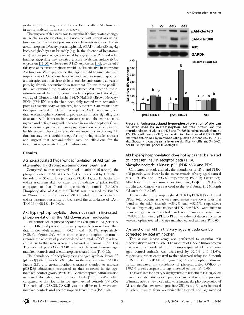
Aging-associated hyper-phosphorylation of Akt can beattenuated by chronic acetaminophen treatment
Compared to that observed in adult animals (6-month), the
phosphorylation of Akt at the Ser473 was increased by 114.3% in
the soleus of 33-month aged rats (P,0.05; Figure 1). Acetamin-
ophen treatment did not alter the abundance of pAkt-Ser473
compared to that found in age-matched controls (P.0.05).
Phosphorylation of Akt at the Thr308 was increased by 450.9%
in 33-month control animals (P,0.05), while chronic acetamin-
ophen treatment significantly decreased the abundance of pAkt-
Thr308 (268.1%, P,0.05).
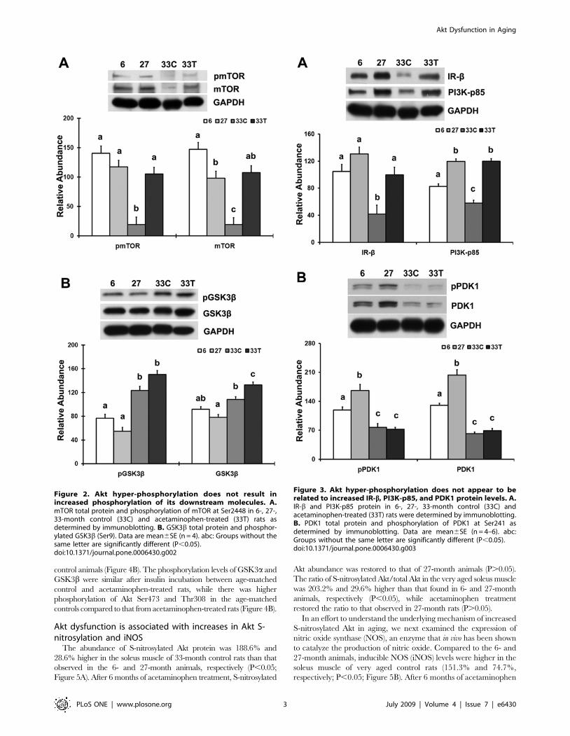
Akt hyper-phosphorylation does not result in increasedphosphorylation of the Akt downstream molecules
The abundance of phosphorylated mTOR (pmTOR) (Ser2448)
and mTOR total protein in the very aged soleus were lower than
that in the adult animals (286.3% and 286.8%, respectively;
P,0.05; Figure 2A), while chronic acetaminophen treatment
restored the amount of phosphorylated and total mTOR to a level
equivalent to that seen in 6- and 27-month old animals (P.0.05).
The ratio of pmTOR/mTOR was not different between age-
matched controls and acetaminophen-treated rats (P.0.05).
The abundance of phosphorylated glycogen synthase kinase 3b(pGSK3b) (Ser9) was 61.7% higher in the very age rats (P,0.05;
Figure 2B), and acetaminophen treatment trended to increase
pGSK3b abundance compared to that observed in the age-
matched control group (P = 0.06). Acetaminophen administration
increased the abundance of total GSK3b by 22.4% when
compared to that found in the age-matched controls (P,0.05).
The ratio of pGSK3b/GSK3b was not different between age-
matched controls and acetaminophen-treated rats (P.0.05).
Akt hyper-phosphorylation does not appear to be relatedto increased insulin receptor beta (IR-b),phosphoinositide 3-kinase p85 (PI3K-p85) and PDK1
Compared to adult animals, the abundance of IR-b and PI3K-
p85 protein were lower in the soleus muscle of very aged control
rats (260.0% and 229.7%, respectively; P,0.05; Figure 3A).
After 6 months of acetaminophen treatment, IR-b and PI3K-p85
protein abundances were restored to the level found in 27-month
old animals (P.0.05).
The abundance of phosphorylated PDK1 (pPDK1) (Ser241) and
PDK1 total protein in the very aged soleus were lower than that
found in the adult animals (235.2% and 252.5%, respectively;
P,0.05; Figure 3B), while neither pPDK1 nor PDK1 were different
between age-matched controls and acetaminophen-treated rats
(P.0.05). The ratio of pPDK1/PDK1 was also not different between
acetaminophen-treated and age-matched control animals (P.0.05).
Dysfunction of Akt in the very aged muscle can becorrected by acetaminophen
The in vitro kinase assay was performed to examine Akt
functionality in aged muscle. The amount of GSK-3 fusion protein
that was phosphorylated by immunoprecipitated Akt from very
aged control animals was decreased by 32.8% and 34.6%,
respectively, when compared to that observed using the 6-month
or 27-month rats (P,0.05; Figure 4A). Acetaminophen adminis-
tration increased the abundance of phosphorylated GSK-3 by
170.5% when compared to age-matched control (P,0.05).
To investigate the ability of aging muscle to respond to insulin, ex vivo
muscle incubation studies were performed in the absence and presence
of insulin. After ex vivo incubation with insulin, the phosphorylation of
Akt and the Akt downstream proteins, GSK-3a and 3b, were increased
in soleus muscles from acetaminophen-treated and age-matched
Figure 1. Aging-associated hyper-phosphorylation of Akt canbe attenuated by acetaminophen. Akt total protein and thephosphorylation of Akt at Ser473 and Thr308 in soleus muscle from 6-,27-, 33-month control (33C) and acetaminophen-treated (33T) F344BNrats were determined by immunoblotting. Data are mean6SE (n = 4–6).abc: Groups without the same letter are significantly different (P,0.05).doi:10.1371/journal.pone.0006430.g001
Akt Dysfunction in Aging
PLoS ONE | www.plosone.org 2 July 2009 | Volume 4 | Issue 7 | e6430
control animals (Figure 4B). The phosphorylation levels of GSK3a and
GSK3b were similar after insulin incubation between age-matched
control and acetaminophen-treated rats, while there was higher
phosphorylation of Akt Ser473 and Thr308 in the age-matched
controls compared to that from acetaminophen-treated rats (Figure 4B).
Akt dysfunction is associated with increases in Akt S-nitrosylation and iNOS
The abundance of S-nitrosylated Akt protein was 188.6% and
28.6% higher in the soleus muscle of 33-month control rats than that
observed in the 6- and 27-month animals, respectively (P,0.05;
Figure 5A). After 6 months of acetaminophen treatment, S-nitrosylated
Akt abundance was restored to that of 27-month animals (P.0.05).
The ratio of S-nitrosylated Akt/total Akt in the very aged soleus muscle
was 203.2% and 29.6% higher than that found in 6- and 27-month
animals, respectively (P,0.05), while acetaminophen treatment
restored the ratio to that observed in 27-month rats (P.0.05).
In an effort to understand the underlying mechanism of increased
S-nitrosylated Akt in aging, we next examined the expression of
nitric oxide synthase (NOS), an enzyme that in vivo has been shown
to catalyze the production of nitric oxide. Compared to the 6- and
27-month animals, inducible NOS (iNOS) levels were higher in the
soleus muscle of very aged control rats (151.3% and 74.7%,
respectively; P,0.05; Figure 5B). After 6 months of acetaminophen
Figure 2. Akt hyper-phosphorylation does not result inincreased phosphorylation of its downstream molecules. A.mTOR total protein and phosphorylation of mTOR at Ser2448 in 6-, 27-,33-month control (33C) and acetaminophen-treated (33T) rats asdetermined by immunoblotting. B. GSK3b total protein and phosphor-ylated GSK3b (Ser9). Data are mean6SE (n = 4). abc: Groups without thesame letter are significantly different (P,0.05).doi:10.1371/journal.pone.0006430.g002
Figure 3. Akt hyper-phosphorylation does not appear to berelated to increased IR-b, PI3K-p85, and PDK1 protein levels. A.IR-b and PI3K-p85 protein in 6-, 27-, 33-month control (33C) andacetaminophen-treated (33T) rats were determined by immunoblotting.B. PDK1 total protein and phosphorylation of PDK1 at Ser241 asdetermined by immunoblotting. Data are mean6SE (n = 4–6). abc:Groups without the same letter are significantly different (P,0.05).doi:10.1371/journal.pone.0006430.g003
Akt Dysfunction in Aging
PLoS ONE | www.plosone.org 3 July 2009 | Volume 4 | Issue 7 | e6430
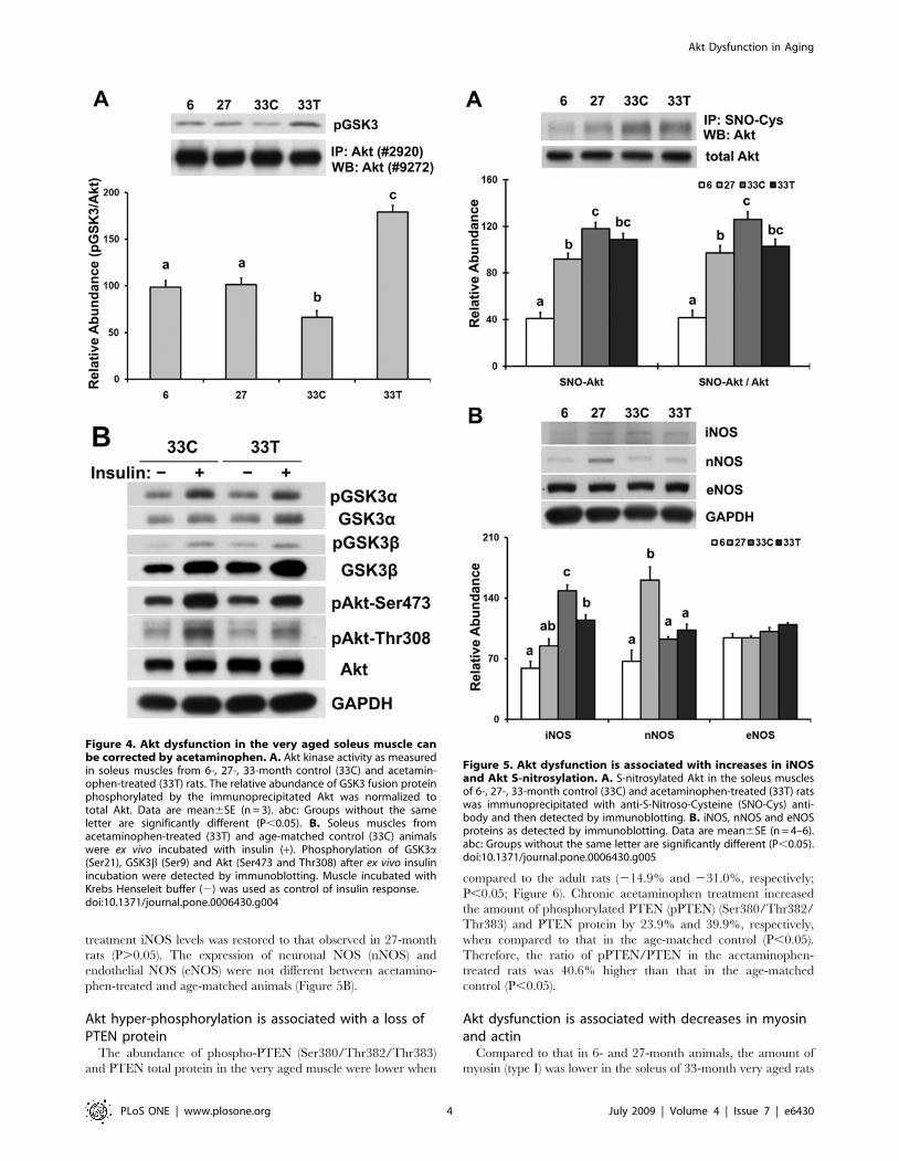
treatment iNOS levels was restored to that observed in 27-month
rats (P.0.05). The expression of neuronal NOS (nNOS) and
endothelial NOS (eNOS) were not different between acetamino-
phen-treated and age-matched animals (Figure 5B).
Akt hyper-phosphorylation is associated with a loss ofPTEN protein
The abundance of phospho-PTEN (Ser380/Thr382/Thr383)
and PTEN total protein in the very aged muscle were lower when
compared to the adult rats (214.9% and 231.0%, respectively;
P,0.05; Figure 6). Chronic acetaminophen treatment increased
the amount of phosphorylated PTEN (pPTEN) (Ser380/Thr382/
Thr383) and PTEN protein by 23.9% and 39.9%, respectively,
when compared to that in the age-matched control (P,0.05).
Therefore, the ratio of pPTEN/PTEN in the acetaminophen-
treated rats was 40.6% higher than that in the age-matched
control (P,0.05).
Akt dysfunction is associated with decreases in myosinand actin
Compared to that in 6- and 27-month animals, the amount of
myosin (type I) was lower in the soleus of 33-month very aged rats
Figure 4. Akt dysfunction in the very aged soleus muscle canbe corrected by acetaminophen. A. Akt kinase activity as measuredin soleus muscles from 6-, 27-, 33-month control (33C) and acetamin-ophen-treated (33T) rats. The relative abundance of GSK3 fusion proteinphosphorylated by the immunoprecipitated Akt was normalized tototal Akt. Data are mean6SE (n = 3). abc: Groups without the sameletter are significantly different (P,0.05). B. Soleus muscles fromacetaminophen-treated (33T) and age-matched control (33C) animalswere ex vivo incubated with insulin (+). Phosphorylation of GSK3a(Ser21), GSK3b (Ser9) and Akt (Ser473 and Thr308) after ex vivo insulinincubation were detected by immunoblotting. Muscle incubated withKrebs Henseleit buffer (2) was used as control of insulin response.doi:10.1371/journal.pone.0006430.g004
Figure 5. Akt dysfunction is associated with increases in iNOSand Akt S-nitrosylation. A. S-nitrosylated Akt in the soleus musclesof 6-, 27-, 33-month control (33C) and acetaminophen-treated (33T) ratswas immunoprecipitated with anti-S-Nitroso-Cysteine (SNO-Cys) anti-body and then detected by immunoblotting. B. iNOS, nNOS and eNOSproteins as detected by immunoblotting. Data are mean6SE (n = 4–6).abc: Groups without the same letter are significantly different (P,0.05).doi:10.1371/journal.pone.0006430.g005
Akt Dysfunction in Aging
PLoS ONE | www.plosone.org 4 July 2009 | Volume 4 | Issue 7 | e6430
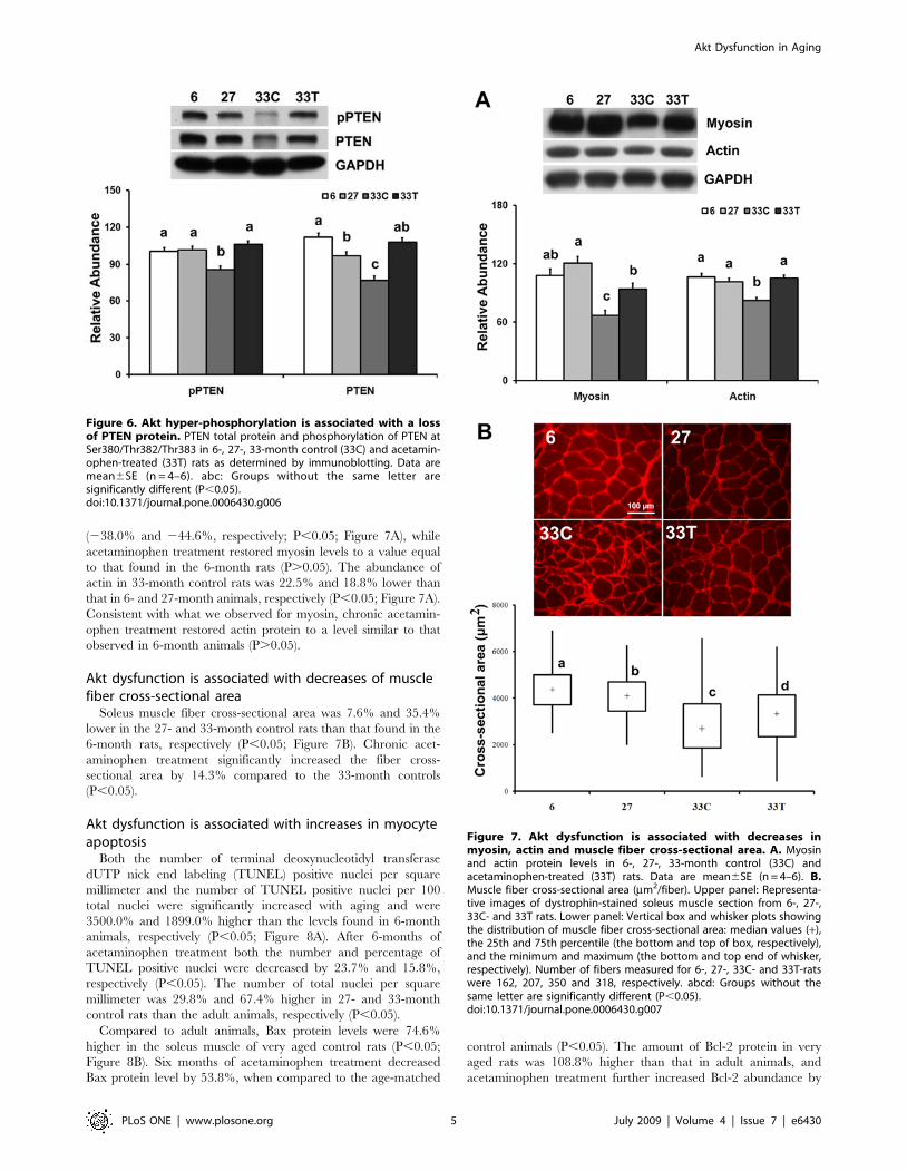
(238.0% and 244.6%, respectively; P,0.05; Figure 7A), while
acetaminophen treatment restored myosin levels to a value equal
to that found in the 6-month rats (P.0.05). The abundance of
actin in 33-month control rats was 22.5% and 18.8% lower than
that in 6- and 27-month animals, respectively (P,0.05; Figure 7A).
Consistent with what we observed for myosin, chronic acetamin-
ophen treatment restored actin protein to a level similar to that
observed in 6-month animals (P.0.05).
Akt dysfunction is associated with decreases of musclefiber cross-sectional area
Soleus muscle fiber cross-sectional area was 7.6% and 35.4%
lower in the 27- and 33-month control rats than that found in the
6-month rats, respectively (P,0.05; Figure 7B). Chronic acet-
aminophen treatment significantly increased the fiber cross-
sectional area by 14.3% compared to the 33-month controls
(P,0.05).
Akt dysfunction is associated with increases in myocyteapoptosis
Both the number of terminal deoxynucleotidyl transferase
dUTP nick end labeling (TUNEL) positive nuclei per square
millimeter and the number of TUNEL positive nuclei per 100
total nuclei were significantly increased with aging and were
3500.0% and 1899.0% higher than the levels found in 6-month
animals, respectively (P,0.05; Figure 8A). After 6-months of
acetaminophen treatment both the number and percentage of
TUNEL positive nuclei were decreased by 23.7% and 15.8%,
respectively (P,0.05). The number of total nuclei per square
millimeter was 29.8% and 67.4% higher in 27- and 33-month
control rats than the adult animals, respectively (P,0.05).
Compared to adult animals, Bax protein levels were 74.6%
higher in the soleus muscle of very aged control rats (P,0.05;
Figure 8B). Six months of acetaminophen treatment decreased
Bax protein level by 53.8%, when compared to the age-matched
control animals (P,0.05). The amount of Bcl-2 protein in very
aged rats was 108.8% higher than that in adult animals, and
acetaminophen treatment further increased Bcl-2 abundance by
Figure 6. Akt hyper-phosphorylation is associated with a lossof PTEN protein. PTEN total protein and phosphorylation of PTEN atSer380/Thr382/Thr383 in 6-, 27-, 33-month control (33C) and acetamin-ophen-treated (33T) rats as determined by immunoblotting. Data aremean6SE (n = 4–6). abc: Groups without the same letter aresignificantly different (P,0.05).doi:10.1371/journal.pone.0006430.g006
Figure 7. Akt dysfunction is associated with decreases inmyosin, actin and muscle fiber cross-sectional area. A. Myosinand actin protein levels in 6-, 27-, 33-month control (33C) andacetaminophen-treated (33T) rats. Data are mean6SE (n = 4–6). B.Muscle fiber cross-sectional area (mm2/fiber). Upper panel: Representa-tive images of dystrophin-stained soleus muscle section from 6-, 27-,33C- and 33T rats. Lower panel: Vertical box and whisker plots showingthe distribution of muscle fiber cross-sectional area: median values (+),the 25th and 75th percentile (the bottom and top of box, respectively),and the minimum and maximum (the bottom and top end of whisker,respectively). Number of fibers measured for 6-, 27-, 33C- and 33T-ratswere 162, 207, 350 and 318, respectively. abcd: Groups without thesame letter are significantly different (P,0.05).doi:10.1371/journal.pone.0006430.g007
Akt Dysfunction in Aging
PLoS ONE | www.plosone.org 5 July 2009 | Volume 4 | Issue 7 | e6430
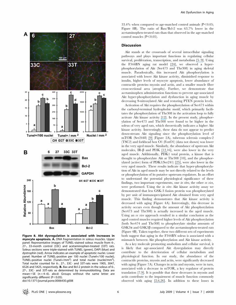
33.4% when compared to age-matched control animals (P,0.05;
Figure 8B). The ratio of Bax/Bcl-2 was 63.7% lower in the
acetaminophen-treated rats than that observed in the age-matched
control muscles (P,0.05).
Discussion
Akt stands at the crossroads of several intracellular signaling
pathways and plays important functions in regulating cellular
survival, proliferation, transcription, and metabolism [5–9]. Using
the F344BN aging rat model [24], we observed a hyper-
phosphorylation of Akt (Ser473 and Thr308) in aging skeletal
muscle. Paradoxically, this increased Akt phosphorylation is
associated with lower Akt kinase activity, diminished response to
insulin, higher levels of myocyte apoptosis, lower abundance of
contractile proteins myosin and actin, and a smaller muscle fiber
cross-sectional area (atrophy). Further, we demonstrate that
acetaminophen administration functions to prevent age-associated
Akt hyper-phosphorylation and dysfunction in aging muscle by
decreasing S-nitrosylated Akt and restoring PTEN protein levels.
Activation of Akt requires the phosphorylation of Ser473 within
the carboxyl-terminal hydrophobic motif, which primarily facili-
tates the phosphorylation of Thr308 in the activation loop to fully
activate Akt kinase activity [12]. In the present study, phosphor-
ylation of Ser473 and Thr308 were found to be higher in the
soleus of very aged rats, which theoretically indicates a higher Akt
kinase activity. Interestingly, these data do not appear to predict
down-stream Akt signaling since the phosphorylation level of
mTOR (Ser2448) [9] (Figure 2A), tuberous sclerosis complex-2
(TSC2) and forkhead box O1 (FoxO1) (data not shown) was lower
in the very aged muscle. Similarly, the abundance of upstream Akt
molecules, IR-b and PI3K [12,16], were also lower in the very
aged muscle. Additionally, PDK1 total protein, a kinase that is
thought to phosphorylate Akt at Thr308 [10], and the phosphor-
ylated (active) form of PDK1(Ser241) [25], were also lower in the
very aged muscle. These results indicate that hyper-phosphoryla-
tion of Akt in aged muscle may be not directly related to the levels
or phosphorylation of its putative upstream regulators. In an effort
to understand the potential physiological significance of these
findings, two important experiments, one in vitro, the other ex vivo,
were performed. Using the in vitro Akt kinase activity assay we
demonstrated that less GSK-3 fusion protein was phosphorylated
by per unit of immunoprecipitated Akt obtained from very aged
muscle. This finding demonstrates that Akt kinase activity is
decreased with aging (Figure 4A). Interestingly, this decrease in
activity occurs even though the amount of Akt phosphorylation
(Ser473 and Thr308) is actually increased in the aged muscle.
Using an ex vivo approach resulted in a similar conclusion as the
aged control muscles required higher levels of Akt phosphorylation
(both Ser474 and Thr308) to phosphorylate similar amounts of
GSK3a and GSK3b compared to the acetaminophen-treated rats
(Figure 4B). Taken together, these two different sets of experiments
both suggest that aging in the F344BN soleus is associated with a
mismatch between Akt phosphorylation and Akt kinase activity.
As a key molecule promoting anabolism and cellular survival, it
is likely that age-associated Akt dysregulation may directly
contribute to the deterioration of cellular metabolism and
physiological function. In our study, the abundance of the
contractile proteins, myosin and actin, were significantly decreased
with aging (Figure 7A). Changes in actin and myosin, were in turn,
associated with a decrease in mTOR, a key regulator of protein
translation [7,9]. It is possible that these decreases in myosin and
actin contribute to the impairment of muscle function commonly
observed with aging [2,6,26]. In addition to these losses in
Figure 8. Akt dysregulation is associated with increases inmyocyte apoptosis. A. DNA fragmentation in soleus muscles. Upperpanel: Representative images of TUNEL-stained soleus muscle from 6-,27-, 33-month control (33C) and acetaminophen-treated (33T) rats.Soleus sections were triple-stained with TUNEL (green), DAPI (blue) anddystrophin (red). Arrow indicates an example of apoptotic nuclei. Lowerpanel: Number of TUNEL-positive per 100 nuclei (Tunel+/100 nuclei),TUNEL-positive nuclei (Tunel+/mm2) and total nuclei (nuclei/mm2).Total nuclei counted for 6-, 27-, 33C- and 33T-rats were 1905, 3047,6526 and 5425, respectively. B. Bax and Bcl-2 protein in the soleus of 6-,27-, 33C- and 33T-rats as determined by immunoblotting. Data aremean6SE (n = 4–6). abcd: Groups without the same letter aresignificantly different (P,0.05).doi:10.1371/journal.pone.0006430.g008
Akt Dysfunction in Aging
PLoS ONE | www.plosone.org 6 July 2009 | Volume 4 | Issue 7 | e6430
contractile proteins, we also observed a loss of myocyte size, a
dramatic increase in the abundance of Bax, a shift in the Bax/Bcl-
2 ratio and an increase in the number of myocytes undergoing
apopotosis (Figures 7B, 8A and 8B). Interestingly, the restoration
of Akt kinase activity by chronic acetaminophen intervention was
associated with increases in the amount of contractile proteins and
myocyte size, and a decrease in the incidence of myocyte apoptosis
(Figures 7 and 8). Taken together these data suggest that an
impairment in Akt signaling might be related to the development
of age-related metabolic and functional disorders in skeletal muscle
and importantly, that the re-establishment of Akt functionality
may be a useful strategy for diminishing the deleterious effects of
aging on muscle structure and function.
As an important signaling molecule involved in many
physiological processes, nitric oxide regulates protein structure
and function by interacting with cysteine sulfhydryls and inhibiting
the formation of disulfide bonds [15–17,27]. Increases in the
amount of cellular S-nitrosylated proteins have been shown to be
associated with the disruption of protein structure along with
increases in tissue damage and mortality [17,27]. S-nitrosylated
proteins, including Akt, are involved in pathogenesis of insulin
resistance [15,16]. We found that both the abundance of S-
nitrosylated Akt and ratio of S-nitrosylated Akt/total Akt were
substantially increased with aging (Figure 5A). These increases
occurred concomitant to increases in the amount of iNOS, an
enzyme that catalyzes the production of nitric oxide needed for the
S-nitrosylation reaction [18,19]. Although it is unclear why aging
might increase iNOS levels, other reports have suggested that
increased extracellular glucose levels can induce iNOS expression
[19,20]. A previous study by our laboratory demonstrated that
age-associated hyperglycemia and decreases in muscle glucose
transporter-4 (Glut4) can be reversed by acetaminophen interven-
tion [23], while other work using cultured cell has shown that
acetaminophen can directly inhibit NO production and iNOS
expression through its ability to diminish NF-kappaB binding to
the iNOS gene promoter [28]. Consistent with these findings, we
show that age-associated increases in iNOS expression were
reversed after acetaminophen treatment, and further that this
decrease in iNOS expression coincided with decreases in the
amount of S-nitrosylated Akt (Figure 5A). As expected, this
decrease in S-nitrosylated Akt was found to parallel the
normalization of Akt phosphorylation and increases in Akt kinase
activity. As such, it is likely that the S-nitrosylation of Akt is
involved in contributing to age-associated Akt dysfunction. Unlike
that observed for iNOS, acetaminophen did not appear to affect
the regulation of nNOS or eNOS expression (Figure 5B)
suggesting perhaps that the normalization of Akt S-nitrosylation
by acetaminophen may be dependent on the reduced expression
of iNOS. Why acetaminophen may target iNOS instead of other
NOS isoforms is currently unclear. Further studies designed to
examine the effects of aging on individual cysteine residues within
the Akt protein will no doubt be useful in furthering our
understanding on how S-nitrosylation might compromise Akt
kinase activity.
The phosphatidylinositol 39-phosphatase PTEN is considered a
key negative regulator of Akt signaling [13,14]. PTEN catalyzes
phosphatidylinositol 3,4,5-trisphosphate (PIP3), a key mediator of
PI3K activity, into phosphatidylinositol 4,5-trisphosphate (PIP2),
resulting in the attenuation of phosphorylation (activation) of Akt
[13,14]. It has been reported that the phosphorylation of Thr308
of Akt, but not Ser473, is regulated by PTEN in adipocytes [13].
In present study, the abundance of pAkt-Ser473 was higher in
muscles obtained from both the 33-month control and acetamin-
ophen-treated rats, while pAkt-Thr308 was dramatically increased
in control rats. These results are consistent with the alterations in
PTEN protein levels and suggest that PTEN may regulate the
phosphorylation of Akt Thr308 in skeletal muscle using a
mechanism similar to that previously observed in adipocytes
[13]. Interestingly, 6 months of acetaminophen intervention
restored PTEN protein levels similar to that found in 6- and 27-
month rats. This increase in PTEN protein appeared to parallel
decreases in the amount of Akt-Thr308 phosphorylation which
support the notion that a loss of PTEN protein with aging may
contribute to the hyper-phosphorylation of Akt, and that
acetaminophen intervention may function in reducing Akt
phosphorylation by increasing PTEN levels.
Why aging decreases PTEN protein levels is not clear. It is
thought that high glucose levels can result in decreased PTEN
expression and decreases in PTEN phosphatase activity [14]. Our
previous study found that the aging-associated hyperglycemia can
be reversed by acetaminophen intervention [23]. It is possible that
the normalization of blood glucose by acetaminophen may
contribute to the increased PTEN expression. It has also been
documented that phosphorylation of the C-terminal tail of PTEN
decreases the degradation of PTEN protein by increasing its
stability [29]. Our data also show that pPTEN levels were
decreased with aging and that acetaminophen treatment restored
pPTEN comparable to that observed in 6- and 27-month rats
(Figure 6). Therefore, aging-associated hyperglycemia and de-
creases in pPTEN level may result in decreased PTEN protein
levels, which could act to increase the phosphorylation of Akt at
Thr308 in aging muscle. Whether changes in PTEN expression
alone or if the presence of other factors is required to explain the
effects of aging on Akt expression and phosphorylation will require
further investigation.
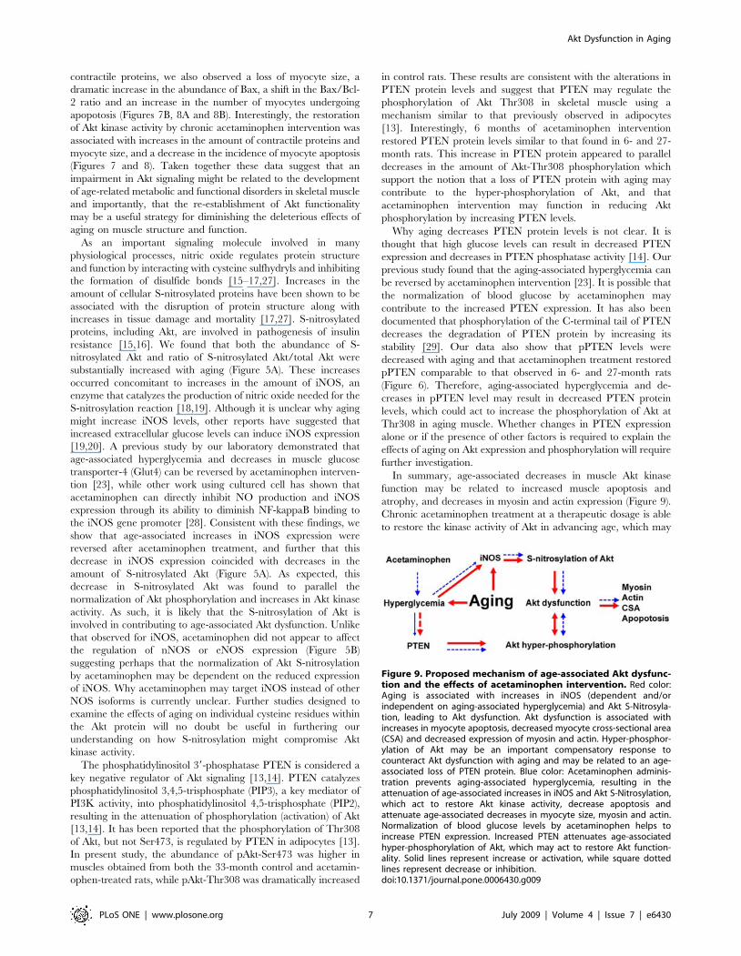
In summary, age-associated decreases in muscle Akt kinase
function may be related to increased muscle apoptosis and
atrophy, and decreases in myosin and actin expression (Figure 9).
Chronic acetaminophen treatment at a therapeutic dosage is able
to restore the kinase activity of Akt in advancing age, which may
Figure 9. Proposed mechanism of age-associated Akt dysfunc-tion and the effects of acetaminophen intervention. Red color:Aging is associated with increases in iNOS (dependent and/orindependent on aging-associated hyperglycemia) and Akt S-Nitrosyla-tion, leading to Akt dysfunction. Akt dysfunction is associated withincreases in myocyte apoptosis, decreased myocyte cross-sectional area(CSA) and decreased expression of myosin and actin. Hyper-phosphor-ylation of Akt may be an important compensatory response tocounteract Akt dysfunction with aging and may be related to an age-associated loss of PTEN protein. Blue color: Acetaminophen adminis-tration prevents aging-associated hyperglycemia, resulting in theattenuation of age-associated increases in iNOS and Akt S-Nitrosylation,which act to restore Akt kinase activity, decrease apoptosis andattenuate age-associated decreases in myocyte size, myosin and actin.Normalization of blood glucose levels by acetaminophen helps toincrease PTEN expression. Increased PTEN attenuates age-associatedhyper-phosphorylation of Akt, which may act to restore Akt function-ality. Solid lines represent increase or activation, while square dottedlines represent decrease or inhibition.doi:10.1371/journal.pone.0006430.g009
Akt Dysfunction in Aging
PLoS ONE | www.plosone.org 7 July 2009 | Volume 4 | Issue 7 | e6430
be due to decreases in iNOS and diminished S-nitrosylation of
Akt. The data of the present study show that a loss of PTEN
protein in aging may contribute to the increased phosphorylation
of Akt, and that acetaminophen intervention can restore both
PTEN and pPTEN levels. This report provides evidence that Akt
may be a key target molecule for interventions designed to
attenuate the effects of aging on skeletal muscle and that
acetaminophen may be useful for the treatment of age-related
muscle disorders.
Materials and Methods
MaterialsPrimary antibodies against Akt (#9272 and #2920), phospho-
Akt (Thr308) (pAkt-Thr308, #9275), phospho-Akt (Ser473) (pAkt-
Ser473, #9271), GAPDH (#2118), GSK3a (#9338), GSK3b(#9315), phospho-GSK3a/b (Ser21/9) (#9331), IR-b (#3025),
mTOR (#2972), phospho-mTOR (Ser2448)(pmTOR, #2971),
PI3K-p85 (#4257), PTEN (#9552) and phospho-PTEN (Ser380/
Thr382/383) (pPTEN, #9554), PDK1 (#3062), phospho-PDK1
(Ser241)(pPDK1, #3438), nNOS (#4231), eNOS (#9572),
secondary antibodies conjugated with horseradish peroxidase
(HRP) (anti-rabbit (#7074) or anti-mouse (#7076)) and nonra-
dioactive Akt kinase assay kit (#9840) were purchased from Cell
Signaling Technology (Beverly, MA). Primary antibodies against
Bcl-2 (sc-7382) and Bax (sc-493) were from Santa Cruz
Biotechnology (Santa Cruz, CA). Dystrophin antibody (NCL-
DYS2) was from Novocastra Vector Laboratories (Burlingame,
CA). Actin antibody (MCA1906) was from Antibodies Direct
(Raleigh, NC). S-Nitroso-Cysteine (SNO-Cys, #N5411) and
myosin (M8421) primary antibodies, and acetaminophen
(#A5000) were from Sigma-Aldrich, Inc. (St. Louis, MO). Human
recombinant insulin was from SAFC Biosciences (Lenexa, KS).
The in situ cell death detection kit was from Roche Diagnostics
(Mannheim, Germany). VECTASHIELD HardSet Mounting
Medium with DAPI was from Vector Laboratories (Burlingame,
CA). Pierce Tissue Protein Extraction Reagent (T-PER) and
Pierce 660nm protein assay reagent (#22660) were from Thermo
Fisher Scientific Inc. (Rockford, IL). The PAGEr Gold Precast gel
(10%) was from Lonza (Rockland, ME). The Amersham ECLTM
Western Blotting reagent (RPN 2106) was from GE Healthcare
Bio-Sciences Corp. (Piscataway, NJ).
AnimalsThe F344BN rats (National Institute on Aging, Bethesda, MD)
were housed in an Association for Assessment and Accreditation of
Laboratory Animal Care International (AAALAC) approved
vivarium with a 12:12 h light/dark cycle at 2262uC. Food
(LabDiet 5001, PMI Nutrition International, LLC, Brentwood,
MO) and water were provided ad libitum. All procedures were
approved by the Marshall University Institutional Animal Care
and Use Committee, and the ‘‘Principles of laboratory animal
care’’ (NIH publication No. 86-23, revised 1985) were followed.
Acetaminophen treatment and tissue collectionTwenty seven months old F344BN rats were given acetamin-
ophen daily (30 mg/kg body weight/day) for 6 months in their
drinking water. Age-matched rats were maintained as controls.
Soleus muscles were removed from anesthetized animals, frozen in
liquid nitrogen, and stored at 2 80uC.
Ex vivo muscle incubation with insulinTwo strips of muscle were isolated from each soleus muscle as
previously described [30]. To avoid tissue hypoxia that may
compromise structure and function of muscle, ex vivo muscle
incubation were performed at 25uC in Krebs Henseleit buffer
(KHB, pH 7.4) containing 5.5 mM glucose, 2 mM pyruvate,
5 mM HEPES and 0.1% bovine serum albumin, and equilibrated
with 95% O2 and 5% CO2 as outlined elsewhere [31]. Muscles
were mounted to plastic plates to keep them at their in situ resting
length. After pre-incubation in KHB for 15 min, muscles were
incubated in KHB for either another 30 min (control), or in KHB
containing human recombinant insulin (100 mU/mL) for 30 min.
Muscle samples were then washed with KHB, immediately frozen
in liquid nitrogen, and stored at 2 80uC.
In vitro Akt kinase activity assayTo assess Akt kinase activity, total Akt protein was immuno-
precipitated with anti-Akt antibody (#2920) overnight at 4uC, and
then the kinase activity assay was performed using a nonradioac-
tive Akt kinase assay kit as outlined by the manufacturer (#9840).
Briefly, after washing with cell lysis buffer and kinase buffer, the
immunoprecipitates were incubated with a GSK-3 fusion protein
substrate and ATP for 30 min at 30uC. The phosphorylation of
the GSK-3 fusion protein at Ser21/Ser9 by the immunoprecip-
itated Akt was detected by immunoblotting. The immunoprecip-
itated Akt protein was detected using anti-Akt antibody (#9272).
The abundance of p-GSK-3 was normalized to the amount of
total Akt.
Analysis of S-nitrosylated AktTo detect S-nitrosylated Akt, soleus lysates were immunopre-
cipitated by anti-S-Nitroso-Cysteine antibody overnight at 4uC.
Akt in the immunoprecipitates were detected by immunoblotting.
Immunoblotting analysisProtein samples was prepared, separated on a 10% PAGEr
Gold Precast gel, and then transferred to nitrocellulose mem-
branes as previously described [23]. After incubated with primary
antibody overnight at 4uC and secondary antibody for 1 h at room
temperature, target proteins were visualized following reaction
with Amersham ECL reagent. Target protein levels were
quantified by an AlphaEaseFC image analysis software (Alpha
Innotech, San Leandro, CA) and normalized to GAPDH.
TUNEL, Dystrophin and DAPI triple-stainingDNA fragmentation associated with apoptosis was detected by
TUNEL, using a Roche in situ cell death detection kit according to
manufacturer instructions. Soleus sections were blocked with 3%
BSA and incubated with anti-dystrophin antibody at the dilution
of 1:200 to visualize the cell membrane. Nuclei were co-stained
using DAPI in a VECTASHIELD HardSet Mounting Medium.
Sections were visualized under an Olympus BX51 microscope
equipped with Olympus WH 106widefield eyepieces and
Olympus UPlanF1 406/0.75 objective lens.
Quantification of muscle fiber cross-sectional areaMuscle fiber cross-sectional area was determined by tracing the
outline of dystrophin-stained fibers using the ImageJ program
(http://rsb.info.nih.gov/ij/). The distribution of muscle fiber
cross-sectional area was plotted with vertical box and whisker
plots, and multiple comparisons were performed to determine
differences of means between groups (See ‘‘Data analysis’’).
Data analysisResults are presented as mean6SE. The effects of age and
acetaminophen treatment were analyzed using the GLM proce-
Akt Dysfunction in Aging
PLoS ONE | www.plosone.org 8 July 2009 | Volume 4 | Issue 7 | e6430
dure (SAS 9.1 for Windows, SAS Institute Inc., Cary, NC). Means
were calculated by the LSMEANS procedure and multiple
comparisons were performed using the Tukey-Kramer test to
determine differences between groups. Values of P,0.05 were
considered to be statistically significant.
Acknowledgments
We thank Mr. Brent Kidd, Madhukar Kolli, and Siva Nalabotu for their
technical assistance.
Author Contributions
Conceived and designed the experiments: MW ERB. Performed the
experiments: MW AK MKG HL SKK JF SP RKA KMR YW. Analyzed
the data: MW ERB. Wrote the paper: MW ERB.
References
1. Jejurikar SS, Henkelman EA, Cederna PS, Marcelo CL, Urbanchek MG, et al.
(2006) Aging increases the susceptibility of skeletal muscle derived satellite cells
to apoptosis. Exp Gerontol 41: 828–836.
2. Balagopal P, Rooyackers OE, Adey DB, Ades PA, Nair KS (1997) Effects of
aging on in vivo synthesis of skeletal muscle myosin heavy-chain and
sarcoplasmic protein in humans. Am J Physiol 273: E790–800.
3. Brown M (1987) Change in fibre size, not number, in ageing skeletal muscle. Age
Ageing 16: 244–248.
4. Faulkner JA, Larkin LM, Claflin DR, Brooks SV (2007) Age-related changes in
the structure and function of skeletal muscles. Clin Exp Pharmacol Physiol 34:
1091–1096.
5. Gupte AA, Bomhoff GL, Geiger PC (2008) Age-related differences in skeletal
muscle insulin signaling: the role of stress kinases and heat shock proteins. J Appl
Physiol 105: 839–848.
6. Leger B, Derave W, De Bock K, Hespel P, Russell AP (2008) Human sarcopenia
reveals an increase in SOCS-3 and myostatin and a reduced efficiency of Akt
phosphorylation. Rejuvenation Res 11: 163–175B.
7. Bodine SC, Stitt TN, Gonzalez M, Kline WO, Stover GL, et al. (2001) Akt/
mTOR pathway is a crucial regulator of skeletal muscle hypertrophy and can
prevent muscle atrophy in vivo. Nat Cell Biol 3: 1014–1019.
8. Cross DA, Alessi DR, Cohen P, Andjelkovich M, Hemmings BA (1995)
Inhibition of glycogen synthase kinase-3 by insulin mediated by protein kinase B.
Nature 378: 785–789.
9. Nave BT, Ouwens M, Withers DJ, Alessi DR, Shepherd PR (1999) Mammalian
target of rapamycin is a direct target for protein kinase B: identification of a
convergence point for opposing effects of insulin and amino-acid deficiency on
protein translation. Biochem J 344 Pt 2: 427–431.
10. Alessi DR, James SR, Downes CP, Holmes AB, Gaffney PR, et al. (1997)
Characterization of a 3-phosphoinositide-dependent protein kinase which
phosphorylates and activates protein kinase Balpha. Curr Biol 7: 261–269.
11. Sarbassov DD, Guertin DA, Ali SM, Sabatini DM (2005) Phosphorylation and
regulation of Akt/PKB by the rictor-mTOR complex. Science 307: 1098–1101.
12. Scheid MP, Marignani PA, Woodgett JR (2002) Multiple phosphoinositide 3-
kinase-dependent steps in activation of protein kinase B. Mol Cell Biol 22:
6247–6260.
13. Ono H, Katagiri H, Funaki M, Anai M, Inukai K, et al. (2001) Regulation of
phosphoinositide metabolism, Akt phosphorylation, and glucose transport by
PTEN (phosphatase and tensin homolog deleted on chromosome 10) in 3T3-L1
adipocytes. Mol Endocrinol 15: 1411–1422.
14. Mahimainathan L, Das F, Venkatesan B, Choudhury GG (2006) Mesangial cell
hypertrophy by high glucose is mediated by downregulation of the tumor
suppressor PTEN. Diabetes 55: 2115–2125.
15. Yasukawa T, Tokunaga E, Ota H, Sugita H, Martyn JA, et al. (2005) S-
nitrosylation-dependent inactivation of Akt/protein kinase B in insulin
resistance. J Biol Chem 280: 7511–7518.
16. Carvalho-Filho MA, Ueno M, Hirabara SM, Seabra AB, Carvalheira JB, et al.
(2005) S-nitrosation of the insulin receptor, insulin receptor substrate 1, and
protein kinase B/Akt: a novel mechanism of insulin resistance. Diabetes 54:959–967.
17. Lu XM, Lu M, Tompkins RG, Fischman AJ (2005) Site-specific detection of S-nitrosylated PKB alpha/Akt1 from rat soleus muscle using CapLC-Q-
TOF(micro) mass spectrometry. J Mass Spectrom 40: 1140–1148.
18. Wu MD, Kimura M, Inafuku S, Ishigami H (1997) Effect of aging on theexpression of iNOS and cell death in the mouse cochlear spiral ganglion.
Okajimas Folia Anat Jpn 74: 155–165.19. Fujimoto M, Shimizu N, Kunii K, Martyn JA, Ueki K, et al. (2005) A role for
iNOS in fasting hyperglycemia and impaired insulin signaling in the liver ofobese diabetic mice. Diabetes 54: 1340–1348.
20. Pacheco ME, Beltran A, Redondo J, Manso AM, Alonso MJ, et al. (2006) High
glucose enhances inducible nitric oxide synthase expression. Role of proteinkinase C-betaII. Eur J Pharmacol 538: 115–123.
21. Kurowski TG, Lin Y, Luo Z, Tsichlis PN, Buse MG, et al. (1999) Hyperglycemiainhibits insulin activation of Akt/protein kinase B but not phosphatidylinositol 3-
kinase in rat skeletal muscle. Diabetes 48: 658–663.
22. Oku A, Nawano M, Ueta K, Fujita T, Umebayashi I, et al. (2001) Inhibitoryeffect of hyperglycemia on insulin-induced Akt/protein kinase B activation in
skeletal muscle. Am J Physiol Endocrinol Metab 280: E816–824.23. Wu M, Desai DH, Kakarla SK, Katta A, Paturi S, et al. (2009) Acetaminophen
prevents aging-associated hyperglycemia in aged rats: effect of aging-associatedhyperactivation of p38-MAPK and ERK1/2. Diabetes Metab Res Rev 25:
279–286.
24. Rice KM, Wu M, Blough ER (2008) Aortic Aging in the Fischer 344/NNiaHSdx Brown Norway/BiNia Rat. J Pharmacol Sci 108: 393–398.
25. Casamayor A, Morrice NA, Alessi DR (1999) Phosphorylation of Ser-241 isessential for the activity of 3-phosphoinositide-dependent protein kinase-1:
identification of five sites of phosphorylation in vivo. Biochem J 342 (Pt 2):
287–292.26. Caiozzo VJ, Baker MJ, Herrick RE, Tao M, Baldwin KM (1994) Effect of
spaceflight on skeletal muscle: mechanical properties and myosin isoformcontent of a slow muscle. J Appl Physiol 76: 1764–1773.
27. Simon DI, Mullins ME, Jia L, Gaston B, Singel DJ, et al. (1996) Polynitrosylatedproteins: characterization, bioactivity, and functional consequences. Proc Natl
Acad Sci U S A 93: 4736–4741.
28. Ryu YS, Lee JH, Seok JH, Hong JH, Lee YS, et al. (2000) Acetaminopheninhibits iNOS gene expression in RAW 264.7 macrophages: differential
regulation of NF-kappaB by acetaminophen and salicylates. Biochem BiophysRes Commun 272: 758–764.
29. Birle D, Bottini N, Williams S, Huynh H, deBelle I, et al. (2002) Negative
feedback regulation of the tumor suppressor PTEN by phosphoinositide-inducedserine phosphorylation. J Immunol 169: 286–291.
30. Crettaz M, Prentki M, Zaninetti D, Jeanrenaud B (1980) Insulin resistance insoleus muscle from obese Zucker rats. Involvement of several defective sites.
Biochem J 186: 525–534.31. Segal SS, Faulkner JA (1985) Temperature-dependent physiological stability of
rat skeletal muscle in vitro. Am J Physiol 248: C265–270.
Akt Dysfunction in Aging
PLoS ONE | www.plosone.org 9 July 2009 | Volume 4 | Issue 7 | e6430
Related Documents
![Paracetamol(acetaminophen)ornon-steroidalanti ... (acetaminophen) … · [Intervention Review] Paracetamol (acetaminophen) or non-steroidal anti-inflammatory drugs, alone or combined,](https://static.cupdf.com/doc/110x72/5f387cbd43d51a2eb45648f8/paracetamolacetaminophenornon-steroidalanti-acetaminophen-intervention.jpg)