AGA Technical Review on the Evaluation and Management of Chronic Diarrhea This literature review and the recommendations therein were prepared for the American Gastroenterological Association Clinical Practice and Practice Economics Committee. The paper was approved by the committee on September 27, 1998. C hronic diarrhea is a common complaint of patients presenting to family practitioners, internists, and gastroenterologists. The differential diagnosis is com- plex, and the variety of tests applicable to these patients can be bewildering. Accurate diagnosis may be elusive, and treatment can be frustrating. The purpose of this review is to summarize the medical literature pertinent to the clinical evaluation and treatment of patients with chronic diarrhea to provide a sound basis for dealing with these patients and to identify issues that could benefit from further research. The computerized MEDLINE database for 1966–1997 was queried using the keywords ‘‘chronic,’’ ‘‘diarrhea,’’ and ‘‘diarrhea, diagnostic evaluation,’’ and articles in English were selected for review. Pertinent papers that appeared in peer-reviewed journals were read and were used. In this literature search, several points became obvious: (1) properly designed epidemiological and out- come studies are scarce; (2) there is a lack of large, controlled studies of diagnostic techniques and empirical treatment; (3) most recommendations for evaluation and treatment are based on expert opinion, not evidence- based reasoning; and (4) experts vary in their opinions (probably because of referral bias and the absence of convincing data), and relatively little consensus about evaluation and treatment exists at present. This situation is unlikely to change until more definitive information is provided by appropriate studies. This technical review first focuses on problems with the definition of ‘‘diarrhea’’ and ‘‘chronic.’’ The limited information available about prevalence, final diagnoses, and economic impact is then reviewed, and the clinical utility of various diagnostic tests is analyzed. An ap- proach to the complaint of chronic diarrhea is presented, and empirical therapy is reviewed briefly. Finally, direc- tions for future research are outlined. Definition of Chronic Diarrhea Diarrhea is defined in the dictionary as ‘‘an intestinal disorder characterized by an abnormal fre- quency and liquidity of fecal evacuations.’’ 1 Although increased frequency of stools (.3/day) is considered part of the definition of diarrhea, 1–3 patients generally do not consider increased frequency of defecation alone as diar- rhea. 4 On the other hand, increased liquidity holds up as a criterion used by patients. 5 Although stool weight has been cited frequently as a scientific definition of diarrhea, diarrhea should not be defined solely in terms of fecal weight (e.g., above the upper limit of normal fecal weight, 200 g/day). Some individuals have increased fecal weight (sometimes as high as 300 g/day) but have normal stool consistency and do not complain of diarrhea. Others have normal fecal weight and complain of diarrhea because their stools are loose or watery. 5 A recent study has shed some light on objective determinants of decreased fecal consistency. 5 Considering a variety of potential factors, it was reported that the presence of water-insoluble fecal solids (such as those that might be derived from some forms of dietary fiber or bacterial cell walls) and their ability to hold or bind water in terms of the total amount of water present in the stool correlated best with fecal consistency as measured objec- tively. If there was too little water-holding capacity to bind all of the water present, stool consistency was loose, eventually to the point of having the pouring properties of water. On the other hand, when fecal solids had enough water-holding capacity and there was only a scant amount of nonbound (‘‘free’’) water, stools remained thick or formed. (This is similar to runny pancake batter that thickens progressively as more flour is added.) The fact that fecal consistency best relates to this ratio (i.e., water-holding capacity of insoluble solids/total water) rather than the amount of water present per se further supports the concept that stool weight should not be considered in the definition of diarrhea. Having defined diarrhea as a decrease in fecal consis- tency, we must consider the duration of symptoms necessary to define chronic diarrhea. Unfortunately, there has been and still is no consensus definition of chronic diarrhea. 6 The optimal definition will depend on the purpose for which it is proposed, i.e., whether it is for a review such as this, for a clinical study, or for a particular GASTROENTEROLOGY 1999;116:1464–1486

Welcome message from author
This document is posted to help you gain knowledge. Please leave a comment to let me know what you think about it! Share it to your friends and learn new things together.
Transcript
AGA Technical Review on the Evaluation and Managementof Chronic Diarrhea
This literature review and the recommendations therein were prepared for the American Gastroenterological Association ClinicalPractice and Practice Economics Committee. The paper was approved by the committee on September 27, 1998.
Chronic diarrhea is a common complaint of patientspresenting to family practitioners, internists, and
gastroenterologists. The differential diagnosis is com-plex, and the variety of tests applicable to these patientscan be bewildering. Accurate diagnosis may be elusive,and treatment can be frustrating. The purpose of thisreview is to summarize the medical literature pertinent tothe clinical evaluation and treatment of patients withchronic diarrhea to provide a sound basis for dealing withthese patients and to identify issues that could benefitfrom further research.
The computerized MEDLINE database for 1966–1997was queried using the keywords ‘‘chronic,’’ ‘‘diarrhea,’’and ‘‘diarrhea, diagnostic evaluation,’’ and articles inEnglish were selected for review. Pertinent papers thatappeared in peer-reviewed journals were read and wereused. In this literature search, several points becameobvious: (1) properly designed epidemiological and out-come studies are scarce; (2) there is a lack of large,controlled studies of diagnostic techniques and empiricaltreatment; (3) most recommendations for evaluation andtreatment are based on expert opinion, not evidence-based reasoning; and (4) experts vary in their opinions(probably because of referral bias and the absence ofconvincing data), and relatively little consensus aboutevaluation and treatment exists at present. This situationis unlikely to change until more definitive information isprovided by appropriate studies.
This technical review first focuses on problems withthe definition of ‘‘diarrhea’’ and ‘‘chronic.’’ The limitedinformation available about prevalence, final diagnoses,and economic impact is then reviewed, and the clinicalutility of various diagnostic tests is analyzed. An ap-proach to the complaint of chronic diarrhea is presented,and empirical therapy is reviewed briefly. Finally, direc-tions for future research are outlined.
Definition of Chronic Diarrhea
Diarrhea is defined in the dictionary as ‘‘anintestinal disorder characterized by an abnormal fre-quency and liquidity of fecal evacuations.’’1 Although
increased frequency of stools (.3/day) is considered partof the definition of diarrhea,1–3 patients generally do notconsider increased frequency of defecation alone as diar-rhea.4 On the other hand, increased liquidity holds up asa criterion used by patients.5 Although stool weight hasbeen cited frequently as a scientific definition of diarrhea,diarrhea should not be defined solely in terms of fecalweight (e.g., above the upper limit of normal fecalweight, 200 g/day). Some individuals have increasedfecal weight (sometimes as high as 300 g/day) but havenormal stool consistency and do not complain of diarrhea.Others have normal fecal weight and complain of diarrheabecause their stools are loose or watery.5
A recent study has shed some light on objectivedeterminants of decreased fecal consistency.5 Consideringa variety of potential factors, it was reported that thepresence of water-insoluble fecal solids (such as those thatmight be derived from some forms of dietary fiber orbacterial cell walls) and their ability to hold or bind waterin terms of the total amount of water present in the stoolcorrelated best with fecal consistency as measured objec-tively. If there was too little water-holding capacity tobind all of the water present, stool consistency was loose,eventually to the point of having the pouring propertiesof water. On the other hand, when fecal solids had enoughwater-holding capacity and there was only a scant amountof nonbound (‘‘free’’) water, stools remained thick orformed. (This is similar to runny pancake batter thatthickens progressively as more flour is added.) The factthat fecal consistency best relates to this ratio (i.e.,water-holding capacity of insoluble solids/total water)rather than the amount of water present per se furthersupports the concept that stool weight should not beconsidered in the definition of diarrhea.
Having defined diarrhea as a decrease in fecal consis-tency, we must consider the duration of symptomsnecessary to define chronic diarrhea. Unfortunately, therehas been and still is no consensus definition of chronicdiarrhea.6 The optimal definition will depend on thepurpose for which it is proposed, i.e., whether it is for areview such as this, for a clinical study, or for a particular
GASTROENTEROLOGY 1999;116:1464–1486
patient being evaluated in clinical practice. Whatever thesetting, the definition should include a period longenough to allow most cases of acute diarrhea to run theircourses. This is because the causes of acute diarrhea(mostly self-limited infections) and chronic diarrhea(mostly noninfectious etiologies) differ. Four weeks prob-ably is the shortest duration of diarrhea that could beconsidered chronic by this criterion, and 6–8 weekswould provide even more of a distinction. We use 4 weeksas our cutoff for clinical purposes.
Differentiation of Chronic Diarrhea FromIrritable Bowel Syndrome and FecalIncontinence
The irritable bowel syndrome (IBS) is currentlydefined by consensus as the combination of abdominalpain and abnormal bowel habits (constipation, diarrhea,or variable bowel movements) in the absence of otherdefined illnesses.7 For many years, painless chronicdiarrhea was included as a variation of IBS, but this is nolonger tenable, given the emphasis on abdominal pain inthe current definition of IBS. Patients with painlessdiarrhea may have a functional process (i.e., without aknown organic cause) but should not be characterized ashaving IBS. ‘‘Functional diarrhea’’ should not be consid-ered a final diagnosis; many of these patients have aspecific, definable problem that can be discovered byappropriate testing and can be treated effectively.
Fecal incontinence poses another problem.8 Manypatients will not volunteer this symptom and insteadexplain it to the physician as diarrhea. Although many ofthese patients have loose stools, their major problem iswith the mechanisms of continence and not with intesti-nal fluid and electrolyte absorption. Tests designed toevaluate the symptom of diarrhea may not help patientswith disorders of continence. All patients with diarrheashould be queried about the presence of fecal inconti-nence; if incontinence is present frequently, especiallywith low-volume stools, these patients should be evalu-ated for incontinence and not for diarrhea.
Prevalence of Chronic Diarrhea andIts Causes
The precise prevalence of chronic diarrhea isunknown. According to the World Health Organization,the prevalence of chronic diarrhea in children worldwideranges from 3% to 20%.9 Reliable international data foradults are lacking. Surveys of Americans have yieldedvarying prevalence rates. For example, a recent survey of144 randomly selected individuals from a large metropoli-tan area indicated that 4% had loose or watery stools at
least 3 days a week for 6 continuous months.10 Whenchronic diarrhea was less stringently defined as passage ofmore than three bowel movements per day and/or loosestools at least 25% of the time, the prevalence increasedto 14%–18%.3,11 Many of these people had abdominalpain compatible with the IBS. When only patientswithout abdominal pain are considered, the number withmore than three bowel movements daily was 3%.12
Diarrhea persisted 12–20 months in 94% of theseindividuals. Although population differences may in partbe responsible for the wide range of reported prevalencerates, the major factor is probably differing definitions ofchronic diarrhea.6 Based on excessive stool frequency (themost widely used criterion), the prevalence of chronicdiarrhea in the United States seems to be approximately5%.10–12
The main causes of chronic diarrhea seem to depend onthe socioeconomic status of the population surveyed. Indeveloped countries, the most frequent diagnoses made inpatients with chronic diarrhea are IBS, idiopathic inflam-matory bowel disease, malabsorption syndrome, chronicinfections, and idiopathic secretory diarrhea (which alsomay be a chronic, but eventually self-limited, infec-tion).13–18 In less developed countries, chronic bacterial,mycobacterial, and parasitic infections are the mostcommon causes of chronic diarrhea; functional disorders,inflammatory bowel disease, and malabsorption (from avariety of unspecified causes) are also common in thissetting.19–24
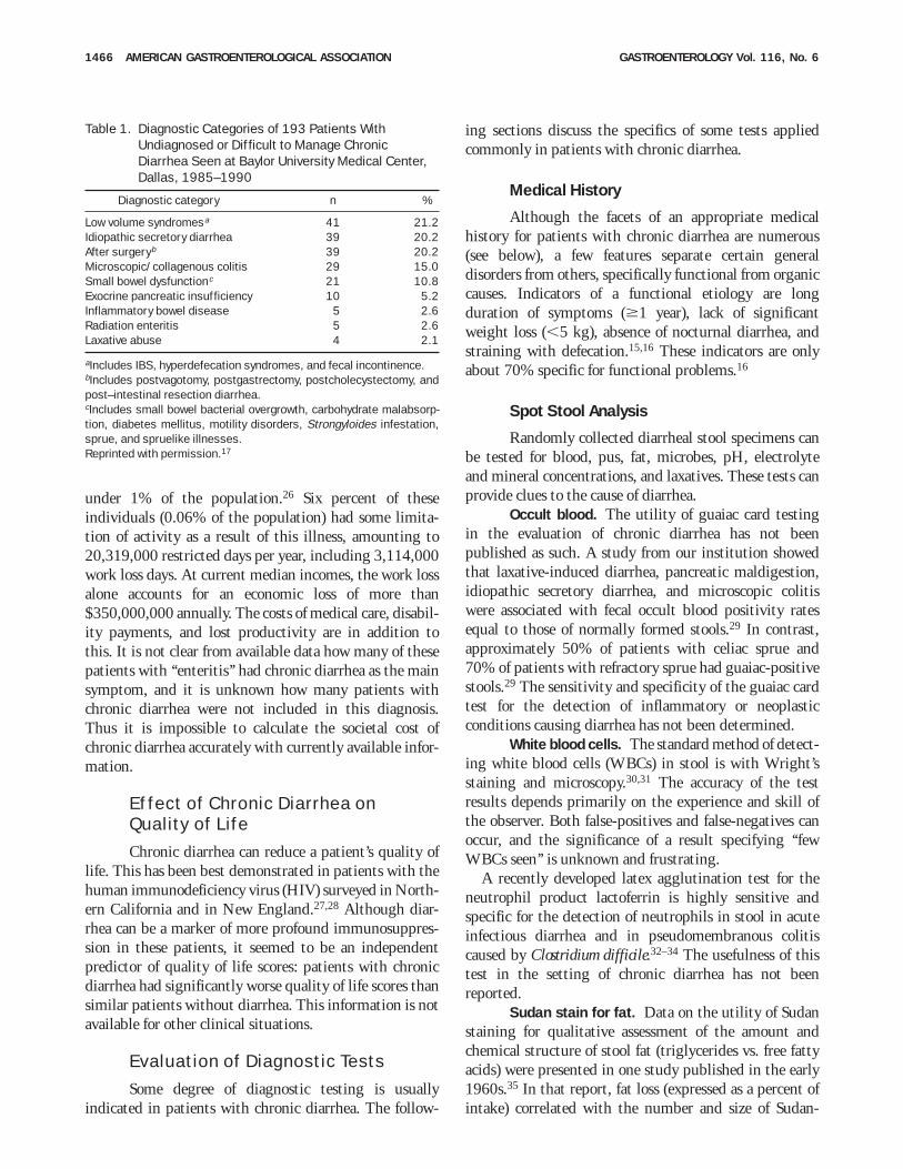
Within a given population or country, the prevalenceof different causes of chronic diarrhea is influenced by thelevel of subspecialization and the referral base of theparticular institution reporting its findings. For example,surreptitious laxative abuse is common in tertiary carereferral centers with a focused interest in diarrhealdisorders but may be uncommon in primary care set-tings.13,25 The frequency of various diagnoses in patientswith undiagnosed or refractory diarrhea seen at ourinstitution illustrates this sort of referral bias (Table 1).17
Economic Impact of ChronicDiarrhea
Observation of patients with chronic diarrheasuggests that chronic diarrhea can be a disabling prob-lem. Many patients cannot maintain employment be-cause of the need for or threat of frequent trips to thetoilet. In the absence of credible incidence or prevalencedata for diarrhea per se, it is difficult to estimate theeconomic impact of disability due to chronic diarrhea.For example, the National Health Interview Survey,1983–1987, reported a prevalence of ‘‘enteritis’’ of just
June 1999 AMERICAN GASTROENTEROLOGICAL ASSOCIATION 1465
under 1% of the population.26 Six percent of theseindividuals (0.06% of the population) had some limita-tion of activity as a result of this illness, amounting to20,319,000 restricted days per year, including 3,114,000work loss days. At current median incomes, the work lossalone accounts for an economic loss of more than$350,000,000 annually. The costs of medical care, disabil-ity payments, and lost productivity are in addition tothis. It is not clear from available data how many of thesepatients with ‘‘enteritis’’ had chronic diarrhea as the mainsymptom, and it is unknown how many patients withchronic diarrhea were not included in this diagnosis.Thus it is impossible to calculate the societal cost ofchronic diarrhea accurately with currently available infor-mation.
Effect of Chronic Diarrhea onQuality of Life
Chronic diarrhea can reduce a patient’s quality oflife. This has been best demonstrated in patients with thehuman immunodeficiency virus (HIV) surveyed in North-ern California and in New England.27,28 Although diar-rhea can be a marker of more profound immunosuppres-sion in these patients, it seemed to be an independentpredictor of quality of life scores: patients with chronicdiarrhea had significantly worse quality of life scores thansimilar patients without diarrhea. This information is notavailable for other clinical situations.
Evaluation of Diagnostic Tests
Some degree of diagnostic testing is usuallyindicated in patients with chronic diarrhea. The follow-
ing sections discuss the specifics of some tests appliedcommonly in patients with chronic diarrhea.
Medical History
Although the facets of an appropriate medicalhistory for patients with chronic diarrhea are numerous(see below), a few features separate certain generaldisorders from others, specifically functional from organiccauses. Indicators of a functional etiology are longduration of symptoms ($1 year), lack of significantweight loss (,5 kg), absence of nocturnal diarrhea, andstraining with defecation.15,16 These indicators are onlyabout 70% specific for functional problems.16
Spot Stool Analysis
Randomly collected diarrheal stool specimens canbe tested for blood, pus, fat, microbes, pH, electrolyteand mineral concentrations, and laxatives. These tests canprovide clues to the cause of diarrhea.
Occult blood. The utility of guaiac card testingin the evaluation of chronic diarrhea has not beenpublished as such. A study from our institution showedthat laxative-induced diarrhea, pancreatic maldigestion,idiopathic secretory diarrhea, and microscopic colitiswere associated with fecal occult blood positivity ratesequal to those of normally formed stools.29 In contrast,approximately 50% of patients with celiac sprue and70% of patients with refractory sprue had guaiac-positivestools.29 The sensitivity and specificity of the guaiac cardtest for the detection of inflammatory or neoplasticconditions causing diarrhea has not been determined.
White blood cells. The standard method of detect-ing white blood cells (WBCs) in stool is with Wright’sstaining and microscopy.30,31 The accuracy of the testresults depends primarily on the experience and skill ofthe observer. Both false-positives and false-negatives canoccur, and the significance of a result specifying ‘‘fewWBCs seen’’ is unknown and frustrating.
A recently developed latex agglutination test for theneutrophil product lactoferrin is highly sensitive andspecific for the detection of neutrophils in stool in acuteinfectious diarrhea and in pseudomembranous colitiscaused by Clostridium difficile.32–34 The usefulness of thistest in the setting of chronic diarrhea has not beenreported.
Sudan stain for fat. Data on the utility of Sudanstaining for qualitative assessment of the amount andchemical structure of stool fat (triglycerides vs. free fattyacids) were presented in one study published in the early1960s.35 In that report, fat loss (expressed as a percent ofintake) correlated with the number and size of Sudan-
Table 1. Diagnostic Categories of 193 Patients WithUndiagnosed or Difficult to Manage ChronicDiarrhea Seen at Baylor University Medical Center,Dallas, 1985–1990
Diagnostic category n %
Low volume syndromesa 41 21.2Idiopathic secretory diarrhea 39 20.2After surgeryb 39 20.2Microscopic/collagenous colitis 29 15.0Small bowel dysfunctionc 21 10.8Exocrine pancreatic insufficiency 10 5.2Inflammatory bowel disease 5 2.6Radiation enteritis 5 2.6Laxative abuse 4 2.1
aIncludes IBS, hyperdefecation syndromes, and fecal incontinence.bIncludes postvagotomy, postgastrectomy, postcholecystectomy, andpost–intestinal resection diarrhea.cIncludes small bowel bacterial overgrowth, carbohydrate malabsorp-tion, diabetes mellitus, motility disorders, Strongyloides infestation,sprue, and spruelike illnesses.Reprinted with permission.17
1466 AMERICAN GASTROENTEROLOGICAL ASSOCIATION GASTROENTEROLOGY Vol. 116, No. 6
stained fat droplets viewed microscopically. The test was86% specific for a fat output of #5% of intake and87%–100% sensitive for fat outputs of 6%–15% ofintake.35 However, the use of percent of intake as the unitof fat excretion (as opposed to the current standard, gramsexcreted per day) and qualitative expression of the results(as normal, slight increase, and definite increase) have ledto confusion in interpretation of the significance of apositive qualitative fat test result. Furthermore, a highlevel of observer skill and experience is critical to theaccuracy of the microscopic interpretation. In a morerecent study, the number of stained fat droplets countedin a hematocytometer correlated well with fat outputmeasured chemically.36 However, this method was evalu-ated in only 41 patients and has not been applied widelyelsewhere. The origin and types of fats that yield positiveresults on Sudan staining have been explored in anotherpublication.37 An alternative, semiquantitative measureof stool fat content, the steatocrit, has been used mainlyin children and correlates well with quantitative fatoutput as measured using the van de Kamer method.38–40
Fecal cultures. Because bacterial infections arerarely the cause of chronic diarrhea in immunocompetentpatients, routine fecal cultures usually are not obtained inmost individuals with chronic diarrhea. However, at leastone fecal culture should be performed at some point inthe evaluation of these patients.41,42 Cultures on specialmedia and under specific environmental conditions arerequired to look for Aeromonas or Pleisiomonas species.43–48
The epidemiological clues raising suspicion for thepresence of these organisms include consumption ofuntreated well water and swimming in fresh water pondsand streams.44
In immunocompromised patients, but only rarely innormal hosts, common infectious causes of acute diarrhea,such as Campylobacter or Salmonella, can cause persistentdiarrhea.49,50 In this population, bacterial cultures oughtto be part of the initial diagnostic evaluation.
Infections with yeast and fungi, mainly Candidaalbicans, have been reported as causes of both nosocomialand community-acquired chronic diarrhea, even in immu-nocompetent individuals.51–54 Increasing use of broad-spectrum antibiotics with greater ‘‘killing power’’ may beselecting for overgrowth of what had previously beenviewed as part of the normal flora. The yield of gramstains of stool and fungal cultures and the appropriatenessof their use in patients with chronic diarrhea of unknownorigin have not been studied.
Protozoa and parasites are endemic in third worldcountries but can also affect both immigrants to andnatives of developed countries, including the UnitedStates. Poorly sensitive cytological and pathological tests
for detection of Giardia lamblia are being replaced bymore sensitive and specific methods of detection, such asfecal enzyme-linked immunosorbent assay (ELISA) forGiardia-specific antigen.55 Although the old-fashionedstool examination for ‘‘O and P’’ remains popular, itspositive and negative predictive value in developedcountries is undefined. Observer skill is essential to thesuccess of this stool examination.56 Detection of somepathogens, such as Strongyloides larva, may be of clinicalimportance; however, cysts and ova of other organisms,including Entamoeba histolytica, may be innocent colonistsrather than pathogens, especially in inhabitants of thirdworld countries.57 Special techniques are required todetect cryptosporidia and microsporidia in stool; theseorganisms may cause diarrhea in immunocompetent aswell as immunocompromised people. Chronic viral infec-tions (a diagnostic consideration practically limited toimmunocompromised hosts) usually are diagnosed fromgastrointestinal mucosal biopsy specimens rather thanstool samples.
pH, electrolytes and minerals, and laxatives. Theutility of these measurements on a spot stool specimen isthe same as when they are measured in a quantitativelycollected specimen, as discussed below.
Quantitative Stool Collection andAnalysis
A 48- or 72-hour quantitative stool collection isuseful in the work-up of chronic diarrhea. Although thistest is not necessary in every case, it can be helpful incharacterizing the volume of diarrhea and segregatinglikely diagnostic possibilities from less likely ones (e.g.,by finding significant steatorrhea). However, the poten-tial benefits of obtaining these measurements (e.g., lesscostly directed work-up or fewer patient complicationsfrom invasive procedures) have not been established. Thenecessary duration of the collection has not been definedscientifically for clinical purposes. In general, the higherthe daily stool weight is, the more accurate and represen-tative shorter collection periods can be. Practical consid-erations mandate a 48-hour collection for most inpatientsand outpatients. In patients in whom 48-hour collectionyields a small or unrepresentative sample, the collectioncan be extended.
General principles. Full analysis of the collectionincludes measurement of weight, fat content, osmolality,electrolyte concentrations, magnesium concentration andoutput, pH, occult blood, and when appropriate, fecalchymotrypsin or elastase activity (for assessment ofpancreatic function) and screening for laxatives. If theseanalyses cannot be performed locally, many clinical
June 1999 AMERICAN GASTROENTEROLOGICAL ASSOCIATION 1467
laboratories and referral hospital laboratories can analyzea representative aliquot of stool (approximately 200 g)taken from a homogenized collection after it is weighed.(The aliquot should be frozen immediately; if mailing isnecessary, it should be mailed in a container packed withdry ice, along with a record of the total weight of the 48-or 72-hour collection.)
Quantitative stool collection can be done easily andsuccessfully at home or in the hospital. Some simpleequipment can facilitate collection, including a dispos-able collection unit that fits onto the commode andallows separation of stool and urine, several preweighedcontainers for the collected stool (e.g.,plastic or metalcontainers with airtight lids that hold at least 1 or 2 Leach), and a receptacle to keep these collection containerscold during the collection period, such as a small,portable refrigerator (usually used for inpatients) or apicnic cooler containing refreezable ‘‘blue ice’’ packs(ideal for outpatients).
Several days before and during the collection period,the patient should eat a regular diet of moderately highfat content. It may be useful to prescribe a fixed diet forsome patients to ensure that adequate amounts of fat andcalories are consumed. We encourage patients to consume80–100 g of fat during the collection, but fractionalabsorption can be calculated for any intake. Duringcollection, the patient should keep a record of bowelactivity and a diary of food and liquid intake (so thatcalorie, fat, carbohydrate, and fiber intake can be esti-mated from dietary tables). During the collection period,no diagnostic tests should be done that would disturb thenormal eating pattern, aggravate diarrhea (e.g., lactose–or D-xylose–absorption tests or tests using enteral iodin-ated contrast media), diminish diarrhea (by requiringfasting or use of opiates), add foreign material to the gut(e.g., barium radiography studies), or risk an episode ofincontinence. All but essential medications should beavoided, and any antidiarrheal medication begun beforethe collection period should be held.
Stool output may vary considerably from day to dayand week to week. When evaluating the results of aquantitative stool analysis, the physician needs to knowwhether the submitted stool was collected during a timethat the patient was having what he or she considered tobe diarrhea. It is advantageous for the physician to look atthe collected specimen and assess stool consistency visu-ally because the definition of diarrhea depends on the factthat stools are abnormally loose or liquid.
Fecal weight. Knowledge of stool weight mayhelp to clarify the nature of the patient’s problem and tolocalize the region of the intestine most likely to beresponsible for diarrhea (although the reliability of this is
untested). In some instances, knowledge of stool weightis of direct help in diagnosis and management. Forexample, stool weights greater than 500 g/day are rarelyif ever seen in patients with IBS,58,59 and stool weights ofless than 1000 g/day are evidence against pancreaticcholera syndrome. Also, very high stool weights alert thephysician to the possible need for vigorous fluid replace-ment; patients with stool weights greater than 2000g/day usually require supplemental intravenous fluids.Low stool weight in a patient complaining of ‘‘severediarrhea’’ suggests that incontinence or pain may be thedominant problem.
On rare occasions (e.g., when fecal volumes are extraor-dinarily high or there appears to be both a malabsorptiveand a secretory component to the diarrhea), it is useful todetermine the degree to which diarrhea persists duringfasting. Continuation of diarrhea during a 48-hour fast isone criterion for classification of the diarrhea as asecretory (nonosmotic) process.60 (Fecal weight maydecrease some with fasting, even in secretory diarrhea,because fluid input to the intestine decreases; however,continued diarrhea on the second day of fasting is anindicator of a secretory process.) Alternatively, completecessation of diarrhea during fasting is strong evidencethat the mechanism of diarrhea involves somethingingested (which could be a nonabsorbable substance ornutrient causing osmotic diarrhea, or unabsorbed fattyacids or laxatives causing secretory diarrhea). The re-sponse to fasting is more helpful for determination ofpathophysiology, thereby limiting the spectrum of poten-tial diagnoses, rather than for making a specific diagno-sis.61,62
Electrolytes and calculation of an osmotic gap.Fecal electrolyte concentrations are measured in stoolwater after homogenization of the entire specimen (bymanual stirring or in a mechanical blender) and centrifu-gation of an aliquot to obtain supernatant for analysis.Placement of dialysis bags in the stool is another reportedbut less commonly used method of obtaining stool waterfor analysis.63 The osmotic gap of fecal fluid can be usedto estimate the contribution of electrolytes and nonelec-trolytes to retention of water in the intestinal lumen. Insecretory diarrhea, unabsorbed electrolytes retain water inthe lumen; in osmotic diarrhea, nonelectrolytes causewater retention. The osmotic gap is calculated fromelectrolyte concentrations in stool water by the followingformula: 290 2 2([Na1] 1 [K1]). The sum of thesodium and potassium concentrations is multiplied by afactor of 2 to account for associated anions. The osmolal-ity of stool within the distal intestine (estimated as 290mOsm/kg because it equilibrates with plasma osmolality)should be used for this calculation rather than the
1468 AMERICAN GASTROENTEROLOGICAL ASSOCIATION GASTROENTEROLOGY Vol. 116, No. 6
osmolality measured in fecal fluid, because measured fecalosmolality begins to increase in the collection containeralmost immediately when carbohydrates are converted bybacterial fermentation to osmotically active organic acids.The advantages of using 290 mOsm/kg instead ofmeasured fecal osmolality for calculation of the fecalosmotic gap have been substantiated in two studies.64,65
The osmotic gap should be large (.125 mOsm/kg) inpure osmotic diarrhea, in which nonelectrolytes accountfor most of the osmolality of stool water, and small (,50mOsm/kg) in pure secretory diarrhea, in which electro-lytes account for most of stool osmolality.64 In mixedosmotic and secretory processes and in cases of modestcarbohydrate malabsorption (in which most of the carbo-hydrate load is converted to organic anions that obligatethe fecal excretion of cations including Na1 and K 1), theosmotic gap may lie between 50 and 125.64
Measured osmolality. Although measured fecalfluid osmolality should not be used to calculate theosmotic gap, measurement of fecal fluid osmolality maybe useful in patients with unexplained diarrhea. Lowosmolalities (,290 mOsm/kg) indicate contamination ofstool with water or dilute urine66 or the presence of agastrocolic fistula and ingestion of hypotonic fluid. Asmentioned previously, osmolalities of .290 mOsm/kgare common because of bacterial metabolism of fecalcarbohydrate during storage of the stool sample (up to600 mOsm/kg). Even higher values for fecal osmolalitycan be observed with ingestion of large amounts of poorlyabsorbable carbohydrate or dietary fiber, with fecalcontamination by concentrated urine, or with a gastro-colic fistula and ingestion of hypertonic fluids.
Fecal pH. A low fecal pH is characteristic ofdiarrhea caused solely by carbohydrate malabsorption.The results of measurement of fecal pH in experimentallyinduced diarrhea showed that a fecal pH of ,5.3indicates that carbohydrate malabsorption (such as thatassociated with lactulose or sorbitol ingestion) is a majorcause of diarrhea, whereas a pH of .5.6 argues againstcarbohydrate malabsorption as the only cause of diar-rhea.64 In generalized malabsorption syndrome that in-volves fecal loss of amino acids and fatty acids in additionto carbohydrate, the fecal pH usually is higher (e.g.,6.0–7.5).67
Fecal fat concentration and output. The concen-tration of fat per 100 g of stool can be quantitated byeither titration or gravimetric methods,68–72 and the dailyexcretion rate is obtained by multiplying this concentra-tion by the average daily weight of the 2- or 3- day stoolspecimen. In most clinical laboratories, the upper limit ofnormal for daily fecal fat output measured in normalsubjects (without diarrhea) ingesting normal amounts of
dietary fat is approximately 7 g/day (9% of dietary fatintake). By definition, values greater than this areabnormal and signify the presence of steatorrhea. How-ever, in a study of normal subjects with induced diarrhea(stool weights up to 1400 g/day), 35% had fecal fatexcretion measured above the upper limit of normal, withvalues as high as 13.6 g/day.73 Thus, even when mecha-nisms of digesting and absorbing dietary fat are intact,diarrhea itself causes steatorrhea (‘‘secondary steator-rhea’’). Therefore, in patients with diarrhea, an abnormalfecal fat value between 7 and 14 g/day has low specificityfor the diagnosis of primary defects of fat digestion orabsorption. On the other hand, abnormal values of 14g/day or higher are more specific for diseases that impairfat digestion or absorption (i.e., diseases of the exocrinepancreas, the small intestinal mucosa, or the enterohe-patic circulation of bile salts).
Dietary fat intake during the stool collection should beestimated from a diet diary. Most patients with diarrhea,especially patients with malabsorption, curb their foodintake (particularly of fatty foods) in an attempt to lessentheir diarrhea. Nausea and anorexia may also limit dietaryfat intake. If this is done during quantitative stoolcollection, patients with malabsorptive disorders mayhave fecal fat outputs lower than expected for theirsyndrome. Stool fat excretion normally should be ,9% ofdietary intake.
Although the reported fat concentration in stool (i.e.,fat per 100 g of stool) frequently is ignored, fecal fatconcentration may provide a clue to the cause of steator-rhea. In one study a fecal fat concentration of ,9.5 g/100g of stool was more likely to be seen in small intestinalmalabsorptive syndromes because of the diluting effectsof coexisting fluid malabsorption, whereas fecal fatconcentrations of $9.5 g/100 g of stool were seen inpancreatic and biliary steatorrhea, in which fluid absorp-tion in the small bowel is intact.74 Although in this studythe test was 100% sensitive, a second study found asensitivity of only 42%.75 However, specificity was highin both studies (80%–92%), meaning that, when present,a high fecal fat concentration should suggest the presenceof pancreatic or biliary steatorrhea.
Tests for fecal carbohydrate. Although tests forcarbohydrate content are not done routinely on collectedstool, qualitative tests for carbohydrates can be used toidentify malabsorbed carbohydrates. However, these tests,originally designed for measuring urinary carbohydrates,have not been standardized for stool analysis. Based onthe reagents in these products, the following conclusionsare likely: dipsticks based on glucose oxidase should givea positive reaction with glucose and should be negativewith all other sugars; Clinitest tablets (Ames Division,
June 1999 AMERICAN GASTROENTEROLOGICAL ASSOCIATION 1469
Miles Laboratories, Elkhart, IN) should give a positivereaction with glucose, galactose, fructose, maltose, andlactose (reducing sugars) but a negative result withsucrose, lactulose, sorbitol, and mannitol (nonreducingsugars or sugar alcohols). Anthrone reagent, used as aresearch tool to quantitate fecal carbohydrate,76 is sensi-tive to the presence of starches, oligosaccharides, disaccha-rides, and all hexoses but does not detect sorbitol andmannitol (sugar alcohols).
Analysis for laxatives. Diagnosis of factitiousdiarrhea requires a high index of suspicion. Analysis forlaxatives should be done early in the evaluation ofdiarrhea of unknown etiology.23,77–79 Because patientsmay ingest laxatives intermittently, negative studies mayhave to be repeated.77,80 The simplest test for a laxative isalkalinization of 3 mL of stool supernatant or urine withone drop of concentrated (1N) sodium hydroxide. Thiswill result in a pink or red color with a maximalspectrophotometric absorption of 550–555 nm if phenol-phthalein is present.81 (Phenolphthalein has been with-drawn from the market in the United States because offears of carcinogenicity but may be available elsewhere.)Stool water can be analyzed specifically for phenolphtha-lein, emetine (one component of ipecac syrup),82 andbisacodyl and its metabolites,83 using chromatographicor chemical tests. Urine can be analyzed for anthraqui-none derivatives.84,85
In searching for surreptitious laxative ingestion, stoolwater should be analyzed for osmolality and electro-lytes.86 If findings suggest secretory diarrhea (osmoticgap ,50), the patient may have ingested a laxativecapable of causing secretory diarrhea. Diarrhea caused bysodium sulfate or sodium phosphate ingestion alsoappears to be a secretory diarrhea by electrolyte analysis,even though pathophysiologically it is an osmotic diar-rhea. This occurs because the negative charges of unab-sorbed sulfate or phosphate obligate sodium, potassium,and other cations remain in the colonic lumen.64,87 Thefecal concentration and daily output of sulfate andphosphate can be measured by chemical testing, but theupper limits of normal have not been established. A highfecal sodium concentration in the presence of a low fecalchloride concentration should also raise the suspicion ofingestion of sodium sulfate or sodium phosphate.64
If stool electrolyte analysis suggests osmotic diarrhea(osmotic gap .125 mOsm/kg), magnesium (Mg21)laxatives may have been ingested. A soluble fecal Mgconcentration greater than 45 mmol/L (90 mEq/L) or adaily fecal Mg output much above 15 mmol/day (30mEq/day) strongly suggests Mg-induced diarrhea.88 Fac-titious diarrhea may be caused by deliberate contamina-tion of a stool collection with water urine. If fecal
osmolality is significantly less than 290 mOsm/kg (theosmolality of plasma), water or hypotonic urine has beenadded to the stool.66 If the osmolality is far above that ofplasma, hypertonic urine may have been added to stool(although this finding also may be caused by theproduction of fermentation products in vitro). Urinarycontamination can be confirmed by a finding of highmonovalent cation concentration (e.g., [Na1] 1 [K1] .165, physiologically impossible in stool water) and a highconcentration of urea or creatinine in stool water.
Because many institutions lack analytical methods forall available laxatives, searching the patient’s hospitalroom or home for hidden laxatives has been used toestablish the diagnosis of factitious diarrhea. Discovery ofcaches of laxatives can also be helpful in convincingrelatives that the diarrhea is caused by laxative ingestion.In one series, a room search had a higher diagnostic yieldfor factitious diarrhea than any other test.89 However,some physicians think that it is unethical invasion ofprivacy to search a patient’s belongings for laxatives anddiuretics without permission. Others believe that a searchshould be viewed as a diagnostic study, requiring in-formed consent and including a discussion with thepatient of the procedure, its risks and benefits, andalternatives.90 On the other hand, failure to discoverlaxative abuse may lead to needless hazardous tests andtreatment, such as insertion of central venous catheters,administration of total parenteral nutrition, and diagnos-tic and/or ‘‘therapeutic’’ operations, such as partial pancre-atectomy and total colectomy. Even more importantly,laxative abuse may be fatal in children whose caregiversare poisoning them with laxatives.87,91 Despite thepotential of protecting the patient from self-inducedharm, the legality of searching a patient’s belongingswithout permission is questionable, and such searches arediscouraged by most attorneys. Because laxative assays arereadily available from reference laboratories, room searchesshould be done only under exceptional circumstances.
Tests for protein-losing enteropathy. A diagno-sis of protein-losing enteropathy should be consideredwhen a patient has hypoalbuminemia but does not havenephrotic syndrome or hepatic dysfunction. Confirma-tion of enteric protein loss can come from measurement ofthe fecal clearance of a1-antitrypsin.92 Clearance of thisprotein from plasma via the intestinal tract is based onthe same concepts as renal inulin clearance and iscalculated in similar fashion. Radioimmunoassay is usedto measure a1-antitrypsin concentrations in stool andplasma; total fecal output is calculated from concentra-tion and volume and is divided by plasma concentration.Measurement of the concentration of a1-antitrypsin inrandomly passed stools has been tried as a simpler
1470 AMERICAN GASTROENTEROLOGICAL ASSOCIATION GASTROENTEROLOGY Vol. 116, No. 6
method to measure intestinal protein loss in children buthas had only moderate success.93 Fecal excretion ofradioiodinated albumin and immunoglobulin G adminis-tered parenterally has also been reported as a method ofmeasuring enteric protein loss but is not availableroutinely.94,95
Blood and Urine Tests
Analysis of urine. Urine collections may be help-ful for laxative identification and for measurement ofexcretion of 5-hydroxyindole acetic acid (for carcinoidsyndrome), vanillylmandelic acid (VMA; for pheochromo-cytoma, metanephrine (for pheochromocytoma), and his-tamine (for mast cell disease and foregut carcinoids). Ifvolume depletion or hypokalemia are present, analysis ofurine electrolytes can determine whether renal conserva-tion of sodium and potassium is appropriate. If theurinary concentration or output of sodium or potassium isinappropriately high, surreptitious diuretic use may bepresent and may suggest coexisting laxative abuse. Also,measurement of urine electrolytes and aldosterone maydistinguish hypervolemia from volume depletion in thesetting of hypernatremia caused by ingestion of sodium-containing laxatives.88
Vasoactive intestinal polypeptide and other pep-tide hormones. Pancreatic cholera syndrome is a rarecause of secretory diarrhea attributable to secretion ofvasoactive intestinal polypeptide (VIP) by a neuroendo-crine tumor. It should be suspected if diarrhea ofunknown origin has lasted longer than 4 weeks, has theclinical features of secretory diarrhea, has a volumegreater than 1 L/day, is associated with hypokalemia, andcauses clinically significant volume depletion and ifsurreptitious laxative and diuretic abuse and organicdisease of the gastrointestinal tract have been excluded. Itis only in this rare subgroup of patients that serum assayfor VIP is likely to be useful. Measurement of a few otherspecific peptides can be helpful in the diagnosis of otherneuroendocrine tumors. These include measurement ofcalcitonin for the diagnosis of medullary carcinoma of thethyroid, gastrin for suspected Zollinger–Ellison syn-drome, and glucagon for the rare patient with a gluca-gonoma.17 Measurement of large panels of enteric pep-tides not specific for particular tumor syndromes, such asmotilin, neurotensin, pancreatic polypeptide, substanceP, and gastrin-releasing peptide, should not be done inpatients with chronic diarrhea because of their poorspecificity and extremely low positive predictive value,17
which is attributable to the rarity of these tumors and thehigh frequency of false-positive assays.
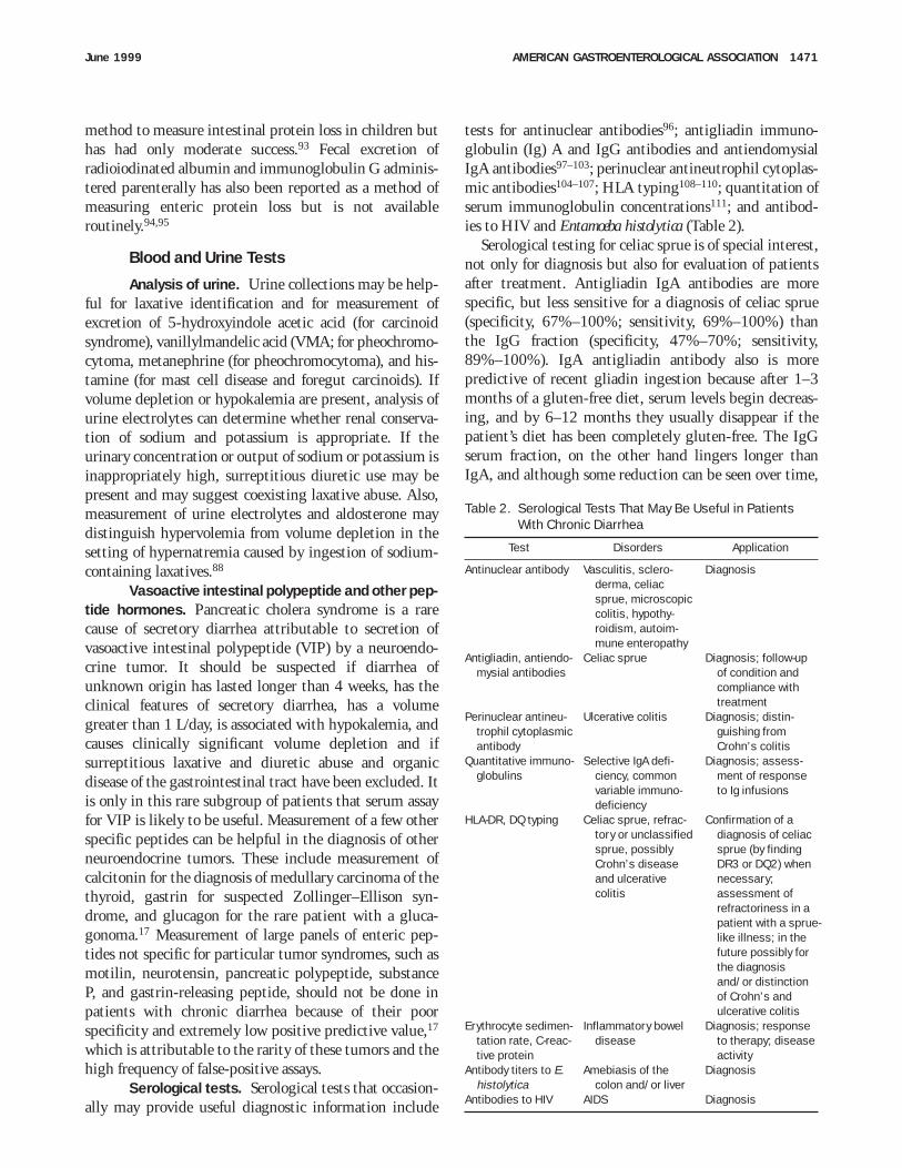
Serological tests. Serological tests that occasion-ally may provide useful diagnostic information include
tests for antinuclear antibodies96; antigliadin immuno-globulin (Ig) A and IgG antibodies and antiendomysialIgA antibodies97–103; perinuclear antineutrophil cytoplas-mic antibodies104–107; HLA typing108–110; quantitation ofserum immunoglobulin concentrations111; and antibod-ies to HIV and Entamoeba histolytica (Table 2).
Serological testing for celiac sprue is of special interest,not only for diagnosis but also for evaluation of patientsafter treatment. Antigliadin IgA antibodies are morespecific, but less sensitive for a diagnosis of celiac sprue(specificity, 67%–100%; sensitivity, 69%–100%) thanthe IgG fraction (specificity, 47%–70%; sensitivity,89%–100%). IgA antigliadin antibody also is morepredictive of recent gliadin ingestion because after 1–3months of a gluten-free diet, serum levels begin decreas-ing, and by 6–12 months they usually disappear if thepatient’s diet has been completely gluten-free. The IgGserum fraction, on the other hand lingers longer thanIgA, and although some reduction can be seen over time,
Table 2. Serological Tests That May Be Useful in PatientsWith Chronic Diarrhea
Test Disorders Application
Antinuclear antibody Vasculitis, sclero-derma, celiacsprue, microscopiccolitis, hypothy-roidism, autoim-mune enteropathy
Diagnosis
Antigliadin, antiendo-mysial antibodies
Celiac sprue Diagnosis; follow-upof condition andcompliance withtreatment
Perinuclear antineu-trophil cytoplasmicantibody
Ulcerative colitis Diagnosis; distin-guishing fromCrohn’s colitis
Quantitative immuno-globulins
Selective IgA defi-ciency, commonvariable immuno-deficiency
Diagnosis; assess-ment of responseto Ig infusions
HLA-DR, DQ typing Celiac sprue, refrac-tory or unclassifiedsprue, possiblyCrohn’s diseaseand ulcerativecolitis
Confirmation of adiagnosis of celiacsprue (by findingDR3 or DQ2) whennecessary;assessment ofrefractoriness in apatient with a sprue-like illness; in thefuture possibly forthe diagnosisand/or distinctionof Crohn’s andulcerative colitis
Erythrocyte sedimen-tation rate, C-reac-tive protein
Inflammatory boweldisease
Diagnosis; responseto therapy; diseaseactivity
Antibody titers to E.histolytica
Amebiasis of thecolon and/or liver
Diagnosis
Antibodies to HIV AIDS Diagnosis
June 1999 AMERICAN GASTROENTEROLOGICAL ASSOCIATION 1471
IgG antigliadin antibodies may never disappear com-pletely.
Antiendomysial antibodies are the most specific of theserological tests for celiac sprue, with a specificity invillous atrophic disease of nearly 100%. However, theirsensitivity has ranged from as low as 74% up to 100%.There is inadequate information regarding the sensitivityof antiendomysial antibodies in less severe forms ofgluten sensitivity, but it may well be lower. Until now,the only test to detect endomysial antibodies has beenindirect immunofluorescence in monkey esophagus orhuman umbilical cord tissue substrates. As with allimmunofluorescent tests, correct interpretation of resultsis highly dependent on the skill and experience of atechnician interpreting the fluorescence pattern in tis-sues, and quantitation of the amount of antibody presentrelies on repeat examinations after serial dilutions ofserum. An ELISA test that can detect and quantitateserum endomysial antibody probably will be availablesoon for clinical use.
Endoscopic Examination andMucosal Biopsy
Sigmoidoscopy and colonoscopy. Examinationof the mucosa of the colon and rectum and mucosalbiopsy may be useful in patients with chronic diarrhea,but it is unclear whether the initial procedure shouldinvolve a 60-cm flexible sigmoidoscopy or a full colonos-copy. The advantages of the former include its ease (i.e.,simple preparation, no need for sedation, shorter proce-dure, and greater chance for successful completion), lowerrisk of perforation, and lower cost. Patient acceptancemay be better or worse than with colonoscopy (becausepatients are usually sedated for colonoscopy). The mainconcern with sigmoidoscopy is that the causative diseasemay be present only in the proximal colon or terminalileum and will be missed by a limited examination.Although this can occur in Crohn’s disease and other rareidiopathic inflammatory conditions,112 the pathologicalprocess occurs diffusely throughout the colon in mostdiseases that can be diagnosed by lower endoscopy. Forexample, microscopic and collagenous colitis are usuallydiffuse processes, but inflammatory changes or subepithe-lial collagen band thickening may occur only in theproximal colon in approximately 10% of patients.113,114
Thus relatively few cases remain undiagnosed withlimited examination. Therefore, in light of the advan-tages of flexible sigmoidoscopy over colonoscopy listedabove, sigmoidoscopy can be recommended as the bestinitial test. During sigmoidoscopy, random biopsy speci-mens should be obtained from the descending colon, thesigmoid colon, and the rectum (e.g., four biopsy speci-
mens taken every 10–20 cm). When results of otherdiagnostic tests raise a strong suspicion of a colonicprocess (e.g., when leukocytes or lactoferrin are present instool),when the presence of inflammatory bowel disease issuggested by specific symptoms or signs, or when biopsyspecimens from the distal colon are equivocal, colonos-copy may provide additional helpful information. Whenthere is significant weight loss or gross or occult bleedingto suggest malignancy, or when an abnormality of theterminal ileum or proximal colon has been seen on animaging study or radiogram, it is appropriate to beginendoscopic evaluation of the colon with a full colonos-copy. However, no prospective study has assessed theutility and costs of limited vs. complete examination ofthe colon in patients with chronic diarrhea, although itseems that complete colonoscopy would be more expen-sive and rarely leads to an additional diagnosis.115
Chronic disorders that can be diagnosed by inspectionof the colonic mucosa include melanosis coli, ulceration,polyps, tumors, Crohn’s disease, ulcerative colitis, andamebiasis.116–118 Diseases in which the mucosa appearsnormal endoscopically but that can be diagnosed histologi-cally include microscopic colitis (lymphocytic and collag-enous colitis), amyloidosis, Whipple’s disease, granu-lomatous infections, and schistosomiasis in its chronicform.
Upper tract endoscopy. Upper endoscopy hasbecome the standard method for obtaining biopsy speci-mens from the upper small intestine.119,120 If a smallintestinal malabsorptive disorder is strongly suspected,the procedure is probably best performed with anendoscope that allows specimens to be obtained from thedistal duodenum and/or proximal jejunum as well as fromthe proximal duodenum, although duodenal biopsiesmay be adequate to discover most diffuse mucosaldiseases. An aspirate of small intestinal contents can besent for quantitative aerobic and anaerobic bacterialculture (using techniques used for quantitative urineculture) if bacterial overgrowth is suspected and formicroscopic examination for parasites. Diseases that maybe diagnosed by small intestinal biopsy include Crohn’sdisease, giardiasis, celiac sprue, intestinal lymphomawith or without villous atrophy, eosinophilic gastroenteri-tis, hypogammaglobulinemic sprue (with or withoutnodular lymphoid hyperplasia), Whipple’s disease, lym-phangiectasia, abetalipoproteinemia, amyloidosis, masto-cytosis, and various mycobacterial, fungal, protozoal, andparasitic infections.121–123 The presence of steatorrhea orfecal occult blood increases the likelihood of making oneof these diagnoses by upper endoscopy.
1472 AMERICAN GASTROENTEROLOGICAL ASSOCIATION GASTROENTEROLOGY Vol. 116, No. 6
Radiography
Barium radiography. There have been no formalstudies of the utility of radiography in the diagnosticevaluation of chronic diarrhea. However, because most ofthe small intestine (including most of the terminal ileum)cannot be approached with standard endoscopes, ana-tomic changes are best assessed with barium radiography.There are situations in which a previously unsuspecteddiagnosis is made by small intestinal radiography (such asCrohn’s disease or jejunal diverticulosis). In other situa-tions, abnormal findings will lead to further investigationand, ultimately, a diagnosis. For example, a ‘‘malabsorp-tion pattern’’ consisting of excess luminal fluid, dilation,and an irregular mucosal surface may lead to a diagnosisof celiac sprue, Whipple’s disease, or intestinal lym-phoma, although this may be less common with modernbarium preparations than in the past.124 Other diseasesthat might be diagnosed with small intestinal radiogra-phy are carcinoid tumors and scleroderma.
Although it has not been tested specifically in patientswith chronic diarrhea, the diagnostic yield of small bowelradiography is about the same whether barium is admin-istered orally (‘‘small bowel follow-through examina-tion’’) or by an enteroclysis tube, provided that the smallbowel follow-through study is performed by a ‘‘dedi-cated’’ radiologist who personally watches the column ofbarium and uses fluoroscopy intermittently (rather than atechnologist who performs overhead radiographs at someset time intervals).125,126 Thus, for the study of patientswith chronic diarrhea, enteroclysis probably has nospecial role.
Radiographic studies of the stomach and colon may becomplementary to endoscopy and colonoscopy becausebarium-contrast radiograms can better detect fistulas andstrictures. Radiography of the gastrointestinal tract alsohelps to delineate anatomy after previous surgical resec-tion or bypass.
Mesenteric angiography. Small intestinal isch-emia is a rare cause of chronic diarrhea.127,128 In theappropriate clinical setting, mesenteric or celiac angiogra-phy may show evidence of intestinal ischemia caused byatherosclerosis or vasculitis. The utility of magneticresonance imaging or spiral computed tomographicangiography in this setting is not clear.
Computed tomography. Computed tomographyis performed in patients with chronic diarrhea to examinefor pancreatic cancer or evidence of chronic pancreatitis inthe presence of malabsorption or when the results ofpancreatic function tests are abnormal. Inflammatorybowel disease, chronic infections such as tuberculosis,intestinal lymphoma, carcinoid syndrome, and other
neuroendocrine tumors are additional diagnoses that canbe revealed by computed tomography. In the case of thetumors mentioned last, rapid computed tomographyscanning with thin (5 mm or less) sections through thepancreas following a bolus of intravenous contrast isrecommended, although the degree to which sensitivityis increased over standard computed tomographic meth-ods is unknown.
Physiological Tests
Mucosal absorption. Tests of monosaccharide ab-sorption have been used classically to distinguish smallintestinal mucosal absorptive defects from pancreaticdigestive defects in the setting of malabsorption. In the1950s, the oral glucose tolerance test was replaced by theD-xylose–absorption test because of better reliability andthe lack of interference from endogenous serum glu-cose.129,130 Subsequently, an abnormal D-xylose–absorp-tion test result became synonymous with a diseased smallintestine (barring confounding renal dysfunction or urinecollection problems) and was used to determine whowould be served best by capsule biopsies of the smallbowel. Today, with the widespread use of endoscopicbiopsies, the role of this test has become less clear,although it still yields information about small intestinalabsorptive function. Some clinicians still use this test forscreening or for following up the response to treatment,but the utility of this approach has not been assessedformally.
Most verification studies of the D-xylose test involvepatients with celiac sprue or inflammatory bowel diseaseof the small bowel.129,130 Urinary excretion of less than5 g in the 5 hours following ingestion of 25 g of D-xyloseis considered abnormal. A plasma concentration of lessthan 1.3 mmol/L per 1.73 m2 body surface area (20mg/dL for an average adult) 1 hour after a 25-g oral doseis considered abnormal.131
Tests of ileal absorptive function. The terminalileum has evolved three specific and unique absorptivefunctions: absorption of vitamin B12, absorption ofsodium chloride against steep electrochemical gradients,and absorption of bile acids. Disruption of any one ofthese, particularly of all three, can be seen in patientswith diarrhea. Therefore, tests have been developed toassess these specialized ileal absorptive functions.
The time-honored test of vitamin B12 absorption is theSchilling test.132 In patients being evaluated for chronicdiarrhea (as opposed to those being evaluated for vitaminB12 deficiency or macrocytic anemia), radiolabeled vita-min B12 is given with intrinsic factor (the so-called‘‘Schilling II test’’). The positive and negative predictivevalues of the test for ileal dysfunction in a large group of
June 1999 AMERICAN GASTROENTEROLOGICAL ASSOCIATION 1473
patients with chronic diarrhea of unknown origin has notbeen determined. With the widespread availability ofcolonoscopy, which can visualize the terminal ileum,sophisticated radiographic imaging techniques, and stan-dard barium radiography, the Schilling test is now lessimportant in the investigation of chronic diarrhea than inthe past.
The only method developed to study fluid and electro-lyte absorption in the ileum is intestinal perfusion.Perfusion of a segment of ileum can be carried out butrequires painstaking effort by the patient and investiga-tor to place the tube in the distal small bowel. Alterna-tively, total gastrointestinal perfusion (with the infusionport in the stomach and the effluent collected from therectum) can be performed. To assess ileal function, totalgut perfusion is carried out first with a balanced electro-lyte solution, and then a solution containing a lowconcentration of sodium chloride compared with plasma,which requires an intact functional ileum for normalactive absorption.133 Although this technique has uncov-ered specific absorptive defects in patients with ileoco-lonic resection and rare patients with idiopathic ilealdysfunction, intestinal perfusion is not clinically useful inmost cases of chronic diarrhea.134
Tests for bile acid malabsorption can be done in twoways. The first method measures the quantity of endog-enous bile acids excreted during a quantitative stoolcollection. The second method involves measurement ofthe turnover of radiolabeled bile acid. This can be done intwo ways. The first involves the use of a gamma camera todetect the retained fraction of an orally administeredsynthetic radiolabeled bile acid, selena-homocholic acidconjugated with taurine (commonly abbreviated 75Se-HCAT).135–140 The second involves measurement of fecalrecovery of an oral load of [14C]glycocholate during a48- or 72-hour stool collection and calculation of aretention half-life.141,142 Measurement of serum concen-trations of an intermediate of bile acid synthesis, 7a-hydroxy-4-cholesten-3-one, is an alternate method forevaluating bile acid metabolism, with a reported sensitiv-ity and specificity of 80% and 85%, and positive andnegative predictive values of 74% and 98%, respectively,for excessive losses of bile acids from the body.143,144
These tests are not widely available to clinicians and havebeen complicated by lack of standardization of referencevalues.145 Furthermore, an abnormal test result is notnecessarily specific for pathological bile acid malabsorp-tion but may occur as the result of diarrhea perse.141,142,146,147 Therefore, many clinicians use a therapeu-tic trial of cholestyramine as an indirect test for thepossibility that malabsorbed bile acids are the cause ofdiarrhea. However, the extent to which a good therapeu-
tic response to cholestyramine denotes the presence ofbile acid malabsorption as the main cause of diarrhea is anunsettled issue.141,142
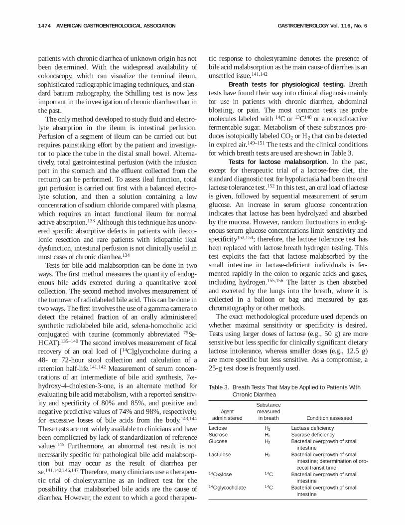
Breath tests for physiological testing. Breathtests have found their way into clinical diagnosis mainlyfor use in patients with chronic diarrhea, abdominalbloating, or pain. The most common tests use probemolecules labeled with 14C or 13C148 or a nonradioactivefermentable sugar. Metabolism of these substances pro-duces isotopically labeled CO2 or H2 that can be detectedin expired air.149–151 The tests and the clinical conditionsfor which breath tests are used are shown in Table 3.
Tests for lactose malabsorption. In the past,except for therapeutic trial of a lactose-free diet, thestandard diagnostic test for hypolactasia had been the orallactose tolerance test.152 In this test, an oral load of lactoseis given, followed by sequential measurement of serumglucose. An increase in serum glucose concentrationindicates that lactose has been hydrolyzed and absorbedby the mucosa. However, random fluctuations in endog-enous serum glucose concentrations limit sensitivity andspecificity153,154; therefore, the lactose tolerance test hasbeen replaced with lactose breath hydrogen testing. Thistest exploits the fact that lactose malabsorbed by thesmall intestine in lactase-deficient individuals is fer-mented rapidly in the colon to organic acids and gases,including hydrogen.155,156 The latter is then absorbedand excreted by the lungs into the breath, where it iscollected in a balloon or bag and measured by gaschromatography or other methods.
The exact methodological procedure used depends onwhether maximal sensitivity or specificity is desired.Tests using larger doses of lactose (e.g., 50 g) are moresensitive but less specific for clinically significant dietarylactose intolerance, whereas smaller doses (e.g., 12.5 g)are more specific but less sensitive. As a compromise, a25-g test dose is frequently used.
Table 3. Breath Tests That May be Applied to Patients WithChronic Diarrhea
Agentadministered
Substancemeasuredin breath Condition assessed
Lactose H2 Lactase deficiencySucrose H2 Sucrase deficiencyGlucose H2 Bacterial overgrowth of small
intestineLactulose H2 Bacterial overgrowth of small
intestine; determination of oro-cecal transit time
14C-xylose 14C Bacterial overgrowth of smallintestine
14C-glycocholate 14C Bacterial overgrowth of smallintestine
1474 AMERICAN GASTROENTEROLOGICAL ASSOCIATION GASTROENTEROLOGY Vol. 116, No. 6
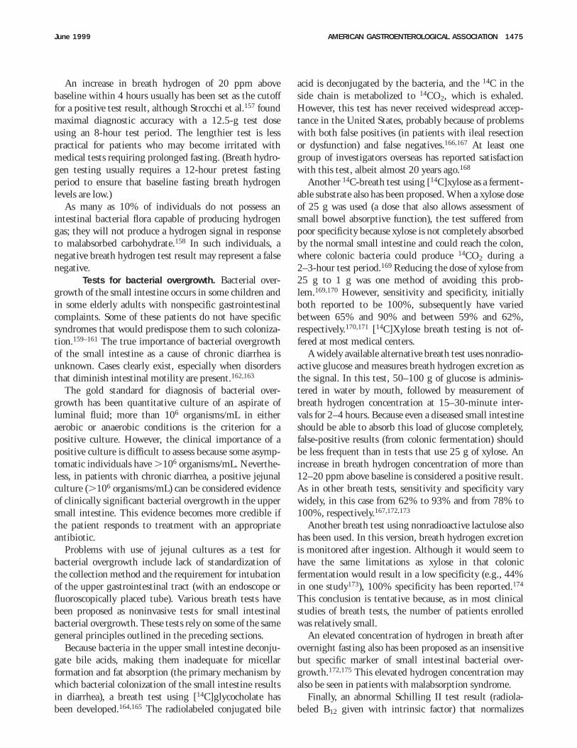
An increase in breath hydrogen of 20 ppm abovebaseline within 4 hours usually has been set as the cutofffor a positive test result, although Strocchi et al.157 foundmaximal diagnostic accuracy with a 12.5-g test doseusing an 8-hour test period. The lengthier test is lesspractical for patients who may become irritated withmedical tests requiring prolonged fasting. (Breath hydro-gen testing usually requires a 12-hour pretest fastingperiod to ensure that baseline fasting breath hydrogenlevels are low.)
As many as 10% of individuals do not possess anintestinal bacterial flora capable of producing hydrogengas; they will not produce a hydrogen signal in responseto malabsorbed carbohydrate.158 In such individuals, anegative breath hydrogen test result may represent a falsenegative.
Tests for bacterial overgrowth. Bacterial over-growth of the small intestine occurs in some children andin some elderly adults with nonspecific gastrointestinalcomplaints. Some of these patients do not have specificsyndromes that would predispose them to such coloniza-tion.159–161 The true importance of bacterial overgrowthof the small intestine as a cause of chronic diarrhea isunknown. Cases clearly exist, especially when disordersthat diminish intestinal motility are present.162,163
The gold standard for diagnosis of bacterial over-growth has been quantitative culture of an aspirate ofluminal fluid; more than 106 organisms/mL in eitheraerobic or anaerobic conditions is the criterion for apositive culture. However, the clinical importance of apositive culture is difficult to assess because some asymp-tomatic individuals have .106 organisms/mL. Neverthe-less, in patients with chronic diarrhea, a positive jejunalculture (.106 organisms/mL) can be considered evidenceof clinically significant bacterial overgrowth in the uppersmall intestine. This evidence becomes more credible ifthe patient responds to treatment with an appropriateantibiotic.
Problems with use of jejunal cultures as a test forbacterial overgrowth include lack of standardization ofthe collection method and the requirement for intubationof the upper gastrointestinal tract (with an endoscope orfluoroscopically placed tube). Various breath tests havebeen proposed as noninvasive tests for small intestinalbacterial overgrowth. These tests rely on some of the samegeneral principles outlined in the preceding sections.
Because bacteria in the upper small intestine deconju-gate bile acids, making them inadequate for micellarformation and fat absorption (the primary mechanism bywhich bacterial colonization of the small intestine resultsin diarrhea), a breath test using [14C]glycocholate hasbeen developed.164,165 The radiolabeled conjugated bile
acid is deconjugated by the bacteria, and the 14C in theside chain is metabolized to 14CO2, which is exhaled.However, this test has never received widespread accep-tance in the United States, probably because of problemswith both false positives (in patients with ileal resectionor dysfunction) and false negatives.166,167 At least onegroup of investigators overseas has reported satisfactionwith this test, albeit almost 20 years ago.168
Another 14C-breath test using [14C]xylose as a ferment-able substrate also has been proposed. When a xylose doseof 25 g was used (a dose that also allows assessment ofsmall bowel absorptive function), the test suffered frompoor specificity because xylose is not completely absorbedby the normal small intestine and could reach the colon,where colonic bacteria could produce 14CO2 during a2–3-hour test period.169 Reducing the dose of xylose from25 g to 1 g was one method of avoiding this prob-lem.169,170 However, sensitivity and specificity, initiallyboth reported to be 100%, subsequently have variedbetween 65% and 90% and between 59% and 62%,respectively.170,171 [14C]Xylose breath testing is not of-fered at most medical centers.
A widely available alternative breath test uses nonradio-active glucose and measures breath hydrogen excretion asthe signal. In this test, 50–100 g of glucose is adminis-tered in water by mouth, followed by measurement ofbreath hydrogen concentration at 15–30-minute inter-vals for 2–4 hours. Because even a diseased small intestineshould be able to absorb this load of glucose completely,false-positive results (from colonic fermentation) shouldbe less frequent than in tests that use 25 g of xylose. Anincrease in breath hydrogen concentration of more than12–20 ppm above baseline is considered a positive result.As in other breath tests, sensitivity and specificity varywidely, in this case from 62% to 93% and from 78% to100%, respectively.167,172,173
Another breath test using nonradioactive lactulose alsohas been used. In this version, breath hydrogen excretionis monitored after ingestion. Although it would seem tohave the same limitations as xylose in that colonicfermentation would result in a low specificity (e.g., 44%in one study173), 100% specificity has been reported.174
This conclusion is tentative because, as in most clinicalstudies of breath tests, the number of patients enrolledwas relatively small.
An elevated concentration of hydrogen in breath afterovernight fasting also has been proposed as an insensitivebut specific marker of small intestinal bacterial over-growth.172,175 This elevated hydrogen concentration mayalso be seen in patients with malabsorption syndrome.
Finally, an abnormal Schilling II test result (radiola-beled B12 given with intrinsic factor) that normalizes
June 1999 AMERICAN GASTROENTEROLOGICAL ASSOCIATION 1475
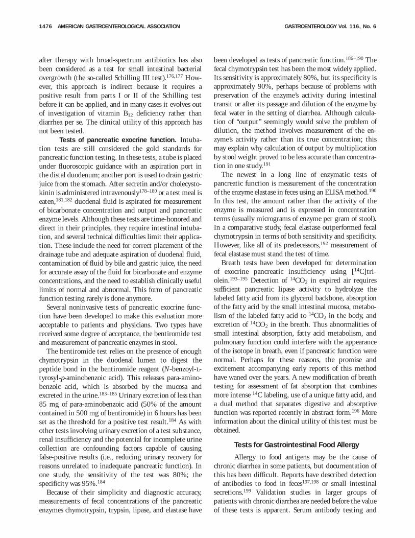
after therapy with broad-spectrum antibiotics has alsobeen considered as a test for small intestinal bacterialovergrowth (the so-called Schilling III test).176,177 How-ever, this approach is indirect because it requires apositive result from parts I or II of the Schilling testbefore it can be applied, and in many cases it evolves outof investigation of vitamin B12 deficiency rather thandiarrhea per se. The clinical utility of this approach hasnot been tested.
Tests of pancreatic exocrine function. Intuba-tion tests are still considered the gold standards forpancreatic function testing. In these tests, a tube is placedunder fluoroscopic guidance with an aspiration port inthe distal duodenum; another port is used to drain gastricjuice from the stomach. After secretin and/or cholecysto-kinin is administered intravenously178–180 or a test meal iseaten,181,182 duodenal fluid is aspirated for measurementof bicarbonate concentration and output and pancreaticenzyme levels. Although these tests are time-honored anddirect in their principles, they require intestinal intuba-tion, and several technical difficulties limit their applica-tion. These include the need for correct placement of thedrainage tube and adequate aspiration of duodenal fluid,contamination of fluid by bile and gastric juice, the needfor accurate assay of the fluid for bicarbonate and enzymeconcentrations, and the need to establish clinically usefullimits of normal and abnormal. This form of pancreaticfunction testing rarely is done anymore.
Several noninvasive tests of pancreatic exocrine func-tion have been developed to make this evaluation moreacceptable to patients and physicians. Two types havereceived some degree of acceptance, the bentiromide testand measurement of pancreatic enzymes in stool.
The bentiromide test relies on the presence of enoughchymotrypsin in the duodenal lumen to digest thepeptide bond in the bentiromide reagent (N-benzoyl-L-tyrosyl-p-aminobenzoic acid). This releases para-amino-benzoic acid, which is absorbed by the mucosa andexcreted in the urine.183–185 Urinary excretion of less than85 mg of para-aminobenzoic acid (50% of the amountcontained in 500 mg of bentiromide) in 6 hours has beenset as the threshold for a positive test result.184 As withother tests involving urinary excretion of a test substance,renal insufficiency and the potential for incomplete urinecollection are confounding factors capable of causingfalse-positive results (i.e., reducing urinary recovery forreasons unrelated to inadequate pancreatic function). Inone study, the sensitivity of the test was 80%; thespecificity was 95%.184
Because of their simplicity and diagnostic accuracy,measurements of fecal concentrations of the pancreaticenzymes chymotrypsin, trypsin, lipase, and elastase have
been developed as tests of pancreatic function.186–190 Thefecal chymotrypsin test has been the most widely applied.Its sensitivity is approximately 80%, but its specificity isapproximately 90%, perhaps because of problems withpreservation of the enzyme’s activity during intestinaltransit or after its passage and dilution of the enzyme byfecal water in the setting of diarrhea. Although calcula-tion of ‘‘output’’ seemingly would solve the problem ofdilution, the method involves measurement of the en-zyme’s activity rather than its true concentration; thismay explain why calculation of output by multiplicationby stool weight proved to be less accurate than concentra-tion in one study.191
The newest in a long line of enzymatic tests ofpancreatic function is measurement of the concentrationof the enzyme elastase in feces using an ELISA method.190
In this test, the amount rather than the activity of theenzyme is measured and is expressed in concentrationterms (usually micrograms of enzyme per gram of stool).In a comparative study, fecal elastase outperformed fecalchymotrypsin in terms of both sensitivity and specificity.However, like all of its predecessors,192 measurement offecal elastase must stand the test of time.
Breath tests have been developed for determinationof exocrine pancreatic insufficiency using [14C]tri-olein.193–195 Detection of 14CO2 in expired air requiressufficient pancreatic lipase activity to hydrolyze thelabeled fatty acid from its glycerol backbone, absorptionof the fatty acid by the small intestinal mucosa, metabo-lism of the labeled fatty acid to 14CO2 in the body, andexcretion of 14CO2 in the breath. Thus abnormalities ofsmall intestinal absorption, fatty acid metabolism, andpulmonary function could interfere with the appearanceof the isotope in breath, even if pancreatic function werenormal. Perhaps for these reasons, the promise andexcitement accompanying early reports of this methodhave waned over the years. A new modification of breathtesting for assessment of fat absorption that combinesmore intense 14C labeling, use of a unique fatty acid, anda dual method that separates digestive and absorptivefunction was reported recently in abstract form.196 Moreinformation about the clinical utility of this test must beobtained.
Tests for Gastrointestinal Food Allergy
Allergy to food antigens may be the cause ofchronic diarrhea in some patients, but documentation ofthis has been difficult. Reports have described detectionof antibodies to food in feces197,198 or small intestinalsecretions.199 Validation studies in larger groups ofpatients with chronic diarrhea are needed before the valueof these tests is apparent. Serum antibody testing and
1476 AMERICAN GASTROENTEROLOGICAL ASSOCIATION GASTROENTEROLOGY Vol. 116, No. 6
skin testing are not of proven value in detection ofgastrointestinal food allergies.
Recommended Approach to PatientsWith Chronic Diarrhea
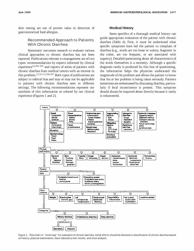
Systematic outcomes research to evaluate variousclinical approaches to chronic diarrhea has not beenreported. Publications relevant to management are of twotypes: recommendations by experts informed by clinicalexperience14,200–205 and reports of series of patients withchronic diarrhea from medical centers with an interest inthis problem.15,16,115,206,207 Both types of publications aresubject to referral bias and may or may not be applicableto patients with chronic diarrhea seen in differentsettings. The following recommendations represent oursynthesis of this information as colored by our clinicalexperience (Figures 1 and 2).
Medical History
Some specifics of a thorough medical history canguide appropriate evaluation of the patient with chronicdiarrhea (Table 4). First, it must be understood whatspecific symptoms have led the patient to complain ofdiarrhea (e.g., stools are too loose or watery, fragment inthe toilet, are too frequent, or are associated withurgency). Detailed questioning about all characteristics ofthe stools themselves is a necessity. Although a specificdiagnosis rarely is produced by this line of questioning,the information helps the physician understand themagnitude of the problem and allows the patient to knowthat his or her problem is being taken seriously. Patientssometimes are embarrassed by discussing diarrhea, particu-larly if fecal incontinence is present. This symptomshould always be inquired about directly because it rarelyis volunteered.
Figure 1. Flow chart or ‘‘mind map’’ for evaluation of chronic diarrhea. Initial efforts should be directed to classification of chronic diarrhea basedon history, physical examination, basic laboratory test results, and stool analysis.
June 1999 AMERICAN GASTROENTEROLOGICAL ASSOCIATION 1477
The characteristics of the stool may suggest a potentialpathophysiological mechanism (e.g., malodorous, float-ing, greasy stools containing food particles suggestingmalabsorption; or gross blood suggesting inflammationor neoplasm). The patient’s perception of the volume ofdiarrhea may help to localize the disease process in thegastrointestinal tract. Diarrhea that is watery and volumi-nous suggests a disorder of the small bowel or proximalcolon, whereas frequent, small-volume diarrhea may beassociated with disorders of the left colon or rectum. Thelatter disorders are often accompanied by tenesmus andpassage of dark, mushy stools that may contain mucus,pus, or blood.
Fever or weight loss may herald a diagnosis ofinflammatory bowel disease, amebiasis, intestinal lym-
phoma, other malignancies, Whipple’s disease, tuberculo-sis, other enteric infections, or thyrotoxicosis.
The patient’s medical history may be important.Seronegative spondyloarthropathy may precede the recog-nition of inflammatory bowel disease by many years. Ahistory of diabetes, thyroid problems, and other autoim-mune phenomena may be pertinent. Previous surgery tothe gastrointestinal or biliary tracts may be the cause ofdiarrhea. Other diseases associated with diarrhea includerheumatic diseases with or without vasculitis, immuno-globulin deficiency, and peptic ulcer disease if caused bythe Zollinger–Ellison syndrome or systemic mastocyto-sis.
All current medications (including ‘‘over-the-counter’’drugs), nutritional supplements, illicit drugs, alcohol,
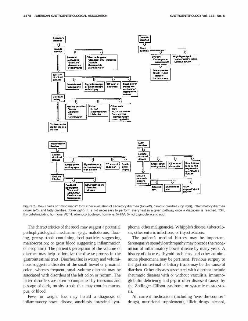
Figure 2. Flow charts or ‘‘mind maps’’ for further evaluation of secretory diarrhea (top left), osmotic diarrhea (top right), inflammatory diarrhea(lower left), and fatty diarrhea (lower right). It is not necessary to perform every test in a given pathway once a diagnosis is reached. TSH,thyroid-stimulating hormone; ACTH, adrenocorticotropic hormone; 5-HIAA, 5-hydroxyindole acetic acid.
1478 AMERICAN GASTROENTEROLOGICAL ASSOCIATION GASTROENTEROLOGY Vol. 116, No. 6
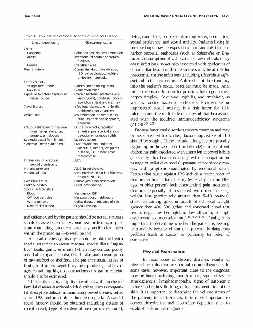
and caffeine used by the patient should be noted. Patientsshould be asked specifically about new medicines, magne-sium-containing products, and any antibiotics takenwithin the preceding 6–8-week period.
A detailed dietary history should be obtained withspecial attention to recent changes; special diets; ‘‘sugar-free’’ foods, gums, or mints (which may contain poorlyabsorbable sugar alcohols); fiber intake; and consumptionof raw seafood or shellfish. The patient’s usual intake offruits, fruit juices, vegetables, milk products, and bever-ages containing high concentrations of sugar or caffeineshould also be estimated.
The family history may disclose others with diarrhea orfamilial diseases associated with diarrhea, such as congeni-tal absorptive defects, inflammatory bowel disease, celiacsprue, IBS, and multiple endocrine neoplasia. A carefulsocial history should be obtained including details ofrecent travel, type of residential area (urban vs. rural),
living conditions, sources of drinking water, occupation,sexual preference, and sexual activity. Patients living inrural settings may be exposed to farm animals that canharbor bacterial pathogens (such as Salmonella or Bru-cella). Consumption of well water or raw milk also maycause infections, sometimes associated with epidemics ofchronic diarrhea. Health-care workers may be at risk fornosocomial enteric infections (including Clostridium diffi-cile) and factitious diarrhea . A discrete but direct inquiryinto the patient’s sexual practices must be made. Analintercourse is a risk factor for proctitis due to gonorrhea,herpes simplex, Chlamydia, syphilis, and amebiasis, aswell as routine bacterial pathogens. Promiscuous orunprotected sexual activity is a risk factor for HIVinfection and the multitude of causes of diarrhea associ-ated with the acquired immunodeficiency syndrome(AIDS).200–205
Because functional disorders are very common and maybe associated with diarrhea, factors suggestive of IBSshould be sought. These include a long history (usuallybeginning in the second or third decade) of intermittentabdominal pain associated with alteration of bowel habits(classically diarrhea alternating with constipation orpassage of pellet-like stools), passage of nonbloody mu-cus, and symptoms exacerbated by emotional stress.Factors that argue against IBS include a recent onset ofdiarrhea without a long history (especially in a middle-aged or older person), lack of abdominal pain, nocturnaldiarrhea (especially if associated with incontinence),weight loss (particularly greater than 5–10 pounds),stools containing gross or occult blood, fecal weightgreater than 400–500 g/day, and abnormal blood testresults (e.g., low hemoglobin, low albumin, or higherythrocyte sedimentation rate).15,16,208,209 Finally, it isimportant to determine whether the patient is seekinghelp mainly because of fear of a potentially dangerousproblem (such as cancer) or primarily for relief ofsymptoms.
Physical Examination
In most cases of chronic diarrhea, results ofphysical examination are normal or nondiagnostic. Insome cases, however, important clues to the diagnosismay be found including mouth ulcers, signs of severeatherosclerosis, lymphadenopathy, signs of autonomicfailure, and rashes, flushing, or hyperpigmentation of theskin. It is important to determine the volume status ofthe patient; in all instances, it is more important tocorrect dehydration and electrolyte depletion than toestablish a definitive diagnosis.
Table 4. Implications of Some Aspects of Medical History
Line of questioning Clinical implication
OnsetCongenital Chloridorrhea, Na1 malabsorptionAbrupt Infections, idiopathic secretory
diarrheaGradual Everything else
Family history Congenital absorptive defects,IBD, celiac disease, multipleendocrine neoplasia
Dietary history‘‘Sugar-free’’ foods Sorbitol, mannitol ingestionRaw milk Brainerd diarrhea
Exposure to potentially impurewater source
Chronic bacterial infections (e.g.,Aeromonas), giardiasis, crypto-sporidiosis, Brainerd diarrhea
Travel history Infectious diarrhea, chronic idio-pathic secretory diarrhea
Weight loss Malabsorption, pancreatic exo-crine insufficiency, neoplasm,anorexia
Previous therapeutic interven-tions (drugs, radiation,surgery, antibiotics)
Drug side effects, radiationenteritis, postsurgical status,pseudomembranous colitis
Secondary gain from illness Laxative abuseSystemic illness symptoms Hyperthyroidism, diabetes,
vasculitis, tumors, Whipple’sdisease, IBD, tuberculosis,mastocytosis
Intravenous drug abuse,sexual promiscuity
AIDS
Immune problems AIDS, Ig deficienciesAbdominal pain Mesenteric vascular insufficiency,
obstruction, IBSExcessive flatus Carbohydrate malabsorptionLeakage of stool Fecal incontinenceStool characteristics
Blood Malignancy, IBDOil/food particles Malabsorption, maldigestionWhite/tan color Celiac disease, absence of bileNocturnal diarrhea Organic etiology
June 1999 AMERICAN GASTROENTEROLOGICAL ASSOCIATION 1479
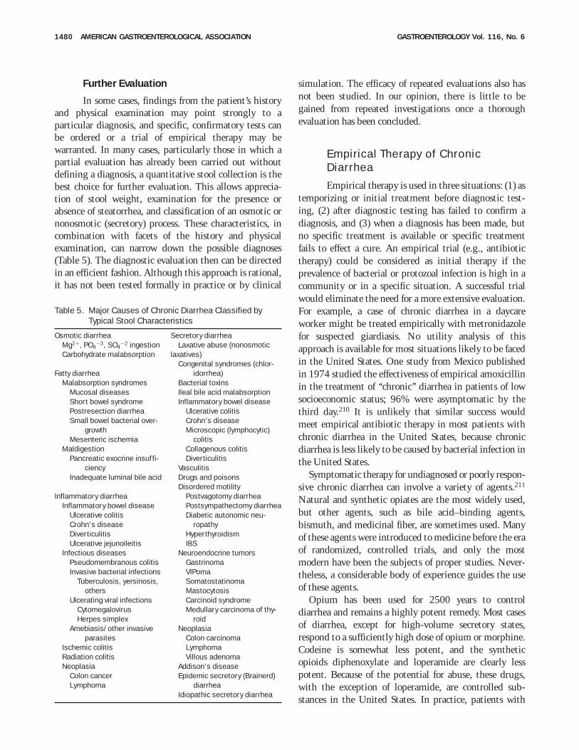
Further Evaluation
In some cases, findings from the patient’s historyand physical examination may point strongly to aparticular diagnosis, and specific, confirmatory tests canbe ordered or a trial of empirical therapy may bewarranted. In many cases, particularly those in which apartial evaluation has already been carried out withoutdefining a diagnosis, a quantitative stool collection is thebest choice for further evaluation. This allows apprecia-tion of stool weight, examination for the presence orabsence of steatorrhea, and classification of an osmotic ornonosmotic (secretory) process. These characteristics, incombination with facets of the history and physicalexamination, can narrow down the possible diagnoses(Table 5). The diagnostic evaluation then can be directedin an efficient fashion. Although this approach is rational,it has not been tested formally in practice or by clinical
simulation. The efficacy of repeated evaluations also hasnot been studied. In our opinion, there is little to begained from repeated investigations once a thoroughevaluation has been concluded.
Empirical Therapy of ChronicDiarrhea
Empirical therapy is used in three situations: (1) astemporizing or initial treatment before diagnostic test-ing, (2) after diagnostic testing has failed to confirm adiagnosis, and (3) when a diagnosis has been made, butno specific treatment is available or specific treatmentfails to effect a cure. An empirical trial (e.g., antibiotictherapy) could be considered as initial therapy if theprevalence of bacterial or protozoal infection is high in acommunity or in a specific situation. A successful trialwould eliminate the need for a more extensive evaluation.For example, a case of chronic diarrhea in a daycareworker might be treated empirically with metronidazolefor suspected giardiasis. No utility analysis of thisapproach is available for most situations likely to be facedin the United States. One study from Mexico publishedin 1974 studied the effectiveness of empirical amoxicillinin the treatment of ‘‘chronic’’ diarrhea in patients of lowsocioeconomic status; 96% were asymptomatic by thethird day.210 It is unlikely that similar success wouldmeet empirical antibiotic therapy in most patients withchronic diarrhea in the United States, because chronicdiarrhea is less likely to be caused by bacterial infection inthe United States.
Symptomatic therapy for undiagnosed or poorly respon-sive chronic diarrhea can involve a variety of agents.211
Natural and synthetic opiates are the most widely used,but other agents, such as bile acid–binding agents,bismuth, and medicinal fiber, are sometimes used. Manyof these agents were introduced to medicine before the eraof randomized, controlled trials, and only the mostmodern have been the subjects of proper studies. Never-theless, a considerable body of experience guides the useof these agents.
Opium has been used for 2500 years to controldiarrhea and remains a highly potent remedy. Most casesof diarrhea, except for high-volume secretory states,respond to a sufficiently high dose of opium or morphine.Codeine is somewhat less potent, and the syntheticopioids diphenoxylate and loperamide are clearly lesspotent. Because of the potential for abuse, these drugs,with the exception of loperamide, are controlled sub-stances in the United States. In practice, patients with
Table 5. Major Causes of Chronic Diarrhea Classified byTypical Stool Characteristics
Osmotic diarrhea Secretory diarrheaMg21, PO4
23, SO422 ingestion
Carbohydrate malabsorption
Fatty diarrheaMalabsorption syndromes
Mucosal diseasesShort bowel syndromePostresection diarrheaSmall bowel bacterial over-
growthMesenteric ischemia
MaldigestionPancreatic exocrine insuffi-
ciencyInadequate luminal bile acid
Inflammatory diarrheaInflammatory bowel disease
Ulcerative colitisCrohn’s diseaseDiverticulitisUlcerative jejunoileitis
Infectious diseasesPseudomembranous colitisInvasive bacterial infections
Tuberculosis, yersinosis,others
Ulcerating viral infectionsCytomegalovirusHerpes simplex
Amebiasis/other invasiveparasites
Ischemic colitisRadiation colitisNeoplasia
Colon cancerLymphoma
Laxative abuse (nonosmoticlaxatives)
Congenital syndromes (chlor-idorrhea)
Bacterial toxinsIleal bile acid malabsorptionInflammatory bowel disease
Ulcerative colitisCrohn’s diseaseMicroscopic (lymphocytic)
colitisCollagenous colitisDiverticulitis
VasculitisDrugs and poisonsDisordered motility
Postvagotomy diarrheaPostsympathectomy diarrheaDiabetic autonomic neu-
ropathyHyperthyroidismIBS
Neuroendocrine tumorsGastrinomaVIPomaSomatostatinomaMastocytosisCarcinoid syndromeMedullary carcinoma of thy-
roidNeoplasia
Colon carcinomaLymphomaVillous adenoma
Addison’s diseaseEpidemic secretory (Brainerd)
diarrheaIdiopathic secretory diarrhea
1480 AMERICAN GASTROENTEROLOGICAL ASSOCIATION GASTROENTEROLOGY Vol. 116, No. 6
chronic diarrhea seldom abuse these agents, and it isunusual to require ever higher doses to control diarrheaonce an effective dose is reached. Potent narcotics areprobably underused in the treatment of severe chronicdiarrhea. Lesser opioids, such as diphenoxylate andloperamide, are satisfactory for control of less severediarrhea and ought to be tried before resorting to morepotent drugs. The prodrug loperamide-N-oxide and theenkephalinase inhibitor acetorphan are under develop-ment and may have some therapeutic advantages overcurrently available agents.212–214
The somatostatin analogue octreotide has proven effec-tiveness in carcinoid tumors and other peptide-secretingtumors, dumping syndrome, and chemotherapy-induceddiarrhea. It has had limited success in patients withAIDS-associated diarrhea and short bowel syndrome.211
Octreotide does not seem to have any advantage overopiates in the treatment of chronic idiopathic diarrheaand probably should be a second-line agent for thisindication because of the need for administration byinjection and its expense.
Intraluminal agents include adsorbants, such as clays,activated charcoal, and binding resins; bismuth; and stoolmodifiers, such as medicinal fiber.211 Few controlledstudies have been conducted with these agents, andresults have been equivocal. Cholestyramine and othersimilar binding resins have reduced stool weight inEuropean studies in which patients with chronic idio-pathic diarrhea have had a high frequency of bile acidmalabsorption.135–139,215 In an American series, cholestyr-amine had little effect in these patients, even when bileacid malabsorption was present.141 Bismuth subsalicylatehas been shown to be effective in acute travelers’ diarrhea,but its effectiveness in chronic diarrhea is unproven.216
Stool modifiers, such as psyllium, may alter stool consis-tency but do not reduce stool weight.217
Oral rehydration solutions that include glucose orother nutrients and salt are useful for repletion of bodyfluids.211 Cereal-based oral rehydration solutions havegained acceptance in recent years. They can be life-savingtherapy for dehydrating, acute secretory diarrheas, such ascholera, but have limited application in most chronicdiarrheal states and have not been well studied in thesesituations. Although these solutions increase net salt andwater absorption, they are not designed to reduce stoolweight, and diarrhea (defined by stool weight or fre-quency) may worsen with their use.
Directions for Future Research
Study of clinical outcomes in patients with chronicdiarrhea has been fragmentary. Utility parameters areavailable for some diagnostic tests, but in the absence ofsolid epidemiological data about the prevalence of differ-ent causes of chronic diarrhea, these cannot be appliedaccurately to clinical decision making. Algorithmic man-agement is untested, and the economic aspects of differ-ent approaches to the diagnosis and treatment of chronicdiarrhea are unstudied. Opportunities for clinical re-search in patients with chronic diarrhea are voluminous.
KENNETH D. FINELAWRENCE R. SCHILLERGastroenterology SectionDepartment of Internal MedicineBaylor University Medical CenterDallas, Texas
References1. Flexner SB, ed. The Random House Dictionary of the English
Language, Unabridged. 2nd ed. New York: Random House,1987:548.
2. Dorland’s Illustrated Medical Dictionary. 27th ed. Philadelphia:Saunders, 1988:464.
3. Talley NJ, Zinsmeister AR, Van Dyke C, Melton LJ III. Epidemiol-ogy of colonic symptoms and the irritable bowel syndrome.Gastroenterology 1991;101:927–934.
4. Talley NJ, Weaver AL, Zinsmeister AR, Melton LJ III. Self-reporteddiarrhea: what does it mean? Am J Gastroenterol 1994;89:1160–1164.
5. Wenzl HH, Fine KD, Schiller LR, Fordtran JS. Determinants ofdecreased fecal consistency in patients with diarrhea. Gastroen-terology 1995;108:1729–1738.
6. Stanton B, Clemens JD. Chronic diarrhoea: a methodologicbasis for its apparent heterogeneity. Trop Geogr Med 1989;41:100–107.
7. Thompson WG, Creed F, Drossman DA, Mazzacca G. Functionalbowel disorders and chronic abdominal pain. Gastroenterol Int1992;5:75–91.
8. Schiller LR. Fecal incontinence. In: Feldman M, Scharschmidt B,Sleisenger MH, eds. Sleisenger & Fordtran’s gastrointestinaland hepatic disease: pathophysiology, diagnosis, management.6th ed. Philadelphia: Saunders, 1998:160–173.
9. WHO CDD/DDM/85.1 Diarrhoeal Diseases Control Programme.Persistent diarrhoea in children—research priorities.
10. Fine KD, Meyer RL, Lee EL. The prevalence of chronic diarrhea inpatients with celiac sprue treated with a gluten-free diet.Gastroenterology 1997;112:1830–1838.
11. Talley NJ, O’Keefe EA, Zinsmeister AR, Melton LJ III. Prevalenceof gastrointestinal symptoms in the elderly: a population-basedstudy. Gastroenterology 1992;102:895–901.
12. Talley NJ, Weaver AL, Zinsmeister AR, Melton LJ III. Onset anddisappearance of gastrointestinal symptoms and functionalgastrointestinal disorders. Am J Epidemiol 1992;136:165–177.
13. Read NW, Krejs GJ, Read MG, Santa Ana CA, Morawski SG,Fordtran JS. Chronic diarrhea of unknown origin. Gastroenterol-ogy 1980;78:264–271.
14. Bayless TM. Chronic diarrhea: newly appreciated syndromes.Hosp Pract (Off Ed) 1989;24:117–122,124–126,131–132.
15. Bytzer P, Stokholm M, Andersen I, Lund-Hansen B, SchaffalitzkyDe Muckadell OB. Aetiology, medical history, and fecal weight in
June 1999 AMERICAN GASTROENTEROLOGICAL ASSOCIATION 1481
adult patients referred for diarrhoea: a prospective study. ScandJ Gastroenterol 1990;25:572–578.
16. Bertomeu A, Ros E, Barragan V, Sachje L, Navarro S. Chronicdiarrhea with normal stool and colonic examinations: organic orfunctional? J Clin Gastroenterol 1991;13:531–536.
17. Schiller LR, Rivera LM, Santangelo W, Little K, Fordtran JS.Diagnostic value of fasting plasma peptide concentrations inpatients with chronic diarrhea. Dig Dis Sci 1994;39:2216–2222.
18. Afzalpurkar RG, Schiller LR, Little KH, Santangelo WC, FordtranJS. The self-limited nature of chronic idiopathic diarrhea. N EnglJ Med 1992;327:1849–1852.
19. Tandon BN, Tandon HD, Prakash OM. A study of chronic colonicdiarrhoea and dysentery. I. Clinical, endoscopic and coprologicalstudy. Indian J Med Res 1966;54:623–628.
20. Chatterjee H. Chronic diarrhoeas in adults. J Indian Med Assoc1977;69:259–261.
21. Awori NW, Rees PH, Roy AD. Causes of chronic diarrhoea inKenya and their relationship to ulcerative colitis. East Afr Med J1972;49:604–613.
22. Ahmed MU, Sarker NC, Haque E, Hasan MA. Chronic diarrhoealdisease in adults: a preliminary report. Bangladesh Med ResCounc Bull 1976;2:8–11.
23. Kotwal MR, Durrani HA, Shah SN. Chronic colonic diarrhoea inNorth-West India: a clinical study with special reference to thesyndrome of irritable colon. J Indian Med Assoc 1978;70:77–80.
24. Manatsathit S, Israsena S, Kladcharoen N, Sithicharoenchai P,Roenprayoon S, Suwanakul P. Chronic diarrhoea: a prospectivestudy in Thai patients at Chulalongkorn University Hospital,Bangkok. Southeast Asian J Trop Med Public Health 1985;16:447–452.
25. Bytzer P, Stokholm M, Andersen I, Klitgaard NA, Schaffalitzky DeMuckadell OB. Prevalence of surreptitious laxative abuse inpatients with diarrhoea of uncertain origin: a cost-benefit analy-sis of a screening procedure. Gut 1989;30:1379–1384.
26. Everhart JE, ed. Digestive Disease in the United States: epidemi-ology and impact. NIH Publ 94-1447. Bethesda, MD: NationalInstitutes of Health, 1994.
27. Lubeck DP, Bennett CL, Mazonson PD, Fifer SK, Fries JF. Qualityof life and health service use among HIV-infected patients withchronic diarrhea. J Acquir Immune Defic Syndr Hum Retrovirol1993;6:478–484.
28. Watson A, Samore MH, Wanke CA. Diarrhea and quality of life inambulatory HIV-infected patients. Dig Dis Sci 1996;41:1794–1800.
29. Fine KD. The prevalence of occult gastrointestinal bleeding inceliac sprue. N Engl J Med 1996;334:1163–1167.
30. Anthonisen P, Riis P. A new diagnostic approach to mucosalinflammation in proctocolitis. Lancet 1961;2:81–82.
31. Harris JC, DuPont HL, Hornick RB. Fecal leukocytes in diarrhealillness. Ann Intern Med 1972;76:697–703.
32. Guerrant RL, Araujo V, Soares E, Kotloff K, Lima AAM, CooperWH, Lee AG. Measurement of fecal lactoferrin as a marker offecal leukocytes. J Clin Microbiol 1992;30:1238–1242.
33. Yong WH, Mattia AR, Ferraro MJ. Comparison of fecal lactoferrinlatex agglutination assay and methylene blue microscopy fordetection of fecal leukocytes in Clostridium difficile–associateddisease. J Clin Microbiol 1994;32:1360–1361.
34. Miller JR, Barrett LJ, Kotloff K, Guerrant RL. A rapid test forinfectious and inflammatory enteritis. Arch Intern Med 1994;154:2660–2664.
35. Drummey GD, Benson JA, Jones CM. Microscopical examinationof the stool for steatorrhea. N Engl J Med 1961;264:85–87.
36. Simko V. Fecal fat microscopy. Acceptable predictive value inscreening for steatorrhea. Am J Gastroenterol 1981;75:204–208.
37. Khouri MR, Huang G, Shiau YF. Sudan stain of fecal fat: newinsight into an old test. Gastroenterology 1989;96:421–427.
38. Guarino A, Tarallo L, Greco L, Cesarano L, Guandalini S, RubinoA. Reference values of the steatocrit and its modifications indiarrheal diseases. J Pediatr Gastroenterol Nutr 1992;14:268–274.
39. Sugai E, Srur G, Vazquez H, Benito F, Maurino E, Boerr LA, BaiJC. Steatocrit: a reliable semiquantitative method for detectionof steatorrhea. J Clin Gastroenterol 1994;19:206–209.
40. Tran M, Forget P, Van den Neucker A, van Kreel B. Improvedsteatocrit results obtained by acidification of fecal homogenatesare due to improved fat extraction. J Pediatr Gastroenterol Nutr1996;22:157–160.
41. Blaser MJ. Infectious diarrheas: acute, chronic, and iatrogenic.Ann Intern Med 1986;105:785–787.
42. Horing E, Gopfert D, Schroter G, von Gaisberg U. Frequency andspectrum of microorganisms isolated from biopsy specimens inchronic colitis. Endoscopy 1991;23:325–327.
43. George WL, Nakata MW, Thompson J, White ML. Aeromonas-related diarrhea in adults. Arch Intern Med 1985;145:2207–2211.
44. Holmberg SD, Schell WL, Fanning GR, Wachsmuth IK, Hickmann-Brenner FW, Blake PA, Brenner DJ, Farmer JJ III. Aeromonasintestinal infections in the United States. Ann Intern Med1986;105:683–689.
45. Jesudason MV, Koshi G. Aeromonas species in human septicae-mia and diarrhoea. Indian J Med Res 1990;91:174–176.
46. Del Val A, Moles JR, Garrigues V. Very prolonged diarrheaassociated with Aeromonas hydrophila (letter). Am J Gastroen-terol 1990;85:1535.
47. Rautelin H, Hanninen ML, Sivonen A, Turunen U, Valtonen V.Chronic diarrhea due to a single strain of Aeromonas caviae. EurJ Clin Microbiol Infect Dis 1995;14:51–53.
48. Penn RG, Giger DK, Knoop FC, Preheim LC. Plesiomonasshigelloides overgrowth in the small intestine. J Clin Microbiol1982;15:869–872.
49. Tabibian N, Clarridge JE, Smith JL, Alpert E, Shaw I, Graham DY.Clinical impact of stool cultures for Campylobacter in adults withacute or chronic diarrhea. South Med J 1987;80:709–711.
50. Paulet P, Coffernils M. Very long term diarrhoea due to Campylo-bacter jejuni (letter). Postgrad Med J 1990;66:410–411.
51. Caselli M, Trevisani L, Bighi S, Aleotti A, Balboni PG, Gaiani R,Bovolenta MR, Stabellini G. Dead fecal yeasts and chronicdiarrhea. Digestion 1988;41:142–148.
52. Gupta TP, Ehrinpreis MN. Candida-associated diarrhea in hospi-talized patients. Gastroenterology 1990;98:780–785.
53. Talwar P, Chakrabarti A, Chawla A, Mehta S, Walia BN, Kumar L,Chugh KS. Fungal diarrhoea: association of different fungi andseasonal variation in their incidence. Mycopathologia 1990;110:101–105.
54. Zaidi M, Ponce de Leon S, Ortiz RM, Ponce de Leon S, Calva JJ,Ruiz-Palacios G, Camorlinga M, Cervantes LE, Ojeda F. Hospital-aquired diarrhea in adults: a prospective case-controlled studyin Mexico. Infect Control Hosp Epidemiol 1991;12:349–355.
55. Rosenblatt JE, Sloan LM, Schneider SK. Evaluation of anenzyme-linked immunosorbent assay for the detection of Giardialamblia in stool specimens. Diagn Microbiol Infect Dis 1993;16:337–341.
56. Koontz F, Weinstock JV. The approach to stool examination forparasites. Gastroenterol Clin North Am 1996;25:435–449.
57. Azad Khan AK, Islam MS, Haque S. Stool findings in ‘‘chronicdysentery.’’ Bangladesh Med Res Counc Bull 1981;7:7–11.
58. Goy JA, Eastwood MA, Mitchell WD, Pritchard JL, Smith AN. Fecalcharacteristics contrasted in the irritable bowel syndrome anddiverticular disease. Am J Clin Nutr 1976;29:1480–1484.
59. Pimparkar BD, Tulsky EG, Kalser MH, Bockus HL. Correlation ofradioactive and chemical fecal fat determinations in the malab-
1482 AMERICAN GASTROENTEROLOGICAL ASSOCIATION GASTROENTEROLOGY Vol. 116, No. 6
sorption syndrome. I. Studies in normal man and in functionaldisorders of the gastrointestinal tract. Am J Med 1961;30:910–926.
60. Fordtran JS. Speculations on the pathogenesis of diarrhea. FedProc 1967;26:1405–1414.
61. Shiau YF, Feldman GM, Resnick MA, Coff PM. Stool electrolyteand osmolality measurements in the evaluation of diarrhealdisorders. Ann Intern Med 1985;102:773–775.
62. Ladefoged K, Schaffalitzky de Muckadell OB, Jarnum S. Faecalosmolality and electrolyte concentrations in chronic diarrhoea:do they provide diagnostic clues? Scand J Gastroenterol 1987;22:813–820.
63. Vernia P, Breuer RI, Gnaedinger A, Latella G, Santoro ML.Composition of fecal water: comparison of in vitro dialysis withultrafiltration. Gastroenterology 1984;86:1557–1561.
64. Eherer AJ, Fordtran JS. Fecal osmotic gap and pH in experimen-tal diarrhea of various causes. Gastroenterology 1992;103:545–551.
65. Duncan A, Robertson C, Russell RI. The fecal osmotic gap:technical aspects regarding its calculation. J Lab Clin Med1992;119:359–363.
66. Topazian M, Binder HJ. Factitious diarrhea detected by measure-ment of stool osmolality. N Engl J Med 1994;330:1418–1419.
67. Phillips S, Donaldson L, Geisler K, Pera A, Kochar R. Stoolcomposition in factitial diarrhea: a 6-year experience with stoolanalysis. Ann Intern Med 1995;123:97–100.
68. van de Kamer JH, ten Bokkel Huinink H, Weyers HA. Rapidmethod for the determination of fat in feces. J Biol Chem1949;177:347–355.
69. Wollaeger EE, Comfort MW, Osterberg AE. Total solids, fat andnitrogen in the feces: III. A study of normal persons taking a testdiet containing a moderate amount of fat; comparison withresults obtained with normal persons taking a test diet contain-ing a large amount of fat. Gastroenterology 1947;9:272–283.
70. Anneger JH, Boutwell JH, Ivy AC. The effect of dietary fat on fecalfat excretion and subjective symptoms in man. Gastroenterology1948;10:486–495.
71. Braddock LI, Fleisher DR, Barbero GJ. A physical chemical studyof the van de Kamer method for fecal fat analysis. Gastroenterol-ogy 1968;55:165–172.
72. Arora S, Kassajian Z, Krasinski SD, Croffey B, Kaplan MM,Russell RM. Effect of age on tests of intestinal and hepaticfunction in healthy humans. Gastroenterology 1989;96:1560–1565.
73. Fine KD, Fordtran JS. The effect of diarrhea on fecal fatexcretion. Gastroenterology 1992;102:1936–1939.
74. Bo-Linn GW, Fordtran JS. Fecal fat concentration in patients withsteatorrhea. Gastroenterology 1984;87:319–322.
75. Roberts IM, Poturich C, Wald A. Utility of fecal fat concentrationsas screening test in pancreatic insufficiency. Dig Dis Sci 1986;31:1021–1024.
76. Ameen VZ, Powell GK. A simple spectrophotometric method forquantitative fecal carbohydrate measurement. Clin Chim Acta1985;152:3–9.
77. Duncan A, Morris AJ, Cameron A, Stewart MJ, Brydon WG,Russell RI. Laxative induced diarrhoea—a neglected diagnosis.J R Soc Med 1992;85:203–205.
78. Morris AI, Turnberg LA. Surreptitious laxative abuse. Gastroenter-ology 1979;77:780–786.
79. Ewe K, Karbach U. Factitious diarrhoea. Clin Gastroenterol1986;15:723–740.
80. Slugg PH, Carey WD. Clinical features and follow-up of surrepti-tious laxative users. Cleve Clin Q 1984;51:167–171.
81. Fine KD, Krejs GJ, Fordtran JS. Diarrhea. In: Sleisenger MH,Fordtran JS, eds. Gastrointestinal disease: pathophysiology,diagnosis, management. 4th ed. Philadelphia: Saunders, 1989:307.
82. Santangelo WC, Richey JE, Rivera L, Fordtran JS. Surreptitiousipecac administration simulating intestinal pseudo-obstruction.Ann Intern Med 1989;110:1031–1032.
83. Kacere RD, Srivatsa SS, Tremaine WJ, Ebnet LE, Batts KP.Chronic diarrhea due to surreptitious use of bisacodyl: casereports and methods for detection. Mayo Clin Proc 1993;68:355–357.
84. Morton J. The detection of laxative abuse. Ann Clin Biochem1987;24:107–108.
85. de Wolff FA, de Hass EJM, Verweij M. A screening method forestablishing laxative abuse. Clin Chem 1981;27:914–917.
86. Sladen GE. Effects of chronic purgative abuse. Proc R Soc Lond[Biol] 1972;65:288–291.
87. Carlson J, Fernlund P, Ivarsson SA, Jakobsson I, Neiderud J,Nilsson KO, Svensson M, Swanstein U. Munchausen syndromeby proxy: an unexpected cause of severe chronic diarrhoea in achild. Acta Paediatr 1994;83:119–121.
88. Fine KD, Santa Ana CA, Fordtran JS. Diagnosis of magnesium-induced diarrhea. N Engl J Med 1991;324:1012–1017.
89. Cummings JH, Sladen GE, James OF, Sarner M, Misiewicz JJ.Laxative-induced diarrhoea: a continuing clinical problem. BMJ1974;1:537–541.
90. Plumeri PA. Gastroenterology and the law: the room search. JClin Gastroenterol 1984;6:181–185.
91. Sofinowski RE, Butler PM. Munchausen syndrome by proxy: areview. Texas Med 1991;87:66–70.
92. Strygler B, Nicar MJ, Santangelo WC, Porter JL, Fordtran JS.Alpha 1-antitrypsin excretion in stool in normal subjects and inpatients with gastrointestinal disorders. Gastroenterology 1990;99:1380–1387.
93. Thomas DW, Sinatra FR, Merritt RJ. Random fecal alpha-1-antitrypsin concentration in children with gastrointestinal dis-ease. Gastroenterology 1981;80:776–782.
94. Jarnum S, Jensen KB. Fecal radioiodide excretion followingintravenous injection of 131-I-albumin and 125-I-immunoglobu-lin G in chronic inflammatory bowel disease. An aid to topo-graphic diagnosis. Gastroenterology 1975;68:1433–1444.
95. Waldmann TA. Gastrointestinal protein loss demonstrated by 51Cr- labelled albumin. Lancet 1961;2:121–123.
96. Unsworth DJ, Walker-Smith JA. Autoimmunity in diarrhoealdisease. J Pediatr Gastroenterol Nutr 1985;4:375–380.
97. Savilahti E, Perkkio M, Kalimo K, Viander M, Vainio E, Reunala T.IgA antigliadin antibodies: a marker of mucosal damage inchildhood coeliac disease. Lancet 1983;1:320–322.
98. Hallstrom O. Comparison of IgA-class reticulin and endomysiumantibodies in coeliac disease and dermatitis herpetiformis. Gut1989;30:1225–1232.
99. Ferreira M, Lloyd Davies S, Butler M, Scott D, Clark D, Kumar P.Endomysial antibody: is it the best screening test for coeliacdisease? Gut 1992;33:1633–1637.
100. Valentini RA, Andreani ML, Corazza GR, Gasbarrini G. IgAendomysium antibody: a valuable tool in the screening of coeliacdisease but not its follow-up. Ital J Gastroenterol 1994;26:279–282.
101. Ferfoglia G, Pulitano R, Sategna-Guidetti C. Do dietary antibod-ies still play a role in the diagnosis and follow-up of coeliacdisease? A comparison among different serological tests.Panminerva Med 1995;37:55–59.
102. Valdimarsson T, Franzen L, Grodzinsky E, Skogh T, Strom M. Issmall bowel biopsy necessary in adults with suspected celiacdisease and IgA anti-endomysium antibodies? Dig Dis Sci1996;41:83–87.
103. Ascher H, Hahn-Zoric M, Hanson LÅ, Kilander AF, Nilsson L-Å,Tlaskalova H. Value of serologic markers for clinical diagnosisand population studies of coeliac disease. Scand J Gastroen-terol 1996;31:61–67.
104. Saxon A, Shanahan F, Landers C, Ganz T, Targan S. A distinct
June 1999 AMERICAN GASTROENTEROLOGICAL ASSOCIATION 1483
subset of antineutrophil cytoplasmic antibodies is associatedwith inflammatory bowel disease. J Allergy Clin Immunol 1990;86:202–210.
105. Duerr RH, Targan SR, Landers CJ, Sutherland LR, Shanahan F.Anti-neutrophil cytoplasmic antibodies in ulcerative colitis. Com-parison with other colitides/diarrheal illnesses. Gastroenterol-ogy 1991;100:1590–1596.
106. Duerr RH, Targan SR, Landers CJ, LaRusso NF, Lindsay KL,Weisner RH, Shanahan F. Neutrophil cytoplasmic antibodies: alink between primary sclerosing cholangitis and ulcerativecolitis. Gastroenterology 1991;100:1385–1391.
107. Arslan S, Bakkaloglu A, Oksuzoglu G, Kadayifci A, Kansu E,Uzunalimoglu B, Kayhan B. The value of p-ANCA as a serologicalmarker in diagnosing the coexistence of chronic active invasiveamebic colitis and ulcerative colitis (letter). Am J Gastroenterol1995;90:2265–2266.
108. Sollid LM, Markussen G, Ek J, Gjerde H, Vartdal F, Thorsby E.Evidence for a primary association of celiac disease to aparticular HLA-DQ alpha/beta heterodimer. J Exp Med 1989;169:345–350.
109. Sollid LM, Thorsby E. HLA susceptibility genes in celiac disease:genetic mapping and role in pathogenesis. Gastroenterology1993;105:910–922.
110. Danze P-M, Colombel J-F, Jacquot S, Loste M-N, Heresbach D,Ategbo S, Khamassi S, Perichon B, Semana G, Charron D,Cezard J-P. Association of HLA class II genes with susceptibilityto Crohn’s disease. Gut 1996:39:69–72.
111. Collins JR, Isselbacher KJ. The occurrence of severe smallintestinal mucosal damage in conditions other than celiacdisease (nontropical sprue). Gastroenterology 1965;49:425–432.
112. Halphen M, Galian A, Certin M, Ink F, Filali A, Rambaud J-C.Clinicopathological study of a patient with idiopathic villousatrophy and small vessel alterations of the ileum. Dig Dis Sci1989;34:111–117.
113. Zins BJ, Tremaine WJ, Carpenter HA. Collagenous colitis: muco-sal biopsies and association with fecal leukocytes. Mayo ClinProc 1995;70:430–433.
114. Tanaka M, Mazzoleni G, Riddell RH. Distribution of collagenouscolitis: utility of flexible sigmoidoscopy. Gut 1992;33:65–70.
115. Marshall JB, Singh R, Diaz-Arias AA. Chronic, unexplaineddiarrhea: are biopsies necessary if colonoscopy is normal? Am JGastroenterol 1995;90:372–376.
116. Surawicz CM, Meisel JL, Ylvisaker T, Saunders DR, Rubin CE.Rectal biopsy in the diagnosis of Crohn’s disease: value ofmultiple biopsies and serial sectioning. Gastroenterology 1981;80:66–71.
117. Candreviotis N. The pathology of chronic amebic colitis inGreece studied by colon biopsy. Am J Proctol 1966;17:39–47.
118. Nostrant TT, Kumar NB, Appleman HD. Histopathology differenti-ates acute self-limited colitis from ulcerative colitis. Gastroenter-ology 1987;92:318–328.
119. Mee AS, Burke M, Vallon AG, Newman J, Cotton PB. Small bowelbiopsy for malabsorption: comparison of the diagnostic ad-equacy of endoscopic forceps and capsule biopsy specimens.BMJ 1985;291:769–772.
120. Achkar E, Carey WD, Petras R, Sivak MV, Revta R. Comparison ofsuction capsule and endoscopic biopsy of small bowel mucosa.Gastrointest Endosc 1986;32:278–281.
121. Rubin CE, Dobbins WO. Peroral biopsy of the small intestine.Gastroenterology 1965;49:676–697.
122. Perera DR, Weinstein WM, Rubin CE. Small intestinal biopsy.Hum Pathol 1975;6:157–217.
123. Whitehead R. Jejunal biopsy In: Whitehead R, ed. Mucosalbiopsy of the gastrointestinal tract. 3rd ed. London: Saunders,1985:139–164.
124. Chatterjee H, Adhikari GN. Clinical and radiological aspects ofchronic diarrhoeas. J Indian Med Assoc 1984;82:194–196.
125. Ott DJ, Chen YM, Gelfand DW, Van Swearingen F, Munitz HA.Detailed per-oral small bowel examination vs. enteroclysis.Radiology 1985;155:29–31.
126. Diner WC, Hoskins EOL, Navab F. Radiologic examination of thesmall intestine: review of 402 cases and discussion of indica-tions and methods. South Med J 1984;77:68–74.
127. Jones MP, Pandak WM, Moxley GF. Chronic diarrhea in essentialmixed cryoglobulinemia: a manifestation of visceral vasculitis?Am J Gastroenterol 1991;86:522–524.
128. Cipolla DM, Boley SJ, Luchs S, Pasternak B, Floch C, DiCoratoM, Floch MH. Chronic mesenteric ischemia presenting aschronic diarrhea and weight loss with pneumatosis intestinalis.Gastroenterologist 1996;4:134–141.
129. Benson JA, Culver PJ, Ragland S, Jones CM, Drummey GD,Bougas E. The D-xylose absorption test in malabsorption syn-dromes. N Engl J Med 1957;256:335–339.
130. Shiner M, Vakil BJ, Wilcox PB. Urinary xylose excretion insteatorrhoea. Gut 1962;3:240–244.
131. Haeney MR, Culank LS, Montgomery RD, Sammons HG. Evalua-tion of xylose as measured in blood and urine. A one-hour bloodxylose screening test for malabsorption. Gastroenterology 1978;75:393–400.
132. Schilling FR. Intrinsic factor studies. II. The effect of gastric juiceon the urinary excretion of radioactivity after the oral administra-tion of radioactive vitamin B12. J Lab Clin Med 1953;42:860–866.
133. Arrambide KA, Santa Ana CA, Schiller LR, Little KH, SantangeloWC, Fordtran JS. Loss of absorptive capacity for sodium chlorideas a cause of diarrhea following partial ileal and right colonresection. Dig Dis Sci 1989;34:193–201.
134. Fordtran JS, Santa Ana CA, Morawski SG, Bo-Linn GW, SchillerLR. Pathophysiology of chronic diarrhoea: insights derived fromintestinal perfusion studies in 31 patients. J Clin Gastroenterol1986;15:477–490.
135. Sciarretta G, Vicini G, Fagioli G, Verri A, Ginevra A, Malaguti P.Use of 23-selena-25-homocholyltaurine to detect bile acid mal-absorption in patients with ileal dysfunction or diarrhea. Gastro-enterology 1986;91:1–9.
136. Sciarretta G, Fagioli G, Furno A, Vicini G, Cecchetti L, Grigolo B,Verri A, Malaguti P. 75Se HCAT test in the detection of bile acidmalabsorption in functional diarrhoea and its correlation withsmall bowel transit. Gut 1987;28:970–975.
137. Suhr O, Danielsson A, Nyhlin H, Truedsson H. Bile acid malab-sorption demonstrated by SeHCAT in chronic diarrhoea, withspecial reference to the impact of cholecystectomy. Scand JGastroenterol 1988;23:1187–1194.
138. Anonymous. Bile acids, diarrhoea, and SeHCAT (editorial).Lancet 1991;338:1563–1564.
139. Rudberg U, Nylander B. Radiological bile acid absorption test75SeHCAT in patients with diarrhoea of unknown cause. ActaRadiol 1996;37:672–675.
140. Scheurlen C, Kruis W, Bull U, Stellaard F, Lang P, PaumgartnerG. Comparison of 75SeHCAT retention half-life and fecal contentof individual bile acids in patients with chronic diarrheal disor-ders. Digestion 1986;35:102–108.
141. Schiller LR, Hogan RB, Morawski SG, Santa CA, Bern MJ,Norgaard RP, Fordtran JS. Studies of the prevalence andsignificance of bile acid malabsorption in a group of patientswith idiopathic chronic diarrhea. Gastroenterology 1987;92:151–160.
142. Schiller LR, Bilhartz LE, Santa Ana CA, Fordtran JS. Comparisonof endogenous and radiolabeled bile acid excretion in patientswith idiopathic chronic diarrhea. Gastroenterology 1990;98:1036–1043.
143. Eusufzai S, Axelson M, Angelin B, Einarsson K. Serum 7a-hydroxy-
1484 AMERICAN GASTROENTEROLOGICAL ASSOCIATION GASTROENTEROLOGY Vol. 116, No. 6
4-cholesten-3-one concentrations in the evaluations of bile acidmalabsorption in patients with diarrhoea: correlation to SeHCATtest. Gut 1993;34:698–701.
144. Brydon WG, Nyhlin H, Eastwood MA, Merrick MV. Serum 7a-hydroxy-4-cholesten-3-one and selenohomocholyltaurine (SeH-CAT) whole body retention in the assessment of bile acidinduced diarrhoea. Eur J Gastroenterol Hepatol 1996;8:117–123.
145. van Tilburg AJP, de Rooij FWM, van den Berg JWO, Kooij PP, vanBlankenstein M. The selenium-75-homocholic acid taurine testreevaluated: combined measurement of fecal selenium75 activ-ity and 3 alpha-hydroxy bile acids in 211 patients. J Nucl Med1991;32:1219–1224.
146. Ferguson J, Walker K, Thomson AB. Limitations in the use of14C-glycocholate breath and stool bile acid determinations inpatients with chronic diarrhea. J Clin Gastroenterol 1986;8:258–262.
147. Otte JJ, Andersen JR. The clinical value of faecal bile aciddetermination in patients with chronic diarrhoea of unknownorigin. Scand J Gastroenterol 1986;21:585–588.
148. Hiele M, Ghoos Y, Rutgeerts P, Vantrappen G, Carchon H,Eggermont E. 13CO2 breath test using naturally 13C-enrichedlactose for detection of lactase deficiency in patient withgastrointestinal symptoms. J Lab Clin Med 1988;112:193–200.
149. Levitt MD, Donaldson RM. Use of respiratory hydrogen (H2)excretion to detect carbohydrate malabsorption. J Lab Clin Med1970;75:937–945.
150. Bond JH Jr, Levitt MD. Use of pulmonary hydrogen (H2) measure-ments to quantitate carbohydrate malabsorption: study of par-tially gastrectomized patients. J Clin Invest 1972;51:1219–1225.
151. Solomons NW. Evaluation of carbohydrate absorption: the hydro-gen breath test in clinical practice. Clin Nutr 1984;3:71–78.
152. Newcomer AD, McGill DG. Lactose tolerance tests in adults withnormal lactase activity. Gastroenterology 1966;50:340–346.
153. Newcomer AD, McGill DB, Thomas PJ, Hofmann AF. Prospectivecomparison of indirect methods for detecting lactase deficiency.N Engl J Med 1975;293:1232–1236.
154. Arvanitakis C, Chen G-H, Folscroft J, Klotz AP. Lactase defi-ciency—a comparative study of diagnostic methods. Am J ClinNutr 1977;30:1597–1602.
155. Metz G, Jenkins DJ, Peters TJ, Newman A, Blendis LM. Breathhydrogen as a diagnostic method for hypolactasia. Lancet1975;1:1155–1157.
156. Hassan NA, al-Ani MR, Lafta AM, Kassir ZA. Value of breathhydrogen test in detection of hypolactasia in patients withchronic diarrhoea. J Chromatogr 1990;530:102–107.
157. Strocchi A, Corazza G, Ellis CJ, Gasbarrini G, Levitt MD.Detection of malabsorption of low doses of carbohydrate:accuracy of various breath H2 criteria. Gastroenterology 1993;105:1404–1410.
158. Gilat T, Ben Hur H, Gelman-Malachi E, Terdiman R, Peled J.Alterations of colonic flora and their effect of the hydrogenbreath test. Gut 1978;19:602–605.
159. Bhatnagar S, Bhan MK, George C, Gupta U, Kumar R, Bright D,Saini S. Is small bowel bacterial overgrowth of pathogenicsignificance in persistent diarrhea? Acta Paediatr 1992;381(suppl):108–113.
160. de Boissieu D, Chaussain M, Badoual J, Raymond J, Dupont C.Small-bowel bacterial overgrowth in children with chronic diar-rhea, abdominal pain, or both. J Pediatr 1996;128:203–207.
161. Riordan SM, McIver CJ, Wakefield D, Bolin TD, Duncombe VM,Thomas MC. Small intestinal bacterial overgrowth in the symp-tomatic elderly. Am J Gastroenterol 1997;92:47–51.
162. Wengrower D. Diarrhea in hypothyroidism: bacterial overgrowthas a possible etiology. J Clin Gastroenterol 1990;12:98–99.
163. Kaye SA, Lim SG, Taylor M, Patel S, Gillespie S, Black CM. Smallbowel bacterial overgrowth in systemic sclerosis: detectionusing direct and indirect methods and treatment outcome. Br JRheumatol 1995;34:265–269.
164. Fromm H, Hofmann AF. Breath test for altered bile-acid metabo-lism. Lancet 1971;1:621–625.
165. Sherr HP, Sasaki Y, Newman A, Banwell JG, Wagner HN Jr,Hendrix TR. Detection of bacterial deconjugation of bile salts bya convenient breath-analysis technic. N Engl J Med 1971;285:656–661.
166. Fromm H, Thomas PJ, Hofmann AF. Sensitivity and specificity intests of distal ileal function: prospective comparison of bile acidand vitamin B12 absorption in ileal resection patients. Gastroen-terology 1973;64:1077–1090.
167. Metz G, Gassull MA, Drasar BS, Jenkins DJ, Blendis LM.Breath-hydrogen test for small-intestinal bacterial colonisation.Lancet 1976;1:668–669.
168. Huibregtse K, Hoek F, Samson G, Tytgat GN. Clinical value of the14C- cholylglycine breath test. Tijdschr Gastroenterol 1978;21:389–401.
169. King CE, Toskes PP, Spivey JC, Lorenz E, Welkos S. Detection ofsmall intestine bacterial overgrowth by means of a 14C-d-xylosebreath test. Gastroenterology 1979;77:75–82.
170. Schneider A, Novis B, Chen V, Leichtman G. Value of the14C-d-xylose breath test in patients with intestinal bacterialovergrowth. Digestion 1985;32:86–91.
171. Rumessen JJ, Gudmand-Hoyer E, Bachmann E, Justesen T.Diagnosis of bacterial overgrowth of the small intestine: compari-son of the 14C-d-xylose breath test and jejunal cultures in 60patients. Scand J Gastroenterol 1985;20:1267–1275.
172. Kerlin P, Wong L. Breath hydrogen testing in bacterial overgrowthof the small intestine. Gastroenterology 1988;95:982–988.
173. Corazza GR, Menozzi MG, Strocchi A, Rasciti L, Vaira D, LecchiniR, Avanzini P, Chezzi C, Gasbarrini G. The diagnosis of smallbowel bacterial overgrowth. Reliability of jejunal culture andinadequacy of breath hydrogen testing. Gastroenterology 1990;98:302–309.
174. Rhodes JM, Middleton P, Jewell DP. The lactulose hydrogenbreath test as a diagnostic test for small-bowel bacterialovergrowth. Scand J Gastroenterol 1979;14:333–336.
175. Perman JA, Modler S, Barr RG, Rosenthal P. Fasting breathhydrogen concentration: normal values and clinical application.Gastroenterology 1984;87:1358–1363.
176. Today’s tests: Schilling test of vitamin B12 absorption. BMJ1969;1:300–301
177. Silberstein EB. The Schilling test. JAMA 1969;208:2325–2326.178. Wormsley KG. Response to secretin in man. Gastroenterology
1968;54:197–209.179. Wormsley KG. The response to infusion of a combination of
secretin and pancreozymin in health and disease. Scand JGastroenterol 1969;4:623–632.
180. Wormsley KG. Further studies of the response to secretin andpancreozymin in man. Scand J Gastroenterol 1971;6:343–350.
181. Lundh G. Pancreatic exocrine function in neoplastic and inflam-matory disease: a simple and reliable new test. Gastroenterol-ogy 1962;42:275–280.
182. Moeller DD, Dunn GD, Klotz AP. Comparison of the pancreozymin-secretin test and Lundh test meal. Am J Dig Dis 1972;17:799–805.
183. Arvanitakis C, Greenberger NJ. Diagnosis of pancreatic diseaseby a synthetic peptide—a new test of exocrine pancreaticfunction. Lancet 1976;1:663–666.
184. Toskes PP. Bentiromide as a test of exocrine pancreatic functionin adult patients with pancreatic exocrine insufficiency: determi-nation of appropriate dose and urinary collection interval.Gastroenterology 1983;85:565–569.
185. Weizman Z, Forstner G, Gaskin KJ, Kopelman H, Wong S, Durie
June 1999 AMERICAN GASTROENTEROLOGICAL ASSOCIATION 1485
PR. Bentiromide test for assessing pancreatic dysfunction usinganalysis of para-aminobenzoic acid in plasma and urine: studiesin cystic fibrosis and Schwachman’s syndrome. Gastroenterol-ogy 1985;89:596–604.
186. Amman RW, Tagwercher E, Kashiwagi H, Rosenmund H. Diagnos-tic value of fecal chymotrypsin and trypsin assessment fordetection of pancreatic disease: a comparative study. Am J DigDis 1968;13:123–146.
187. Muller L, Wisniewski ZS, Hansky J. The measurement of fecalchymotrypsin as screening test for pancreatic exocrine insuffi-ciency. Australas Ann Med 1970;19:47–49.
188. Mbonda E, Forget P, Saye Z, Leclercq-Foucart J. Usefulness ofrandom fecal alpha 1-antitrypsin and chymotrypsin determina-tions in children. J Pediatr Gastroenterol Nutr 1989;8:85–88.
189. Muench R, Ammann R. Fecal immunoreactive lipase: a newtubeless pancreatic function test. Scand J Gastroenterol 1992;27:289–294.
190. Loser C, Mollgaard A, Folsch UR. Faecal elastase 1: a novel,highly sensitive, and specific tubeless pancreatic function test.Gut 1996;39:580–586.
191. Stockbrugger RW, Armbrecht U, Muller E, Heusinger A. Determi-nation of faecal chymotrypsin concentration and 72-hour faecalchymotrypsin output in the detection of pancreatic steator-rhoea. Scand J Gastroenterol 1991;26:13–19.
192. Arvanitakis C, Cooke AR. Diagnostic tests of exocrine pancre-atic function and disease. Gastroenterology 1978;74:932–948.
193. Newcomer AD, Hofmann AF, DiMagno EP, Thomas PJ, CarlsonGL. Triolein breath test. A sensitive and specific test for fatmalabsorption. Gastroenterology 1979;76:6–13.
194. Mylvaganam K, Hudson PR, Ross A, Williams CP. 14C trioleinbreath test: a routine test in the gastroenterology clinic? Gut1986;27:1347–1352.
195. Mylvaganam K, Hudson PR, Herring A, Williams CP. 14C trioleinbreath test: an assessment in the elderly. Gut 1989;30:1082–1086.
196. Amann ST, Cintron M, Curington C, Roush W, Bishop M, ToskesPP. 13C triolein/neolate breath test for fat maldigestion/malabsorption—finally a fat breath test that works (abstr).Gastroenterology 1995;108:A270.
197. Self TW, Herskovic T, Czapek E, Caplan D, Schonberger T,Gryboski JD. Gastrointestinal protein allergy: immunologic con-siderations. JAMA 1969;207:2393–2396.
198. Meillet D, Raichvarg D, Tallet F, Savel J, Yonger J, Gobert JG.Measurement of total, monomeric and polymeric IgA in humanfaeces by electroimmunodiffusion. Clin Exper Immunol 1987;69:142–147.
199. Raithel M, Schwab D, Ell C, Hahn EG. Identification of foodspecific IgE- antibodies in patients with chronic diarrhea byintestinal lavage (abstr). Gastroenterology 1995;108:A315.
200. Bruckstein AH. Diagnosis and therapy of acute and chronicdiarrhea. Postgrad Med 1989;86:151–159.
201. Greenberger NJ. Diagnostic approach to the patient with achronic diarrheal disorder. Dis Mon 1990;36:131–179.
202. Fedorak RN, Rubinoff MJ. Basic investigation of a patient withdiarrhea. In: Field M, ed. Diarrheal diseases. New York: Elsevier,1991:191–218.
203. Donowitz M, Kokke FT, Saidi R. Evaluation of patients withchronic diarrhea. N Engl J Med 1995;332:725–729.
204. Powell DW. Approach to the patient with diarrhea. In: Yamada T,Alpers DH, Owyang C, Powell DW, Silverstein FE, eds. Textbookof gastroenterology. 2nd ed. Philadelphia: Lippincott, 1995:813–863.
205. Fine KD. Diarrhea. In: Feldman M, Scharschmidt B, SleisengerMH, eds. Sleisenger & Fordtran’s gastrointestinal and hepaticdisease: pathophysiology, diagnosis, management. 6th ed.Philadelphia: Saunders, 1998:128–152.
206. Read NW, Krejs GJ, Read MG, Santa Ana CA, Morawski SG,Fordtran JS. Chronic diarrhea of unknown origin. Gastroenterol-ogy 1980;78:264–271.
207. Geraedts AAM, Esseveld MR, Tytgat GNJ. The value of non-invasive examinations of patients with chronic diarrhoea. ScandJ Gastroenterol 1988;23(Suppl 154):46–56.
208. Bertomeu A, Ros E, Barragan V, Sachje L, Navarro S. Chronicdiarrhea with normal stool and colonic examinations: organic orfunctional? J Clin Gastroenterol 1991;13:531–536.
209. Marshall JB, Singh R, Diaz-Arias AA. Chronic, unexplaineddiarrhea: are biopsies necessary if colonoscopy is normal? Am JGastroenterol 1995;90:372–376.
210. Santoscoy G, Grannell J, Romero M. Treatment of chronicdiarrhea with amoxicillin. J Infect Dis 1974;129(suppl):S228–S230.
211. Schiller LR. Review article: anti-diarrhoeal pharmacology andtherapeutics. Aliment Pharmacol Ther 1995;9:87–106.
212. Yeoh EK, Horowitz M, Russo A, Muecke T, Robb T, ChattertonBE. Gastrointestinal function in chronic radiation enteritis-effects of loperamide-N-oxide. Gut 1993;34:476–482.
213. Baumer P, Danquechin-Dorval E, Bertrand J, Vetel JM, SchwartzJC, Lecomte JM. Effects of acetorphan, an enkephalinaseinhibitor, on experimental and acute diarrhoea. Gut 1992;33:753–758.
214. Roge J, Baumer P, Berard H, Schwartz J-C, Lecomte J-M. Theenkephalinase inhibitor, acetorphan, in acute diarrhea. A double-blind, controlled clinical trial versus loperamide. Scand J Gastro-enterol 1993;28:352–354.
215. Thaysen EH. Idiopathic bile acid diarrhea reconsidered. Scand JGastroenterol 1985;20:452–456.
216. DuPont HL. Bismuth subsalicylate in the treatment and preven-tion of diarrheal disease. Drug Intell Clin Pharm 1987;21:687–693.
217. Eherer AJ, Santa Ana CA, Porter J, Fordtran JS. Effect ofpsyllium, calcium polycarbophil, and wheat bran on secretorydiarrhea induced by phenolphthalein. Gastroenterology 1993;104:1007–1012.
Address requests for reprints to: Chair, Clinical Practice andEconomics Committee, AGA National Office, c/o Membership Depart-ment, 7910 Woodmont Avenue, 7th Floor, Bethesda, Maryland20814. Fax: (301) 654-5920.
The Clinical Practice and Practice Economics Committee acknowl-edges the following individuals whose critiques of this review paperprovided valuable guidance to the authors: Mark Donowitz, M.D.,Jeffrey L. Barnett, M.D., and Don W. Powell, M.D.
r 1999 by the American Gastroenterological Association0016-5085/99/$10.00
1486 AMERICAN GASTROENTEROLOGICAL ASSOCIATION GASTROENTEROLOGY Vol. 116, No. 6
Related Documents