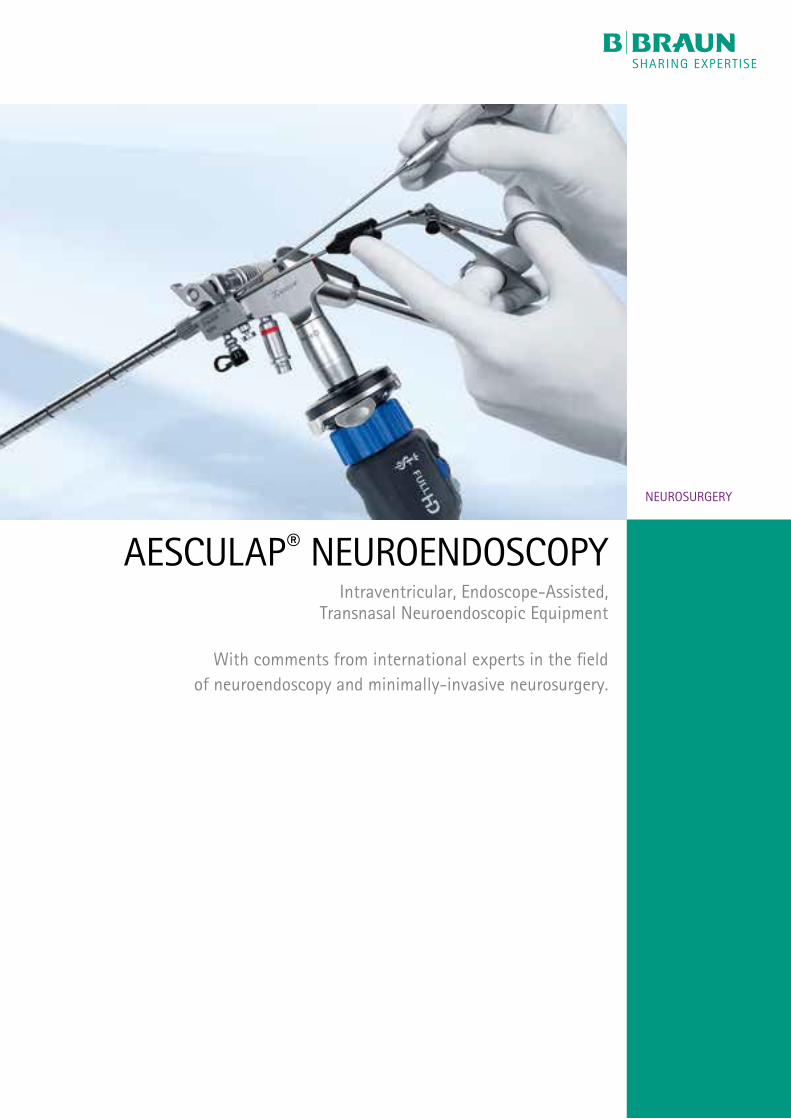
NEUROSURGERY AESCULAP ® NEUROENDOSCOPY Intraventricular, Endoscope-Assisted, Transnasal Neuroendoscopic Equipment With comments from international experts in the field of neuroendoscopy and minimally-invasive neurosurgery.

Welcome message from author
This document is posted to help you gain knowledge. Please leave a comment to let me know what you think about it! Share it to your friends and learn new things together.
Transcript
NEUROSURGERY
AESCULAP® NEUROENDOSCOPYIntraventricular, Endoscope-Assisted,
Transnasal Neuroendoscopic Equipment
With comments from international experts in the field of neuroendoscopy and minimally-invasive neurosurgery.
2
AESCULAP® NEUROENDOSCOPY
Recent improvements in preoperative imaging and surgical instrumentation allow neurosurgeons to treat more complex pathologies through customized less invasive approaches.
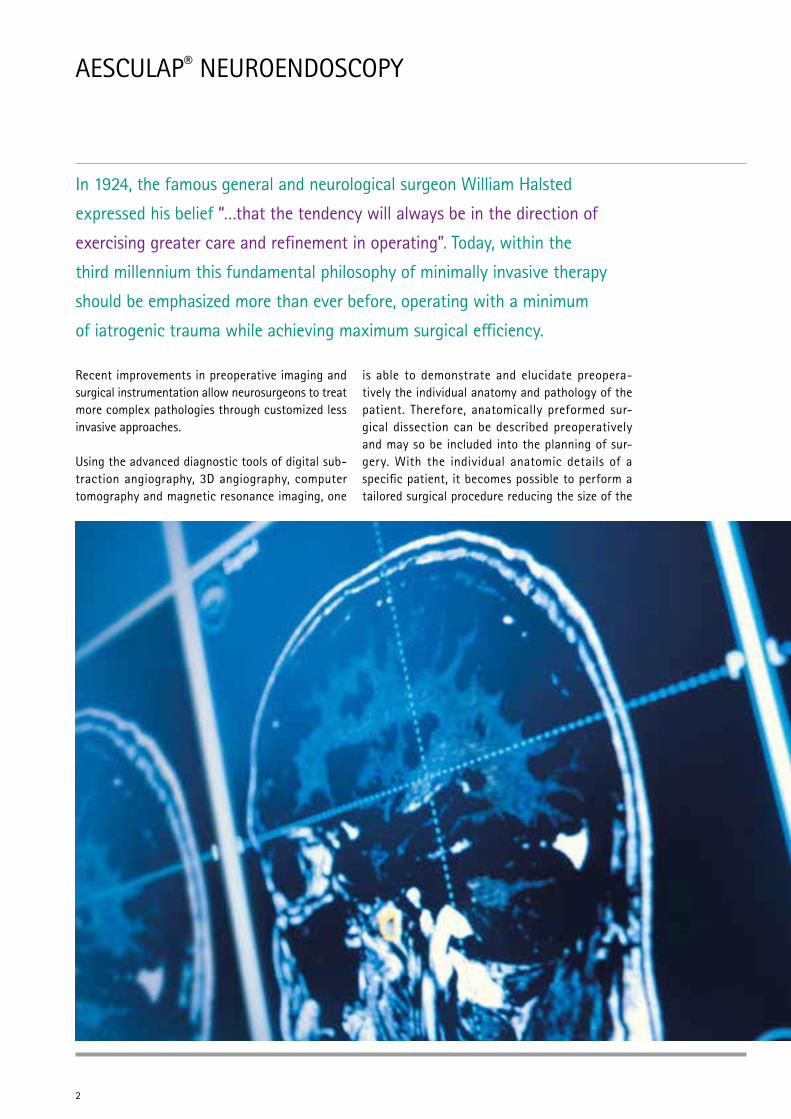
Using the advanced diagnostic tools of digital sub-traction angiography, 3D angiography, computer tomography and magnetic resonance imaging, one
is able to demonstrate and elucidate preopera-tively the individual anatomy and pathology of the patient. Therefore, anatomically preformed sur-gical dissection can be described preoperatively and may so be included into the planning of sur-gery. With the individual anatomic details of a specific patient, it becomes possible to perform a tailored surgical procedure reducing the size of the
In 1924, the famous general and neurological surgeon William Halsted
expressed his belief “…that the tendency will always be in the direction of
exercising greater care and refinement in operating”. Today, within the
third millennium this fundamental philosophy of minimally invasive therapy
should be emphasized more than ever before, operating with a minimum
of iatrogenic trauma while achieving maximum surgical efficiency.
3
Charles TeoSydney, Australia
Mark SouweidaneNew York, USA
André Grotenhuis Nijmegen, Netherlands
Michael FritschNeubrandenburg, Germany
Jeremy GreenleeIowa City, USA
Peter NakajiPhoenix, USA
skin incision, the craniotomy, and the extent of brain surface traumatization and retraction to a necessary minimum limit. These advantages of minimally inva-sive microsurgery contribute to improved postoper-ative results, including shorter hospitalization time because of reduction of the risk for complications.
However, small sized minimally invasive ap-proaches cause two important limitations: the significant loss of optical control and limited maneuverability of microsurgical instruments. The intraoperative use of endoscopes and dedicated minimally invasive instruments overcome these restrictions, thus enabling neurosurgeons to achieve deep seated regions without approach related traumatization of sensitive neurovascular structures.
The endoscopic image allows illumination and inspection of angles in hidden parts of the surgi-cal field with the and clear depiction of anatomical details. In addition, due to the enormous optical depth of field of modern endoscopes, endoscopes provide a three dimensional aspect of anatomic structures. Recently, the intraoperative use of full high definition (HD) image quality offers a new area in endoscopic neurosurgery with an increased range of indications in minimally invasive neurosurgery.
There are three main indications of endoscopic neurosurgery: the intraventricular, transcranial and transnasal application. In this brochure, contem-porary endoscopic equipment and instrumenta-tion is presented in a comprehensive way. Interna-tional experts in the field of minimally invasive and endoscopic neurosurgery comment the different applications, giving remarks with important tips and ideas, thus providing valuable instructions for the use of endoscopes in the field of minimally invasive neu-rosurgery.
Michael Fritsch, Neubrandenburg, GermanyJeremy Greenlee, Iowa City, USAAndré Grotenhuis, Nijmegen, NetherlandsPeter Nakaji, Phoenix, USAMark Souweidane, New York, USACharles Teo, Sydney, Australia

4
5
Intr
aven
tric
ular
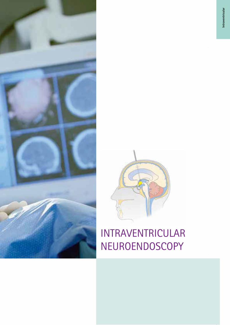
INTRAVENTRICULAR NEUROENDOSCOPY
6
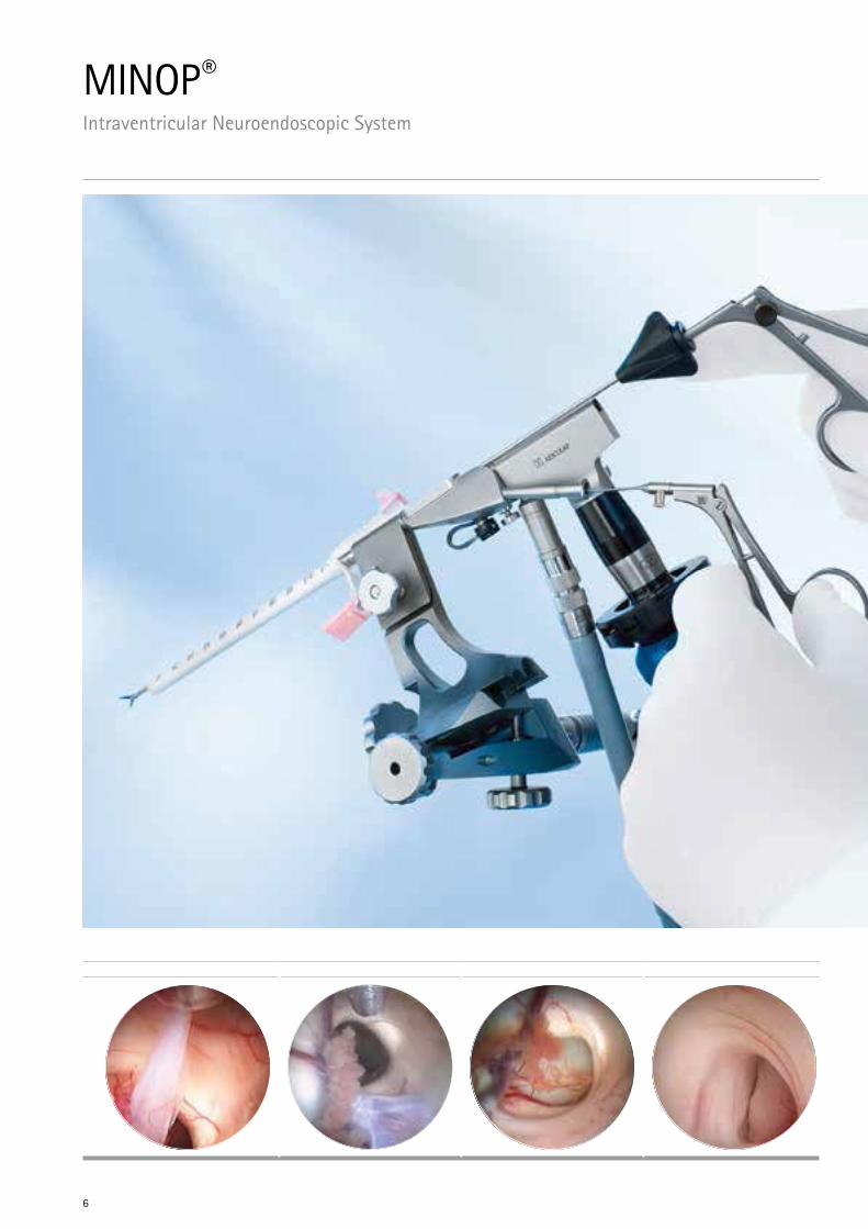
MINOP® Intraventricular Neuroendoscopic System
7
Intr
aven
tric
ular
Charles TeoSydney, Australia
Mark SouweidaneNew York, USA
The genesis of endoscopic surgery within the ventricular compartment can be attributed to the development of small caliber rod lens optics, fiberoptic light transmission and dedicated instrumentation. Since the advent of intraven-tricular endoscopic surgery, neurosurgeons have applied the technology to treat a number of disorders. While the enthusiasm has been great and the full potential not yet realized, a major benefit to the patient has been proven for selected conditions. Most notably the treatment of non-communicating hydrocephalus, management of patients with pineal region tumors, fenestra-tion of intracranial cysts, and removal of colloid cysts have all been shown to provide significant benefit and reduced morbidity compared with conventional treatment strategies.
The benefit in minimally invasive endoscopic procedures is analogous to that of any endoscopic procedure, namely minimal tissue disruption, en-hanced visualization, improved cosmetic results, shorter hospital stay, and less surgical morbidity.The surgeon willing to utilize intraventricular endoscopic surgery is first responsible for attain-ing a considerable degree of familiarity with the technology, relevant anatomy, and the surgical procedures. Given the relative nascence of the field, the discipline is only now being commonly implemented in training programs. Hence, for those that have not had the opportunity to have endoscopic surgery as part of their formal training, it is strongly recommended that the surgeon partici-pates in established practical courses in endoscopic neurosurgery, such as the courses from the Aesculap Academy.
Once fluent with the endoscopic equipment, more advanced procedures can be performed with greater familiarity and experience. It is antici- pated with future generations of neurosurgeons that the endoscope will be an indispensable part of the neurosurgeon‘s armamentarium given the unmatched image resolution and minimally invasive qualities.
This foreseeable integration will expectantly be paralleled with continued evolution in compat-ible equipment to suit the needs of an expanding repertoire.
Few neurosurgical procedures demand a degree of familiarity with equipment as do neuroen-dos-copic techniques. This feature is somewhat explained by the recent introduction of the neuroendoscope as well as the delicate nature of the equipment. The basic components of any neuroendoscopic procedure include the endoscope and trocar, a camera with light source and monitor, as well as compatible instrumentation.
Charles TeoMark Souweidane
8
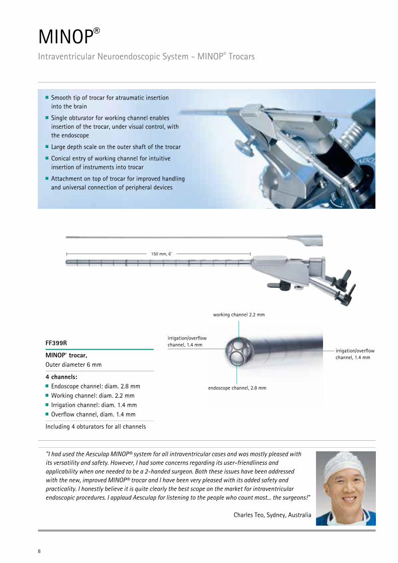
MINOP® Intraventricular Neuroendoscopic System - MINOP® Trocars
FF399R
MINOP® trocar, Outer diameter 6 mm
4 channels: Endoscope channel: diam. 2.8 mm Working channel: diam. 2.2 mm Irrigation channel: diam. 1.4 mm Overflow channel, diam. 1.4 mm
Including 4 obturators for all channels
150 mm, 6”
Smooth tip of trocar for atraumatic insertion into the brain
Single obturator for working channel enables insertion of the trocar, under visual control, with the endoscope
Large depth scale on the outer shaft of the trocar
Conical entry of working channel for intuitive insertion of instruments into trocar
Attachment on top of trocar for improved handling and universal connection of peripheral devices
endoscope channel, 2.8 mm
working channel 2.2 mm
irrigation/overflow channel, 1.4 mm
irrigation/overflow channel, 1.4 mm
"I had used the Aesculap MINOP® system for all intraventricular cases and was mostly pleased with its versatility and safety. However, I had some concerns regarding its user-friendliness and applicability when one needed to be a 2-handed surgeon. Both these issues have been addressed with the new, improved MINOP® trocar and I have been very pleased with its added safety and practicality. I honestly believe it is quite clearly the best scope on the market for intraventricular endoscopic procedures. I applaud Aesculap for listening to the people who count most... the surgeons!"
Charles Teo, Sydney, Australia
9
Intr
aven
tric
ular
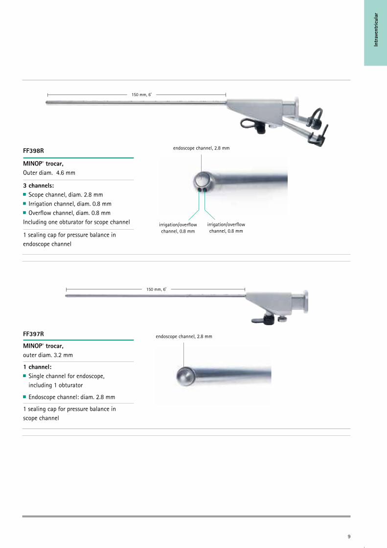
FF398R
MINOP® trocar, Outer diam. 4.6 mm
3 channels: Scope channel, diam. 2.8 mm Irrigation channel, diam. 0.8 mm Overflow channel, diam. 0.8 mm
Including one obturator for scope channel
1 sealing cap for pressure balance in endoscope channel
150 mm, 6”
150 mm, 6”
FF397R
MINOP® trocar, outer diam. 3.2 mm
1 channel: Single channel for endoscope,
including 1 obturator
Endoscope channel: diam. 2.8 mm
1 sealing cap for pressure balance in scope channel
endoscope channel, 2.8 mm
irrigation/overflow channel, 0.8 mm
irrigation/overflow channel, 0.8 mm
endoscope channel, 2.8 mm
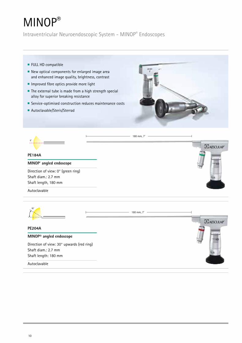
MINOP® Intraventricular Neuroendoscopic System - MINOP® Endoscopes
PE184A
MINOP® angled endoscope
Direction of view: 0° (green ring) Shaft diam.: 2.7 mm Shaft length, 180 mm
Autoclavable
10
180 mm, 7”
PE204A
MINOP® angled endoscope
Direction of view: 30° upwards (red ring) Shaft diam.: 2.7 mm Shaft length: 180 mm
Autoclavable
180 mm, 7”
FULL HD compatible
New optical components for enlarged image area and enhanced image quality, brightness, contrast
Improved fibre optics provide more light
The external tube is made from a high strength special alloy for superior breaking resistance
Service-optimised construction reduces maintenance costs
Autoclavable/Steris/Sterrad
Intr
aven
tric
ular
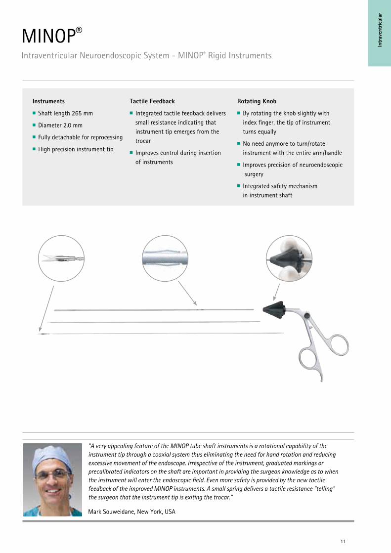
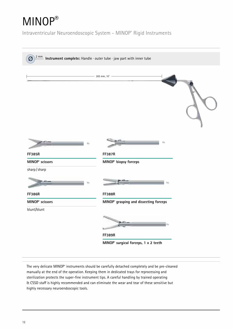
MINOP® Intraventricular Neuroendoscopic System - MINOP® Rigid Instruments
11
Rotating Knob
By rotating the knob slightly with index finger, the tip of instrument turns equally
No need anymore to turn/rotate instrument with the entire arm/handle
Improves precision of neuroendoscopic surgery
Integrated safety mechanism in instrument shaft
Instruments
Shaft length 265 mm
Diameter 2.0 mm
Fully detachable for reprocessing
High precision instrument tip
Tactile Feedback
Integrated tactile feedback delivers small resistance indicating that instrument tip emerges from the trocar
Improves control during insertion of instruments
"A very appealing feature of the MINOP tube shaft instruments is a rotational capability of the instrument tip through a coaxial system thus eliminating the need for hand rotation and reducing excessive movement of the endoscope. Irrespective of the instrument, graduated markings or precalibrated indicators on the shaft are important in providing the surgeon knowledge as to when the instrument will enter the endoscopic field. Even more safety is provided by the new tactile feedback of the improved MINOP instruments. A small spring delivers a tactile resistance "telling" the surgeon that the instrument tip is exiting the trocar."
Mark Souweidane, New York, USA
12
2⁄1 2⁄1
2⁄1
2⁄1
The very delicate MINOP® instruments should be carefully detached completely and be pre-cleaned manually at the end of the operation. Keeping them in dedicated trays for reprocessing and sterilization protects the super-fine instrument tips. A careful handling by trained operating & CSSD staff is highly recommended and can eliminate the wear and tear of these sensitive but highly necessary neuroendoscopic tools.
265 mm, 10”
FF385R
MINOP® scissors
sharp / sharp
FF386R
MINOP® scissors
blunt/blunt
FF387R
MINOP® biopsy forceps
FF388R
MINOP® grasping and dissecting forceps
FF389R
MINOP® surgical forceps, 1 x 2 teeth
2⁄1
MINOP® Intraventricular Neuroendoscopic System - MINOP® Rigid Instruments
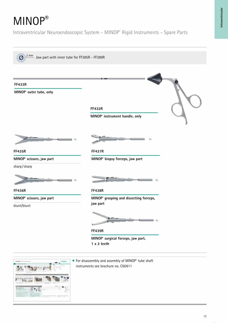
Instrument complete: Handle · outer tube · jaw part with inner tube Ø 2 mm
13
Intr
aven
tric
ular
2⁄1
2⁄1
2⁄1
2⁄1
2⁄1
FF435R
MINOP® scissors, jaw part
sharp / sharp
FF436R
MINOP® scissors, jaw part
blunt/blunt
FF437R
MINOP® biopsy forceps, jaw part
FF438R
MINOP® grasping and dissecting forceps, jaw part
FF432R
MINOP® instrument handle, only
FF433R
MINOP® outer tube, only
FF439R
MINOP® surgical forceps, jaw part, 1 x 2 teeth
Aesculap® Neuroendoscopy
D I S A S S E M B L I N G
Push and hold down the black button. At the same time, slide off the moveable ring towards the lower end.I Pull back the rotation wheel towards the
handle. Hold the rotation wheel at its positive stop and extract the shaft. Ensure that the moveable ring is kept downward.
II Remove the outer tube from the working insert.
Remove the inner tube from the working insert.IV
A S S E M B L I N G
III
REPAIRS Reprocess the product immediately after it has been contaminated.
Please follow the instructions for use TA012978 for further information on cleaning, disinfection and sterilization.
Please see also www.aesculap-extra.net for current information about processing.
For accessories and spare parts see brochure C35502.
Please find further information at www.aesculap-neuro.com
PROCESSING - ACCESSORIES
Please oil the instruments at the movable parts with Aesculap Sterilit® after each cleaning.
Repairs to the product must be carried out by personnel authorized by AESCULAP only. Only in this way warranties and guarantees will remain valid.
If any repairs are needed, please send the product to: Aesculap Technischer Service, Am Aesculap-Platz, 78532 Tuttlingen/Germany.
Local service addresses can be obtained from the address indicated above or from [email protected]
These instructions apply for all MINOP® and MINOP® InVent tube shaft instruments (FF385R - FF389R, FH635R - FH639R).!
III Hold the shaft at its closed working end. Ensure that the outer tube has been pushed over the inner tube with the working insert up to its positive stop.
Slide back and hold the rotation wheel at its positive stop. At the same time, slide off the moveable ring towards the lower end. Hold the shaft at its closed working end and reinsert it to its positive stop.
V Allow the rotation wheel to slide forward. (The shaft must be connected securely to the handle and must not come loose even when pulled.)
I Push the inner tube towards the working end of the working insert and align the slots of the inner tube so they slide over the two nubbs of the working insert (see „Disassambling IV“)
Slide the outer tube over the inner tube with the working insert. Ensure that the arrows (circum ferential markings) point towards the handle.
II VI Slide the moveable ring upward until the button clicks into place.IV
outer tubeblack button
circumferential markings rotation wheel
working end
inner tubemoveable ring
working insert
CARE
Aesculap AG | Am Aesculap-Platz | 78532 Tuttlingen | Germany Phone +49 7461 95-0 | Fax +49 7461 95-2600 | www.aesculap.com
Aesculap – a B. Braun company Brochure No. 60911 1114/0.1/1
Subject to technical changes. All rights reserved. This brochure may only be used for the exclusive purpose of obtaining information about our products. Reproduction in any form partial or otherwise is not permitted.
The main product trademark ‘Aesculap’ and the product trademarks ‘Sterilit’ and ‘Minop’ are registered trademarks of Aesculap AG.
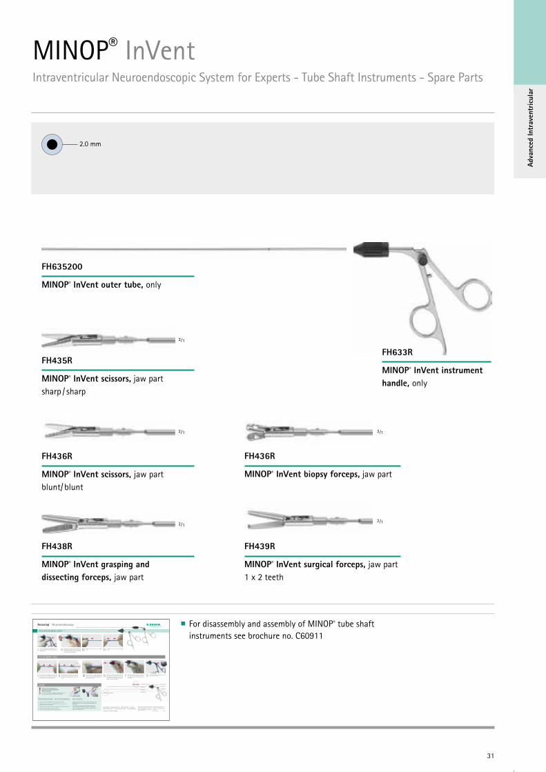
For disassembly and assembly of MINOP® tube shaft instruments see brochure no. C60911
MINOP® Intraventricular Neuroendoscopic System - MINOP® Rigid Instruments - Spare Parts
Jaw part with inner tube for FF385R - FF389RØ 2 mm
14
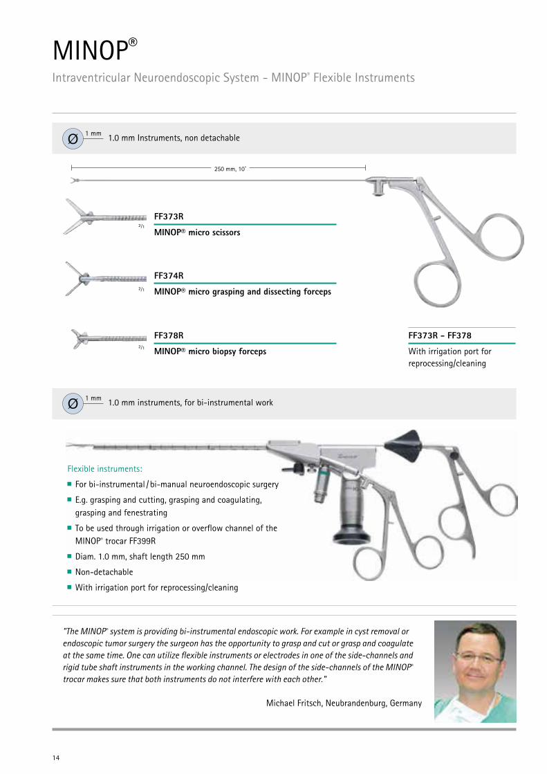
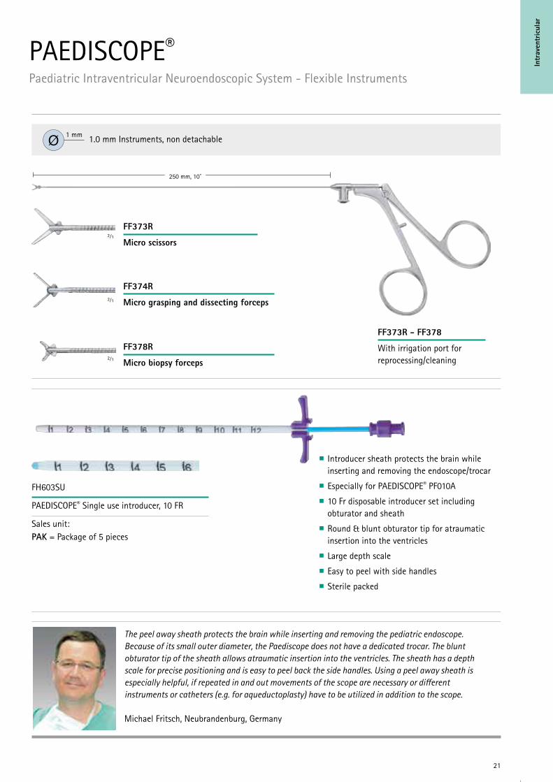
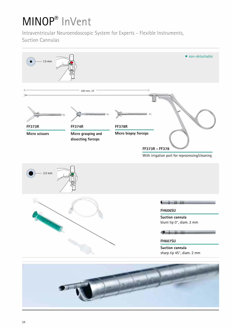
FF373R
MINOP® micro scissors
FF374R
MINOP® micro grasping and dissecting forceps
FF378R
MINOP® micro biopsy forceps
FF373R - FF378
With irrigation port for reprocessing/cleaning
250 mm, 10”
Flexible instruments:
For bi-instrumental / bi-manual neuroendoscopic surgery
E.g. grasping and cutting, grasping and coagulating, grasping and fenestrating
To be used through irrigation or overflow channel of the MINOP® trocar FF399R
Diam. 1.0 mm, shaft length 250 mm
Non-detachable
With irrigation port for reprocessing/cleaning
1.0 mm instruments, for bi-instrumental workØ 1 mm
"The MINOP® system is providing bi-instrumental endoscopic work. For example in cyst removal or endoscopic tumor surgery the surgeon has the opportunity to grasp and cut or grasp and coagulate at the same time. One can utilize flexible instruments or electrodes in one of the side-channels and rigid tube shaft instruments in the working channel. The design of the side-channels of the MINOP® trocar makes sure that both instruments do not interfere with each other."
Michael Fritsch, Neubrandenburg, Germany
MINOP® Intraventricular Neuroendoscopic System - MINOP® Flexible Instruments
1.0 mm Instruments, non detachableØ 1 mm
2⁄1
2⁄1
2⁄1
15
Intr
aven
tric
ular
1:1
1:1
1:1
1:1
255 mm, 10”
1:1
1:1255 mm, 10”
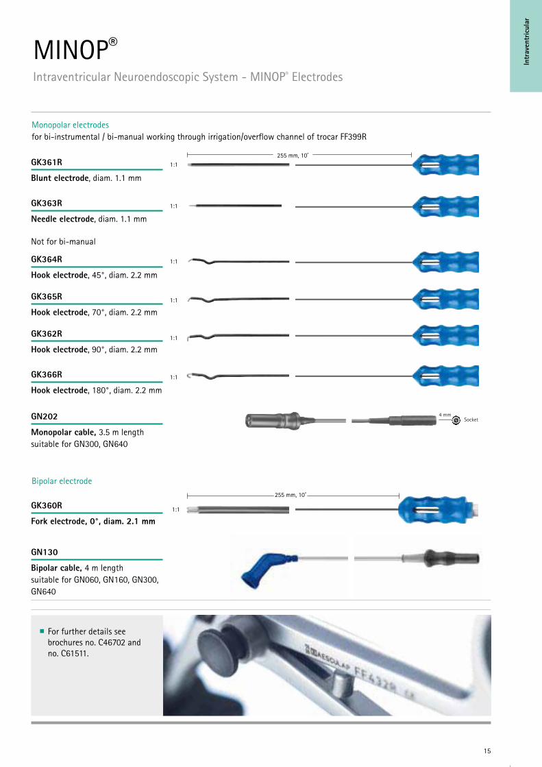
Bipolar electrode
Monopolar electrodes for bi-instrumental / bi-manual working through irrigation/overflow channel of trocar FF399R
Ø4 mm
Socket
MINOP® Intraventricular Neuroendoscopic System - MINOP® Electrodes
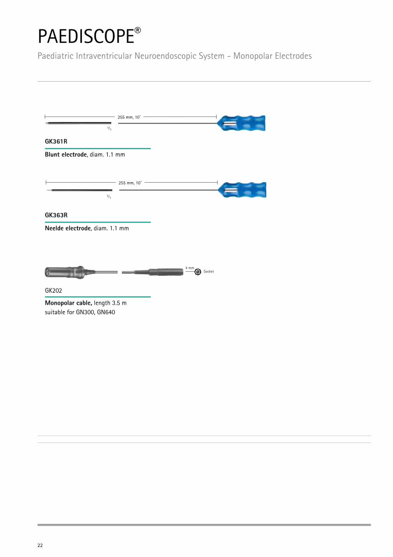
GK361R
Blunt electrode, diam. 1.1 mm
GK363R
Needle electrode, diam. 1.1 mm
GK364R
Hook electrode, 45°, diam. 2.2 mm
GK365R
Hook electrode, 70°, diam. 2.2 mm
GK362R
Hook electrode, 90°, diam. 2.2 mm
GK366R
Hook electrode, 180°, diam. 2.2 mm
GN202
Monopolar cable, 3.5 m length suitable for GN300, GN640
GN130
Bipolar cable, 4 m length suitable for GN060, GN160, GN300, GN640
GK360R
Fork electrode, 0°, diam. 2.1 mm
1:1
For further details see brochures no. C46702 and no. C61511.
Not for bi-manual
16
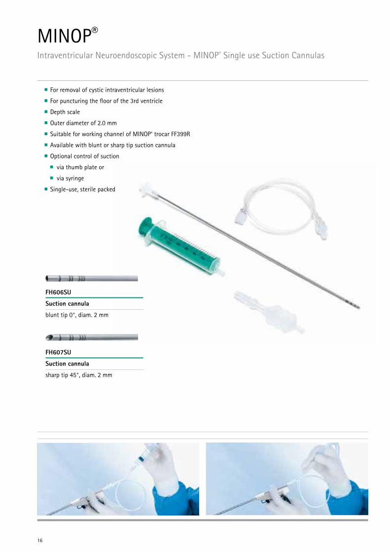
For removal of cystic intraventricular lesions
For puncturing the floor of the 3rd ventricle
Depth scale
Outer diameter of 2.0 mm
Suitable for working channel of MINOP® trocar FF399R
Available with blunt or sharp tip suction cannula
Optional control of suction
via thumb plate or
via syringe
Single-use, sterile packed
FH606SU
Suction cannula
blunt tip 0°, diam. 2 mm
FH607SU
Suction cannula
sharp tip 45°, diam. 2 mm
MINOP® Intraventricular Neuroendoscopic System - MINOP® Single use Suction Cannulas
17
Intr
aven
tric
ular
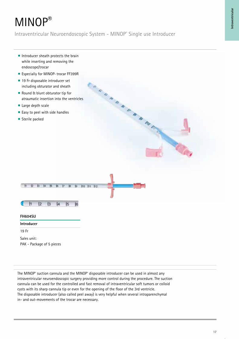
Introducer sheath protects the brain while inserting and removing the endoscope/trocar
Especially for MINOP® trocar FF399R
19 Fr disposable introducer set including obturator and sheath
Round & blunt obturator tip for atraumatic insertion into the ventricles
Large depth scale
Easy to peel with side handles
Sterile packed
FH604SU
Introducer
19 Fr
Sales unit: PAK - Package of 5 pieces
The MINOP® suction cannula and the MINOP® disposable introducer can be used in almost any intraventricular neuroendoscopic surgery providing more control during the procedure. The suction cannula can be used for the controlled and fast removal of intraventricular soft tumors or colloid cysts with its sharp cannula tip or even for the opening of the floor of the 3rd ventricle. The disposable introducer (also called peel away) is very helpful when several intraparenchymal in- and out-movements of the trocar are necessary.
MINOP® Intraventricular Neuroendoscopic System - MINOP® Single use Introducer
18
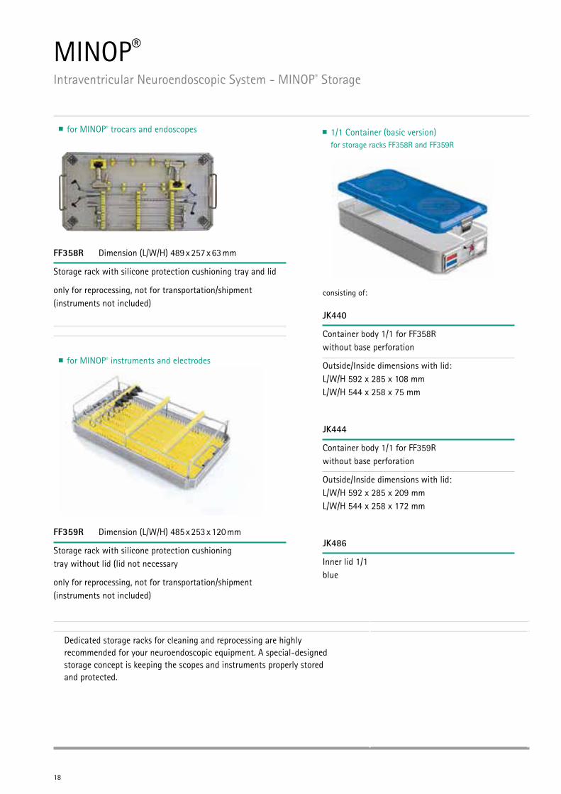
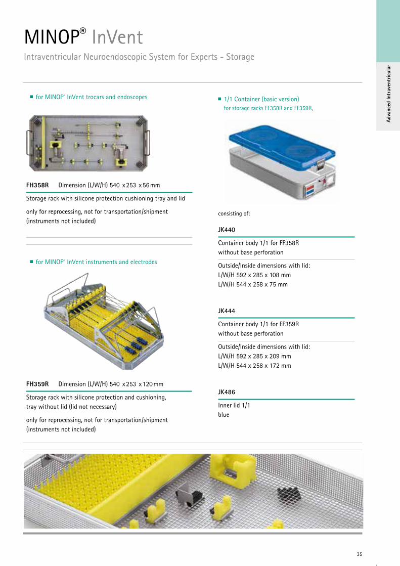
FF358R Dimension (L/W/H) 489 x 257 x 63 mm
Storage rack with silicone protection cushioning tray and lid
only for reprocessing, not for transportation/shipment (instruments not included)
FF359R Dimension (L/W/H) 485 x 253 x 120 mm
Storage rack with silicone protection cushioning tray without lid (lid not necessary
only for reprocessing, not for transportation/shipment (instruments not included)
Dedicated storage racks for cleaning and reprocessing are highly recommended for your neuroendoscopic equipment. A special-designed storage concept is keeping the scopes and instruments properly stored and protected.
MINOP® Intraventricular Neuroendoscopic System - MINOP® Storage
for MINOP® trocars and endoscopes
for MINOP® instruments and electrodes
1/1 Container (basic version) for storage racks FF358R and FF359R
consisting of:
JK440
Container body 1/1 for FF358R without base perforation
Outside/Inside dimensions with lid: L/W/H 592 x 285 x 108 mm L/W/H 544 x 258 x 75 mm
JK444
Container body 1/1 for FF359R without base perforation
Outside/Inside dimensions with lid: L/W/H 592 x 285 x 209 mm L/W/H 544 x 258 x 172 mm
JK486
Inner lid 1/1 blue
19
Intr
aven
tric
ular
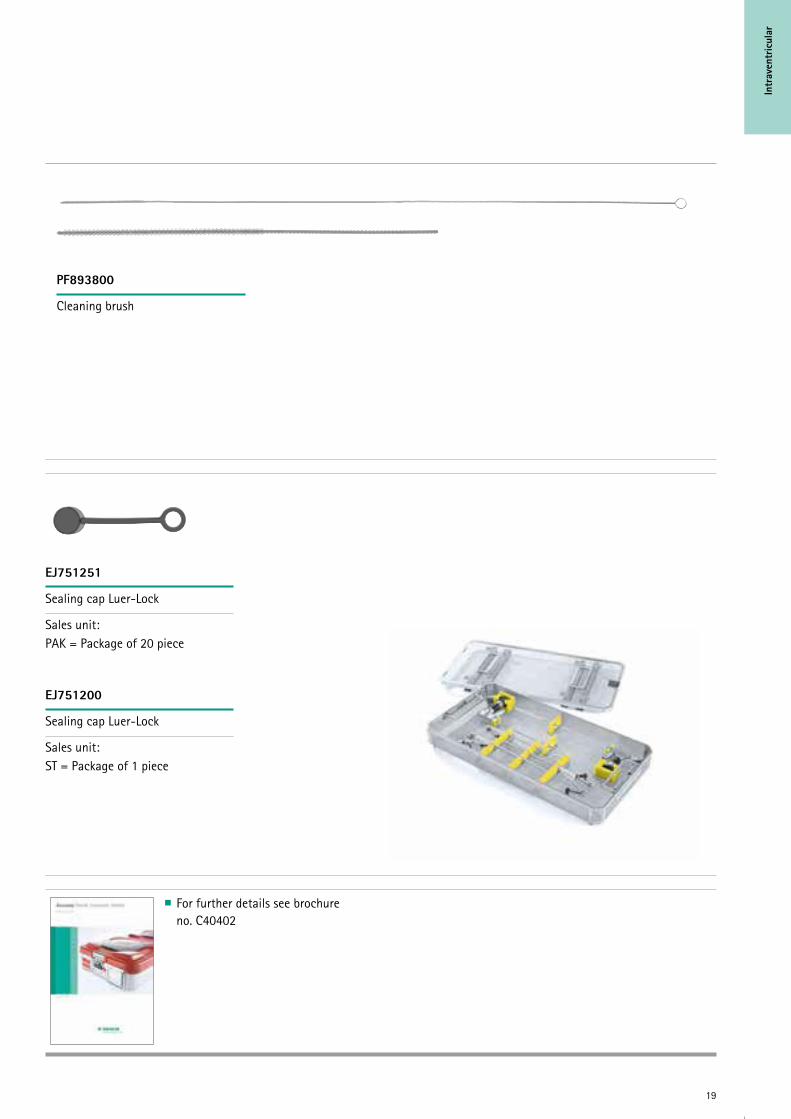
PF893800
Cleaning brush
EJ751251
Sealing cap Luer-Lock
Sales unit: PAK = Package of 20 piece
EJ751200
Sealing cap Luer-Lock
Sales unit: ST = Package of 1 piece
For further details see brochure no. C40402
20
150 mm, 6”
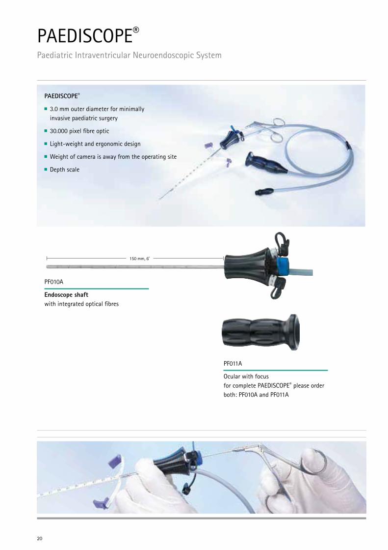
PAEDISCOPE® Paediatric Intraventricular Neuroendoscopic System
PAEDISCOPE®
3.0 mm outer diameter for minimally invasive paediatric surgery
30.000 pixel fibre optic
Light-weight and ergonomic design
Weight of camera is away from the operating site
Depth scale
PF010A
Endoscope shaft with integrated optical fibres
PF011A
Ocular with focus for complete PAEDISCOPE® please order both: PF010A and PF011A
21
Intr
aven
tric
ular
250 mm, 10”
The peel away sheath protects the brain while inserting and removing the pediatric endoscope. Because of its small outer diameter, the Paediscope does not have a dedicated trocar. The blunt obturator tip of the sheath allows atraumatic insertion into the ventricles. The sheath has a depth scale for precise positioning and is easy to peel back the side handles. Using a peel away sheath is especially helpful, if repeated in and out movements of the scope are necessary or different instruments or catheters (e.g. for aqueductoplasty) have to be utilized in addition to the scope.
Michael Fritsch, Neubrandenburg, Germany
Introducer sheath protects the brain while inserting and removing the endoscope/trocar Especially for PAEDISCOPE® PF010A 10 Fr disposable introducer set including obturator and sheath
Round & blunt obturator tip for atraumatic insertion into the ventricles
Large depth scale Easy to peel with side handles Sterile packed
PAEDISCOPE® Paediatric Intraventricular Neuroendoscopic System - Flexible Instruments
1.0 mm Instruments, non detachableØ 1 mm
FF373R
Micro scissors
FF374R
Micro grasping and dissecting forceps
FF378R
Micro biopsy forceps
FF373R - FF378
With irrigation port for reprocessing/cleaning
2⁄1
2⁄1
2⁄1
FH603SU
PAEDISCOPE® Single use introducer, 10 FR
Sales unit: PAK = Package of 5 pieces
22
255 mm, 10”
255 mm, 10”
Ø4 mm
Socket
PAEDISCOPE® Paediatric Intraventricular Neuroendoscopic System - Monopolar Electrodes
GK361R
Blunt electrode, diam. 1.1 mm
GK363R
Neelde electrode, diam. 1.1 mm
GK202
Monopolar cable, length 3.5 m suitable for GN300, GN640
1⁄₁
1⁄₁
23
Intr
aven
tric
ular
For futher details see brochure no. C29202 and no. C40402
PAEDISCOPE® Paediatric Intraventricular Neuroendoscopic System - Storage
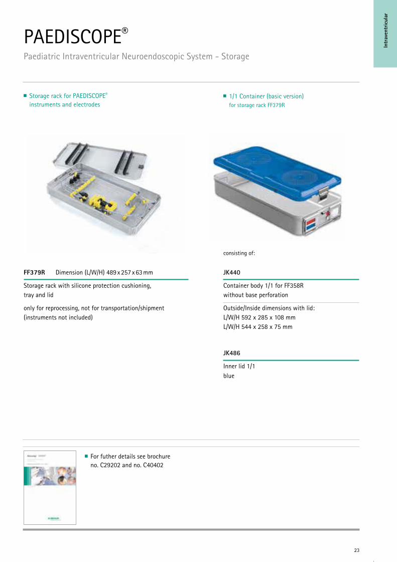
Storage rack for PAEDISCOPE® instruments and electrodes
FF379R Dimension (L/W/H) 489 x 257 x 63 mm
Storage rack with silicone protection cushioning, tray and lid
only for reprocessing, not for transportation/shipment (instruments not included)
1/1 Container (basic version) for storage rack FF379R
consisting of:
JK440
Container body 1/1 for FF358R without base perforation
Outside/Inside dimensions with lid: L/W/H 592 x 285 x 108 mm L/W/H 544 x 258 x 75 mm
JK486
Inner lid 1/1 blue
24
25
Adva
nced
Intr
aven
tric
ular
26
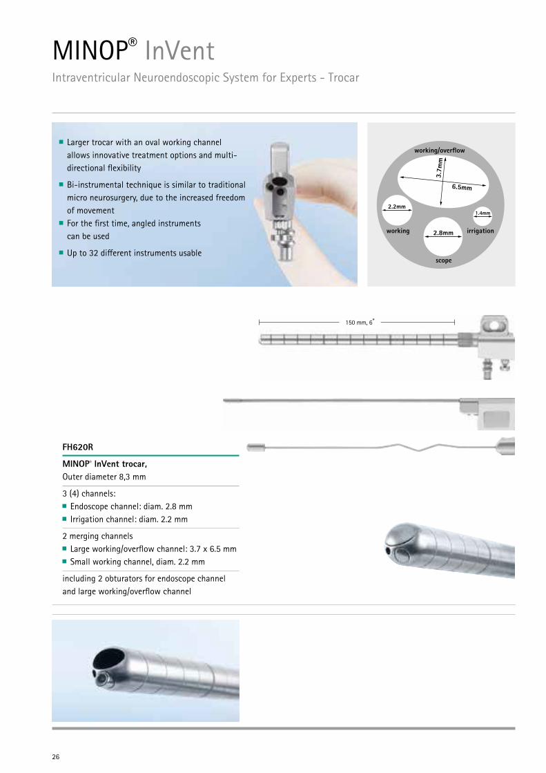
Larger trocar with an oval working channel allows innovative treatment options and multi- directional flexibility
Bi-instrumental technique is similar to traditional micro neurosurgery, due to the increased freedom of movement
For the first time, angled instruments can be used
Up to 32 different instruments usable
150 mm, 6”
MINOP® InVentIntraventricular Neuroendoscopic System for Experts - Trocar
2.2mm
2.8mm
1.4mm
working
working/over�ow
irrigation
scope
3.7m
m
6.5mm
FH620R
MINOP® InVent trocar, Outer diameter 8,3 mm
3 (4) channels: Endoscope channel: diam. 2.8 mm Irrigation channel: diam. 2.2 mm
2 merging channels Large working/overflow channel: 3.7 x 6.5 mm Small working channel, diam. 2.2 mm
including 2 obturators for endoscope channel and large working/overflow channel
27
Adva
nced
Intr
aven
tric
ular
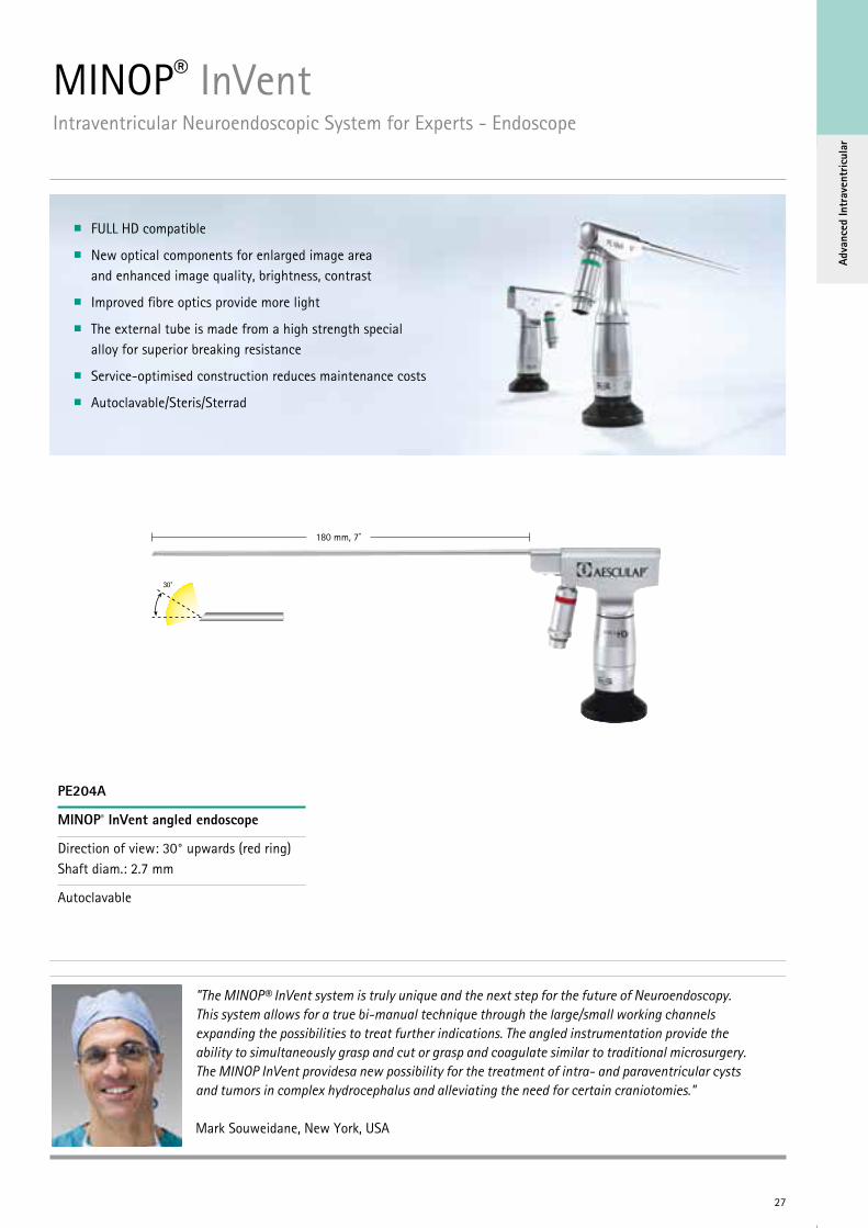
"The MINOP® InVent system is truly unique and the next step for the future of Neuroendoscopy. This system allows for a true bi-manual technique through the large/small working channels expanding the possibilities to treat further indications. The angled instrumentation provide the ability to simultaneously grasp and cut or grasp and coagulate similar to traditional microsurgery. The MINOP InVent providesa new possibility for the treatment of intra- and paraventricular cysts and tumors in complex hydrocephalus and alleviating the need for certain craniotomies."
Mark Souweidane, New York, USA
180 mm, 7”
FULL HD compatible
New optical components for enlarged image area and enhanced image quality, brightness, contrast
Improved fibre optics provide more light
The external tube is made from a high strength special alloy for superior breaking resistance
Service-optimised construction reduces maintenance costs
Autoclavable/Steris/Sterrad
MINOP® InVentIntraventricular Neuroendoscopic System for Experts - Endoscope
PE204A
MINOP® InVent angled endoscope
Direction of view: 30° upwards (red ring) Shaft diam.: 2.7 mm
Autoclavable
28
355 mm, 14”
FH629R
MINOP® InVent dissector, tip width 2.2 mm
FH623R
MINOP® InVent hook, 90° blunt, hook deflection width 3.5 mm
FH623R
MINOP® InVent knife, backwards cutting, knife deflection width 3.0 mm
FH630R
MINOP® InVent dissector, tip width 1.7 mm
FH631R
MINOP® InVent dissector, tip width 1.0 mm
²⁄₁
²⁄₁
²⁄₁
²⁄₁
²⁄₁
2 mm
MINOP® InVentIntraventricular Neuroendoscopic System for Experts - Dissectors, Hook and Knife
29
Adva
nced
Intr
aven
tric
ular
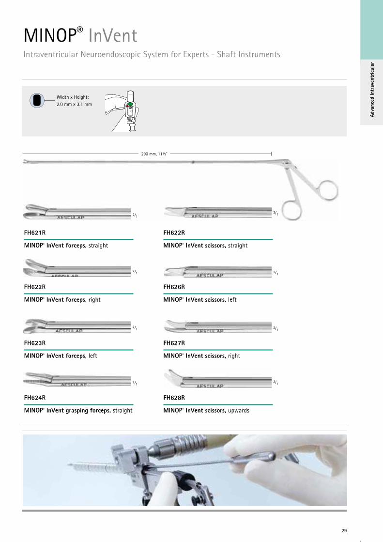
290 mm, 11½”
FH622R
MINOP® InVent scissors, straight
FH621R
MINOP® InVent forceps, straight
FH626R
MINOP® InVent scissors, left
FH622R
MINOP® InVent forceps, right
FH627R
MINOP® InVent scissors, right
FH623R
MINOP® InVent forceps, left
FH628R
MINOP® InVent scissors, upwards
FH624R
MINOP® InVent grasping forceps, straight
Width x Height: 2.0 mm x 3.1 mm
²⁄₁ ²⁄₁
²⁄₁ ²⁄₁
²⁄₁ ²⁄₁
²⁄₁ ²⁄₁
MINOP® InVentIntraventricular Neuroendoscopic System for Experts - Shaft Instruments
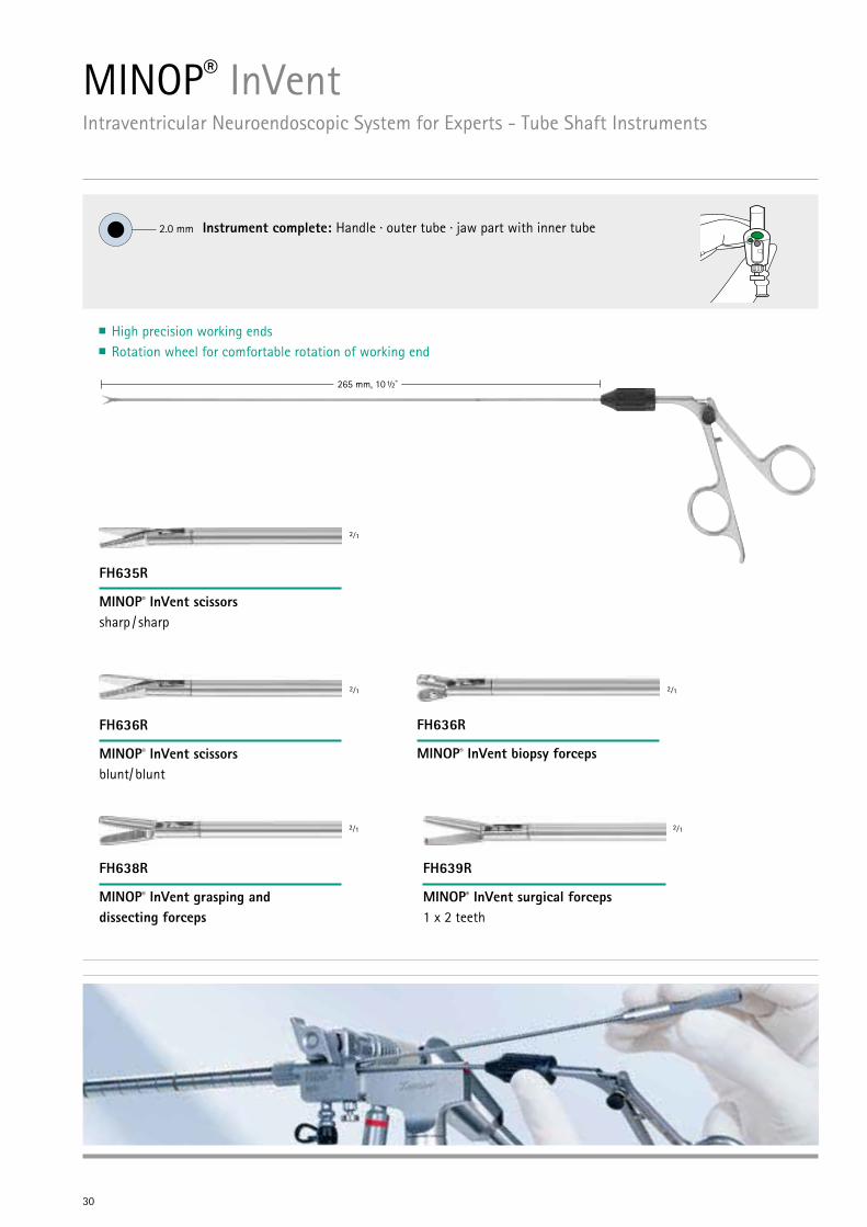
30
265 mm, 10 ½”
Instrument complete: Handle · outer tube · jaw part with inner tube 2.0 mm
MINOP® InVentIntraventricular Neuroendoscopic System for Experts - Tube Shaft Instruments
FH635R
MINOP® InVent scissors sharp / sharp
FH638R
MINOP® InVent grasping and dissecting forceps
FH639R
MINOP® InVent surgical forceps 1 x 2 teeth
FH636R
MINOP® InVent scissors blunt/ blunt
FH636R
MINOP® InVent biopsy forceps
2⁄1
2⁄1
2⁄1
2⁄1
2⁄1
High precision working endsRotation wheel for comfortable rotation of working end
31
Adva
nced
Intr
aven
tric
ular
2.0 mm
Aesculap® Neuroendoscopy
D I S A S S E M B L I N G
Push and hold down the black button. At the same time, slide off the moveable ring towards the lower end.I Pull back the rotation wheel towards the
handle. Hold the rotation wheel at its positive stop and extract the shaft. Ensure that the moveable ring is kept downward.
II Remove the outer tube from the working insert.
Remove the inner tube from the working insert.IV
A S S E M B L I N G
III
REPAIRS Reprocess the product immediately after it has been contaminated.
Please follow the instructions for use TA012978 for further information on cleaning, disinfection and sterilization.
Please see also www.aesculap-extra.net for current information about processing.
For accessories and spare parts see brochure C35502.
Please find further information at www.aesculap-neuro.com
PROCESSING - ACCESSORIES
Please oil the instruments at the movable parts with Aesculap Sterilit® after each cleaning.
Repairs to the product must be carried out by personnel authorized by AESCULAP only. Only in this way warranties and guarantees will remain valid.
If any repairs are needed, please send the product to: Aesculap Technischer Service, Am Aesculap-Platz, 78532 Tuttlingen/Germany.
Local service addresses can be obtained from the address indicated above or from [email protected]
These instructions apply for all MINOP® and MINOP® InVent tube shaft instruments (FF385R - FF389R, FH635R - FH639R).!
III Hold the shaft at its closed working end. Ensure that the outer tube has been pushed over the inner tube with the working insert up to its positive stop.
Slide back and hold the rotation wheel at its positive stop. At the same time, slide off the moveable ring towards the lower end. Hold the shaft at its closed working end and reinsert it to its positive stop.
V Allow the rotation wheel to slide forward. (The shaft must be connected securely to the handle and must not come loose even when pulled.)
I Push the inner tube towards the working end of the working insert and align the slots of the inner tube so they slide over the two nubbs of the working insert (see „Disassambling IV“)
Slide the outer tube over the inner tube with the working insert. Ensure that the arrows (circum ferential markings) point towards the handle.
II VI Slide the moveable ring upward until the button clicks into place.IV
outer tubeblack button
circumferential markings rotation wheel
working end
inner tubemoveable ring
working insert
CARE
Aesculap AG | Am Aesculap-Platz | 78532 Tuttlingen | Germany Phone +49 7461 95-0 | Fax +49 7461 95-2600 | www.aesculap.com
Aesculap – a B. Braun company Brochure No. 60911 1114/0.1/1
Subject to technical changes. All rights reserved. This brochure may only be used for the exclusive purpose of obtaining information about our products. Reproduction in any form partial or otherwise is not permitted.
The main product trademark ‘Aesculap’ and the product trademarks ‘Sterilit’ and ‘Minop’ are registered trademarks of Aesculap AG.
For disassembly and assembly of MINOP® tube shaft instruments see brochure no. C60911
MINOP® InVentIntraventricular Neuroendoscopic System for Experts - Tube Shaft Instruments - Spare Parts
FH435R
MINOP® InVent scissors, jaw part sharp / sharp
FH438R
MINOP® InVent grasping and dissecting forceps, jaw part
FH439R
MINOP® InVent surgical forceps, jaw part 1 x 2 teeth
FH436R
MINOP® InVent scissors, jaw part blunt/ blunt
FH436R
MINOP® InVent biopsy forceps, jaw part
FH635200
MINOP® InVent outer tube, only
FH633R
MINOP® InVent instrument handle, only
2⁄1 2⁄1
2⁄1 2⁄1
2⁄1
32
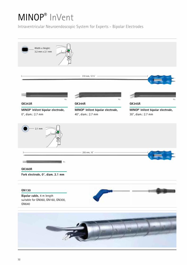
310 mm, 12 ¼”
255 mm, 10”
Width x Height: 3.2 mm x 2.1 mm
2.1 mm
GN130
Bipolar cable, 4 m length suitable for GN060, GN160, GN300, GN640
GK360R
Fork electrode, 0°, diam. 2.1 mm
GK343R
MINOP® InVent bipolar electrode, 0°, diam.: 2.7 mm
GK344R
MINOP® InVent bipolar electrode, 40°, diam.: 2.7 mm
GK345R
MINOP® InVent bipolar electrode, 30°, diam.: 2.7 mm
MINOP® InVentIntraventricular Neuroendoscopic System for Experts - Bipolar Electrodes
2⁄1
2⁄1 2⁄1 2⁄1
33
Adva
nced
Intr
aven
tric
ular
255 mm, 10”
Ø4 mm
Socket
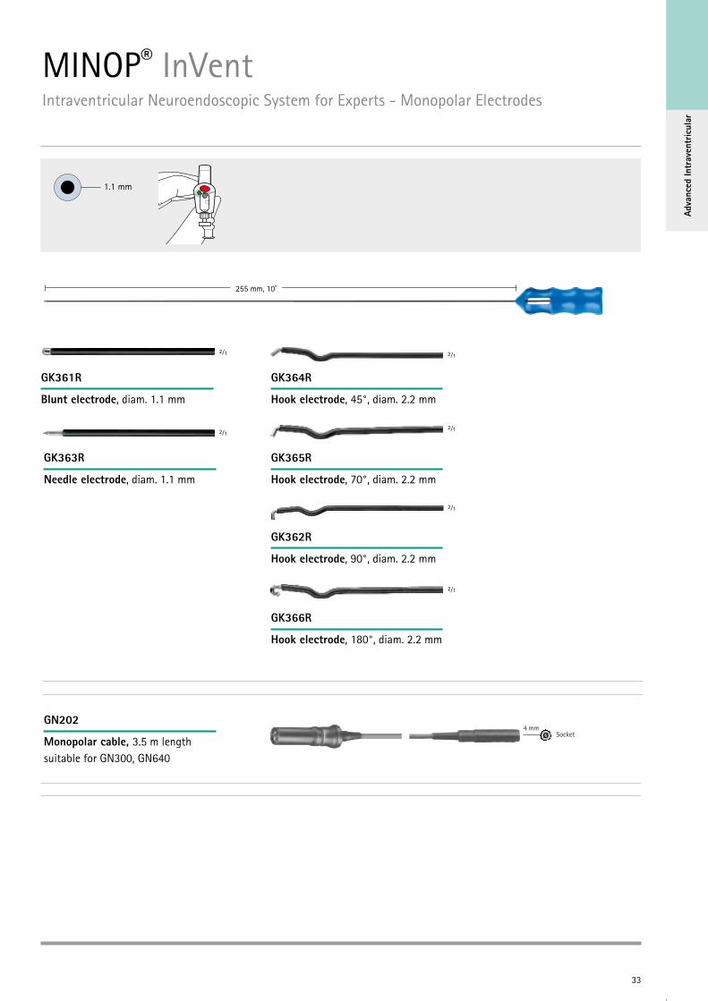
1.1 mm
MINOP® InVentIntraventricular Neuroendoscopic System for Experts - Monopolar Electrodes
2⁄1 2⁄1
2⁄1 2⁄1
2⁄1
2⁄1
GK361R
Blunt electrode, diam. 1.1 mm
GK363R
Needle electrode, diam. 1.1 mm
GK364R
Hook electrode, 45°, diam. 2.2 mm
GK365R
Hook electrode, 70°, diam. 2.2 mm
GK362R
Hook electrode, 90°, diam. 2.2 mm
GK366R
Hook electrode, 180°, diam. 2.2 mm
GN202
Monopolar cable, 3.5 m length suitable for GN300, GN640
34
1.0 mm
2.0 mm
MINOP® InVentIntraventricular Neuroendoscopic System for Experts - Flexible Instruments, Suction Cannulas
250 mm, 10”
FF373R
Micro scissors
FF374R
Micro grasping and dissecting forceps
FF378R
Micro biopsy forceps
FF373R - FF378
With irrigation port for reprocessing/cleaning
2⁄1 2⁄1 2⁄1
FH606SU
Suction cannula blunt tip 0°, diam. 2 mm
FH607SU
Suction cannula sharp tip 45°, diam. 2 mm
non-detachable
35
Adva
nced
Intr
aven
tric
ular
MINOP® InVentIntraventricular Neuroendoscopic System for Experts - Storage
FH358R Dimension (L/W/H) 540 x 253 x 56 mm
Storage rack with silicone protection cushioning tray and lid
only for reprocessing, not for transportation/shipment (instruments not included)
FH359R Dimension (L/W/H) 540 x 253 x 120 mm
Storage rack with silicone protection and cushioning, tray without lid (lid not necessary)
only for reprocessing, not for transportation/shipment (instruments not included)
for MINOP® InVent trocars and endoscopes
for MINOP® InVent instruments and electrodes
1/1 Container (basic version) for storage racks FF358R and FF359R,
consisting of:
JK440
Container body 1/1 for FF358R without base perforation
Outside/Inside dimensions with lid: L/W/H 592 x 285 x 108 mm L/W/H 544 x 258 x 75 mm
JK444
Container body 1/1 for FF359R without base perforation
Outside/Inside dimensions with lid: L/W/H 592 x 285 x 209 mm L/W/H 544 x 258 x 172 mm
JK486
Inner lid 1/1 blue

36

ENDOSCOPE-ASSISTED MICRO NEUROSURGERY
Endo
scop
e-As
siste
d
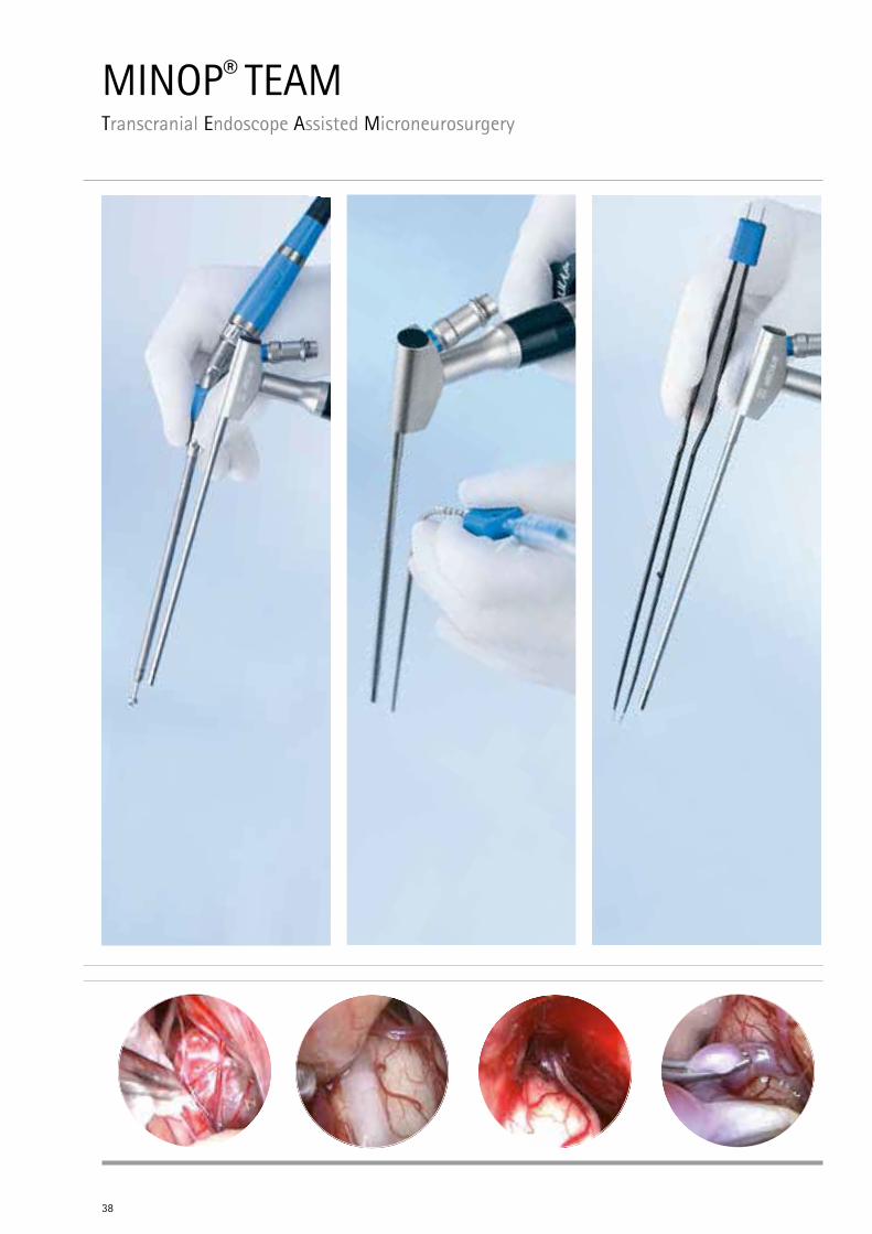
MINOP® TEAMTranscranial Endoscope Assisted Microneurosurgery
38
Endo
scop
e-As
siste
d
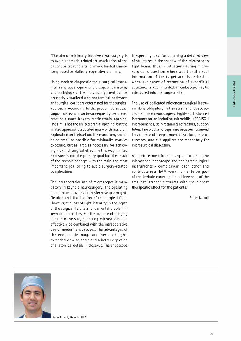
Peter Nakaji, Phoenix, USA
39
"The aim of minimally invasive neurosurgery is to avoid approach-related traumatization of the patient by creating a tailor-made limited cranio-tomy based on skilled preoperative planning.
Using modern diagnostic tools, surgical instru-ments and visual equipment, the specific anatomy and pathology of the individual patient can be precisely visualized and anatomical pathways and surgical corridors determined for the surgical approach. According to the predefined access, surgical dissection can be subsequently performed creating a much less traumatic cranial opening. The aim is not the limited cranial opening, but the limited approach associated injury with less brain exploration and retraction. The craniotomy should be as small as possible for minimally invasive exposure, but as large as necessary for achiev-ing maximal surgical effect. In this way, limited exposure is not the primary goal but the result of the keyhole concept with the main and most important goal being to avoid surgery-related complications.
The intraoperative use of microscopes is man-datory in keyhole neurosurgery. The operating microscope provides both stereoscopic magni-fication and illumination of the surgical field. However, the loss of light intensity in the depth of the surgical field is a fundamental problem in keyhole approaches. For the purpose of bringing light into the site, operating microscopes can effectively be combined with the intraoperative use of modern endoscopes. The advantages of the endoscopic image are increased light, extended viewing angle and a better depiction of anatomical details in close-up. The endoscope
is especially ideal for obtaining a detailed view of structures in the shadow of the microscope‘s light beam. Thus, in situations during micro- surgical dissection where additional visual information of the target area is desired or when avoidance of retraction of superficial structures is recommended, an endoscope may be introduced into the surgical site.
The use of dedicated microneurosurgical instru-ments is obligatory in transcranial endoscope- assisted microneurosurgery. Highly sophisticated instrumentation including microdrills, KERRISON micropunches, self-retaining retractors, suction tubes, fine bipolar forceps, microscissors, diamond knives, microforceps, microdissectors, micro- curettes, and clip appliers are mandatory for microsurgical dissection.
All before mentioned surgical tools - the microscope, endoscope and dedicated surgical instruments - complement each other and contribute in a TEAM-work manner to the goal of the keyhole concept: the achievement of the smallest iatrogenic trauma with the highest therapeutic effect for the patients."
Peter Nakaji
MINOP® TEAMTranscranial Endoscope Assisted Microneurosurgery - Angled “PERNECZKY” Endoscopes
40
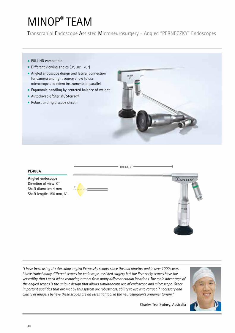
FULL HD compatible
Different viewing angles (0°, 30°, 70°)
Angled endoscope design and lateral connection for camera and light source allow to use microscope and micro instruments in parallel
Ergonomic handling by centered balance of weight
Autoclavable / Steris® / Sterrad®
Robust and rigid scope sheath
150 mm, 6”
"I have been using the Aesculap angled Perneczky scopes since the mid nineties and in over 1000 cases. I have trialed many different scopes for endoscope-assisted surgery but the Perneczky scopes have the versatility that I need when removing tumors from many different cranial locations. The main advantage of the angled scopes is the unique design that allows simultaneous use of endoscope and microscope. Other important qualities that are met by this system are robustness, ability to use it to retract if necessary and clarity of image. I believe these scopes are an essential tool in the neurosurgeon’s armamentarium."
Charles Teo, Sydney, Australia
PE486A
Angled endoscope Direction of view: 0° Shaft diameter: 4 mm Shaft length: 150 mm, 6“
Endo
scop
e-As
siste
d
41
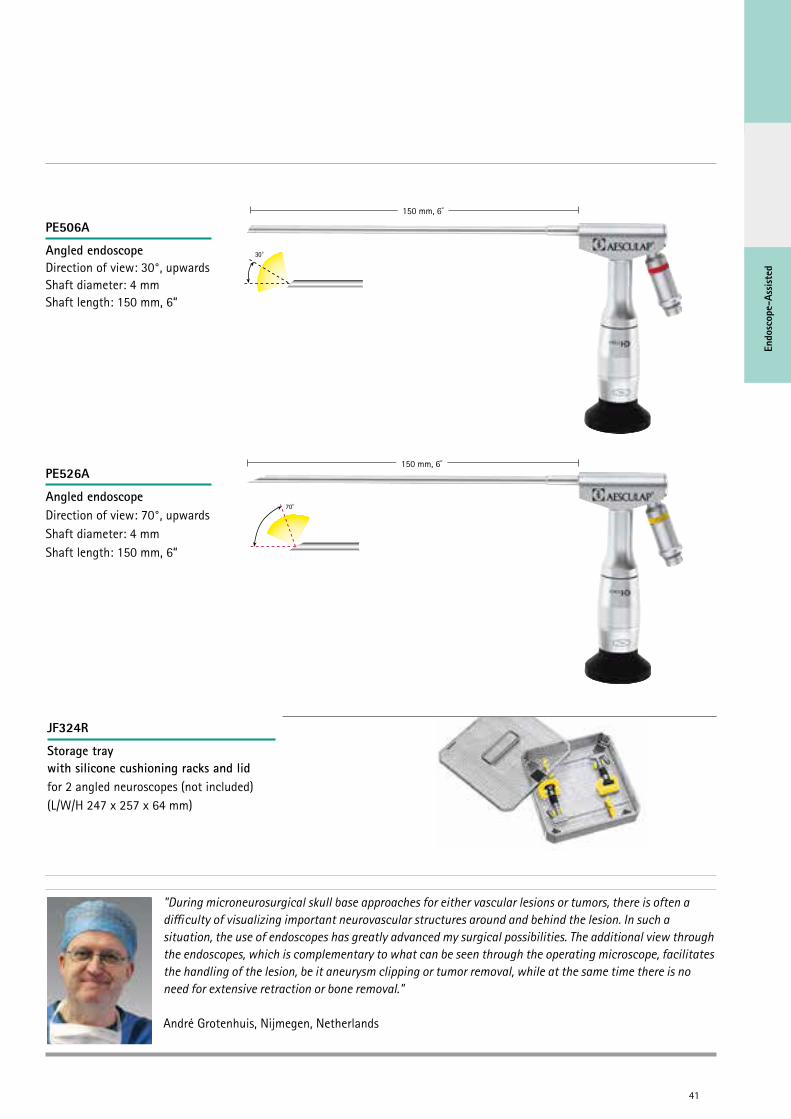
PE506A
Angled endoscope Direction of view: 30°, upwards Shaft diameter: 4 mm Shaft length: 150 mm, 6“
150 mm, 6”
150 mm, 6”
"During microneurosurgical skull base approaches for either vascular lesions or tumors, there is often a difficulty of visualizing important neurovascular structures around and behind the lesion. In such a situation, the use of endoscopes has greatly advanced my surgical possibilities. The additional view through the endoscopes, which is complementary to what can be seen through the operating microscope, facilitates the handling of the lesion, be it aneurysm clipping or tumor removal, while at the same time there is no need for extensive retraction or bone removal."
André Grotenhuis, Nijmegen, Netherlands
PE526A
Angled endoscope Direction of view: 70°, upwards Shaft diameter: 4 mm Shaft length: 150 mm, 6“
JF324R
Storage tray with silicone cushioning racks and lid for 2 angled neuroscopes (not included) (L/W/H 247 x 257 x 64 mm)
MINOP® TEAMTranscranial Endoscope Assisted Microneurosurgery - Aesculap Micro Instruments
Small craniotomies or narrow operative sites require especially designed fine and slender micro instruments
Experience our three different lines of minimally invasive Micro Instruments
42
For more information about XS Micro Instruments please see our brochure C77011
page 43 -48
For more information about SENSATION Micro Instruments please see our brochure C84902
page 49 - 58
For more information about MIN Set please see our brochure C92011
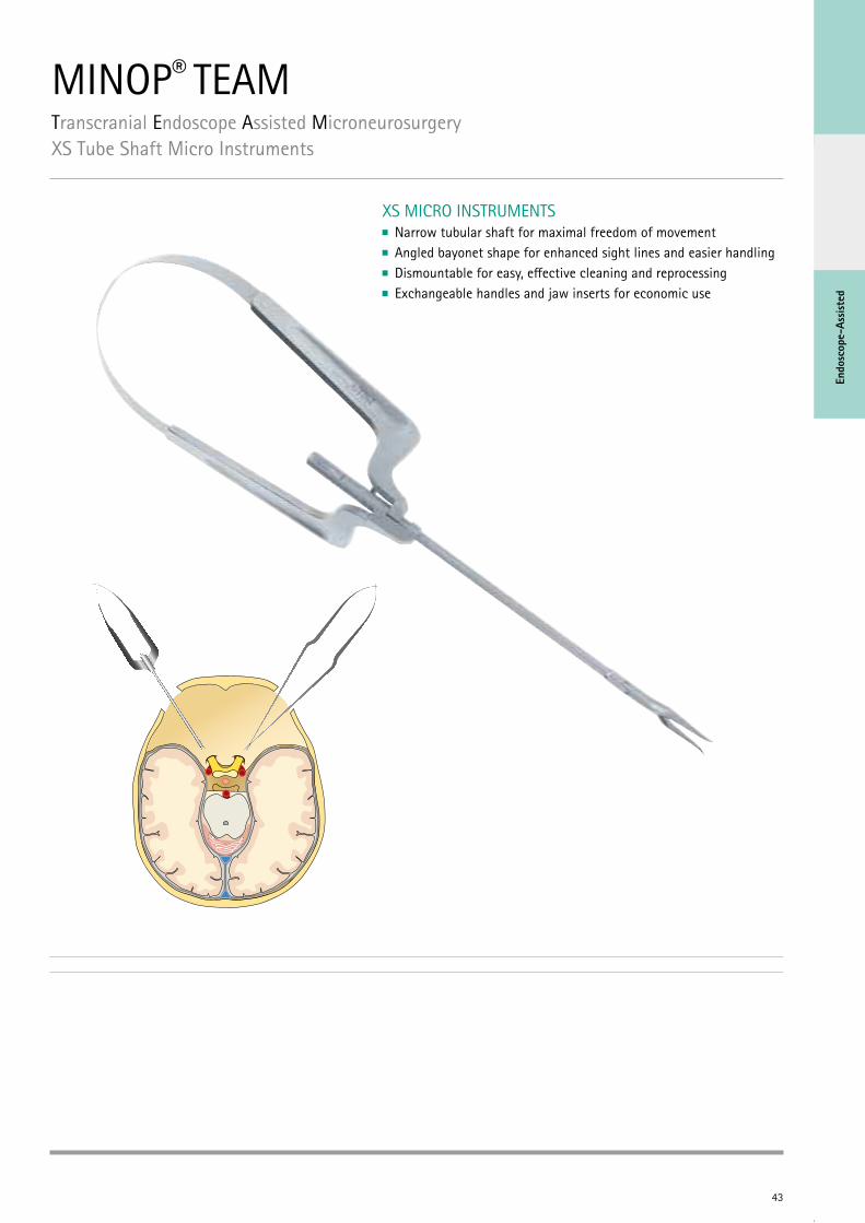
MINOP® TEAMTranscranial Endoscope Assisted MicroneurosurgeryXS Tube Shaft Micro Instruments
43
Endo
scop
e-As
siste
d
XS MICRO INSTRUMENTS Narrow tubular shaft for maximal freedom of movement Angled bayonet shape for enhanced sight lines and easier handling Dismountable for easy, effective cleaning and reprocessing Exchangeable handles and jaw inserts for economic use
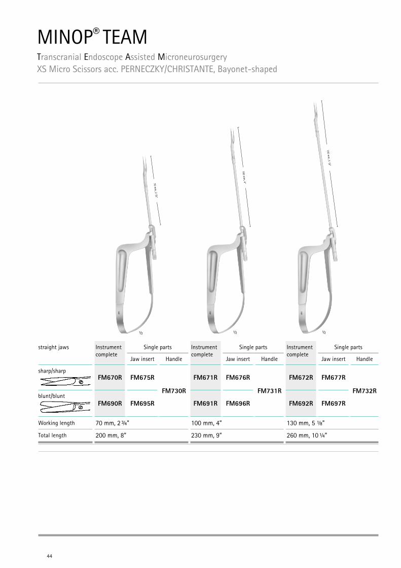
MINOP® TEAMTranscranial Endoscope Assisted MicroneurosurgeryXS Micro Scissors acc. PERNECZKY/CHRISTANTE, Bayonet-shaped
44
70 mm
, 2 3/4“
½
100 mm
, 4“
½
130 mm
, 5 1/8“
½
straight jaws Instrument complete
Single parts Instrument complete
Single parts Instrument complete
Single parts
Jaw insert Handle Jaw insert Handle Jaw insert Handle
sharp/sharpFM670R FM675R
FM730R
FM671R FM676R
FM731R
FM672R FM677R
FM732Rblunt/blunt
FM690R FM695R FM691R FM696R FM692R FM697R
Working length 70 mm, 2 ¾” 100 mm, 4” 130 mm, 5 ⅛”
Total length 200 mm, 8” 230 mm, 9” 260 mm, 10 ¼”
Endo
scop
e-As
siste
d
45
70 mm
, 2 3/4“
100 mm
, 4“
130 mm
, 5 1/8“
½ ½ ½
curved jaws Instrument complete
Single parts Instrument complete
Single parts Instrument complete
Single parts
Jaw insert Handle Jaw insert Handle Jaw insert Handle
sharp/sharpFM680R FM685R
FM730R
FM681R FM686R
FM731R
FM682R FM687R
FM732Rblunt/blunt
FM700R FM705R FM701R FM706R FM702R FM707R
Working length 70 mm, 2 ¾” 100 mm, 4” 130 mm, 5 ⅛”
Total length 200 mm, 8” 230 mm, 9” 260 mm, 10 ¼”
46
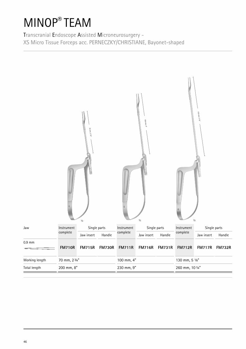
MINOP® TEAMTranscranial Endoscope Assisted Microneurosurgery - XS Micro Tissue Forceps acc. PERNECZKY/CHRISTIANE, Bayonet-shaped
70 mm
, 2 3/4“
½
100 mm
, 4“
½
130 mm
, 5 1/8“
½
Jaw Instrument complete
Single parts Instrument complete
Single parts Instrument complete
Single parts
Jaw insert Handle Jaw insert Handle Jaw insert Handle
0.9 mmFM710R FM715R FM730R FM711R FM716R FM731R FM712R FM717R FM732R
Working length 70 mm, 2 ¾” 100 mm, 4” 130 mm, 5 ⅛”
Total length 200 mm, 8” 230 mm, 9” 260 mm, 10 ¼”
47
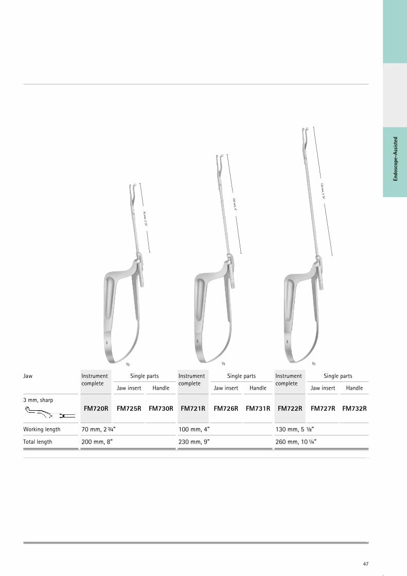
Endo
scop
e-As
siste
d
70 mm
, 2 3/4“
½
100 mm
, 4“
½
130 mm
, 5 1/8“
½
Jaw Instrument complete
Single parts Instrument complete
Single parts Instrument complete
Single parts
Jaw insert Handle Jaw insert Handle Jaw insert Handle
3 mm, sharpFM720R FM725R FM730R FM721R FM726R FM731R FM722R FM727R FM732R
Working length 70 mm, 2 ¾” 100 mm, 4” 130 mm, 5 ⅛”
Total length 200 mm, 8” 230 mm, 9” 260 mm, 10 ¼”
48
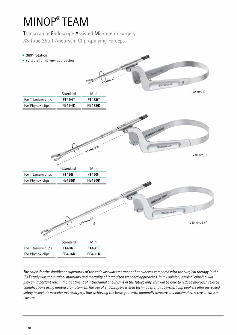
MINOP® TEAMTranscranial Endoscope Assisted Microneurosurgery XS Tube Shaft Aneurysm Clip Applying Forceps
360° rotation suitable for narrow approaches
90 mm, 3 ½“
230 mm, 9“
110 mm, 4 3⁄8“
250 mm, 9 ¾“
The cause for the significant superiority of the endovascular treatment of aneurysms compared with the surgical therapy in the ISAT study was the surgical morbidity and mortality of large sized standard approaches. In my opinion, surgical clipping will play an important role in the treatment of intracranial aneurysms in the future only, if it will be able to reduce approach related complications using limited craniotomies. The use of endoscope-assisted techniques and tube-shaft clip appliers offer increased safety in keyhole vascular neurosurgery, thus achieving the basic goal with minimally invasive and maximal effective aneurysm closure.
50 mm, 2”
190 mm, 7”Standard MiniFor Titanium clips FT494T FT489TFor Phynox clips FE494K FE489K
Standard MiniFor Titanium clips FT495T FT490TFor Phynox clips FE495K FE490K
Standard MiniFor Titanium clips FT496T FT491TFor Phynox clips FE496K FE491K
Endo
scop
e-As
siste
d
49
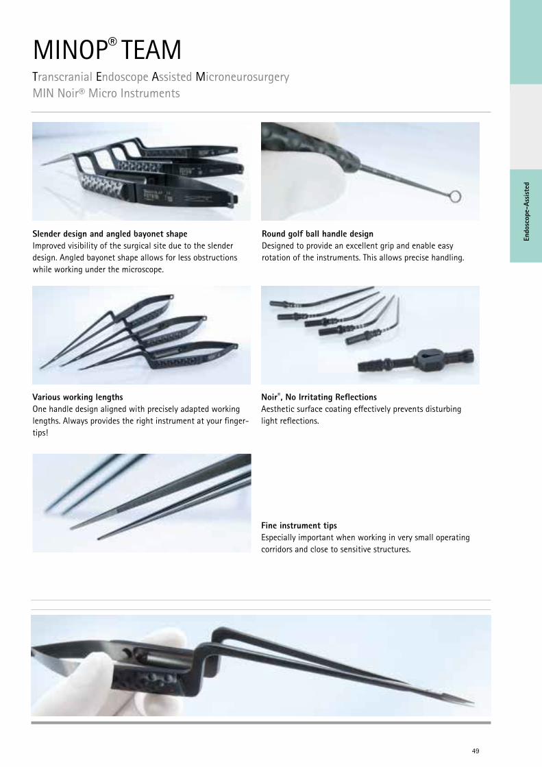
MINOP® TEAMTranscranial Endoscope Assisted MicroneurosurgeryMIN Noir® Micro Instruments
Fine instrument tips Especially important when working in very small operating corridors and close to sensitive structures.
Various working lengths One handle design aligned with precisely adapted working lengths. Always provides the right instrument at your finger-tips!
Slender design and angled bayonet shape Improved visibility of the surgical site due to the slender design. Angled bayonet shape allows for less obstructions while working under the microscope.
Noir®, No Irritating Reflections Aesthetic surface coating effectively prevents disturbing light reflections.
Round golf ball handle design Designed to provide an excellent grip and enable easy rotation of the instruments. This allows precise handling.
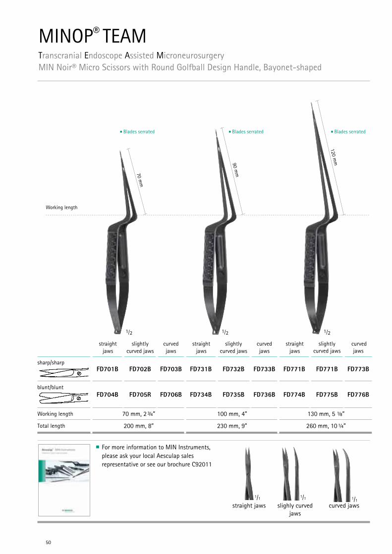
MINOP® TEAMTranscranial Endoscope Assisted MicroneurosurgeryMIN Noir® Micro Scissors with Round Golfball Design Handle, Bayonet-shaped
50
1/2 1/2 1/2
Working length
70 mm
90 mm
120 mm
For more information to MIN Instruments, please ask your local Aesculap sales representative or see our brochure C92011
1/1 1/1 1/1straight jaws slighly curved
jawscurved jaws
straight jaws
slightly curved jaws
curved jaws
straight jaws
slightly curved jaws
curved jaws
straight jaws
slightly curved jaws
curved jaws
sharp/sharpFD701B FD702B FD703B FD731B FD732B FD733B FD771B FD771B FD773B
blunt/bluntFD704B FD705R FD706B FD734B FD735B FD736B FD774B FD775B FD776B
Working length 70 mm, 2 ¾” 100 mm, 4” 130 mm, 5 ⅛”
Total length 200 mm, 8” 230 mm, 9” 260 mm, 10 ¼”
Blades serrated Blades serrated Blades serrated
Endo
scop
e-As
siste
d
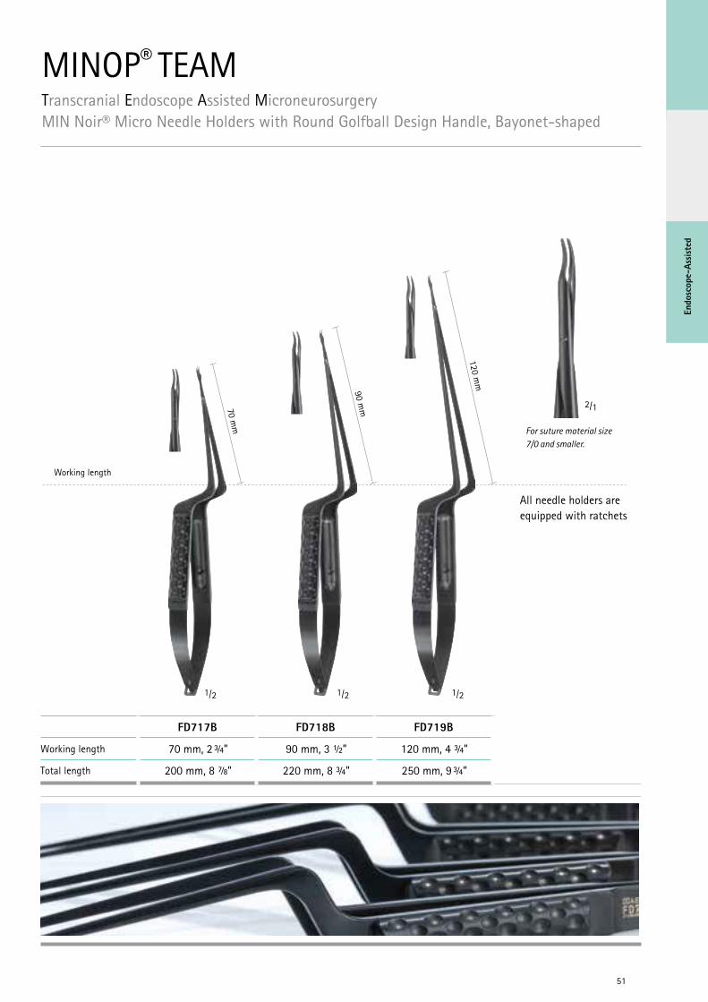
MINOP® TEAMTranscranial Endoscope Assisted MicroneurosurgeryMIN Noir® Micro Needle Holders with Round Golfball Design Handle, Bayonet-shaped
51
1/2 1/21/2
70 mm
90 mm
120 mm
Working length
For suture material size 7/0 and smaller.
2/1
All needle holders are equipped with ratchets
FD717B FD718B FD719B
Working length 70 mm, 2 ¾” 90 mm, 3 ½” 120 mm, 4 ¾”
Total length 200 mm, 8 ⅞” 220 mm, 8 ¾” 250 mm, 9 ¾”
52
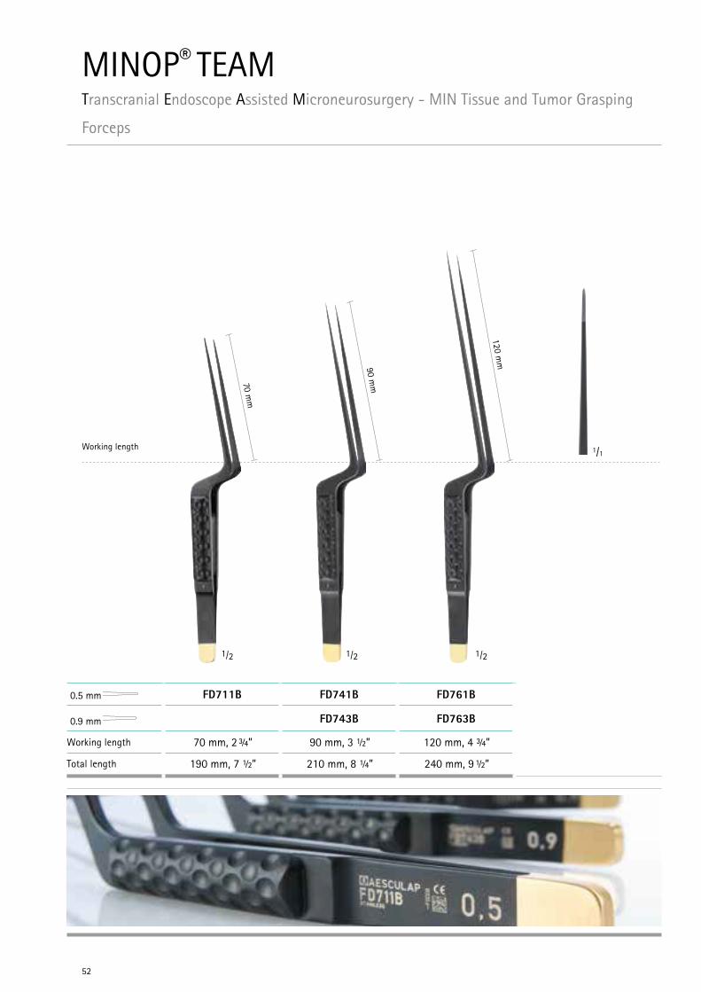
MINOP® TEAMTranscranial Endoscope Assisted Microneurosurgery - MIN Tissue and Tumor Grasping
Forceps
1/21/21/2
Working length70 m
m
90 mm
120 mm
0.5 mm FD711B FD741B FD761B
0.9 mm FD743B FD763B
Working length 70 mm, 2 ¾” 90 mm, 3 ½” 120 mm, 4 ¾”
Total length 190 mm, 7 ½” 210 mm, 8 ¼” 240 mm, 9 ½”
1/1
Endo
scop
e-As
siste
d
53
1/2
Working length
90 mm
120 mm
90 mm
120 mm
90 mm
120 mm
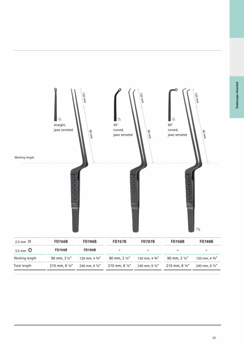
2.5 mm FD766B FD786B FD767B FD787B FD768B FD788B
3.5 mm FD769B FD789B - - - -
Working length 90 mm, 3 ½” 120 mm, 4 ¾" 90 mm, 3 ½” 120 mm, 4 ¾" 90 mm, 3 ½” 120 mm, 4 ¾"
Total length 210 mm, 8 ¼” 240 mm, 9 ½" 210 mm, 8 ¼” 240 mm, 9 ½" 210 mm, 8 ¼” 240 mm, 9 ½"
1/1
straight,jaws serrated
1/1
45° curved, jaws serrated
1/1
90° curved,jaws serrated
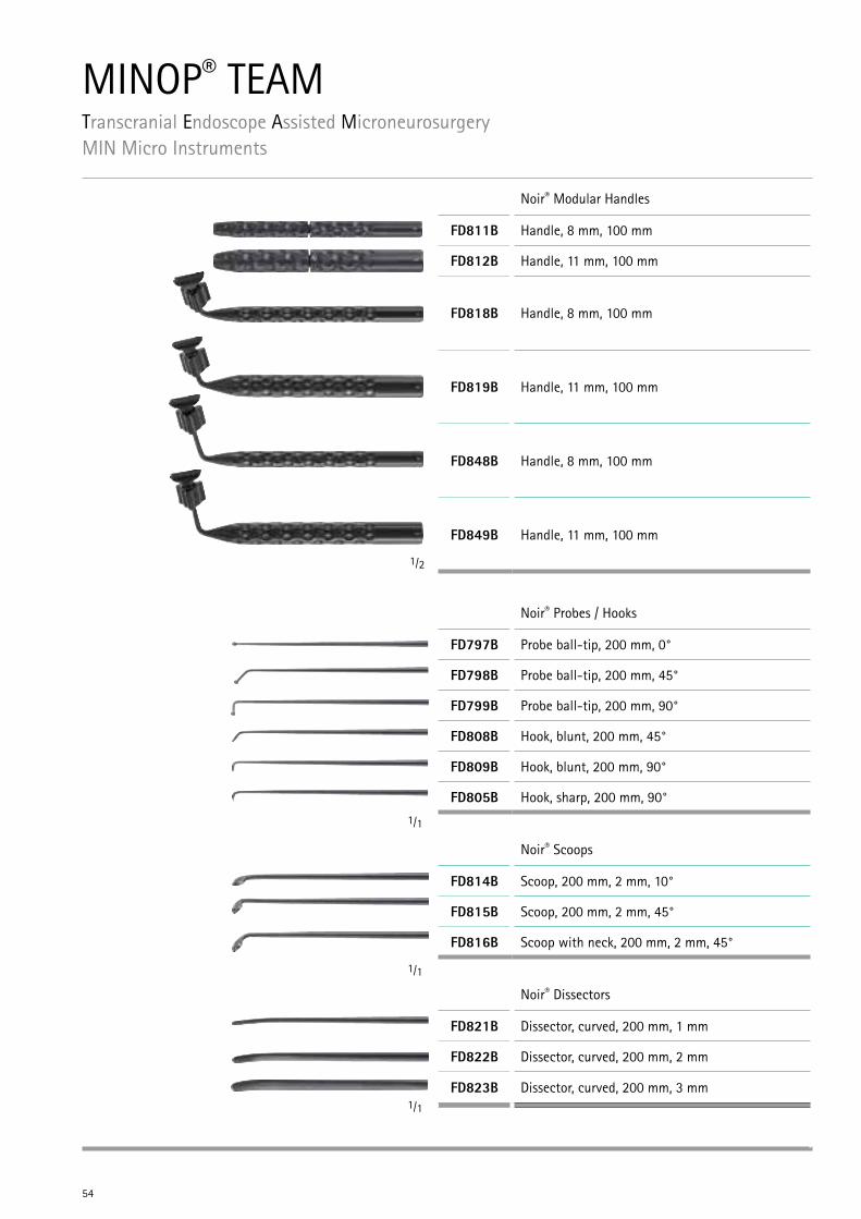
MINOP® TEAMTranscranial Endoscope Assisted Microneurosurgery MIN Micro Instruments
54
Noir® Modular Handles
FD811B Handle, 8 mm, 100 mm
FD812B Handle, 11 mm, 100 mm
FD818B Handle, 8 mm, 100 mm
FD819B Handle, 11 mm, 100 mm
FD848B Handle, 8 mm, 100 mm
FD849B Handle, 11 mm, 100 mm
Noir® Probes / Hooks
FD797B Probe ball-tip, 200 mm, 0°
FD798B Probe ball-tip, 200 mm, 45°
FD799B Probe ball-tip, 200 mm, 90°
FD808B Hook, blunt, 200 mm, 45°
FD809B Hook, blunt, 200 mm, 90°
FD805B Hook, sharp, 200 mm, 90°
Noir® Scoops
FD814B Scoop, 200 mm, 2 mm, 10°
FD815B Scoop, 200 mm, 2 mm, 45°
FD816B Scoop with neck, 200 mm, 2 mm, 45°
Noir® Dissectors
FD821B Dissector, curved, 200 mm, 1 mm
FD822B Dissector, curved, 200 mm, 2 mm
FD823B Dissector, curved, 200 mm, 3 mm
1/1
1/1
1/1
1/2
55
Endo
scop
e-As
siste
d
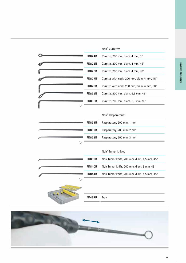
Noir® Currettes
FD824B Curette, 200 mm, diam. 4 mm, 0°
FD825B Curette, 200 mm, diam. 4 mm, 45°
FD826B Curette, 200 mm, diam. 4 mm, 90°
FD827B Curette with neck, 200 mm, diam. 4 mm, 45°
FD828B Curette with neck, 200 mm, diam. 4 mm, 90°
FD835B Curette, 200 mm, diam. 6,5 mm, 45°
FD836B Curette, 200 mm, diam. 6,5 mm, 90°
Noir® Rasparatories
FD831B Rasparatory, 200 mm, 1 mm
FD832B Rasparatory, 200 mm, 2 mm
FD833B Rasparatory, 200 mm, 3 mm
Noir® Tumor knives
FD839B Noir Tumor knife, 200 mm, diam. 1,5 mm, 45°
FD840B Noir Tumor knife, 200 mm, diam. 3 mm, 45°
FD841B Noir Tumor knife, 200 mm, diam. 4,5 mm, 45°
FD467R Tray
1/1
1/1
1/1
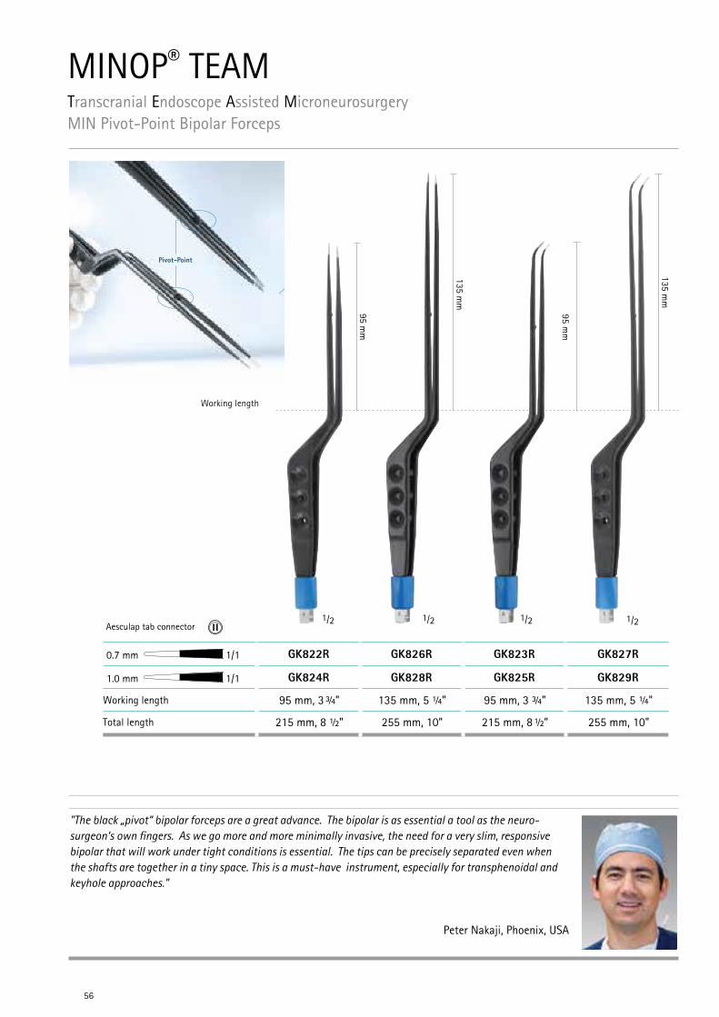
"The black „pivot“ bipolar forceps are a great advance. The bipolar is as essential a tool as the neuro-surgeon‘s own fingers. As we go more and more minimally invasive, the need for a very slim, responsive bipolar that will work under tight conditions is essential. The tips can be precisely separated even when the shafts are together in a tiny space. This is a must-have instrument, especially for transphenoidal and keyhole approaches."
Peter Nakaji, Phoenix, USA
MINOP® TEAMTranscranial Endoscope Assisted MicroneurosurgeryMIN Pivot-Point Bipolar Forceps
56
1/2
Working length
Aesculap tab connector1/21/2 1/2
95 mm
95 mm
135 mm
135 mm
Pivot-Point
0.7 mm 1/1 GK822R GK826R GK823R GK827R
1.0 mm 1/1 GK824R GK828R GK825R GK829R
Working length 95 mm, 3 ¾” 135 mm, 5 ¼” 95 mm, 3 ¾” 135 mm, 5 ¼"
Total length 215 mm, 8 ½” 255 mm, 10” 215 mm, 8 ½” 255 mm, 10"
Endo
scop
e-As
siste
d
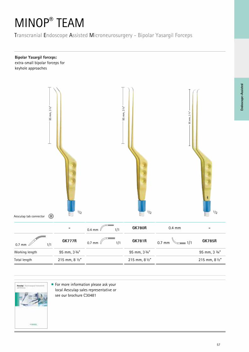
MINOP® TEAMTranscranial Endoscope Assisted Microneurosurgery - Bipolar Yasargil Forceps
57
Bipolar Yasargil forceps:extra-small bipolar forceps for keyhole approaches
95 m
m, 3
½“
95 m
m, 3
½“
1/2 1/2 1/2Aesculap tab connector
For more information please ask your local Aesculap sales representative or see our brochure C30481
- 0.4 mm 1/1 GK780R 0.4 mm -
0.7 mm 1/1GK777R 0.7 mm 1/1 GK781R 0.7 mm 1/1 GK785R
Working length 95 mm, 3 ¾” 95 mm, 3 ¾” 95 mm, 3 ¾”
Total length 215 mm, 8 ½” 215 mm, 8 ½” 215 mm, 8 ½”
95 m
m, 3
3 /4“
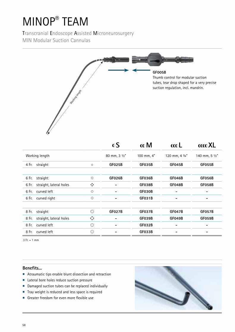
MINOP® TEAMTranscranial Endoscope Assisted MicroneurosurgeryMIN Modular Suction Cannulas
58
S M L XLWorking length 80 mm, 3 ½" 100 mm, 4" 120 mm, 4 ¾" 140 mm, 5 ½"
4 Fr. straight GF025B GF035B GF045B GF055B
6 Fr. straight GF026B GF036B GF046B GF056B
6 Fr. straight, lateral holes - GF038B GF048B GF058B
6 Fr. curved left - GF030B - -
6 Fr. curved right - GF031B - -
8 Fr. straight GF027B GF037B GF047B GF057B
8 Fr. straight, lateral holes - GF039B GF049B GF059B
8 Fr. curved left - GF032B - -
8 Fr. curved left - GF033B - -
3 Fr. = 1 mm
GF005BThumb control for modular suction tubes, tear drop shaped for a very precise suction regulation, incl. mandrin.
Benefits...n Atraumatic tips enable blunt dissection and retractionn Lateral bore holes reduce suction pressuren Damaged suction tubes can be replaced individuallyn Tray weight is reduced and less space is requiredn Greater freedom for even more flexible use
Wor
king l
engt
h
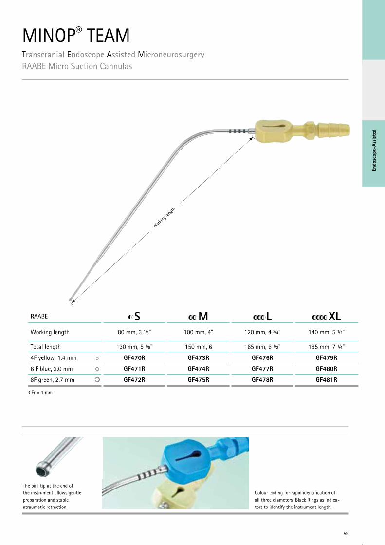
MINOP® TEAMTranscranial Endoscope Assisted MicroneurosurgeryRAABE Micro Suction Cannulas
59
Endo
scop
e-As
siste
d
The ball tip at the end of the instrument allows gentle preparation and stable atraumatic retraction.
Colour coding for rapid identification of all three diameters. Black Rings as indica-tors to identify the instrument length.
3 Fr = 1 mm
RAABE S M L XLWorking length 80 mm, 3 ⅛" 100 mm, 4" 120 mm, 4 ¾" 140 mm, 5 ½"
Total length 130 mm, 5 ⅛" 150 mm, 6 165 mm, 6 ½" 185 mm, 7 ¼"
4F yellow, 1.4 mm GF470R GF473R GF476R GF479R
6 F blue, 2.0 mm GF471R GF474R GF477R GF480R
8F green, 2.7 mm GF472R GF475R GF478R GF481R
Working
leng
th
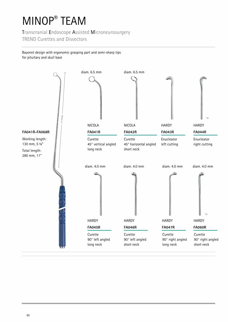
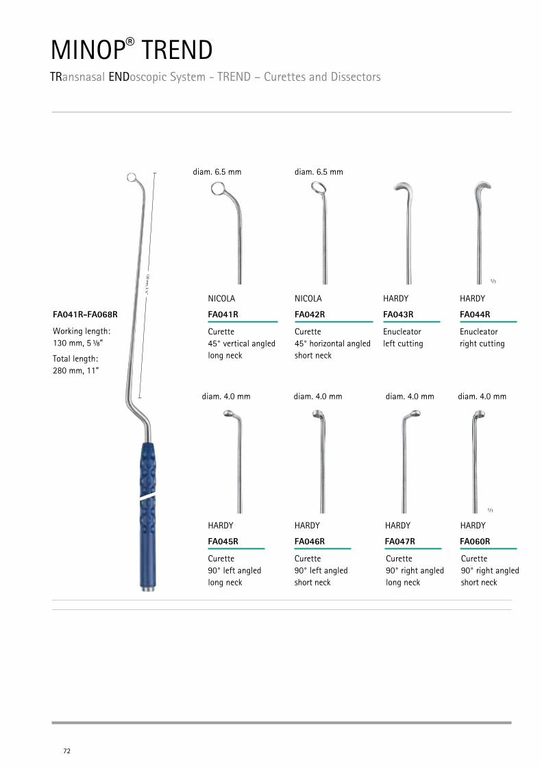
MINOP® TEAMTranscranial Endoscope Assisted MicroneurosurgeryTREND Curettes and Dissectors
60
FA041R-FA068R FA041R FA042R FA043R FA044R
FA045R FA046R FA047R
1⁄1
1⁄1
FA060R
Curette 45° vertical angled long neck
NICOLA
Working length: 130 mm, 5 ⅛”
Total length: 280 mm, 11”
Curette 45° horizontal angled short neck
NICOLA
Enucleator left cutting
HARDY
Enucleator right cutting
HARDY
Curette 90° left angled long neck
HARDY
Curette 90° left angled short neck
HARDY
Curette 90° right angled long neck
HARDY
Curette 90° right angled short neck
HARDY
1/8
diam. 6.5 mm
diam. 4.0 mm diam. 4.0 mm diam. 4.0 mm diam. 4.0 mm
diam. 6.5 mm
Bayonet design with ergonomic grasping part and semi-sharp tips for pituitary and skull base
Endo
scop
e-As
siste
d
61
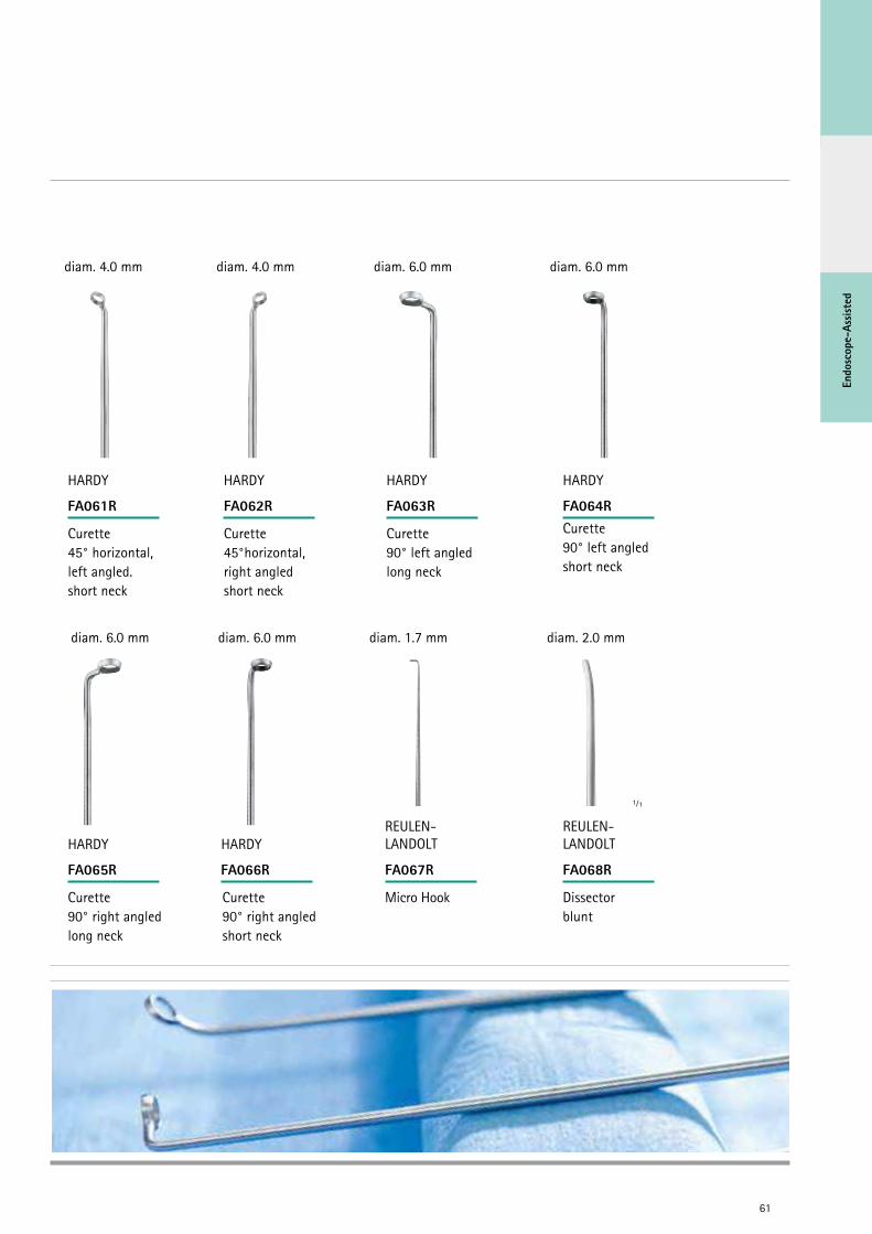
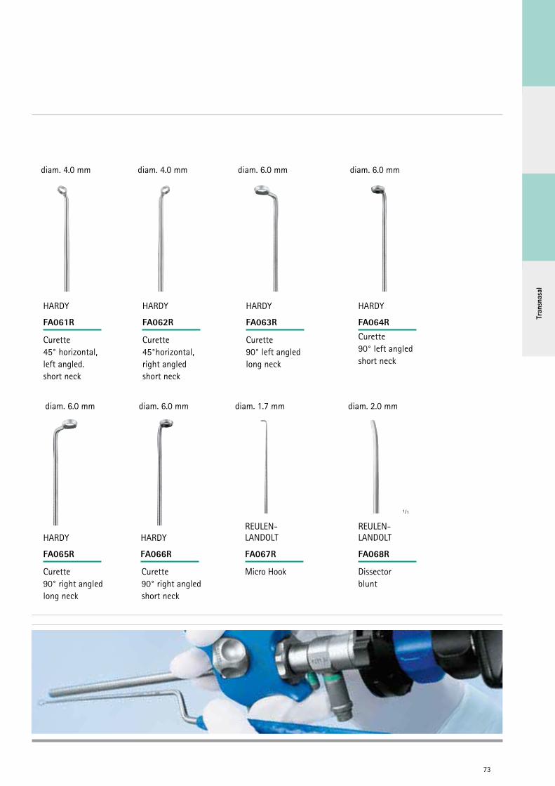
1⁄1
FA061R FA062R FA063R FA064R
FA065R FA066R FA067R FA068R
Curette 45° horizontal, left angled. short neck
HARDY
Curette 45°horizontal, right angled short neck
HARDY
Curette 90° left angled long neck
HARDY
Curette 90° left angled short neck
HARDY
Curette 90° right angled long neck
HARDY
Curette 90° right angled short neck
HARDY
Micro Hook
REULEN- LANDOLT
Dissector blunt
REULEN- LANDOLT
diam. 6.0 mm diam. 6.0 mm diam. 1.7 mm diam. 2.0 mm
diam. 4.0 mm diam. 4.0 mm diam. 6.0 mm diam. 6.0 mm
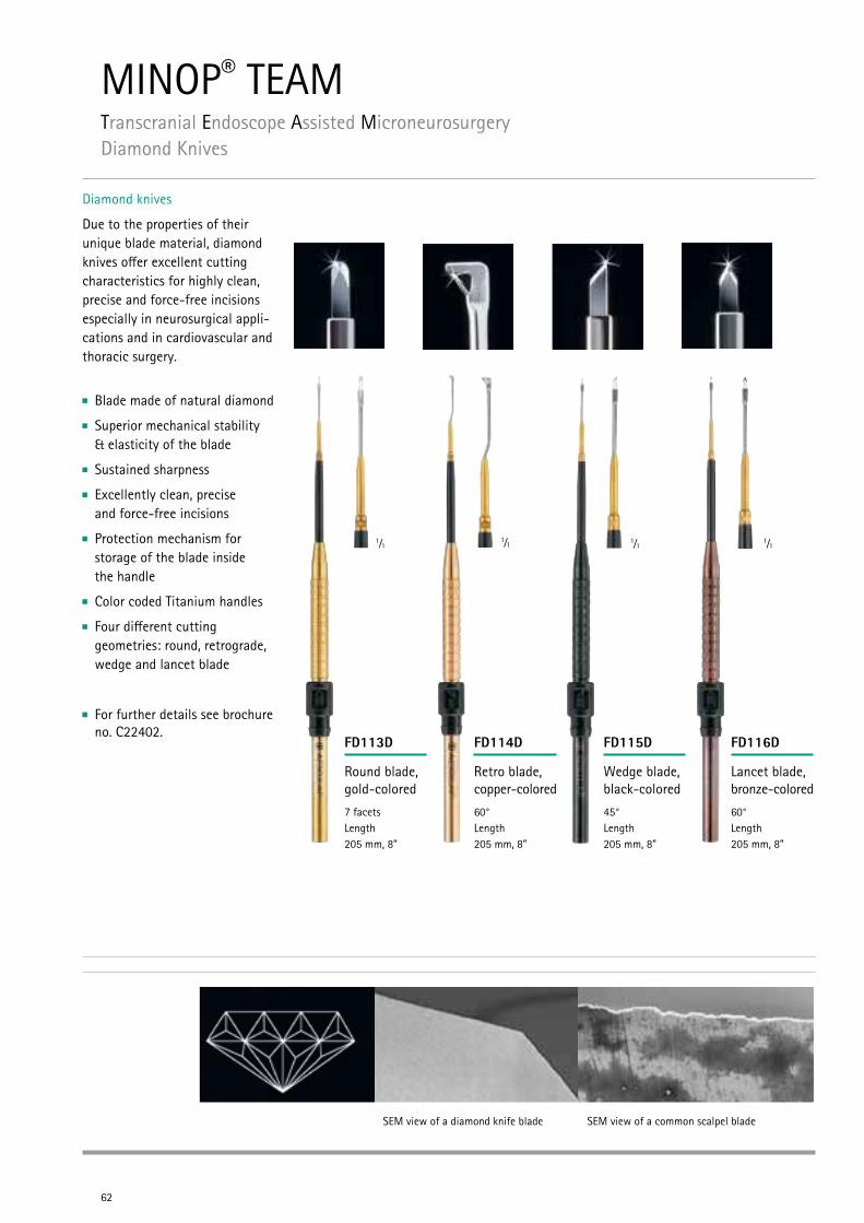
SEM view of a diamond knife blade SEM view of a common scalpel blade
MINOP® TEAMTranscranial Endoscope Assisted MicroneurosurgeryDiamond Knives
62
FD113D FD114D FD115D FD116D
Diamond knives
Due to the properties of their unique blade material, diamond knives offer excellent cutting characteristics for highly clean, precise and force-free incisions especially in neurosurgical appli-cations and in cardiovascular and thoracic surgery.
Blade made of natural diamond
Superior mechanical stability & elasticity of the blade
Sustained sharpness
Excellently clean, precise and force-free incisions
Protection mechanism for storage of the blade inside the handle
Color coded Titanium handles
Four different cutting geometries: round, retrograde, wedge and lancet blade
For further details see brochure no. C22402.
Round blade, gold-colored
7 facets Length 205 mm, 8”
1⁄1
Retro blade, copper-colored
60° Length 205 mm, 8”
1⁄1
Wedge blade, black-colored
45° Length 205 mm, 8”
1⁄1
Lancet blade, bronze-colored
60° Length 205 mm, 8”
1⁄1
Endo
scop
e-As
siste
d
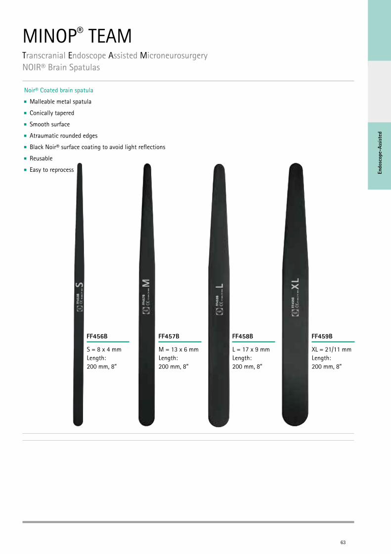
MINOP® TEAMTranscranial Endoscope Assisted MicroneurosurgeryNOIR® Brain Spatulas
63
Noir® Coated brain spatula
Malleable metal spatula
Conically tapered
Smooth surface
Atraumatic rounded edges
Black Noir® surface coating to avoid light reflections
Reusable
Easy to reprocess
FF456B
S = 8 x 4 mmLength: 200 mm, 8”
FF457B
M = 13 x 6 mmLength: 200 mm, 8”
FF458B
L = 17 x 9 mmLength: 200 mm, 8”
FF459B
XL = 21/11 mmLength: 200 mm, 8”
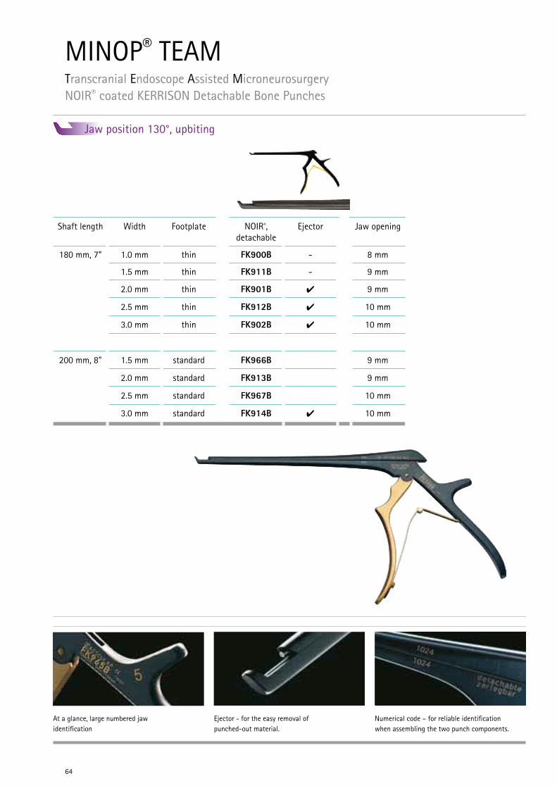
Ejector - for the easy removal of punched-out material.
At a glance, large numbered jaw identification
Numerical code – for reliable identification when assembling the two punch components.
64
MINOP® TEAMTranscranial Endoscope Assisted Microneurosurgery NOIR® coated KERRISON Detachable Bone Punches
Jaw position 130°, upbiting
Shaft length Width Footplate NOIR®, detachable
Ejector Jaw opening
180 mm, 7” 1.0 mm thin FK900B - 8 mm
1.5 mm thin FK911B - 9 mm
2.0 mm thin FK901B 4 9 mm
2.5 mm thin FK912B 4 10 mm
3.0 mm thin FK902B 4 10 mm
200 mm, 8" 1.5 mm standard FK966B 9 mm
2.0 mm standard FK913B 9 mm
2.5 mm standard FK967B 10 mm
3.0 mm standard FK914B 4 10 mm
Endo
scop
e-As
siste
d
65
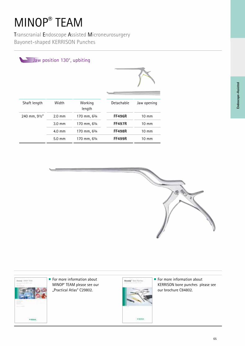
For more information about MINOP® TEAM please see our „Practical Atlas“ C29802.
For more information about KERRISON bone punches please see our brochure C84802.
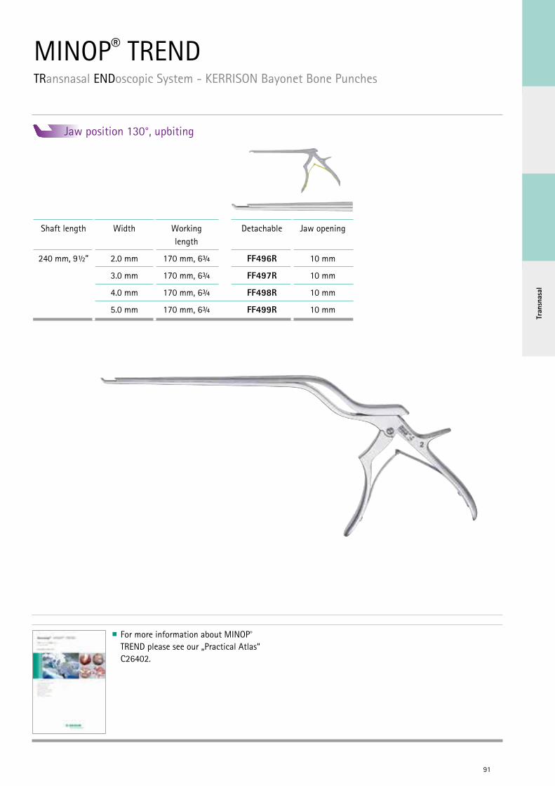
MINOP® TEAMTranscranial Endoscope Assisted MicroneurosurgeryBayonet-shaped KERRISON Punches
Jaw position 130°, upbiting
Shaft length Width Working length
Detachable Jaw opening
240 mm, 9½” 2.0 mm 170 mm, 6¾ FF496R 10 mm
3.0 mm 170 mm, 6¾ FF497R 10 mm
4.0 mm 170 mm, 6¾ FF498R 10 mm
5.0 mm 170 mm, 6¾ FF499R 10 mm

66

Tran
snas
al
67
TRANSNASAL NEUROENDOSCOPY
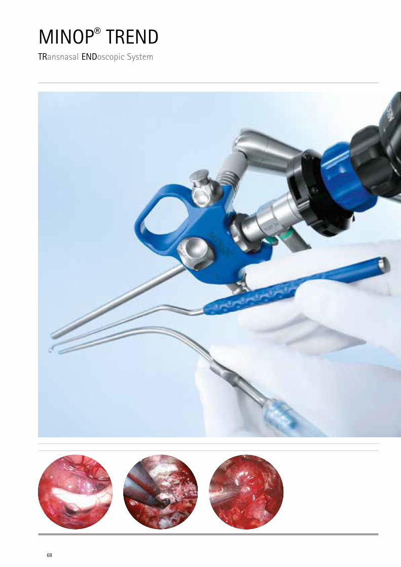
MINOP® TRENDTRansnasal ENDoscopic System
68
Tran
snas
al
André Grotenhuis Nijmegen, Netherlands
"When looking at recent publications on trans-sphenoidal surgery, it will be clear that TRans-sphenoidal ENDoscopy is TREND-setting! How-ever, this endoscopic technique is not in routine use everywhere and neurosurgeons are often reluctant to use it: One is often cautious about an endoscopic endonasal dissection because the permanent contamination of the endoscope with blood and nasal secretions hinders orientation. In addition, the para-endoscopic and biportal dissection is very unfamiliar requiring an un- acceptably steep learning curve.
Nevertheless, endoscopic visualization and para-endoscopic dissection without using the surgical microscope offers several undisputable advantages. Advantages in visualization increases light intensity in the deep-seated surgical field and clearly displays patho-anatomical details. In addition, the extended viewing angle of endo-scopes enables surgeons to observe hidden parts of the surgical field. The major benefit in surgical dissection is the unhindered approach to these clearly visible structures: Without using a nasal speculum, surgical manipulation is not impeded and the instruments are freely mobile. In addi-tion, a pure endoscopic technique avoids the need
for rhinoseptal submucosal dissection providing a direct and quicker approach to the sphenoid sinus. This method avoids the need for postop-erative nasal packing, thus causing less pain and discomfort after surgery, providing better nasal airflow and a shorter hospital stay.
Pre-conditions of transsphenoidal endoscopy are the basic endoscopic experience and ana-tomical studies in the laboratory; however, it is indispensable to use a dedicated endoscopic system to further shorten the learning phase. The endoscope for transsphenoidal skull base surgery must provide a brilliant image quality with true colors, high contrast and highly realistic im-ages. This simplifies the differentiation between healthy or pathological structures. It is essential to have an effective cleaning function in order to free the endoscope lens from fog, blood or mu-cosal secretions. The endoscope must offer a high-ly ergonomic design and sufficient working length for extended approaches. For selected cases, it is also necessary to connect the endoscope to a navigation system or a holding device."
André Grotenhuis
69
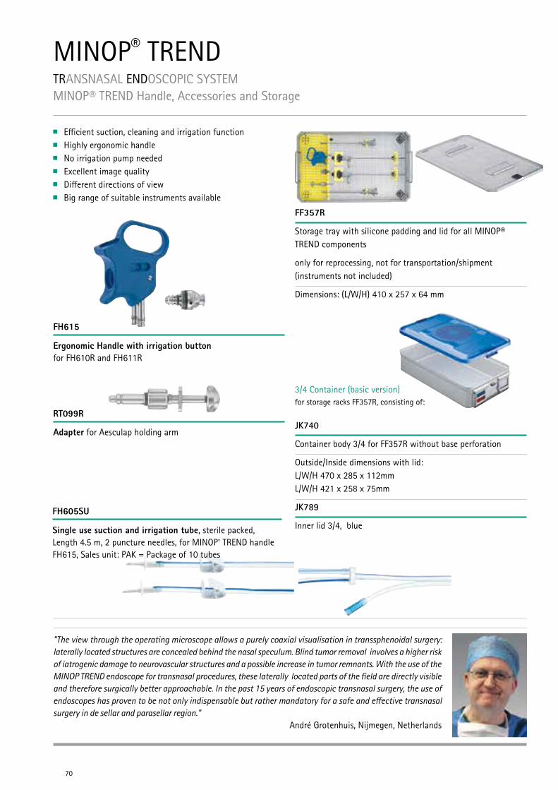
MINOP® TRENDTRANSNASAL ENDOSCOPIC SYSTEMMINOP® TREND Handle, Accessories and Storage
Ergonomic Handle with irrigation button for FH610R and FH611R
FH615
FF357R
Storage tray with silicone padding and lid for all MINOP® TREND components
only for reprocessing, not for transportation/shipment (instruments not included)
Dimensions: (L/W/H) 410 x 257 x 64 mm
Adapter for Aesculap holding arm
RT099R
Single use suction and irrigation tube, sterile packed, Length 4.5 m, 2 puncture needles, for MINOP® TREND handle FH615, Sales unit: PAK = Package of 10 tubes
FH605SU
70
"The view through the operating microscope allows a purely coaxial visualisation in transsphenoidal surgery: laterally located structures are concealed behind the nasal speculum. Blind tumor removal involves a higher risk of iatrogenic damage to neurovascular structures and a possible increase in tumor remnants. With the use of the MINOP TREND endoscope for transnasal procedures, these laterally located parts of the field are directly visible and therefore surgically better approachable. In the past 15 years of endoscopic transnasal surgery, the use of endoscopes has proven to be not only indispensable but rather mandatory for a safe and effective transnasal surgery in de sellar and parasellar region."
André Grotenhuis, Nijmegen, Netherlands
Efficient suction, cleaning and irrigation function Highly ergonomic handle No irrigation pump needed Excellent image quality Different directions of view Big range of suitable instruments available
3/4 Container (basic version) for storage racks FF357R, consisting of:
JK740
Container body 3/4 for FF357R without base perforation
Outside/Inside dimensions with lid: L/W/H 470 x 285 x 112mm L/W/H 421 x 258 x 75mm
JK789
Inner lid 3/4, blue
Tran
snas
al
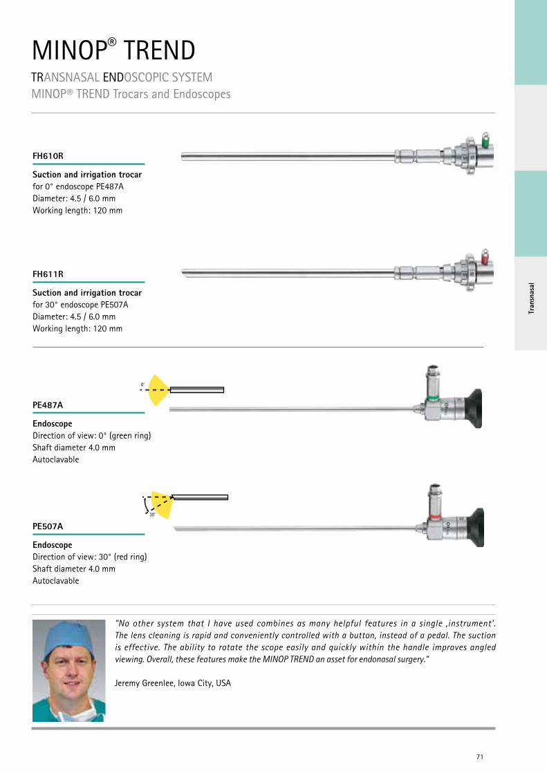
MINOP® TRENDTRANSNASAL ENDOSCOPIC SYSTEMMINOP® TREND Trocars and Endoscopes
Suction and irrigation trocar for 0° endoscope PE487ADiameter: 4.5 / 6.0 mmWorking length: 120 mm
FH610R
Suction and irrigation trocar for 30° endoscope PE507ADiameter: 4.5 / 6.0 mmWorking length: 120 mm
FH611R
EndoscopeDirection of view: 0° (green ring)Shaft diameter 4.0 mmAutoclavable
PE487A
EndoscopeDirection of view: 30° (red ring)Shaft diameter 4.0 mmAutoclavable
PE507A
71
"No other system that I have used combines as many helpful features in a single ‚instrument‘. The lens cleaning is rapid and conveniently controlled with a button, instead of a pedal. The suction is effective. The ability to rotate the scope easily and quickly within the handle improves angled viewing. Overall, these features make the MINOP TREND an asset for endonasal surgery."
Jeremy Greenlee, Iowa City, USA
0o
30o
MINOP® TRENDTRansnasal ENDoscopic System - TREND – Curettes and Dissectors
72
FA041R-FA068R FA041R FA042R FA043R FA044R
FA045R FA046R FA047R
1⁄1
1⁄1
FA060R
Curette 45° vertical angled long neck
NICOLA
Working length: 130 mm, 5 ⅛”
Total length: 280 mm, 11”
Curette 45° horizontal angled short neck
NICOLA
Enucleator left cutting
HARDY
Enucleator right cutting
HARDY
Curette 90° left angled long neck
HARDY
Curette 90° left angled short neck
HARDY
Curette 90° right angled long neck
HARDY
Curette 90° right angled short neck
HARDY
1/8
diam. 6.5 mm
diam. 4.0 mm diam. 4.0 mm diam. 4.0 mm diam. 4.0 mm
diam. 6.5 mm
Tran
snas
al
73
1⁄1
FA061R FA062R FA063R FA064R
FA065R FA066R FA067R FA068R
Curette 45° horizontal, left angled. short neck
HARDY
Curette 45°horizontal, right angled short neck
HARDY
Curette 90° left angled long neck
HARDY
Curette 90° left angled short neck
HARDY
Curette 90° right angled long neck
HARDY
Curette 90° right angled short neck
HARDY
Micro Hook
REULEN- LANDOLT
Dissector blunt
REULEN- LANDOLT
diam. 6.0 mm diam. 6.0 mm diam. 1.7 mm diam. 2.0 mm
diam. 4.0 mm diam. 4.0 mm diam. 6.0 mm diam. 6.0 mm
FA032R
MINOP® TRENDTRansnasal ENDoscopic System - TREND – Curettes and Dissectors
74
FA030R-FA040R
Working length: 140 mm, 5 ½”
Total length: 265 mm, 10 ½”
Straight design with ergonomic grasping part and semi-sharp tips
1⁄1
FA030R FA031R FA033R FA034R FA035R
1⁄1
FA036R FA037R FA038R FA039R FA040R
Curette 45° vertical angled, long neck
diam. 6.5 mm diam. 6.5 mm diam. 4.0 mm
diam. 4.0 mm diam. 6.0 mm diam. 6.0 mm diam. 1.7 mm diam. 2.0 mm
diam. 4.0 mm
NICOLA
Curette 45° horizontal angled, short neck
NICOLA
Enucleator left cutting
HARDY
Enucleatorright cutting
HARDY
Curette 90° angled long neck
HARDY
Curette 90° angled short neck
HARDY
Curette 45° angled short neck
HARDY
Curette 90° angled long neck
HARDY
Curette 90° angled short neck
HARDY
Micro Hook
LANDOLT-REULEN
Dissector blunt
LANDOLT-REULEN

Tran
snas
al
75
MINOP® TRENDTRansnasal ENDoscopic System - Nasal Specula
76
OK105R
1/2
1/1
COTTLE
OK105R-OK108R OK090R
with aseptic joint, set-screw, with extra thin blades 140 mm, 5 ½“
OK107R
56 mm
OK108R
OK090R
1/1
1/1
1/1
1/1
52 mm
75 mm
33 mm
90 mm
OK106R
Tran
snas
al
77
1/2 1/2
KILLIAN
OK081R Fig. 1
OK082R Fig. 2
OK083R Fig. 3
OK084R Fig. 4
with aseptic joint145 mm, 5 ¾“
OK091R Fig. 1
OK092R Fig. 2
OK093R Fig. 3
OK094R Fig. 4
with screw joint140 mm, 5 ½“
Fig.1
1/1
36 mm
Fig.2
56 mm
75 mm
Fig.3
Fig.4
90 mm
1/1
1/1
1/1
MINOP® TRENDTRansnasal ENDoscopic System - Pituitary Instruments / Sinus Punches
78
FA076R
Antrum punch for removal ofposterior nasal septum, Rotating sheath 360°, Working length: 120 mm, 4 ¾“ Backwards
cutting½
6 x 1.5 mm 8 x 3 mm 11.5 x 3.5 mm
stra
ight
OK608Rforward through cutting
MACKAY-GRUNEWALD
OK602Rforward through cutting
MACKAY-GRUNEWALD
OK603Rforward through cutting
45°
upw
ards
an
gled
OK609Rforward through cutting
MACKAY-GRUNEWALD
OK606Rforward through cutting
MACKAY-GRUNEWALD
OK607Rforward through cutting
130 mm,5 ⅛"OK602R-OK609R
2/1 2/1 2/1
1⁄1
Sinus Punches
½
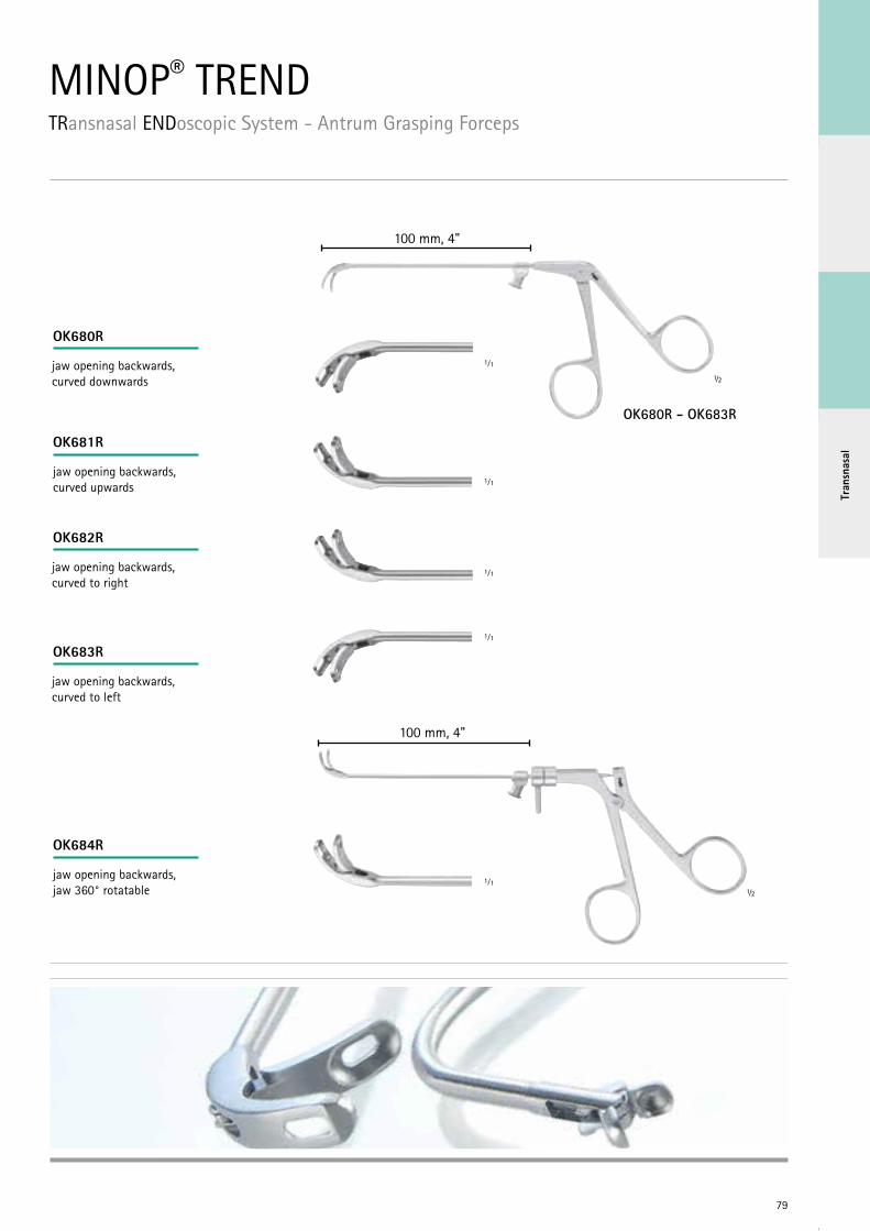
MINOP® TRENDTRansnasal ENDoscopic System - Antrum Grasping Forceps
79
Tran
snas
al
OK680R - OK683R
100 mm, 4"
jaw opening backwards, curved downwards
jaw opening backwards, curved to left
jaw opening backwards, jaw 360° rotatable
100 mm, 4"
jaw opening backwards, curved to right
jaw opening backwards, curved upwards
½
½
1⁄1
1⁄1
1⁄1
1⁄1
1⁄1
OK680R
OK681R
OK682R
OK683R
OK684R
80
FF345R
½
1⁄1
1⁄1
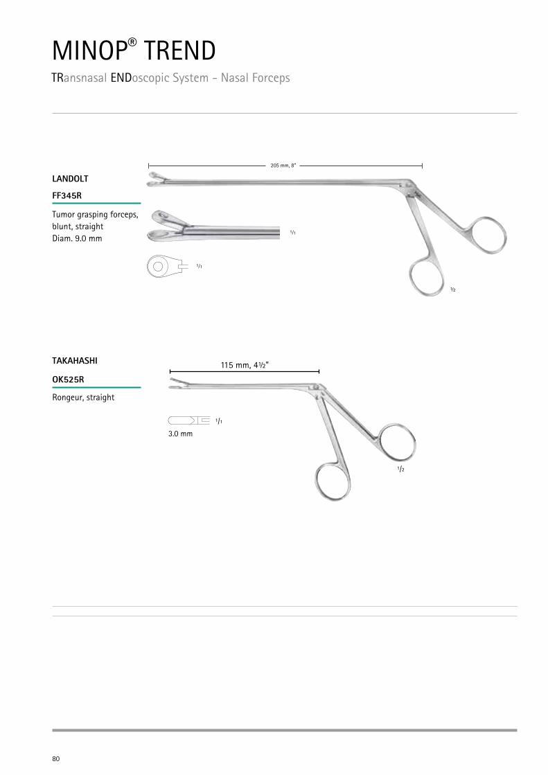
MINOP® TRENDTRansnasal ENDoscopic System - Nasal Forceps
205 mm, 8”
LANDOLT
Tumor grasping forceps, blunt, straightDiam. 9.0 mm
TAKAHASHI
Rongeur, straight
115 mm, 4½"
1/1
1/2
3.0 mm
OK525R
81
Tran
snas
al
MINOP® TRENDTRansnasal ENDoscopic System - Nasal Forceps
Angled positions for rongeurs
BLAKESLEY-WILDE
OK505R-OK509R
Ethmoidal forceps, straight
120 mm, 4¾"
½
½
3.0 mm
3.6 mm
4.2 mm
4.8 mm
5.6 mm
BLAKESLEY-WILDE
OK520R-OK522R
Ethmoidal forceps, upwards curved, 140°
110 mm, 4½"
1/1
3.6 mm
4.2 mm
4.8 mm
1/1
1/1
1⁄1
1⁄1
1⁄1
1⁄1
1⁄1
OK505R
OK506R
OK507R
OK508R
OK509R
OK520R
OK521R
OK522R
For more information about instruments for Functional Endoscopic Sinus Surgery (FESS), please ask your local Aesculap sales representative or see brochure no. C87511.
82
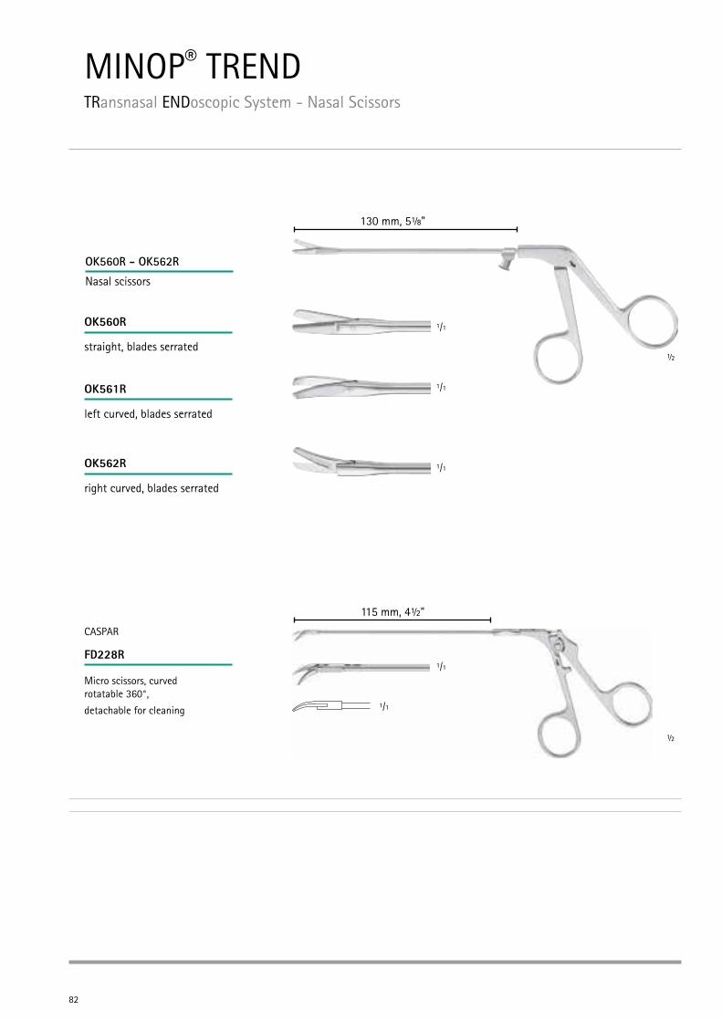
MINOP® TRENDTRansnasal ENDoscopic System - Nasal Scissors
OK560R
OK561R
OK562R
OK560R - OK562R
Nasal scissors
130 mm, 5⅛"
1/1
1/1
1/1
straight, blades serrated
left curved, blades serrated
right curved, blades serrated
115 mm, 4½"
1/1
1/1
CASPAR
Micro scissors, curved rotatable 360°,
detachable for cleaning
½
½
FD228R
83
Tran
snas
al
MINOP® TRENDTRansnasal ENDoscopic System - Pituitary Scissors
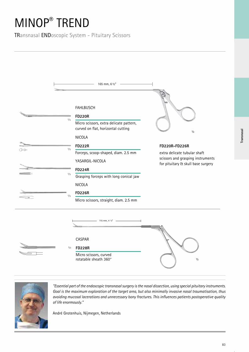
115 mm, 4 ” 1/2
CASPAR
FD228R
Micro scissors, curvedrotatable sheath 360°
1⁄1
FD220R-FD226R
extra delicate tubular shaft scissors and grasping instruments for pituitary & skull base surgery
Micro scissors, extra delicate pattern, curved on flat, horizontal cutting
Grasping forceps with long conical jaw
Micro scissors, straight, diam. 2.5 mm
FAHLBUSCH
1⁄1
1⁄1
NICOLA
Forceps, scoop-shaped, diam. 2.5 mm
1⁄1
1⁄1
YASARGIL-NICOLA
NICOLA
FD220R
FD222R
FD224R
FD226R
165 mm, 6 ½”
½
½
"Essential part of the endoscopic transnasal surgery is the nasal dissection, using special pituitary instruments. Goal is the maximum exploration of the target area, but also minimally invasive nasal traumatisation, thus avoiding mucosal lacerations and unnecessary bony fractures. This influences patients postoperative quality of life enormously."
André Grotenhuis, Nijmegen, Netherlands
180 mm, 7”
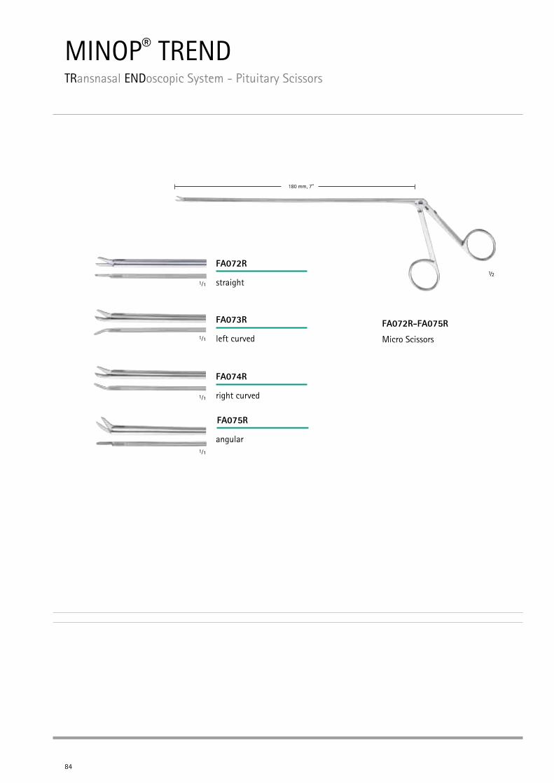
MINOP® TRENDTRansnasal ENDoscopic System - Pituitary Scissors
84
FA072R-FA075R
Micro Scissors
FA074R
right curved
FA075R
angular
FA072R
straight
FA073R
left curved
1⁄1
1⁄1
1⁄1
1⁄1
½
Tran
snas
al
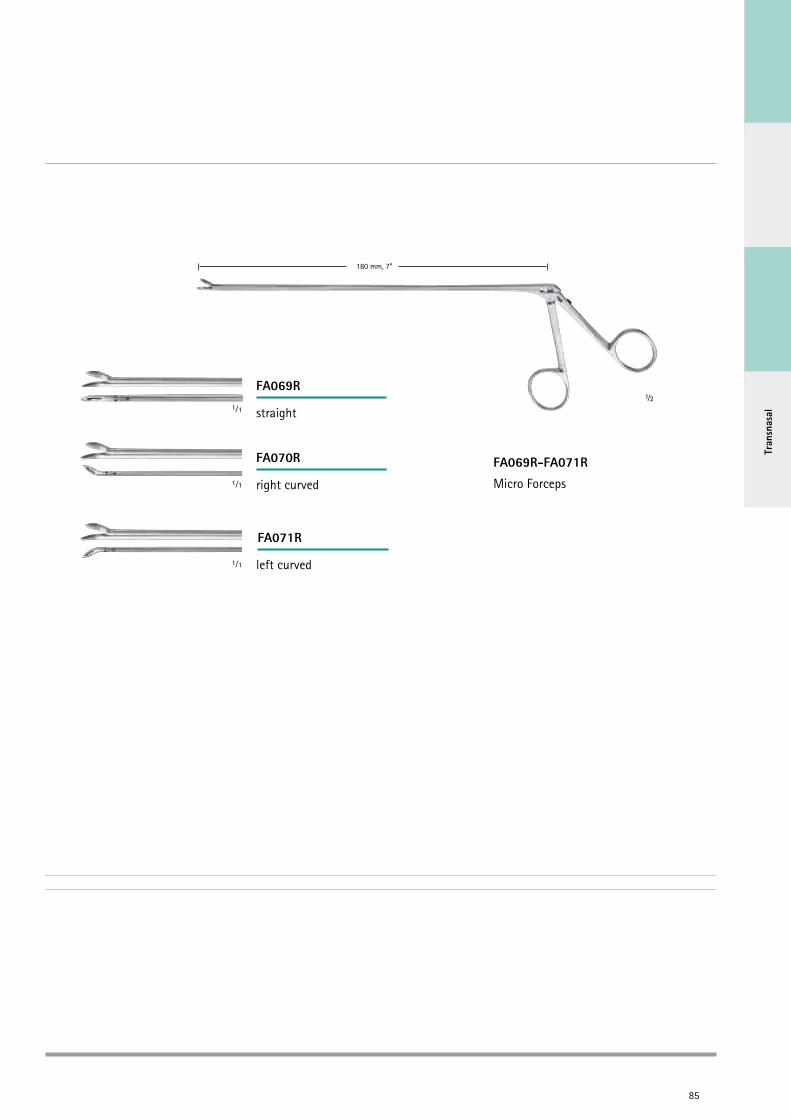
180 mm, 7”
85
FA069R-FA071R
Micro Forceps
FA070R
right curved
FA071R
left curved
FA069R
straight1⁄1
1⁄1
1⁄1
½
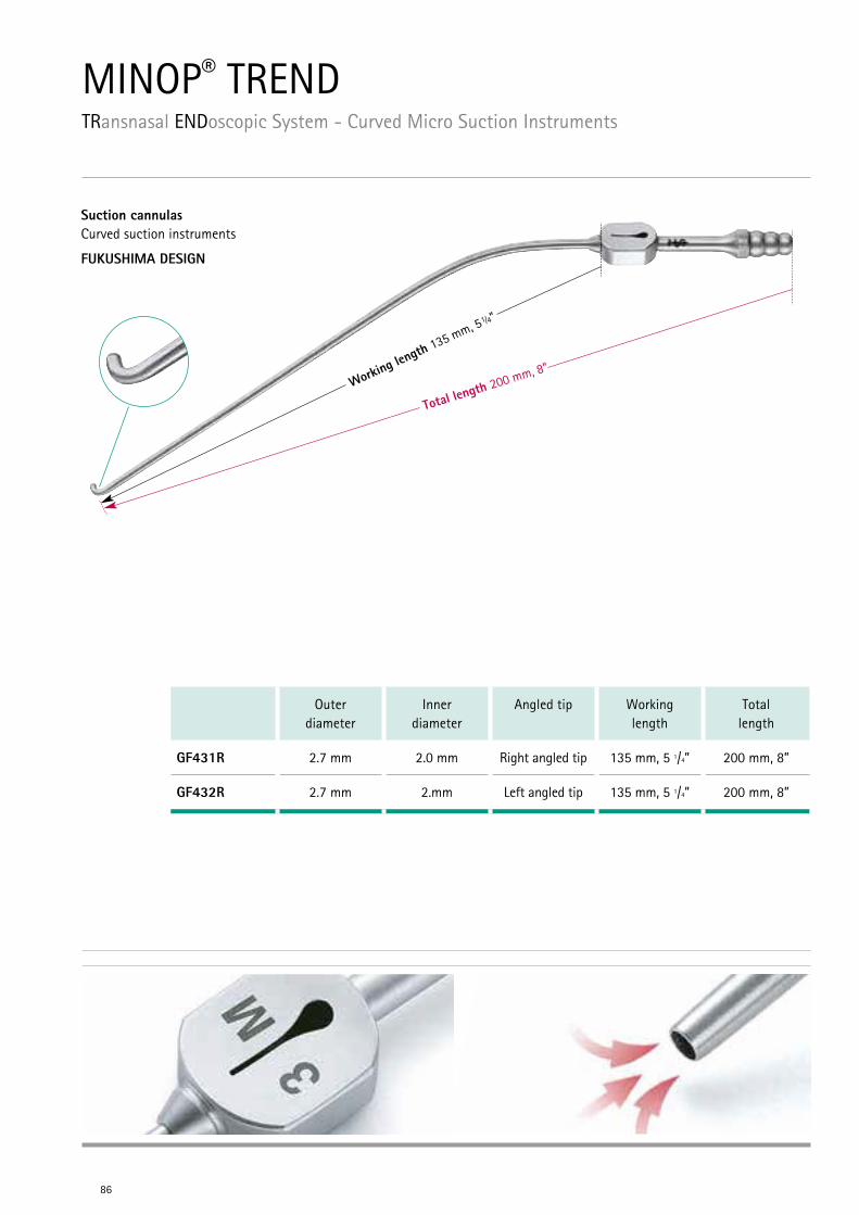
MINOP® TRENDTRansnasal ENDoscopic System - Curved Micro Suction Instruments
86
Total length 200 mm, 8”Workin
g length 135 mm, 5 ”
Suction cannulas Curved suction instruments
FUKUSHIMA DESIGN
Outer diameter
Inner diameter
Angled tip Working length
Total length
GF431R 2.7 mm 2.0 mm Right angled tip 135 mm, 5 1/4” 200 mm, 8”
GF432R 2.7 mm 2.mm Left angled tip 135 mm, 5 1/4” 200 mm, 8”
Tran
snas
al
MINOP® TRENDTRansnasal ENDoscopic System - Micro Suction Instruments
87
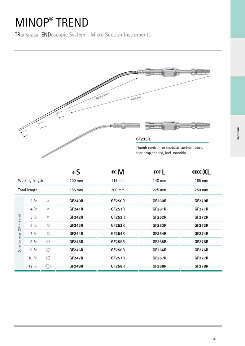
S M L XLWorking length 100 mm 115 mm 140 mm 165 mm
Total length 185 mm 200 mm 225 mm 250 mm
3 Fr. GF240R GF250R GF260R GF270R
4 Fr. GF241R GF251R GF261R GF271R
5 Fr. GF242R GF252R GF262R GF272R
6 Fr. GF243R GF253R GF263R GF273R
7 Fr. GF244R GF254R GF264R GF274R
8 Fr. GF245R GF255R GF265R GF275R
9 Fr. GF246R GF256R GF266R GF276R
10 Fr. GF247R GF257R GF267R GF277R
12 Fr. GF248R GF258R GF268R GF278R
GF235R
Working length
Total length
Oute
r dia
met
er (
3Fr =
1 m
m)
Thumb control for modular suction tubes, tear drop shaped, incl. mandrin
88
T-coagulation forceps with blunt, t-shaped tips
Total length 255 mm, 10” Working length 135 mm, 5 ¼“
Aesculap tab connector
GK800R
1/1
GK801R
Bipolar coagulation forceps with slender jaws and higher spring tension
Total length 255 mm, 10” Working length 135 mm, 5 ¼“
Aesculap tab connector
Special pin between the branches opens the tip of the forceps by additional compression of the handle – allowing coagulation in narrow and deep seated surgical field.
135 mm, 5 ” 1/4
135 mm, 5 ¼“
135 mm, 5 ¼“
MINOP® TRENDTRansnasal ENDoscopic System - Nasal Forceps
GK560R
GK580R
½
1⁄1
1⁄1
150 mm, 6“
Coagulation forceps for hypophysectomy, 90°
Coagulation forceps for hypophysectomy, 120°
LANDOLT
LANDOLT
89
Tran
snas
al
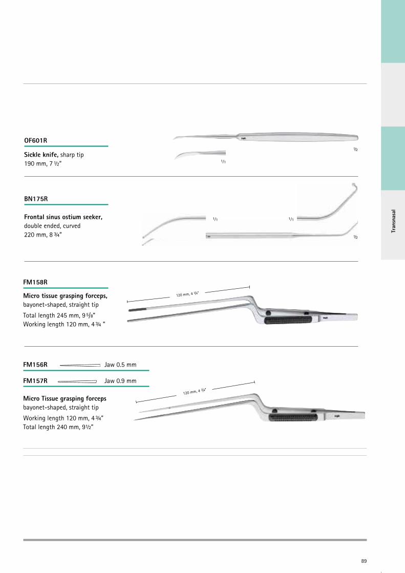
OF601R
BN175R
Micro tissue grasping forceps, bayonet-shaped, straight tip
Total length 245 mm, 9 5/8” Working length 120 mm, 4 ¾ ”
120 mm, 4 3/4“
FM158R
120 mm, 4 3/4“
FM156R Jaw 0.5 mm
FM157R Jaw 0.9 mm
Micro Tissue grasping forceps bayonet-shaped, straight tip
Working length 120 mm, 4 ¾” Total length 240 mm, 9½”
Frontal sinus ostium seeker, double ended, curved220 mm, 8 ¾“
Sickle knife, sharp tip190 mm, 7 ½“ 1⁄1
1⁄1 1⁄1
½
½
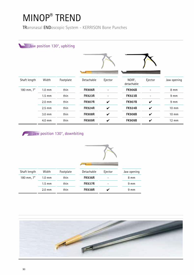
MINOP® TRENDTRansnasal ENDoscopic System - KERRISON Bone Punches
90
Jaw position 130°, downbiting
Jaw position 130°, upbiting
Shaft length Width Footplate Detachable Ejector NOIR®, detachable
Ejector Jaw opening
180 mm, 7” 1.0 mm thin FK906R - FK906B - 8 mm
1.5 mm thin FK923R - FK923B - 9 mm
2.0 mm thin FK907R 4 FK907B 4 9 mm
2.5 mm thin FK924R 4 FK924B 4 10 mm
3.0 mm thin FK908R 4 FK908B 4 10 mm
4.0 mm thin FK909R 4 FK909B 4 12 mm
Shaft length Width Footplate Detachable Ejector Jaw opening
180 mm, 7” 1.0 mm thin FK936R - 8 mm
1.5 mm thin FK937R - 9 mm
2.0 mm thin FK938R 4 9 mm
Tran
snas
al
MINOP® TRENDTRansnasal ENDoscopic System - KERRISON Bayonet Bone Punches
91
Jaw position 130°, upbiting
For more information about MINOP® TREND please see our „Practical Atlas“ C26402.
Shaft length Width Working length
Detachable Jaw opening
240 mm, 9½” 2.0 mm 170 mm, 6¾ FF496R 10 mm
3.0 mm 170 mm, 6¾ FF497R 10 mm
4.0 mm 170 mm, 6¾ FF498R 10 mm
5.0 mm 170 mm, 6¾ FF499R 10 mm
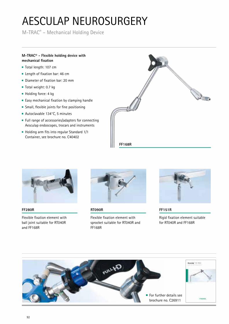
M-TRAC® - Flexible holding device with mechanical fixation
Total length: 107 cm
Length of fixation bar: 46 cm
Diameter of fixation bar: 20 mm
Total weight: 0.7 kg
Holding force: 4 kg
Easy mechanical fixation by clamping handle
Small, flexible joints for fine positioning
Autoclavable 134°C, 5 minutes
Full range of accessories/adapters for connecting Aesculap endoscopes, trocars and instruments
Holding arm fits into regular Standard 1/1 Container, see brochure no. C40402
AESCULAP NEUROSURGERYM-TRAC® – Mechanical Holding Device
Flexible fixation element with sprocket suitable for RT040R and FF168R
Flexible fixation element with ball joint suitable for RT040R and FF168R
Rigid fixation element suitable for RT040R and FF168R
FF280R RT090R FF151R
FF168R
92
For further details see brochure no. C26911
Hold
ing
Devi
ces
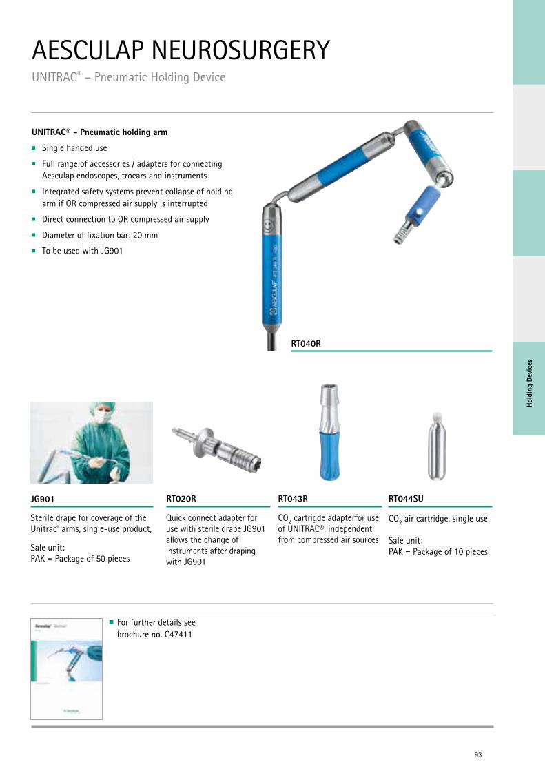
AESCULAP NEUROSURGERYUNITRAC® – Pneumatic Holding Device
UNITRAC® - Pneumatic holding arm
Single handed use
Full range of accessories / adapters for connecting Aesculap endoscopes, trocars and instruments
Integrated safety systems prevent collapse of holding arm if OR compressed air supply is interrupted
Direct connection to OR compressed air supply
Diameter of fixation bar: 20 mm
To be used with JG901
RT040R
RT020R RT043R RT044SUJG901
93
Quick connect adapter for use with sterile drape JG901 allows the change of instruments after draping with JG901
Sterile drape for coverage of the Unitrac® arms, single-use product,
Sale unit: PAK = Package of 50 pieces
CO2 cartrigde adapterfor use of UNITRAC®, independent from compressed air sources
CO2 air cartridge, single use Sale unit: PAK = Package of 10 pieces
For further details see brochure no. C47411
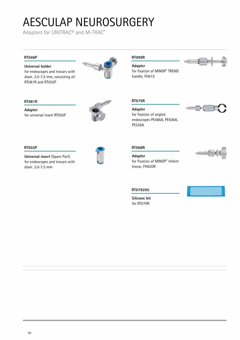
AESCULAP NEUROSURGERY Adapters for UNITRAC® and M-TRAC®
94
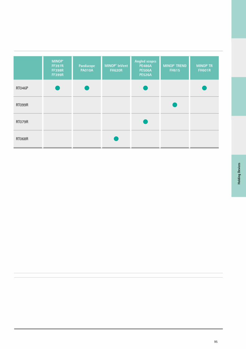
Adapter for universal insert RT055P
Universal holder for endoscopes and trocars with diam. 3.0-7.5 mm, consisting of: RT081R and RT055P
Adapter for fixation of angled endoscopes PE486A, PE506A, PE526A
RT079R
Adapter for fixation of MINOP® InVent trocar, FH620R
RT068R
Silicone bit for RT079R
RT079205
RT081R
RT046P
Universal insert (Spare Part) for endoscopes and trocars with diam. 3.0-7.5 mm
RT055P
Adapter for fixation of MINOP® TREND handle, FH615
RT099R
Hold
ing
Devi
ces
MINOP®
FF397RFF398RFF399R
PaediscopePA010A
MINOP® InVentFH620R
Angled scopesPE486APE506APE526A
MINOP® TRENDFH615
MINOP® TRFH601R
RT046P
RT099R
RT079R
RT068R
95
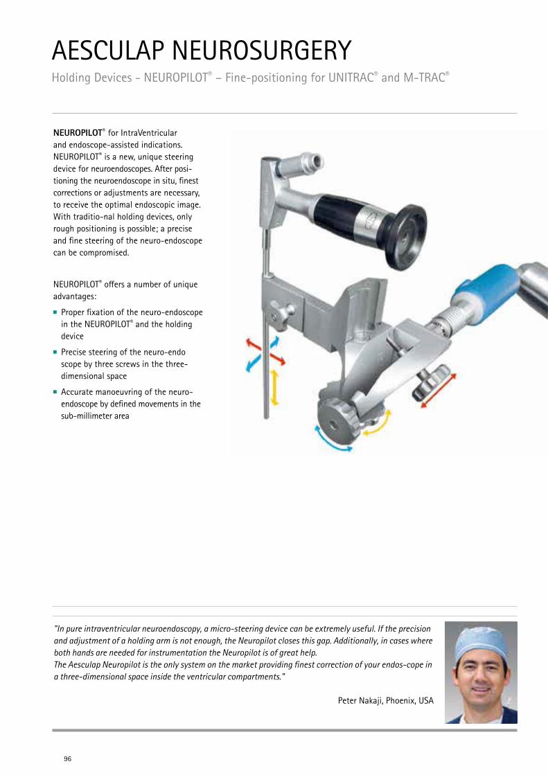
"In pure intraventricular neuroendoscopy, a micro-steering device can be extremely useful. If the precision and adjustment of a holding arm is not enough, the Neuropilot closes this gap. Additionally, in cases where both hands are needed for instrumentation the Neuropilot is of great help. The Aesculap Neuropilot is the only system on the market providing finest correction of your endos-cope in a three-dimensional space inside the ventricular compartments."
Peter Nakaji, Phoenix, USA
AESCULAP NEUROSURGERY Holding Devices - NEUROPILOT® – Fine-positioning for UNITRAC® and M-TRAC®
NEUROPILOT® for IntraVentricular and endoscope-assisted indications. NEUROPILOT® is a new, unique steering device for neuroendoscopes. After posi-tioning the neuroendoscope in situ, finest corrections or adjustments are necessary, to receive the optimal endoscopic image. With traditio-nal holding devices, only rough positioning is possible; a precise and fine steering of the neuro-endoscope can be compromised.
NEUROPILOT® offers a number of unique advantages:
Proper fixation of the neuro-endoscope in the NEUROPILOT® and the holding device
Precise steering of the neuro-endo scope by three screws in the three- dimensional space
Accurate manoeuvring of the neuro- endoscope by defined movements in the sub-millimeter area
96
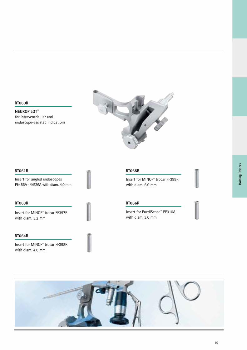
Hold
ing
Devi
ces
NEUROPILOT® for intraventricular and endoscope-assisted indications
Insert for angled endoscopes PE486A - PE526A with diam. 4.0 mm
Insert for MINOP® trocar FF397R with diam. 3.2 mm
Insert for MINOP® trocar FF398R with diam. 4.6 mm
RT060R
RT061R
RT063R
RT064R
Insert for MINOP® trocar FF399R with diam. 6.0 mm
Insert for PaediScope® PF010A with diam. 3.0 mm
RT065R
RT066R
97
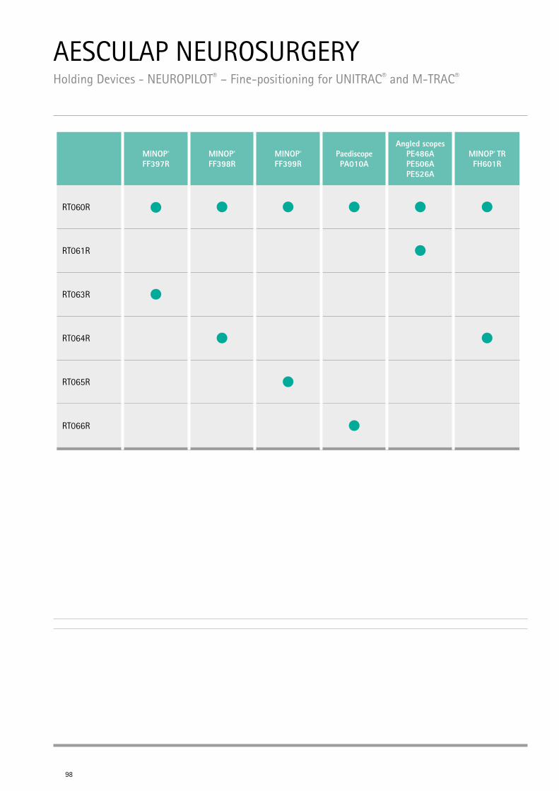
AESCULAP NEUROSURGERY Holding Devices - NEUROPILOT® – Fine-positioning for UNITRAC® and M-TRAC®
98
MINOP®
FF397RMINOP®
FF398RMINOP®
FF399RPaediscopePA010A
Angled scopesPE486APE506APE526A
MINOP® TRFH601R
RT060R
RT061R
RT063R
RT064R
RT065R
RT066R
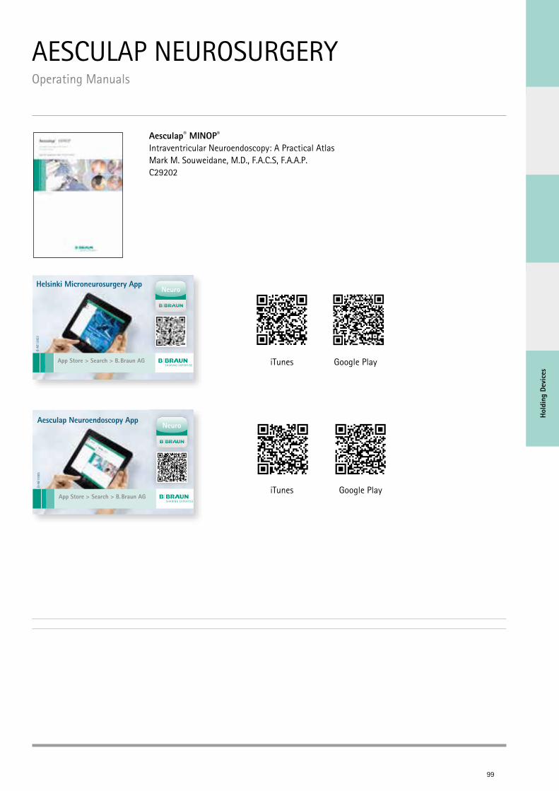
Hold
ing
Devi
ces
AESCULAP NEUROSURGERY Operating Manuals
99
Helsinki Microneurosurgery App
App Store > Search > B.Braun AG
D-N
E120
03
D-NE12003 Karte Helsinki App_Layout 1 29.03.12 13:36 Seite 1
Aesculap Neuroendoscopy App
App Store > Search > B.Braun AG
D-N
E110
05
D-NE11005 NeuroApp-Infokarte_Layout 1 27.09.11 16:20 Seite 1
Aesculap® MINOP®
Intraventricular Neuroendoscopy: A Practical AtlasMark M. Souweidane, M.D., F.A.C.S, F.A.A.P.C29202
iTunes
iTunes
Google Play
Google Play
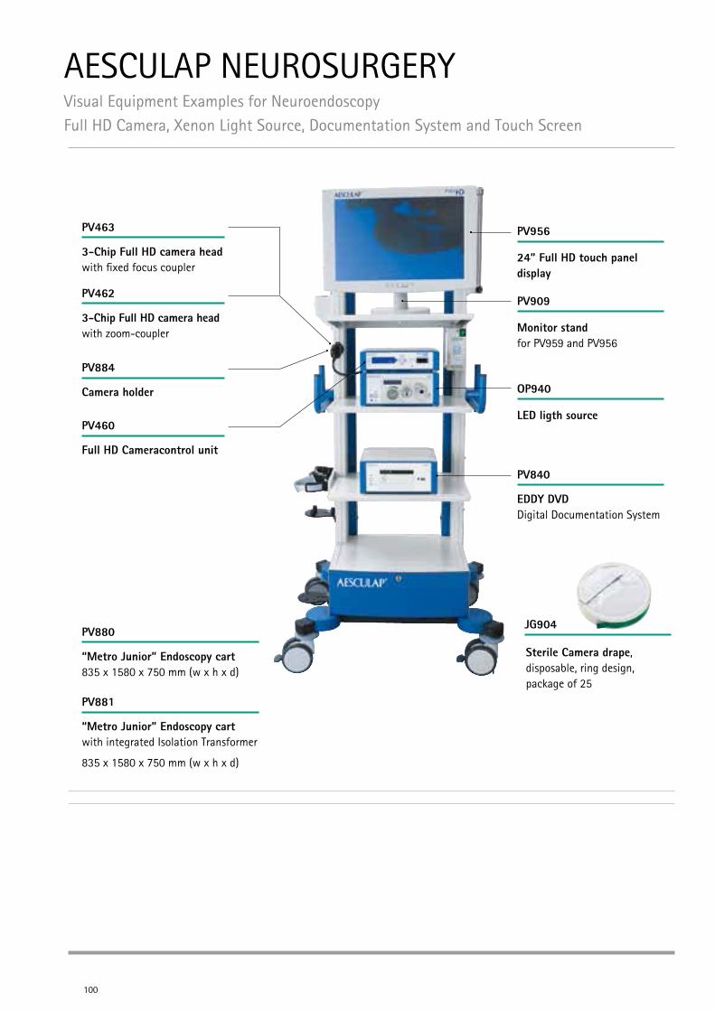
AESCULAP NEUROSURGERY Visual Equipment Examples for NeuroendoscopyFull HD Camera, Xenon Light Source, Documentation System and Touch Screen
Camera holder
100
Full HD Cameracontrol unit
3-Chip Full HD camera head with fixed focus coupler
3-Chip Full HD camera head with zoom-coupler
PV463
PV462
PV884
EDDY DVD Digital Documentation System
“Metro Junior” Endoscopy cart 835 x 1580 x 750 mm (w x h x d)
“Metro Junior” Endoscopy cart with integrated Isolation Transformer
835 x 1580 x 750 mm (w x h x d)
24” Full HD touch panel display
PV956
Monitor stand for PV959 and PV956
PV909
LED ligth source
OP940
PV840
PV880
PV881
PV460
JG904
Sterile Camera drape, disposable, ring design, package of 25
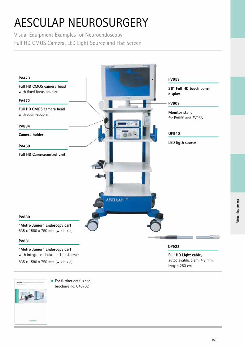
Visu
al E
quip
men
t
AESCULAP NEUROSURGERY Visual Equipment Examples for NeuroendoscopyFull HD CMOS Camera, LED Light Source and Flat Screen
Camera holder
101
Full HD Cameracontrol unit
Full HD CMOS camera head with fixed focus coupler
Full HD CMOS camera head with zoom-coupler
PV473
PV472
PV884
“Metro Junior” Endoscopy cart 835 x 1580 x 750 mm (w x h x d)
“Metro Junior” Endoscopy cart with integrated Isolation Transformer
835 x 1580 x 750 mm (w x h x d)
26” Full HD touch panel display
PV959
Monitor stand for PV959 and PV956
PV909
LED ligth source
OP940
PV880
PV881
For further details see brochure no. C46702
PV460
OP923
Full HD Light cable, autoclavable, diam. 4.8 mm, length 250 cm
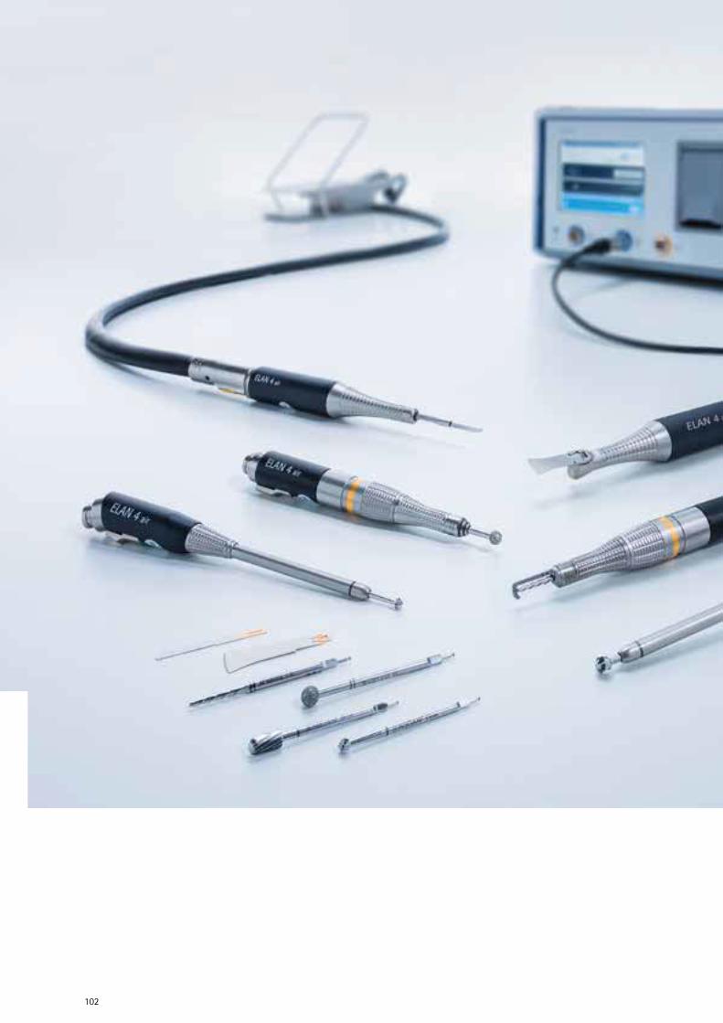
102
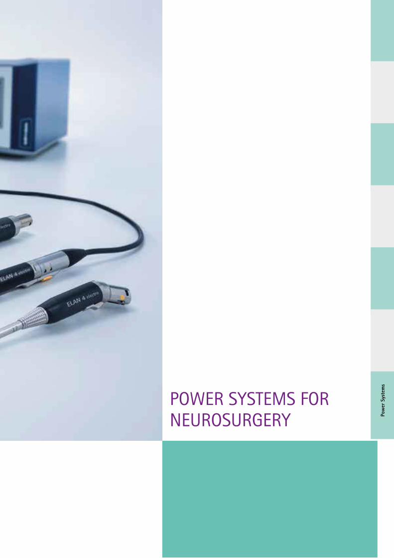
Pow
er S
yste
ms
103
POWER SYSTEMS FOR NEUROSURGERY
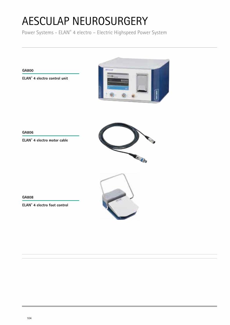
AESCULAP NEUROSURGERY Power Systems - ELAN® 4 electro – Electric Highspeed Power System
104
ELAN® 4 electro control unit
ELAN® 4 electro motor cable
ELAN® 4 electro foot control
GA800
GA806
GA808
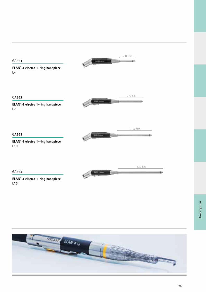
Pow
er S
yste
ms
105
~ 40 mm
~ 70 mm
~ 100 mm
~ 130 mm
ELAN® 4 electro 1-ring handpiece L4
ELAN® 4 electro 1-ring handpiece L7
ELAN® 4 electro 1-ring handpiece L10
ELAN® 4 electro 1-ring handpiece L13
GA861
GA862
GA863
GA864
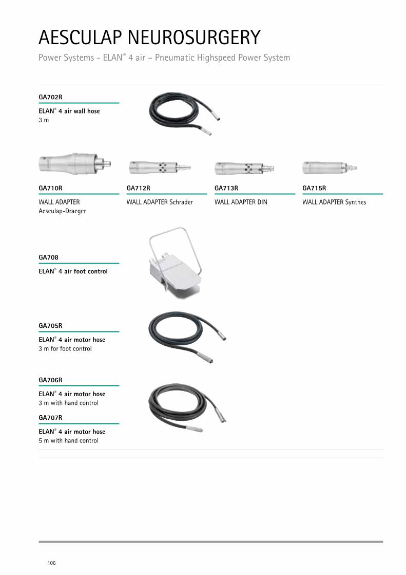
AESCULAP NEUROSURGERY Power Systems - ELAN® 4 air – Pneumatic Highspeed Power System
106
ELAN® 4 air wall hose 3 m
ELAN® 4 air foot control
ELAN® 4 air motor hose 3 m for foot control
ELAN® 4 air motor hose 3 m with hand control
ELAN® 4 air motor hose 5 m with hand control
WALL ADAPTER Aesculap-Draeger
WALL ADAPTER Schrader WALL ADAPTER DIN WALL ADAPTER Synthes
GA702R
GA708
GA705R
GA706R
GA707R
GA710R GA712R GA713R GA715R
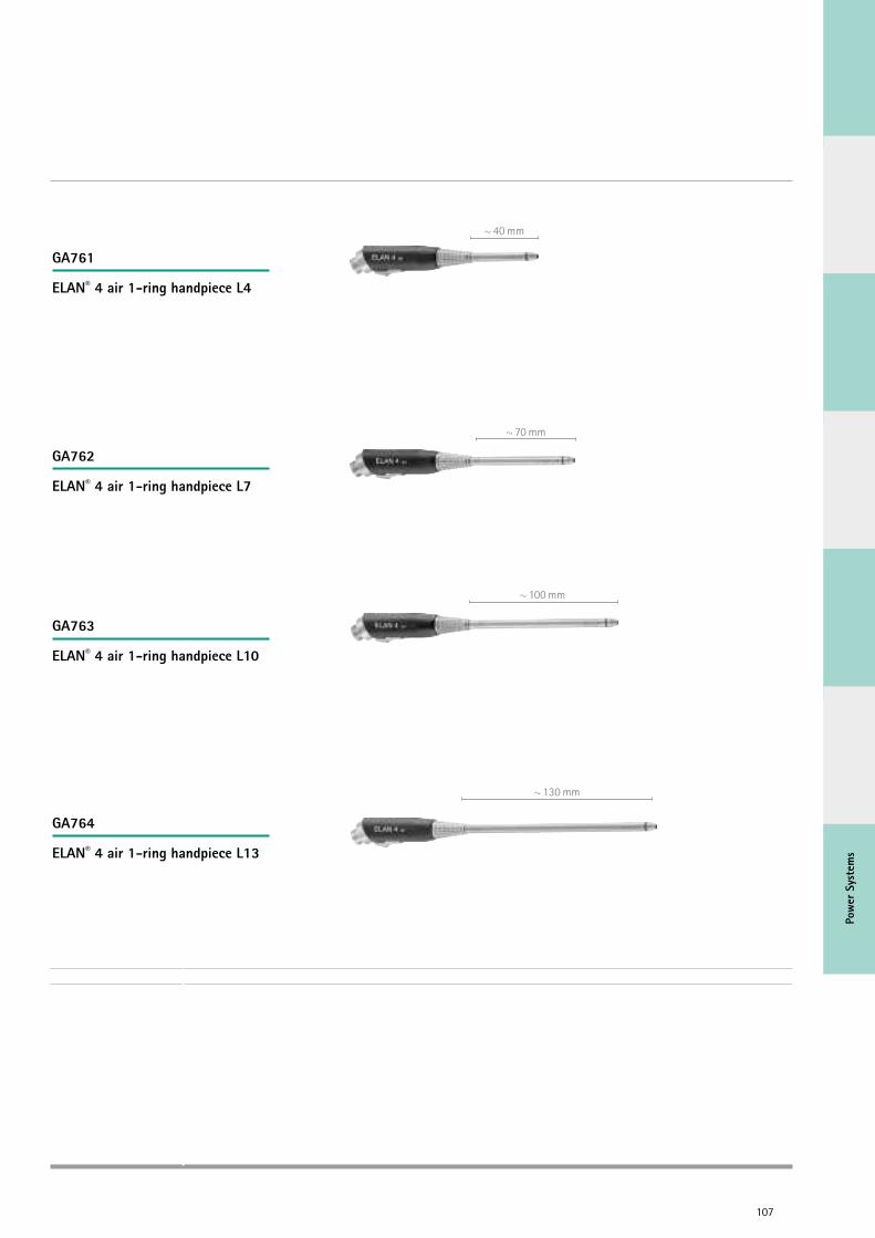
Pow
er S
yste
ms
107
~ 40 mm
~ 70 mm
~ 100 mm
~ 130 mm
ELAN® 4 air 1-ring handpiece L4
ELAN® 4 air 1-ring handpiece L7
ELAN® 4 air 1-ring handpiece L10
ELAN® 4 air 1-ring handpiece L13
GA761
GA762
GA763
GA764
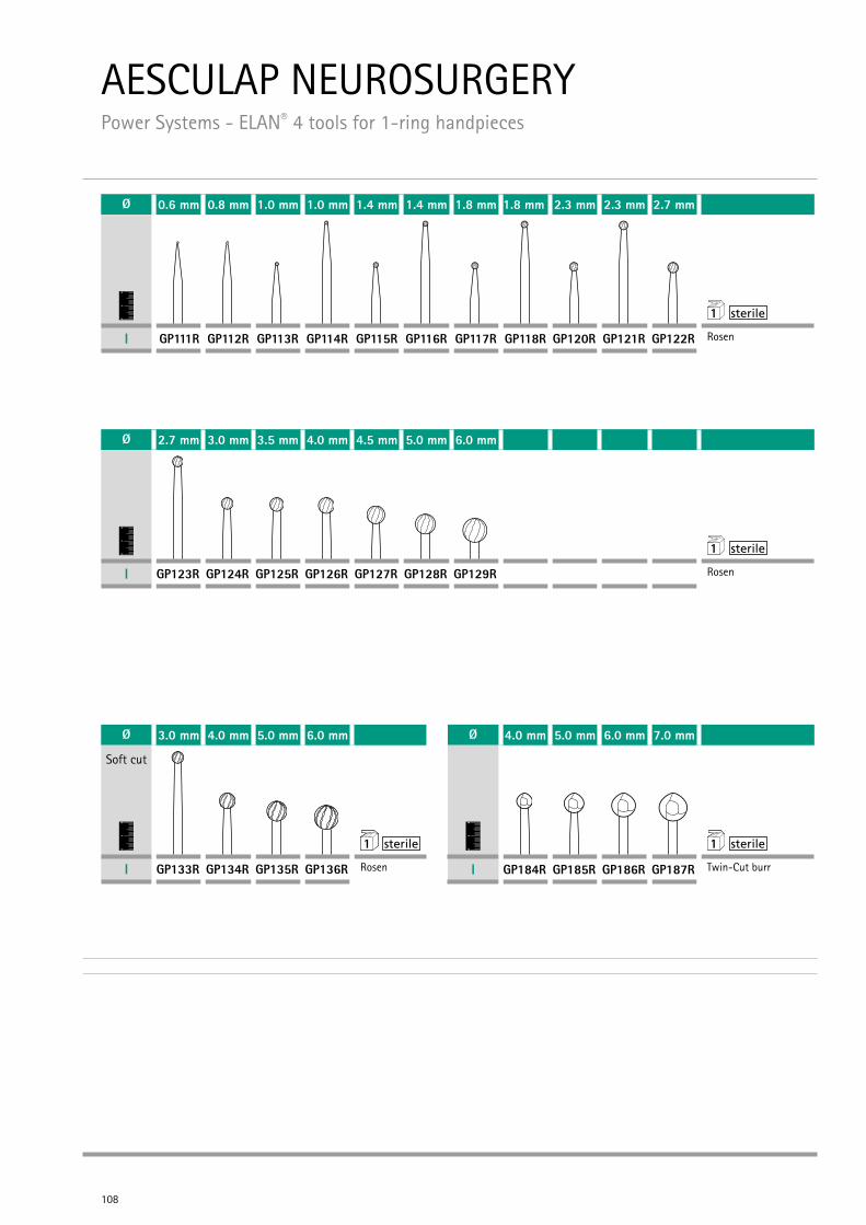
AESCULAP NEUROSURGERY Power Systems - ELAN® 4 tools for 1-ring handpieces
108
Ø 3.0 mm 4.0 mm 5.0 mm 6.0 mm
Soft cut
01
2 1
I GP133R GP134R GP135R GP136R Rosen
Ø 4.0 mm 5.0 mm 6.0 mm 7.0 mm
01
2 1
I GP184R GP185R GP186R GP187R Twin-Cut burr
Ø 0.6 mm 0.8 mm 1.0 mm 1.0 mm 1.4 mm 1.4 mm 1.8 mm 1.8 mm 2.3 mm 2.3 mm 2.7 mm
01
2 1
I GP111R GP112R GP113R GP114R GP115R GP116R GP117R GP118R GP120R GP121R GP122R Rosen
Ø 2.7 mm 3.0 mm 3.5 mm 4.0 mm 4.5 mm 5.0 mm 6.0 mm
01
2 1
I GP123R GP124R GP125R GP126R GP127R GP128R GP129R Rosen
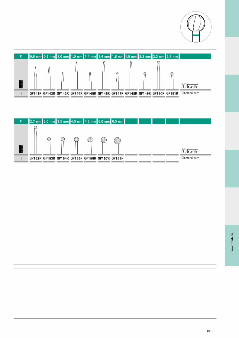
Pow
er S
yste
ms
109
Ø 0.6 mm 0.8 mm 1.0 mm 1.0 mm 1.4 mm 1.4 mm 1.8 mm 1.8 mm 2.3 mm 2.3 mm 2.7 mm
01
2 1
I GP141R GP142R GP143R GP144R GP145R GP146R GP147R GP148R GP149R GP150R GP151R Diamond burr
Ø 2.7 mm 3.0 mm 3.5 mm 4.0 mm 4.5 mm 5.0 mm 6.0 mm
01
2 1
I GP152R GP153R GP154R GP155R GP156R GP157R GP158R Diamond burr
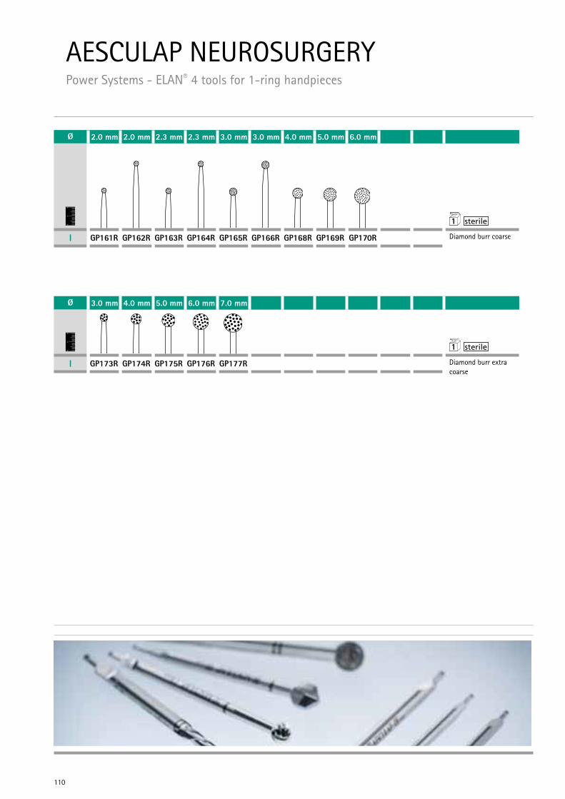
110
Ø 2.0 mm 2.0 mm 2.3 mm 2.3 mm 3.0 mm 3.0 mm 4.0 mm 5.0 mm 6.0 mm
01
2 1
I GP161R GP162R GP163R GP164R GP165R GP166R GP168R GP169R GP170R Diamond burr coarse
Ø 3.0 mm 4.0 mm 5.0 mm 6.0 mm 7.0 mm
01
2 1
I GP173R GP174R GP175R GP176R GP177R Diamond burr extra coarse
AESCULAP NEUROSURGERY Power Systems - ELAN® 4 tools for 1-ring handpieces
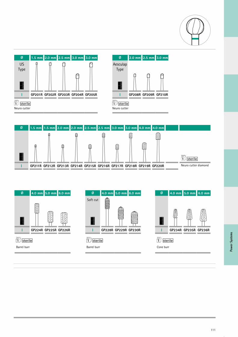
111
Pow
er S
yste
ms
Ø 1.5 mm 1.5 mm 2.0 mm 2.0 mm 2.5 mm 2.5 mm 3.0 mm 3.0 mm 4.0 mm 4.0 mm
01
2 1
I GP211R GP212R GP213R GP214R GP215R GP216R GP217R GP218R GP219R GP220R Neuro cutter diamond
Ø 4.0 mm 5.0 mm 6.0 mm Ø 4.0 mm 5.0 mm 6.0 mm Ø 4.0 mm 5.0 mm 6.0 mm
01
2
Soft cut
01
2
01
2
I GP224R GP225R GP226R I GP228R GP229R GP230R I GP234R GP235R GP236R
1 1 1 Barrel burr Barrel burr Cone burr
Ø 1.5 mm 2.0 mm 2.5 mm 3.0 mm 3.0 mm
US Type
01
2
I GP201R GP202R GP203R GP204R GP205R
1 Neuro cutter
Ø 2.0 mm 2.5 mm 3.0 mm
Aesculap Type
01
2
I GP208R GP209R GP210R
1 Neuro cutter
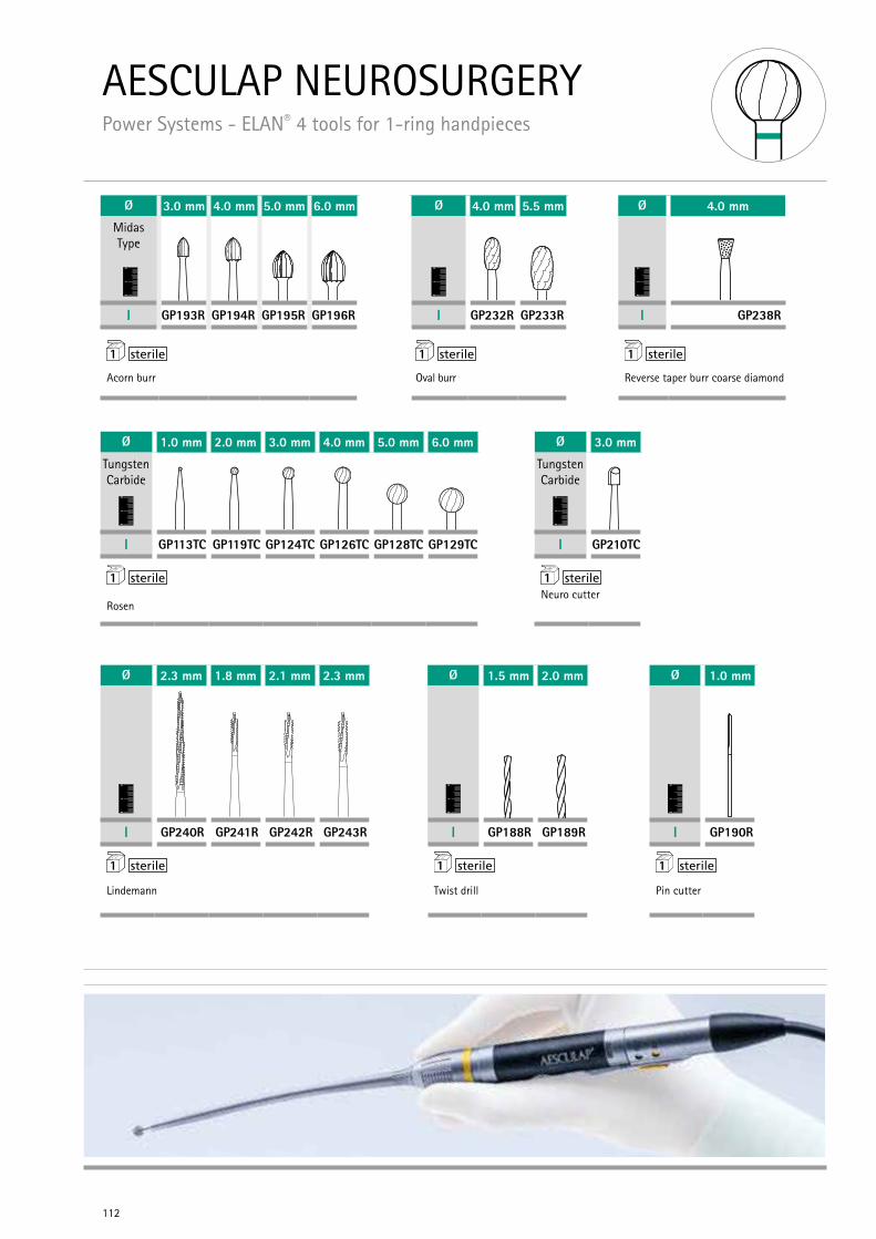
112
Ø 3.0 mm 4.0 mm 5.0 mm 6.0 mm Ø 4.0 mm 5.5 mm Ø 4.0 mm
Midas Type
01
2
01
2
01
2
I GP193R GP194R GP195R GP196R I GP232R GP233R I GP238R
1 1 1 Acorn burr Oval burr Reverse taper burr coarse diamond
Ø 1.0 mm 2.0 mm 3.0 mm 4.0 mm 5.0 mm 6.0 mm
Tungsten Carbide
01
2
I GP113TC GP119TC GP124TC GP126TC GP128TC GP129TC
1
Rosen
Ø 3.0 mm
Tungsten Carbide
01
2
I GP210TC
1 Neuro cutter
Ø 2.3 mm 1.8 mm 2.1 mm 2.3 mm
01
2
I GP240R GP241R GP242R GP243R
1 Lindemann
Ø 1.5 mm 2.0 mm
01
2
I GP188R GP189R
1 Twist drill
Ø 1.0 mm
01
2
I GP190R
1 Pin cutter
AESCULAP NEUROSURGERY Power Systems - ELAN® 4 tools for 1-ring handpieces
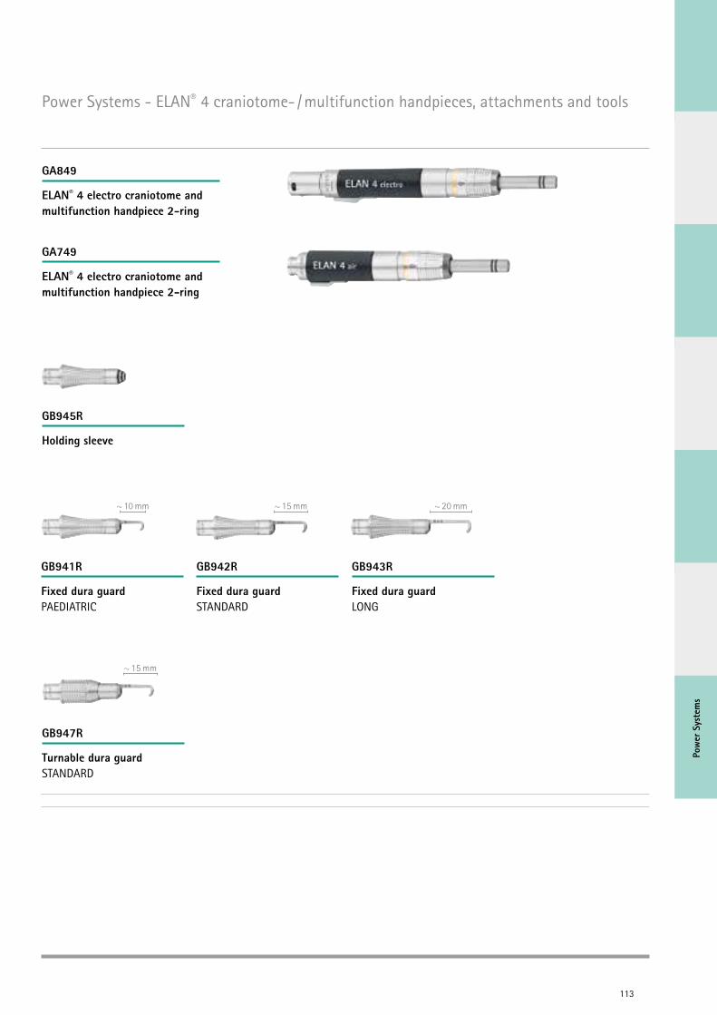
Pow
er S
yste
ms
113
~ 10 mm ~ 15 mm ~ 20 mm
~ 15 mm
Power Systems - ELAN® 4 craniotome- / multifunction handpieces, attachments and tools
ELAN® 4 electro craniotome and multifunction handpiece 2-ring
ELAN® 4 electro craniotome and multifunction handpiece 2-ring
GA849
GA749
Holding sleeve
Fixed dura guard PAEDIATRIC
Turnable dura guard STANDARD
Fixed dura guard STANDARD
Fixed dura guard LONG
GB945R
GB941R
GB947R
GB942R GB943R
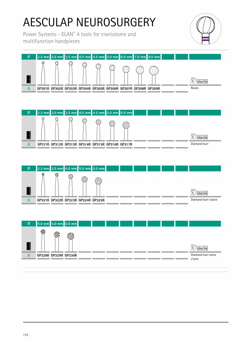
AESCULAP NEUROSURGERY Power Systems - ELAN® 4 tools for craniotome and multifunction handpieces
114
Ø 2.3 mm 3.0 mm 3.5 mm 4.0 mm 4.5 mm 5.0 mm 6.0 mm 7.0 mm 8.0 mm
01
2 1
II GP301R GP302R GP303R GP304R GP305R GP306R GP307R GP308R GP309R Rosen
Ø 2.3 mm 3.0 mm 3.5 mm 4.0 mm 4.5 mm 5.0 mm 6.0 mm
01
2 1
II GP311R GP312R GP313R GP314R GP315R GP316R GP317R Diamond burr
Ø 2.3 mm 3.0 mm 4.0 mm 5.0 mm 6.0 mm
01
2 1
II GP321R GP322R GP323R GP324R GP325R Diamond burr coarse
Ø 4.0 mm 5.0 mm 6.0 mm
01
2 1
II GP328R GP329R GP330R Diamond burr extra cOarse
115
Pow
er S
yste
ms
The whole burr range for 2-ring handpieces can be found in the Burrs & Blades catalogue O17599.
Ø 1.0 mm 1.1 mm 1.2 mm 1.5 mm4.0 mm 4.0 mm 4.0 mm 4.0 mm
01
2
II GP344R GP345R GP346R GP347R
1
Twist drill
Ø 1.5 mm 2.0 mm
01
2
II GP348R GP349R
1
Twist drill
Ø 5.0 mm 6.0 mm 7.5 mm 9.0 mm Ø 5.0 mm 6.0 mm 7.0 mm 8.0 mm 9.0 mm
Midas Type
01
2
01
2
II GP355R GP356R GP357R GP358R II GP335R GP336R GP337R GP338R GP339R
1 1
Acorn burr Twin-Cut burr
L10 L15 L20
01
2
II GP341R GP342R GP343R
1
Craniotome cutter - spiral type
L10 L15 L20
01
2
II GP351R GP352R GP353R
1
Craniotome cutter - straight type
116
AESCULAP NEUROSURGERY Power Systems - ELAN® 4 perforator drivers and tools & ELAN® 4 lowspeed motors
1 Ø/mm
GB300R 6/9
GB302R 9/12
GB304R 12/15
Hudson
1 Ø/mm
TE561 6/9
TE562 9/12
TE563 12/15
ELAN® 4 electro perforator driver
ELAN® 4 air perforator driver
ELAN® 4 electro lowspeed motor
ELAN® 4 air lowspeed motor
Skull perforators
GA822
GA722
GA824
GA724
117
Pow
er S
yste
ms
0
1
2
0 1 2
GP491R 14 mm 13 mm 0.3 mm 0.3 mm
GP492R 15 mm 5 mm 0.3 mm 0.3 mm
GP493R 20 mm 5 mm 0.3 mm 0.3 mm
GP494R 20 mm 10 mm 0.3 mm 0.3 mm
GP495R 20 mm 15 mm 0.3 mm 0.3 mm
GP496R 25 mm 5 mm 0.3 mm 0.3 mm
GP497R 25 mm 12 mm 0.3 mm 0.3 mm
GP491R
GP492R
GP493R
GP494R
GP495R
GP496R
GP497R
ELAN® 4 electro micro sagittal saw
ELAN® 4 air micro sagittal saw
GA836
GA736
118
0
1
2
GP542R 13 mm 0.3 mm 0.3 mm
GP543R 20 mm 0.3 mm 0.3 mm
GP544R 13 mm 0.3 mm 0.3 mm
GP545R 20 mm 0.3 mm 0.3 mm
GP542R
GP543R
GP544R
GP545R
AESCULAP NEUROSURGERY Power Systems - ELAN® 4 micro reciprocating saws and saw blades
ELAN® 4 electro micro reciproca-ting saw
ELAN® 4 air micro reciprocating saw
GA837
GA737
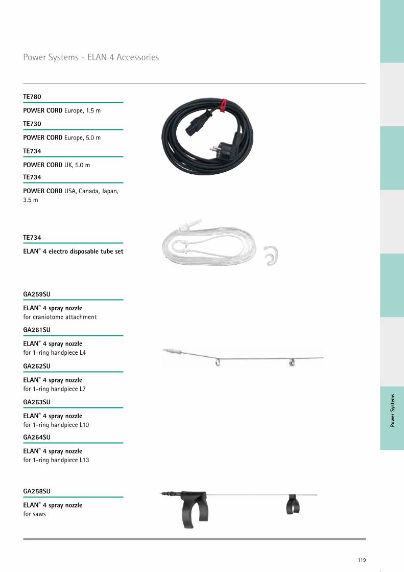
119
Pow
er S
yste
ms
Power Systems - ELAN 4 Accessories
POWER CORD Europe, 1.5 m
POWER CORD Europe, 5.0 m
POWER CORD UK, 5.0 m
POWER CORD USA, Canada, Japan, 3.5 m
ELAN® 4 electro disposable tube set
ELAN® 4 spray nozzle for craniotome attachment
ELAN® 4 spray nozzle for saws
ELAN® 4 spray nozzle for 1-ring handpiece L4
ELAN® 4 spray nozzle for 1-ring handpiece L7
ELAN® 4 spray nozzle for 1-ring handpiece L10
ELAN® 4 spray nozzle for 1-ring handpiece L13
TE780
TE730
TE734
TE734
TE734
GA259SU
GA258SU
GA261SU
GA262SU
GA263SU
GA264SU
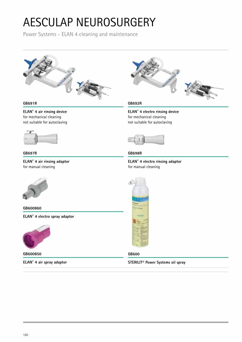
120
AESCULAP NEUROSURGERY Power Systems - ELAN 4 cleaning and maintenance
ELAN® 4 air rinsing device for mechanical cleaning not suitable for autoclaving
ELAN® 4 air rinsing adaptor for manual cleaning
ELAN® 4 electro spray adaptor
STERILIT® Power Systems oil sprayELAN® 4 air spray adaptor
ELAN® 4 electro rinsing adaptor for manual cleaning
ELAN® 4 electro rinsing device for mechanical cleaning not suitable for autoclaving
GB691R
GB697R
GB600860
GB600GB600850
GB698R
GB692R
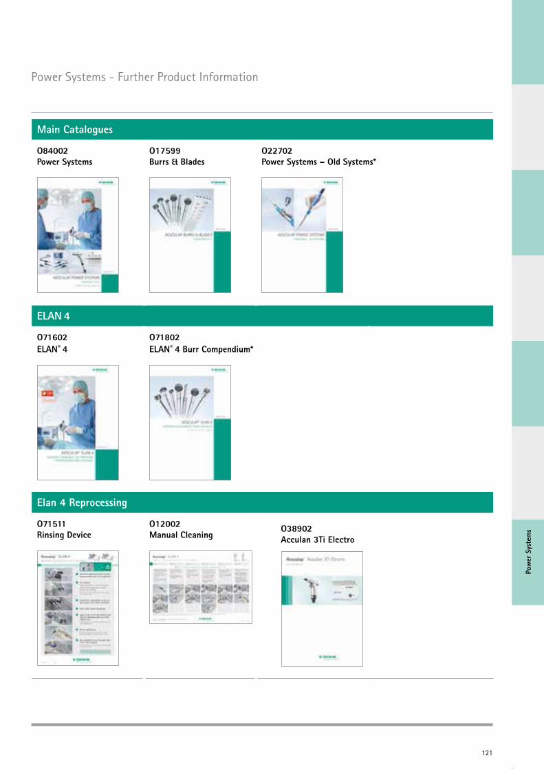
121
Pow
er S
yste
ms
Power Systems - Further Product Information
Main Catalogues
O84002 Power Systems
O17599 Burrs & Blades
O22702 Power Systems – Old Systems*
ELAN 4
O71602 ELAN® 4
O71802 ELAN® 4 Burr Compendium*
Elan 4 Reprocessing
O71511 Rinsing Device
O12002 Manual Cleaning
O38902 Acculan 3Ti Electro
122
Technical developments, new treatment methods, hospital management – the requirements placed on today’s medical professionals are varied, which is why high-quality continuing professional development is more important than ever. This is precisely what Aesculap Academy stands for.
Its aim is clear: The Aesculap Academy wants to keep medical and specialist staff in hospitals and practices fit for the future. Founded in 1995 under the B. Braun Group, the Aesculap Academy is seen today around the world as an important forum for medical training and further professional education. It works with an interdisciplinary, independent and international approach and it strives for long-term partnerships. Thanks to tailored and interrelated modules, the participants can continually develop and build up their knowledge and skills throughout their career. This means they are always as well prepared as possible for their daily work and future tasks.
Take part in one of our international Neuroendoscopy courses.
For detailed information and registration please visit our website „www.aesculap-neuro.com“ or „www.aesculap-academy.com“ or contact your local B. Braun Aesculap representative.
Credit PointsAESCULAP ACADEMY Am Aesculap-Platz 78532 Tuttlingen Phone +49 7461 95-2001www.aesculap-academy.com
AESCULAP ACADEMYForum for contemporary medicine.
Tuttlingen | Berlin | Bochum
Aesc
ulap
Aca
dem
y
123
AESCULAP NEUROSURGERYLiterature
M. M. Souweidane, P. F. Morgenstern, S. Kang et al.
Endoscopic Third Ventriculostomy in Patients with a Diminished Prepontine Interval
Journal of Neurosurgery: Pediatrics, Vol. 5, 250-254, March 2010
O. Sacko, S. Boetto, V. Lauwers-Cances, et al.
Endoscopic Third Ventriculostomy: Outcome Analysis in 368 Procedures
Journal of Neurosurgery: Pediatrics, Vol. 5, 68-74, January 2010
N. Luther, W. R. Stetler Jr., Ira. J. Dunkel, et al.
Subarachnoid Dissemination of Intraventricular Tumors Following Simultaneous Endoscopic Biopsy and Third Ventriculostomy
Journal of Neurosurgery: Pediatrics, Vol. 5, 61-67, January 2010
G. P. Lekovic, J. F. Kerrigan, S. Wait, et al.
In Situ Single-Unit Recording of Hypothalamic Hamartomas Under Endoscopic Direct Visualization
Neurosurgery, Vol. 65, Nr. 6, E1195-E1196, December 2009
B. D. Kollroy, F. A. Ponce, Scott D. Wait, et al.
Endoscopic Intraventricular Biopsy of Infundibular Langerhana Cell Histiocytosis: Case Report
Neurosurgery, Vol. 65, Nr. 1, E214-E215, July 2009
P. Pillai, M. N. Baig, Ch. S. Karas, et al.
Endoscopic Image-Guided Transoral Approach to the Craniover-tebral Junction: An Anatomic Study Comparing Surgical Exposure and Surgical Freedom Obtained with the Endoscope and the Operating Microscope
Neurosurgery, Operative Neurosurgery 2, Vol. 64, ONS437-ONS444, May 2009
N. J. Hopf, A. Stadie, R. Reisch, et al.
Surgical Management of Bilateral Middle Cerebral Artery Aneurysms via a Unilateral Supraorbital Key-Hole Craniotomy
Minimally Invasive Neurosurgery, Vol. 52, 126-131, 2009
R. Reisch, A. Stadie, R. Kockro, et al.
The Minimally Invasive Supraorbital Subfrontal Key-Hole Approach for Surgical Treatment of Temporomesial Lesions of the Dominant Hemisphere
Minimally Invasive Neurosurgery, Vol. 52, 163-169, 2009
J. Leonardo, R. A. Hanel, W. Grand
Endoscopic Tracking of a Ventricular Catheter for Entry into the Lateral Ventricle: Technical Note
Minimally Invasive Neurosurgery, Vol. 52, 287-289, 2009
A. T. Stadie, R. Reisch, R. A. Kockro, et al.
Minimally Invasive Cerebral Cavernoma Surgery Using Keyhole Approaches – Solutions for Technique-Related Limitations
Minimally Invasive Neurosurgery, Vol. 52, 9-16, 2009
P. Pillai, M. Lubow, A. Ortega, et al.
Endoscopic Transconjunctival Surgical Approach to the Optic Nerve and Medial Intraconal Space: A Cadaver Study
Neurosurgery, Operative Neurosurgery 2, Vol. 63, OBS204-ONS209, October 2008
S. C. Froelich, K. M. Abdel Aziz, P. D. Cohen, et al.
Microsurgical and Endoscopic Anatomy of Liliequist’s Membrane: A Complex and Variable Structure of the Basal Cisterns
Neurosurgery, Operative Neurosurgery 1, Vol. 63, ONS1-ONS9, July 2008
B. C. Ong, P. A. Gore, M. B. Donnellan, et al.
Endoscopic Sublabial Transmaxillary Approach to the Rostral Middle Fossa
Neurosurgery, Operative Neurosurgery 1, Vol. 62, 30-37, March 2008
J. D. W. Greenlee, C. Teo, A. Ghahreman, et al.
Purely Endoscopic Resection of Colloid Cysts
Neurosurgery, Operative Neurosurgery 1, Vol. 62, ONS51-ONS56, March 2008
AESCULAP NEUROSURGERYLiterature
124
P. A. Gore, L. F. Gonzalez, H. L. Rekate, et al.
Endoscopic Supracerebellar Infratentorial Approach for Pineal Cyst Resection: Technical Case Report
Neurosurgery, Operative Neurosurgery 1, Vol. 62, March 2008
P. Cappabianca, G. Cinalli, M. Gangemi, et al.
Application of Neuroendoscopy to Intraventricular Lesions
Neurosurgery, Supplement, Vol. 62, No. 2, SHC575-SHC598, February 2008
J. P. Greenfield, L. Z. Leng, U. Chaudhry, et al.
Combined Simultaneous Endoscopic Transsphenoidal and Endosco-pic Transventricular Resection of a Giant Pituitary Macroadenoma
Minimally Invasive Neurosurgery, Vol. 51, 306-309, 2008
P. Y. Hwang, C. Long Ho
Neuronavigation Using an Image-Guided Endoscopic Transnasal-Sphenoethmoidal Approach to Clival Chordomas
Neurosurgery, Operative Neurosurgery 2, Vol. 61, ONS212-ONS218, November 2007
B. Depreitere, N. Dasi, J. Rutka, et al.
Endoscopic Biopsy for Intraventricular Tumors in Children
Journal of Neurosurgery: Pediatrics, Vol. 106, 340-346, May 2007
M. Gangemi, F. Maiuri, G. Colella, et al.
Is Endoscopic Third Ventriculostomy an Internal Shunt Alone?
Minimally Invasive Neurosurgery, Vol. 50, 47-50, 2007
M. Husain, M. Rastogi, B. K. Ojha, et al.
Endoscopic Transoral Surgery for Craniovertebral Junction Anomalies
Journal of Neurosurgery: Spine, Vol. 5, 367-373, October 2006
C. Teo, P. Nakaji, R. J. Mobbs
Endoscope-Assisted Microvascular Decompression for Trigeminal Neu-ralgia: Technical Case Report
Neurosurgery, Operative Neurosurgery 4, Vol. 59, October 2006
A. Weyerbrock, T. Mainprize, J. T. Rutka
Endoscopic Fenestration of a Symptomatic Cavum Septum Pellucidum: Technical Case Report
Neurosurgery, Operative Neurosurgery 4, Vol. 59, October 2006
R. Moftakhar, M. S. Salamat, S. Sahin, et al.
Endoscopically-Assisted Resection of a Choroid Plexus Vascular Malformation Traversing the Cerebral Aqueduct: Technical Case Report
Neurosurgery, Operative Neurosurgery 1, Vol. 59, July 2006
J. van Beijnum, P. W. Hanlo, K. Sen Han, et al.
Navigated Laser-Assisted Endoscopic Fenestration of a Suprasellar Arachnoid Cyst in a 2-Year-Old Child with Bobble-Head Doll Syndrome
Journal of Neurosurgery: Pediatrics, Vol. 104, 348-351, May 2006
F. T. Mangano, D. D. Limbrick, J. R. Leonard, et al.
Simultaneous Image-Guided and Endoscopic Navigation without Rigid Cranial Fixation: Application in Infants: Technical Case Report
Neurosurgery, Operative Neurosurgery 2, Vol. 58, ONS-377-ONS-378, April 2006
G. P. Lekovic, L. F. Gonzalez, I. Feiz-Erfan, et al.
Endoscopic Resection of Hypothalamic Hamartoma using a Novel Va-riable Aspiration Tissue Resector
Neurosurgery, Operative Neurosurgery 1, vol. 58, ONS166-ONS169, Febru-ary 2006
A. Morita, M. Shin, L. N. Sekhar, et al.
Endoscopic Microneurosurgery: Usefulness and Cost-Effectiveness in the Consecutive Experience of 210 Patients
Neurosurgery, Vol. 58, No. 2, 315-321, February 2006

Lite
ratu
re
125
A. A. Figaji, A. G. Fieggen, P. L. Semple, et al.
Intracranial Endoscopy
Samj Forum, Vol. 96, No. 1, 32-37, January 2006
A. Bussarsky, M. Marinov, V. Bussarsky, et al.
Virtual Simulation of Neuroendoscopic Procedures: Early Clinical Experience with Ventricular Lesions
Central European Neurosurgery, Vol. 67, 129-136, 2006
J. Zhao, Y. Wang, Y. Zhao, et al.
Neuroendoscope-Assisted Minimally Invasive Microsurgery for Clipping Intracranial Aneurysms
Minimally Invasive Neurosurgery, Vol. 49, 335-341, 2006
Y. Arakawa, K. Nakazawa, H. Kataoka, et al.
Microfiberscope Coaxial Technique in Neuroendoscopic Surgery
Minimally Invasive Neurosurgery, Vol. 49, 380-383, 2006
I. Gawish, R. Reisch, A. Perneczky
Endoscopic Aqueductoplasty through a Tailored Craniocervical Approach
Journal of Neurosurgery, Vol. 103, 778-782, November 2005
R. Reisch, A. Perneczky
Ten-Year Experience with the Supraorbital Subfrontal Approach through an Eyebrow Skin Incision
Neurosurgery, Operative Neurosurgery 4, Vol. 57, ONS242-ONS255, Oc-tober 2005
M. M. Souweidane
Endoscopic Surgery for Intraventricular Brain Tumors in Patients without Hydrocephalus
Neurosurgery, Operative Neurosurgery 4, Vol. 57, ONS312-ONS318, Oc-tober 2005
K. Schmidt, C. Coimbra
Endoscopic Treatment of Thalamic Neuroepithelial Cysts
Journal of Neurosurgery, Vol. 103, 342-346, August 2005
N. Luther, A. Cohen, M. M. Souweidane
Hemorrhagic Sequelae from Intracranial Neuroendoscopic Proce-dures for Intraventricular Tumors
Neurosurgical Focus, Vol. 19 (1), E9, 1-4, July 2005
P. D. Purdy, T. Fujimoto, R. E. Replogle, et al.
Percutaneous Intraspinal Navigation for Access to the Subarachnoid Space: Use of Another Natural Conduit for Neurosurgical Procedures
Neurosurgical Focus, Vol. 19 (1), E11, 1-5, July 2005
M. M. Souweidane
Endoscopic Management of Pediatric Brain Tumors
Neurosurgical Focus, Vol. 18 (6a), E1, June 2005
N. Luther, M. M. Souweidane
Neuroendoscopic Resection of Posterior Third Ventricular Ependymoma
Neurosurgical Focus, Vol. 18 (6a), E3, 1-2, June 2005
E. Nathal, J. L. Gomez-Amador
Anatomic and Surgical Basis of the Sphenoid Ridge Keyhole Approach for Cerebral Aneurysms
Neurosurgery, Operative Neurosurgery 1, Vol. 56, ONS178-ONS185, January 2005
J. Martin, C. Neal, I. Moores, et al.
Use of a Nitrogen Arm-Stabilized Endoscopic Microdriver in Neuroendoscopic Surgery
Minimally Invasive Neurosurgery, Vol. 48, 63-65, 2005
M.J. Fritsch, L. Dörner, S. Kienke, et al.
Hydrocephalus in children with posterior fossa tumors: The role of Endoscopic Third Ventriculostomy (ETV)
Journal of Neurosurgery: Pediatrics, Vol. 103: 40-42, 2005
AESCULAP NEUROSURGERYLiterature
126
M.J. Fritsch, S. Kienke, T. Ankermann, et al.
Endoscopic Third Ventriculostomy (ETV) in infants
Journal of Neurosurgery: Pediatrics, Vol. 103: 50-53, 2005
M. Taniguchi, A. Kato, T. Taki, et al.
Endoscope Assisted Removal of Jugular Foramen Schwannoma; Report of 3 Cases
Minimally Invasive Neurosurgery, Vol. 48, 365-368, 2005
H. Kinouchi, T. Yanagisawa, A. Suzuki, et al.
Simultaneous Microscopic and Endoscopic Monitoring During Surgery for Internal Carotid Artery Aneurysms
Journal of Neurosurgery, Vol. 101, 989-995, December 2004
J. C. Wang, L. Heier, M. M. Souweidane
Advances in the Endoscopic Management of Suprasellar Arachnoid Cysts in Children
Journal of Neurosurgery: Pediatrics, Vol. 100, 418-426, May 2004
M.J. Fritsch, S. Kienke, H.M. Mehdorn
Endoscopic aqueductoplasty: stent or not to stent?
Childs Nerv System, Vol. 20, 137-142, 2004
M.J. Fritsch, K.H. Manwaring, S. Kienke, et al.
Endoscopic treatment of isolated 4th ventricle in children
Neurosurgery, Vol. 55, 372-379, 2004
C. Trantakis, J. Helm, M. Keller, et al.
Third Ventriculostomy in Communicating Hydrocephalus in Adult Patients – The Role of Lumbar and Cranial Cerebrospinal Fluid Outflow Measurement
Minimally Invasive Neurosurgery, Vol. 48, 140-144, 2004
S. Wolfsberger, M.-T. Forster, M. Donat, et al.
Virtual Endoscopy is a Useful Device for Training and Preoperative Planning of Transsphenoidal Endoscopic Pituitary Surgery
Minimally Invasive Neurosurgery, Vol. 47, 214-220, 2004
Y. Lin, Y. Qiu
Microanatomy of Endoscope-Assisted Glabellar Nasal Keyhole Approach
Minimally Invasive Neurosurgery, Vol. 46, 155-160, 2003
T. G. Psarros, J. Krumerman, C. Coimbra
Endoscopic Management of Supratentorial Ventricular Neurocysticercosis: Case Series and Review of the Literature
Minimally Invasive Neurosurgery, Vol. 46, 331-334, 2003
M. A. Barajas, G. Ramirez-Guzmán, C. Rodríguez-Vazquez, et al.
Multimodal Management of Craniopharyngiomas: Neuroendoscopy, Microsurgery, and Radiosurgery
Journal of Neurosurgery (Supplement 5), Vol. 97, 607-609, December 2002
Z. Horváth, F. Vetö, I. Balás, et al.
Biportal Endoscopic Removal of a Primary Intraventricular Hematoma: Case Report
Minimally Invasive Neurosurgery, Vol. 43, 4-8, 2000
A. Rieger, N. G. Rainov, M. Brucke, et al.
Endoscopic Third Ventriculostomy is the Treatment of Choice for Obstructive Hydrocephalus due to Pediatric Tumors
Minimally Invasive Neurosurgery, Vol. 43. 83-86, 2000
V. Rohde, J. M. Gilsbach
Anomalies and Variants of the Endoscopic Anatomy for Third Ventriculostomy
Minimally Invasive Neurosurgery, Vol. 43, 111-117, 2000
J. Paladino K. Rotim, D. Štimac, et al.
Endoscopic Third Ventriculostomy with Ultrasonic Contact Microprobe
Minimally Invasive Neurosurgery, Vol. 43, 132-134, 2000

Lite
ratu
re
127
T. Menovsky, J. A. Grotenhuis, J. de Vries, et al.
Endoscope-Assisted Supraorbital Craniotomy for Lesions of the Interpeduncular Fossa Technique and Application
Neurosurgery, Vol. 44, No. 1, 106-112, January 1999
P. Wieneke, T. Lutze
Technologies for Microendoscopes of the Future: The MINOP Project
Minimally Invasive Therapy & Allied Technology, Vol. 7/3, 233-239, 1998
E. van Lindert, N. Hopf, A. Perneczky
Endoscopic Treatment of Mesencephalic Ependymal Cysts: Technical Case Report
Neurosurgery, Vol. 43, No. 5, November 1998
F. Vetõ, Z. Horváth, T. Dóczi
Biportal Endoscopic Management of Third Ventricle Tumors in Patients with Occlusive Hydrocephalus: Technical Note
Neurosurgery, Vol. 40, No. 4, 871-877, April 1997
J. A. Grotenhuis
Endoscope-Assisted Craniotomy
Techniques in Neurosurgery, Vol. 1, No. 3, 201-212, 1996
G. Fries, R. Reisch
Biportal Neuroendoscopic Microsurgical Approaches to the Subarachnoid Cisterns: A Cadaver Study
Minimally Invasive Neurosurgery, Vol. 39, 99-104, 1996
A. Perneczky
Planning Strategies for the Suprasellar Region
Neurosurgeons 11, 343-348, 1992
128
AESCULAP NEUROSURGERY Numerical Index
BN175R 89
FA030R 74
FA031R 74
FA032R 74
FA033R 74
FA034R 74
FA035R 74
FA036R 74
FA037R 74
FA038R 74
FA039R 74
FA040R 74
FA041R 60
FA041R 72
FA042R 60
FA042R 72
FA043R 60
FA043R 72
FA044R 60
FA044R 72
FA045R 60
FA045R 72
FA046R 60
FA046R 72
FA047R 60
FA047R 72
FA060R 60
FA060R 72
FA061R 61
FA061R 73
FA062R 61
FA062R 73
FA063R 61
FA063R 73
FA064R 61
FA064R 73
FA065R 61
FA065R 73
FA066R 61
FA066R 73
FA067R 61
FA067R 73
FA068R 61
FA068R 73
FA069R 85
FA070R 85
FA071R 85
FA072R 84
FA073R 84
FA074R 84
FA075R 84
FA076R 78
FD113D 62
FD114D 62
FD115D 62
FD116D 62
FD220R 83
FD222R 83
FD224R 83
FD226R 83
FD228R 82
FD228R 83
FD467R 55
FD701B 50
FD702B 50
FD703B 50
FD704B 50
FD705R 50
FD706B 50
FD711B 52
FD717B 51
FD718B 51
FD719B 51
FD731B 50
FD732B 50
FD733B 50
FD734B 50
FD735B 50
FD736B 50
FD741B 52
FD743B 52
FD761B 52
FD763B 52
FD766B 53
FD767B 53
FD768B 53
FD769B 53
FD771B 50
FD771B 50
FD773B 50
FD774B 50
FD775B 50
FD776B 50
FD786B 53
FD787B 53
FD788B 53
FD789B 53
FD797B 54
FD798B 54
FD799B 54
FD805B 54
FD808B 54
FD809B 54
FD811B 54
FD812B 54
FD814B 54
FD815B 54
FD816B 54
FD818B 54
FD819B 54
FD821B 54
FD822B 54
FD823B 54
FD824B 55
FD825B 55
FD826B 55
FD827B 55
FD828B 55
FD831B 55
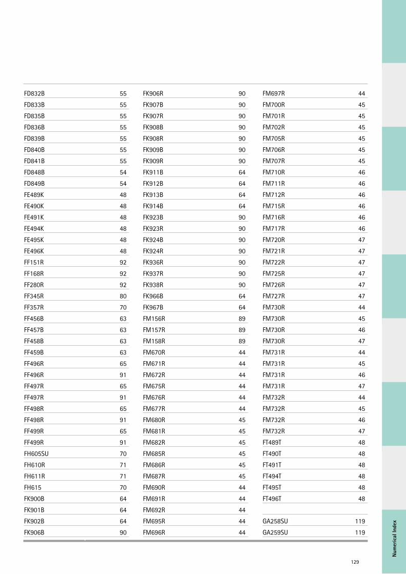
Num
eric
al In
dex
129
FD832B 55
FD833B 55
FD835B 55
FD836B 55
FD839B 55
FD840B 55
FD841B 55
FD848B 54
FD849B 54
FE489K 48
FE490K 48
FE491K 48
FE494K 48
FE495K 48
FE496K 48
FF151R 92
FF168R 92
FF280R 92
FF345R 80
FF357R 70
FF456B 63
FF457B 63
FF458B 63
FF459B 63
FF496R 65
FF496R 91
FF497R 65
FF497R 91
FF498R 65
FF498R 91
FF499R 65
FF499R 91
FH605SU 70
FH610R 71
FH611R 71
FH615 70
FK900B 64
FK901B 64
FK902B 64
FK906B 90
FK906R 90
FK907B 90
FK907R 90
FK908B 90
FK908R 90
FK909B 90
FK909R 90
FK911B 64
FK912B 64
FK913B 64
FK914B 64
FK923B 90
FK923R 90
FK924B 90
FK924R 90
FK936R 90
FK937R 90
FK938R 90
FK966B 64
FK967B 64
FM156R 89
FM157R 89
FM158R 89
FM670R 44
FM671R 44
FM672R 44
FM675R 44
FM676R 44
FM677R 44
FM680R 45
FM681R 45
FM682R 45
FM685R 45
FM686R 45
FM687R 45
FM690R 44
FM691R 44
FM692R 44
FM695R 44
FM696R 44
FM697R 44
FM700R 45
FM701R 45
FM702R 45
FM705R 45
FM706R 45
FM707R 45
FM710R 46
FM711R 46
FM712R 46
FM715R 46
FM716R 46
FM717R 46
FM720R 47
FM721R 47
FM722R 47
FM725R 47
FM726R 47
FM727R 47
FM730R 44
FM730R 45
FM730R 46
FM730R 47
FM731R 44
FM731R 45
FM731R 46
FM731R 47
FM732R 44
FM732R 45
FM732R 46
FM732R 47
FT489T 48
FT490T 48
FT491T 48
FT494T 48
FT495T 48
FT496T 48
GA258SU 119
GA259SU 119
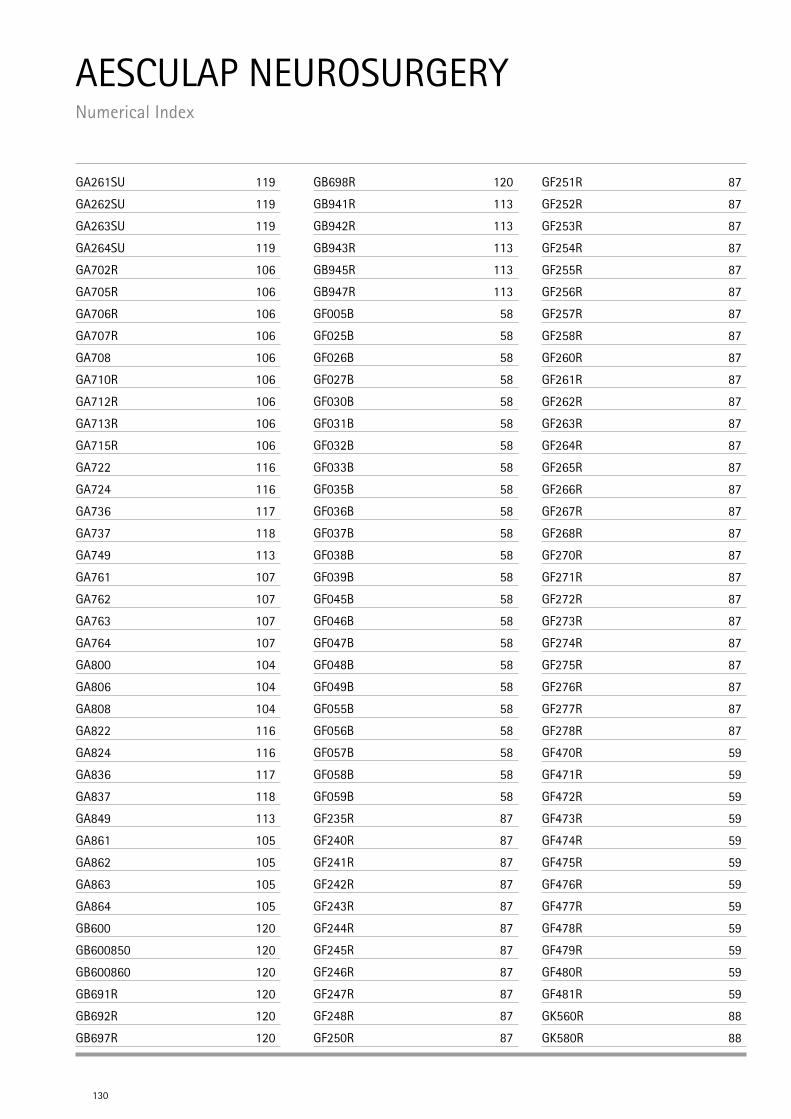
AESCULAP NEUROSURGERY Numerical Index
130
GA261SU 119
GA262SU 119
GA263SU 119
GA264SU 119
GA702R 106
GA705R 106
GA706R 106
GA707R 106
GA708 106
GA710R 106
GA712R 106
GA713R 106
GA715R 106
GA722 116
GA724 116
GA736 117
GA737 118
GA749 113
GA761 107
GA762 107
GA763 107
GA764 107
GA800 104
GA806 104
GA808 104
GA822 116
GA824 116
GA836 117
GA837 118
GA849 113
GA861 105
GA862 105
GA863 105
GA864 105
GB600 120
GB600850 120
GB600860 120
GB691R 120
GB692R 120
GB697R 120
GB698R 120
GB941R 113
GB942R 113
GB943R 113
GB945R 113
GB947R 113
GF005B 58
GF025B 58
GF026B 58
GF027B 58
GF030B 58
GF031B 58
GF032B 58
GF033B 58
GF035B 58
GF036B 58
GF037B 58
GF038B 58
GF039B 58
GF045B 58
GF046B 58
GF047B 58
GF048B 58
GF049B 58
GF055B 58
GF056B 58
GF057B 58
GF058B 58
GF059B 58
GF235R 87
GF240R 87
GF241R 87
GF242R 87
GF243R 87
GF244R 87
GF245R 87
GF246R 87
GF247R 87
GF248R 87
GF250R 87
GF251R 87
GF252R 87
GF253R 87
GF254R 87
GF255R 87
GF256R 87
GF257R 87
GF258R 87
GF260R 87
GF261R 87
GF262R 87
GF263R 87
GF264R 87
GF265R 87
GF266R 87
GF267R 87
GF268R 87
GF270R 87
GF271R 87
GF272R 87
GF273R 87
GF274R 87
GF275R 87
GF276R 87
GF277R 87
GF278R 87
GF470R 59
GF471R 59
GF472R 59
GF473R 59
GF474R 59
GF475R 59
GF476R 59
GF477R 59
GF478R 59
GF479R 59
GF480R 59
GF481R 59
GK560R 88
GK580R 88
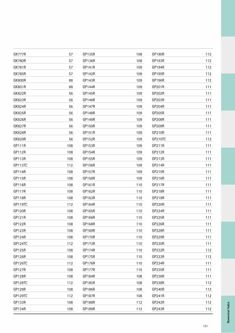
Num
eric
al In
dex
131
GK777R 57
GK780R 57
GK781R 57
GK785R 57
GK800R 88
GK801R 88
GK822R 56
GK823R 56
GK824R 56
GK825R 56
GK826R 56
GK827R 56
GK828R 56
GK829R 56
GP111R 108
GP112R 108
GP113R 108
GP113TC 112
GP114R 108
GP115R 108
GP116R 108
GP117R 108
GP118R 108
GP119TC 112
GP120R 108
GP121R 108
GP122R 108
GP123R 108
GP124R 108
GP124TC 112
GP125R 108
GP126R 108
GP126TC 112
GP127R 108
GP128R 108
GP128TC 112
GP129R 108
GP129TC 112
GP133R 108
GP134R 108
GP135R 108
GP136R 108
GP141R 109
GP142R 109
GP143R 109
GP144R 109
GP145R 109
GP146R 109
GP147R 109
GP148R 109
GP149R 109
GP150R 109
GP151R 109
GP152R 109
GP153R 109
GP154R 109
GP155R 109
GP156R 109
GP157R 109
GP158R 109
GP161R 110
GP162R 110
GP163R 110
GP164R 110
GP165R 110
GP166R 110
GP168R 110
GP169R 110
GP170R 110
GP173R 110
GP174R 110
GP175R 110
GP176R 110
GP177R 110
GP184R 108
GP185R 108
GP186R 108
GP187R 108
GP188R 112
GP189R 112
GP190R 112
GP193R 112
GP194R 112
GP195R 112
GP196R 112
GP201R 111
GP202R 111
GP203R 111
GP204R 111
GP205R 111
GP208R 111
GP209R 111
GP210R 111
GP210TC 112
GP211R 111
GP212R 111
GP213R 111
GP214R 111
GP215R 111
GP216R 111
GP217R 111
GP218R 111
GP219R 111
GP220R 111
GP224R 111
GP225R 111
GP226R 111
GP228R 111
GP229R 111
GP230R 111
GP232R 112
GP233R 112
GP234R 111
GP235R 111
GP236R 111
GP238R 112
GP240R 112
GP241R 112
GP242R 112
GP243R 112
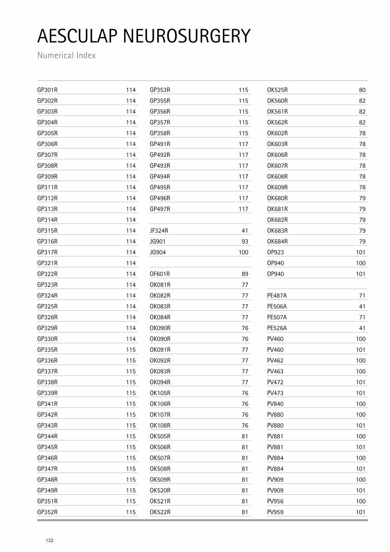
AESCULAP NEUROSURGERY Numerical Index
132
GP301R 114
GP302R 114
GP303R 114
GP304R 114
GP305R 114
GP306R 114
GP307R 114
GP308R 114
GP309R 114
GP311R 114
GP312R 114
GP313R 114
GP314R 114
GP315R 114
GP316R 114
GP317R 114
GP321R 114
GP322R 114
GP323R 114
GP324R 114
GP325R 114
GP328R 114
GP329R 114
GP330R 114
GP335R 115
GP336R 115
GP337R 115
GP338R 115
GP339R 115
GP341R 115
GP342R 115
GP343R 115
GP344R 115
GP345R 115
GP346R 115
GP347R 115
GP348R 115
GP349R 115
GP351R 115
GP352R 115
GP353R 115
GP355R 115
GP356R 115
GP357R 115
GP358R 115
GP491R 117
GP492R 117
GP493R 117
GP494R 117
GP495R 117
GP496R 117
GP497R 117
JF324R 41
JG901 93
JG904 100
OF601R 89
OK081R 77
OK082R 77
OK083R 77
OK084R 77
OK090R 76
OK090R 76
OK091R 77
OK092R 77
OK093R 77
OK094R 77
OK105R 76
OK106R 76
OK107R 76
OK108R 76
OK505R 81
OK506R 81
OK507R 81
OK508R 81
OK509R 81
OK520R 81
OK521R 81
OK522R 81
OK525R 80
OK560R 82
OK561R 82
OK562R 82
OK602R 78
OK603R 78
OK606R 78
OK607R 78
OK608R 78
OK609R 78
OK680R 79
OK681R 79
OK682R 79
OK683R 79
OK684R 79
OP923 101
OP940 100
OP940 101
PE487A 71
PE506A 41
PE507A 71
PE526A 41
PV460 100
PV460 101
PV462 100
PV463 100
PV472 101
PV473 101
PV840 100
PV880 100
PV880 101
PV881 100
PV881 101
PV884 100
PV884 101
PV909 100
PV909 101
PV956 100
PV959 101
Num
eric
al In
dex
133
RT020R 93
RT040R 93
RT043R 93
RT044SU 93
RT046P 94
RT055P 94
RT060R 97
RT061R 97
RT063R 97
RT064R 97
RT065R 97
RT066R 97
RT068R 94
RT079205 94
RT079R 94
RT081R 94
RT090R 92
RT099R 70
RT099R 94
TE730 119
TE734 119
TE734 119
TE734 119
TE780 119

134
AESCULAP NEUROSURGERY Notes

135

AESCULAP® – a B. Braun brand
Aesculap AG | Am Aesculap-Platz | 78532 Tuttlingen | Germany
Phone +49 7461 95-0 | Fax +49 7461 95-2600 | www.aesculap.com
The main product trademark “Aesculap” is a registered trademark of Aesculap AG. Subject to technical changes. All rights reserved. This brochure may only be used for the exclusive purpose of obtaining information about our products. Repro-duction in any form partial or otherwise is not permitted. C35502 0617/0.5/17
Related Documents