Advances in Clinical and Biomedical Applications of Photoacoustic Imaging Jimmy L. Su, Bo Wang, Katheryne E. Wilson, Carolyn L. Bayer, Yun-Sheng Chen, Seungsoo Kim, Kimberly A. Homan, and Stanislav Y. Emelianov * Department of Biomedical Engineering, University of Texas at Austin, Austin, TX 78712 USA Abstract Importance of the field—Photoacoustic imaging is an imaging modality that derives image contrast from the optical absorption coefficient of the tissue being imaged. The imaging technique is able to differentiate between healthy and diseased tissue with either deeper penetration or higher resolution than other functional imaging modalities currently available. From a clinical standpoint, photoacoustic imaging has demonstrated safety and effectiveness in diagnosing diseased tissue regions using either endogenous tissue contrast or exogenous contrast agents. Furthermore, the potential of photoacoustic imaging has been demonstrated in various therapeutic interventions ranging from drug delivery and release to image-guided therapy and monitoring. Areas covered in this review—This article reviews the current state of photoacoustic imaging in biomedicine from a technological perspective, highlights various biomedical and clinical applications of photoacoustic imaging, and gives insights on future directions. What the reader will gain—Readers will learn about the various applications of photoacoustic imaging, as well as the various contrast agents that can be used to assist photoacoustic imaging. This review will highlight both pre-clinical and clinical uses for photoacoustic imaging, as well as discuss some of the challenges that must be addressed to move photoacoustic imaging into the clinical realm. Take home message—Photoacoustic imaging offers unique advantages over existing imaging modalities. The imaging field is broad with many exciting applications for detecting and diagnosing diseased tissue or processes. Photoacoustics is also used in therapeutic applications to identify and characterize the pathology and then to monitor the treatment. Although the technology is still in its infancy, much work has been done in the pre-clinical arena, and photoacoustic imaging is fast approaching the clinical setting. Keywords Atherosclerosis; cancer; contrast agents; drug delivery; molecular imaging; nanoparticles; optoacoustics; photoacoustics; therapy; ultrasound * [email protected]. Declaration of Interest The authors were supported by an NIH grant (# HL096981). NIH Public Access Author Manuscript Expert Opin Med Diagn. Author manuscript; available in PMC 2011 November 1. Published in final edited form as: Expert Opin Med Diagn. 2010 November 1; 4(6): 497–510. doi:10.1517/17530059.2010.529127. NIH-PA Author Manuscript NIH-PA Author Manuscript NIH-PA Author Manuscript

Welcome message from author
This document is posted to help you gain knowledge. Please leave a comment to let me know what you think about it! Share it to your friends and learn new things together.
Transcript

Advances in Clinical and Biomedical Applications ofPhotoacoustic Imaging
Jimmy L. Su, Bo Wang, Katheryne E. Wilson, Carolyn L. Bayer, Yun-Sheng Chen,Seungsoo Kim, Kimberly A. Homan, and Stanislav Y. Emelianov*Department of Biomedical Engineering, University of Texas at Austin, Austin, TX 78712 USA
AbstractImportance of the field—Photoacoustic imaging is an imaging modality that derives imagecontrast from the optical absorption coefficient of the tissue being imaged. The imaging techniqueis able to differentiate between healthy and diseased tissue with either deeper penetration or higherresolution than other functional imaging modalities currently available. From a clinical standpoint,photoacoustic imaging has demonstrated safety and effectiveness in diagnosing diseased tissueregions using either endogenous tissue contrast or exogenous contrast agents. Furthermore, thepotential of photoacoustic imaging has been demonstrated in various therapeutic interventionsranging from drug delivery and release to image-guided therapy and monitoring.
Areas covered in this review—This article reviews the current state of photoacoustic imagingin biomedicine from a technological perspective, highlights various biomedical and clinicalapplications of photoacoustic imaging, and gives insights on future directions.
What the reader will gain—Readers will learn about the various applications of photoacousticimaging, as well as the various contrast agents that can be used to assist photoacoustic imaging.This review will highlight both pre-clinical and clinical uses for photoacoustic imaging, as well asdiscuss some of the challenges that must be addressed to move photoacoustic imaging into theclinical realm.
Take home message—Photoacoustic imaging offers unique advantages over existing imagingmodalities. The imaging field is broad with many exciting applications for detecting anddiagnosing diseased tissue or processes. Photoacoustics is also used in therapeutic applications toidentify and characterize the pathology and then to monitor the treatment. Although thetechnology is still in its infancy, much work has been done in the pre-clinical arena, andphotoacoustic imaging is fast approaching the clinical setting.
KeywordsAtherosclerosis; cancer; contrast agents; drug delivery; molecular imaging; nanoparticles;optoacoustics; photoacoustics; therapy; ultrasound
*[email protected] of InterestThe authors were supported by an NIH grant (# HL096981).
NIH Public AccessAuthor ManuscriptExpert Opin Med Diagn. Author manuscript; available in PMC 2011 November 1.
Published in final edited form as:Expert Opin Med Diagn. 2010 November 1; 4(6): 497–510. doi:10.1517/17530059.2010.529127.
NIH
-PA Author Manuscript
NIH
-PA Author Manuscript
NIH
-PA Author Manuscript

1. Introduction1.1. Basic Fundamentals
Biomedical photoacoustic (also known as optoacoustic) imaging is an imaging modality thatderives its contrast from the optical absorption properties of tissue and other objects beingimaged [1,2]. Imaging is performed through the detection of photoacoustic signals generatedfrom the energy absorption events caused by of pulsed laser illumination. Sincephysiological and pathological changes often alter tissue composition and its associatedoptical absorption, the magnitude of the received photoacoustic signal can reveal differentcharacteristics of living tissue. Many imaging applications have taken advantage of thestrong potential of photoacoustic imaging from detection of cancer [3] to diagnosis ofvulnerable atherosclerotic plaques [4,5]. Since image contrast is obtained from opticalabsorption properties, imaging can be performed using endogenous or exogenous contrast intissue. The primary endogenous optical absorber in tissue in the near infrared spectrum ishemoglobin. The absorption coefficient of hemoglobin is several orders of magnitudegreater than the absorption of surrounding tissues [6]. In vivo photoacoustic imaging ofblood vasculature [7] has been used to monitor tumor angiogenesis, vasa vasorum inatheroscelerotic plaques, blood oxygenation [8], functional brain mapping [9], and also skinmelanomas [10]. At wavelengths above 1100 nm, optical absorption of other tissuecomponents such as lipid may dominate, which is helpful in imaging other tissue types[5,11]. To further improve photoacoustic contrast, various exogenous contrast agents havebeen introduced to target specific regions or pathologies. These contrast agents include theuse of dyes [7,12], nanoparticles [13,14] or other absorbers [15] targeted to various receptorson the surface of diseased cells.
Photoacoustic imaging operates through the use of a pulsed energy irradiated into tissue.Energy absorbed by tissue is converted into heat, whereupon rapid thermal expansion occursand causes emission of an acoustic pressure wave proportional to the fluence of theirradiated energy, Φ, the absorption coefficient of the medium being irradiated, μa, and theGrüneisen coefficient of tissue, Γ. This relationship is described in equation 1, where μa isdependent on the wavelength of light, and fluence is dependent on the wavelength of lightand the depth to which the photons propagate. The resulting acoustic transients arewideband and can be detected by an ultrasonic transducer. Reconstruction of the resultantimage is performed by inversely calculating the photoacoustic signal source both temporallyand spatially. Two-dimensional images or 3D tomographic images can be constructeddepending on the scan parameters used in data acquisition.
(Eq. 1)
Multi-wavelength, or spectroscopic, photoacoustic imaging can be used to reconstruct thelocal optical absorption spectrum in the imaged regions of interest [5]. The opticalabsorption coefficient can be backcalculated at corresponding wavelengths by normalizingthe acquired photoacoustic signals to their fluence, the Grüneisen coefficient of theenvironment, and also accounting for wavelength-dependent scattering [3]. Examining theunique absorption spectra from the corresponding photoacoustic image can determine thecomposition of lipid-filled plaques or cancerous neoplasia growth. The ability to image atdifferent wavelengths and fluences, as well as perform different signal processing methodson the photoacoustic signal, provide the necessary framework for developing new clinicalmethods to accurately image in vivo tissue in real-time.
Su et al. Page 2
Expert Opin Med Diagn. Author manuscript; available in PMC 2011 November 1.
NIH
-PA Author Manuscript
NIH
-PA Author Manuscript
NIH
-PA Author Manuscript

In this paper, we review the current state of photoacoustic imaging in terms of technologyand implementation, biomedical applications, and future directions.
1.2. Components/SystemsSystems for photoacoustic imaging require two primary components: a pulsed energy sourceand an acoustic detector. Usually, a pulsed nanosecond laser is used as the energy source.The laser pulse is delivered to the tissue through a combination of optical fibers and/ormirrors. Acoustic detectors can be single element or array-based ultrasound transducers ofvarious center frequencies. An ultrasound receiver is required to receive photoacousticsignals, and a microprocessor is used to handle data acquisition, image reconstruction anddisplay. With these basic components in place, a photoacoustic imaging system can take onmany different configurations.
The simplest photoacoustic imaging setup uses a focused single-element ultrasoundtransducer. Typically seen in photoacoustic microscopy (PAM) systems, this configurationis able to detect photoacoustic signals from a single A-line (i.e., the axial line along theultrasound beam of the transducer). Imaging requires the transducer to be mechanicallyscanned if a 2-D imaging plane is desired. This type of imaging requires minimal imagereconstruction, as the amplitude at every position along the A-line is mapped to acorresponding image pixel. Photoacoustic microscopy systems use transducers with highcenter frequencies and have the advantage of high spatial resolution at relatively highimaging depth compared to optical imaging modalities. Additional scanning geometries canbe used to generate photoacoustic tomographic (PAT) images, which rely on reconstructionmethods to form images [16]. To improve image acquisition speed and reduce the need formechanical scanning, ultrasound array transducers can be used for real-time photoacousticimaging to be performed [17]. Image reconstruction, called beamforming, is required toprocess signals captured by array elements. Beamforming methods have been developed toimprove spatial resolution and increase image contrast [17]. Real-time or near real-timephotoacoustic images can be obtained as long as the pulse repetition rate of the laser sourceis fast enough (i.e., 30 pulses per second). Many array-based systems have been used for invivo photoacoustic imaging [18,19].
In addition to photoacoustic imaging from the surface of the skin, several applications ofphotoacoustic imaging are based on endoscopic probes. Endoscopic imaging requires theuse of a catheter-based probe for both light delivery and acoustic detection similar toendoscopic ultrasound or optical coherence tomography [20]. A combined catheter probehas proven to be technically challenging, with several groups trying different techniques toimplement a light delivery system, ultrasound transducer and a mechanical scanning systemin one probe [16,21,22].
1.3. Hybrid Imaging ModalitiesSeveral other imaging modalities have been proposed to complement the absorption-basedcontrast modality of photoacoustic imaging. The most common hybrid imaging technologycombined with photoacoustic imaging is ultrasound. This combination of ultrasound andphotoacoustic imaging is based on the complementary nature of these imaging modalities[23,24]. Ultrasound has been in widespread clinical practice for many years. It is a relativelyinexpensive, easy to use, real-time imaging technology. Its non-invasiveness has allowed itto be extensively used in many diagnostic settings such as obstetrics, mammography,cardiology, and also for needle guidance in tissue biopsy. Ultrasound is very successful atimaging tissue structure and morphology. However, ultrasound is limited by its ability toidentify all abnormalities in tissue. Acoustic contrast between healthy and diseased tissuecan be very limited, hindering effective diagnosis. Photoacoustic imaging is able to
Su et al. Page 3
Expert Opin Med Diagn. Author manuscript; available in PMC 2011 November 1.
NIH
-PA Author Manuscript
NIH
-PA Author Manuscript
NIH
-PA Author Manuscript
complement and enhance ultrasound imaging by providing physiological and functionalassessment of tissue based on changes in optical absorption [25]. The shared use of anultrasound transducer for signal detection increases the ease of integration and decreases thecost for combined imaging. Since ultrasound is already readily available in clinical settings,all the necessary prerequisites for combined imaging are in place, allowing for ease ofintegration into the diagnostic procedure.
Magneto-photoacoustic imaging is a technique based on the combination of magneto-motiveultrasound (MMUS) and photoacoustic imaging. MMUS is capable of detecting thepresence of hybrid magnetic particles [26]. In situations where photoacoustic signal is usedto detect the aggregation of nanoparticles [27], the absence of photoacoustic signal canindicate either the lack of particle aggregation or the complete absence of particles.Magneto-photoacoustic imaging, using hybrid magnetic plasmonic nanoparticles, canconfirm the presence or absence of these particles and photoacoustic imaging can furtheridentify the state of the nanoparticles [28,29].
Photoacoustic imaging has also been combined with optical coherence tomography (OCT)to provide complementary contrast for imaging tissue [30,31]. The combination ofphotoacoustic and OCT provides comprehensive anatomical and functional information ofbiological tissues. The capabilities of this technique was demonstrated by imaging theanatomy and microvasculature of a mouse ear in vivo [30]. Furthermore, functional imagingcould be measured together [31]: photoacoustics can be used to measure oxygen saturation[32], while OCT can be used to measure blood flow as slow as 10 μm/s [33].
1.4. Comparisons with Other Imaging ModalitiesOne of the primary benefits of photoacoustic imaging is its ability to image functionalinformation due to the strong wavelength-dependent optical absorption oxygenated anddeoxygenated blood, or contrast agents. Many functional imaging modalities, such aspositron emission tomography (PET) and single photon emission computed tomography(SPECT), rely on the use of nuclear radioisotopes for gamma-ray detection but have poorspatial resolution. Optical imaging techniques can be used to obtain functional information,but suffer from poor spatial resolution at depths beyond 0.5 mm due to the high opticalscattering properties of tissue. Acoustic scattering in tissue, however, is several orders ofmagnitude weaker than optical scattering, and therefore, photoacoustic imaging overcomesthe depth limitation of optical techniques by detecting acoustic phonons instead of ballisticphotons. Photoacoustics can visualize light-tissue interaction with high spatial resolution atdepths of several tens of millimeters, depending on the laser wavelength and transducerfrequency used. Generally, spatial resolution of photoacoustic imaging beyond 0.5 mm isdetermined by the ultrasound transducer. However, within the penetration depth of ballisticphotons, incorporating fine optical focusing results in optical-resolution photoacousticmicroscopy (OR-PAM) [34]. In OR-PAM imaging, lateral resolution is increased, whileaxial resolution is still determined by the transducer bandwidth.
1.5. Broad Applications of Photoacoustic ImagingPhotoacoustic imaging is ideal for imaging in areas where differences in optical absorptionexist (Fig. 1a). These contrast differences can occur naturally, such as in atheroscleroticplaques or angiogenesis in tumor. In atherosclerosis, the presence of a large lipid pool or athin fibrous cap is a strong indicator of vulnerable plaques and can be investigated withmulti-wavelength intravascular photoacoustic (IVPA) imaging [5,11,25]. Plaque formationis usually accompanied by the development of a micro-network of blood vessels that serveto feed the plaque. This angiogenic growth can serve as an indicator of plaque growth and
Su et al. Page 4
Expert Opin Med Diagn. Author manuscript; available in PMC 2011 November 1.
NIH
-PA Author Manuscript
NIH
-PA Author Manuscript
NIH
-PA Author Manuscript

vulnerability. Tumor growth is also dependent on angiogenesis which can be an earlyindicator of various cancers, such as breast cancer [35].
Contrast can also come from exogenous sources (Fig. 1b). For example, contrast agentlabeled antibodies can be injected to specifically target regions of interest for photoacousticimaging. Photoacoustics has also been proposed as a detection method for guiding metalneedles or detecting foreign objects embedded in vivo [36].
2. Diagnostic Imaging2.1. Endogenous Contrast Agents
The intrinsic optical absorption property of tissue constituents can be used to identify tissuecomponents. Because the amplitude of the photoacoustic response is proportional to theoptical absorption coefficient, photoacoustic imaging can differentiate tissue types based onthe endogenous contrast in tissue. Figure 1a shows the optical absorption spectrum ofcommon tissue constituents [6, 37, 38]. Blood (oxygenated and de-oxygenated hemoglobin)dominates in the visible to near infrared wavelength range. The absorption of water is verylow compared to blood in the same wavelength range, but starts to increase and becomesdominant in the infrared wavelengths.
Some types of tissue, such as blood and melanoma cells, can be imaged at a singlewavelength in the visible range because of their high optical absorption contrast compared tosurrounding tissues [39]. However, to differentiate tissue types with lower opticalabsorption, multi-wavelength imaging is necessary. Spectroscopic photoacoustic imaginghas been widely investigated to detect the oxygen saturation in blood vessels. Changes ofoxygen saturation in the brain, skin or tumors usually indicate changes in their physiologicalcondition [2,40,41]. For example, the irregular vasculature in a tumor region causesinsufficient blood perfusion and leads to hypoxemia that can be detected by spectroscopicphotoacoustic imaging [41].
Fatty tissue, as another example, can also be differentiated from other tissue types based onthe optical absorption peak of fatty acid around 1210 nm. At wavelengths higher than 1000nm, the optical absorption from blood is low and overall background photoacoustic signalfrom native tissue is more homogeneous [42]. The relatively sharp absorption peak of fattyacid at 1210 nm makes detection within a small wavelength range possible, reducing theeffect of wavelength-dependent optical property changes on the spectroscopic analysis (suchas optical scattering). Moreover, photoacoustic imaging may detect fatty tissue with highersensitivity in vivo because fatty tissues have higher Grüneisen coefficients than water [43].
Spectroscopic photoacoustic detection of fatty tissue is of interest in atherosclerotic plaquedetection because lipid is one of the most important pathological relevant constituents inatherosclerosis [11]. Plaques with a large lipid pool and thin fibrous cap are typicallyclassified as rupture-prone plaques [44]. In Figure 2a, lipid in an atherosclerotic plaque isimaged using an intravascular photoacoustic (IVPA) imaging system operating from 1200–1230 nm [11]. The identified lipid regions were color-coded in red and overlaid onto theintravascular ultrasound (IVUS) image to illustrate the morphology of the vessel wall. Lipidis primarily located in the thickened intimal layer of the aorta. H&E and Oil Red O stains fortissue morphology and lipid (Figs. 2b and 2c) confirmed the finding from spectroscopicIVPA imaging. In contrast, no lipid was detected in the normal, healthy vessel (Figure 2d–f).
Spectroscopic photoacoustic imaging can detect tissues with intrinsically different opticalabsorption contrast. However, data on the optical absorption properties of certain tissueconstituents are limited. In the visible to NIR wavelength range, blood inside of the tissues
Su et al. Page 5
Expert Opin Med Diagn. Author manuscript; available in PMC 2011 November 1.
NIH
-PA Author Manuscript
NIH
-PA Author Manuscript
NIH
-PA Author Manuscript
greatly affects the measurements of their optical properties. For example, the opticalabsorption spectra of the vessel intima, media and adventitia resemble that of blood (Figure1a). It is challenging to differentiate tissue from blood in this wavelength range. As a result,exogenous contrast is usually required for additional contrast in the NIR region.
The main challenge in spectroscopic photoacoustic imaging is that the optical properties oftissue are unknown, which greatly affects laser fluence compensation for quantitativemeasurements [45,46]. Without a priori knowledge of the tissue properties through whichthe light passes, imaging in a small wavelength range can reduce errors in the reconstructionprocess [46]. However, inverse algorithms have been developed to estimate both theultrasonic measurement and the photoacoustic image reconstruction [47–49].
2.2. Exogenous Contrast AgentsExogenous contrast agents can produce photoacoustic contrast with several fold highermagnitude than intrinsic endogenous contrast alone. Plasmonic noble metal nanoparticles,primarily made from gold [27,50–52] and silver [53,54], are the most frequently usedexogenous contrast agents. The superior optical absorption properties of plamonicnanoparticles are due to their ability to undergo local surface plasmon resonance. When anelectromagnetic wave interacts with a plasmonic nanoparticle, which has a wavelength andsize of similar magnitude, the free surface electrons oscillate with the polarity of the incitingwave. The energy from the electromagnetic wave is absorbed, causing oscillation of theelectrons, and is released as heat, resulting in high attenuation (absorption) of the incidentelectromagnetic wave. Multiple absorption peaks at different wavelengths arise from thedifferent orientations in which the nanoparticle could interact with the inciting wave.Plasmonic nanoparticles are highly optically absorbing, and their optical absorption can beincreased by shape/composition modifications, due to surface area and volume effects [55].The synthesis of these nanoparticles can be easily tuned to produce a variety of differentshapes, including nanospheres [27,50], nanoshells [51,56], nanorods [52,57], and nanocages[54,58]. Additionally, the peak absorption wavelength may be varied by changing synthesisconditions to control the size and shape of the nanoparticle as shown in Figure 1b.
Other categories of exogenous contrast agents exist, such as organic dyes [59,60]. As withplasmonic nanoparticles, organic dyes, such as indocyanine green [59,60] and Evan’s blue[61], have distinct absorption properties. However, organic dyes are prone tophotobleaching, which limits their ability to produce sustained photoacoustic transients.Fluorescent proteins also allow for photoacoustic imaging of cellular phenomenon in vivo[62].
With respect to contrast agent toxicity and immunogenicity, research on gold nanoparticleshas advanced to the in vivo clinical trial stage [63]. Previous studies have examined the invitro cytotoxicity of nanoparticles [64] many of which can be used as contrast agents forphotoacoustic imaging. As with most biomedical nanoparticle applications, the size, shape,and surface properties affect the biodistribution and efficacy of nanoparticle contrast agentsin vivo [65,66], and each parameter should be designed for optimal uptake, distribution andretention in the targeted tissue or organ to be imaged.
With respect to the optical properties, a strong optical absorption in the near infrared oftenallows for deep imaging within biological tissues; however, many contrast agents used inphotoacoustic imaging possess strong visible optical absorption spectra. A contrast agentwith tunable optical absorption properties is useful for multiplex applications, wheremultiple contrast agents may be targeted to differing tissue or cell types.
Su et al. Page 6
Expert Opin Med Diagn. Author manuscript; available in PMC 2011 November 1.
NIH
-PA Author Manuscript
NIH
-PA Author Manuscript
NIH
-PA Author Manuscript

The strong optical absorption of a contrast agent can result in a large temperature increase ofthe particle and surrounding tissue when illuminated with a laser of sufficient power andappropriate pulse duration. In diagnostic photoacoustic imaging the increase in bulk tissuetemperature does not exceed a small fraction of a degree. However, the use of a continuouswave laser can produce a large thermal heating effect due to energy absorption bynanoparticles and could cause cell injury—an effect that can be used for photothermaltherapy [67,68]. Unique to photoacoustic imaging or photothermal therapies, the thermalstability of nanoparticles should also be considered. Gold nanorods, for example, maychange shape and become more spherical, thereby shifting their peak absorption wavelengthas they absorb laser fluence and heat [69]. Silica-coatings have been found to enhancethermal stability of gold nanorods (Fig. 3a).
Applications of exogeneous contrast agents for photoacoustic imaging take advantage of theunique targeting capabilities of nanoparticles. Diseases, such as cancer [3,70] andatherosclerosis [71], may be imaged more effectively using a photoacoustic contrast agent.Imaging of specific tumor cell types using targeted nanoparticles [3,70], and of themetastasis of tumor cells [72] have been demonstrated. Additionally, physiology, such asdetailed microvasculature [59], may be imaged easily with a contrast agent injected into thebloodstream. Most promisingly, exogeneous contrast agents may provide molecular imagingcapabilities in vivo at depths not possible with other imaging modalities. This possibility hasled to the demonstration of exogenous contrast agents combined with photoacoustic imagingto analyze gene expression [73], enzyme concentrations [74], the differential uptake oftargeted nanoparticles in tumor cells [3,27], and the pharmacokinetics of drug delivery [75].Gold nanoparticles have been used to target epithelial growth factor receptor (EGFR), afactor that is over-expressed in epithelial cancer cells [27]. The clustering of targeted goldnanoparticles interacting with cancer cells causes plasmon resonance coupling to occurbetween nanoparticles, resulting in a red-shift in the absorbance spectra of the nanoparticles(Fig. 4). This spectral shift is detectable using spectroscopic photoacoustic imaging, and canbe used to differentiate cancer cells from surrounding benign cells [27].
Exogenous contrast agents for photoacoustic imaging and therapy have been combined withtherapeutic functionality. These multimodal contrast agents have been demonstrated forphotoacoustic imaging combined with MRI with gadolinium doped, gold speckled silicananoparticles [76], as well as with ferromagnetic cobalt and gold particles [77]. Multimodalcontrast agents for photoacoustic and ultrasound imaging have been created using acombination of perfluorocarbon droplets and plasmonic nanoparticles [53] (Fig. 3b), andpolymer spheres loaded with organic dye [78].
3. Image-Guided TherapyPhotoacoustic imaging has also been proposed as a tool for guiding therapy in vivo inaddition to diagnosing pathologies. Changes over time can be investigated in vivo to trackthe effectiveness of therapy.
3.1. Thermal ImagingPhotoacoustic imaging can be applied to noninvasively measure a temperature map bytracking the temperature-induced changes in photoacoustic signal amplitude [79]. Thisphotoacoustic-based thermal imaging can be used for photothermal and high-intensityfocused ultrasound (HIFU) therapies where accurate and real-time measurements oftemperature distributions are necessary to maximize therapy outcomes and minimize normaltissue damage [14].
Su et al. Page 7
Expert Opin Med Diagn. Author manuscript; available in PMC 2011 November 1.
NIH
-PA Author Manuscript
NIH
-PA Author Manuscript
NIH
-PA Author Manuscript
The combination of ultrasound, photoacoustic, and thermal imaging has a great synergisticeffect to plan, guide and monitor the outcome of photothermal therapies [3,14]. Combinedultrasound and photoacoustic imaging can first be used prior to therapy to identify the sizeand location of the tumor, and confirm the uptake of optical contrast agents, such asnanoparticles in the tumor. Photoacoustic-based thermal imaging can then be performedduring therapy to monitor temperature. This concept has been demonstrated in vivo using amouse model of cancer [80]. Gold nanorods were directly injected into the subcutaneoustumor in a nude mouse prior to performing photothermal therapy. In vivo ultrasound andphotoacoustic imaging performed after the injection showed both tumor location (Fig. 5a)and the presence of nanoparticles (Fig. 5b) [81]. Photoacoustic-based thermal imagingshowed significant temperature increases within the tumor which can cause tumor necrosis(Fig. 5c).
Aside from this, noninvasive photoacoustic temperature measurements can be used forophthalmology applications such as photocoagulation, photo dynamic therapy, selectiveretina treatment (SRT), and transpupillary thermotherapy (TTT). For example,photoacoustic-based temperature measurements were successfully performed for SRT [82]and TTT [83].
3.2. Drug Delivery/ReleaseResearch using photoacoustic imaging to guide photothermal therapy or trigger drug releaseis still in its infancy. Initial strategies proposed to combine drug delivery with photoacousticimaging involve using photoacoustic contrast agents as multiplexed drug carriers.
Gold nanorods are photoacoustic contrast agents that can be easily multiplexed byconjugating drugs, targeting moieties, or other molecules of interest to their surface [84].Drugs can be chemically modified with a linker that allows for facile attachment to the goldsurface. The linker can have a thiol group that readily attaches to the gold surface viathiolate bonds, or other photocleavable linkers. For example, gold nanorods conjugated toEtanercept (an rheumatic drug) were intra-articularly injected into ex vivo rat tail joints, andphotoacoustic tomography (PAT) was used to visualize the distribution of the conjugates.The sensitivity of photoacoustic imaging to detect the gold nanorods was determined to be10 pM in joint connective tissue [85].
Other photoacoustic contrast agents with potential as drug carriers are hollow goldstructures. Both hollow gold nanocages and nanoshells absorb NIR light and have thecapacity to house large payloads of drugs in their interior while leaving their exterioravailable for surface functionalization with targeting moieties [86,87] (Fig. 6a, b). It wasshown that 63% by weight doxorubicin (a breast and ovarian chemotherapeutic agent) couldbe loaded by weight in hollow gold nanospheres [88]. NIR light was used to trigger releaseof the drug by using the light for heat conversion at the nanosphere surface. After release,the doxorubicin exhibited cytotoxic effects on MDA-MB-231 breast cancer cells. Othersdemonstrated that the gold hollow nanospheres could double as photothermal agents,demonstrating an enhanced killing effect in vitro when both drugs and photothermal therapywere used in combination. These hollow gold structures have been used to enhance contrastin photoacoustic imaging [89].
Though using noble metal structures with drugs conjugated directly to their surface is acommonly used example of a multiplexed drug carrier, new hybrid carriers are also beingexplored. Traditionally, nano-forms of medicine involved encapsulation of drugs inpolymers or liposomal complexes for delivery. Now hybrid polymer-silver or polymer-goldstructures are being explored for this purpose where the drugs are housed in the polymer andthe metal is used to heat the structure and trigger drug release [90–92] (Fig. 6c, d). In one
Su et al. Page 8
Expert Opin Med Diagn. Author manuscript; available in PMC 2011 November 1.
NIH
-PA Author Manuscript
NIH
-PA Author Manuscript
NIH
-PA Author Manuscript

approach, hollow gold nanoshells were either encapsulated in the liposome with drugs ortethered to the lipid bilayer via a linker, and it was shown that NIR light could be used todisrupt the stability of the lipid membrane and cause the inner contents to be released [93]. Ifthe ratio of the noble metal content to the drugs and polymer or lipid system is known,photoacoustic imaging can be used to monitor drug release processes using these hybridsystems.
The combination of photoacoustics with nanocarriers is the most common way researchershave approached multiplexed strategies of imaging and drug therapy, but photoacoustics canalso be used without nanocarriers to monitor drug diffusion. Several examples of thisapproach come from dermatology and optometry. In one study, photoacoustic spectroscopywas used to monitor nitroglycerin concentration transdermally in an in vitro model [94].Others used photoacoustics to monitor the absorption kinetics of different types of sunscreenapplied to skin samples. Their results showed that light absorption by the sunscreen plusskin system stabilized between 25 and 45 minutes after sunscreen application [95].Photoacoustics was also used to monitor diffusion of dyes in ocular tissue-bearingphantoms, showing the potential of photoacoustics to be sensitive to specific analytes in theeye while using laser pulses below the threshold for retinal damage [96].
3.3. Imaging of Coronary Artery StentsPhotoacoustic imaging has been used to intravascularly image arterial stents deployed invessels containing severe stenosis. These stenotic vessels can be due to atheroscleroticplaques, which can also be detected with photoacoustic imaging [4]. While stentingprocedures are largely successful, they bring about several issues including restenosis,hyperplasia and stent drift, all of which require additional follow-up visits to track changesover time. These changes require imaging that can visualize stent longitudinal location andcross-sectional apposition as it relates to the lumen wall. There exist several imagingmodalities that can qualitatively assess the location of the stent in vivo. However, issues withmetallic susceptibility make it difficult to accurately visualize individual stent struts inrelation to the lumen wall. Imaging modalities such as OCT are able to visualize stent strutsbut are unable to penetrate greater than 1–2 mm through the lumen wall.
Coronary stents have been shown to be well-visualized with IVPA imaging combined withIVUS [97]. Using a high frequency transducer, images showed good depth penetration andsufficient resolution (on the order of tens of micrometers) (Fig. 7). Three-dimensionaltomography can be obtained by performing pullback imaging within the vessel. Imaging in3D is important since recent studies have shown that stent positioning can drift over time,leading to the need to detect stent shape and location with respect to the site ofatherosclerosis while also determining the progression of plaque vulnerability [98]. Thismakes the combined IVUS/IVPA imaging a natural and feasible method in the diagnosis andtreatment of atherosclerosis. This initial study showed that IVUS/IVPA is a promisingmodality to image stents in vivo.
Though photoacoustic imaging is well-suited for imaging stents, it is also effective forimaging other clinical metal implants [36]. Many of these devices, such as needles, staples,and brachytherapy seeds, are manufactured out of stainless steel which has strongbiocompatibility, durability and high resistance to degradability in the body. Photoacousticimaging of metal implants in vivo is highly advantageous due to the high optical absorptioncoefficient of stainless steel relative to that of background tissue [6,99]. The differences inoptical absorption at physiological temperatures between metal and tissue are upwards oftwo to three orders of magnitude in the near infrared region.
Su et al. Page 9
Expert Opin Med Diagn. Author manuscript; available in PMC 2011 November 1.
NIH
-PA Author Manuscript
NIH
-PA Author Manuscript
NIH
-PA Author Manuscript

4. ConclusionsDue to the ability to measure differences in optical absorption, photoacoustic imaging iswell suited to measure functional information in vivo. The added benefit of photoacousticimaging is that it can penetrate to deep tissue structures with better resolution than otherfunctional imaging modalities. Furthermore, the functional measurements that photoacousticimaging can provide have increased interest in combining photoacoustic with othercomplementary modalities. This has led to an explosion in hybrid imaging technologies thathave included photoacoustic imaging for diagnosing specific pathologies.
Since photoacoustic imaging obtains its contrast from differences in optical absorption, thereare a wide variety of endogenous and exogenous contrast agents available. Generallyspeaking, endogenous contrast agents are less absorbing than exogenous ones, but the easeof imaging naturally occurring contrast in vivo is an attractive proposition for both cliniciansand patients. Endogenous contrast agents include, but are not limited to, hemoglobin foundin blood, and lipid in atherosclerotic plaques. Though these endogenous contrast agents maydiffer only slightly in their absorption coefficient compared with healthy background tissueat a given wavelength, the optical spectra of these endogenous agents are unique and can bedifferentiated through the use of multi-wavelength photoacoustic imaging. Multi-wavelengthphotoacoustic imaging allows for differentiation of several tissue constituents in the sameimage. However, from a hardware perspective, this requires a tunable wavelength lightsource which can increase the hardware costs.
The use of exogenous contrast agents can help simplify the imaging hardware, sincegenerally these contrast agents are highly absorbing and can be tuned to a specificwavelength. Various contrast agents can be targeted to specific tissue with surfacemodifiers. The benefit to targeted contrast agents is a higher signal-to-noise ratio andcellular/molecular photoacoustic imaging. As with any exogenous contrast agent, safety andbiocompatibility are very important and must be dealt with. Many of the dyes in use today,such as indocyanine green or methylene blue, are already FDA-approved contrast agentssuitable for photoacoustic imaging.
Several photoacoustic image-guided therapy applications were discussed. Photoacousticimaging features the ability to image foreign objects with high optical absorbing properties.Temperature-based imaging is also capable due to the temperature dependence on thephotoacoustic signal. The exogenous contrast agents can also be multiplexed to function asdrug delivery carriers which are triggered remotely.
5. Expert OpinionPhotoacoustic imaging is a relatively new but rapidly developing field which has enjoyedtremendous growth in recent years. The ability to image differences in optical absorption intissue makes it an effective strategy for differentiating between healthy and diseased tissue.Current imaging modalities for diagnosing diseased tissue suffer from lack of penetrationdepth, appropriate resolution or sufficient contrast in several of the applications mentionedin this paper. Studies have shown the effectiveness of photoacoustic imaging for detectingrelevant functional features of tissue. Furthermore, real-time photoacoustic imaging can beperformed in vivo. Real time imaging is critical in many diagnostic and therapeuticapplications of photoacoustic imaging in clinical environments.
From a clinical standpoint, a combined ultrasound and photoacoustic imaging system can beeasily implemented due to the presence of a shared detector and associated electronics.Furthermore, such a system will be readily accepted by clinicians familiar with ultrasoundimaging. However, the addition of a tunable pulsed laser source with a large spectral range
Su et al. Page 10
Expert Opin Med Diagn. Author manuscript; available in PMC 2011 November 1.
NIH
-PA Author Manuscript
NIH
-PA Author Manuscript
NIH
-PA Author Manuscript

would add significant cost to ultrasound systems. A more cost-effective option would be touse a single-wavelength laser with repetition rates sufficient for imaging in real-time.
Photoacoustic imaging can reliably detect regions with high optical absorption which meansdiseased tissue can be imaged with high contrast in the presence of surrounding healthytissue. When combined with ultrasound, the photoacoustic image can be visualized in thecontext of the ultrasound image. The ultrasound image can provide depth and positioninformation of the optical absorber when overlaid with the photoacoustic image. Clinicallyspeaking, the high resolution of photoacoustic imaging can allow accurate localization ofdiseased tissue such as cancerous tumors. Another application is the imaging and tracking ofmetal objects in vivo. Photoacoustic images of metal objects co-registered with ultrasoundbackground image can be a useful tool to monitor and localize implanted metal that mayotherwise be lost due to lack of contrast with present imaging modalities. The potential isthere to identify other foreign objects in vivo by using spectroscopic imaging to differentiatebetween non-tissue materials. Secondly, quantitative information from photoacoustic signalsmust take into account light attenuation from depth-dependence and optical properties. Thephotoacoustic information alone cannot measure this light attenuation. Work has been doneutilizing other methods to inversely calculate or to estimate the light attenuation of specifictissue or other absorbers. Monte Carlo simulations offer rough calculations for fluenceestimations but do not take into account minute changes in optical properties, or fluenceattenuations due to other absorbers. Since these parameters can change based onwavelength, multi-wavelength imaging across a wide optical spectra can introduce largeerrors in the calculated absorption spectrum. For simple geometries and tissue structures,ultrasound may assist by providing additional information about the background. However,this could only provide a very limited estimation of optical properties, and could only beapplied for small, uniform tissue volumes where the optical properties would not changemuch. Techniques for quantifying chromophore concentrations may truly requireapplication-specific methods that take into account prior knowledge of particular imagingscenarios to simplify the calculations.
Design of combined imaging probes contains challenges that still need to be met. Thetransducer’s size is a major factor in endoscopic applications where size constraints exist.Intravascular imaging catheters are heavily limited by size which affects light deliverythrough an optical fiber to the vessel lumen. Small fibers can burn out if used to deliver highfluence. Though ANSI standards exist for laser safety levels, light attenuation means thatdeeply penetrating landmarks require high fluences to image these landmarks with sufficientcontrast. Larger external use transducers may not have size constraints, but must still bedeveloped to appropriately deliver light into tissue at the imaging plane. These concernsmust be addressed in the near future for photoacoustic imaging to be used effectively.
The combination of ultrasound-guided photoacoustic imaging with molecularly targetedcontrast agents can be used to non-invasively study the development and treatment of cancerand other pathologies in preclinical settings. Indeed, this imaging system, capable ofsimultaneous anatomical, functional, cellular and molecular visualization of cancer in smallanimals, will have a significant impact on many aspects of cancer research. However,significant challenges with the interface between nanotechnology (targeted contrast agents)and biology cannot be ignored. In most studies to date only 5 to 15% of a systemicallyinjected dose of nanoparticles actually accumulates in a tumor or other pathological regionof interest in mice. The majority of the dose accumulates in other filtering organs such as thespleen and liver. Sufficient renal clearance of nanoparticles has only been demonstrated forparticles below 5.5 nm [100]. Thus, the plasmonic nanoparticles discussed extensively ascontrast agents here will have limited renal clearance and the majority of the dose willaccumulate in the liver, spleen, and kidneys. Even though only a limited amount of the
Su et al. Page 11
Expert Opin Med Diagn. Author manuscript; available in PMC 2011 November 1.
NIH
-PA Author Manuscript
NIH
-PA Author Manuscript
NIH
-PA Author Manuscript

injected dose accumulates in the pathological region of interest, that concentration has beenmore than sufficient to provide high imaging contrast or therapy of the pathology asrequired. The long term effect of accumulated nanoparticles in other areas of the body is asubject of intense investigation by researchers. Most metal nanoparticles formulations areproven non-toxic in in vitro testing, but long term in vivo studies are still pending. In fact,the two biggest challenges with nanoparticles are delivery in vivo and toxicity. Research onimproving the delivery in vivo spans from: (1) making smaller and/or biodegradablenanoformulations, (2) using ultrasound to enhance nanoparticle extravasation through bloodvessels or uptake in cells, and (3) using magnet fields to guide nanoparticle accumulation inspecific regions. Toxicity is also being investigated. Overall, while nanotechnology has itschallenges in vivo, researchers are finding ways to meet these challenges since nanoparticlecontrast agents can provide unprecedented information about functional, cellular, andmolecular changes non-invasively.
Indeed, research in the photoacoustic field is expanding towards imaging targeted contrastagents that are multi-functional—capable of functional imaging along with deliveringtherapy. Multiplexed contrast agents have evolved from the need to treat the diseased tissueafter detection and diagnosis has been made. Therefore, the future of photoacoustic imagingincludes targeted nanoparticles that can be triggered to deliver drugs remotely, as needed.There is also a need to use nanoparticles that can absorb light in order to deliverphotothermal therapy to specific tissue regions. In the coming years, we expect image-guided drug delivery using a combination of photoacoustics with multiplexed nanoparticlesto be a rapidly expanding research area. Future challenges involve creating bioconjugatednanoparticles that can be specifically targeted and aggregated, but cleared after imaging andtherapy is complete.
Article Highlights
• Photoacoustic imaging is an imaging modality capable of mapping the opticalabsorption coefficient of tissue. It is implemented by pairing a pulsed lightsource with an acoustic detector. Various types of lasers and ultrasoundreceivers can be integrated into a photoacoustic system depending on theimaging application.
• Though well-suited for combined imaging with several other imagingmodalities, photoacoustic imaging is complementary in nature and synergeticwith ultrasound; therefore, the two imaging technologies are often combined.
• The photoacoustic imaging modality can be used for diagnostic imaging byutilizing endogenous contrast between various tissues in vivo. Furthermore, theendogenous tissue contrast can be used to uniquely differentiate tissuecomposition through multi-wavelength, or spectroscopic, imaging.
• Exogenous contrast agents such as free or encapsulated dyes and plasmonicnanoparticles, characterized by their high absorption coefficients, can beemployed in photoacoustic imaging. The properties of these contrast agents willaffect the biodistribution and efficacy of nanoparticle contrast agents in vivo.Each parameter should be designed for optimal uptake, distribution andretention of the contrast agent in the targeted tissue or organ to be imaged.
• Photoacoustic imaging is also well-suited for visualizing foreign objects in vivosuch as stents, needles, and brachytherapy seeds, due to the high absorptioncoefficient of the metal or composite materials. This ability makesphotoacoustic imaging an excellent tool to guide diagnostic procedures or tomonitor therapeutic interventions where metal implants are used.
Su et al. Page 12
Expert Opin Med Diagn. Author manuscript; available in PMC 2011 November 1.
NIH
-PA Author Manuscript
NIH
-PA Author Manuscript
NIH
-PA Author Manuscript

• Photoacoustic signal changes with temperature. Therefore, photoacousticimaging can be used in thermal medicine. For example, the use of combinedultrasound and photoacoustic imaging is demonstrated in photothermal therapyto plan therapeutic procedures, to monitor the effectiveness of the treatment planand to assess the outcome of the therapy.
• Photoacoustic imaging can also be used to monitor drug delivery and release.Initial strategies combining drug delivery with photoacoustic imaging involveusing hybrid particles as dual photoacoustic imaging contrast agents and drugcarriers.
References1. Kruger RA. Photoacoustic ultrasound. Med Phys 1994 Jan;21(1):127–31. [PubMed: 8164577]2. Wang X, Pang Y, Ku G, et al. Noninvasive laser-induced photoacoustic tomography for structural
and functional in vivo imaging of the brain. Nat Biotechnol 2003 Jul;21(7):803–6. [PubMed:12808463]
3. Mallidi S, Larson T, Tam J, et al. Multiwavelength Photoacoustic Imaging and Plasmon ResonanceCoupling of Gold Nanoparticles for Selective Detection of Cancer. Nano Lett 2009 Aug;9(8):2825–31. [PubMed: 19572747]
4. Sethuraman S, Amirian JH, Litovsky SH, et al. Ex vivo Characterization of Atherosclerosis usingIntravascular Photoacoustic Imaging. Opt Express 2007 Dec 10;15(25):16657–66. [PubMed:19550952]
5. Sethuraman S, Amirian JH, Litovsky SH, et al. Spectroscopic intravascular photoacoustic imagingto differentiate atherosclerotic plaques. Opt Express 2008 Mar 3;16(5):3362–7. [PubMed:18542427]
6. Prahl, SA. Optical properties spectra compiled by Scott Prahl. 2001. [cited July 1, 2010]; Availablefrom: http://omlc.ogi.edu/spectra/
7. Hu S, Wang LV. Photoacoustic imaging and characterization of the microvasculature. Journal ofBiomedical Optics 2010;15(1):011101–15. [PubMed: 20210427]
8. Laufer JG, Elwell CE, Delpy DT, et al. Spatially resolved blood oxygenation measurements usingtime-resolved photoacoustic spectroscopy. Adv Exp Med Biol 2006;578:155–60. [PubMed:16927686]
9. Stein EW, Maslov K, Wang LV. Noninvasive, in vivo imaging of blood-oxygenation dynamicswithin the mouse brain using photoacoustic microscopy. J Biomed Opt 2009 Mar-Apr;14(2):020502. [PubMed: 19405708]
10. Oh JT, Li ML, Zhang HF, et al. Three-dimensional imaging of skin melanoma in vivo by dual-wavelength photoacoustic microscopy. J Biomed Opt 2006 May-Jun;11(3):34032. [PubMed:16822081]
11. Wang B, Su JL, Amirian J, et al. Detection of lipid in atherosclerotic vessels using ultrasound-guided spectroscopic intravascular photoacoustic imaging. Opt Express 2010 Mar 1;18(5):4889–97. [PubMed: 20389501]
12. McDonald MA, Jankovic L, Shahzad K, et al. Acoustic fingerprints of dye-labeled proteinsubmicrosphere photoacoustic contrast agents. J Biomed Opt 2009 May-Jun;14(3):034032.[PubMed: 19566325]
13. Li PC, Wei CW, Liao CK, et al. Photoacoustic imaging of multiple targets using gold nanorods.IEEE Trans Ultrason Ferroelectr Freq Control 2007 Aug;54(8):1642–7. [PubMed: 17703668]
14. Shah J, Park S, Aglyamov S, et al. Photoacoustic imaging and temperature measurement forphotothermal cancer therapy. J Biomed Opt 2008 May-Jun;13(3):034024. [PubMed: 18601569]
15. Shashkov EV, Everts M, Galanzha EI, et al. Quantum dots as multimodal photoacoustic andphotothermal contrast agents. Nano Lett 2008 Nov;8(11):3953–8. [PubMed: 18834183]
16. Wang LV. Prospects of photoacoustic tomography. Med Phys 2008 Dec;35(12):5758–67.[PubMed: 19175133]
Su et al. Page 13
Expert Opin Med Diagn. Author manuscript; available in PMC 2011 November 1.
NIH
-PA Author Manuscript
NIH
-PA Author Manuscript
NIH
-PA Author Manuscript

17. Park S, Karpiouk AB, Aglyamov SR, et al. Adaptive beamforming for photoacoustic imaging. OptLett 2008 Jun 15;33(12):1291–3. [PubMed: 18552935]
18. Kharine A, Manohar S, Seeton R, et al. Poly(vinyl alcohol) gels for use as tissue phantoms inphotoacoustic mammography. Phys Med Biol 2003 Feb 7;48(3):357–70. [PubMed: 12608612]
19. Song L, Kim C, Maslov K, et al. High-speed dynamic 3D photoacoustic imaging of sentinel lymphnode in a murine model using an ultrasound array. Med Phys 2009 Aug;36(8):3724–9. [PubMed:19746805]
20. Yang JM, Maslov K, Yang HC, et al. Photoacoustic endoscopy. Opt Lett 2009 May 15;34(10):1591–3. [PubMed: 19448831]
21. Hsieh, Bao-Yu; Chen, Sung-Liang; Ling, Tao, et al. Design and fabrication of an integratedintravascular ultrasound/photoacoustic scan head. In: Oraevsky, AA.; Wang, LV., editors. PhotonsPlus Ultrasound: Imaging and Sensing. San Francisco, CA: 2010.
22. Karpiouk AB, Wang B, Emelianov SY. Development of a catheter for combined intravascularultrasound and photoacoustic imaging. Rev Sci Instrum 2010 Jan;81(1):014901. [PubMed:20113121]
23. Emelianov, SY.; Aglyamov, SR.; Karpiouk, AB., et al. Synergy and applications of combinedultrasound, elasticity, and photoacoustic imaging. IEEE Ultrasonics Symposium; 2006;Vancouver, British Columbia, Canada. 2006. p. 405-15.
24. Niederhauser JJ, Jaeger M, Lemor R, et al. Combined ultrasound and optoacoustic system for real-time high-contrast vascular imaging in vivo. IEEE Trans Med Imaging 2005 Apr;24(4):436–40.[PubMed: 15822801]
25. Sethuraman S, Aglyamov SR, Amirian JH, et al. Intravascular photoacoustic imaging using anIVUS imaging catheter. IEEE Trans Ultrason Ferroelectr Freq Control 2007 May;54(5):978–86.[PubMed: 17523562]
26. Mehrmohammadi M, Oh J, Aglyamov SR, et al. Pulsed magneto-acoustic imaging. Conf ProcIEEE Eng Med Biol Soc 2009;2009:4771–4. [PubMed: 19964848]
27. Mallidi S, Larson T, Aaron J, et al. Molecular specific optoacoustic imaging with plasmonicnanoparticles. Opt Express 2007 May 28;15(11):6583–8. [PubMed: 19546967]
28. Jia, C.; Huang, S-W.; Jin, Y., et al. Integration of Photoacoustic, Ultrasound and MagnetomotiveSystem. In: Oraevsky, AA.; Wang, LV., editors. Photons Plus Ultrasound: Imaging and Sensing.San Francisco, CA, USA: 2010.
29. Qu, M.; Kim, S.; Mehrmohammadi, M., et al. Combined photoacoustic and magneto-motiveultrasound imaging. In: Oraevsky, AA.; Wang, LV., editors. Photons Plus Ultrasound: Imagingand Sensing. San Francisco, CA, USA: 2010. p. 756433
30. Jiao S, Xie Z, Zhang HF, et al. Simultaneous multimodal imaging with integrated photoacousticmicroscopy and optical coherence tomography. Opt Lett 2009 Oct 1;34(19):2961–3. [PubMed:19794782]
31. Li L, Maslov K, Ku G, et al. Three-dimensional combined photoacoustic and optical coherencemicroscopy for in vivo microcirculation studies. Opt Express 2009 Sep 14;17(19):16450–5.[PubMed: 19770860]
32. Zhang HF, Maslow, Sivaramakrishnan M, et al. Imaging of hemoglobin oxygen saturationvariations in single vessels in vivo using photoacoustic microscopy. Appl Phys Lett 2007;90(5):053901.
33. Zhao Y, Chen Z, Saxer C, et al. Phase-resolved optical coherence tomography and optical Dopplertomography for imaging blood flow in human skin with fast scanning speed and high velocitysensitivity. Opt Lett 2000 Jan 15;25(2):114–6. [PubMed: 18059800]
34. Maslov K, Zhang HF, Hu S, et al. Optical-resolution photoacoustic microscopy for in vivo imagingof single capillaries. Opt Lett 2008 May 1;33(9):929–31. [PubMed: 18451942]
35. Folkman J. Angiogenesis in cancer, vascular, rheumatoid and other disease. Nat Med 1995 Jan;1(1):27–31. [PubMed: 7584949]
36. Su JL, Karpiouk AB, Wang B, et al. Photoacoustic imaging of clinical metal needles in tissue. JBiomed Opt 2010 Mar-Apr;15(2):021309. [PubMed: 20459231]
37. Tsai CL, Chen JC, Wang WJ. Near-infrared absorption property of biological soft tissueconstituents. Journal of Medical and Biological Engineering 2001;21(1):7–14.
Su et al. Page 14
Expert Opin Med Diagn. Author manuscript; available in PMC 2011 November 1.
NIH
-PA Author Manuscript
NIH
-PA Author Manuscript
NIH
-PA Author Manuscript

38. Anderson RR, Farinelli W, Laubach H, et al. Selective photothermolysis of lipid-rich tissues: a freeelectron laser study. Lasers Surg Med 2006;38(10):913–9. [PubMed: 17163478]
39. Viator JA, Au G, Paltauf G, et al. Clinical testing of a photoacoustic probe for port wine staindepth determination. Lasers Surg Med 2002;30(2):141–8. [PubMed: 11870794]
40. Zhang HF, Maslov K, Stoica G, et al. Functional photoacoustic microscopy for high- resolutionand noninvasive in vivo imaging. Nature Biotechnology 2006;24:848–51.
41. Meng-Lin L, Jung-Taek O, Xueyi X, et al. Simultaneous Molecular and Hypoxia Imaging of BrainTumors In Vivo Using Spectroscopic Photoacoustic Tomography. Proceedings of the IEEE2008;96(3):481–89.
42. Homan K, Kim S, Chen Y-S, et al. Prospects of molecular photoacoustic imaging at 1064 nmwavelength. Opt Lett 2010;35(15):2663–65. [PubMed: 20680092]
43. Oraevsky, AA.; Karabutov, AA. Optoacoustic Tomography. CRC Press; 2003.44. Virmani R, Burke AP, Farb A, et al. Pathology of the unstable plaque. Progress in Cardiovascular
Diseases 2002;44(5):349–56. [PubMed: 12024333]45. Laufer J, Delpy D, Elwell C, et al. Quantitative spatially resolved measurement of tissue
chromophore concentrations. Physics in Medicine and Biology 2007;52:141–68. [PubMed:17183133]
46. Maslov K, et al. Effects of wavelength-dependent fluence attenuation on the noninvasivephotoacoustic imaging of hemoglobin oxygen saturation in subcutaneous vasculature in vivo.Inverse Problems 2007;23(6):S113.
47. Xu M, Wang LV. Photoacoustic imaging in biomedicine. Review of Scientific Instruments2006;77(4):041101–22.
48. Cox BT, Arridge SR, Beard PC. Estimating chromophore distributions from multiwavelengthphotoacoustic images. J Opt Soc Am A Opt Image Sci Vis 2009 Feb;26(2):443–55. [PubMed:19183699]
49. Modgil D, Anastasio MA, Riviere PJL. Image reconstruction in photoacoustic tomography withvariable speed of sound using a higher-order geometrical acoustics approximation. Journal ofBiomedical Optics 2010;15(2):021308. [PubMed: 20459230]
50. Oraevsky, AA.; Karabutov, AA.; Savateeva, EV. Hybrid and Novel Imaging and New OpticalInstrumentation for Biomedical Applications (SPIE); 2001. Munich, Germany: 2001.Enhancement of optoacoustic tissue contrast with absorbing nanoparticles; p. 60-69.
51. Wang YW, Xie XY, Wang XD, et al. Photoacoustic tomography of a nanoshell contrast agent inthe in vivo rat brain. Nano Letters 2004 Sep;4(9):1689–92.
52. Eghtedari M, Oraevsky A, Copland JA, et al. High sensitivity of in vivo detection of gold nanorodsusing a laser optoacoustic imaging system. Nano Letters 2007 Jul;7(7):1914–18. [PubMed:17570730]
53. Wilson, K.; Homan, K.; Emelianov, S. Synthesis of a dual contrast agent for ultrasound andphotoacoustic imaging. In: Samuel, A.; Ramesh, R., editors. Reporters, Markers, Dyes,Nanoparticles, and Molecular Probes for Biomedical Applications II; 2010. San Francisco, CA,USA: 2010. p. 75760M
54. Homan, K.; Shah, J.; Gomez, S., et al. Photons Plus Ultrasound: Imaging and Sensing 2009. SanJose, CA, USA: SPIE; 2009. Combined ultrasound and photoacoustic imaging of pancreaticcancer using nanocage contrast agents; p. 71771M-6.
55. Burda C, Chen X, Narayanan R, et al. Chemistry and properties of nanocrystals of different shapes.Chem Rev 2005 Apr;105(4):1025–102. [PubMed: 15826010]
56. Lu W, Huang Q, Ku G, et al. Photoacoustic imaging of living mouse brain vasculature usinghollow gold nanospheres. Biomaterials 2010;31(9):2617–26. [PubMed: 20036000]
57. Li, PC.; Wei, CW.; Liao, CK., et al. Multiple targeting in photoacoustic imaging usingbioconjugated gold nanorods - art. no. 60860M. In: Oraevsky, AA.; Wang, LV., editors. PhotonsPlus Ultrasound: Imaging and Sensing 2006. Bellingham: Spie-Int Soc Optical Engineering; 2006.p. M860-M60.
58. Yang XM, Skrabalak SE, Li ZY, et al. Photoacoustic tomography of a rat cerebral cortex in vivowith au nanocages as an optical contrast agent. Nano Letters 2007 Dec;7(12):3798–802. [PubMed:18020475]
Su et al. Page 15
Expert Opin Med Diagn. Author manuscript; available in PMC 2011 November 1.
NIH
-PA Author Manuscript
NIH
-PA Author Manuscript
NIH
-PA Author Manuscript

59. Wang XD, Ku G, Wegiel MA, et al. Noninvasive photoacoustic angiography of animal brains invivo with near-infrared light and an optical contrast agent. Opt Lett 2004 Apr;29(7):730–32.[PubMed: 15072373]
60. Ku G, Wang LHV. Deeply penetrating photoacoustic tomography in biological tissues enhancedwith an optical contrast agent. Opt Lett 2005 Mar;30(5):507–09. [PubMed: 15789718]
61. Yao J, Maslov K, Hu S, et al. Evans blue dye-enhanced capillary-resolution photoacousticmicroscopy in vivo. J Biomed Opt 2009;14(5):054049. [PubMed: 19895150]
62. Razansky D, Distel M, Vinegoni C, et al. Multispectral opto-acoustic tomography of deep-seatedfluorescent proteins in vivo. Nat Photon 2009;3(7):412–17.
63. Goel R, Shah N, Visaria R, et al. Biodistribution of TNF-alpha-coated gold nanoparticles in an invivo model system. Nanomedicine 2009;4(4):401–10. [PubMed: 19505243]
64. Lewinski, N.; Colvin, V.; Drezek, R. Small. Vol. 4. 2008. Cytotoxicity of nanoparticles.65. Owens DE III, Peppas NA. Opsonization, biodistribution, and pharmacokinetics of polymeric
nanoparticles. International Journal of Pharmaceutics 2006;307(1):93–102. [PubMed: 16303268]66. Phillips MA, Gran ML, Peppas NA. Targeted nanodelivery of drugs and diagnostics. Nano Today
2010;5(2):143–59. [PubMed: 20543895]67. Pitsillides CM, Joe EK, Wei X, et al. Selective Cell Targeting with Light-Absorbing Microparticles
and Nanoparticles. Biophysical Journal 2003;84(6):4023–32. [PubMed: 12770906]68. Gobin AM, Lee MH, Halas NJ, et al. Near-infrared resonant nanoshells for combined optical
imaging and photothermal cancer therapy. Nano Lett 2007 Jul;7(7):1929–34. [PubMed: 17550297]69. Chen YS, Frey W, Kim S, et al. Enhanced thermal stability of silica-coated gold nanorods for
photoacoustic imaging and image-guided therapy. Opt Express 2010 Apr 26;18(9):8867–78.[PubMed: 20588732]
70. Copland JA, Eghtedari M, Popov VL, et al. Bioconjugated gold nanoparticles as a molecular basedcontrast agent: implications for imaging of deep tumors using optoacoustic tomography. MolecularImaging & Biology 2004;6(5):341–49. [PubMed: 15380744]
71. Wang B, Yantsen E, Larson T, et al. Plasmonic Intravascular Photoacoustic Imaging for Detectionof Macrophages in Atherosclerotic Plaques. Nano Letters 2009 Jun;9(6):2212–17. [PubMed:18844426]
72. Galanzha EI, Kokoska MS, Shashkov EV, et al. In vivo fiber-based multicolor photoacousticdetection and photothermal purging of metastasis in sentinel lymph nodes targeted bynanoparticles. J Biophotonics 2009 Sep;2(8–9):528–39. [PubMed: 19743443]
73. Li, L.; Zemp, RJ.; Lungu, G., et al. Imaging of gene expression in vivo with photoacoustictomography - art. no. 608608. In: Oraevsky, AA.; Wang, LV., editors. Photons Plus Ultrasound:Imaging and Sensing 2006. Bellingham: Spie-Int Soc Optical Engineering; 2006. p. 8608-08.
74. La Riviere, PJ.; Green, A.; Norris, JR. Development of a protease-sensitive molecular imagingagent for optoacoustic tomography. In: Oraevsky, AA.; Wang, LV., editors. Photons PlusUltrasound: Imaging and Sensing; 2007. San Jose, CA, USA: 2007. p. K4370-K70.
75. Chamberland DL, Agarwal A, Kotov N, et al. Photoacoustic tomography of joints aided by anEtanercept-conjugated gold nanoparticle contrast agentóan ex vivo preliminary rat study.Nanotechnology 2008;19(9):095101.
76. Sharma P, Brown SC, Bengtsson N, et al. Gold-Speckled Multimodal Nanoparticles forNoninvasive Bioimaging. Chemistry of Materials 2008;20(19):6087–94. [PubMed: 19466201]
77. Bouchard L-S, Anwar MS, Liu GL, et al. Picomolar sensitivity MRI and photoacoustic imaging ofcobalt nanoparticles. Proceedings of the National Academy of Sciences March 17;2009 106(11):4085–89.
78. Kim C, Qin R, Xu JS, et al. Multifunctional microbubbles and nanobubbles for photoacoustic andultrasound imaging. J Biomed Opt 2010;15(1):010510–3. [PubMed: 20210423]
79. Larina IV, Larin KV, Esenaliev RO. Real-time optoacoustic monitoring of temperature in tissues. JPhys D: Appl Phys 2005;38:2633–39.
80. Shah J, Karpiouk AB, Emelianov SY. Real-Time photoacoustic and ultrasound imaging to monitorphotothermal therapy in mice. Abstract and presentation at the 2009 SPIE Photonics WestSymposium: Photons Plus Ultrasound: Imaging and Sensing. 2009
Su et al. Page 16
Expert Opin Med Diagn. Author manuscript; available in PMC 2011 November 1.
NIH
-PA Author Manuscript
NIH
-PA Author Manuscript
NIH
-PA Author Manuscript

81. Mallidi, S.; Wang, B.; Mehrmohammadi, M., et al. Ultrasound-based imaging of nanoparticles:from molecular and cellular imaging to therapy guidance. Proceedings of the 2009 IEEEUltrasonics Symposium; 2009. p. 27-36.
82. Schule G, Huttmann G, Framme C, et al. Noninvasive optoacoustic temperature determination atthe fundus of the eye during laser irradiation. J Biomed Opt 2004 Jan-Feb;9(1):173–9. [PubMed:14715070]
83. Kandulla J, Elsner H, Birngruber R, et al. Noninvasive optoacoustic online retinal temperaturedetermination during continuous-wave laser irradiation. J Biomed Opt 2006 Jul-Aug;11(4):041111. [PubMed: 16965139]
84. Huang X, Neretina S, El-Sayed M. Gold Nanorods: From Synthesis and Properties to Biologicaland Biomedical Applications. Advanced Materials 2009;21(48):4880–910.
85. Chamberland D, Agarwal A, Kotov N, et al. Photoacoustic tomography of joints aided by anEtanercept-conjugated gold nanoparticle contrast agentoan ex vivo preliminary rat study.Nanotechnology 2008;19:095101.
86. Skrabalak SE, Chen J, Sun Y, et al. Gold nanocages: synthesis, properties, and applications. AccChem Res 2008 Dec;41(12):1587–95. [PubMed: 18570442]
87. Loo C, Lowery A, Halas N, et al. Immunotargeted Nanoshells for Integrated Cancer Imaging andTherapy. Nano Letters 2005;5(4):709–11. [PubMed: 15826113]
88. You J, Zhang G, Li C. Exceptionally high payload of doxorubicin in hollow gold nanospheres fornear-infrared light-triggered drug release. ACS Nano 2010 Feb 23;4(2):1033–41. [PubMed:20121065]
89. Yang X, Skrabalak S, Li Z, et al. Photoacoustic tomography of a rat cerebral cortex in vivo withAu nanocages as an optical contrast agent. Nano Lett 2007;7(12):3798–802. [PubMed: 18020475]
90. Cobley CM, Au L, Chen J, et al. Targeting gold nanocages to cancer cells for photothermaldestruction and drug delivery. Expert Opin Drug Deliv 2010 May;7(5):577–87. [PubMed:20345327]
91. Das M, Sanson N, Fava D, et al. Microgels loaded with gold nanorods: photothermally triggeredvolume transitions under physiological conditions. Langmuir 2007 Jan 2;23(1):196–201.[PubMed: 17190504]
92. Homan K, Shah J, Gomez S, et al. Silver nanosystems for photoacoustic imaging and image-guided therapy. J Biomed Opt 2010 Mar-Apr;15(2):021316. [PubMed: 20459238]
93. Wu G, Mikhailovsky A, Khant HA, et al. Remotely triggered liposome release by near-infraredlight absorption via hollow gold nanoshells. Journal of the American Chemical Society 2008 Jul2;130(26):8175–7. [PubMed: 18543914]
94. Mitchem L, Mio C, Snook R. Diffusion of transdermally delivered nitroglycerin through skinmimetics using photoacoustic and attenuated total reflectance spectrometry. Analytica ChimicaActa 2004;511(2):281–88.
95. dos Anjos F, Rompe P, Mansanares A, et al. Sunscreen effects in skin analyzed by photoacousticspectroscopy. J Phys IV France 2005;125:797–99.
96. Maswadi, S.; Glickman, R.; Barslou, N., et al. Investigational detection of pharmacological agentsin the eye using photoacoustic spectroscopy. In: Oraevsky, AA.; Wang, LV., editors. Photons PlusUltrasound: Imaging and Sensing 2007: The Eighth Conference on Biomedical Thermoacoustics,Optoacoustics, and Acousto-optics; 2007; San Jose, CA, USA. 2007. p. 643706
97. Su JL, Wang B, Emelianov SY. Photoacoustic imaging of coronary artery stents. Opt Express 2009Oct 26;17(22):19894–901. [PubMed: 19997212]
98. Maintz D, Botnar RM, Fischbach R, et al. Coronary magnetic resonance angiography forassessment of the stent lumen: a phantom study. J Cardiovasc Magn Reson 2002;4(3):359–67.[PubMed: 12234107]
99. Karlsson B, Ribbing CG. Optical constants and spectral selectivity of stainless steel and its oxides.Journal of Applied Physics 1982;53(9):6340–46.
100. Yoon, SJ.; Mallidi, S.; Tam, JM., et al. Biodegradable plasmonic nanoclusters as contrast agentfor photoacoustic imaging. In: Oraevsky, AA.; Wang, LV., editors. Photons Plus Ultrasound:Imaging and Sensing; 2010. San Francisco, CA, USA: 2010.
Su et al. Page 17
Expert Opin Med Diagn. Author manuscript; available in PMC 2011 November 1.
NIH
-PA Author Manuscript
NIH
-PA Author Manuscript
NIH
-PA Author Manuscript

Figure 1.(a) Optical absorption spectrum of different tissue types which could be used as endogenouscontrast agents in vivo. (b) Exogenous contrast agents such as gold nanospheres (16 nmdiameter), silver nanotriangles (200 nm on edge), and gold nanorods (10 nm by 40 nm) andtheir corresponding extinction spectrum. Tunable peaks show that nanoparticles can be usedas contrast agents in various applications.
Su et al. Page 18
Expert Opin Med Diagn. Author manuscript; available in PMC 2011 November 1.
NIH
-PA Author Manuscript
NIH
-PA Author Manuscript
NIH
-PA Author Manuscript
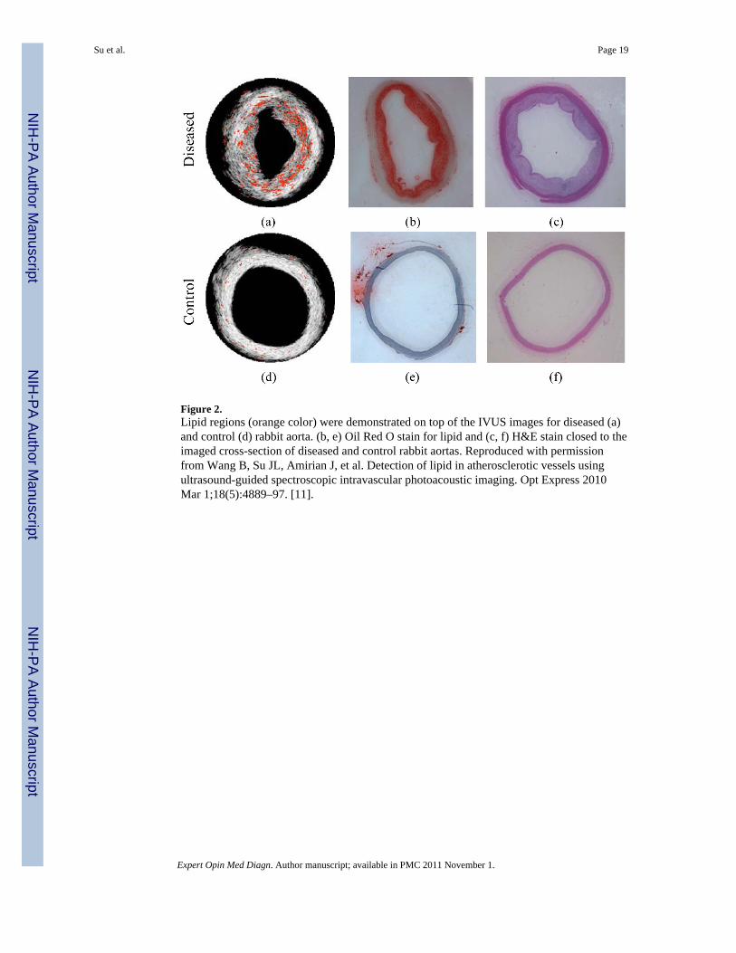
Figure 2.Lipid regions (orange color) were demonstrated on top of the IVUS images for diseased (a)and control (d) rabbit aorta. (b, e) Oil Red O stain for lipid and (c, f) H&E stain closed to theimaged cross-section of diseased and control rabbit aortas. Reproduced with permissionfrom Wang B, Su JL, Amirian J, et al. Detection of lipid in atherosclerotic vessels usingultrasound-guided spectroscopic intravascular photoacoustic imaging. Opt Express 2010Mar 1;18(5):4889–97. [11].
Su et al. Page 19
Expert Opin Med Diagn. Author manuscript; available in PMC 2011 November 1.
NIH
-PA Author Manuscript
NIH
-PA Author Manuscript
NIH
-PA Author Manuscript

Figure 3.(a) Silica-coated gold nanorods with enhanced thermal stability. Reproduced withpermission from[69]. (b) cTEM image of a dual ultrasound and photoacoustic contrastagent. Perfluorocarbon droplets loaded with silver nanotriangles. Reproduced withpermission from Wilson K, Homan K, Emelianov S. Synthesis of a dual contrast agent forultrasound and photoacoustic imaging. In: Samuel A, Ramesh R, editors. Reporters,Markers, Dyes, Nanoparticles, and Molecular Probes for Biomedical Applications II; 2010;San Francisco, CA, USA; 2010. p. 75760M [53]. (b) Silica-coated gold nanorods withenhanced thermal stability. Reproduced with permission from Chen YS, Frey W, Kim S, etal. Enhanced thermal stability of silica-coated gold nanorods for photoacoustic imaging andimage-guided therapy. Opt Express 2010 Apr 26;18(9):8867–78. [69].
Su et al. Page 20
Expert Opin Med Diagn. Author manuscript; available in PMC 2011 November 1.
NIH
-PA Author Manuscript
NIH
-PA Author Manuscript
NIH
-PA Author Manuscript

Figure 4.Darkfield, ultrasound and photoacoustic images (λ = 532 nm and 680 nm) of control,targeted and non-targeted tissue phantoms. The darkfield images measure 440 μm by 340μm field of view. The ultrasound and optoacoustic images measure 2 mm by 1.67 mm. Thetargeted EGFR cells show high photoacoustic signal due to the plasmon resonance couplingresulting from clustering gold nanoparticles. Reproduced from Mallidi S, Larson T, Aaron J,et al. Molecular specific optoacoustic imaging with plasmonic nanoparticles. Opt Express2007 May 28;15(11):6583–8 with permission of the Optical Society of America. [27]
Su et al. Page 21
Expert Opin Med Diagn. Author manuscript; available in PMC 2011 November 1.
NIH
-PA Author Manuscript
NIH
-PA Author Manuscript
NIH
-PA Author Manuscript

Figure 5.(a) Ultrasound, (b) photoacoustic and (c) thermal image of a subcutaneous tumor in nudemouse. Reprinted with permission from Mallidi S, Wang B, Mehrmohammadi M, et al.Ultrasound-based imaging of nanoparticles: from molecular and cellular imaging to therapyguidance. Proceedings of the 2009 IEEE Ultrasonics Symposium 2009:27–36 (2009) [81].
Su et al. Page 22
Expert Opin Med Diagn. Author manuscript; available in PMC 2011 November 1.
NIH
-PA Author Manuscript
NIH
-PA Author Manuscript
NIH
-PA Author Manuscript

Figure 6.Examples of several multiplexed nanoparticles used as an imaging contrast agent, as a drugdelivery vehicle, and/or for image-guided therapy. (a)_SEM image of hollow nanocagesused to contain therapy products. Reproduced with permission from Skrabalak SE, Chen J,Sun Y, et al. Gold nanocages: synthesis, properties, and applications. Acc Chem Res 2008Dec;41(12):1587–95 [86] (b) Gold nanoshells for use in photothermal applications.Reproduced with permission from Loo C, Lowery A, Halas N, et al. ImmunotargetedNanoshells for Integrated Cancer Imaging and Therapy. Nano Letters 2005;5(4):709–11.[87]. (c) Diagram demonstrating a multifunctional nanosystem platform utilizing a silvercage with a silica core. Reproduced with permission from Homan K, Shah J, Gomez S, et al.Silver nanosystems for photoacoustic imaging and image-guided therapy. J Biomed Opt2010 Mar-Apr;15(2):021316 [92]. (d) Silver coated PLGA as a carrier for imaging contrastagents. Image courtesy of K.A. Homan. All figures used with permission.
Su et al. Page 23
Expert Opin Med Diagn. Author manuscript; available in PMC 2011 November 1.
NIH
-PA Author Manuscript
NIH
-PA Author Manuscript
NIH
-PA Author Manuscript

Figure 7.3D-reconstruction of a stent embedded in a PVA phantom. Individual cross sections canshow the position of the stent within the vessel. (a) Ultrasound 3D reconstruction of thephantom showing the structure of the vessel. (b) Photoacoustic reconstruction of the stentstructure which can be used to assess the condition of the stent. (c) Photoacoustic image ofthe stent overlaid with the ultrasound image of the vessel can show the position of both. (d)Cutaway image of the reconstruction, allowing for accurate assessment of the stent withinthe vessel. Reproduced with permission from Su JL, Wang B, Emelianov SY. Photoacousticimaging of coronary artery stents. Opt Express 2009 Oct 26;17(22):19894–901 [97].
Su et al. Page 24
Expert Opin Med Diagn. Author manuscript; available in PMC 2011 November 1.
NIH
-PA Author Manuscript
NIH
-PA Author Manuscript
NIH
-PA Author Manuscript
Related Documents