d Original Contribution ADVANCED CHARACTERIZATION AND REFINEMENT OF POLY N-BUTYL CYANOACRYLATE MICROBUBBLES FOR ULTRASOUND IMAGING STANLEY FOKONG,* MONICA SIEPMANN, y ZHE LIU,* GEORG SCHMITZ, y F ABIAN KIESSLING,* and JESSICA G€ A TJENS* *Department of Experimental Molecular Imaging, Medical Faculty, RWTH Aachen University, Aachen, Germany; and y Department of Medical Engineering, Ruhr-University Bochum, Bochum, Germany (Received 16 February 2011; revised 30 June 2011; in final form 1 July 2011) Abstract—We aimed to develop and characterize poly n-butylcyanoacrylate (PBCA) microbubbles (MBs) with a narrow size distribution. MBs were synthesized by established emulsion polymerization techniques, size- isolated by centrifugation and functionalized for molecular imaging by coating their surface with streptavidin. The physical and acoustic properties of the parent solution, different-size isolated populations and functionalized MBs were measured and compared. As expected from negative zeta potentials at pH 7, cryo scanning electron microscopy showed no aggregates. In phantoms MBs were destructible at high mechanical indices and showed a frequency-dependent attenuation and backscattering. The MBs were stable in solution for more than 14 weeks and could be lyophilized without major damage. However, for injection, small needle diameters and high injection rates are shown to be critical because both lead to MB destruction. In summary, when being handled correctly, size-isolated PBCA MBs are promising candidates for preclinical functional and molecular ultrasound imaging. (E-mail: [email protected]) Ó 2011 World Federation for Ultrasound in Medicine & Biology. Key Words: Ultrasound, Contrast agents, Flow cytometry, Emulsion polymerization, Microbubbles, Poly n-butyl cyanoacrylate, Molecular imaging, Streptavidin, Bioconjugation. INTRODUCTION Ultrasound contrast enhancement by gas microbubbles (MBs) has been known since 1968 (Gramiak and Shah 1968). Contrast enhancement by these MBs is caused by high acoustic backscattering resulting from the acoustic impedance difference between the gas in the MBs and the tissue. The compressibility of MBs also causes nonlinear backscattering, which can be used for contrast-specific imaging. Over the years, engineering of gas MBs has led to their stabilization by shells made of proteins, phospholipids or polymers. The shell serves the purposes of: 1. Optimizing their pharmacokinetic properties, e.g., increasing the circulation time 2. Preventing the MBs from coalescing, which could lead to undesired physiological side effects (e.g., embolisms) 3. Presenting a site for modification and conjugation of target specific ligands 4. Optimizing the acoustic response of the MBs for contrast specific imaging In a bid to further increase the in vivo stability, low-solubility gases, mostly perfluorocarbons, have been used in place of air as the MB core (Cui et al. 2005). Gas-encapsulated MBs can be synthesized by either mechanical agitation, sonication or the use of microfluidic devices. Although mechanical agitation and sonication are the preferred methods to generate gas-encapsulated MBs, they generate highly polydisperse populations. MBs with a narrow size distribution have been mostly produced by microfluidic techniques (T-junction) (Pancholi et al. 2008), electrodynamic atom- ization (Farook et al. 2009), flow focussing (Talu et al. 2008a) and ink jet printing (B€ ohmer et al. 2006). Although these techniques produce MBs with a very narrow size distribution, they are more expensive and produce much less MBs per unit time compared with mechanical agitation and sonication. Alternatively, the isolation of a narrow size distribution of MBs from poly- dispersed parent solutions has been achieved by flotation (Kvale et al. 1996) and by differential centrifugation (Feshitan et al. 2009). Imaging with size-isolated MBs Address correspondence to: Dr. Jessica G€ atjens, Pauwelsstrasse 20,52074 Aachen, Germany. E-mail: [email protected] 1622 Ultrasound in Med. & Biol., Vol. 37, No. 10, pp. 1622–1634, 2011 Copyright Ó 2011 World Federation for Ultrasound in Medicine & Biology Printed in the USA. All rights reserved 0301-5629/$ - see front matter doi:10.1016/j.ultrasmedbio.2011.07.001

Welcome message from author
This document is posted to help you gain knowledge. Please leave a comment to let me know what you think about it! Share it to your friends and learn new things together.
Transcript
Ultrasound in Med. & Biol., Vol. 37, No. 10, pp. 1622–1634, 2011Copyright � 2011 World Federation for Ultrasound in Medicine & Biology
Printed in the USA. All rights reserved0301-5629/$ - see front matter
asmedbio.2011.07.001
doi:10.1016/j.ultrd Original Contribution
ADVANCED CHARACTERIZATION AND REFINEMENT OF POLY N-BUTYLCYANOACRYLATE MICROBUBBLES FOR ULTRASOUND IMAGING
STANLEY FOKONG,* MONICA SIEPMANN,y ZHE LIU,* GEORG SCHMITZ,y FABIAN KIESSLING,*and JESSICA G€ATJENS*
*Department of Experimental Molecular Imaging, Medical Faculty, RWTH Aachen University, Aachen, Germany; andyDepartment of Medical Engineering, Ruhr-University Bochum, Bochum, Germany
(Received 16 February 2011; revised 30 June 2011; in final form 1 July 2011)
A20,520
Abstract—We aimed to develop and characterize poly n-butylcyanoacrylate (PBCA) microbubbles (MBs) witha narrow size distribution. MBs were synthesized by established emulsion polymerization techniques, size-isolated by centrifugation and functionalized for molecular imaging by coating their surface with streptavidin.The physical and acoustic properties of the parent solution, different-size isolated populations and functionalizedMBs were measured and compared. As expected from negative zeta potentials at pH 7, cryo scanning electronmicroscopy showed no aggregates. In phantoms MBs were destructible at high mechanical indices and showeda frequency-dependent attenuation and backscattering. The MBs were stable in solution for more than 14 weeksand could be lyophilized without major damage. However, for injection, small needle diameters and high injectionrates are shown to be critical because both lead to MB destruction. In summary, when being handled correctly,size-isolated PBCA MBs are promising candidates for preclinical functional and molecular ultrasound imaging.(E-mail: [email protected]) � 2011 World Federation for Ultrasound in Medicine & Biology.
Key Words: Ultrasound, Contrast agents, Flow cytometry, Emulsion polymerization, Microbubbles, Poly n-butylcyanoacrylate, Molecular imaging, Streptavidin, Bioconjugation.
INTRODUCTION
Ultrasound contrast enhancement by gas microbubbles(MBs) has been known since 1968 (Gramiak and Shah1968). Contrast enhancement by these MBs is causedby high acoustic backscattering resulting from theacoustic impedance difference between the gas in theMBs and the tissue. The compressibility of MBs alsocauses nonlinear backscattering, which can be used forcontrast-specific imaging. Over the years, engineeringof gas MBs has led to their stabilization by shells madeof proteins, phospholipids or polymers.
The shell serves the purposes of:
1. Optimizing their pharmacokinetic properties, e.g.,increasing the circulation time
2. Preventing the MBs from coalescing, which couldlead to undesired physiological side effects (e.g.,embolisms)
3. Presenting a site for modification and conjugation oftarget specific ligands
ddress correspondence to: Dr. Jessica G€atjens, Pauwelsstrasse74 Aachen, Germany. E-mail: [email protected]
1622
4. Optimizing the acoustic response of the MBs forcontrast specific imaging
In a bid to further increase the in vivo stability,low-solubility gases, mostly perfluorocarbons, havebeen used in place of air as the MB core (Cui et al. 2005).
Gas-encapsulated MBs can be synthesized byeither mechanical agitation, sonication or the use ofmicrofluidic devices. Although mechanical agitationand sonication are the preferred methods to generategas-encapsulatedMBs, they generate highly polydispersepopulations. MBs with a narrow size distribution havebeen mostly produced by microfluidic techniques(T-junction) (Pancholi et al. 2008), electrodynamic atom-ization (Farook et al. 2009), flow focussing (Talu et al.2008a) and ink jet printing (B€ohmer et al. 2006).Although these techniques produce MBs with a verynarrow size distribution, they are more expensive andproduce much less MBs per unit time compared withmechanical agitation and sonication. Alternatively, theisolation of a narrow size distribution of MBs from poly-dispersed parent solutions has been achieved by flotation(Kvale et al. 1996) and by differential centrifugation(Feshitan et al. 2009). Imaging with size-isolated MBs
Refinement of PBCA MBs for US imaging d S. FOKONG et al. 1623
is advantageous because the size influences importantparameters such as their resonance frequency, backscatterintensities, pharmacokinetics, and biodistribution.
Depending on their shell properties, MBs have beenclassified into hard- (mostly protein and polymer) andsoft- (mostly lipid) shelled MBs (Kiessling et al. 2009).The stiffness of the shell is important because it definesthe behaviour of the MBs in the ultrasound field andthus the contrast enhancement effect by the MBs. Forexample, a soft-shelled MB made of phospholipids willoscillate more to ultrasound pressure waves than a hard-shelled MB. This will lead to more nonlinear ultrasoundbackscattering by the soft-shelled MB compared withtheir hard-shelled counterparts. On the other hand,hard-shelled MBs are in principle easier to destroy byhigh mechanical index ultrasound waves, making thembetter suited for destructive imaging than soft-shelledMBs. Furthermore, they often have a higher in vivo(intravasal) stability, which is important for their use asmolecular imaging agents.
In this study, hard-shelled (poly n-bultylcyanoacry-late [PBCA]) MBs with an air core were used. Theyhave been reported to be excellent candidates for prec-linical contrast-enhanced ultrasound perfusion imagingfor cardiovascular applications and in cancer diagnosis(Puls et al. 2000; Palmowski et al. 2008a, 2008b).These MBs are biocompatible and in vitro experimentsshowed their degradation depends on pH value andtemperature of the buffer solution in which they arestored. Enhanced degradation was also found in serum,most probably caused by enzymatic cleavage (Stein andHamacher 1992; Olbrich et al. 2006).
PBCAMBs carrying streptavidin on their surface, towhich biotinylated antibodies can be attached, have alsobeen used for molecular imaging of rodents (Joseph et al.2005; Palmowski et al. 2008a). Injection of these target-specific MBs in vivo leads to their accumulation in areaswhere the target markers are expressed. Quantification ofthe amount of bound MBs in a region gives a measure ofthe marker expression (Reinhardt et al. 2005; Siepmannet al. 2010).
However, PBCA MBs like every other MB synthe-sized by mechanical agitation or sonication, are polydis-perse. Because of this, only a small amount of MBs willshow suitable acoustic characteristics such as the optimalresonance frequency. For example, Hoff et al. (2000)reported an increase in resonance frequency withdecreasing MB diameter for polymer-shelled MBs.Because MBs give a maximum backscatter signal at theirresonance frequency, MBs with a resonance frequencyaround that of the transducer are desired. Furthermore,the pharmacokinetics, gas release profile, induced bioef-fects, dose of injected MB to contrast enhancement ratio,as well as the biodistribution after intravenous injection
of the MBs also depend on their size, thus necessitatingthe use of size-isolated MBs for biomedical applications(Talu et al. 2007).
Apart from the size distribution, a detailed (chemi-cal, physical and acoustic) characterization of PBCAMBs has never been reported, which is important toimprove their contrast enhancement effect and to opti-mize their binding kinetics, which is particularly crucialfor molecular imaging.
Therefore, it was the aim of this study to develop andcharacterize PBCA MBs with a narrow size distributionfor preclinical functional and molecular ultrasoundimaging. After synthesis, they were size-isolated bycentrifugation. The effects of centrifugation on theMBs’ size distribution and on their acoustic properties(resonance frequency and backscattering) were investi-gated. The amount of streptavidin molecules on thesurface of MBs functionalized for molecular imagingwas also investigated by quantitative flow cytometry. Ifthe development of MBs for clinical use is intended, theirstorability becomes an important quality characteristic,which is often neglected. The stability of PBCA MBs insolution as well as the effects of freeze-drying (for storagepurposes) on the MBs were therefore analyzed. Theirpersistence during insonication and their stability whenbeing injected through cannulae with different innerdiameters at different velocities were also studied.
MATERIALS AND METHODS
Materialsn-butylcyanoacrylate (BCA) was purchased from
Sichel Werke (Henkel Sichel Werke GmbH, Hannover,Germany), Triton X-100 and Polyvinylpyrrolidon(PVP) from Sigma-Aldrich (Munich, Germany), gelatinefrom Merck (Darmstadt, Germany) and Dulbecco’sphosphate-buffered saline (PBS) was obtained fromLonza (Walkersville, MD, USA). Deionized water wasused for all experiments and all chemicals were usedwithout any further purification.
Microbubble synthesisPBCA-stabilized air-filled MBs were prepared by
established emulsion polymerization techniques asdescribed in Palmowski et al. (2008b). After synthesis,the MBs were purified by flotation (which usually takes6 d) and their size distribution measured using a Multi-sizer 3 (Beckmann Coulter GmbH, Krefeld, Germany).
Target-specific MBs for molecular imaging wereprepared by coupling streptavidin to the surface of pre-formed PBCA MBs as reported in Joseph et al. (2005).In short, a 10% hydrolysis of PBCA MBs was performedusing 0.1 N NaOH solution. 53108 of the hydrolysedMBs were diluted in 15 mL sodium acetate buffer
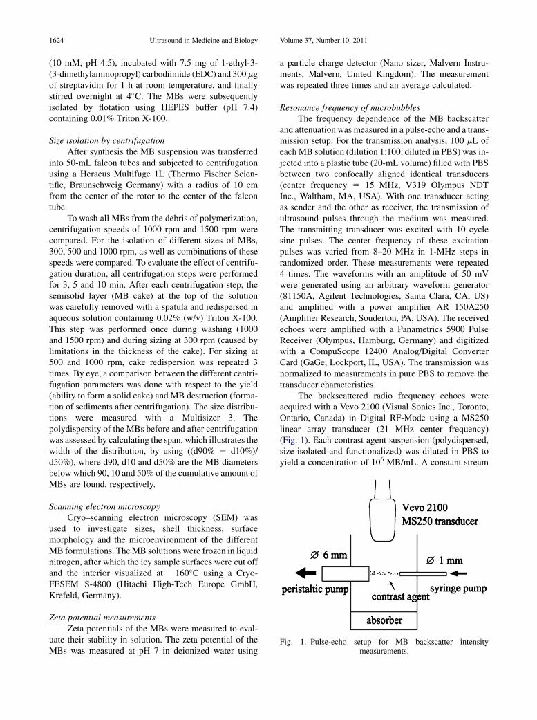
Fig. 1. Pulse-echo setup for MB backscatter intensitymeasurements.
1624 Ultrasound in Medicine and Biology Volume 37, Number 10, 2011
(10 mM, pH 4.5), incubated with 7.5 mg of 1-ethyl-3-(3-dimethylaminopropyl) carbodiimide (EDC) and 300 mgof streptavidin for 1 h at room temperature, and finallystirred overnight at 4�C. The MBs were subsequentlyisolated by flotation using HEPES buffer (pH 7.4)containing 0.01% Triton X-100.
Size isolation by centrifugationAfter synthesis the MB suspension was transferred
into 50-mL falcon tubes and subjected to centrifugationusing a Heraeus Multifuge 1L (Thermo Fischer Scien-tific, Braunschweig Germany) with a radius of 10 cmfrom the center of the rotor to the center of the falcontube.
To wash all MBs from the debris of polymerization,centrifugation speeds of 1000 rpm and 1500 rpm werecompared. For the isolation of different sizes of MBs,300, 500 and 1000 rpm, as well as combinations of thesespeeds were compared. To evaluate the effect of centrifu-gation duration, all centrifugation steps were performedfor 3, 5 and 10 min. After each centrifugation step, thesemisolid layer (MB cake) at the top of the solutionwas carefully removed with a spatula and redispersed inaqueous solution containing 0.02% (w/v) Triton X-100.This step was performed once during washing (1000and 1500 rpm) and during sizing at 300 rpm (caused bylimitations in the thickness of the cake). For sizing at500 and 1000 rpm, cake redispersion was repeated 3times. By eye, a comparison between the different centri-fugation parameters was done with respect to the yield(ability to form a solid cake) and MB destruction (forma-tion of sediments after centrifugation). The size distribu-tions were measured with a Multisizer 3. Thepolydispersity of the MBs before and after centrifugationwas assessed by calculating the span, which illustrates thewidth of the distribution, by using ((d90% 2 d10%)/d50%), where d90, d10 and d50% are the MB diametersbelow which 90, 10 and 50% of the cumulative amount ofMBs are found, respectively.
Scanning electron microscopyCryo–scanning electron microscopy (SEM) was
used to investigate sizes, shell thickness, surfacemorphology and the microenvironment of the differentMB formulations. TheMB solutions were frozen in liquidnitrogen, after which the icy sample surfaces were cut offand the interior visualized at 2160�C using a Cryo-FESEM S-4800 (Hitachi High-Tech Europe GmbH,Krefeld, Germany).
Zeta potential measurementsZeta potentials of the MBs were measured to eval-
uate their stability in solution. The zeta potential of theMBs was measured at pH 7 in deionized water using
a particle charge detector (Nano sizer, Malvern Instru-ments, Malvern, United Kingdom). The measurementwas repeated three times and an average calculated.
Resonance frequency of microbubblesThe frequency dependence of the MB backscatter
and attenuation was measured in a pulse-echo and a trans-mission setup. For the transmission analysis, 100 mL ofeach MB solution (dilution 1:100, diluted in PBS) was in-jected into a plastic tube (20-mL volume) filled with PBSbetween two confocally aligned identical transducers(center frequency 5 15 MHz, V319 Olympus NDTInc., Waltham, MA, USA). With one transducer actingas sender and the other as receiver, the transmission ofultrasound pulses through the medium was measured.The transmitting transducer was excited with 10 cyclesine pulses. The center frequency of these excitationpulses was varied from 8–20 MHz in 1-MHz steps inrandomized order. These measurements were repeated4 times. The waveforms with an amplitude of 50 mVwere generated using an arbitrary waveform generator(81150A, Agilent Technologies, Santa Clara, CA, US)and amplified with a power amplifier AR 150A250(Amplifier Research, Souderton, PA, USA). The receivedechoes were amplified with a Panametrics 5900 PulseReceiver (Olympus, Hamburg, Germany) and digitizedwith a CompuScope 12400 Analog/Digital ConverterCard (GaGe, Lockport, IL, USA). The transmission wasnormalized to measurements in pure PBS to remove thetransducer characteristics.
The backscattered radio frequency echoes wereacquired with a Vevo 2100 (Visual Sonics Inc., Toronto,Ontario, Canada) in Digital RF-Mode using a MS250linear array transducer (21 MHz center frequency)(Fig. 1). Each contrast agent suspension (polydispersed,size-isolated and functionalized) was diluted in PBS toyield a concentration of 106 MB/mL. A constant stream
Refinement of PBCA MBs for US imaging d S. FOKONG et al. 1625
of agent was generated in a water tank using two differentpumps: a syringe pump (Aitecs 2016, Aitecs, Vilnius,Lithuania) supplies agent suspension at a flow rate of200 mL/h through a 1-mm inner diameter glass tube, anda peristaltic pump (Ecoline VC-Easy-Load, Ismatec,Wertheim-Mondfeld, Germany) draws fluid at 110mL/minthrough a 6-mm-diameter inlet. The outlet and inlet of thepumps were spaced 1 cm apart. Using this setup, the agentresponse can be measured without any attenuating tubing.The imaging plane was positioned perpendicular to theflow direction with the transducer focus, set to 13 mm (inthe center of the flow). Five hundred frames were acquiredin B-mode at 21-MHz transmit frequency and 10% or 1%power to prevent substantial MB destruction. A referencemeasurement was performed with 1 g of silica gel particles(Merck Chemicals KG, Darmstadt, Germany) with anaverage particle size of 15 mm diluted in 100 mL PBS.All experiments were repeated five times. For each MBtype, the spectra of the MB echoes were calculated andtheir magnitude averaged. The results are then normalizedto the reference to remove the system characteristics.
Stability in solutionPolydispersed and size-isolated MBs were stored in
0.02% Triton X-100 solution, both at room temperatureand at 4�C. The mean diameters of the samples weremeasured with the Multisizer weekly over a period of14 weeks.
Changes in size distribution of the MBs over timewere investigated, elucidating effects like MB destructionand gas loss, which could occur forMBs stored in solutionover prolonged periods of time. All MB solutions weregently mixed to ensure a homogenous solution beforemeasurements. For each sample, the measurements wereperformed in triplicate and an average was calculated.Functionalized MBs are usually prepared in small quanti-ties before use, making their stability in solution overextended periods of time irrelevant for investigation.
Freeze-drying of microbubblesMB storage as powder is desired because it can
substantially increase the storage time, make it easier tokeep the MBs sterile, and reduces the effects of MBdestruction and gas loss, which could take place in MBsstored in solution. 10% (w/w) polyvinylpyrrolidone(Sigma-Aldrich) were therefore added to the MB solu-tion, and the mixture was frozen in liquid nitrogen. Thesample was subsequently freeze-dried (Lyovac GT2,Amsco Finn-Aqua, Huerth, Germany). The dry samplewas redispersed using isotonic sodium chloride solution(Deltaselect, Pfullingen, Germany) and the MB popula-tion distribution measured using a Multisizer 3 andcompared with that before freeze-drying. Changes infrequency dependence of the acoustic response were
compared using the backscatter setup for resonancemeasurements described before. To avoid the influenceof MB destruction during the measurement, the reductionin contrast intensity for different ultrasound powers wasinvestigated. Therefore, the same concentration of MB aswas used for the resonance measurements (106 MB/mL)was injected in Ibidi m-slides (Ibidi, Martinsried,Germany) and the chamber was placed in the transducerfocus. The reduction in contrast intensity after 100 frameswas measured to investigate contrast agent destruction.Furthermore, we compared the results for redispersedMBs (after freeze-drying) with size-isolatedMBs (havingcomparable mean diameters and size distribution) thatdid not undergo the freeze-drying process.
Evaluation of MB stabilityTo confirm the MB-induced contrast enhancement
and to investigate the dependency of PBCA MB destruc-tion with ultrasound power, ultrasound imaging (linearcontrast mode) was performed on a 2.5% (w/v) gelatinphantom loaded with size-isolated and functionalizedPBCA MBs. The floor of the phantom was coveredwith a piece of cloth to reduce acoustic reflections fromthe bottom. Ultrasound imaging was performed usingthe preclinical ultrasound scanner Vevo 770 (VisualSon-ics Inc., Toronto, Ontario, Canada) with a 25-MHz trans-ducer at powers between 3% and 79%. A predefinedimaging sequence made continuous images for 30 s afterwhich it applied a destructive pulse (high mechanicalindex) and measured a reference signal. For differentportions of the phantom, different ultrasound powerswere used for imaging. An identical region of interest(25.11 mm2) drawn at the focus of the transducer wasused for plotting the contrast intensity over time curves,for the different powers used for isonication.
Effect of injection parametersIn this experiment, the numbers of MBs (size-
isolated and functionalized) before and after transitthrough needles with different inner diameters atdifferent flow velocities were investigated. The MBsolution was diluted using isotonic NaCl solution (Del-taselect GmbH, Pfullingen, Germany) to a concentra-tion of 1 3 107 MB/mL. Using a perfusor (B. Braun,Melsungen, Germany), the MB solution was injectedat speeds of 10, 30, 50, 70 and 90 mL/h through 25-,27- and 30-G needles connected to a 6-inch catheter.The size distribution and concentration of the MBsolution were measured three times before and afterinjection using the Multisizer 3.
Flow cytometryFlow cytometry was used to quantify the amount of
streptavidin molecules on the surface of the MBs that
1626 Ultrasound in Medicine and Biology Volume 37, Number 10, 2011
were functionalized for molecular imaging. Fifty mLof streptavidin-coated MBs (5 3 108 MB/mL) wereincubated with different concentrations (1, 5, 25 and150 ng/mL) of biotinylated fluoresceinisothiocyanate(Biotin-FITC) molecules for 20 min, washed by centrifu-gation using a Heraeus Fresco 21 (Thermo Fischer Scien-tific, Braunschweig, Germany) (2000 rpm for 2 min) andsubjected to flow cytometry using a BD fluorescenceactivated cell sorter (FACS) Calibur (BD Biosciences,Heidelberg, Germany). Using the same instrumentsettings, eight peak rainbow calibration particles (BDBiosciences) with a known amount of fluorescence(molecules of equivalent fluorescein [MEFL]) were alsomeasured. The calibration particles solution consistedof beads with diameters between 3 and 3.4 mm, whichwere dyed to eight known different fluorescence intensi-ties. The mean fluorescence channel number (MFCN) foreach fluorescence intensity on the calibration beads wasdetermined from its FACS histograms and used for plot-ting a calibration line with the MEFL (given by the manu-facturer of the calibration particles) on the particles.Using this calibration plot, and given the MFCN of theMBs from their measurements, the MEFL on the MBscould be determined and translated to the number ofstreptavidin molecules.
Statistical analysisDependency between injection speed and MB count
was analyzed using Pearson’s correlation. The effect ofthe needle diameter on the MB count was tested witha two-tailed paired t-test. Unless otherwise stated,p-values , 0.05 were considered statistically significant.
RESULTS
Synthesis and size isolation by centrifugationThe centrifugation time influenced the size of the
solid layer (cake) and the amount of sediments. Threeminutes were insufficient to yield a cake independent ofthe centrifugation speed. Centrifugation for 5 min onlyyielded a cake at 1000 and 1500 rpm. However, smallamounts of sediment were observed at all speeds except300 rpm. Centrifugation for 10 min resulted in increasedsedimentation, which was strongest at 1500 rpm. Never-theless, for all speeds, 10 min of centrifugation yieldedthe largest MB cake. Therefore, all experiments on sizeisolation were performed with 10-min centrifugation. Aslight shift toward smaller MBs after centrifugation with1500 rpm compared with 1000 rpm was observed(Fig. 2a). MB size distributions after centrifugation with500 and 1000 rpm were not substantially different(Table 1e, 1f), whereas 300 rpm generated larger MBs(Table 1a, distribution shown in Fig. 2b).
The infranatant obtained by centrifugation at 300 rpm(point A) is freed of the largest MBs and may now be usedto isolate smaller populations. This is achieved by addi-tional centrifugation at 500 rpm (point B) taking eitherthe cake (Table 1b) or the infranatant (Table 1g,Fig. 2b). Size isolation at 500 rpm after a washing stepat 1000 rpm generated slightly larger MBs (mean diam-eter in mm md 5 1.89 6 0.45, SPAN 5 0.54) comparedwith a prewashing at 1500 rpm (md 5 1.71 6 0.45,SPAN 5 0.63) and no washing (md 5 1.50 6 0.44SPAN 5 0.75) (Table 1c, 1d, 1f; Fig. 2c). Similar resultswere observed for 300 rpm (data not shown).
Figure 2d shows the size distributions of theMBs usedin the rest of the study consisting of the parent MB solution(prepared by mechanical agitation and flotation, whichtook 6 d, md 5 1.50 6 0.78, SPAN 5 1.00) comparedwith size-isolatedMBs (took 4 h for synthesis and purifica-tion by centrifugation, md 5 2.00 6 0.41, SPAN 5 0.56).The narrow size distribution (SPAN 5 0.54) was main-tained even after coating the MBs with streptavidin(functionalized MBs).
Scanning electron microscopyPolydispersed MBs had a broader size distribution
compared with size-isolated MBs (Fig. 3), and noaggregation was observed for both MB solutions. Highermagnification images showed a rough and seeminglytight shell structure with a thickness of approximately50 nm. No visible differences were found betweensize-isolated and functionalized MBs (data not shown),as well as between the shell structure of the differentMB formulations.
Resonance frequencyA frequency-dependent transmission was obser-
ved for the PBCA MB solutions with a minimum at11 MHz for the size-isolated and functionalized MBs,and 10 MHz for the polydispersed MB suspension(Fig. 4). The dependency of backscatter intensity onfrequency exhibited similar characteristics: The peakintensity occurred at 10 MHz for the larger (�2 mm)size-isolated MBs, and around 9.5 MHz for the polydis-persed and functionalized MBs (Fig. 5). An increase inbackscattered intensity is observed for both the size-isolated and the functionalized MBs compared with thepolydispersed MBs.
Stability in solutionOver a period of 14 weeks, very little variations in
the mean MB diameters were observed for the polydis-persed and size-isolated MBs stored in solution atdifferent temperatures (Table 2). There was also nosubstantial change in the MB concentration and noobservation of sediments over this time period.
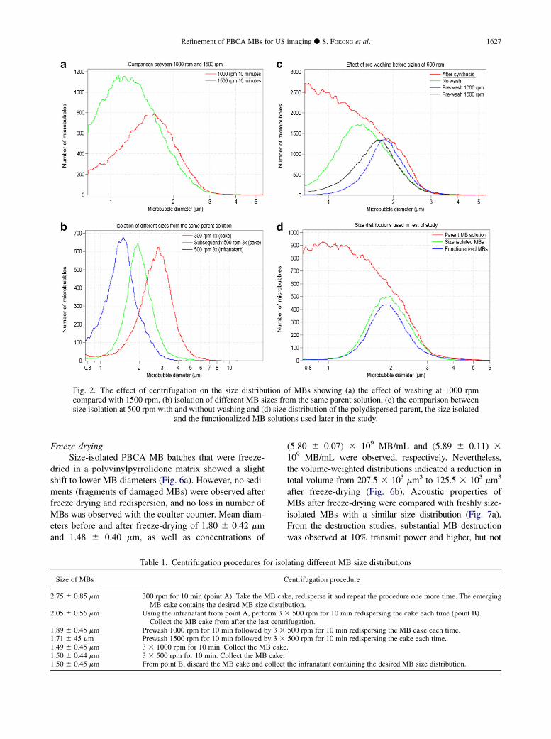
Fig. 2. The effect of centrifugation on the size distribution of MBs showing (a) the effect of washing at 1000 rpmcompared with 1500 rpm, (b) isolation of different MB sizes from the same parent solution, (c) the comparison betweensize isolation at 500 rpm with and without washing and (d) size distribution of the polydispersed parent, the size isolated
and the functionalized MB solutions used later in the study.
Refinement of PBCA MBs for US imaging d S. FOKONG et al. 1627
Freeze-dryingSize-isolated PBCA MB batches that were freeze-
dried in a polyvinylpyrrolidone matrix showed a slightshift to lower MB diameters (Fig. 6a). However, no sedi-ments (fragments of damaged MBs) were observed afterfreeze drying and redispersion, and no loss in number ofMBs was observed with the coulter counter. Mean diam-eters before and after freeze-drying of 1.80 6 0.42 mmand 1.48 6 0.40 mm, as well as concentrations of
Table 1. Centrifugation procedures for iso
Size of MBs C
2.75 6 0.85 mm 300 rpm for 10 min (point A). Take the MB cakMB cake contains the desired MB size distrib
2.05 6 0.56 mm Using the infranatant from point A, perform 3 3Collect the MB cake from after the last centr
1.89 6 0.45 mm Prewash 1000 rpm for 10 min followed by 3 31.71 6 45 mm Prewash 1500 rpm for 10 min followed by 3 31.49 6 0.45 mm 3 3 1000 rpm for 10 min. Collect the MB cake1.50 6 0.44 mm 3 3 500 rpm for 10 min. Collect the MB cake.1.50 6 0.45 mm From point B, discard the MB cake and collect
(5.80 6 0.07) 3 109 MB/mL and (5.89 6 0.11) 3109 MB/mL were observed, respectively. Nevertheless,the volume-weighted distributions indicated a reduction intotal volume from 207.5 3 103 mm3 to 125.5 3 103 mm3
after freeze-drying (Fig. 6b). Acoustic properties ofMBs after freeze-drying were compared with freshly size-isolated MBs with a similar size distribution (Fig. 7a).From the destruction studies, substantial MB destructionwas observed at 10% transmit power and higher, but not
lating different MB size distributions
entrifugation procedure
e, redisperse it and repeat the procedure one more time. The emergingution.500 rpm for 10 min redispersing the cake each time (point B).
ifugation.500 rpm for 10 min redispersing the MB cake each time.500 rpm for 10 min redispersing the cake each time..
the infranatant containing the desired MB size distribution.
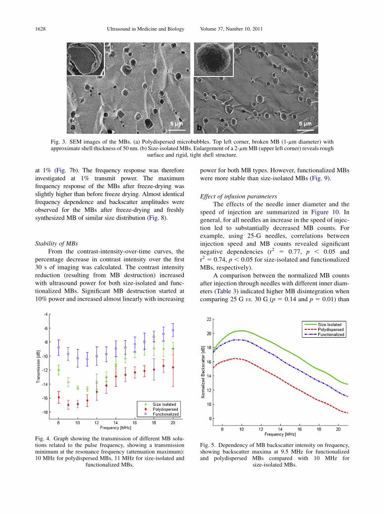
Fig. 3. SEM images of the MBs. (a) Polydispersed microbubbles. Top left corner, broken MB (1-mm diameter) withapproximate shell thickness of 50 nm. (b) Size-isolatedMBs. Enlargement of a 2-mmMB (upper left corner) reveals rough
surface and rigid, tight shell structure.
1628 Ultrasound in Medicine and Biology Volume 37, Number 10, 2011
at 1% (Fig. 7b). The frequency response was thereforeinvestigated at 1% transmit power. The maximumfrequency response of the MBs after freeze-drying wasslightly higher than before freeze drying. Almost identicalfrequency dependence and backscatter amplitudes wereobserved for the MBs after freeze-drying and freshlysynthesized MB of similar size distribution (Fig. 8).
Stability of MBsFrom the contrast-intensity-over-time curves, the
percentage decrease in contrast intensity over the first30 s of imaging was calculated. The contrast intensityreduction (resulting from MB destruction) increasedwith ultrasound power for both size-isolated and func-tionalized MBs. Significant MB destruction started at10% power and increased almost linearly with increasing
Fig. 4. Graph showing the transmission of different MB solu-tions related to the pulse frequency, showing a transmissionminimum at the resonance frequency (attenuation maximum):10 MHz for polydispersed MBs, 11 MHz for size-isolated and
functionalized MBs.
power for both MB types. However, functionalized MBswere more stable than size-isolated MBs (Fig. 9).
Effect of infusion parametersThe effects of the needle inner diameter and the
speed of injection are summarized in Figure 10. Ingeneral, for all needles an increase in the speed of injec-tion led to substantially decreased MB counts. Forexample, using 25-G needles, correlations betweeninjection speed and MB counts revealed significantnegative dependencies (r2 5 0.77, p , 0.05 andr2 5 0.74, p , 0.05 for size-isolated and functionalizedMBs, respectively).
A comparison between the normalized MB countsafter injection through needles with different inner diam-eters (Table 3) indicated higher MB disintegration whencomparing 25 G vs. 30 G (p 5 0.14 and p 5 0.01) than
Fig. 5. Dependency of MB backscatter intensity on frequency,showing backscatter maxima at 9.5 MHz for functionalizedand polydispersed MBs compared with 10 MHz for
size-isolated MBs.
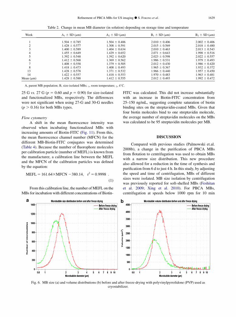
Table 2. Change in mean MB diameter (in solution) depending on storage time and temperature
Week A1 6 SD (mm) A2 6 SD (mm) B1 6 SD (mm) B2 6 SD (mm)
1 1.504 6 0.785 1.504 6 0.406 2.010 6 0.406 2.002 6 0.4062 1.428 6 0.577 1.308 6 0.591 2.015 6 0.569 2.018 6 0.4803 1.400 6 0.589 1.404 6 0.634 2.010 6 0.463 2.013 6 0.5434 1.455 6 0.649 1.429 6 0.652 2.071 6 0.643 1.998 6 0.5165 1.392 6 0.548 1.392 6 0.620 2.023 6 0.598 2.022 6 0.5576 1.412 6 0.568 1.369 6 0.562 1.986 6 0.531 1.979 6 0.4937 1.408 6 0.558 1.379 6 0.505 2.012 6 0.430 1.986 6 0.4208 1.418 6 0.473 1.408 6 0.493 1.965 6 0.367 1.932 6 0.37213 1.428 6 0.578 1.371 6 0.532 1.966 6 0.440 1.957 6 0.45014 1.422 6 0.557 1.418 6 0.553 1.970 6 0.483 1.963 6 0.481
Mean (mm) 1.428 6 0.588 1.412 6 0.555 2.012 6 0.493 1.992 6 0.472
A, parent MB population; B, size-isolated MBs; 1, room temperature; 2, 4�C.
Refinement of PBCA MBs for US imaging d S. FOKONG et al. 1629
25 G vs. 27 G (p 5 0.60 and p 5 0.90) for size-isolatedand functionalized MBs, respectively. The differenceswere not significant when using 27-G and 30-G needles(p . 0.16) for both MBs types.
Flow cytometryA shift in the mean fluorescence intensity was
observed when incubating functionalized MBs withincreasing amounts of Biotin-FITC (Fig. 11). From this,the mean fluorescence channel number (MFCN) for thedifferent MB-Biotin-FITC conjugates was determined(Table 4). Because the number of fluorophore moleculesper calibration particle (number of MEFL) is known fromthe manufacturer, a calibration line between the MEFLand the MFCN of the calibration particles was definedby the equation:
MEFL5 161:643MFCN 2380:14;�r2 5 0:9998
�:
(1)
From this calibration line, the number ofMEFL on theMBs for incubation with different concentrations of Biotin-
Fig. 6. MB size (a) and volume distributions (b) before and aftecryostabil
FITC was calculated. This did not increase substantiallywith an increase in Biotin-FITC concentration from25–150 ng/mL, suggesting complete saturation of biotinbinding sites on the streptavidin-coated MBs. Given thatfour biotin molecules bind to one streptavidin molecule,the average number of streptavidin molecules on the MBswas calculated to be 95 streptavidin molecules per MB.
DISCUSSION
Compared with previous studies (Palmowski et.al.2008b), a change in the purification of PBCA MBsfrom flotation to centrifugation was used to obtain MBswith a narrow size distribution. This new procedurealso allowed for a reduction in the time of synthesis andpurification from 6 d to just 4 h. In this study, by adjustingthe speed and time of centrifugation, MBs of differentsizes were isolated. MB size isolation by centrifugationwas previously reported for soft-shelled MBs (Feshitanet al. 2009; Xing et al. 2010). For PBCA MBs,centrifugation at speeds below 1000 rpm for 10 min
r freeze-drying with polyvinylpyrrolidone (PVP) used asizer.
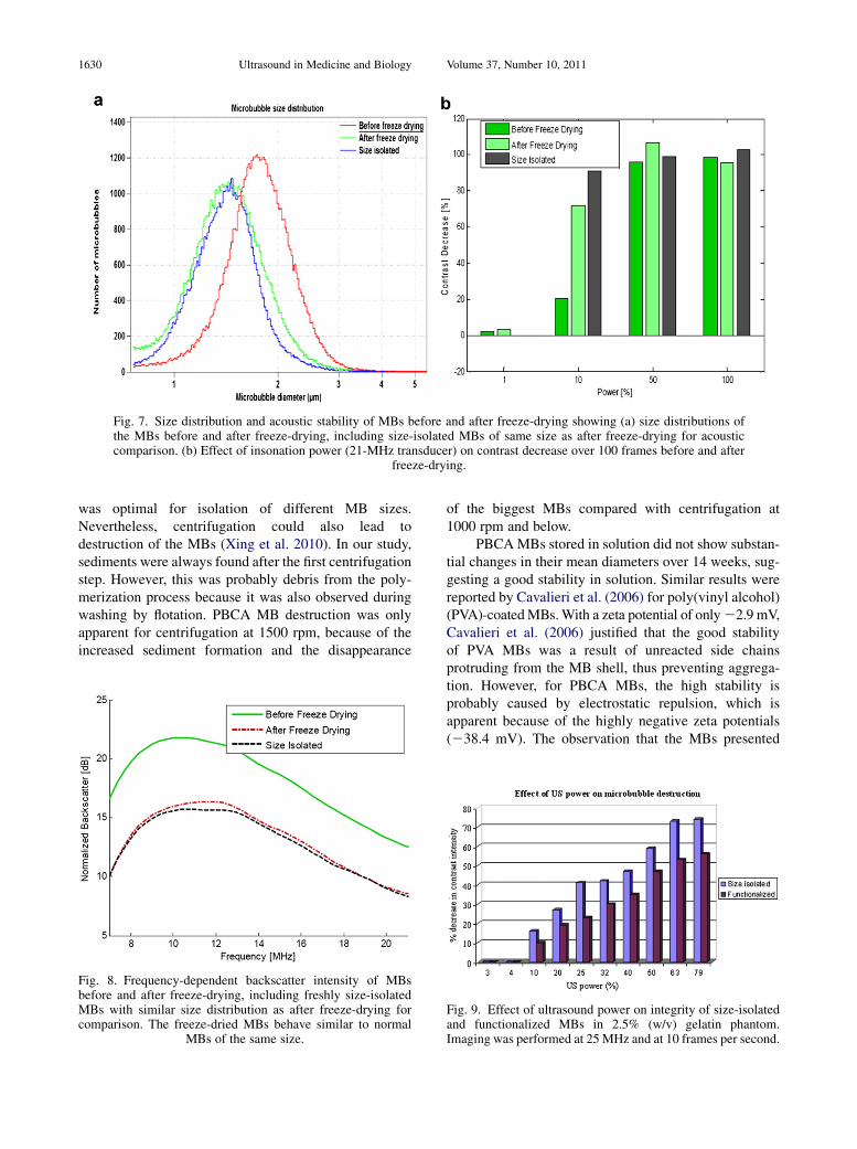
Fig. 7. Size distribution and acoustic stability of MBs before and after freeze-drying showing (a) size distributions ofthe MBs before and after freeze-drying, including size-isolated MBs of same size as after freeze-drying for acousticcomparison. (b) Effect of insonation power (21-MHz transducer) on contrast decrease over 100 frames before and after
freeze-drying.
1630 Ultrasound in Medicine and Biology Volume 37, Number 10, 2011
was optimal for isolation of different MB sizes.Nevertheless, centrifugation could also lead todestruction of the MBs (Xing et al. 2010). In our study,sediments were always found after the first centrifugationstep. However, this was probably debris from the poly-merization process because it was also observed duringwashing by flotation. PBCA MB destruction was onlyapparent for centrifugation at 1500 rpm, because of theincreased sediment formation and the disappearance
Fig. 8. Frequency-dependent backscatter intensity of MBsbefore and after freeze-drying, including freshly size-isolatedMBs with similar size distribution as after freeze-drying forcomparison. The freeze-dried MBs behave similar to normal
MBs of the same size.
of the biggest MBs compared with centrifugation at1000 rpm and below.
PBCAMBs stored in solution did not show substan-tial changes in their mean diameters over 14 weeks, sug-gesting a good stability in solution. Similar results werereported by Cavalieri et al. (2006) for poly(vinyl alcohol)(PVA)-coatedMBs.With a zeta potential of only22.9 mV,Cavalieri et al. (2006) justified that the good stabilityof PVA MBs was a result of unreacted side chainsprotruding from the MB shell, thus preventing aggrega-tion. However, for PBCA MBs, the high stability isprobably caused by electrostatic repulsion, which isapparent because of the highly negative zeta potentials(238.4 mV). The observation that the MBs presented
Fig. 9. Effect of ultrasound power on integrity of size-isolatedand functionalized MBs in 2.5% (w/v) gelatin phantom.Imaging was performed at 25 MHz and at 10 frames per second.
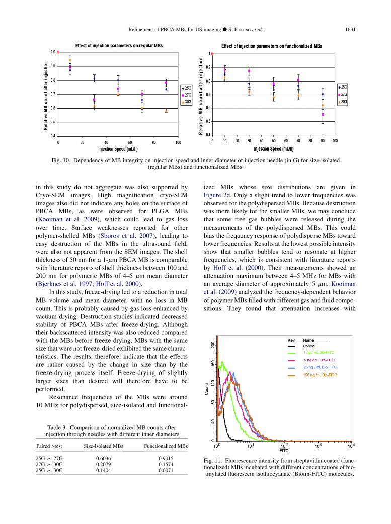
Fig. 10. Dependency of MB integrity on injection speed and inner diameter of injection needle (in G) for size-isolated(regular MBs) and functionalized MBs.
Refinement of PBCA MBs for US imaging d S. FOKONG et al. 1631
in this study do not aggregate was also supported byCryo-SEM images. High magnification cryo-SEMimages also did not indicate any holes on the surface ofPBCA MBs, as were observed for PLGA MBs(Kooiman et al. 2009), which could lead to gas lossover time. Surface weaknesses reported for otherpolymer-shelled MBs (Sboros et al. 2007), leading toeasy destruction of the MBs in the ultrasound field,were also not apparent from the SEM images. The shellthickness of 50 nm for a 1-mm PBCA MB is comparablewith literature reports of shell thickness between 100 and200 nm for polymeric MBs of 4–5 mm mean diameter(Bjerknes et al. 1997; Hoff et al. 2000).
In this study, freeze-drying led to a reduction in totalMB volume and mean diameter, with no loss in MBcount. This is probably caused by gas loss enhanced byvacuum-drying. Destruction studies indicated decreasedstability of PBCA MBs after freeze-drying. Althoughtheir backscattered intensity was also reduced comparedwith the MBs before freeze-drying, MBs with the samesize that were not freeze-dried exhibited the same charac-teristics. The results, therefore, indicate that the effectsare rather caused by the change in size than by thefreeze-drying process itself. Freeze-drying of slightlylarger sizes than desired will therefore have to beperformed.
Resonance frequencies of the MBs were around10 MHz for polydispersed, size-isolated and functional-
Table 3. Comparison of normalized MB counts afterinjection through needles with different inner diameters
Paired t-test Size-isolated MBs Functionalized MBs
25G vs. 27G 0.6036 0.901527G vs. 30G 0.2079 0.157425G vs. 30G 0.1404 0.0071
ized MBs whose size distributions are given inFigure 2d. Only a slight trend to lower frequencies wasobserved for the polydispersed MBs. Because destructionwas more likely for the smaller MBs, we may concludethat some free gas bubbles were released during themeasurements of the polydispersed MBs. This couldbias the frequency response of polydisperse MBs towardlower frequencies. Results at the lowest possible intensityshow that smaller bubbles tend to resonate at higherfrequencies, which is consistent with literature reportsby Hoff et al. (2000). Their measurements showed anattenuation maximum between 4–5 MHz for MBs withan average diameter of approximately 5 mm. Kooimanet al. (2009) analyzed the frequency-dependent behaviorof polymerMBs filled with different gas and fluid compo-sitions. They found that attenuation increases with
Fig. 11. Fluorescence intensity from streptavidin-coated (func-tionalized) MBs incubated with different concentrations of bio-tinylated fluorescein isothiocyanate (Biotin-FITC) molecules.
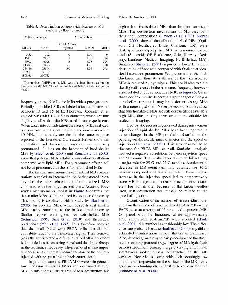
Table 4. Determination of streptavidin loading on MBsurfaces by flow cytometry
Calibration beads Microbubbles
MFCN MEFLBio-FITC conc.
(ng/mL) MFCN MEFL
5.32 692 0 1.09 015.03 2192 1 2.50 2439.03 6028 5 3.75 226113.82 17493 25 4.70 380234.89 35674 150 4.78 393769.1 1269071808.63 290983
The number of MEFL on the MBs was calculated from a calibrationline between the MFCN and the number of MEFL of the calibrationbeads.
1632 Ultrasound in Medicine and Biology Volume 37, Number 10, 2011
frequency up to 15 MHz for MBs with a pure gas core.Partially fluid-filled MBs exhibited attenuation maximabetween 10 and 15 MHz. However, Kooiman et al.studied MBs with 1.2–1.3-mm diameter, which are thusslightly smaller than the MBs used in our experiments.When taken into consideration the sizes ofMBs analyzed,one can say that the attenuation maxima observed at10 MHz in this study are thus in the same range asreported in the literature. Our results further show thatattenuation and backscatter maxima are not verypronounced. Studies on the behavior of hard-shelledMBs by Bloch et al. (2004) and Bouakaz et al. (2005)show that polymer MBs exhibit lower radius oscillationscompared with lipid MBs. Thus, resonance effects willnot be as pronounced as those for soft-shelled MBs.
Backscatter measurements of identical MB concen-trations revealed an increase in the backscattered inten-sity for the size-isolated and functionalized MBscompared with the polydispersed ones. Acoustic back-scatter measurements shown in Figure 8 confirm thatthe smaller MBs exhibit reduced backscattered intensity.This finding is consistent with a study by Bloch et al.(2005) on polymer MBs, which suggests that smallerMBs hardly contribute to the backscattered intensity.Similar reports were given for soft-shelled MBs(Schneider 1999; Sirsi et al. 2010) and theoreticalpredictions (Man et al. 1997). It is therefore possiblethat the small (,1.5 mm) PBCA MBs also did notcontribute much to the backscatter signal. Their removal(as in the size-isolated and functionalized MBs) thereforeled to little loss in scattering signal and thus little changein the resonance frequency. Their removal is also impor-tant because it will greatly reduce the dose of the polymerinjected with no great loss in backscatter signal.
In gelatin phantoms, PBCAMBs were echogenic atlow mechanical indices (MIs) and destroyed at highMIs. In this context, the degree of MB destruction was
higher for size-isolated MBs than for functionalizedMBs. The destruction mechanisms of MB vary withtheir shell composition (Dayton et al. 1999). Moranet al. (2000) showed that albumin-shelled MBs (Opti-son, GE Healthcare, Little Chalfont, UK) weredestroyed more rapidly than MBs with a more flexibleshell (Sonazoid, GE Healthcare, Oslo, Norway; Defi-nity, Lantheus Medical Imaging, N. Billerica, MA).Similarly, Shi et al. (2001) reported a lower fractionaldestruction of Sonazoid compared with Optison at iden-tical insonation parameters. We presume that the shellthickness and thus its stiffness of the size-isolatedMBs is reduced by hydrolysis. This could also explainthe slight difference in the resonance frequency betweensize-isolated and functionalized MBs in Figure 5. Giventhat more flexible shells permit larger changes of the gascore before rupture, it may be easier to destroy MBswith a more rigid shell. Nevertheless, our studies showthat functionalized MBs are still destructible at suitablyhigh MIs, thus making them even more suitable formolecular imaging.
Hydrostatic pressures generated during intravenousinjection of lipid-shelled MBs have been reported tocause changes in the MB population distribution de-pending on the needle inner diameter and the speed ofinjection (Talu et al. 2008b). This was observed to bethe case for PBCA MBs as well. Statistical analysisshowed a negative correlation between injection speedand MB count. The needle inner diameter did not playa major role for 25-G and 27-G needles. A substantialdecrease in MB count was only obtained for 30-Gneedles compared with 25-G and 27-G. Nevertheless,increase in the injection speed led to comparativelymore MB damage than decrease in needle inner diam-eter. For human use, because of the larger needlesused, MB destruction will mostly be related to thespeed of injection.
Quantification of the number of streptavidin mole-cules on the surface of functionalized PBCA MBs usingFACS gave an average of 95 streptavidin proteins/MB.Compared with the literature, where approximately1900 streptavidin proteins/MB were reported (Hauffet al. 2004), this number is considerably low. The differ-ences are probably because Hauff et al. (2004) only did anestimated quantification without the use of a standard.Also, depending on the synthesis procedure and the strep-tavidin coating protocol (e.g., degree of MB hydrolysisbefore streptavidin coating), largely varying amounts ofstreptavidin molecules can be attached to the MBsurfaces. Nevertheless, even with such seemingly lowamounts of streptavidin on the surface of the MBs, verygood in vivo binding characteristics have been reported(Palmowski et al. 2008a).
Refinement of PBCA MBs for US imaging d S. FOKONG et al. 1633
CONCLUSION
In this manuscript, an improved production methodfor PBCA MBs with a narrow size distribution is pre-sented followed by a characterization of their physicalproperties. The MBs show a good stability in solutionfor more than four months, and even longer as a powder.They also proved to be stable against the hydrostatic pres-sures generated during injection through needles withsmall inner diameters. Phantom experiments demon-strated the MBs’ suitability for nondestructive anddestructive ultrasound imaging.
In general, PBCA-MBs are promising candidates forpreclinical functional and also molecular ultrasoundimaging because they allow the attachment of biotiny-lated ligands onto their surface. Nevertheless, if there isthe intention to further develop this molecular ultrasoundcontrast agent for clinical use, modifications on thecoupling of ligands to the MB shell have to be made toavoid immunological reactions in humans.
Acknowledgments—The authors wish to gratefully thank Prof. A. Pichand Miss Balaceanu from the group of ‘‘Interactive and FunctionalPolymers’’ at RWTH Aachen University for making zeta potential- andSEM-measurements possible. Many thanks go to Felix Gremse,Jabadurai Jayapaul, NataschaMertens, Anne Rix and Sebastian Schwarzfor valuable discussions.—This research was supported by DFG (KI1072/5-1, SCHM1171/3-1), ForSaTum (co-financed by the state of NorthRhine-Westphalia and the EUwith grant 005 0908 0117) and theGermanFederal Ministry of Education and Research (0315481).
REFERENCES
Bjerknes K, Sontum PC, Smistad G, Agerkvist I. Preparation of poly-meric microbubbles: Formulation studies and product characterisa-tion. Int J Pharm 1997;158:129–136.
Bloch SH, Short RE, Ferrara KW, Wisner ER. The effect of size on theacoustic response of polymer-shelled contrast agents. UltrasoundMed Biol 2005;31:439–444.
Bloch SH, Wan M, Dayton PA, Ferrara KW. Optical observation oflipid- and polymer-shelled ultrasound microbubble contrast agents.Appl Phys Lett 2004;84:631–633.
B€ohmer MR, Schroeders R, Steenbackkers JAM, de Winter SHPM,Duineveld PA, Lub J, Nijssen WPM, Pikkemaat JA, Stapert HR.Preparation of monodisperse polymer particles and capsules byink-jet printing. Colloids and Surfaces A: Physiochemical andEngineering Aspect 2006;289:96–104.
Bouakaz A, Versluis M, De Jong N. High-speed optical observations ofcontrast agent destruction. Ultrasound Med Biol 2005;31:391–399.
Cavalieri F, El Hamassi A, Chiessi E, Paradossi G. Tethering functionalligands onto shell of ultrasound active polymeric microbubbles.Biomacromolecules 2006;7:604–611.
CuiW, Bei J,Wang S, Zhi G, ZhaoY, ZhouX, ZhangH, XuY. Preparationand evaluation of poly(L-lactide-co-glycolide)(PLGA) microbubblesas a contrast agent formyocardial contrast echocardiography. JBiomedMat Res B Appl Biomat 2005;73B:171–178.
Dayton PA,Morgan KE, Klibanov AL, Brandenburger GH, Ferrara KW.Optical and acoustical observations of the effects of ultrasound oncontrast agents. IEEE Trans Ultrason Ferroelectr Freq Control1999;46:220–232.
Farook U, Stride E, Edirisinghe MJ. Preparation of suspensions ofphospholipid-coated microbubbles by coaxial electrodynamic atom-ization. Interface 2009;6:271–277.
Feshitan J, Chen C, Kwan J, Borden M. Microbubble size isolation bydifferential centrifugation. J Colloid Interface Sci 2009;329:316–324.
Gramiak R, Shah P. Echocardiography of the aortic root. Invest Radiol1968;3:356–366.
Hauff P, Reinhardt M, Briel A, Debus N, Schirner M. Molecular target-ing of lymph nodes with L-selectin ligand-specific US contrastagent: A feasibility study in mices and dogs. Radiology 2004;231:667–673.
Hoff L, Sontum PC, Hovem JM. Oscillations of polymeric microbub-bles: Effect of the encapsulating shell. J Acoust Soc Am 2000;107:2272–2280.
Joseph S, Marten T, Gronewold A, Schlensog M, Olbrich C, Quandt E,Famulok M, Schirner M. Specific targeting of ultrasound contrastagent (USCA) for diagnostic application: An in vitro feasibilitystudy based on SAW biosensor. Biosens Bioelectr 2005;20:1829–1835.
Kiessling F, Huppert J, Palmowski M. Functional and molecular ultra-sound imaging: Concepts and contrast agents. Curr Med Chem2009;16:627–642.
KooimanK, B€ohmerMR, EmmerM, Vos HJ, Chlon C, ShiWT, Hall CS,de Winter SHPM, Schroen K, Versluis M, de Jong N, van Wamel A.Oil-filled polymermicrocapsules for ultrasound-mediated delivery oflipophilic drugs. J Control Rel 2009;133:109–118.
Kvale S, Jakobsen H, Asbjornsen O, Omtveit T. Size fractionation ofgas-filled microspheres by flotation. Separation Technology 1996;6:219–226.
Man C, Kawan S, Motoyoshi O. Simulations of contrast effects fromfree microbubbles in relation to their size, concentration andacoustic properties. Jpn J Appl Phys 1997;36:3242–3245.
Moran C, Anderson T, Pye S, Sboros V, McDicken W. Quantification ofmicrobubble destruction of three fluorocarbon-filled ultrasoundcontrast agents. Ultrasound Med Biol 2000;26:629–639.
Olbrich C, Hauff P, Scholle F, Schmidt W, Bakowsky U, Briel A,Schirner M. The in vitro stability of air-filled polybutylcyanoacry-late microparticles. Biomaterials 2006;27:3546–3559.
Palmowski M, Huppert J, Ladewig G, Hauff P, Reinhardt M,Mueller MM, Woenne EC, Jenne JW, Maurer M, Kauffmann GW,Semmler W, Kiessling F. Molecular profilling of angiogenesiswith targeted ultrasound imaging: Early assesment of antiangiogenictherapy effects. Mol Cancer Ther 2008a;7:101–109.
Palmowski M, Morgenstern B, Hauff P, Reinhardt M, Huppert J,Maurer M, Woenne EC, Doerk S, Ladewig G, Jenne JW,Delorme S, Grenacher L, Hallscheidt P, Kauffmann GW,Semmler W, Kiessling F. Pharmacodynamics of streptavidin-coated cyanoacrylate microbubbles designed for molecular ultra-sound imaging. Invest Radiol 2008b;43:162–169.
Pancholi K, Stride E, Edirisinghe M. Generation of microbubbles fordiagnostic and therapeutic applications using a novel device.J Drug Target 2008;16:494–501.
Puls R, Hosten N, Lemke M, Teichgraber U, Steinkamp H, Felix R.Perfusion abnormalities of kidney parenchyma: Microvascularimaging with contrast-enhanced color and power Doppler ultraso-nography. J Ultrasound Med 2000;19:817–821.
Reinhardt M, Hauff P, Briel A, Uhlendorf V, Linker R, Maeurer M,SchirnerM. Sensitive particle acoustic quantification (SPAQ). InvestRadiol 2005;40:2–7.
Sboros V, Glynos E, Pye S, Moran C, Butler M, Ross J, McDicken W,Koutsos V. Nanomechanical probing of microbubbles using theatomic force microscope. Ultrasonics 2007;46:349–354.
Schneider M. Characteristics of SonoVue. Echocardiography 1999;16:743–746.
Shi WT, Forsberg F, Tornes A, Ostensen J, Goldberg BB. Experimentalinvestigation of contrast microbubble destruction. IEEE UltrasonSymp 2001;1691–1694.
Siepmann M, Reinhardt M, Schmitz G. A statistical model for the quan-tification of microbubbles in destructive imaging. Invest Radiol2010;45:592–599.
Sirsi S, Feshitan J, Homma S, Borden M. Effect of microbubble size onfundamental mode high frequency ultrasound imaging in mice.Ultrasound Med Biol 2010;36:935–948.
1634 Ultrasound in Medicine and Biology Volume 37, Number 10, 2011
Stein M, Hamacher E. Degradation of polybutyl 2-cyanoacrylate
microparticles. Int J Pharm 1992;80:R11–R13.Talu E, Hettiarachchi K, Powell RL, Lee AP, Dayton PA, Longo ML.
Maintaining monodispersity in a microbubble population formed
by flow-focusing. Langmuir 2008a;25:1745–1749.Talu E, Hettiarachchi K, Zhao S, Powell R, Lee AP, LongoM, Dayton P.
Tailoring the size distribution of ultrasound contrast agents: Possible
method for improving sensitivity in molecular imaging. MolImaging 2007;6:384–392.
Talu E, powell R, Longo M, Dayton P. Needle size and injection rateimpact microbubble contrast agent population. Ultrasound MedBiol 2008b;34:1182–1185.
Xing Z, Wang J, Ke H, Zhao B, Yue X, Dai Z, Liu J. The fabrication ofnovel nanobubble ultrasound contrast agent for potential tunorimaging. Nanotechnology 2010;21:14–21.
Related Documents