1 Adult Advanced Cardiovascular Life Support 2015 American Heart Association Guidelines for Cardiopulmonary Resuscitation and Emergency Cardiovascular Care

Welcome message from author
This document is posted to help you gain knowledge. Please leave a comment to let me know what you think about it! Share it to your friends and learn new things together.
Transcript

1
Adult Advanced Cardiovascular Life Support 2015
American Heart Association Guidelines for Cardiopulmonary Resuscitation
and Emergency Cardiovascular Care

2
DR. Alireza Abootalebi
Assistant Professor Of Emergency Medicine Isfahan Univercity Of
Medical Science

3
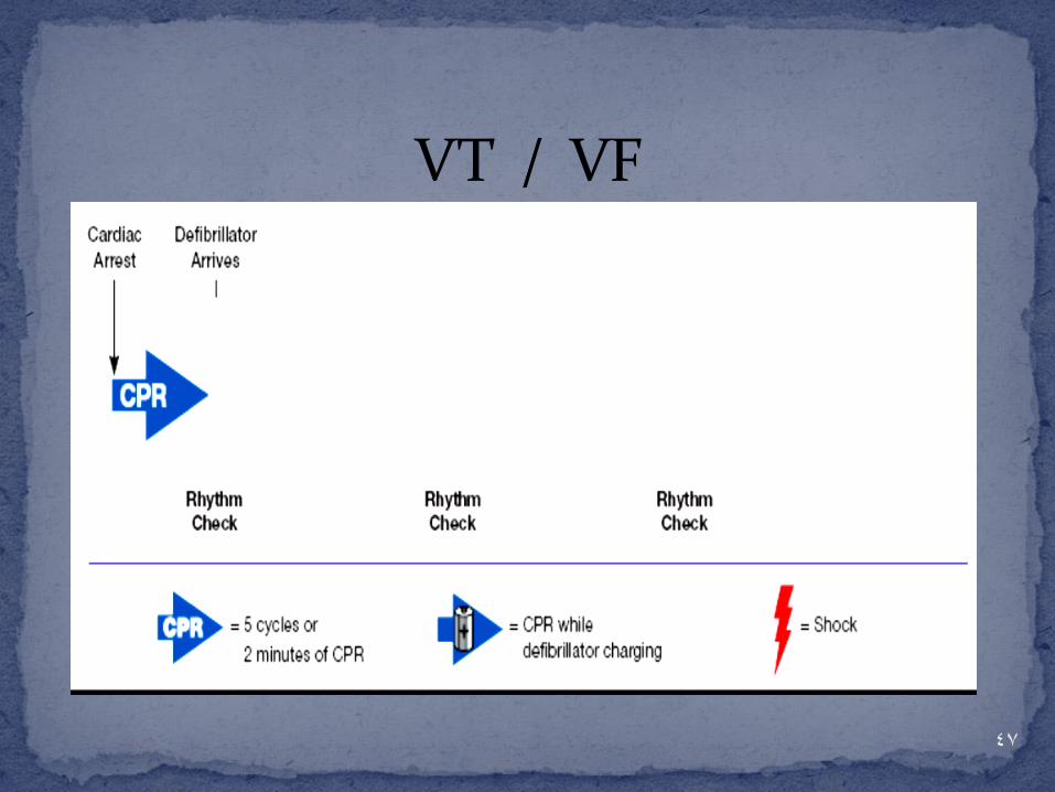
Pulseless Arrest

4 rhythms produce
pulseless cardiac arrest:
Ventricular fibrillation (VF)
Rapid ventricular tachycardia (VT)
Pulseless electrical activity (PEA)
Asystole
4
Survival from these arrest rhythms requires both :
Basic life support (BLS)
and
Advanced cardiovascular life support (ACLS)
5
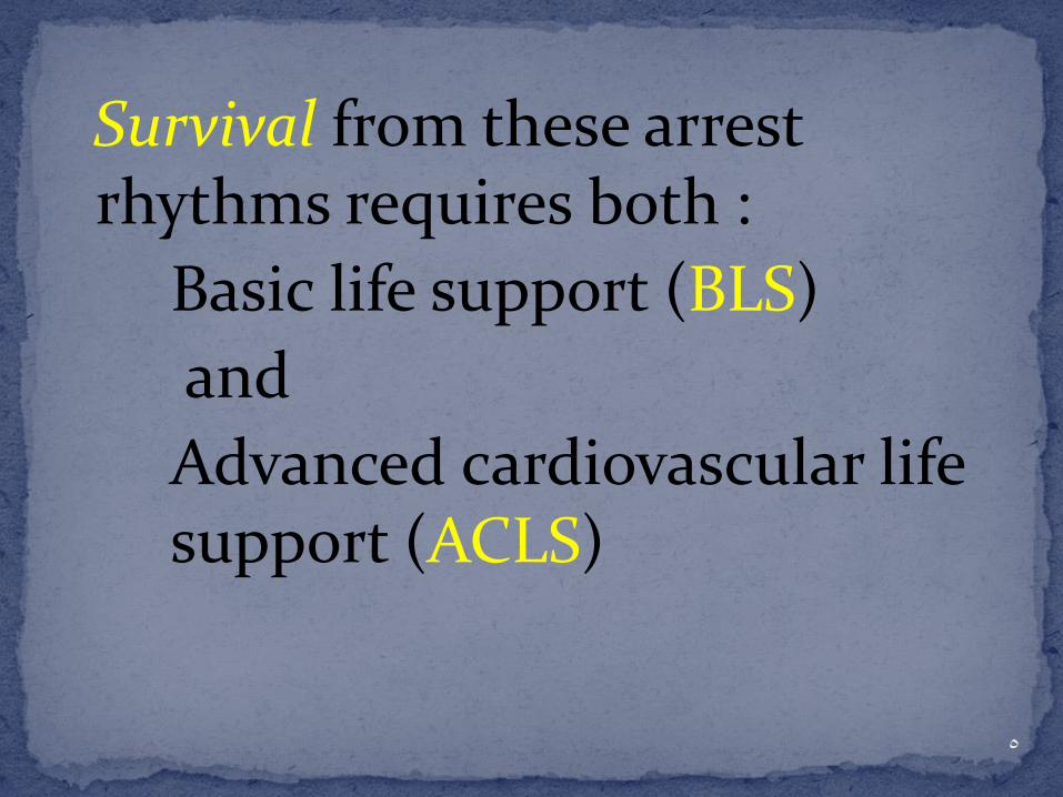
For victims of witnessed VF arrest,
prompt bystander :
1.CPR
2.Early defibrillation
can significantly increase the chance for
survival to hospital discharge. 6
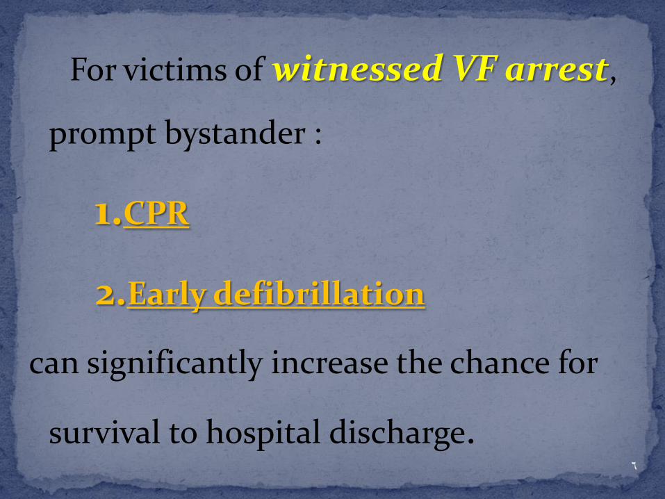
In comparison, typical ACLS therapies, such as:
insertion of advanced airways and
pharmacologic support of the circulation,
have not been shown to increase rate of survival to hospital discharge.
7
8
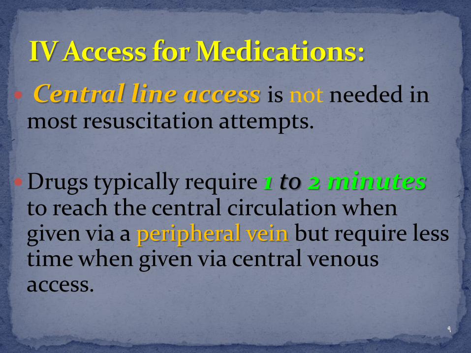
Central line access is not needed in most resuscitation attempts.
Drugs typically require 1 to 2 minutes to reach the central circulation when given via a peripheral vein but require less time when given via central venous access.
9
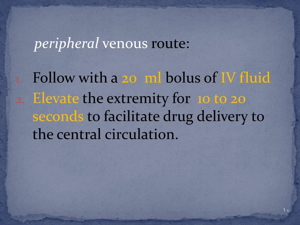
peripheral venous route:
1. Follow with a 20 ml bolus of IV fluid
2. Elevate the extremity for 10 to 20 seconds to facilitate drug delivery to the central circulation.
10

Intraosseous (IO) cannulation provides access to a noncollaps-ible venous plexus, enabling
drug delivery similar to that achieved by central venous access.
11

If IV and IO access cannot be established, some resuscitation drugs may be administered by the endotracheal route
12

Lidocaine
Epinephrine
Atropine
Naloxone
Vasopressin
13
E T route:

The optimal endotracheal dose of most drugs is unknown, but typically the dose given by
the endotracheal route is 2 to
2.5 times the recommended IV
dose.
14

Providers should dilute the recommended dose in
5 to 10 mL of water or normal saline
15

16

17

Copyright ©2010 American Heart Association
Neumar, R. W. et al. Circulation 2010;122:S729-S767
ACLS Cardiac Arrest Algorithm 2010

19

20

21
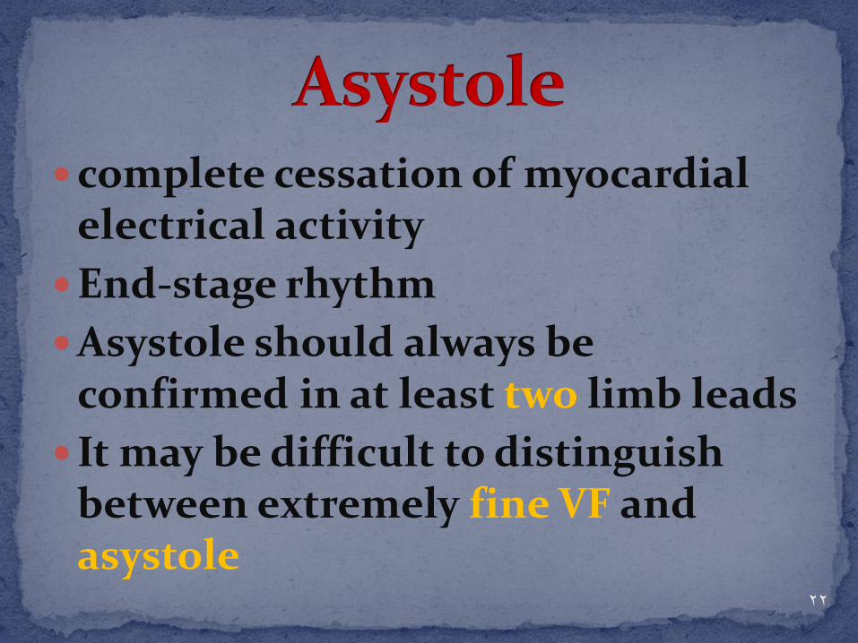
complete cessation of myocardial electrical activity
End-stage rhythm
Asystole should always be confirmed in at least two limb leads
It may be difficult to distinguish between extremely fine VF and asystole 22

23

24

PEA is defined as non-coordinated groups of electrical activity of the heart (other than VT/VF) without a palpable pulse: EMD + pseudo EMD
EMD = Electro Mechanical Dissociation : no myocardial contractions occur
Pseudo-EMD : myocardial contractions occur but no pulse can be palpated
25
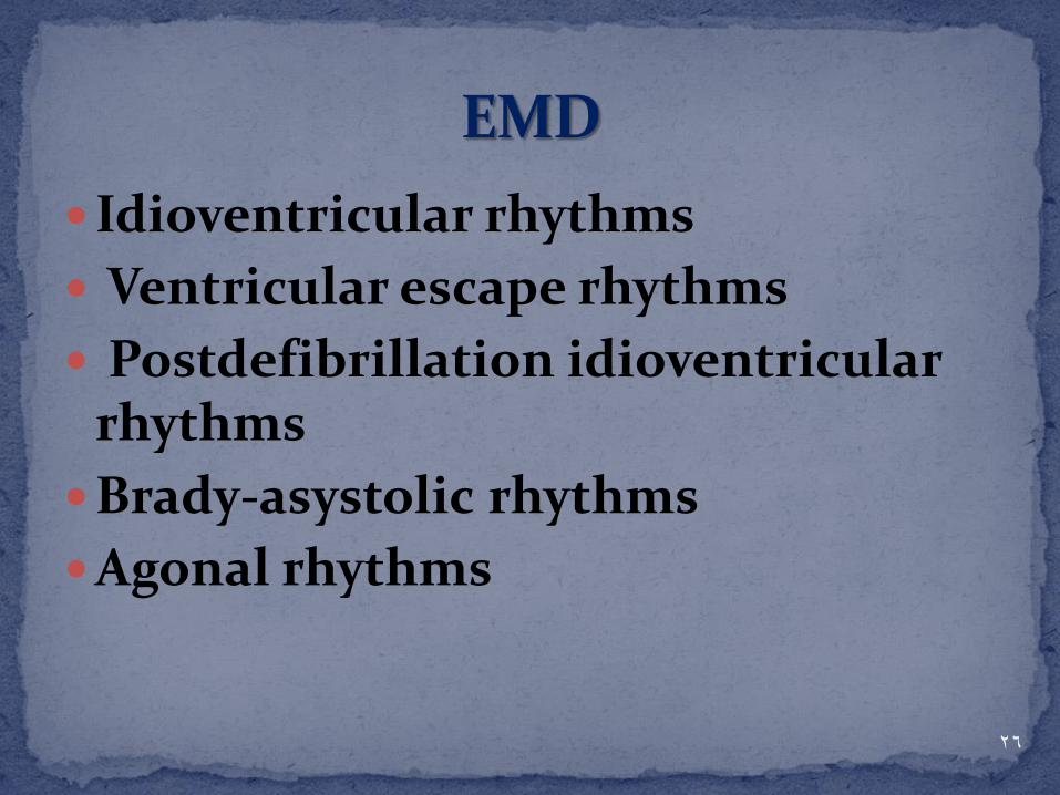
Idioventricular rhythms
Ventricular escape rhythms
Postdefibrillation idioventricular rhythms
Brady-asystolic rhythms
Agonal rhythms
26

Global myocardial dysfunction
Papillary muscle and myocardial wall rupture
Primary supraventricular tachycardia (SVT (
Hypovolemia, tension pneumothorax, pericardial tamponade, and massive PE
27

Patients who have either asystole or PEA will not benefit from defibrillation attempts
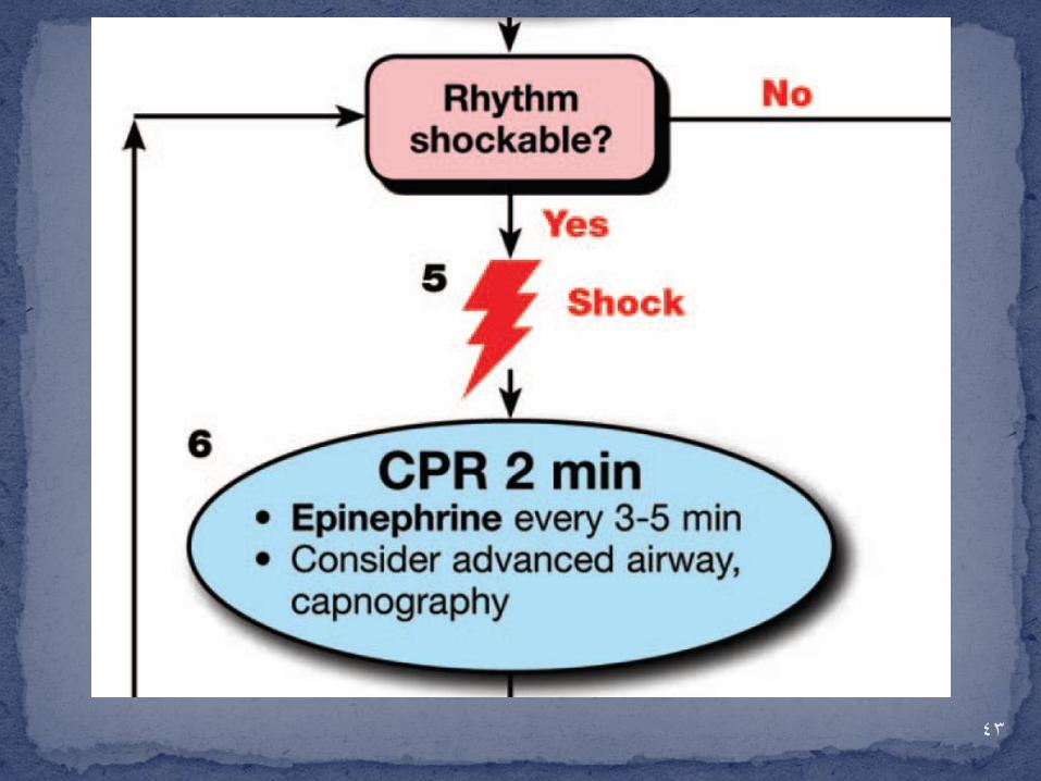
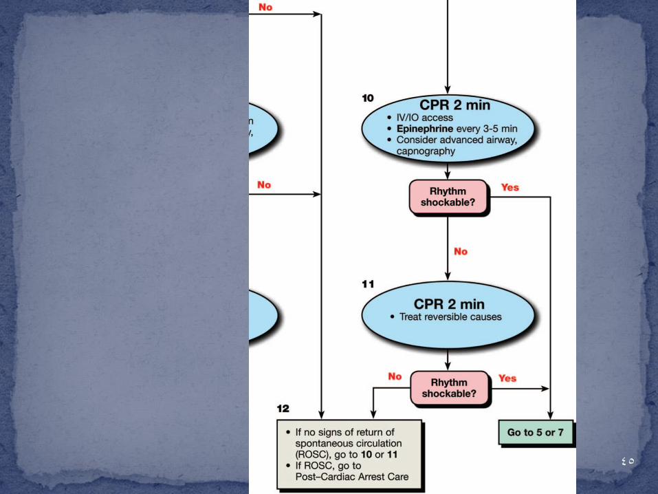
A vasopressor (epinephrine or vasopressin) may be administered at this time.
Epinephrine can be administered
approximately every 3 to 5 minutes during cardiac arrest; one dose of vasopressin may be substituted for either the first or second epinephrine dose (2010)
28
ASYSTOLE/PEA MANAGEMENT

Vasopressin in combination with epinephrine offers no advantage as a substitute for standard-dose epinephrine in cardiac arrest .
The removal of vasopressin has been noted in the Adult Cardiac Arrest Algorithm.
29
2015 Recommendation

It may be reasonable for the provider to deliver 1 breath every 6 seconds (10 breaths per minute) while continuous chest compressions are being performed (ie, during CPR with an advanced airway
This simple single rate for adults, children, and infants—rather than a range of breaths per minute—should be easier to learn, remember, and perform.
30
2015 Recommendation

31

32

33

34

35

36

37
38

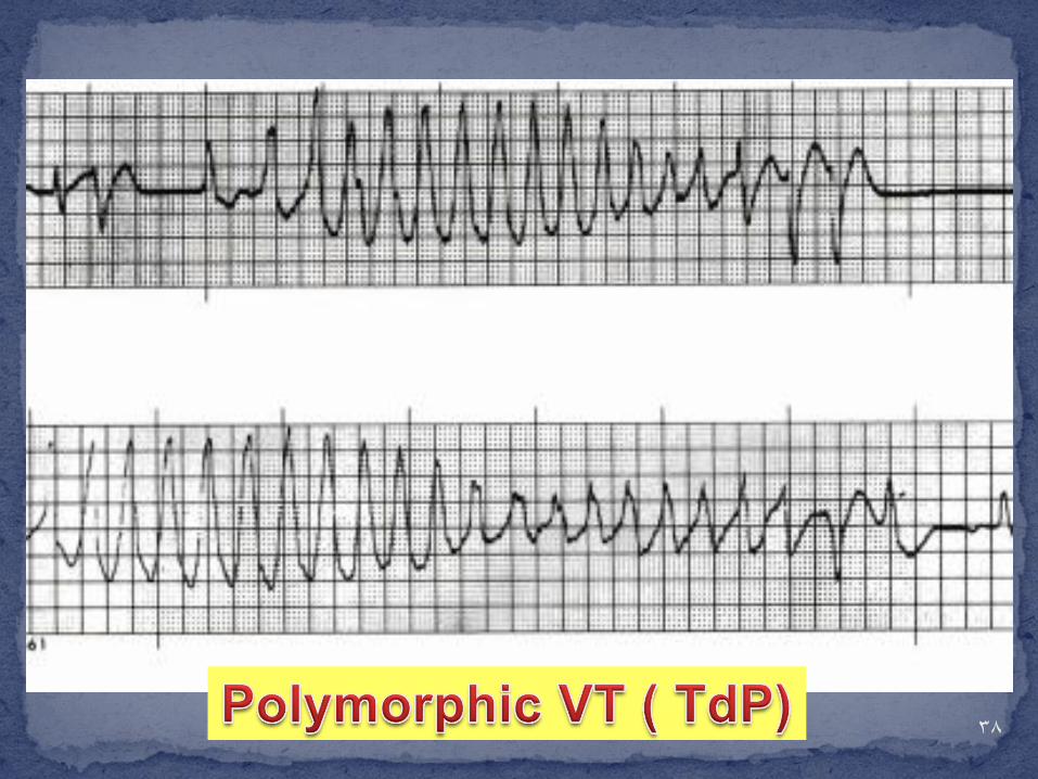
1. Ventricular rate is greater than 200 beats/min.
2. QRS structure displays an undulating axis, with the polarity of the complexes appearing to shift about the baseline.
3. Occurrences are often in short episodes of less than 90 seconds, although sustained runs can be seen.
39

40

Copyright ©2010 American Heart Association
Neumar, R. W. et al. Circulation 2010;122:S729-S767
ACLS Cardiac Arrest Circular Algorithm

42
43

44
45

46
47
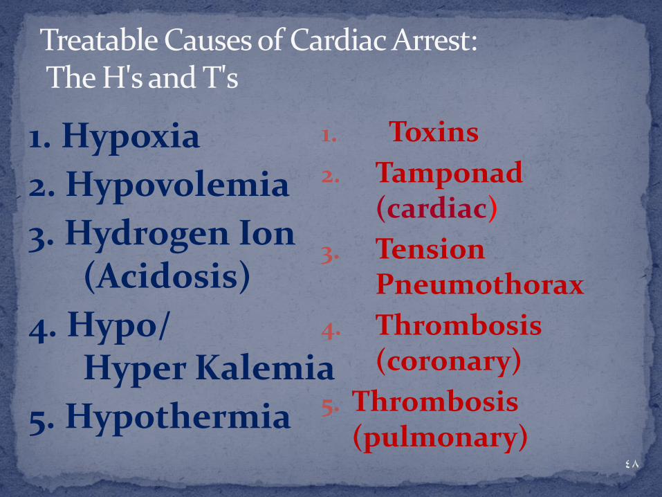
Treatable Causes of Cardiac Arrest: The H's and T's
1. Hypoxia
2. Hypovolemia
3. Hydrogen Ion (Acidosis)
4. Hypo/ Hyper Kalemia
5. Hypothermia
1. Toxins
2. Tamponad (cardiac)
3. Tension Pneumothorax
4. Thrombosis (coronary)
5. Thrombosis (pulmonary)
48

There is inadequate evidence to support the routine use of lidocaine after cardiac arrest. However, the initiation or continuation of lidocaine may be considered immediately after ROSC from cardiac arrest due to VF/pVT .
49
2015 Recommendation

50

Emphasis on effective chest compression
One universal compression-to-ventilation 30/2
Recommendation for 1-second breaths during all CPR
51

Rescuers should change compressors every 2 min
Compression should ideally be interrupted only for rhythm check and shock delivery
52

Providers do not attempt a pulse or check the rhythm after shock delivery
Drug should be delivered during CPR, as soon as possible after rhythm check
53
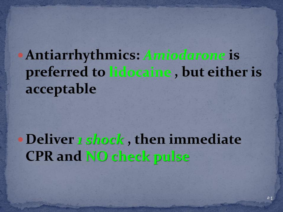
Antiarrhythmics: Amiodarone is preferred to lidocaine , but either is acceptable
Deliver 1 shock , then immediate CPR and NO check pulse
54

55

1-Epinephrine
2-Atropine
3-Amiodarone
4-Lidocaine
5-Magnesium
56

When VF/pulseless VT cardiac arrest is associated with torsades de pointes, providers may administer magnesium sulfate at a dose of 1 - 2 g diluted in 10 mL D5W IV/IO push, typically over
5 - 20 minutes
57

When torsades is present in the patient with pulses, the same 1 - 2 g is mixed in 50 to 100 mL of D5W and given as a loading dose.
It can be given more slowly (eg, over 5 to 60 minutes IV)
58

59

Resuscitation of the Pregnant Patient

Key Points During resuscitation there are two patients, mother &
fetus
The best hope of fetal survival is maternal survival
Consider the physiologic changes due to pregnancy
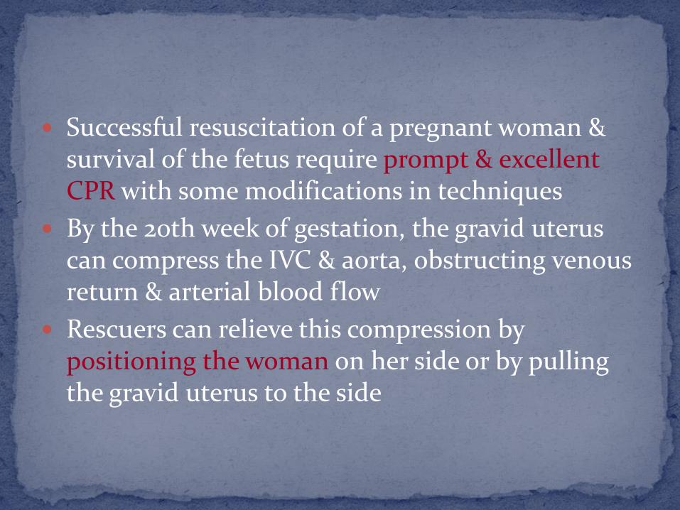
Successful resuscitation of a pregnant woman & survival of the fetus require prompt & excellent CPR with some modifications in techniques
By the 20th week of gestation, the gravid uterus can compress the IVC & aorta, obstructing venous return & arterial blood flow
Rescuers can relieve this compression by positioning the woman on her side or by pulling the gravid uterus to the side

Defibrillation
Defibrillate using standard ACLS defibrillation doses
There is no evidence that shocks from a direct current defibrillator have adverse effects on the heart of the fetus
If fetal or uterine monitors are in place, remove them before delivering shocks
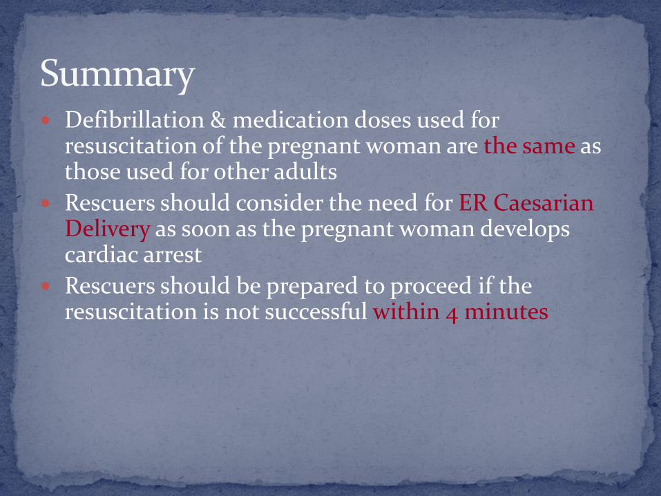
Summary Defibrillation & medication doses used for
resuscitation of the pregnant woman are the same as those used for other adults
Rescuers should consider the need for ER Caesarian Delivery as soon as the pregnant woman develops cardiac arrest
Rescuers should be prepared to proceed if the resuscitation is not successful within 4 minutes

65

DEFIBRILLATION


68
Some AEDs will automatically switch them-selves on when the lid is opened

69

70

71
Stand clear
Deliver shock
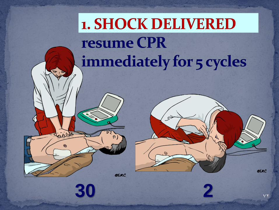
72 30 2
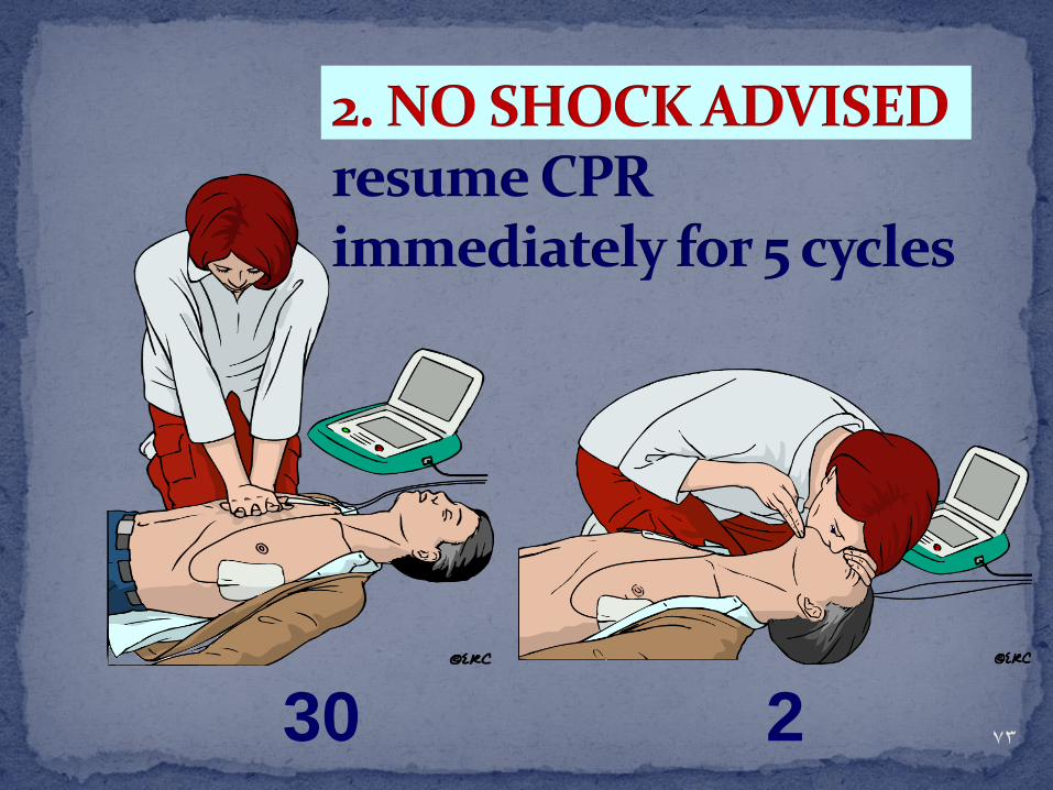
73 30 2

74

defibrillation
CPR prior to defibrillation
Lack of success for in-hospital resuscitation appears to result from delays in time to first shock from collapse.

Defibrillation Equipment

List of Materials for Defibrillation
Defibrillator/ECG monitor
Handheld defibrillation electrodes “quick-look” paddles
Patient interface cables; multifunctional for ECG monitoring and defibrillation
Electrodes and pads for ECG signal acquisition and defibrillation
Conductive gel (not ultrasound gel)

Additional “Equipment” (Pertinent to VF/VT)
ACLS Medications • Epinephrine • Vasopressin • Amiodarone • Lidocaine • Magnesium sulfate
• Procainamide • Atropine Miscellaneous
• IV access equipment, central line kits, and the like

“Code cart” with defibrillation equipment.

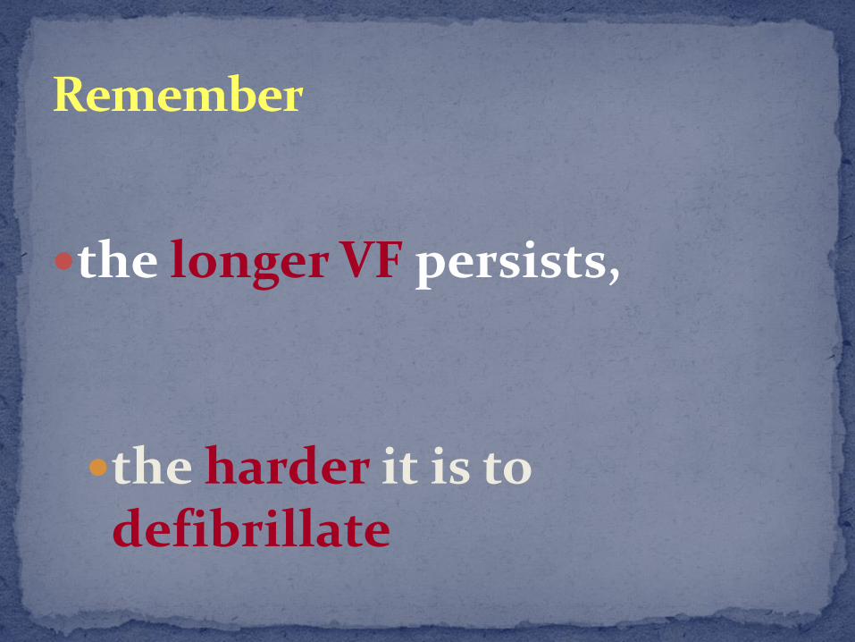
Remember
the longer VF persists,
the harder it is to defibrillate

Multifunction defibrillator/monitor

Defibrillator monitor capable of 12-lead ECG/cardioversion/pacing/limited ECG interpretation.

Defibrillator Types

Defibrillators (operational characteristics)
Manual Semiautomated
fully automatic

Monophasic damped sinusoidal (MDS) and monophasic truncated exponential(MTE) waveforms

Biphasic waveforms.
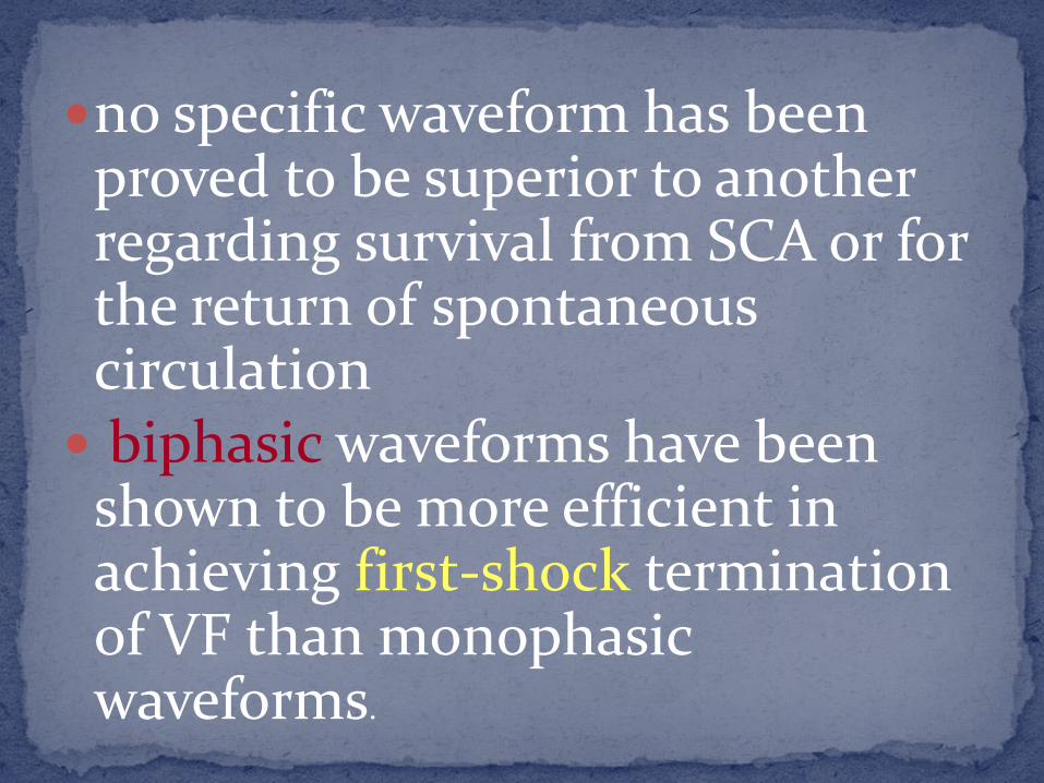
no specific waveform has been proved to be superior to another regarding survival from SCA or for the return of spontaneous circulation biphasic waveforms have been
shown to be more efficient in achieving first-shock termination of VF than monophasic waveforms.

Monophasic Defibrillators/Energy Selection
an energy level of 360 J be used for the first shock

Biphasic Defibrillators
An optimal energy level for first-shock for VF has not been established, several studies have demonstrated that using relatively low energy of 200 J or less
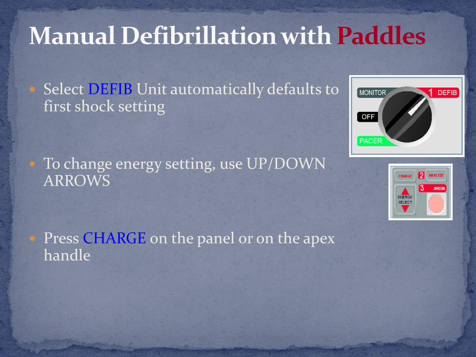
Manual Defibrillation with Paddles
Select DEFIB Unit automatically defaults to first shock setting
To change energy setting, use UP/DOWN ARROWS
Press CHARGE on the panel or on the apex handle

Manual Defibrillation with Paddles
Apply electrolyte gel to the paddles and apply paddles to chest
Make sure everyone is clear
When SHOCK button lights, place paddles on chest with 25 lb pressure and simultaneously press SHOCK on both paddles
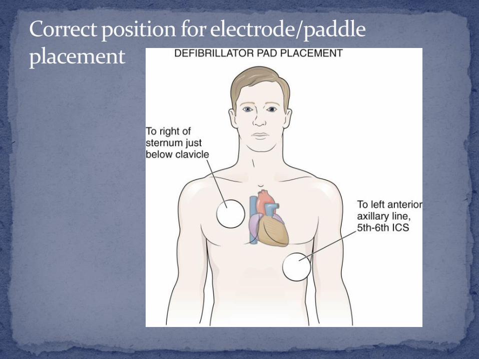
Correct position for electrode/paddle placement

Use of quick-look paddle electrodes for rhythm (ECG) determination and defibrillation
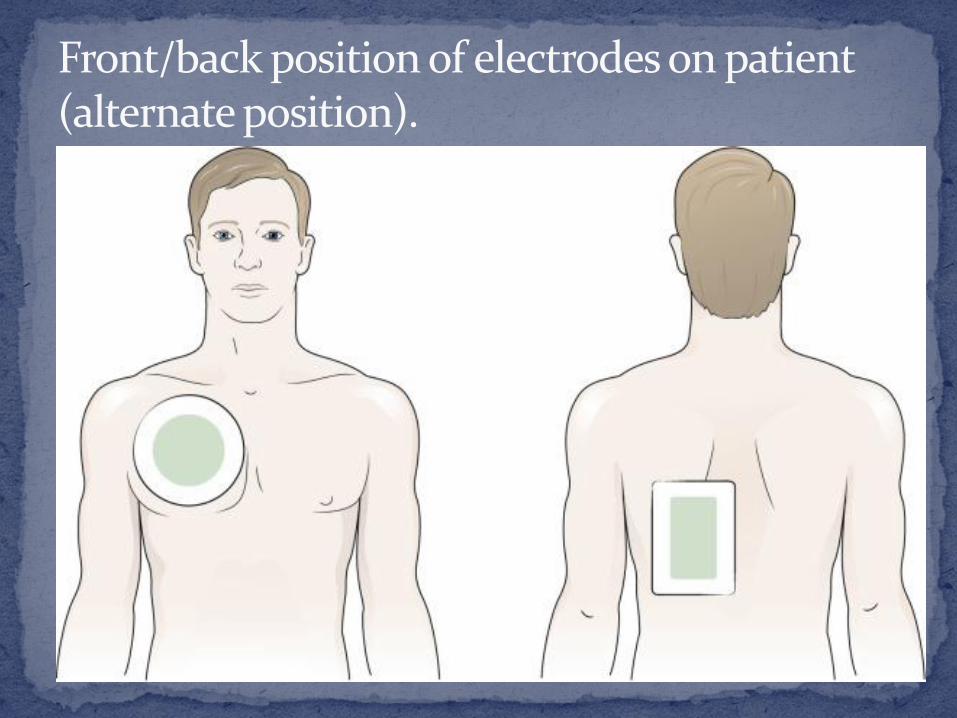
Front/back position of electrodes on patient (alternate position).

Complications soft tissue injury
myocardial injury
Cardiac dysrhythmias
multifunctional electrode pads
better applicators for electrode gels have decreased the potential for soft tissue injuries such as burns to the chest
biphasic defibrillation

98
Related Documents