HAL Id: hal-02295579 https://hal-amu.archives-ouvertes.fr/hal-02295579 Submitted on 25 Feb 2021 HAL is a multi-disciplinary open access archive for the deposit and dissemination of sci- entific research documents, whether they are pub- lished or not. The documents may come from teaching and research institutions in France or abroad, or from public or private research centers. L’archive ouverte pluridisciplinaire HAL, est destinée au dépôt et à la diffusion de documents scientifiques de niveau recherche, publiés ou non, émanant des établissements d’enseignement et de recherche français ou étrangers, des laboratoires publics ou privés. Distributed under a Creative Commons Attribution - NonCommercial - NoDerivatives| 4.0 International License Adsorption properties, the pH-sensitive release of 5-fluorouracil and cytotoxicity studies of mesoporous silica drug delivery matrix Eva Beňová, David Bergé-Lefranc, Vladimír Zeleňák, Miroslav Almáši, Veronika Huntosova, Virginie Hornebecq To cite this version: Eva Beňová, David Bergé-Lefranc, Vladimír Zeleňák, Miroslav Almáši, Veronika Huntosova, et al.. Adsorption properties, the pH-sensitive release of 5-fluorouracil and cytotoxicity studies of mesoporous silica drug delivery matrix . Applied Surface Science, Elsevier, 2021, 504, pp.144028. 10.1016/j.apsusc.2019.144028. hal-02295579

Welcome message from author
This document is posted to help you gain knowledge. Please leave a comment to let me know what you think about it! Share it to your friends and learn new things together.
Transcript

HAL Id: hal-02295579https://hal-amu.archives-ouvertes.fr/hal-02295579
Submitted on 25 Feb 2021
HAL is a multi-disciplinary open accessarchive for the deposit and dissemination of sci-entific research documents, whether they are pub-lished or not. The documents may come fromteaching and research institutions in France orabroad, or from public or private research centers.
L’archive ouverte pluridisciplinaire HAL, estdestinée au dépôt et à la diffusion de documentsscientifiques de niveau recherche, publiés ou non,émanant des établissements d’enseignement et derecherche français ou étrangers, des laboratoirespublics ou privés.
Distributed under a Creative Commons Attribution - NonCommercial - NoDerivatives| 4.0International License
Adsorption properties, the pH-sensitive release of5-fluorouracil and cytotoxicity studies of mesoporous
silica drug delivery matrix Eva Beňová, David Bergé-Lefranc, Vladimír Zeleňák, Miroslav Almáši,
Veronika Huntosova, Virginie Hornebecq
To cite this version:Eva Beňová, David Bergé-Lefranc, Vladimír Zeleňák, Miroslav Almáši, Veronika Huntosova, etal.. Adsorption properties, the pH-sensitive release of 5-fluorouracil and cytotoxicity studies ofmesoporous silica drug delivery matrix . Applied Surface Science, Elsevier, 2021, 504, pp.144028.�10.1016/j.apsusc.2019.144028�. �hal-02295579�

Contents lists available at ScienceDirect
Applied Surface Science
journal homepage: www.elsevier.com/locate/apsusc
Full Length Article
Adsorption properties, the pH-sensitive release of 5-fluorouracil andcytotoxicity studies of mesoporous silica drug delivery matrix
Eva Beňováa,b, David Bergé-Lefrancc, Vladimír Zeleňáka,⁎, Miroslav Almášia,Veronika Huntošovád, Virginie Hornebecqb,⁎
a Department of Inorganic Chemistry, Faculty of Science, P.J. Šafárik University, Moyzesova 11, SK-041 54 Košice, SlovakiabAix Marseille Univ, CNRS, MADIREL, Marseille, Francec Aix Marseille Univ, CNRS, IMBE, Faculté de Pharmacie, Marseille, Franced Center for Interdisciplinary Biosciences, Technology and Innovation Park, P. J. Šafárik University in Košice, Jesenna 5, 041 54 Košice, Slovakia
A R T I C L E I N F O
Keywords:Mesoporous silicaDrug deliverypH responsivity5-fluorouracilCytotoxicityAdsorption enthalpy
A B S T R A C T
Mesoporous silica materials were investigated as the carriers for pH-sensitive drug delivery systems. Poroussilica SBA-15 was first functionalized by anchoring N-[3-(trimethoxysilyl) propyl] aniline groups on the surface.After loading of an antineoplastic agent 5-fluorouracil, the pores were capped by β-cyclodextrin molecules. Thestudied samples were characterized by N2 adsorption/desorption measurements, thermal analysis, powder X-RayDiffraction, and Transmission Electron Microscopy. Adsorption properties of 5-fluorouracil were explored via theconstruction of adsorption isotherms. The amount of 5-FU adsorbed on amine-functionalized SBA-15 was 60mgper 1 g of solid. The adsorption of 5-fluorouracil on silica was also monitored by microcalorimetry, showing lowadsorption enthalpies. Drug release properties from matrices were studied using UV–Visible spectroscopy withthe un-blocked and blocked pores configuration to demonstrate the efficiency of the pH-responsive nanovalvesand evaluated using different kinetic models. It was shown that no drug release occurred at neutral pH and thatmore than 80% of drug adsorbed amount was released at pH=5. Working at the equilibrium, the initial burst ofthe drug from the silica surface, usually observed in other porous silica drug delivery systems, was avoided. Theinteraction between the β-cyclodextrin molecules and grafted amine functions were also studied as a function ofpH. Finally, the cytotoxicity tests were performed using human glioma U87 MG cells.
1. Introduction
The unfavorable physicochemical properties of many drug com-pounds affect their bioavailability and consequently the pharmacoki-netic efficiency of the medical treatment. For example, drug moleculeswith a lack of specificity and solubility lead patients to take high dosesof the drug to achieve sufficient therapeutic effects. This is a leadingcause of adverse drug reactions, particularly for drugs with a narrowtherapeutic window or cytotoxic chemotherapeutics [1]. Lack of spe-cificity of a drug molecule leads to the high-dosage regimen and causeundesired interactions of a drug with healthy tissues or cells [2]. Thepossible solution to overcome these problems is to design the efficientDrug Delivery Systems (DDSs) that enable to control the rate, time, andplace of drugs release in the body [3]. The DDSs improve the phar-macological properties of drug molecules by modifying their pharma-cokinetic profile, solubility and bio-distribution [4]. Among structurallystable materials that have been investigated, mesoporous silica
materials (MSMs) have become apparent as a promising drug vehicledue to their unique mesoporous structure that preserves a level ofchemical stability, surface functionality, and biocompatibility. TheMSMs have been used for delivery of a variety of drug molecules, e.g.:chemotherapeutics agents [5–7], antibiotics [8,9], antimicrobial drugs[10], anti-inflammatory molecules [11–14]. One of their main ad-vantages is their ability to be used for both hydrophilic active agentsand poorly water-soluble drugs by increasing their solubility [15]. Forexample, Mellaerts et al. reported the increase of the oral bioavailabilityof the poorly water-soluble drug, itraconazole using mesoporous silicananoparticles as drug delivery agents [16].
Another advantage of porous silica drug delivery system is thepossibility to design zero-premature cargo release nanosystems byblocking the pore openings using various gatekeepers [17–19]. Lately,most of the research effort for drug delivery has been committed tocancer therapy and internal stimuli such as pH, redox potential, en-zymes, etc. that are typical of the treated pathology [20–22]. pH-
https://doi.org/10.1016/j.apsusc.2019.144028Received 1 May 2019; Received in revised form 10 September 2019; Accepted 13 September 2019
⁎ Corresponding authors.E-mail addresses: [email protected] (V. Zeleňák), [email protected] (V. Hornebecq).
$SSOLHG�6XUIDFH�6FLHQFH������������������
$YDLODEOH�RQOLQH����2FWREHU�����������������������(OVHYLHU�%�9��$OO�ULJKWV�UHVHUYHG�
7

responsive MSMs have attracted extensive research interest becausecancer cells cause the decrease of pH environment to more acidic valuescompared to the pH environment of healthy cells, which provides anefficient way to control the drug release behavior by pH variations[23,24]. There are many strategies to construct pH-responsive drugdelivery systems, e.g. using polyelectrolyte gatekeepers [25], pH-sen-sitive linkers (such as acetal bond, hydrazine bond, ester bond) [26],acid-decomposable inorganic gatekeepers [27] and supramolecularnanovalves [28]. The latter includes an immobilized stalk moleculecovalently attached to silica surface and a mobile cyclic molecule en-circling the stalk via non-covalent interactions [29]. A lot of cucurbi-turil and cyclodextrin (CD) molecules have been demonstrated to beeffective supramolecular pore gatekeepers for mesoporous silicas[30,31]. Particularly for CD, different molecules were grafted on theporous silica surface and then tested as pH-responsive gatekeepers.Meng et al. reported a novel MSM delivery system for doxorubicin, achemotherapeutic agent, based on β-cyclodextrin nanovalves testingdifferent aromatic amines. Best results were obtained using N-methyl-benzimidazole as stalks with a maximum release percentage of 40% inacidic conditions (pH=5) [32]. The delivery of doxorubicin was alsotested in the work of Bai et al. who constructed a controlled releasesystem using p-anisidine stalks. Even if the pH-dependency of releaseproperties were successfully studied for short release time (< 6 h), theloading efficiency of doxorubicin was very low (4,5%), that represented2.44mg of doxorubicin per 1 g of silica support [33].
Considering silica DDSs, several important parameters have to beoptimized all together both in terms of loading and release properties.In this way, the loading efficiency i.e. the amount of available drug thatis incorporated in and only in the porous volume should be as high aspossible and the release profile in time should be as progressive aspossible, avoiding the burst effect. Therefore, in the present paper, bothadsorption and pH-sensitive release properties of a model drug onmesoporous silica material SBA-15 were studied and optimized in thiscontext. The antineoplastic agent 5-fluorouracil (5-FU), that is a first-line anti-cancer drug, commonly used to treat colorectal, gastro-intestinal and breast cancer, was employed as a drug molecule in ourstudy. However, the efficiency of 5-FU is limited by high rate of me-tabolism in the body, short biological half-life, non-uniform oral ad-sorption and cytotoxicity. To be effective it has to be administrated athigher concentrations or more frequent doses. In this work, we assumethat its encapsulation in a host delivery system can reduce the fre-quency of dosing or it could help the drug to be more effective even atlower concentrations.
In our DDSs, supramolecular nanovalves based on β-CD units wereused as gatekeeper and three different amines were grafted onto themesoporous silica surface and used as stalk molecules. For the grafting,we have used aliphatic amines namely propylamine (AP), tetra-ethylenepentamine (TEPA) aromatic amine propyl aniline (N-ANI). Ourstudy showed, that the DDSs based on studied aliphatic amines and β-cyclodextrin molecules do not work effectively and showed poortightness and premature drug release (see Supporting information, Fig.S1). The more promising results were obtained using a gatekeepersystem composed of aromatic propyl aniline based stalk (N-ANI) and β-CD units, which properties are described in the present study. It is note,that combination of N-ANI ligands and β-CD molecules in constructionof pH-responsive DDSs have been already reported [34,35]. However,in our study adsorption properties of drug were studied on pure silicamatrix and functionalized one from a thermodynamic point of view.Moreover, the construction of adsorption isotherms and their modelingas well as the determination of enthalpies of adsorption using calori-metric measurements were realized. As far as our knowledge, such anextensive study concerning thermodynamic description has not beenreported in the literature yet. Moreover, the cytotoxicity of the pre-pared DDS using U87 MG cancer cells complete our study.
2. Experimental section
All chemicals used in the syntheses were obtained by Sigma-Aldrichand Across Organics companies in the highest purities and used withoutfurther purification. Anhydrous solvents were obtained after standardprocedures described in the literature [36] and stored over molecularsieves.
2.1. Synthesis and functionalization mesoporous silica SBA-15
The synthesis of porous silica was carried out according to theprocedure reported in [37]. 4 g of triblock copolymer P123, (Poly(ethylene glycol)-block-poly(propylene glycol)-block-poly(ethyleneglycol)) was dissolved in 30 g of distilled water at T=308 K under highacidic conditions produced by the addition of 120 g of HCl (2M). Themixture was stirred until it became homogenous. Then, 8.5 g of TEOSwas added dropwise to the solution followed by continuous stirring for20 h at T= 308 K. After this time, the solution was aged at 80 °C for24 h. The white solid product was filtered off, washed with distilledwater and dried at room temperature.
Porous SBA-15 matrix was obtained by calcination of as-synthesizedSBA-15 in airflow at 600 °C. The calcination procedure was as follows:the sample was heated to 150 °C with a heating rate 3 °C/min and thesample was held at this temperature for 3 h. Subsequently, the tem-perature was increased to 600 °C by the rate 1 °C/min and the samplewas held at this temperature for 7 h. In the last step, the sample wascooled down to room temperature.
The surface modification (functionalization) of silica was carriedout by a post-synthetic grafting procedure. Typically, 1 g of calcinedSBA-15 was dispersed in anhydrous toluene, mixed with 3mL of N-[3-(trimethoxysilyl) propyl] aniline (12,5 mmol) and refluxed under N2 for20 h. The obtained product was centrifuged, several times washed withtoluene and ethanol and dried at T=313 K for 24 h. The material wasdenoted as SBA-15_N-ANI.
2.2. Drug loading and pores capping
5-fluorouracil (5-FU) was taken as a model drug to estimate theloading and release performance from both SBA-15 and SBA-15_N-ANI.Various information concerning the solubility of 5-FU in water are re-ported in the literature, i.e.: 5 mg/ml [38], 8.62mg/ml [39], 12mg/ml[40]. Since the drug 5-FU is sparingly soluble in the water, the upperconcentration limit of 5-FU in our experiments was set to 6.5 mg/ml.
2.2.1. Adsorption experimentsAdsorption isotherm experiments were carried out using a solution
depletion method. The experimental process consisted in the followingsteps: 20mg of samples were dispersed in a series of plastic vials con-taining 2mL of 5-FU aqueous solution with different initial con-centrations (from 0.1 to 6.5 mg/ml). Then, the adsorption process wascarried out, with continuous stirring, at T=310 K for 24 h to ensureequilibrium. The separation of a solid phase from the supernatant liquidwas achieved by centrifugation at 10 000 rpm (corresponding to a re-lative centrifugal force of 7400 g) for several minutes. The supernatantwas then analyzed by using UV–Vis spectroscopy (wavelength range200 – 350 nm) to determine the equilibrium concentration of 5-Fu (Ceq)at the maximum of the absorption band (λ=266 nm). Prior to thedetermination of the concentration of 5-FU, a calibration curve hasbeen established based on solutions with different concentrations of 5-FU (see Fig. S2 in the Supplementary information). The linearity of thecalibration curve was confirmed by the correlation coefficientR2=0.99996. Then, the amount adsorbed, QADS was calculated asfollows:
= −Q
V C Cm
( )ADS
i eq
s (1)
E. Beňová, et al. $SSOLHG�6XUIDFH�6FLHQFH������������������
�

where Ci is the initial concentration in mol⋅dm−3 (abbreviation M), Ceq
is the equilibrium concentration (M), V is the volume of the drug so-lution (dm3 or L), and ms is the mass of adsorbent used (g).
Microcalorimetric experiments were performed on TAM 2277 mi-crocalorimeter in order to determine the adsorption enthalpies. Duringthese experiments, solids (SBA-15 and SBA-15_N-ANI) were maintainedin water suspension using a stirring system and a stock solution of 5-FUwas added by 5 µL aliquots step by step (20 injections). Next, the heatflow peaks were time-integrated and after correction of dilution effects,the integral enthalpies were obtained.
2.2.2. Preparation of β-CD capped SBA-15_N-ANI_5-FU materialA solution of 5-FU (6,5 mg/ml) was prepared in distilled water at
room temperature. Then, 100mg of the SBA-15_N-ANI and 10mL of 5-FU solution were mixed and stirred at T=310 K until the mesoporoussilica stopped adsorbing the drug, which was monitored by UV–Visabsorption spectroscopy. It was found that 24 h of the adsorption pro-cess was sufficient to achieve equilibrium. Then, the pH of the solutionwas adjusted to 7.4 with the addition of NaOH solution (1M) and600mg of β-CD was added to the mixture and stirred overnight atT= 310 K. The 5-FU-loaded β-CD-capped mesoporous silica was wa-shed with water with pH adjusted to 7.4 by the same NaOH solution,centrifuged and used in further experiments.
2.3. Drug release experiments
The experiments involving 5-FU release by sample SBA-15_N-ANIwere performed by two different procedures. For both, the adsorptionof 5-FU and pores capping were carried out first as described above. Inboth cases, the mixtures were stirred at 37 °C in a tube rotator. Therelease amount of 5-FU was calculated from a standard curve usingUV–Vis spectroscopy at λ=266 nm.
1st procedure: after adsorption of 5-FU and pores blocking using β-CD molecules, the suspension was centrifuged and the supernatant wasremoved to recover the solid phase. Then, 100mg of the solid phasewas dispersed in 10mL solution at pH=7.4 and pH=5. The drugrelease process was checked at fixed time intervals (1 h, 3 h, 5 h, 8 h,18 h, 24 h, and 48 h).
2nd procedure: after adsorption of 5-FU and pores blocking by β-CD,pH was kept at 7.4 The solution was checked every 2 h during 10 hperiod to verify that there is no drug release. Then, pH was adjusted to5 and the drug release process was again checked at fixed time intervals(1 h, 3 h, 5 h, 8 h, 18 h, 24 h, and 48 h).
2.4. Characterization of porous solids and analytical techniques
Nitrogen sorption measurements were carried out using an ASAP2010 Micromeritics apparatus at −196 °C. Prior to adsorption, samples(~80–100mg) were outgassed at 120 °C during 12 h under the vacuumof 2.10−3 mbar. The specific surface area was determined withBrunauer, Emmet, and Teller (BET) method, the pore size distributionwas calculated from the adsorption branch using the Barrett-Joyner-Halenda (BJH) method [41].
TEM micrographs were obtained using a JEOL 2000FX microscope.Samples were ground and afterward suspended in methanol. The sus-pension was added to a carbon grid and dried in air.
UV–Visible spectroscopy measurements in the liquid phase wereperformed on a Varian Cary 300 spectrometer in the 300–800 nmrange.
Thermogravimetric (TGA) measurements were carried out with aTGA Q500 apparatus (TA Instruments) using air as a carrier gas. Thesamples (~5–20mg) were treated up to 800 °C.
Small-angle X-Ray diffraction measurements were recorded on aSiemens D500R XRD diffractometer using Cu Kα radiation in the0.5− 3° 2θ range with a 0.04° step associated with a step time of 4 s.
2.5. The interaction of mesoporous silica SBA-15 and SBA-15_N-ANI_5-FU_β-CD with cancer cells
The human glioma cells U87 MG (Cells Lines Services, Germany)were used as a model of cancer cells for microscopy and cell viabilitystudy. The cell cultures were grown in the complete cell culturemedium Dulbecco’s modified Eagle medium (D-MEM, GlutaMAXTM,Gibco-Invitrogen, Life Technologies Ltd., France) supplemented with10% fetal bovine serum (FBS, biosera, France) in dark at 37 °C, 5% CO2
and 80% humidified atmosphere.
2.5.1. The cytotoxicity of SBA-15 and SBA-15_N-ANI_5-FU_β-CD: MTTassay
The U87 MG cells were seeded into 96-well plates (104 cells/well)and 24 h after seeding the 5-FU, SBA and SBA-15_N-ANI_5-FU_β-CDaliquots (dissolved in the distilled water) were administered into thecomplete cell culture media. The cells were treated during 24 h with the0.02 and 0.05mg/mL of 5-FU, 0.6 and 1.5 mg/mL of SBA-15, and 0.8and 2mg/mL of SBA-15_N-ANI_5-FU_β-CD at pH=7.3 and 6.3. The pHof cell culture media was adjusted with HCl (Sigma-Aldrich, Germany)24 h before administration of the nanoparticles. After the treatment, theMTT (3-(4,5-dimethylthiazol-2-yl)-2,5-diphenyltetrazolium bromide,Sigma-Aldrich, Germany) assay was detected at 560 nm and 750 nm by96-well plate absorption reader (GloMax®-Multi +Detection Systemwith Instinct Software, Promega Corporation, USA). The ten µl of theyellow MTT reagent (5 mg/mL) dissolved in phosphate saline buffersolution (PBS, Sigma-Aldrich Germany) at pH=7.4 were added to100 µL of cell culture media per well. The cells were incubated withMTT in dark and 37 °C during 1 h. After the incubation, the mediumwas replaced with dimethyl sulfoxide (DMSO, Sigma-Aldrich,Germany) to dissolved purple formazan crystals. The results were per-formed in triplicates.
2.5.2. The cytotoxicity of SBA-15 and SBA-15_N-ANI_5-FU_β-CD: Themitochondria membrane potential dissipation
The U87 MG cells were seeded in Petri dishes (35mm) and grown inthe complete cell culture media at pH 7.3 and 6.3. The 1.5mg/mL ofSBA-15 and 2mg/mL of SBA-15_N-ANI_5-FU_β-CD were administeredto cells 24 h before observation. The mitochondria potential probe aMitoTrackerTM Orange CMTM/Ros (MTO, 0.2 µM, ThermoFisherScientific, USA) was administered to cells last 15min of incubation withthe nanoparticles. The cells were detached with the trypsin/ethylene-diaminetetraacetic acid (Gibco-Invitrogen, Life Technologies Ltd.,France), centrifuged at 1200 rpm/10min and resuspended in 0.5mL ofPBS.
Alternatively, cells were harvested with the trypsin/EDTA, cen-trifuged and resuspended in the serum-free D-MEM. Subsequently, cellswere incubated with 0.01 and 0.05mg/mL of 5-FU, 0.3, 0.6 and1.5 mg/mL of SBA-15, and 0.4, 0.8 and 2mg/mL of SBA-15_N-ANI_5-FU_β-CD in the 100 µL of D-MEM during 1 h in dark, at pH=7.3 and37 °C. The mitochondria and lysosomes were stained 15min beforemeasurement with the MTO and the LysoTrackerTM Green DND-26(0.2 µM, ThermoFisher Scientific, USA).
The harvested cells were measured with the flow cytometer(MACSQuant® Analyzer, Miltenyi, Germany) in B1 (525/50 nm) and B3(655–730 nm) channel at the excitation 488 nm.
2.5.3. The intracellular distribution and uptake of SBA-15 and SBA-15_N-ANI_5-FU_β-CD by U87 MG cells
The U87 MG cells were seeded in glass coverslip bottom Petri dishes(35mm, No. 0, MatTek, USA) at the density of 104 cells per Petri dish.The white field and fluorescence images were recorded with the in-verted LSM700 confocal microscope (Zeiss, Germany), equipped with a20X Fluar (NA=0.75, ∞, Zeiss, Germany) and a CCD camera(AxioCam HRm, Zeiss, Germany). The mitochondria were detected withthe MTO. Thy lysosomes were stained with LysoTrackerTM Green DND-
E. Beňová, et al. $SSOLHG�6XUIDFH�6FLHQFH������������������
�

26 and the nuclei of the cells were contrasted with 10 µg/mL Hoechst33,258 (Hoechst, 15min, ThermoFisher Scientific, Slovakia). Thefluorescence of mitochondria and lysosomes was stimulated by 488 nmand 555 nm cw solid-state lasers, and the emission was detected in thespectral range 490–540 nm and>580 nm, respectively. The nucleiwere visualized by 405 nm laser excitation and the emission was de-tected in the range 410–490 nm. The images were analyzed in Zen 2011software (Zeiss, Germany) or ImageJ software (National Institutes ofHealth, USA). The aliquots of 5-FU, SBA and SBA-15_N-ANI_5-FU_β-CDwere administered 24 h before observation with the microscope. ThepH of cell culture media was adjusted with HCl (Sigma-Aldrich,Germany) 24 h before administration of the nanoparticles. The cellswere grown in the complete cell culture media at pH=7.3 and 6.3.
3. Results and discussion
There are different types of periodic mesoporous silicas, like MCM-41, MCM-48, SBA-16, SBA-12, SBA-15 which can be used as matrices inthe design of DDSs. All of these we have tested in our previous studiesas DDS [12–14,42,43]. For example, when we prepared materials,which release the drug using electromagnetic radiation as physicalstimulus, we have used MCM-41 [43] or SBA-12 [12], since these ma-terials have pore size around 3–4 nm, which is suitable for grafting ofphotoactive ligands and reversible formation/splitting of dimers bycycloaddition reaction. When using silica with larger pores, e.g. SBA-15, this cycloaddition reaction would be less favorable to regulate poreclosing/opening. However, in the present study, we have used SBA-15silica with larger pores. In materials like MCM-41, with narrower poresor SBA-16, with ink bottle pores, the grafting of bulky ligands, like N-ANI, would lead to pore blocking. In SBA-15 silica, the pore size is largeenough to adapt both bulky amines as well as the drug. Moreover, SBA-15 material has large pore volume and possesses high stability.
For the silica modification and formation of the stalks on the sur-face, three different amines were grafted onto the mesoporous silicasurface. Aliphatic amines namely propylamine (AP), tetra-ethylenepentamine (TEPA) and the aromatic amine propyl aniline (N-ANI) were used.
In the study, we have used 5-FU as a model drug. This drug is usedin the clinical medicine for cancer treatment, however, at the presenttime, its efficiency is limited by many factors such as high rate of me-tabolism in the body, short biological half-life, non-uniform oral ad-sorption, and cytotoxicity. We expect, that its encapsulation in a hostporous system presenting sustained drug release properties will po-tentially reduce the frequency of administration of the drug, reduce theadverse side effects and prolong its delivery time and elimination in thebody [44].
3.1. Structure and porosity of SBA-15 and SBA15_N-ANI
The porosity of the starting SBA-15 material and its modified formswas studied using Transmission Electron Microscopy (TEM). TEMimages (see Fig. 1a) shows a regular hexagonal array of unimodalchannels perpendicular to the pore direction and parallel to the chan-nels (Fig. 1b). The average pore size and the particle size determinedfrom TEM images were 7 nm and 550× 350 nm, respectively. Aftersurface modification, 5-fluorouracil loading and β-CD capping themesostructured, as evident by TEM, remained unchanged (Fig. 1c-f).
In addition to TEM, the periodicity of the samples was also studiedusing SAXS. Fig. 2 displays the XRD patterns of SBA-15 and SBA-15_N-ANI. For both samples, the XRD patterns are similar and show threeresolved diffraction peaks that can be indexed with (1 0 0), (1 1 0),(2 0 0) reflections of the p6mm hexagonal structure [45].
Textural properties of the prepared mesoporous materials werefurther studied using N2 adsorption/desorption measurements at 77 K.The N2 adsorption/desorption isotherms of the SBA-15 and SBA-15_N-ANI are shown in Fig. 3.
Adsorption isotherms are characterized by an initial sharp uptake ata relative pressure below 0.05 followed by a more gradual uptake be-fore a second sharp upswing in the curves between relative pressures of0.6 and 0.7 depending on samples. A final plateau ends the adsorptionbranch. Such isotherms can be defined by the IUPAC as mixed type Iand type IV isotherms, attributed to samples containing both micro-porosity and mesoporosity [46]. Furthermore, the adsorption isothermsexhibit well-defined H1 hysteresis loops that are typical for SBA-15mesoporous silica. The presence of the H1 hysteresis loop confirms theopen-ended hexagonal cylindrical pore geometry [47]. Hysteresis loopshave sharp adsorption-desorption branches indicating a narrow poresize distribution in prepared mesoporous samples. The equivalent sur-face areas were calculated using the BET method. The values of the BETsurface area can be taken only to estimation because of the presence ofmicroporosity, so the term “equivalent” BET surface area is used. TheseBET values are given in Table 1. The size of the mesopores was esti-mated using the BJH method applied to the adsorption branch of theisotherms and the values obtained are also given in Table 1 with theones of mesopore volumes [41].
As expected, the specific surface areas and total pore volumes de-crease significantly upon functionalization of SBA-15 by N-ANI(400m2⋅g−1, 0.46 cm3⋅g−1) compared to the corresponding values forpure SBA-15 (790m2⋅g−1, 0.57 cm3⋅g−1). The functionalization alsodownshifts the capillary condensation step to lower pressures. The shiftof the adsorption step to lower relative pressures reflected the ‘filling’ ofthe pores. It also decreases the pore size in the modified materials fromthe 7 nm (pure SBA-15) to 6 nm (SBA-15_N-ANI).
Thermogravimetric analysis (TGA) measurements were performedin order to estimate the amounts of the amine groups grafted on thesilica surface (see Fig. 5). As solvents losses occur in the 25–150 °Crange, all TGA curves were normalized at T= 150 °C to not take intoaccount their presence. The original TGA curves are shown inSupplementary information Fig. S3. The TGA curve of calcined SBA-15evidences that the sample is thermally stable and no significant massloss was observed in all range measured. Above 500 °C, a small weightloss (3.6 wt%) was observed corresponding to the silanol groups con-densation and dehydroxylation of the sample. The functionalized me-soporous silica SBA-15_N-ANI is thermally stable up to 200 °C. The massloss step, observed above this temperature, corresponds to the thermaldecomposition of amine N-ANI ligands present on the surface of SBA-15. The total mass loss in the temperature range 200–750 °C is close16.6 wt%. The corresponding amount of the N-ANI molecules graftedon the surface of calcined silica was calculated from the mass differ-ences observed for SBA-15 and SBA-15_N-ANI samples. Amount of N-ANI molecules grafted on the surface of calcined silica material wascalculated to be 1.26mmol.g−1. This amount is similar to the one foundin the literature using the same grafting procedure [48].
3.2. Loading of 5-fluorouracil
The adsorption isotherms of 5-FU both on SBA-15 and SBA15_N-ANIporous solids determined at 25 °C in water conditions are presented inFig. 4 (left). For both solids, similar adsorption isotherms of Langmuirtype were found. The shape of isotherms corresponds to an L-type one[49]. This type of adsorption isotherm is frequently found in the case ofa homogeneous and narrow distribution of energetic adsorption sites. L-type isotherm can be plot by the mathematical expression:
= +q qK C
K C.
.1 .ads max
eq
eq (2)
where qmax (mol.g−1), K (M−1) and Ceq (M) represent the maximumadsorbed amount, the apparent equilibrium constant and the 5-FUequilibrium concentration. Very good correlation coefficients, 0.997 forSBA-15 and 0.993 in the case of SBA-15_N-ANI samples, were foundbetween L-type isotherm expression and experimental results. The fits
E. Beňová, et al. $SSOLHG�6XUIDFH�6FLHQFH������������������
�

of the adsorption isotherms lead to maximum 5-FU adsorbed amountsof (12.8 ± 0.8) 0.10−4 mol⋅g−1 and (8.6 ± 1.8) 0.10−4 mol⋅g−1
corresponding to around 170mg.g−1 and 107mg⋅g−1 for SBA-15 andSBA-15_N-ANI respectively. The affinity of 5-FU for each solid obtainedby the fit appears weak from (29 ± 3) for SBA-15 to (22 ± 7) for SBA-15_N-ANI. These values are clearly evidenced considering the slope ofadsorption isotherms at low concentrations. Thus, the main differencebetween the two solids concerns the maximum adsorbed amount andnot especially the affinity, the former parameter is directly related tothe surface area of solid. To illustrate this point, a direct calculation ofthe adsorbed amount corresponding to a 5-FU equilibrium concentra-tion of 4.5.10−2 mol⋅L−1 leads to values of 7.3.10−4 mol⋅g−1 for SBA-15 versus 4.2.10−4 mol⋅g−1 for SBA-15_N-ANI. This diminution, closeto 45%, is the same than the one observed between the surface areascalculated by BET for the two solids. The loading efficiency of 5-FU was8.46% which represents 55mg of 5-FU per 1 g of solid. Furthermore,the thermodynamic parameters determined from adsorption isothermsare very interesting. Indeed, high adsorbed amounts coupled with weakaffinities should facilitate the release of a sufficient dose of 5-FU. Inorder to complete the adsorption study, calorimetric experiments wereperformed.
Obtained calorimetric results are very similar for both solids andconfirm the interpretation of adsorption isotherms. Indeed, the plots ofthe integral enthalpies of displacement against the adsorbed amountsonto SBA-15 and SBA-15_N-ANI indicate a constant enthalpy of ad-sorption during the adsorption process. The associated values are closeto (–3 ± 1) kJ⋅mol−1 and (–4 ± 1 kJ⋅mol−1) for SBA-15 and SBA-15_N-ANI respectively (Fig. 4 (right)). These weak values of adsorptionenthalpies coupled with the absence of significative evolution along thesurface coverage are characteristic of a narrow and homogenous en-ergetic sites distribution. This set of parameters corresponds to a weakaffinity during the physisorption of 5-FU on the two solids. The specificsurface area limits the adsorbed amount and the presence of N-ANI
Fig. 1. TEM images of (a) and (b) SBA-15 after calcination and its modified forms (c) SBA-15_N-ANI, (d) SBA-15_N-ANI_5-FU, (e) and (f) SBA-15_N-ANI_5-FU_β-CD.
Fig. 2. Small-angle XRD diffractogram of the mesoporous silica SBA-15 andSBA-15_N-ANI.
Fig. 3. Adsorption/desorption isotherms of the prepared samples SBA-15 andSBA-15_N-ANI.
Table 1Surface area, mesopore size and mesopore volume of samples SBA-15 and SBA-15_N-ANI.
SAMPLE SBET [m2⋅g−1] Mesopore volume[cm3⋅g−1]
Mesopore size[nm]
SBA-15 790 0.57 ± 0.3 7 ± 0.4SBA-15_N-ANI 400 0.46 ± 0.3 6 ± 0.4
E. Beňová, et al. $SSOLHG�6XUIDFH�6FLHQFH������������������
�

groups do not seem involved in the interaction.The 5-FU loading was also studied using TGA measurements. First,
the thermal behavior of the pure drug molecule was studied: it de-composes in one single step below 320 °C (Fig. S3, left in theSupplementary information) and in the range of 200–750 °C for the 5-FU loaded SBA-15_N-ANI_5-FU sample (Fig. 5, blue curve). The ex-tended decomposition range for the 5-FU loaded sample is due to theinteractions of a drug molecule with mesoporous channels and N-ANIligands on the surface of silica. The trend of TGA curve of SBA-15_N-ANI_5-FU (Fig. 5, blue curve) is the same as the trend we observed forSBA-15_N-ANI sample (Fig. 5, green curve), but a total mass loss in thesame temperature range is 22.46 wt%. The corresponding amount ofthe loaded 5-FU was calculated from the mass differences observed forSBA-15_N-ANI and SBA-15_N-ANI_5-FU samples. This difference re-presents 5.89 wt% that corresponds to 58,9 mg of 5-FU per 1 g of thecarrier SBA-15_N-ANI_5-FU. This mass of loaded 5-FU is in goodagreement with the one obtained from adsorption isotherms describedabove.
From the results obtained, it looks that the larger amount of the 5-FU was encapsulated in the pure, unmodified SBA-15 (92.63mg⋅g−1,0.712mmol⋅g−1) compare to the modified silica SBA-15_N-ANI(55.3 mg⋅g−1, 0.425mmol⋅g−1). However, these values do not reflectthe ability of the material to adsorb the drug because they exhibitdifferent specific surface areas. Taking into account this fact, the ad-sorbed amounts of the drug are 0.901mmol⋅m−2 for SBA-15 and
1.0625mmol⋅m−2 for SBA-15_N-ANI, thus in the same order of mag-nitude.
3.3. pH-sensitive release of 5-Fluorouracil
3.3.1. Description of the system containing β-cyclodextrinThe typical thermogram (in the air) of the studied β-CD is re-
presented in Fig. S4 (right) in the Supplementary information. Weightloss found at 307 °C was related to the decomposition of β-CD structuredue to the transition from solid to liquid phase and formation of a re-sidue (“char”). At high temperatures> 350 °C complete oxidation(combustion) of the residue occurs (between 350 and 570 °C) with onestep of weight loss [50]. Using the total mass loss for the sample SBA-15_N-ANI_5-FU_β-CD (Fig. S4 in the Supplementary information) theratio of the N-ANI ligands on the external surface of silica and β-CDscapped is close to 1: 4.
The 5-FU loading and β-CD capping were also studied by adsorp-tion/desorption of N2 measurements at 77 K (see Fig. S5 in theSupplementary information). As expected, the specific surface areas andtotal volumes decrease upon 5-FU loading (285m2⋅g−1, 0.33 cm3⋅g−1)and particles capping by β-CD (60m2⋅g−1, 0.06 cm3⋅g−1) compared tothe corresponding values for pure SBA-15_N-ANI (400m2⋅g−1,0.46 cm3⋅g−1). This decrease of around 80% demonstrates the verygood efficiency of the use of β-CD molecules as nanovalves. The porousstructure is almost not accessible after capping pores by β-CD mole-cules.
3.3.2. Zeta potentialZeta (ζ) potential measurements were performed in aqueous sus-
pensions at different pH values to determine the surface charge andstability of the prepared materials (see Fig. 6). The pure SBA-15 showeda negative ζ potential of about −78.8mV due to the presence ofterminal silanol groups on the surface of silica that was deprotonated atneutral pH, which is above the isoelectric point of silica. When pHdecreases to 5 the ζ potential slightly changed to less negative values, inaccordance with data obtained for other silica-based materials [51,52].For the material SBA-15_N-ANI, the deprotonation of amines present onthe surface occurs at pH 7.4, as reflected by the values of ζ potentialwith a negative surface charge −87.7 mV. Furthermore, a high zetapotential above 40mV indicates that the grains of the samples do notagglomerate and are physically stable [53]. The change in pH to 5 led toan increase of ζ potential to positive values +5.7mV (pKa(N-ANI)= 5.04) [32], corresponding to the protonation on the N-anilineligands. Obtained results indicated that material SBA-15_N-ANI, withprotonation/deprotonation of amines on the surface, is promising for
Fig. 4. (a.) Adsorption isotherms of 5-Fluorouracil onto SBA-15 and SBA-15-N-ANI. The line corresponds to the L type model and the shade bands around the fitrepresent the confidence interval of 95%. (b.) Corresponding enthalpograms. Diamonds and circles represent the SBA-15 and the SBA-15_N-ANI, respectively.
Fig. 5. TG curves of all prepared materials.
E. Beňová, et al. $SSOLHG�6XUIDFH�6FLHQFH������������������
�
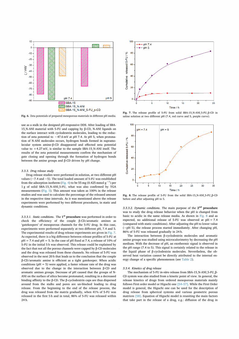
use as a stalk in the designed pH-responsive DDS. After loading of SBA-15_N-ANI material with 5-FU and capping by β-CD, N-ANI ligands onthe surface interact with cyclodextrin molecules, leading to the reduc-tion of zeta potential to −47.6 mV at pH 7.4. At pH 5, when protona-tion of N-ANI molecules occurs, hydrogen bonds formed in supramo-lecular system amine-β-CD disappeared and effected zeta potentialvalue to +4.27mV, is similar to the sample SBA-15_N-ANI itself. Theresults of the zeta potential measurements confirm the mechanism ofgate closing and opening through the formation of hydrogen bondsbetween the amine groups and β-CD driven by pH change.
3.3.3. Drug release studyDrug release studies were performed in solution, at two different pH
values (~7.4 and ~5). The total loaded amount of 5-FU was establishedfrom the adsorption isotherm (Fig. 4) to be 55mg (0.425mmol⋅g−1) per1 g of solid SBA-15_N-ANI_5-FU, what was also confirmed by TGAmeasurements (Fig. 5). This amount was taken as 100% in the releasestudies and was used to calculate the percentage of the released amountin the respective time intervals. As it was mentioned above the releaseexperiments were performed by two different procedures, in static anddynamic conditions.
3.3.3.1. Static conditions. The 1st procedure was performed in order tocheck the efficiency of the couple β-CD/aromatic amines as‘gatekeepers’ of mesoporous channels for the 5-FU drug. In this case,experiments were performed separately at two different pH, 7.4 and 5.The experimental results of drug release experiments are given in Fig. 7.As expected, there is a big difference between release profiles of 5-FU atpH=7.4 and pH=5. In the case of pH fixed at 7.4, a release of 10% of5-FU in the initial 5 h was observed. This release could be explained bythe fact that not all the porous channels were capped by β-CD moleculesand the drug was released from these channels. No release of 5-FU wasobserved in the next 20 h that leads us to the conclusion that the coupleβ-CD/aromatic amine is efficient as a tight gatekeeper. When acidicconditions (pH=5) were applied, a faster release rate of the drug wasobserved due to the change in the interaction between β-CD andaromatic amines groups. Decrease of pH caused that the groups of N-ANI on the surface of silica become protonated, resulting in a decreasedbinding affinity to the β-CD. The β-cyclodextrin caps are thus dispersedaround from the stalks and pores are un-blocked leading to drugrelease. From the beginning to the end of the release process, thedrug was released from the matrix gradually, when 41% of 5-FU wasreleased in the first 5 h and in total, 86% of 5-FU was released within24 h.
3.3.3.2. Dynamic conditions. The main purpose of the 2nd procedurewas to study the drug release behavior when the pH is changed frombasic to acidic in the same release media. As shown in Fig. 8 and asexpected, no additional release of 5-FU was observed at pH=7.4(compared with static conditions). After adjusting the pH to lower value(~pH 5), the release process started immediately. After changing pH,86% of 5-FU was released gradually in 24 h.
The interaction between β-cyclodextrin molecules and aromaticamine groups was studied using microcalorimetry by decreasing the pHmedium. With the decrease of pH, an exothermic signal is observed inthe pH range (7.4 to 5). This signal is certainly related to the release inthe liquid phase of β-cyclodextrin molecules. Nevertheless, the ob-served heat variation cannot be directly attributed to the internal en-ergy change of a specific phenomenon (see Table 2).
3.3.4. Kinetics of drug releaseThe mechanism of 5-FU in-vitro release from SBA-15_N-ANI_5-FU_β-
CD system was also studied from a kinetic point of view. In general, therelease kinetics of drugs from ordered mesoporous materials mainlyfollows First order model or Higuchi one [54–57]. While the First Ordermodel is general, the Higuchi one can be used for the description ofdrug release from spherical systems and various geometric porousmatrices [58]. Equation of Higuchi model is reuniting the main factorsthat take part in the release of a drug, e.g.: diffusion of the drug in
Fig. 6. Zeta potentials of prepared mesoporous materials in different pH media.Fig. 7. The release profile of 5-FU from solid SBA-15_N-ANI_5-FU_β-CD insaline solution at two different pH (7.4, red curve and 5, purple curve).
Fig. 8. The release profile of 5-FU from the solid SBA-15_N-ANI_5-FU_β-CDbefore and after adjusting pH to 5.
E. Beňová, et al. $SSOLHG�6XUIDFH�6FLHQFH������������������
�

dissolvent, the tortuosity factor of the system, the porosity of the ma-trix, total amount of drug in the matrix and the solubility of the drug inthe solvent used [59]. The limitation linked to drug solubility that isone basis of the Higuchi model is not validated in our case. Never-theless, as shown in Table 3, it was found that the in vitro release of 5-FU from SBA-15_N-ANI_5-FU_β-CD at pH 5 could be explained by bothHiguchi’s and First order models, as the plots showed the highest lin-earity with the coefficient of determination (COD) equal to 0.9913 and0.9914 respectively. The values of kinetic constants wereK1= 0.0663 h−1 for First order model and KH=17.486 h−0,5 for Hi-guchi model. The quasi-identical values of COD can be explained con-sidering that the drug kinetics release from the porous system is notaffected by the oversaturation step inherent from the Higuchi model.
3.4. The cytotoxicity of SBA-15 and SBA-15_N-ANI_5-FU_β-CD systems inU87 MG cells
The human glioma U87 MG cells were demonstrated to representthe glycolytic type of the cancer cells model [60]. Since certain tissueand the tumors have more acidic pH than the normal tissue [61], wehave investigated the 5-FU, SBA-15 and SBA-15_N-ANI_5-FU_β-CD atphysiological (pH=7.3) and acidic (pH=6.3) pH of the cell culturemedia.
The morphology of U87 MG cells at both pH in the presence of0.05mg/mL of 5-FU, 1.5mg/mL of SBA-15 and 2mg/mL of SBA-15_N-ANI_5-FU_β-CD is presented in Fig. 9a. The cells were incubated withthe systems 24 h and the significant changes in the morphology of thecells were not found (see white arrows that identify the cells). The SBA-15 and SBA-15_N-ANI_5-FU_β-CD systems were observed in the darkclouds localized in the extracellular media close to the cells. It can beseen that the localization of these clouds is not homogeneous.
The viability of cells in the presence of 0.05mg/mL of 5-FU, 1.5 mg/mL of SBA-15 and 2mg/mL of SBA-15_N-ANI_5-FU_β-CD at pH=7.3and 6.3 was assessed with MTT assay (Fig. 9b). The drop of the extra-cellular pH caused inhibition of the cell proliferation in comparisonwith the cells at pH=7.3. The 24 h treatment with 5-FU significantlydecreased the proliferation. However, more than 80% of the cells werestill active. This finding is in the agreement with previously publishedstudies [62,63]. In those studies, the cytotoxic effect of 5-FU was ob-served at a concentration above 0.5 mg/mL but 48 h after 5-FU ad-ministration.
In the present study, the formation of formazan in the mitochondriaof U87 MG cells was significantly inhibited in the presence of SBA-15and SBA-15_N-ANI_5-FU_β-CD (Fig. 9b). Regarding the cell morphologydetected by microscopy, these observations were unexpected. When wecompare the results from microscopy images and MTT-assay it is evi-dent that the morphology of the cells in the presence of SBA-15 andSBA-15_N-ANI_5-FU_β-CD did not change compared to the morphologyof the controlled cells (Fig. 9a, right). We expected that results of the
MTT-assay would also be similar to the control as it was with micro-scopy results. However, MTT-assay has shown that formation of for-mazan in the cells was significantly inhibited in the presence of SBA-15and SBA-15_N-ANI_5-FU_β-CD (Fig. 9b).
In Hu and Zhao works [62,63], the 5-FU application increased theoxidative stress level in the cancer cells and resulted in the mitochon-dria membrane potential (ΔΨm) dissipation in further. The mitochon-drial probes, especially MTO, can indicate oxidative stress and ΔΨm inliving cells [60,64]. The MTO fluorescence intensity in U87 MG cellsdetected by flow cytometry and fluorescence microscopy is demon-strated in Fig. 9c. While SBA-15 system slightly decreased the MTOfluorescence, the SBA-15_N-ANI_5-FU_β-CD system completely dis-sipated the ΔΨm. The mitochondrial localization of MTO was observedin the control and SBA-15 treated cells. No fluorescence was detected inthe SBA-15_N-ANI_5-FU_β-CD treated cells. The similar results wereobserved at pH=7.3 and 6.3. It suggests that the extracellular pH didnot significantly influence the cell response to treatments.
The U87 MG cells express a higher number of receptors, which fa-cilitate the endocytotic type of drugs transport [65]. It was demon-strated that in a relatively short time (within 1 h), the transport systemcan pass the membrane via endocytosis and subsequently localize inlysosomes of cancer cells [66]. The lysosomes of cells are the cellularcompartments with acidic pH (pH=5.5) [61]. As it was mentionedabove, the SBA-15_N-ANI_5-FU_β-CD system is pH sensitive and 5-FUshould be released from the system at pH=5.5. The 5-FU is notfluorescent. For this reason, the indirect method was used to identifythe release of 5-FU from the SBA-15_N-ANI_5-FU_β-CD.
The representative confocal fluorescence images of U87 MG cells24 h treated with 5-FU, SBA-15, and SBA-15_N-ANI_5-FU_β-CD systemare depicted in Fig. 10a. The cells were stained with MTO to visualizedthe mitochondria and LysoTrackerTM Green to identify the lysosomalcompartments. The nuclei of the cells were contrasted with Hoechst.The control group is represented with tubular mitochondria and glob-ular lysosomes. The application of 0.02mg/mL of 5-FU did not inducesignificant modifications. However, the application of 0.6 mg/mL ofSBA-15 amplified fluorescence of LysoTrackerTM Green. The tubularmitochondria were observed in the SBA-15 treated cells. It should benoted that the clouds of SBA-15 system in the extracellular area en-trapped the number of fluorescent probes and significantly reducedHoechst staining of nuclei. This effect was amplified in the SBA-15_N-ANI_5-FU_β-CD treated cells. The blue (Hoechst) fluorescence wasmostly attributed to the SBA-15_N-ANI_5-FU_β-CD system localization(Fig. 10a). The MTO fluorescence completely disappeared in these cells.The swollen lysosomes can be identified from the images of Lyso-TrackerTM fluorescence.
The cancer cells became highly adaptive to intracellular oxidativestress and revealed the way how to rescue and survive [60,67]. Re-cently, it was shown that the nanoceria applied to the U87 MG cellslocally increased the oxidative stress level in the perinuclear areawithout affection of the cell proliferation [68]. One can expect the samebehavior in cells treated with SBA-1. The MTT-assay, performed in thesame cell as detected in Fig. 10a demonstrates a significant reduction offormazan production in SBA-15 and SBA-15_N-ANI_5-FU_β-CD treatedcells (Fig. 10b). The almost 50% decrease observed could be the resultof ΔΨm dissipation, lysosomal destabilization, and induction of mi-tochondria-mediated apoptosis.
The correlation between MTO and LysoTrackerTM Green fluores-cence was measured by flow cytometry. The characteristic shift into
Table 2Summarization of the values obtained from the thermal analysis, N2 adsorption/desorption measurements and solid/liquid adsorption measurements.
SAMPLE N-ANI grafted, n [mmol⋅g−1] SBET [m2⋅g−1] 5-FU, nADS [mmol⋅g−1] 5-FU, nADS [mmol⋅m−2] 5-FU, mADS [mg⋅g−1] 5-FU, mADS [mg⋅m−2]
SBA-15 790 0.712 0.901 92.63 0.117SBA-15_N-ANI 1.26 400 0.425 1.0625 55.3 0.138
Table 3Coefficient of determination values for the two drug release kinetics models ofSBA-15_N-ANI_5-FU_β-CD.First order Higuchi model
r2 K1 [h−1] r2 Kh [h−0,5]
0.9913 0.06863 0.9914 17.486
E. Beňová, et al. $SSOLHG�6XUIDFH�6FLHQFH������������������
�

upper right quadrant was observed in the non-treated controlled cellslabeled with the probes (Fig. 10c). This character maintained in thecells 1 h treated with 5-FU (0.01 and 0.05mg/mL) and SBA-15 (0.3 and0.6 mg/mL). The reduction of MTO fluorescence was found in the 50%of cells treated with 1.5 mg/mL of SBA-15. Lysosomal integrity haspersevered. In contrast to SBA-15, the SBA-15_N-ANI_5-FU_β-CD systemtriggered dissipation of ΔΨm and destabilization of lysosomes. The79.68% of cells were detected in lower left quadrant (MTO negative andLysoTrackerTM green negative) at 0.4 mg/mL concentration and96.26% was observed at 2mg/mL of the SBA-15_N-ANI_5-FU_β-CD.
These results demonstrated that the 5-FU did not induce cell deathat the studied concentration and during 24 h. Furthermore, SBA-15application to cells slightly decrease mitochondria membrane potentialbut SBA-15 adsorbed the certain number of applied fluorescent probe’smolecules. Due to this effect, it could be assumed that the MTT wassimilarly entrapped into SBA-15 during MTT-assay, and subsequently
resulted in a reduction of formazan production. The mitochondriadysfunction and SBA-15 absorption properties could be the reason forreduced formazan production, which was observed in the present study.For this reason, the direct estimation of the cell viability (MTT-assay)should be supplemented with indirect approaches (ΔΨm and destabili-zation of lysosomes). These indirect approaches confirmed the cytotoxiceffect of the SBA-15_N-ANI_5-FU_β-CD. Regarding to the total loadingconcentration of 5-FU, appropriate concentration of free 5-FU re-presents 0.018mg/mL in 0.4mg/mL SBA-15_N-ANI_5-FU_β-CD. As itwas reported, this concentration of 5-FU was not cytotoxic if appliedinto cell culture media, neither after 48 h [62]. However, when it wasdelivered into the cells by SBA-15_N-ANI_5-FU_β-CD it possessed sig-nificant cytotoxic effects. The degradation of lysosomes was probablyinduced with 5-FU released from SBA-15_N-ANI_5-FU_β-CD within thelysosomes at pH=5.5. The presence of SBA-15 within the cell could besuggested from the elevated levels of oxidative stress, which partially
Fig. 9. The representative a) white-field images of U87 MG cells detected 24 h after application of 0.05mg/mL 5-FU, 1.5 mg/mL SBA-15 and 2mg/mL SBA-15_N-ANI_5-FU_β-CD at pH=7.3 and 6.3 (the white arrows highlighted the cells). The MTT-assay b) was assessed in the cells at the same conditions as in a). The level ofsignificance was calculated from the controls at pH=7.3 (***p < 0.001) and pH=6.3 (••p < 0.01 and •••p < 0.001). The mitochondria membrane potential c)was estimated with MTO staining by flow cytometry (left) and confocal fluorescence microscopy (right) at the same conditions as in a).
E. Beňová, et al. $SSOLHG�6XUIDFH�6FLHQFH������������������
�

Fig. 10. The representative a) confocal fluorescence images of U87 MG cells detected 24 h after application of 0.02mg/mL 5-FU, 0.6mg/mL SBA-15 and 0.8 mg/mLSBA-15_N-ANI_5-FU_β-CD at pH 6.3. Mitochondria were visualized with MTO, the lysosomes with LysoTrackerTM Green and nuclei were contrasted with Hoechst. Thefluorescent images of lysosomes are depicted below the overlapped images. The MTT-assay b) was assessed in the cells at the same conditions as in a). The level ofsignificance was calculated from the controls (***p < 0.001). The correlations of MTO and LysoTrackerTM Green fluorescence c) were estimated by flow cytometryin cells 1 h treated with the 5-FU, SBA-15 and SBA-15_N-ANI_5-FU_β-CD at pH 7.3. The correlation plot was divided into four quadrants. The upper right quadrantrepresents the control cells positive for MTO and LysoTrackerTM Green. The lower left quadrant characterizes the dissipation of the ΔΨm and destabilization oflysosomes. The number of cells is identified by the color-code (blue – minimum, red – maximum).
E. Beňová, et al. $SSOLHG�6XUIDFH�6FLHQFH������������������
��

caused the dissipation of ΔΨm. The SBA-15_N-ANI_5-FU_β-CD systemsseems to be more effective in anti-cancer treatment than sole 5-FU.
4. Conclusions
A system based on mesoporous silica material was studied as apotential pH-sensitive drug delivery system for the antineoplastic agent5-fluorouracil (5-FU). This system is composed of SBA-15-type porousstructure (hexagonal) functionalized with aromatic primary amines (N-ANI) and β-cyclodextrin molecules (β-CD) that are used as nanovalves.
Starting from the synthesis and characterization of SBA-15 material,the successful functionalization of N-ANI groups was evidenced usingthermogravimetric measurements (ngrafted= 1.26mmol/g). Texturalproperties of functionalized samples were investigated using smallangle XRD, TEM, and nitrogen sorption measurements that allow us toconclude that even if a decrease was observed in terms of specificsurface area (−50%) and porous volume (−25%), the whole porousstructure was preserved and available for drug molecules.
Then, the loading of the drug 5-FU in pure SBA-15 and SBA-15_N-ANI was studied from a thermodynamic point of view via the con-struction of adsorption isotherms and calorimetric measurements. Forboth solids, adsorption isotherms are of Langmuir type. The affinity of5-FU molecules (linked to the slope of isotherms at low concentrations)towards both solids is weak. Calorimetric measurements evidenced aweak constant enthalpies of adsorption for both solids (ΔH=−3/−4kJ/mol). Finally, the only difference observed between both solids liesin the maximum adsorbed amounts of 5-FU determined both experi-mentally (7.3 ⋅ 10−4 mol/g vs 4.2 ⋅ 10−4 mol/g); this difference islinked to the difference in specific surface area. The maximum loadingof 5-FU determined from both the adsorption isotherm and thermo-gravimetric measurements for the N-ANI functionalized porous mate-rial was 55mg/g.
The pH-sensitive release of 5-FU from SBA-15_N-ANI solid, using β-CD molecules as nanovalves, was finally investigated in static and dy-namic conditions: it was shown that there was almost no release of 5-FUat pH=7.4 whereas more than 80% of adsorbed 5-FU was progres-sively released at pH=5. The modeling of the 5-FU release was suc-cessfully done using both first order and Higuchi models. The cyto-toxicity tests were performed using human glioma U87 MG cells. Theresults demonstrated that the 5-FU did not induce cell death at thestudied concentration if applied into cell culture media. However, whenit was delivered into the cells by SBA-15_N-ANI_5-FU_β-CD it possessedsignificant cytotoxic effects.
Acknowledgments
This work was supported by the Slovak Research and theDevelopment Agency under the contracts APVV-15-0520 and APVV-15-0485 and national bilateral grant APVV-SK-FR-2017-0011. E.B. thanksto the financial support from VVGS-PG-2017-677 grant. E.B. also thanksto the French Government for the financial support linked to the im-plementation of a joint French-Slovak doctorate. The authors thank theMinistry of Education, Science, Research and Sport of the SlovakRepublic and the Accreditation Commission for the financial support ofthe TRIANGEL team in the frame of the scheme “Top Research Teams inSlovakia”.
Appendix A. Supplementary material
Supplementary data to this article can be found online at https://doi.org/10.1016/j.apsusc.2019.144028.
References
[1] J. Lu, M. Liong, J.I. Zink, F. Tamanoi, Mesoporous silica nanoparticles as a deliverysystem for hydrophobic anticancer drugs, Small 3 (2007) 1341–1346, https://doi.
org/10.1002/smll.200700005.[2] T.P. Lodise, J. Graves, A. Evans, E. Graffunder, M. Helmecke, B.M. Lomaestro,
K. Stellrecht, Relationship between vancomycin MIC and failure among patientswith methicillin-resistant staphylococcus aureus bacteremia treated with vanco-mycin, Antimicrob. Agents Chemother. 52 (2008) 3315–3320, https://doi.org/10.1128/AAC.00113-08.
[3] K.K. Jain, ed., Drug Delivery Systems, Humana Press, 2008. //www.springer.com/us/book/9781588298911 (accessed September 4, 2018).
[4] T.M. Allen, P.R. Cullis, Drug delivery systems: entering the mainstream, Science 303(2004) 1818–1822, https://doi.org/10.1126/science.1095833.
[5] J. Zhou, M. Wang, H. Ying, D. Su, H. Zhang, G. Lu, J. Chen, Extracellular matrixcomponent shelled nanoparticles as dual enzyme-responsive drug delivery vehiclesfor cancer therapy, ACS Biomater. Sci. Eng. 4 (2018) 2404–2411, https://doi.org/10.1021/acsbiomaterials.8b00327.
[6] M. Zhang, F. Wu, W. Wang, J. Shen, N. Zhou, C. Wu, Multifunctional nano-composites for targeted, photothermal, and chemotherapy, Chem. Mater. (2018),https://doi.org/10.1021/acs.chemmater.8b00934.
[7] L. Zhang, Y. Li, J.C. Yu, Chemical modification of inorganic nanostructures fortargeted and controlled drug delivery in cancer treatment, J. Mater. Chem. B. 2(2014) 452–470, https://doi.org/10.1039/C3TB21196G.
[8] S. Carrasco, E. Benito-Peña, F. Navarro-Villoslada, J. Langer, M.N. Sanz-Ortiz,J. Reguera, L.M. Liz-Marzán, M.C. Moreno-Bondi, Multibranched gold-mesoporoussilica nanoparticles coated with a molecularly imprinted polymer for label-freeantibiotic surface-enhanced raman scattering analysis, Chem. Mater. 28 (2016)7947–7954, https://doi.org/10.1021/acs.chemmater.6b03613.
[9] A.L. Doadrio, J.C. Doadrio, J.M. Sánchez-Montero, A.J. Salinas, M. Vallet-Regí, Arational explanation of the vancomycin release from SBA-15 and its derivative bymolecular modelling, Micropor. Mesopor. Mater. 132 (2010) 559–566, https://doi.org/10.1016/j.micromeso.2010.04.010.
[10] M. Moritz, M. Geszke-Moritz, Mesoporous silica materials with different structuresas the carriers for antimicrobial agent. Modeling of chlorhexidine adsorption andrelease, Appl. Surf. Sci. 356 (2015) 1327–1340, https://doi.org/10.1016/j.apsusc.2015.08.138.
[11] E. Beňová, V. Zeleňák, D. Halamová, Miroslav Almáši, Veronika Petrul’ová,M. Psotka, A. Zeleňáková, M. Bačkor, V. Hornebecq, A drug delivery system basedon switchable photo-controlled p-coumaric acid derivatives anchored on meso-porous silica, J. Mater. Chem. B. 5 (2017) 817–825, https://doi.org/10.1039/C6TB02040B.
[12] V. Zeleňák, E. Beňová, M. Almáši, D. Halamová, V. Hornebecq, V. Hronský, Photo-switchable nanoporous silica supports for controlled drug delivery, New J. Chem.42 (2018) 13263–13271, https://doi.org/10.1039/C8NJ00267C.
[13] V. Zeleňák, D. Halamová, M. Almáši, L. Žid, A. Zeleňáková, O. Kapusta, Orderedcubic nanoporous silica support MCM-48 for delivery of poorly soluble drug in-domethacin, Appl. Surf. Sci. 443 (2018) 525–534, https://doi.org/10.1016/j.apsusc.2018.02.260.
[14] D. Halamová, M. Badaničová, V. Zeleňák, T. Gondová, U. Vainio, Naproxen drugdelivery using periodic mesoporous silica SBA-15, Appl. Surf. Sci. 256 (2010)6489–6494, https://doi.org/10.1016/j.apsusc.2010.04.044.
[15] Y. Zhang, J. Wang, X. Bai, T. Jiang, Q. Zhang, S. Wang, Mesoporous silica nano-particles for increasing the oral bioavailability and permeation of poorly watersoluble drugs, Mol. Pharm. 9 (2012) 505–513, https://doi.org/10.1021/mp200287c.
[16] R. Mellaerts, R. Mols, J.A.G. Jammaer, C.A. Aerts, P. Annaert, J. Van Humbeeck,G. Van den Mooter, P. Augustijns, J.A. Martens, Increasing the oral bioavailabilityof the poorly water soluble drug itraconazole with ordered mesoporous silica, Eur.J. Pharm. Biopharm. Off. J. Arbeitsgemeinschaft Pharm. Verfahrenstechnik EV. 69(2008) 223–230, https://doi.org/10.1016/j.ejpb.2007.11.006.
[17] A. Gulzar, S. Gai, P. Yang, C. Li, M.B. Ansari, J. Lin, Stimuli responsive drug deliveryapplication of polymer and silica in biomedicine, J. Mater. Chem. B. 3 (2015)8599–8622, https://doi.org/10.1039/C5TB00757G.
[18] T. Zhao, L. Chen, Q. Li, X. Li, Near-infrared light triggered drug release from me-soporous silica nanoparticles, J. Mater. Chem. B. 6 (2018) 7112–7121, https://doi.org/10.1039/C8TB01548A.
[19] Y. Wang, Y. Cui, J. Huang, D. Di, Y. Dong, X. Zhang, Q. Zhao, N. Han, Y. Gao,T. Jiang, S. Wang, Redox and pH dual-responsive mesoporous silica nanoparticlesfor site-specific drug delivery, Appl. Surf. Sci. 356 (2015) 1282–1288, https://doi.org/10.1016/j.apsusc.2015.07.151.
[20] P. Nadrah, F. Porta, O. Planinšek, A. Kros, M. Gaberšček, Poly(propylene imine)dendrimer caps on mesoporous silica nanoparticles for redox-responsive release:smaller is better, Phys. Chem. Chem. Phys. 15 (2013) 10740–10748, https://doi.org/10.1039/C3CP44614J.
[21] S.H. van Rijt, D.A. Bölükbas, C. Argyo, S. Datz, M. Lindner, O. Eickelberg,M. Königshoff, T. Bein, S. Meiners, Protease-mediated release of chemotherapeuticsfrom mesoporous silica nanoparticles to ex vivo human and mouse lung tumors,ACS Nano. 9 (2015) 2377–2389, https://doi.org/10.1021/nn5070343.
[22] Y. Yang, J. Wan, Y. Niu, Z. Gu, J. Zhang, M. Yu, C. Yu, Structure-dependent andglutathione-responsive biodegradable dendritic mesoporous organosilica nano-particles for safe protein delivery, Chem. Mater. 28 (2016) 9008–9016, https://doi.org/10.1021/acs.chemmater.6b03896.
[23] Q. Yang, S. Wang, P. Fan, L. Wang, Y. Di, K. Lin, F.-S. Xiao, pH-responsive carriersystem based on carboxylic acid modified mesoporous silica and polyelectrolyte fordrug delivery, Chem. Mater. 17 (2005) 5999–6003, https://doi.org/10.1021/cm051198v.
[24] C. Gao, H. Zheng, L. Xing, M. Shu, S. Che, Designable coordination bonding inmesopores as a pH-responsive release system, Chem. Mater. 22 (2010) 5437–5444,https://doi.org/10.1021/cm100667u.
E. Beňová, et al. $SSOLHG�6XUIDFH�6FLHQFH������������������
��

[25] L. Xing, H. Zheng, Y. Cao, S. Che, Coordination polymer coated mesoporous silicananoparticles for ph-responsive drug release, Adv. Mater. 24 (2012) 6433–6437,https://doi.org/10.1002/adma.201201742.
[26] C.-H. Lee, S.-H. Cheng, I.-P. Huang, J.S. Souris, C.-S. Yang, C.-Y. Mou, L.-W. Lo,Intracellular pH-responsive mesoporous silica nanoparticles for the controlled re-lease of anticancer chemotherapeutics, Angew. Chem. Int. Ed. 49 (2010)8214–8219, https://doi.org/10.1002/anie.201002639.
[27] J. Fan, S. Wang, W. Sun, S. Guo, Y. Kang, J. Du, X. Peng, Anticancer drug deliverysystems based on inorganic nanocarriers with fluorescent tracers, AIChE J. 64(2018) 835–859, https://doi.org/10.1002/aic.15976.
[28] H. Li, L.-L. Tan, P. Jia, Q.-L. Li, Y.-L. Sun, J. Zhang, Y.-Q. Ning, J. Yu, Y.-W. Yang,Near-infrared light-responsive supramolecular nanovalve based on mesoporous si-lica-coated gold nanorods, Chem. Sci. 5 (2014) 2804–2808, https://doi.org/10.1039/C4SC00198B.
[29] S.U.N. Yu-Long, Y. Ying-Wei, W.U. Wei, Z.S. Xiao-An, Supramolecular nanovalvesystems based on macrocyclic synthetic receptors, Chem. J. Chin. Univ. 33 (2012)1635–1642, https://doi.org/10.3969/j.issn.0251-0790.2012.08.001.
[30] A.E. Kaziem, Y. Gao, Y. Zhang, X. Qin, Y. Xiao, Y. Zhang, H. You, J. Li, S. He, α-Amylase triggered carriers based on cyclodextrin anchored hollow mesoporous si-lica for enhancing insecticidal activity of avermectin against Plutella xylostella, J.Hazard. Mater. 359 (2018) 213–221, https://doi.org/10.1016/j.jhazmat.2018.07.059.
[31] S. Angelos, Y.-W. Yang, K. Patel, J.F. Stoddart, J.I. Zink, pH-responsive supramo-lecular nanovalves based on cucurbit[6]uril pseudorotaxanes, Angew. Chem. Int.Ed. 47 (2008) 2222–2226, https://doi.org/10.1002/anie.200705211.
[32] H. Meng, M. Xue, T. Xia, Y.-L. Zhao, F. Tamanoi, J.F. Stoddart, J.I. Zink, A.E. Nel,Autonomous in vitro anticancer drug release from mesoporous silica nanoparticlesby ph-sensitive nanovalves, J. Am. Chem. Soc. 132 (2010) 12690–12697, https://doi.org/10.1021/ja104501a.
[33] L. Bai, Q. Zhao, J. Wang, Y. Gao, Z. Sha, D. Di, N. Han, Y. Wang, J. Zhang, S. Wang,Mechanism study on pH-responsive cyclodextrin capped mesoporous silica: effect ofdifferent stalk densities and the type of cyclodextrin, Nanotechnology 26 (2015)165704, , https://doi.org/10.1088/0957-4484/26/16/165704.
[34] A. Mathew, S. Parambadath, S.S. Park, C.-S. Ha, Hydrophobically modified sphe-rical MCM-41 as nanovalve system for controlled drug delivery, Micropor. Mesopor.Mater. 200 (2014) 124–131, https://doi.org/10.1016/j.micromeso.2014.08.033.
[35] X. Zhu, C.-Q. Wang, pH and redox-operated nanovalve for size-selective cargo de-livery on hollow mesoporous silica spheres, J. Colloid Interface Sci. 480 (2016)39–48, https://doi.org/10.1016/j.jcis.2016.06.043.
[36] D.B.G. Williams, M. Lawton, Drying of organic solvents: quantitative evaluation ofthe efficiency of several desiccants, J. Org. Chem. 75 (2010) 8351–8354, https://doi.org/10.1021/jo101589h.
[37] D. Zhao, Q. Huo, J. Feng, B.F. Chmelka, G.D. Stucky, Nonionic triblock and stardiblock copolymer and oligomeric surfactant syntheses of highly ordered, hydro-thermally stable, mesoporous silica structures, J. Am. Chem. Soc. 120 (1998)6024–6036, https://doi.org/10.1021/ja974025i.
[38] Y.-L. Fan, B.-Y. Fan, Q. Li, H.-X. Di, X.-Y. Meng, N. Ling, Preparation of 5-fluor-ouracil-loaded nanoparticles and study of interaction with gastric cancer cells,Asian Pac. J. Cancer Prev. 15 (2014) 7611–7615, https://doi.org/10.7314/APJCP.2014.15.18.7611.
[39] D. Kala, Jijo Thomas, Deepa V. Nair, Effect of pH on aminofunctionalized meso-porous silica nanoparticles loaded with 5-fluorouracil and its optimization, WorldPharm. Pharm. Sci. 7 (2018) 1400–1419.
[40] S. Egodawatte, S. Dominguez, S.C. Larsen, Solvent effects in the development of adrug delivery system for 5-fluorouracil using magnetic mesoporous silica nano-particles, Micropor. Mesopor. Mater. 237 (2017) 108–116, https://doi.org/10.1016/j.micromeso.2016.09.024.
[41] Adsorption by Powders and Porous Solids - 2nd Edition, (n.d.). https://www.elsevier.com/books/adsorption-by-powders-and-porous-solids/rouquerol/978-0-08-097035-6 (accessed November 23, 2018).
[42] V. Zeleňák, D. Halamová, A. Zeleňáková, V. Girman, Periodic 3D nanoporous silicamodified by amine or SPION nanoparticles as NSAID delivery system, J. PorousMater. 23 (2016) 1633–1645, https://doi.org/10.1007/s10934-016-0224-x.
[43] E. Beňová, V. Zeleňák, D. Halamová, M. Almáši, V. Petrul’ová, M. Psotka,A. Zeleňáková, M. Bačkor, V. Hornebecq, A drug delivery system based onswitchable photo-controlled p-coumaric acid derivatives anchored on mesoporoussilica, J. Mater. Chem. B. 5 (2017) 817–825, https://doi.org/10.1039/C6TB02040B.
[44] N.S. Rejinold, K.P. Chennazhi, S.V. Nair, H. Tamura, R. Jayakumar, Biodegradableand thermo-sensitive chitosan-g-poly(N-vinylcaprolactam) nanoparticles as a 5-fluorouracil carrier, Carbohydr. Polym. 83 (2011) 776–786, https://doi.org/10.1016/j.carbpol.2010.08.052.
[45] D. Zhao, J. Sun, Q. Li, G.D. Stucky, Morphological control of highly ordered me-soporous silica SBA-15, Chem. Mater. 12 (2000) 275–279, https://doi.org/10.1021/cm9911363.
[46] M. Thommes, K. Kaneko, A.V. Neimark, J.P. Olivier, F. Rodriguez-Reinoso,J. Rouquerol, K.S.W. Sing, Physisorption of gases, with special reference to theevaluation of surface area and pore size distribution (IUPAC Technical Report),Pure Appl. Chem. 87 (2015) 1051–1069, https://doi.org/10.1515/pac-2014-1117.
[47] Y. Zeng, L. Prasetyo, S.J. Tan, C. Fan, D.D. Do, D. Nicholson, On the hysteresis of
adsorption and desorption of simple gases in open end and closed end pores, Chem.Eng. Sci. 158 (2017) 462–479, https://doi.org/10.1016/j.ces.2016.10.048.
[48] C. Gebald, J.A. Wurzbacher, P. Tingaut, T. Zimmermann, A. Steinfeld, Amine-basednanofibrillated cellulose as adsorbent for CO2 capture from air, Environ. Sci.Technol. 45 (2011) 9101–9108, https://doi.org/10.1021/es202223p.
[49] C.H. Giles, D. Smith, A. Huitson, A general treatment and classification of the soluteadsorption isotherm. I. Theoretical, J. Colloid Interface Sci. 47 (1974) 755–765,https://doi.org/10.1016/0021-9797(74)90252-5.
[50] F. Trotta, M. Zanetti, G. Camino, Thermal degradation of cyclodextrins, Polym.Degrad. Stab. 69 (2000) 373–379, https://doi.org/10.1016/S0141-3910(00)00084-7.
[51] M. Colilla, I. Izquierdo-Barba, S. Sánchez-Salcedo, J.L.G. Fierro, J.L. Hueso,M. Vallet-Regí, Synthesis and characterization of zwitterionic SBA-15 nanos-tructured materials, Chem. Mater. 22 (2010) 6459–6466, https://doi.org/10.1021/cm102827y.
[52] J.M. Rosenholm, M. Lindén, Towards establishing structure-activity relationshipsfor mesoporous silica in drug delivery applications, J. Control. Release Off. J.Control. Release Soc. 128 (2008) 157–164, https://doi.org/10.1016/j.jconrel.2008.02.013.
[53] A. Popat, J. Liu, G.Q. (Max) Lu, S.Z. Qiao, A pH-responsive drug delivery systembased on chitosan coated mesoporous silica nanoparticles, J. Mater. Chem. 22(2012) 11173–11178, https://doi.org/10.1039/C2JM30501A.
[54] J.C. Doadrio, E.M.B. Sousa, I. Izquierdo-Barba, A.L. Doadrio, J. Perez-Pariente,M. Vallet-Regí, Functionalization of mesoporous materials with long alkyl chains asa strategy for controlling drug delivery pattern, J. Mater. Chem. 16 (2006)462–466, https://doi.org/10.1039/B510101H.
[55] L. Polo, N. Gómez-Cerezo, A. García-Fernández, E. Aznar, J.-L. Vivancos, D. Arcos,M. Vallet-Regí, R. Martínez-Máñez, Mesoporous bioactive glasses equipped withstimuli-responsive molecular gates for controlled delivery of levofloxacin againstbacteria, Chem. - Eur. J. (2018), https://doi.org/10.1002/chem.201803301.
[56] S. Pathan, P. Solanki, A. Patel, Functionalized SBA-15 for controlled release ofpoorly soluble drug, Erythromycin, Micropor. Mesopor. Mater. 258 (2018)114–121, https://doi.org/10.1016/j.micromeso.2017.09.012.
[57] M. Hamzehloo, J. Karimi, K. Aghapoor, H. Sayahi, H.R. Darabi, The synergisticcooperation between MCM-41 and azithromycin: a pH responsive system for drugadsorption and release, J. Porous Mater. 25 (2018) 1275–1285, https://doi.org/10.1007/s10934-017-0538-3.
[58] J. Anderson, J. Rosenholm, M., Linden, N. Ashammakhi, Mesoporous silica: Analternative diffusion controlled drug delivery system, in: Top. Multifunct. Biomater.Devices, University of Oulund, Finland, 2008: pp. 1–19.
[59] A. Doadrio, A. Salinas, J. Sánchez-Montero, M. Vallet-Regí, Drug release from or-dered mesoporous silicas, Curr. Pharm. Des. 21 (2015) 6213–6819, https://doi.org/10.2174/1381612822666151106121419.
[60] S. Tomkova, M. Misuth, L. Lenkavska, P. Miskovsky, V. Huntosova, In vitro iden-tification of mitochondrial oxidative stress production by time-resolved fluores-cence imaging of glioma cells, Biochim. Biophys. Acta BBA - Mol. Cell Res. (1865(2018)) 616–628, https://doi.org/10.1016/j.bbamcr.2018.01.012.
[61] X. Chen, X. Yao, L. Chen, Intracellular pH-sensitive dextran-based micelles as effi-cient drug delivery platforms, Polym. Int. 64 (2015) 430–436, https://doi.org/10.1002/pi.4809.
[62] X.-Y. Hu, J.-Y. Liang, X.-J. Guo, L. Liu, Y.-B. Guo, 5-Fluorouracil combined withapigenin enhances anticancer activity through mitochondrial membrane potential(ΔΨm)-mediated apoptosis in hepatocellular carcinoma, Clin. Exp. Pharmacol.Physiol. 42 (2015) 146–153, https://doi.org/10.1111/1440-1681.12333.
[63] H. Zhao, Q. Liu, S. Wang, F. Dai, X. Cheng, X. Cheng, W. Chen, M. Zhang, D. Chen,In vitro additive antitumor effects of dimethoxycurcumin and 5-fluorouracil incolon cancer cells, Cancer Med. 6 (2017) 1698–1706, https://doi.org/10.1002/cam4.1114.
[64] L. Scorrano, V. Petronilli, R. Colonna, F.D. Lisa, P. Bernardi,Chloromethyltetramethylrosamine (Mitotracker OrangeTM) Induces the mi-tochondrial permeability transition and inhibits respiratory complex I implicationsfor the mechanism of cytochrome crelease, J. Biol. Chem. 274 (1999)24657–24663, https://doi.org/10.1074/jbc.274.35.24657.
[65] S. Kascakova, Z. Nadova, A. Mateasik, J. Mikes, V. Huntosova, M. Refregiers,F. Sureau, J.-C. Maurizot, P. Miskovsky, D. Jancura, High level of low-density li-poprotein receptors enhance hypericin uptake by U-87 MG cells in the presence ofLDL, Photochem. Photobiol. 84 (2008) 120–127, https://doi.org/10.1111/j.1751-1097.2007.00207.x.
[66] V. Huntosova, Z. Nadova, L. Dzurova, V. Jakusova, F. Sureau, P. Miskovsky, Celldeath response of U87 glioma cells on hypericin photoactivation is mediated bydynamics of hypericin subcellular distribution and its aggregation in cellular or-ganelles, Photochem. Photobiol. Sci. 11 (2012) 1428–1436, https://doi.org/10.1039/C2PP05409D.
[67] D. Patel, S. Shukla, S. Gupta, Apigenin and cancer chemoprevention: Progress,potential and promise (Review), Int. J. Oncol. 30 (2007) 233–245, https://doi.org/10.3892/ijo.30.1.233.
[68] K. Siposova, V. Huntosova, Y. Shlapa, L. Lenkavska, M. Macajova, A. Belous,A. Musatov, Advances in the study of cerium oxide nanoparticles: new insights intoantiamyloidogenic activity, ACS Appl. Bio Mater. 2 (2019) 1884–1896, https://doi.org/10.1021/acsabm.8b00816.
E. Beňová, et al. $SSOLHG�6XUIDFH�6FLHQFH������������������
��
Related Documents