BioMed Central Page 1 of 12 (page number not for citation purposes) BMC Neuroscience Open Access Research article Adrenalectomy promotes a permanent decrease of plasma corticoid levels and a transient increase of apoptosis and the expression of Transforming Growth Factor β1 (TGF-β1) in hippocampus: effect of a TGF-β1 oligo-antisense Javier A Bravo 1 , Claudio S Parra 1 , Sandor Arancibia 2 , Sergio Andrés 1 , Paola Morales 3 , Mario Herrera-Marschitz 3 , Luisa Herrera 4 , Hernán E Lara 1 and Jenny L Fiedler* 1 Address: 1 Department of Biochemistry and Molecular Biology. Laboratory of Neurobiochemistry. Faculty of Chemical and Pharmaceutical Sciences. Universidad de Chile, Chile, 2 Laboratory of Molecular Mechanisms of Neurodegenerative Diseases, Université de Montpellier, Montpellier, France, 3 Programmes of Molecular & Clinical Pharmacology ICBM, Faculty of Medicine, Universidad de Chile, Chile and 4 Human Genetics, ICBM, Faculty of Medicine, Universidad de Chile, Chile Email: Javier A Bravo - [email protected]; Claudio S Parra - [email protected]; Sandor Arancibia - [email protected]; Sergio Andrés - [email protected]; Paola Morales - [email protected]; Mario Herrera-Marschitz - [email protected]; Luisa Herrera - [email protected]; Hernán E Lara - [email protected]; Jenny L Fiedler* - [email protected] * Corresponding author Abstract Background: Corticosterone reduction produced by adrenalectomy (ADX) induces apoptosis in dentate gyrus (DG) of the hippocampus, an effect related to an increase in the expression of the pro-apoptotic gene bax. However it has been reported that there is also an increase of the anti- apoptotic gene bcl-2, suggesting the promotion of a neuroprotective phenomenon, perhaps related to the expression of transforming growth factor β1 (TGF-β1). Thus, we have investigated whether TGF-β1 levels are induced by ADX, and whether apoptosis is increased by blocking the expression of TGF-β1 with an antisense oligonucleotide (ASO) administered intracerebrally in corticosterone depleted rats. Results: It was observed an increase of apoptosis in DG, 2 and 5 days after ADX, in agreement with a reduction of corticosterone levels. However, the effect of ADX on the number of apoptotic positive cells in DG was decreased 5 days after the lesion. In CA1–CA3 regions, the effect was only observed 2 days after ADX. TGF-β1 mRNA levels were increased 2 days after ADX. The sustained intracerebro-ventricular administration of a TGF-β1 ASO via an osmotic mini pump increased apoptosis levels in CA and DG regions 5 days after ADX as well as sham-operated control animals. No significant effect was observed following a scrambled-oligodeoxynucleotide treatment. Conclusion: The changes in both the pattern and the magnitude of apoptotic-cell morphology observed 2 and 5 days after ADX suggest that, as a consequence of the reduction of corticosteroids, some trophic mechanisms restricting cell death to a particular time window are elicited. Sustained intracerebral administration of TGF-β1 ASO increased the apoptosis promoted by ADX, suggesting that TGF-β1 plays an anti-apoptotic role in vivo in hippocampus. Published: 19 May 2006 BMC Neuroscience 2006, 7:40 doi:10.1186/1471-2202-7-40 Received: 17 February 2006 Accepted: 19 May 2006 This article is available from: http://www.biomedcentral.com/1471-2202/7/40 © 2006 Bravo et al; licensee BioMed Central Ltd. This is an Open Access article distributed under the terms of the Creative Commons Attribution License (http://creativecommons.org/licenses/by/2.0 ), which permits unrestricted use, distribution, and reproduction in any medium, provided the original work is properly cited.

Welcome message from author
This document is posted to help you gain knowledge. Please leave a comment to let me know what you think about it! Share it to your friends and learn new things together.
Transcript
BioMed CentralBMC Neuroscience
ss
Open AcceResearch articleAdrenalectomy promotes a permanent decrease of plasma corticoid levels and a transient increase of apoptosis and the expression of Transforming Growth Factor β1 (TGF-β1) in hippocampus: effect of a TGF-β1 oligo-antisenseJavier A Bravo1, Claudio S Parra1, Sandor Arancibia2, Sergio Andrés1, Paola Morales3, Mario Herrera-Marschitz3, Luisa Herrera4, Hernán E Lara1 and Jenny L Fiedler*1Address: 1Department of Biochemistry and Molecular Biology. Laboratory of Neurobiochemistry. Faculty of Chemical and Pharmaceutical Sciences. Universidad de Chile, Chile, 2Laboratory of Molecular Mechanisms of Neurodegenerative Diseases, Université de Montpellier, Montpellier, France, 3Programmes of Molecular & Clinical Pharmacology ICBM, Faculty of Medicine, Universidad de Chile, Chile and 4Human Genetics, ICBM, Faculty of Medicine, Universidad de Chile, Chile
Email: Javier A Bravo - [email protected]; Claudio S Parra - [email protected]; Sandor Arancibia - [email protected]; Sergio Andrés - [email protected]; Paola Morales - [email protected]; Mario Herrera-Marschitz - [email protected]; Luisa Herrera - [email protected]; Hernán E Lara - [email protected]; Jenny L Fiedler* - [email protected]
* Corresponding author
AbstractBackground: Corticosterone reduction produced by adrenalectomy (ADX) induces apoptosis indentate gyrus (DG) of the hippocampus, an effect related to an increase in the expression of thepro-apoptotic gene bax. However it has been reported that there is also an increase of the anti-apoptotic gene bcl-2, suggesting the promotion of a neuroprotective phenomenon, perhaps relatedto the expression of transforming growth factor β1 (TGF-β1). Thus, we have investigated whetherTGF-β1 levels are induced by ADX, and whether apoptosis is increased by blocking the expressionof TGF-β1 with an antisense oligonucleotide (ASO) administered intracerebrally in corticosteronedepleted rats.
Results: It was observed an increase of apoptosis in DG, 2 and 5 days after ADX, in agreementwith a reduction of corticosterone levels. However, the effect of ADX on the number of apoptoticpositive cells in DG was decreased 5 days after the lesion. In CA1–CA3 regions, the effect was onlyobserved 2 days after ADX. TGF-β1 mRNA levels were increased 2 days after ADX. The sustainedintracerebro-ventricular administration of a TGF-β1 ASO via an osmotic mini pump increasedapoptosis levels in CA and DG regions 5 days after ADX as well as sham-operated control animals.No significant effect was observed following a scrambled-oligodeoxynucleotide treatment.
Conclusion: The changes in both the pattern and the magnitude of apoptotic-cell morphologyobserved 2 and 5 days after ADX suggest that, as a consequence of the reduction ofcorticosteroids, some trophic mechanisms restricting cell death to a particular time window areelicited. Sustained intracerebral administration of TGF-β1 ASO increased the apoptosis promotedby ADX, suggesting that TGF-β1 plays an anti-apoptotic role in vivo in hippocampus.
Published: 19 May 2006
BMC Neuroscience 2006, 7:40 doi:10.1186/1471-2202-7-40
Received: 17 February 2006Accepted: 19 May 2006
This article is available from: http://www.biomedcentral.com/1471-2202/7/40
© 2006 Bravo et al; licensee BioMed Central Ltd.This is an Open Access article distributed under the terms of the Creative Commons Attribution License (http://creativecommons.org/licenses/by/2.0), which permits unrestricted use, distribution, and reproduction in any medium, provided the original work is properly cited.
Page 1 of 12(page number not for citation purposes)
BMC Neuroscience 2006, 7:40 http://www.biomedcentral.com/1471-2202/7/40
BackgroundRecent studies have proposed that cytokines and growthfactors may influence the outcome of the damage inducedby neurodegenerative diseases [1,2]. Transforming growthfactor β1 (TGF-β1) represents the prototype of a large fam-ily of growth factors that regulate cell growth, develop-ment, differentiation and cell death [3,4].
TGF-βs have been detected at high concentrations in post-mortem brain from patients with Parkinson's [5] andAlzheimer's [6]diseases. Also, the presence of TGF-β1 pro-motes an accumulation of cellular mature amyliod pro-tein precursor in a microglial cell line [7]. The expressionof TGF-β1 is induced by hypoxia, ischemia and braintrauma in several brain regions, including the hippocam-pus [8-10]. However, whether the increased TGF-β1expression observed in several neurological diseases has abeneficial or detrimental effect on neurons remainsunclear.
Examples for both pro-apoptotic and neuroprotectiveroles of TGFβ1 have been described. In vitro studies haveshown that immature cerebellar neurons exposed to TGF-β1 die by apoptosis [11]. Also, addition of TGF-β1 to orga-notypic cultures of postnatal mouse retina results in aprominent apoptosis [12,13]. In contrast, pharmacologi-cal administration of TGF-β1 prevents neuronal degener-ation induced by excitotoxic injury in vitro [14] andrescues hippocampal CA1 neurons from post-ischemiccell death in vivo [15].
Targeted deletion of TGF-β1 in mice results in strain-dependent defects and embryonic lethality [16,17].Although TGFβ1 knock-out mice in the NIH genetic back-ground live for a few weeks after birth, they presentincreased numbers of apoptotic neurons in several brainregions including the neocortex, caudate putamen andcerebellum [18]. In addition, TGF-β1 deficiency in adultTgfβ1-/+ mice results in increased neuronal susceptibilityto excitotoxic injury in several structures including thehippocampus [18]. These observations have led to pro-pose that TGF-β1 is a neuroprotective cytokine.
Although the mechanisms underlying the neuroprotectiveaction of TGF-β1 have not been clarified, several reportshave suggested that this cytokine may have a direct effecton apoptosis regulation. Administration of TGF-β1 toneuronal cultures prevents β-amyloid-induced apoptosis,probably by stimulating the expression of anti-apoptoticproteins, such as BCL-2 and BCL-XL [19]. In primary hip-pocampal neuronal cultures, it has been shown that TGF-β1 protects against the excitotoxicity induced by NMDA-dependent Ca2+ conductance, probably via induction ofBCL-2 gene expression [20]. In fact, some apoptotic sig-nals promote mitochondrial membrane permeability, a
process controlled by BCL-2[21], leading to cytochrome Crelease and pro-caspase-3 activation [22]. In agreement, ithas been shown that TGF-β1 can prevent neuronal apop-tosis induced by caspase-3 [23].
Several reports have demonstrated that adrenalectomy(ADX) induces apoptosis in the hippocampus [24-26],probably by depletion of corticosterone levels. Indeed, ithas been found that ADX induces a strong decrease inplasma corticosterone levels and brain changes, includingapoptosis and increased expression of TGF-β1 in hippoc-ampus [27]. It has also been shown that ADX promotesapoptosis in granular cells of the dentate gyrus (DG),which can be prevented by corticosterone or aldosteronereplacement [25]. The adrenalectomy-induced loss of neg-ative feedback at the hypothalamic level, also increasesthe expression of corticotropin releasing hormone (CRH)in several limbic brain regions which could induce neuro-nal death [28]. However, intracerebroventricular infusionof ADX rats with (9–41)-alpha-helical CRH, a CRH antag-onist, does not decrease the number of degenerating gran-ule cells in the hippocampus [28] suggesting that theADX-induced reduction in circulating levels of adrenalsteroids, but not the rise in hypothalamic CRH release, isresponsible for the apoptotic action on DG cells.
We have also reported that the ADX-induced apoptosis inthe DG is associated with an increase in the expression ofthe pro-apoptotic gene bax [29] and activation of caspase-9 [30]. However, ADX also stimulated the expression ofthe anti-apoptotic gene bcl-2 [29]. Bye and collaboratorshave shown that TGF-β1 levels are increased followingADX, suggesting a neuroprotective effect for TGFβ1 [27].
Thus, we have investigated whether (i) TGF-β1 levels areinduced by ADX, (ii) TGF-β1 has a neuroprotective roleand (iii) intracerebral administration of an antisense oli-gonucleotide directed against TGF-β1 in rats depleted ofcorticosterone levels can block the TGF-β1-induced effects
ResultsAdrenalectomyTo evaluate the effectiveness of ADX, serum corticosteroneconcentrations were measured by RIA in all animals. Cor-ticosterone serum levels were significantly decreased afterADX (< 0.1 µg/dL; n = 20) compared to sham-operatedcontrols (22 ± 2.5 µg/dL; n = 10). To decrease the chancesof inducing any stress to the ADX- and sham-operated ani-mals, special care was given to preserve the environmentalconditions where the experimental animals were kept(temperature, light/dark cycle, humidity). The ADX ani-mals also displayed a reduction in body weight gainrespect to sham-operated animals, in agreement with areduced corticosterone condition (data not shown).
Page 2 of 12(page number not for citation purposes)
BMC Neuroscience 2006, 7:40 http://www.biomedcentral.com/1471-2202/7/40
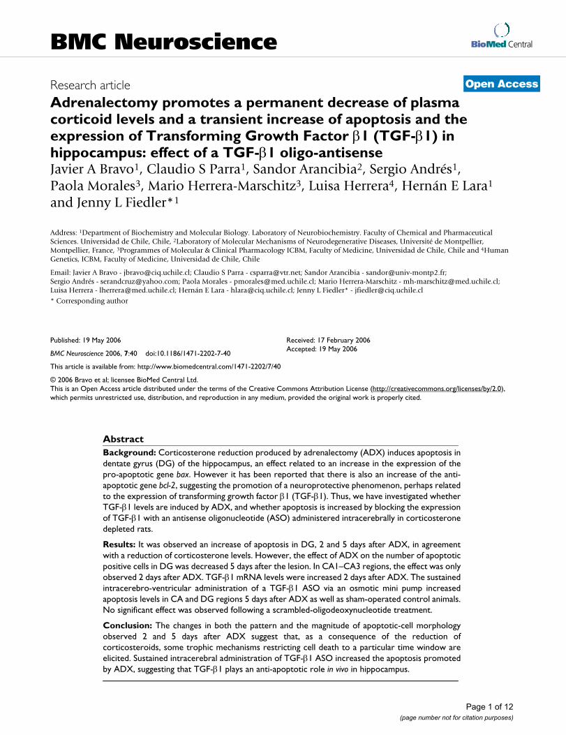
Apotosis induction in the hippocampus after adrenalectomyAs shown in Fig 1A, three morphological criteria wereconsidered for assessing an apoptotic features: (i)pyknotic nucleus, (ii) nuclei with extensive and tight con-densation of nuclear material into darkly stained balls,and/or (iii) segmented nuclei [26]. One of these profileshad to be fulfilled for counting as an apoptotic cell.
The density of cells showing apoptotic features was signif-icantly increased in CA1–CA3 and in both supra- andinfra-pyramidal layers of the dentate gyrus when compar-ing sections from 2-day ADX and sham-operated controlanimals (Fig. 1B). Apoptosis was still prominent in bothblades of the DG 5 days after ADX (Fig. 1B). No significantchanges in the density of apoptotic cells were observed inCA4 either 2 or 5 days after ADX.
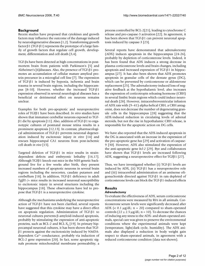
Adrenalectomy induces changes in TGFβ1 mRNA level in the hippocampusTGFβ1 expression as assessed by in situ hybridisation wasobserved in different regions of the brain including neo-cortex and hypothalamus. The hybridisation signal wasobserved in the DG and cornus ammon fields CA1–CA3of the hippocampus. Fig. 2 shows photomicrographs ofthe hippocampal regions after in situ hybridisation forTGF-β1 in brain sections from a sham-operated controlanimal (A), or a 2-day (B) or 5-day (C) ADX animals. Thespecificity of the hybridisation signal was evaluated withan excess of unlabelled probe (D).
Sham-operated control animals showed TGF-β1 mRNApositive hybridisation in the neuropil of the supra- andinfra-pyramidal layers of the DG (Fig. 2A). Expression ofTGF-β1 was also detected in CA1, but the intensity of thehybridisation signal was weaker than in the DG. The CA3and CA4 subfields showed the lowest intensity. Two daysafter ADX, an increase in the intensity of the TGF-β1 signalin the DG and CA1 was observed (Fig. 2B). However, 5days after ADX the intensity of the hybridisation signal inthe hippocampus was similar to that observed in sectionsfrom sham-operated animals (Fig. 2C). Also on the sub-granular layer of DG there were some cells with positiveTGF-β1 mRNA signal, but the method used to measurelabel intensity does not allow us to evaluate changes inthe number positive cells (not shown). No hybridisationsignal was observed when using an excess of unlabelledprobe (Fig. 2D). The results obtained by in situ hybridisa-tion were quantified and the intensity of the hybridisationsignal was normalized to that of CA4, because the hybrid-isation signal did not show any change in that area underany of the experimental conditions tested (see Fig. 3).
Two days after ADX, the levels of TGF-β1 mRNA increasedin CA1 by approximately 30% with respect to sham-oper-
Adrenalectomy (ADX) induced apoptosis in hippocampusFigure 1Adrenalectomy (ADX) induced apoptosis in hippoc-ampus. Brain sections from 2-day (ADX-2) and 5-day (ADX-5) adrenalectomized animals were stained with 1% Cresyl Violet and the density of apoptotic cells in the left hip-pocampus was estimated. Apoptotic cells were counted in ten non-successive 14 µm thick sections at 100× magnifica-tion in Cornus Amonnis area (CA1, CA3, CA4) and supra- (SupDG) and infra- (InfDG) pyramidal layer of the dentate gyrus (DG). (A) Images illustrating the morphology of apop-totic nuclei (small condensed nuclei, extensive and tight con-densation of nuclear material into darkly stained balls or disassembled pyknotic cells). (B) Bar graph represents the mean ± SEM (4–5 animals per group) of the density of apop-totic cells counted in each area for sham-operated animals (black columns), 2 days after ADX (dashed column) or 5 days after ADX (open column); the values obtained in 2-day and 5-day sham-operated animals were pooled because no differ-ences were observed with time after the sham operation. The data was analysed using the non parametric Kruskal-Wallis test, followed by Dunns post-hoc test. * p < 0.05 and ** p < 0.01 compared to the corresponding sham-operated control. Scale bar = 20 µm.
CA1
CA3
CA4
SupDG
InfD
G
0
10
20
30
Control
ADX-2
ADX-5
*
*
*
*
*
*
*
*
**
*
Hippocampal Areas
de
ns
ity
of
ap
op
toti
c c
ell
s /
he
mis
ec
tio
n
B
Page 3 of 12(page number not for citation purposes)
BMC Neuroscience 2006, 7:40 http://www.biomedcentral.com/1471-2202/7/40
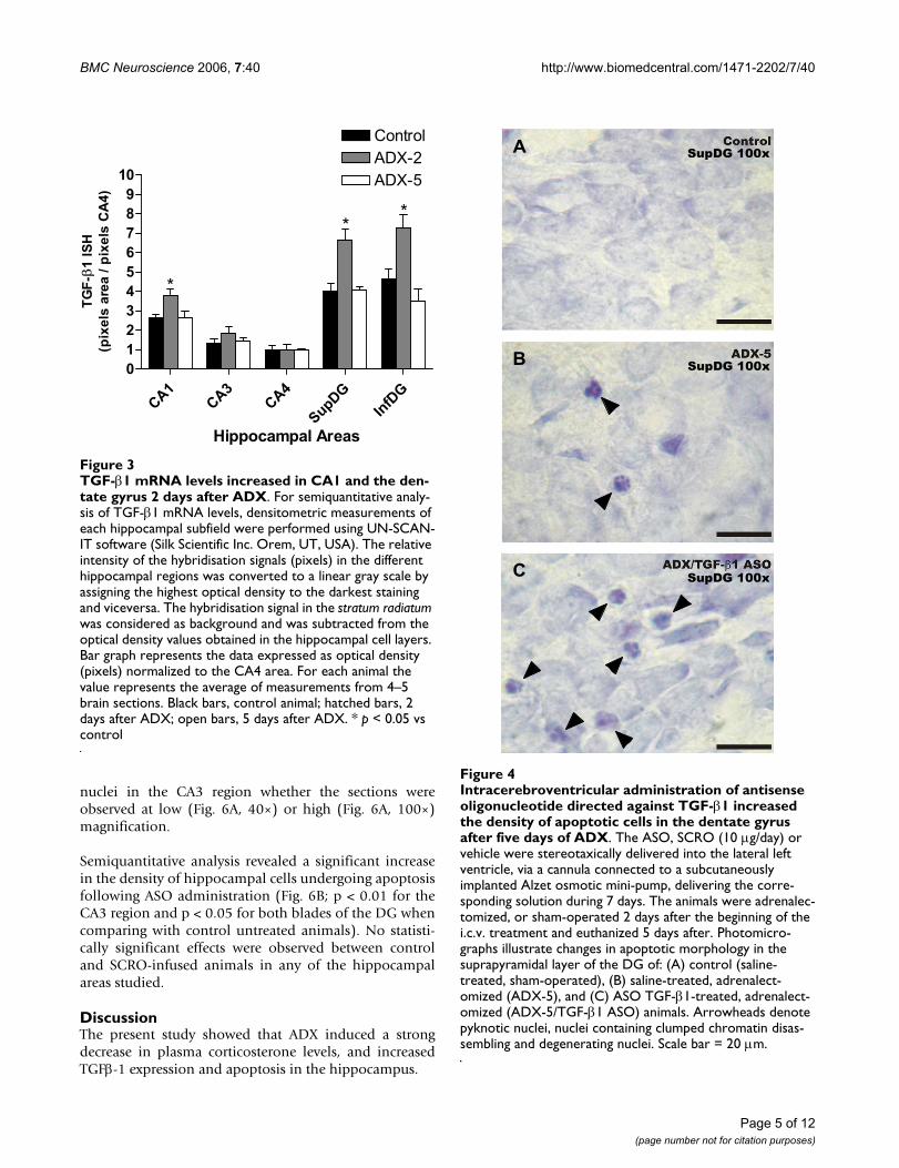
ated animals (Fig. 3). Also, a significant increase in TGF-β1 mRNA was observed in supra- and infra-pyramidal lay-ers of the DG (by 70% and 50%, respectively). In contrast,TGF-β1 mRNA levels in CA3 were not modified by ADX.TGF-β1 mRNA returned to sham-operated baseline levels5 days after ADX in CA1 and the two blades of the DG.
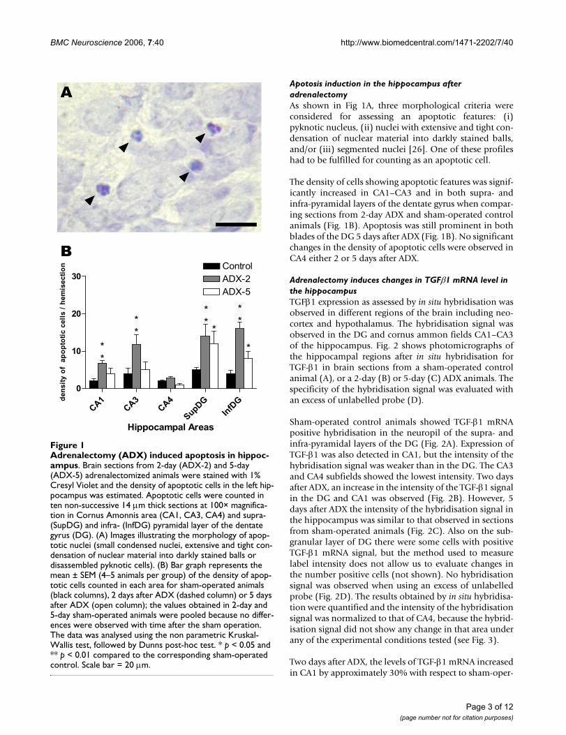
Lateral ventricle administration (i.c.v.) of an oligodeoxynucleotide antisense for TGF-β1 exacerbates hippocampal apoptosis induced by adrenalectomyTo evaluate the role of TGF-β1 in the hippocampus undercontrol and ADX conditions, an antisense oligonucleotideto TGF-β1 that has been successfully used by others [31]was infused into the lateral ventricle starting 2 days beforeADX. TGF-β1 antisense produced an enhancement in thedensity of nuclei with apoptotic features in the DG 5 daysafter ADX (Fig. 4C) when compared to either sham-oper-ated i.c.v. saline-infused (Fig. 4A) or to ADX i.c.v. saline-infused animals (Fig. 4B).
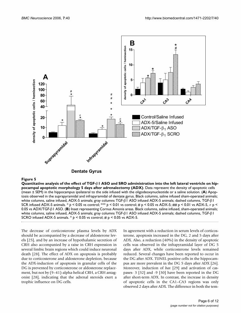
Semiquantitative analysis demonstrated a >2-foldincrease in the density of apoptotic cells in both blades ofthe DG, compared to that observed in sham- or ADX-operated animals infused with saline (Fig. 5A). This effect
was specific because the administration of scrambled oli-gonucleotide (SCRO) did not have any effect compared tothat seen in ADX animals treated with saline (Fig. 5A).Analysis of the results revealed that the density of apop-totic cells in the CA1 or CA4 regions was not statisticallydifferent between ASO- and SCRO- infused ADX rats (Fig.5B). Nevertheless, administration of SCRO but not ASOto ADX animals induced a statistically significant increasein the density of apoptotic cells in the CA3 area comparedto those in saline-infused rats (Fig. 5B). We also tried tocorrelate changes in TGF-β1 mRNA levels with TGF-β1immunoreactivity in the hippocampus. However, theantibodies available did not produce positive immunore-action neither in hippocampal slices nor control tissue(adrenal gland).
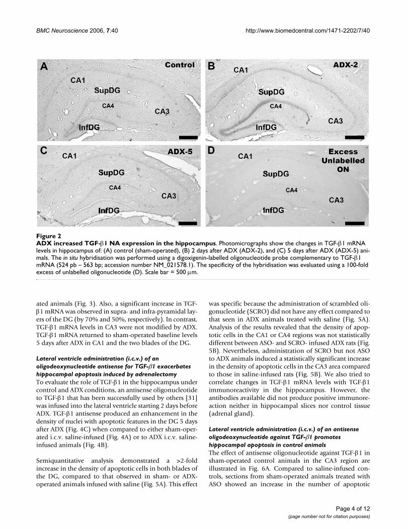
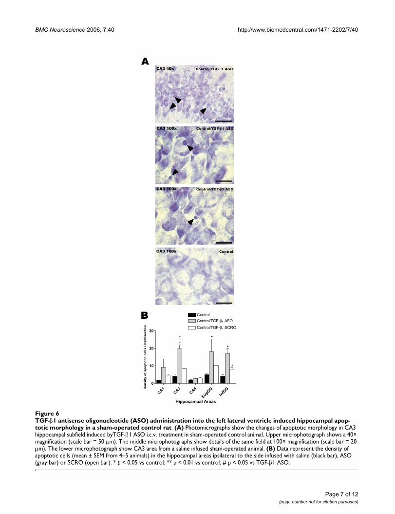
Lateral ventricle administration (i.c.v.) of an antisense oligodeoxynucleotide against TGF-β1 promotes hippocampal apoptosis in control animalsThe effect of antisense oligonucleotide against TGF-β1 insham-operated control animals in the CA3 region areillustrated in Fig. 6A. Compared to saline-infused con-trols, sections from sham-operated animals treated withASO showed an increase in the number of apoptotic
ADX increased TGF-β1 NA expression in the hippocampusFigure 2ADX increased TGF-β1 NA expression in the hippocampus. Photomicrographs show the changes in TGF-β1 mRNA levels in hippocampus of: (A) control (sham-operated), (B) 2 days after ADX (ADX-2), and (C) 5 days after ADX (ADX-5) ani-mals. The in situ hybridisation was performed using a digoxigenin-labelled oligonucleotide probe complementary to TGF-β1 mRNA (524 pb – 563 bp; accession number NM_021578.1). The specificity of the hybridisation was evaluated using a 100-fold excess of unlabelled oligonucleotide (D). Scale bar = 500 µm.
Page 4 of 12(page number not for citation purposes)
BMC Neuroscience 2006, 7:40 http://www.biomedcentral.com/1471-2202/7/40
nuclei in the CA3 region whether the sections wereobserved at low (Fig. 6A, 40×) or high (Fig. 6A, 100×)magnification.
Semiquantitative analysis revealed a significant increasein the density of hippocampal cells undergoing apoptosisfollowing ASO administration (Fig. 6B; p < 0.01 for theCA3 region and p < 0.05 for both blades of the DG whencomparing with control untreated animals). No statisti-cally significant effects were observed between controland SCRO-infused animals in any of the hippocampalareas studied.
DiscussionThe present study showed that ADX induced a strongdecrease in plasma corticosterone levels, and increasedTGFβ-1 expression and apoptosis in the hippocampus.
Intracerebroventricular administration of antisense oligonu-cleotide directed against TGF-β1 increased the density of apoptotic cells in the dentate gyrus after five days of ADXFigure 4Intracerebroventricular administration of antisense oligonucleotide directed against TGF-β1 increased the density of apoptotic cells in the dentate gyrus after five days of ADX. The ASO, SCRO (10 µg/day) or vehicle were stereotaxically delivered into the lateral left ventricle, via a cannula connected to a subcutaneously implanted Alzet osmotic mini-pump, delivering the corre-sponding solution during 7 days. The animals were adrenalec-tomized, or sham-operated 2 days after the beginning of the i.c.v. treatment and euthanized 5 days after. Photomicro-graphs illustrate changes in apoptotic morphology in the suprapyramidal layer of the DG of: (A) control (saline-treated, sham-operated), (B) saline-treated, adrenalect-omized (ADX-5), and (C) ASO TGF-β1-treated, adrenalect-omized (ADX-5/TGF-β1 ASO) animals. Arrowheads denote pyknotic nuclei, nuclei containing clumped chromatin disas-sembling and degenerating nuclei. Scale bar = 20 µm.
SupDG 100x
SupDG 100x
SupDG 100x
A
B
C
TGF-β1 mRNA levels increased in CA1 and the dentate gyrus 2 days after ADXFigure 3TGF-β1 mRNA levels increased in CA1 and the den-tate gyrus 2 days after ADX. For semiquantitative analy-sis of TGF-β1 mRNA levels, densitometric measurements of each hippocampal subfield were performed using UN-SCAN-IT software (Silk Scientific Inc. Orem, UT, USA). The relative intensity of the hybridisation signals (pixels) in the different hippocampal regions was converted to a linear gray scale by assigning the highest optical density to the darkest staining and viceversa. The hybridisation signal in the stratum radiatum was considered as background and was subtracted from the optical density values obtained in the hippocampal cell layers. Bar graph represents the data expressed as optical density (pixels) normalized to the CA4 area. For each animal the value represents the average of measurements from 4–5 brain sections. Black bars, control animal; hatched bars, 2 days after ADX; open bars, 5 days after ADX. * p < 0.05 vs control
CA1
CA3
CA4
SupD
G
InfD
G
0
1
2
3
4
5
6
7
8
9
10
Control
ADX-2
ADX-5
*
**
Hippocampal Areas
TG
F-
1 I
SH
(pix
els
are
a /
pix
els
CA
4)
Page 5 of 12(page number not for citation purposes)
BMC Neuroscience 2006, 7:40 http://www.biomedcentral.com/1471-2202/7/40
The decrease of corticosterone plasma levels by ADXshould be accompanied by a decrease of aldosterone lev-els [25], and by an increase of hypothalamic secretion ofCRH also accompanied by a raise in CRH expression inseveral limbic brain regions which could induce neuronaldeath [28]. The effect of ADX on apoptosis is probablydue to corticosterone and aldosterone depletion, becausethe ADX-induction of apoptosis in granular cells of theDG is prevented by corticosterone or aldosterone replace-ment, but not by (9–41)-alpha-helical CRH, a CRH antag-onist [28], indicating that the adrenal steroids exert atrophic influence on DG cells.
In agreement with a reduction in serum levels of corticos-terone, apoptosis increased in the DG, 2 and 5 days afterADX. Also, a reduction (40%) in the density of apoptoticcells was observed in the infrapyramidal layer of DG 5days after ADX, while corticosterone levels remainedreduced. Several changes have been reported to occur inthe DG after ADX. TUNEL positive cells in the hippocam-pus are more prevalent in the DG 5 days after ADX [26].Moreover, induction of bax [29] and activation of cas-pases- 3 [32] and -9 [30] have been reported in the DGafter short-term ADX. In contrast, the increase in densityof apoptotic cells in the CA1–CA3 regions was onlyobserved 2 days after ADX. The difference in both the tem-
Quantitative analysis of the effect of TGF-β1 ASO and SRO administration into the left lateral ventricle on hippocampal apop-totic morphology 5 days after adrenalectomy (ADX)Figure 5Quantitative analysis of the effect of TGF-β1 ASO and SRO administration into the left lateral ventricle on hip-pocampal apoptotic morphology 5 days after adrenalectomy (ADX). Data represent the density of apoptotic cells (mean ± SEM) in the hippocampus ipsilateral to the side infused with the oligodeoxynucleotide or a saline solution. (A) Apop-tosis observed in the suprapyramidal and infrapyramidal of dentate gyrus. Black columns, saline infused sham-operated animals; white columns, saline infused, ADX-5 animals; gray columns TGF-β1 ASO infused ADX-5 animals; dashed columns, TGF-β1 SCR infused ADX-5 animals. * p < 0.05 vs control; *** p < 0.01 vs control; # p < 0.05 vs ADX-5; ## p < 0.01 vs ADX-5, ≠ p < 0.05 vs ADX/TGF-β1 ASO. (B) Inset representing Cornus Amonis areas. Black columns, saline infused, sham-operated animals; white columns, saline infused, ADX-5 animals; gray columns TGF-β1 ASO infused ADX-5 animals; dashed columns, TGF-β1 SCRO infused ADX-5 animals. * p < 0.05 vs control; # p < 0.05 vs ADX-5.
Supra
pyram
idal
Infrap
yram
idal
0
10
20
ADX/TGF- 1 ASO
ADX/TGF- 1 SCRO
40
60
80
100
120
ADX-5/Saline Infused 1
Control/Saline Infused 1
* #
#
**
*
*
*
*
*
#
Dentate Gyrus
de
ns
ity
of
ap
op
toti
c c
ell
s /
he
mis
ec
tio
n
CA1
CA3
CA4
0
5
10
15
*
*
*#
de
ns
ity
of
ap
op
toti
c c
ell
s /
he
mis
ec
tio
n
A
B
Page 6 of 12(page number not for citation purposes)
BMC Neuroscience 2006, 7:40 http://www.biomedcentral.com/1471-2202/7/40
Page 7 of 12(page number not for citation purposes)
TGF-β1 antisense oligonucleotide (ASO) administration into the left lateral ventricle induced hippocampal apoptotic morphol-ogy in a sham-operated control ratFigure 6TGF-β1 antisense oligonucleotide (ASO) administration into the left lateral ventricle induced hippocampal apop-totic morphology in a sham-operated control rat. (A) Photomicrographs show the changes of apoptotic morphology in CA3 hippocampal subfield induced byTGF-β1 ASO i.c.v. treatment in sham-operated control animal. Upper microphotograph shows a 40× magnification (scale bar = 50 µm). The middle microphotographs show details of the same field at 100× magnification (scale bar = 20 µm). The lower microphotograph show CA3 area from a saline infused sham-operated animal. (B) Data represent the density of apoptotic cells (mean ± SEM from 4–5 animals) in the hippocampal areas ipsilateral to the side infused with saline (black bar), ASO (gray bar) or SCRO (open bar). * p < 0.05 vs control; ** p < 0.01 vs control; # p < 0.05 vs TGF-β1 ASO.
CA1
CA3
CA4
SupDG
InfD
G
0
10
20
30
Control 1
Control/TGF- 1 ASO
*
**
*
Control/TGF- 1 SCRO
#
Hippocampal Areas
den
sit
y o
f ap
op
toti
c c
ell
s /
he
mis
ec
tio
n
A
B
CA3 40x
CA3 100x
CA3 100x
CA3 100x
BMC Neuroscience 2006, 7:40 http://www.biomedcentral.com/1471-2202/7/40
poral pattern and the magnitude of apoptotic-cell mor-phology observed 2 and 5 days after ADX in thesehippocampal regions suggest that under these experimen-tal conditions there are some trophic mechanisms restrict-ing cell death to a particular time window after the insultprovoked by the reduction of corticosteroids resultingfrom ADX. This restriction in cell death is probably relatedto the induction of the anti-apoptotic gene bcl-2 [29]. Inagreement with this hypothesis, it has been reported thatthe removal of corticosteroids (3 days after ADX) triggersa specific gene expression profile in surviving dentategranule cells including the antiapoptotic bcl-2 gene, pro-viding them the chance of survival and probably makingthem more resistant to the apoptosis observed after ADX[33].
Several types of brain injuries are able to induce the syn-thesis and/or the release of different growth factors (e.g.,neurotrophins and cytokines) [1,2] providing neuropro-tection [14,15]. TGF-β1, which is induced in the hippoc-ampus after ADX [27] (and in this study), may alsoparticipate in the neuroprotective mechanisms. TGF-β1 ismarkedly increased in rat cerebral cortex and hippocam-pus after trauma, electrolytic lesion of the entorhinal cor-tex, or ischemia [8-10,34]. Moreover, the administrationof TGF-β1 to neuronal cultures prevents the apoptosisinduced by the β-amyloid through the induction of theantiapoptotic BCL-2 and BCL-XL proteins [19].
The majority of the studies performed to investigate theneuroprotective role of TGF-β1 are based on modelswhere neuronal death is a continuum between necrosisand apoptosis [14,15], making it difficult to establishwhich one of these processes prevails. These processes areassociated with different mechanisms buffering cell death.To differentiate which one of these processes is relevant,we used a model in which apoptosis takes place in a ratherselective manner [24-26,29]. ADX has been proposed as agood in vivo model for hippocampal apoptosis, because itis a non invasive procedure to the brain and, as it is alsoshown in this study, it triggers apoptosis by corticosteroidand aldosterone reduction only [24-26]. Thus, we haveevaluated with the ADX model if the expression of TGF-β1is modified.
Primary rat hippocampal neuronal culture secretes TGF-β1 making feasible an autocrine and/or paracrine role inthe hippocampus [35]. TGF-β1 mRNA and its proteinhave been localised in both hippocampal neurons andglia [32,36]. Hippocampal CA1–CA4 neurons displayimmunoreactivity for active TGF-β1 under physiologicalconditions, although mRNA expression was low [36,37].These evidences suggest basal expression of TGF-β1 in thehippocampus. In the present study, expression of TGF-β1mRNA was observed in the neuropil of the supra and
infra-pyramidal layers of DG and comparatively weaker inthe CA1 and CA3 subfields. Taken together, these resultsindicate the existence of basal expression of TGF-β1 in thehippocampus.
Labelling for TGF-β1 mRNA over OX-42-positive micro-glia with an activated morphology in the dentate gyrusgranule cell layer has been observed 3 days after ADX [32].In our studies, the expression of TGF-β1 mRNA showed atransient increase in both CA1 and supra- and infrapyram-idal-layers of the DG 2 days after ADX with values return-ing to control levels 5 days after ADX. The plastic changesobserved in the present study could be related to the per-manent reduction of corticosteroid plasma levels and/orto cell death promoted by ADX.
It is well established that corticosteroids regulate manyphysiological processes through their binding to gluco-corticoid receptors (GR). The ligand-activated receptordimer activates gene expression by binding to specificDNA sequences (glucocorticoid response element, GRE)in the promoter regions of glucocorticoid-regulated genes[38]. In contrast to the regulation of these classical GREs,the repression of negatively regulated target genes is medi-ated by trans-repression and/or by the binding of the GRto negative GREs (nGREs) [38]. It is possible that nGREsare also present in the TGF-β1 gene promoter region. Thecorticosterone reduction switches off the action of thenGREs, enhancing the expression of TGF-β1. At least inhumans, it is known that human TGF-β1 gene has a GREin its promoter [39], probably acting as a repressor of theTGF-β1 gene expression. Indeed, the increase in theexpression of hippocampal TGF-β1 by ADX is preventedby corticosterone administration [40]. However, thismechanism cannot totally account for the normalizationof TGF-β1 mRNA levels observed 5 days after ADX in ourexperiments.
During the early stages of apoptosis, phosphatidylserine isexposed to the cell surface, triggering cell engulfment byneighboring cells or phagocytes. However, exposure ofphosphatidyl-serine to the outer face of plasma mem-brane in the apoptotic cells acts as a stimulus to induce theexpression of TGF-β1 by microglia [41]. Therefore, the up-regulation of TGF-β1 observed following ADX may resultfrom neuronal death itself, similar to that reported byother scientists [8-10,42]. However, other authors haveobserved an increase in TGF-β1 expression 7 days afterADX although in different rat strains [40]. Recently, thesame authors showed the time course of TGF-β1 mRNAexpression in the DG which is increased 2 days after ADX(similar to our results) reaching maximum 3 days afterADX [32]. Also the levels of expression are mantained 7days after ADX, a period where the apoptosis is still occur-ring. However these authors did not study the TGF-β1
Page 8 of 12(page number not for citation purposes)
BMC Neuroscience 2006, 7:40 http://www.biomedcentral.com/1471-2202/7/40
expression after 5 or more days after ADX. More recently,Battista et al [43] showed an increase in TGF-β1 by RT-PCR in the hippocampus 3 days after ADX. It is possiblethat the ADX-induced expression of TGF-β1 could bebiphasic, i.e. an early generalized induction of TGF-β1mRNA in the hippocampus (CA and DG areas) which ismaintained for a few days after ADX (1–3 days) and there-after the gene is re-expressed between 5–7 days of ADXand persists over ADX period in areas undergoing neuro-nal death, i.e. in the DG. A similar pattern of TGF-β1induction has been reported in the post-ischemic adult rathippocampus [8,36].
Thus, it was interesting to study the effect of blocking TGF-β1 mRNA translation by an antisense oligonucleotide. Weused this approach instead of blocking the TGF-β1 pep-tide using neutralizing antibodies, because the oligonu-cleotide is more diffusible that an antibody. Also theadministration of a neutralizing antibody requires a singleapplication near to the site of interest, i.e. close to the DGor CA areas, making it feasible to induce tissue damageand by this means microglia activation. We used a TGF-β1ASO developed by Le Roy et al. [31], who demonstratedthat antisense prevented the translation of TGF-β1 mRNAby Leydig cells and, consequently, reduced TGF-β1 immu-noreactivity, which was not observed following adminis-tration of a corresponding SCRO
In this study, sustained i.c.v. administration of that TGF-β1 ASO via an osmotic mini pump increased apoptosislevels in CA and DG regions 5 days after ADX, but also insham-operated control animals. Indeed, when TGF-β1ASO was administered to sham-operated animals, apop-totic cells were increased in both CA3 and DG regions,while in ADX-animals the effect was restricted to the DG.The CA3 area shows low levels of TGF-β1 mRNA, both incontrol and ADX animals, indicating that its expression isindependent of corticosterone levels. The finding thatASO administration promotes apoptosis in CA3 led us topropose that low levels of TGF-β1 are enough to protectcells against apoptosis in this hippocampal subfield. AlsoTGF-β1 is capable of suppressing the levels of TNF-α [44],suggesting that the reduction of TGF-β1 by ASO treatmentmight induce the expression of this pro-inflammatorycytokine, that can also promote apoptosis through itsreceptor[45]. It is remarkable that CA3 field is normallyresistant to ischemia [46] and ADX (5 days, Fig. 1) andthis phenomena could be achieved through a tonicexpression of TGF-β1 [36]. Interestingly TGF-β1 defi-ciency in adult Tgfβ1-/+ mice results in increased neuro-nal susceptibility to excitotoxic injury in several structuresincluding the hippocampus [18].
The lack of effect of the TGF-β1 ASO on ADX CA3 may beexplained by other neurotrophic factors elicited by the
lesion. In fact, it has been shown that ADX promotes theinduction of BDNF in pyramidal regions of the hippoc-ampus [47,48].
In the present study, SCRO administration did not induceany effect on apoptosis levels, except in CA3 of ADX ani-mals, where there was an increase of apoptosis levels fol-lowing intracerebral SCRO administration. While we donot have any explanation for this effect, it may be specu-lated that is probably due to the presence of an unmethyl-ated CpG dinucleotide in the SCRO sequence. This amotif that has been reported to stimulate microglia andastrocytes in the CNS, releasing TNF-α, a pro-inflamatorycytokine[49]. This cytokine may exert in turn neuronalinjury in vulnerable regions [50], under conditions wherethere is a decrease of corticosterone levels, like followingADX.
ConclusionADX promotes a permanent decrease of plasma corticoidlevels and transient increase of apoptosis and TGF-β1expression in hippocampus, reaching a maximum 2 daysafter ADX. The effect was mainly observed in supra- andinfra-layers of the DG, where the expression of TGF-β1mRNA was stronger than in any other regions of the hip-pocampus.
Administration of a TGF-β1 ASO promoted hippocampalapoptosis, both in sham- and ADX-animals. In sham-operated animals, apoptosis was increased in both CA3and DG regions, while in ADX-animals the effect wasrestricted to the DG. The lack of effect of the TGF-β1 ASOon ADX CA3 can be explained by other neurotrophic fac-tors elicited by the lesion.
These observations, together with previously publishedreports indicating an increase in the expression of TGF-β1in response to brain injuries, suggest that TGF-β1 exertsneuroprotection in adult nervous system.
MethodsAnimalsAdult (200–220 g) male Sprague-Dawley rats were used inall experiments. The animals were kept under temperature(22°C–24°C), humidity (50%) and 12 h regulated light-dark cycle conditions, with food and water ad libitum. Allprocedures and care of the animals were conducted fol-lowing protocols approved by the local Institutional Ethi-cal Committee of the Faculty of Chemical andPharmaceutical Sciences, University of Chile, in compli-ance with guidelines published in the National Institutesof Health Guide for the care and use of laboratory animals.
Page 9 of 12(page number not for citation purposes)
BMC Neuroscience 2006, 7:40 http://www.biomedcentral.com/1471-2202/7/40
Design and synthesis of oligodeoxynucleotidesAll phosphorothioates oligodeoxynucleotides were pur-chased in purified form from Integrated DNA technolo-gies (INC, Coralville, IA, USA), single-end capped(sulphur modification on the phosphate group betweenthe first and last base pairs), displaying low toxicity for thecentral nervous system [51,52]. Oligodeoxynucleotideswere reconstituted in ultra-filtered saline at a concentra-tion of 417 pg/µL. The antisense to TGF-β1 was a 15 baseoligomer that had been previously used in its full phos-phorothioate form [31]. Its sequence was 5'-G-S-GAG-GGCGGCATGG-S-G-3' (the -S- denotes locations of thesulphur modification) and complementary to the transla-tion-initiation region of the rat TGF-β1 mRNA (410–424pb of rat TGF-β1 cDNA; Genbank accession numberNM_0215781). A scrambled oligodeoxynucleotide(SCRO), which had a randomly ordered but identical basecontent to the antisense was used as a control. Itssequence was 5'-A-S-GGTGGGAGGCGGC-S-G-3'. All oli-godeoxynucleotides used in this study were subjected to aBLASTN search on the National Center for BiotechnologyInformation BLAST server using the Genbank database.The antisense oligodeoxynucleotide sequence had a 100%positive match for its mRNA sequence. The SCRO pro-duced no positive matches.
SurgeryAdrenalectomy (ADX)Animals were anaesthetised with isofluorane (1.5% v/vair) and subjected to a bilateral ADX. The animals werekept with a 0.9% NaCl drinking solution to compensatethe sodium deficit induced by the ADX. The sham-oper-ated rats underwent a skin and lumbar muscle incision,leaving the adrenal glands untouched.
Oligodeoxynucleotide-i.c.v. administrationThe rats were anaesthetised as above and fixed in a DavidKopt stereotaxic frame. Guide cannulae were implantedinto the left lateral ventricle (i.c.v., intra cerebro-ventricu-lar infusion) (Bregma -0.8 mm, lateral 1.5 with a depth of3.7 mm below the dura) according to Paxinos and Watson[53]. The cannula was then connected by a catheter to anALZET® mini-osmotic pump (Model 2001; Palo Alto, CA)implanted subcutaneously in the scapular region deliver-ing at a rate of 10 µg oligodeoxynucleotide/day. Forsaline-treated control animals, the ALZET pump was filledwith sterile saline solution. After 2 days of oligodeoxynu-cleotide infusion, the rats were subjected to either ADX ora sham operation as described above and maintained forfurther 5 days after surgery.
Tissue fixationTwo or five days after ADX, the animals were anesthetizedby intramuscular injection of xylazine (5 mg/kg) and ket-amine (50 mg/kg). A blood sample was taken from the
heart and the obtained serum was kept at -20°C for corti-costerone determination. Animals were then transcar-dially perfused with 100 mL of 0.9% NaCl followed by500 mL of 4% paraformaldehyde in 0.1 M phosphatebuffer (PB) pH 7.4. The brain was dissected from the skull,post-fixed in the same buffer overnight and thenimmersed in 30% sucrose in 0.1 M phosphate bufferedsaline (PBS) at 4°C for 2 days. Coronal sections (14 µmthick) were sliced from the frozen fixed brains and proc-essed for Nissl staining.
Apoptotic morphologySections were dipped in 1% Cresyl Violet for 20 min,dehydrated in alcohol and mounted in Entellan (Merck,Darmstadt, Germany). Apoptotic nuclei were identifiedby using the criteria proposed by Greiner et al [26]. Inshort, one of the following criteria had to be fulfilled tocount for an apoptotic cell: (i) pyknotic nucleus; (ii)extensive and tight condensation of nuclear material intodarkly stained balls, and/or (iii) segmented nuclei [26].The density of apoptotic cells per hemisection was calcu-lated from ten non-successive coronal sections inspectedat 100× for each animal [29], and expressed as the means± SEM of 4–5 animals.
In situ hybridisation for TGFβ1 mRNAThe in situ hybridisation was conducted with an oligode-oxynucleotide probe complementary to TGF-β1 mRNA(524–563 pb access number NM_021578.1) labelled witha digoxigenin oligonucleotide 3'-OH tailing kit (Roche,Molecular Biochemicals, Mannheim, Germany). Thehybridisation was conducted as previously described [29].The specificity of the hybridisation was evaluated by theuse of 100 fold excess of the unlabelled oligodeoxynucle-otide. For semiquantitative analysis, densitometric meas-urements of each hippocampal subfield were analysedusing UN-SCAN-IT software (Silk Scientific Inc. Orem,UT, USA). A grey scale was used for measuring the inten-sity of the signals (pixels) observed in the hippocampalregions, i.e. the darkest staining the highest optical den-sity; the lightest staining the lowest optical density. Thehybridisation signal in the stratum radiatum was consid-ered as background and was subtracted from the opticaldensity values obtained in the hippocampal cell layers.For each animal the value represents the average from 4–5 brain sections. In order to normalize the different exper-imental conditions, all the data are expressed as opticaldensity respect to the CA4, because the intensity in thatregion did not change following any of the experimentalconditions.
Corticosterone radio immunoassayCorticosterone levels were determined by RIA accordingto the instruction of the manufacturer (Sigma, St. Louis,MO, USA), as described previously [26]. In short, the rat
Page 10 of 12(page number not for citation purposes)
BMC Neuroscience 2006, 7:40 http://www.biomedcentral.com/1471-2202/7/40
serum was diluted 1:10 in 0.05 M Tris-HCl, pH 8.0, con-taining 0.1 M NaCl, 0.1% BSA and then warmed at 50°Cfor 30 min. The anti-corticosterone antibody (Sigma-Aldrich, St Louis MO, USA; rabbit) was added to 0.1 mlsample or to 0.1 ml of the corticosterone standard (31–1000 pg). After 30 min of incubation, a 3H-corticosteronetracer (specific activity 100 Ci/mmol, Sigma-Aldrich, StLouis MO, USA) was added to each tube and incubatedfor 1 hour at 37°C. Finally, 0.2 ml cold dextran coatedcharcoal was added to the standard and/or samples, cen-trifuged at 2000 × g for 15 minutes at 4°C. The radioactiv-ity was measured in the supernatant. The quantity ofcorticosterone in serum samples was estimated using thePrism software 4.0 (GraphPad Software, San Diego, CA).Intra and inter assays variation was less than 5%. The min-imal detectable level of corticosterone was 0.1 µg/assay.
Statistical analysisDescriptive statistic for individual parameters are reportedas mean ± SEM. The Prism software 4.0 (GraphPad Soft-ware, San Diego, CA) was used for statistical analysis andgraph representation. Treatment effect was assayed withthe non parametric Kruskal-Wallis test, followed byDunns post-hoc test. Differences were considered to besignificant when the probability of their occurrence as aresult of chance alone was less than 5%.
AbbreviationsADX, Adrenalectomy
ASO, anti-sense oligodeoxynucleotide
CRH, corticotropin releasing hormone
DG, dentate gyrus
i.c.v., intra-cerebro ventricular
SCRO, scrambled oligodeoxynucleotide
TGFβ1, Transforming Growth Factor beta -1
s.c, sub-cutaneous
Authors' contributionsBJA carried out experimental design, sample preparation,in situ hybridisation experiments, statistical analysis,interpretation of data and involved in drafting the manu-script. PCS participated in its design, surgery, data analysisand helped draft the manuscript. AS was involved in draft-ing the article critically. AS, MP, HL, LHE were involved inthe critical review of the manuscript. H-MM was involvedin revising the manuscript critically. FJL conceived,designed and coordinated the study, analysed, interpretedand drafted the manuscript.
AcknowledgementsWe thank Dr. V.M. Berthoud for critical comments. This research was sup-ported by Fondecyt-Chile #1040937 (JF), # 7040157 (JF), #1030541 (MH-M) and Memorias Grant (2002–2003), Facultad de Ciencias Químicas y Far-macéuticas, Universidad de Chile (JF).
References1. Ebadi M, Bashir RM, Heidrick ML, Hamada FM, Refaey HE, Hamed A,
Helal G, Baxi MD, Cerutis DR, Lassi NK: Neurotrophins and theirreceptors in nerve injury and repair. Neurochem Int 1997,30:347-74.
2. Flanders KC, Ren RF, Lippa CF: Transforming growth factor-betas in neurodegenerative disease. Prog Neurobiol 1998,54:71-85.
3. Massague J: How cells read TGF-beta signals. Nat Rev Mol CellBiol 2000, 1:169-78.
4. Schuster N, Krieglstein K: Mechanisms of TGF-beta-mediatedapoptosis. Cell Tissue Res 2002, 307:1-14.
5. Mogi M, Harada M, Kondo T, Narabayashi H, Riederer P, Nagatsu T:Transforming growth factor-beta 1 levels are elevated in thestriatum and in ventricular cerebrospinal fluid in Parkinson'sdisease. Neurosci Lett 1995, 193:129-32.
6. Flanders KC, Lippa CF, Smith TW, Pollen DA, Sporn MB: Alteredexpression of transforming growth factor-beta in Alzhe-imer's disease. Neurology 1995, 45:1561-9.
7. Monning U, Sandbrink R, Banati RB, Masters CL, Beyreuther K:Transforming growth factor beta mediates increase ofmature transmembrane amyloid precursor protein inmicroglial cells. FEBS Lett 1994, 342:267-72.
8. Lehrmann E, Kiefer R, Finsen B, Diemer NH, Zimmer J, Hartung HP:Cytokines in cerebral ischemia: expression of transforminggrowth factor beta-1 (TGF-beta 1) mRNA in the pos-tischemic adult rat hippocampus. Exp Neurol 1995, 131:114-23.
9. Zhao B, Schwartz JP: Involvement of cytokines in normal CNSdevelopment and neurological diseases: recent progress andperspectives. J Neurosci Res 1998, 52:7-16.
10. Bottner M, Krieglstein K, Unsicker K: The transforming growthfactor-betas: structure, signaling, and roles in nervous sys-tem development and functions. J Neurochem 2000, 75:2227-40.
11. de Luca A, Weller M, Fontana A: TGF-beta-induced apoptosis ofcerebellar granule neurons is prevented by depolarization. JNeurosci 1996, 16:4174-85.
12. Duenker N, Valenciano AI, Franke A, Hernandez-Sanchez C, DresselR, Behrendt M, De Pablo F, Krieglstein K, de la Rosa EJ: Balance ofpro-apoptotic transforming growth factor-beta and anti-apoptotic insulin effects in the control of cell death in thepostnatal mouse retina. Eur J Neurosci 2005, 22:28-38.
13. Duenker N: Transforming growth factor-beta (TGF-beta) andprogrammed cell death in the vertebrate retina. Int Rev Cytol2005, 245:17-43.
14. Prehn JH: Marked diversity in the action of growth factors onN-methyl-D-aspartate-induced neuronal degeneration. Eur JPharmacol 1996, 306:81-8.
15. Henrich-Noack P, Prehn JH, Krieglstein J: TGF-beta 1 protectshippocampal neurons against degeneration caused by tran-sient global ischemia. Dose-response relationship and poten-tial neuroprotective mechanisms. Stroke 1996, 27:1609-14.discussion 1615
16. Letterio JJ, AB Roberts: Regulation of immune responses byTGF-beta. Annu Rev Immunol 1998, 16:137-61.
17. Letterio JJ: Murine models define the role of TGF-beta as amaster regulator of immune cell function. Cytokine Growth Fac-tor Rev 2000, 11:81-7.
18. Brionne TC, Tesseur I, Masliah E, Wyss-Coray T: Loss of TGF-beta1 leads to increased neuronal cell death and microgliosis inmouse brain. Neuron 2003, 40:1133-45.
19. Kim ES, Kim RS, Ren RF, Hawver DB, Flanders KC: Transforminggrowth factor-beta inhibits apoptosis induced by beta-amy-loid peptide fragment 25–35 in cultured neuronal cells. BrainRes Mol Brain Res 1998, 62:122-30.
20. Prehn JH, Bindokas VP, Marcuccilli CJ, Krajewski S, Reed JC, Miller RJ:Regulation of neuronal Bcl2 protein expression and calciumhomeostasis by transforming growth factor type beta con-fers wide-ranging protection on rat hippocampal neurons.Proc Natl Acad Sci USA 1994, 91:12599-603.
Page 11 of 12(page number not for citation purposes)
http://www.ncbi.nlm.nih.gov/entrez/query.fcgi?cmd=Retrieve&db=PubMed&dopt=Abstract&list_uids=9106250
http://www.ncbi.nlm.nih.gov/entrez/query.fcgi?cmd=Retrieve&db=PubMed&dopt=Abstract&list_uids=9106250
http://www.ncbi.nlm.nih.gov/entrez/query.fcgi?cmd=Retrieve&db=PubMed&dopt=Abstract&list_uids=9460794
http://www.ncbi.nlm.nih.gov/entrez/query.fcgi?cmd=Retrieve&db=PubMed&dopt=Abstract&list_uids=9460794
http://www.ncbi.nlm.nih.gov/entrez/query.fcgi?cmd=Retrieve&db=PubMed&dopt=Abstract&list_uids=7478158
http://www.ncbi.nlm.nih.gov/entrez/query.fcgi?cmd=Retrieve&db=PubMed&dopt=Abstract&list_uids=7478158
http://www.ncbi.nlm.nih.gov/entrez/query.fcgi?cmd=Retrieve&db=PubMed&dopt=Abstract&list_uids=7478158
http://www.ncbi.nlm.nih.gov/entrez/query.fcgi?cmd=Retrieve&db=PubMed&dopt=Abstract&list_uids=7543987
http://www.ncbi.nlm.nih.gov/entrez/query.fcgi?cmd=Retrieve&db=PubMed&dopt=Abstract&list_uids=7543987
http://www.ncbi.nlm.nih.gov/entrez/query.fcgi?cmd=Retrieve&db=PubMed&dopt=Abstract&list_uids=7543987
http://www.ncbi.nlm.nih.gov/entrez/query.fcgi?cmd=Retrieve&db=PubMed&dopt=Abstract&list_uids=8150082
http://www.ncbi.nlm.nih.gov/entrez/query.fcgi?cmd=Retrieve&db=PubMed&dopt=Abstract&list_uids=8150082
http://www.ncbi.nlm.nih.gov/entrez/query.fcgi?cmd=Retrieve&db=PubMed&dopt=Abstract&list_uids=8150082
http://www.ncbi.nlm.nih.gov/entrez/query.fcgi?cmd=Retrieve&db=PubMed&dopt=Abstract&list_uids=7895806
http://www.ncbi.nlm.nih.gov/entrez/query.fcgi?cmd=Retrieve&db=PubMed&dopt=Abstract&list_uids=7895806
http://www.ncbi.nlm.nih.gov/entrez/query.fcgi?cmd=Retrieve&db=PubMed&dopt=Abstract&list_uids=7895806
http://www.ncbi.nlm.nih.gov/entrez/query.fcgi?cmd=Retrieve&db=PubMed&dopt=Abstract&list_uids=9556025
http://www.ncbi.nlm.nih.gov/entrez/query.fcgi?cmd=Retrieve&db=PubMed&dopt=Abstract&list_uids=9556025
http://www.ncbi.nlm.nih.gov/entrez/query.fcgi?cmd=Retrieve&db=PubMed&dopt=Abstract&list_uids=9556025
http://www.ncbi.nlm.nih.gov/entrez/query.fcgi?cmd=Retrieve&db=PubMed&dopt=Abstract&list_uids=8753879
http://www.ncbi.nlm.nih.gov/entrez/query.fcgi?cmd=Retrieve&db=PubMed&dopt=Abstract&list_uids=8753879
http://www.ncbi.nlm.nih.gov/entrez/query.fcgi?cmd=Retrieve&db=PubMed&dopt=Abstract&list_uids=8813618
http://www.ncbi.nlm.nih.gov/entrez/query.fcgi?cmd=Retrieve&db=PubMed&dopt=Abstract&list_uids=8813618
http://www.ncbi.nlm.nih.gov/entrez/query.fcgi?cmd=Retrieve&db=PubMed&dopt=Abstract&list_uids=8784137
http://www.ncbi.nlm.nih.gov/entrez/query.fcgi?cmd=Retrieve&db=PubMed&dopt=Abstract&list_uids=8784137
http://www.ncbi.nlm.nih.gov/entrez/query.fcgi?cmd=Retrieve&db=PubMed&dopt=Abstract&list_uids=8784137
http://www.ncbi.nlm.nih.gov/entrez/query.fcgi?cmd=Retrieve&db=PubMed&dopt=Abstract&list_uids=9597127
http://www.ncbi.nlm.nih.gov/entrez/query.fcgi?cmd=Retrieve&db=PubMed&dopt=Abstract&list_uids=9597127
http://www.ncbi.nlm.nih.gov/entrez/query.fcgi?cmd=Retrieve&db=PubMed&dopt=Abstract&list_uids=9813276
http://www.ncbi.nlm.nih.gov/entrez/query.fcgi?cmd=Retrieve&db=PubMed&dopt=Abstract&list_uids=9813276
http://www.ncbi.nlm.nih.gov/entrez/query.fcgi?cmd=Retrieve&db=PubMed&dopt=Abstract&list_uids=9813276
BMC Neuroscience 2006, 7:40 http://www.biomedcentral.com/1471-2202/7/40
Publish with BioMed Central and every scientist can read your work free of charge
"BioMed Central will be the most significant development for disseminating the results of biomedical research in our lifetime."
Sir Paul Nurse, Cancer Research UK
Your research papers will be:
available free of charge to the entire biomedical community
peer reviewed and published immediately upon acceptance
cited in PubMed and archived on PubMed Central
yours — you keep the copyright
Submit your manuscript here:http://www.biomedcentral.com/info/publishing_adv.asp
BioMedcentral
21. Hengartner MO: The biochemistry of apoptosis. Nature 2000,407:770-6.
22. Boise LH, Gonzalez-Garcia M, Postema CE, Ding L, Lindsten T, TurkaLA, Mao X, Nunez G, Thompson CB: bcl-x, a bcl-2-related genethat functions as a dominant regulator of apoptotic celldeath. Cell 1993, 74:597-608.
23. Zhu Y, Ahlemeyer B, Bauerbach E, Krieglstein J: TGF-beta1 inhibitscaspase-3 activation and neuronal apoptosis in rat hippoc-ampal cultures. Neurochem Int 2001, 38:227-35.
24. Sloviter RS, Valiquette G, Abrams GM, Ronk EC, Sollas AL, Paul LA,Neubort S: Selective loss of hippocampal granule cells in themature rat brain after adrenalectomy. Science 1989,243:535-8.
25. Woolley CS, Gould E, Sakai RR, Spencer RL, McEwen BS: Effects ofaldosterone or RU28362 treatment on adrenalectomy-induced cell death in the dentate gyrus of the adult rat. BrainRes 1991, 554:312-5.
26. Greiner M, Cardenas S, Parra C, Bravo J, Avalos AM, Paredes A, LaraHE, Fiedler JL: Adrenalectomy regulates apoptotic-associatedgenes in rat hippocampus. Endocrine 2001, 15:323-33.
27. Bye N, Nichols NR: Adrenalectomy-induced apoptosis andglial responsiveness during ageing. Neuroreport 1998, 9:1179-84.
28. Gerth A, Hatalski CG, Avishai-Eliner S, Baram TZ: Corticotropinreleasing hormone antagonist does not prevent adrenalec-tomy-induced apoptosis in the dentate gyrus of the rat hip-pocampus. Stress 1998, 2:159-69.
29. Cardenas SP, Parra C, Bravo J, Morales P, Lara HE, Herrera-MarschitzM, Fiedler JL: Corticosterone differentially regulates bax, bcl-2 and bcl-x mRNA levels in the rat hippocampus. Neurosci Lett2002, 331:9-12.
30. Andrés S, Cárdenas S, Parra C, Bravo J, Greiner M, Rojas P, MoralesP, Lara HFJ: Effects of long-term adrenalectomy on apoptosisand neuroprotection in the rat hippocampus. Endocrine 2006.
31. Le Roy C, Leduque P, Yuan Li J, Saez JM, Langlois D: Antisense oli-gonucleotide targeting the transforming growth factorbeta1 increases expression of specific genes and functions ofLeydig cells. Eur J Biochem 1998, 257:506-14.
32. Nichols NR, Agolley D, Zieba M, Bye N: Glucocorticoid regula-tion of glial responses during hippocampal neurodegenera-tion and regeneration. Brain Res Brain Res Rev 2005, 48:287-301.
33. Nair N, Bedwal RS: Metallothionein in male reproductiveorgans of adrenalectomized and hydrocortisone-treatedWistar rats. Indian J Exp Biol 2004, 42:288-91.
34. Nichols NR, Laping NJ, Day JR, Finch CE: Increases in transform-ing growth factor-beta mRNA in hippocampus duringresponse to entorhinal cortex lesions in intact and adrenal-ectomized rats. J Neurosci Res 1991, 28:134-9.
35. Ren RF, Flanders KC: Transforming growth factors-beta pro-tect primary rat hippocampal neuronal cultures from degen-eration induced by beta-amyloid peptide. Brain Res 1996,732:16-24.
36. Knuckey NW, Finch P, Palm DE, Primiano MJ, Johanson CE, FlandersKC, Thompson NL: Differential neuronal and astrocyticexpression of transforming growth factor beta isoforms inrat hippocampus following transient forebrain ischemia.Brain Res Mol Brain Res 1996, 40:1-14.
37. Zhu Y, Culmsee C, Roth-Eichhorn S, Krieglstein J: Beta(2)-adreno-ceptor stimulation enhances latent transforming growth fac-tor-beta-binding protein-1 and transforming growth factor-beta1 expression in rat hippocampus after transient fore-brain ischemia. Neuroscience 2001, 107:593-602.
38. Dostert A, Heinzel T: Negative glucocorticoid receptorresponse elements and their role in glucocorticoid action.Curr Pharm Des 2004, 10:2807-16.
39. Parrelli JM, Meisler N, Cutroneo KR: Identification of a glucocor-ticoid response element in the human transforming growthfactor beta 1 gene promoter. Int J Biochem Cell Biol 1998,30:623-7.
40. Bye N, Zieba M, Wreford NG, Nichols NR: Resistance of the den-tate gyrus to induced apoptosis during ageing is associatedwith increases in transforming growth factor-beta1 messen-ger RNA. Neuroscience 2001, 105:853-62.
41. De Simone R, Ajmone-Cat MA, Tirassa P, Minghetti L: ApoptoticPC12 cells exposing phosphatidylserine promote the pro-duction of anti-inflammatory and neuroprotective mole-
cules by microglial cells. J Neuropathol Exp Neurol 2003,62:208-16.
42. Pratt BM, McPherson JM: TGF-beta in the central nervous sys-tem: potential roles in ischemic injury and neurodegenera-tive diseases. Cytokine Growth Factor Rev 1997, 8:267-92.
43. Battista D, Ferrari CC, Gage FH, Pitossi FJ: Neurogenic nichemodulation by activated microglia: transforming growth fac-tor beta increases neurogenesis in the adult dentate gyrus.Eur J Neurosci 2006, 23:83-93.
44. Harris-White ME, Chu T, Balverde Z, Sigel JJ, Flanders KC, FrautschySA: Effects of transforming growth factor-beta (isoforms 1–3)on amyloid-beta deposition, inflammation, and cell targetingin organotypic hippocampal slice cultures. J Neurosci 1998,18:10366-74.
45. Kitson J, Raven T, Jiang YP, Goeddel DV, Giles KM, Pun KT, GrinhamCJ, Brown R, Farrow SN: A death-domain-containing receptorthat mediates apoptosis. Nature 1996, 384:372-5.
46. Chen J, Zhu RL, Nakayama M, Kawaguchi K, Jin K, Stetler RA, SimonRP, Graham SH: Expression of the apoptosis-effector gene,Bax, is up-regulated in vulnerable hippocampal CA1 neuronsfollowing global ischemia. J Neurochem 1996, 67:64-71.
47. Chao HM, McEwen BS: Glucocorticoids and the expression ofmRNAs for neurotrophins, their receptors and GAP-43 inthe rat hippocampus. Brain Res Mol Brain Res 1994, 26:271-6.
48. Chao HM, Sakai RR, Ma LY, McEwen BS: Adrenal steroid regula-tion of neurotrophic factor expression in the rat hippocam-pus. Endocrinology 1998, 139:3112-8.
49. Krieg AM: Mechanisms and applications of immune stimula-tory CpG oligodeoxynucleotides. Biochim Biophys Acta 1999,1489:107-16.
50. Iliev AI, Stringaris AK, Nau R, Neumann H: Neuronal injury medi-ated via stimulation of microglial toll-like receptor-9 (TLR9).Faseb J 2004, 18:412-4.
51. Hebb MO, Robertson HA: End-capped antisense oligodeoxynu-cleotides effectively inhibit gene expression in vivo and offera low-toxicity alternative to fully modified phosphorothioateoligodeoxynucleotides. Brain Res Mol Brain Res 1997, 47:223-8.
52. Mikkelsen JD, Karle J, Madsen TM: Intrahippocampal infusion ofantisense oligodeoxynucleotide to the GABA(A) receptorgamma2 subunit enhances neuropeptide Y gene expression.Brain Res Bull 2001, 54:91-9.
53. Paxinos G, Watson C: The rat brain in stereotaxic coordinates.Academic Press, New York; 1986.
Page 12 of 12(page number not for citation purposes)
http://www.ncbi.nlm.nih.gov/entrez/query.fcgi?cmd=Retrieve&db=PubMed&dopt=Abstract&list_uids=8358789
http://www.ncbi.nlm.nih.gov/entrez/query.fcgi?cmd=Retrieve&db=PubMed&dopt=Abstract&list_uids=8358789
http://www.ncbi.nlm.nih.gov/entrez/query.fcgi?cmd=Retrieve&db=PubMed&dopt=Abstract&list_uids=8358789
http://www.ncbi.nlm.nih.gov/entrez/query.fcgi?cmd=Retrieve&db=PubMed&dopt=Abstract&list_uids=2911756
http://www.ncbi.nlm.nih.gov/entrez/query.fcgi?cmd=Retrieve&db=PubMed&dopt=Abstract&list_uids=2911756
http://www.ncbi.nlm.nih.gov/entrez/query.fcgi?cmd=Retrieve&db=PubMed&dopt=Abstract&list_uids=1933312
http://www.ncbi.nlm.nih.gov/entrez/query.fcgi?cmd=Retrieve&db=PubMed&dopt=Abstract&list_uids=1933312
http://www.ncbi.nlm.nih.gov/entrez/query.fcgi?cmd=Retrieve&db=PubMed&dopt=Abstract&list_uids=1933312
http://www.ncbi.nlm.nih.gov/entrez/query.fcgi?cmd=Retrieve&db=PubMed&dopt=Abstract&list_uids=9601690
http://www.ncbi.nlm.nih.gov/entrez/query.fcgi?cmd=Retrieve&db=PubMed&dopt=Abstract&list_uids=9601690
http://www.ncbi.nlm.nih.gov/entrez/query.fcgi?cmd=Retrieve&db=PubMed&dopt=Abstract&list_uids=9787264
http://www.ncbi.nlm.nih.gov/entrez/query.fcgi?cmd=Retrieve&db=PubMed&dopt=Abstract&list_uids=9787264
http://www.ncbi.nlm.nih.gov/entrez/query.fcgi?cmd=Retrieve&db=PubMed&dopt=Abstract&list_uids=9787264
http://www.ncbi.nlm.nih.gov/entrez/query.fcgi?cmd=Retrieve&db=PubMed&dopt=Abstract&list_uids=9826200
http://www.ncbi.nlm.nih.gov/entrez/query.fcgi?cmd=Retrieve&db=PubMed&dopt=Abstract&list_uids=9826200
http://www.ncbi.nlm.nih.gov/entrez/query.fcgi?cmd=Retrieve&db=PubMed&dopt=Abstract&list_uids=9826200
http://www.ncbi.nlm.nih.gov/entrez/query.fcgi?cmd=Retrieve&db=PubMed&dopt=Abstract&list_uids=2041055
http://www.ncbi.nlm.nih.gov/entrez/query.fcgi?cmd=Retrieve&db=PubMed&dopt=Abstract&list_uids=2041055
http://www.ncbi.nlm.nih.gov/entrez/query.fcgi?cmd=Retrieve&db=PubMed&dopt=Abstract&list_uids=2041055
http://www.ncbi.nlm.nih.gov/entrez/query.fcgi?cmd=Retrieve&db=PubMed&dopt=Abstract&list_uids=8891264
http://www.ncbi.nlm.nih.gov/entrez/query.fcgi?cmd=Retrieve&db=PubMed&dopt=Abstract&list_uids=8891264
http://www.ncbi.nlm.nih.gov/entrez/query.fcgi?cmd=Retrieve&db=PubMed&dopt=Abstract&list_uids=8891264
http://www.ncbi.nlm.nih.gov/entrez/query.fcgi?cmd=Retrieve&db=PubMed&dopt=Abstract&list_uids=8840007
http://www.ncbi.nlm.nih.gov/entrez/query.fcgi?cmd=Retrieve&db=PubMed&dopt=Abstract&list_uids=8840007
http://www.ncbi.nlm.nih.gov/entrez/query.fcgi?cmd=Retrieve&db=PubMed&dopt=Abstract&list_uids=9693962
http://www.ncbi.nlm.nih.gov/entrez/query.fcgi?cmd=Retrieve&db=PubMed&dopt=Abstract&list_uids=9693962
http://www.ncbi.nlm.nih.gov/entrez/query.fcgi?cmd=Retrieve&db=PubMed&dopt=Abstract&list_uids=9693962
http://www.ncbi.nlm.nih.gov/entrez/query.fcgi?cmd=Retrieve&db=PubMed&dopt=Abstract&list_uids=9620642
http://www.ncbi.nlm.nih.gov/entrez/query.fcgi?cmd=Retrieve&db=PubMed&dopt=Abstract&list_uids=9620642
http://www.ncbi.nlm.nih.gov/entrez/query.fcgi?cmd=Retrieve&db=PubMed&dopt=Abstract&list_uids=9620642
http://www.ncbi.nlm.nih.gov/entrez/query.fcgi?cmd=Retrieve&db=PubMed&dopt=Abstract&list_uids=9852574
http://www.ncbi.nlm.nih.gov/entrez/query.fcgi?cmd=Retrieve&db=PubMed&dopt=Abstract&list_uids=9852574
http://www.ncbi.nlm.nih.gov/entrez/query.fcgi?cmd=Retrieve&db=PubMed&dopt=Abstract&list_uids=9852574
http://www.ncbi.nlm.nih.gov/entrez/query.fcgi?cmd=Retrieve&db=PubMed&dopt=Abstract&list_uids=8934525
http://www.ncbi.nlm.nih.gov/entrez/query.fcgi?cmd=Retrieve&db=PubMed&dopt=Abstract&list_uids=8934525
http://www.ncbi.nlm.nih.gov/entrez/query.fcgi?cmd=Retrieve&db=PubMed&dopt=Abstract&list_uids=8667027
http://www.ncbi.nlm.nih.gov/entrez/query.fcgi?cmd=Retrieve&db=PubMed&dopt=Abstract&list_uids=8667027
http://www.ncbi.nlm.nih.gov/entrez/query.fcgi?cmd=Retrieve&db=PubMed&dopt=Abstract&list_uids=8667027
http://www.ncbi.nlm.nih.gov/entrez/query.fcgi?cmd=Retrieve&db=PubMed&dopt=Abstract&list_uids=7854057
http://www.ncbi.nlm.nih.gov/entrez/query.fcgi?cmd=Retrieve&db=PubMed&dopt=Abstract&list_uids=7854057
http://www.ncbi.nlm.nih.gov/entrez/query.fcgi?cmd=Retrieve&db=PubMed&dopt=Abstract&list_uids=7854057
http://www.ncbi.nlm.nih.gov/entrez/query.fcgi?cmd=Retrieve&db=PubMed&dopt=Abstract&list_uids=9645683
http://www.ncbi.nlm.nih.gov/entrez/query.fcgi?cmd=Retrieve&db=PubMed&dopt=Abstract&list_uids=9645683
http://www.ncbi.nlm.nih.gov/entrez/query.fcgi?cmd=Retrieve&db=PubMed&dopt=Abstract&list_uids=9645683
http://www.ncbi.nlm.nih.gov/entrez/query.fcgi?cmd=Retrieve&db=PubMed&dopt=Abstract&list_uids=9221920
http://www.ncbi.nlm.nih.gov/entrez/query.fcgi?cmd=Retrieve&db=PubMed&dopt=Abstract&list_uids=9221920
Related Documents