ADNI-GO PET Technical Procedures Manual for FDG and AV-45 Page 1 of 36 V3.8 January 14, 2011 ADNI-GO PET Technical Procedures Manual AV-45 & FDG V3.8.0 January 14, 2011

Welcome message from author
This document is posted to help you gain knowledge. Please leave a comment to let me know what you think about it! Share it to your friends and learn new things together.
Transcript
ADNI-GO PET Technical Procedures Manual for FDG and AV-45 Page 1 of 36 V3.8 January 14, 2011
ADNI-GO PET Technical Procedures Manual
AV-45 & FDG
V3.8.0 January 14, 2011
ADNI-GO PET Technical Procedures Manual for FDG and AV-45 Page 2 of 36 V3.8 January 14, 2011
Table of Contents
General Information.........................................................................................................................3 Contact Information .........................................................................................................................3 Site Qualification .............................................................................................................................4
PET Scanners ...........................................................................................................................4 Regulatory ................................................................................................................................4
Continued Quality Monitoring During Execution Phase.................................................................4 PET Pre-Scan Procedures / General Information ............................................................................6 Participants Pre-screening................................................................................................................6 AV-45 Ordering ...............................................................................................................................6 Subject Preparation ..........................................................................................................................6 Participant Positioning .....................................................................................................................7 Ambient Conditions .........................................................................................................................8
FDG Scans:...............................................................................................................................8 AV-45 Scans: ...........................................................................................................................8
Image File Identification..................................................................................................................8 Documentation.................................................................................................................................9 Assessments and Endpoints for 18F-AV-45: ....................................................................................9 Follow-up post AV45 administration: ...........................................................................................10 PET Imaging Protocols ..................................................................................................................10
AV-45:....................................................................................................................................10 FDG:.......................................................................................................................................12
Appendix A – LONI Access User Registration.............................................................................15 Appendix B – Scanner Specific Reconstruction Parameters…………………………………….17 Appendix C – Example PET Scan Information Sheets .................................................................33
ADNI-GO PET Technical Procedures Manual for FDG and AV-45 Page 3 of 36 V3.8 January 14, 2011
General Information The purpose of this manual is to further explain the PET imaging component of the ADNI-GO protocol. Standard procedures are needed to ensure consistency of data collection in this longitudinal study. This manual contains information for study-site clinical staff involved with the care of study participants during the imaging procedure and those involved with the processing and transfer of PET imaging data. AV-45 PET imaging will be performed on all the participants from ADNI1 who are in the cognitively normal (CN) and mild cognitive impairment (MCI) groups. In addition, 200 participants with early amnestic cognitive Impairment (EMCI) will be enrolled and imaged. FDG PET images will also be acquired in all subjects at the same time point (± 2 weeks). A minimum of 12 hours between scans is required.
Contact Information If you have any questions or concerns regarding the FDG PET imaging please contact
If you have any specific questions regarding AV-45 ordering or imaging please contact: Jason Burns
If you have question regarding the scan uploading to the LONI website please contact
If you have any questions or concerns regarding individual participants please contact the study coordinator at your referral site.
ADNI-GO PET Technical Procedures Manual for FDG and AV-45 Page 4 of 36 V3.8 January 14, 2011
Site Qualification
PET Scanners It is preferable for sites to use existing qualified ADNI scanners for both FDG and AV-45 imaging. If a new scanner must be introduced for AV-45, it will need to be qualified using standard ADNI scanner qualification before imaging can be performed. If a new scanner is used for AV-45 imaging, it is preferable that patients continue to be studied with the existing scanner for FDG. Ideally, no hardware or software upgrades of the PET imaging system should occur during the duration of the study. In the event of such an upgrade, we ask that you inform the PET core prior to the anticipated upgrade. Depending on the nature of the upgrade the site may be asked to repeat the phantom scans prior to scanning any additional subjects.
Regulatory Sites must be appropriately licensed through appropriate state or federal agencies to receive and use AV-45 prior to imaging. Sites must also receive both IRB approval and radiation safety committee (RSC) or radioactive approval, before scanning any subjects.
Continued Quality Monitoring During Execution Phase To ensure scanner/ancillary equipment stability and quality throughout the project, each site is required to perform ongoing quality control procedures.
Dedicated PET Scanner:
PET scanner should have an up to date calibration and normalization on the date of each imaging session.
A daily QC/blank scan (empty port transmission) scan should be done at the
beginning of the day the scanning is to be completed. This scan should be visually inspected for abnormalities. If there is a possibility that the abnormality could impact the quality of the PET scan the study should be rescheduled.
ADNI-GO PET Technical Procedures Manual for FDG and AV-45 Page 5 of 36 V3.8 January 14, 2011
PET/CT Scanner:
PET scanner should have an up to date calibration and normalization on the
date of the imaging session.
A daily QC check should be done at the beginning of the day the scanning is to be completed. This scan should be visually inspected for abnormalities. If there is a possibility that the abnormality could impact the quality of the PET scan the study should be rescheduled.
Daily CT should be performed as recommended by the specific vendor, but
typically should include a "checkup/calibration" procedure and a water phantom scan. The checkup/calibration procedure guarantees optimum image quality by warming up the x-ray tube and should be performed at startup and within 1 hour prior to any scan. The water phantom provides quality measurements of 3 parameters. The parameters are the CRT value of water calculated in Hounsfield units (HU), the pixel noise of images calculated as a standard deviation, and the tube voltages measured directly on the x-ray tubes. These three measurements should be determined for all available kVp values.
Ancillary Equipment:
Quality control of blood glucose meter should be performed according to the manufacturer’s or institution’s procedure to ensure proper functioning.
Quality control of dose calibrator should be performed throughout the course of the study. This typically will include daily constancy, quarterly linearity and annual accuracy.
ADNI-GO PET Technical Procedures Manual for FDG and AV-45 Page 6 of 36 V3.8 January 14, 2011
PET Pre-Scan Procedures / General Information
Participants Pre-screening All participants should have been screened by the study coordinator for the following contraindications
Inability to cooperate/claustrophobia (sedation is not offered for this protocol)
Inability to lie on the scanner bed for 40 minutes
Total radiation dose exposure to the subject in any given year exceed the limits of annual and total dose commitment set forth in the US Code of Federal Regulations (CFR) Title 21 Section 361.1.
AV-45 Ordering
Study coordinators and PET technologists will need to reference the Avid Radiopharmaceuticals, Inc. Clinical Supplies Guidance Document (CSGD) for all relevant documents regarding ordering, shipping and receiving investigational unit doses of 18F-AV-45 for injection. Packaging slips, quality control approval records and dose dispensing logs are included in the CSGD. Study coordinators will coordinate AV-45 ordering with the PET imaging facility using the AV-45 drug request form (DRF). Doses typically require a 2-3 day notification prior to the desired day of imaging to coordinate production and delivery.
Subject Preparation
FDG Scans: Subjects to be imaged in the morning are asked to omit all food and fluids (except water) from midnight the night before the scan until after the imaging is completed. Subjects scanned later in the day are asked to omit food and fluids (except water) for at least 4 hours prior to the imaging session. AV-45 Scans: There are no specific dietary restrictions for the AV-45 PET scans.
ADNI-GO PET Technical Procedures Manual for FDG and AV-45 Page 7 of 36 V3.8 January 14, 2011
Participant Positioning
Proper patient positioning is a key aspect of the successful completion of the PET exam. It is important to take the time necessary to ensure not only that the patient is properly positioned but can comfortably maintain that position throughout the duration of the scanning session. Excessive motion and in particular a difference in the subjects’ position between the emission scan and the transmission (or CT) scan used for attenuation correction is the single most common cause of failed studies.
Have the patient remove any bulky items from their pockets such as billfolds, keys, etc. In addition, they should remove eyeglasses, earrings, hair clips/combs if present. If possible they should try and remove hearing aids also.
Position the patient so that their head/neck are relaxed. It may be necessary to add
additional pads beneath the neck to provide sufficient support. Use the lasers to ensure there is little or no rotation in either plane. The head should be approximately positioned parallel to the imaginary line between the external canthus of the eye and the external auditory meatus.
Use support devices under the back and/or legs to help decrease the strain on
these regions. This also will assist in the stabilization of motion in the lower body.
Once the patient has been positioned foam pads can be placed alongside the head for additional support. Velcro straps and/or tape should also be used to secure the head position. Vacuum bean bags can also be used in this process.
If using a dedicated PET system it is helpful to perform a short emission or
transmission scan to determine optimal axial position. The patients should be offered a “panic button” or be reassured that someone is
watching or able to hear them at all times.
Proper positioning of the subject to get the entire head in the field of view is critical to the success of the project.
Checking the patient positioning and readjusting (if possible) the position of
the subjects’ head should be done often throughout the study.
ADNI-GO PET Technical Procedures Manual for FDG and AV-45 Page 8 of 36 V3.8 January 14, 2011
Ambient Conditions
FDG Scans: Standardization of the environment during the 20-30 minutes following tracer administration is essential.
During the uptake phase, subjects should be asked to remain still and keep awake with eyes open looking straight ahead (not into lights).
Lights should be dimmed to a level similar to twilight. The subjects’ position
(e.g., sitting or lying), their visual environment, and the room’s ambient light should be the same throughout the longitudinal study.
The patient should be monitored periodically to be certain of compliance and to
ensure that the eyes do not close and the patient remains awake.
AV-45 Scans: Contrary to FDG-PET imaging, standardization of the environment during the 50 minute uptake period following AV-45 administration is not essential.
Image File Identification
It is VERY important that each site follow standard file identification so that all scans can be easily identified. The file ID will be assigned by the Clinical Study Coordinator at the clinical site prior to the PET visit. The naming convention is SSS_C_#### where SSS is the three digit site ID, C is either S (subject) or P (phantom), and #### is the unique four digit number assigned by the site. For example, 129_S_0012 is the 12th subject enrolled in ADNI from site 129. Additionally please ensure in the series description, the type of scan is identified being FDG or AV45. Also ensure the header information is complete for each and every scan.
IMPORTANT: The subjects’ position during the uptake period, their visual environment, and the room’s ambient light conditions should be the same across all scans of the longitudinal study. It is important to standardize these conditions as the PET scans are performed over a 2-3 year period.
ADNI-GO PET Technical Procedures Manual for FDG and AV-45 Page 9 of 36 V3.8 January 14, 2011
Documentation
The study coordinator must ensure the PET Technologist has a copy of the 18F-AV-45 and FDG PET Scan Information Forms prior to each scan session. Be sure to complete the metadata sheet as the study is being acquired. A process should be established for transferring this form back to the study coordinator. The study coordinator will then need to ensure the appropriate data is entered online within 24 hours of the scan.
Assessments and Endpoints for 18F-AV-45:
The following assessments will be performed for all AV-45 subjects:
Informed consent for ADNI-GO study; A 370 MBq (10 mCi +/- 10%) bolus injection of AV-45 will be administered and
20 minute continuous brain PET imaging will begin approximately 50 minutes post-injection. The images will be reconstructed immediately after the 20 minute scan, and if motion artifact is detected, another 20 minute continuous scan will be acquired.
Vital signs will be taken in a supine position immediately prior to administration
of AV-45 (within 5 minutes prior to injection) and again at the end of the study visit, prior to discharge (approximately 70 minutes after AV-45 administration).
During the imaging session subjects will be observed continuously for signs of
adverse events or serious adverse events. The injection site will be observed for excessive inflammation or damage to the
surrounding tissue. Either a physician or a person designated by the physician, appropriate by training
and experience, should be present during the AV45 injection and present to approve the discharge of the subject from the PET suite.
ADNI-GO PET Technical Procedures Manual for FDG and AV-45 Page 10 of 36 V3.8 January 14, 2011
Follow-up post AV45 administration:
Each study participant or authorized caregiver will be contacted by phone within one to two days after imaging to confirm their well being and inquire about any adverse events. In the event of a sterility failure during the AV-45 synthesis: Avid will have the following plans for notification and follow-up of a possible sterility failure:
Avid will notify the investigator immediately when the sterility test of a dose of AV-45 injection shows growth (possible failure).
Avid will conduct a sterility test failure investigation (which may take up to two weeks).
Avid will notify the investigator of the outcome of the sterility test failure
investigation (confirmed sterility failure and microbial identification or invalidated first test with a negative retest).
Avid recommends diligent monitoring of subjects who have received a dose having a possible failing or confirmed to have a failing sterility test result. The investigator should exercise appropriate medical judgment regarding treatment for possible or actual infection.
PET Imaging Protocols
AV-45:
Have the patient use the restroom and empty their bladder. Allow them to lie comfortably in a bed or reclining chair in a room. Supply them with
blankets/pillows as needed to maximize their comfort.
Obtain intravenous access using a small angiocath.
Draw 370 MBq (10 mCi +/- 10%) of AV-45 and assay with a dose calibrator. Record the assay time to the nearest minute.
ADNI-GO PET Technical Procedures Manual for FDG and AV-45 Page 11 of 36 V3.8 January 14, 2011
Obtain pre-injection vitals (heart rate, respirations, blood pressure and temperature).
Inject the AV-45. Rinse the syringe and flush the line with at least 10 cc of normal saline. Record the injection time to the nearest minute. The IV line can be discontinued at this time.
Re-assay the dose syringe. If the residual activity is 0.1 mCi or greater, record the amount and correct the amount of the injected dose for the residual activity.
Allow the subject to rest comfortably in the room for approximately 30 minutes for the
incorporation of AV-45 into the brain.
At the end of the 30 minute incorporation period, have the patient use the restroom and empty their bladder.
Position and secure the subject in the scanner using methods previously described.
Alignment marks should be put on the subject using the laser system, which can then be subsequently used to check alignment and reposition the subject as necessary.
Acquire a dynamic, 3D scan consisting of four-5 minute fames. Acquisition must start
50 minutes post injection.
It is crucial that the subject’s position is checked several times throughout the 20 min PET scan. A good idea is to check the patient’s marks using the laser system at the end of each 5 min scan frame. The subject’s position should be returned as closely as possible to the original position just at the beginning of the next scan frame.
All images will need to be corrected using measured attenuation. o PET Only Scanners
Acquire an attenuation correction scan using rod sources for 5-6 minutes after the acquisition of the emission scan. Again it is absolutely crucial that the subject is repositioned “on their marks” prior to acquiring the transmission scan. The single most common reason for unusable PET scans is motion between the emission and transmission scans.
Segmentation and re-projection routines will be applied for attenuation correction.
o PET/CT Scanners Standard CT acquisition parameters The patient must undergo the CT scan starting at around 40 minutes post
injection. Be sure to prepare the subject so that you are ready to press “start” for the PET scan at 50 minutes.
ADNI-GO PET Technical Procedures Manual for FDG and AV-45 Page 12 of 36 V3.8 January 14, 2011
Upon completion the subject can be removed from the scanner and encouraged to void. The subject should also be instructed to drink plenty of fluids and void frequently throughout the day to help reduce radiation exposure.
Obtain post-scan vitals (heart rate, respirations, blood pressure and temperature).
Either a physician or a person designated by the physician, appropriate by training and
experience, should be present to approve the discharge of the subject from the PET suite.
Reconstruct images using parameters specific to the system used for scanning. (See Appendix A in this document).
Upon completion of the reconstruction, review all the images to assess for artifacts and
motion.
Archive ALL raw and processed study data including copies of the normalization and blank scans. It is necessary to archive and store raw and processed data at the imaging site for the duration of the project (approximately 4 years).
Transfer image data to the Laboratory of Neuroimaging (LONI) at UCLA.
FDG: Upon arrival to the imaging center, compliance to the dietary requirements should be
confirmed. If they have not complied with the preparation instructions then the following procedures should apply:
o If < 2 hours have elapsed since food/drink, wait until 2 hours have elapsed from last ingestion.
o Once >2 hour have elapsed since last ingestion, measure the blood glucose levels.
If the blood glucose level is <180 mg/dL (9.9 mmol/L) then proceed with the scan. If not, the subject will need to wait an additional amount of time until the blood glucose levels meet the above criteria or reschedule.
Have the patient use the restroom and empty their bladder. Allow them to lie comfortably in a bed or reclining chair in a room in which the ambient
noise is minimal and the degree of lighting can be controlled and minimized as previously described. Supply them with blankets/pillows as needed to maximize their comfort.
IMPORTANT: Data uploads to LONI should be performed as soon as the images have been acquired & reconstructed as it will be important to promptly QC the data to identify if the scan needs to be repeated. The timeframe should be 1-2 business days from acquisition.
ADNI-GO PET Technical Procedures Manual for FDG and AV-45 Page 13 of 36 V3.8 January 14, 2011
Obtain intravenous access using either a small butterfly needle or angiocath. Obtain
baseline blood glucose level if not already performed. Draw 185 MBq (5 mCi +/- 10%) of [18F]-FDG and assay with a dose calibrator. Record
the assay time to the nearest minute.
Inject the [18F]-FDG. Rinse the syringe and flush the line with at least 10 cc of normal saline. Record the injection time to the nearest minute. The IV line can be discontinued at this time.
Re-assay the dose syringe. If the residual activity is 0.1 mCi or greater, record the amount
and correct the amount of the injected dose for the residual activity.
Allow the subject to rest comfortably in the room for 20 minutes for the incorporation of [18F]-FDG into the brain. During the incorporation period, the patient’s eyes should be open and the ears should remain un-occluded.
At the end of the 20 minutes incorporation period, have the patient use the restroom and
empty their bladder.
Position and secure the subject in the scanner using methods previously described.
Acquire a dynamic, 3D scan consisting of six-5 minute fames.
All images will need to be corrected using measured attenuation.
o PET Only Scanners Acquire an attenuation correction scan using rod sources for 5-6 minutes
after the acquisition of the emission scan. Segmentation and re-projection routines will be applied for attenuation
correction. o PET/CT Scanners
Standard CT acquisition parameters
Upon completion the subject can be removed from the scanner and encouraged to void. The subject should also be instructed to drink plenty of fluids and void frequently throughout the day to help reduce radiation exposure.
IMPORTANT: This should be timed such that the patient will be on the scanner at 30 minutes after injection, ready for acquisition to begin.
IMPORTANT: Biograph PET/CT users should acquire a single 30 minute frame since dynamic scanning capability is not currently available.
ADNI-GO PET Technical Procedures Manual for FDG and AV-45 Page 14 of 36 V3.8 January 14, 2011
Reconstruct images using parameters specific to the system used for scanning. (See Appendix A in this document).
Upon completion of the reconstruction, review all the images to assess for artifacts and
motion.
Archive ALL raw and processed study data including copies of the normalization and blank scans. It is necessary to archive and store raw and processed data at the imaging site for the duration of the ADNI project (approximately 5 years).
Transfer image data to the Laboratory of Neuroimaging (LONI) at UCLA.
IMPORTANT: Data uploads to LONI should be performed as soon as the images have been acquired & reconstructed as it will be important to promptly QC the data to identify if the scan needs to be repeated.
ADNI-GO PET Technical Procedures Manual for FDG and AV-45 Page 15 of 36 V3.8 January 14, 2011
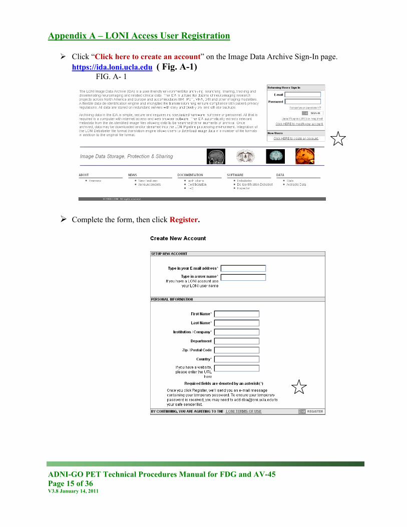
Appendix A – LONI Access User Registration Click “Click here to create an account” on the Image Data Archive Sign-In page.
https://ida.loni.ucla.edu ( Fig. A-1) FIG. A- 1
Complete the form, then click Register.
ADNI-GO PET Technical Procedures Manual for FDG and AV-45 Page 16 of 36 V3.8 January 14, 2011
Send an email to [email protected] requesting to have your permissions set for uploading ADNI data. Please include the email address used when you created your account, the name of your site and the name of your site
ADNI-GO PET Technical Procedures Manual for FDG and AV-45 Page 17 of 36 V3.8 January 14, 2011
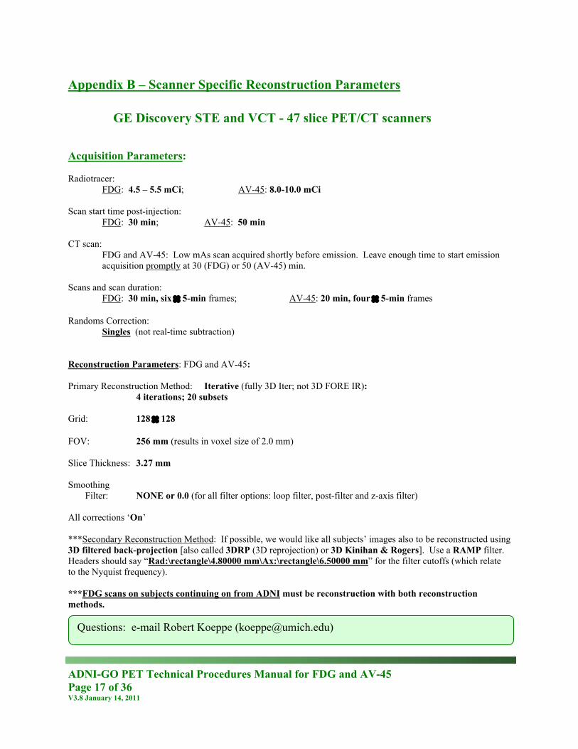
Appendix B – Scanner Specific Reconstruction Parameters
GE Discovery STE and VCT - 47 slice PET/CT scanners Acquisition Parameters: Radiotracer:
FDG: 4.5 – 5.5 mCi; AV-45: 8.0-10.0 mCi Scan start time post-injection:
FDG: 30 min; AV-45: 50 min CT scan:
FDG and AV-45: Low mAs scan acquired shortly before emission. Leave enough time to start emission acquisition promptly at 30 (FDG) or 50 (AV-45) min.
Scans and scan duration:
FDG: 30 min, six × 5-min frames; AV-45: 20 min, four × 5-min frames Randoms Correction:
Singles (not real-time subtraction) Reconstruction Parameters: FDG and AV-45: Primary Reconstruction Method: Iterative (fully 3D Iter; not 3D FORE IR): 4 iterations; 20 subsets Grid: 128 × 128 FOV: 256 mm (results in voxel size of 2.0 mm) Slice Thickness: 3.27 mm Smoothing
Filter: NONE or 0.0 (for all filter options: loop filter, post-filter and z-axis filter) All corrections ‘On’ ***Secondary Reconstruction Method: If possible, we would like all subjects’ images also to be reconstructed using 3D filtered back-projection [also called 3DRP (3D reprojection) or 3D Kinihan & Rogers]. Use a RAMP filter. Headers should say “Rad:\rectangle\4.80000 mm\Ax:\rectangle\6.50000 mm” for the filter cutoffs (which relate to the Nyquist frequency). ***FDG scans on subjects continuing on from ADNI must be reconstruction with both reconstruction methods.
Questions: e-mail Robert Koeppe ([email protected])
ADNI-GO PET Technical Procedures Manual for FDG and AV-45 Page 18 of 36 V3.8 January 14, 2011
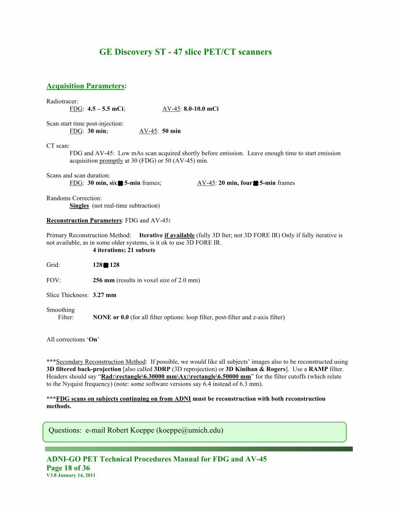
GE Discovery ST - 47 slice PET/CT scanners Acquisition Parameters: Radiotracer:
FDG: 4.5 – 5.5 mCi; AV-45: 8.0-10.0 mCi Scan start time post-injection:
FDG: 30 min; AV-45: 50 min CT scan:
FDG and AV-45: Low mAs scan acquired shortly before emission. Leave enough time to start emission acquisition promptly at 30 (FDG) or 50 (AV-45) min.
Scans and scan duration:
FDG: 30 min, six × 5-min frames; AV-45: 20 min, four × 5-min frames Randoms Correction:
Singles (not real-time subtraction) Reconstruction Parameters: FDG and AV-45: Primary Reconstruction Method: Iterative if available (fully 3D Iter; not 3D FORE IR) Only if fully iterative is not available, as in some older systems, is it ok to use 3D FORE IR. 4 iterations; 21 subsets Grid: 128 × 128 FOV: 256 mm (results in voxel size of 2.0 mm) Slice Thickness: 3.27 mm Smoothing
Filter: NONE or 0.0 (for all filter options: loop filter, post-filter and z-axis filter) All corrections ‘On’ ***Secondary Reconstruction Method: If possible, we would like all subjects’ images also to be reconstructed using 3D filtered back-projection [also called 3DRP (3D reprojection) or 3D Kinihan & Rogers]. Use a RAMP filter. Headers should say “Rad:\rectangle\6.30000 mm\Ax:\rectangle\6.50000 mm” for the filter cutoffs (which relate to the Nyquist frequency) (note: some software versions say 6.4 instead of 6.3 mm). ***FDG scans on subjects continuing on from ADNI must be reconstruction with both reconstruction methods.
Questions: e-mail Robert Koeppe ([email protected])
ADNI-GO PET Technical Procedures Manual for FDG and AV-45 Page 19 of 36 V3.8 January 14, 2011
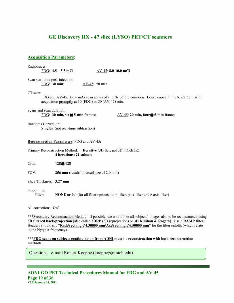
GE Discovery RX - 47 slice (LYSO) PET/CT scanners
Acquisition Parameters: Radiotracer:
FDG: 4.5 – 5.5 mCi; AV-45: 8.0-10.0 mCi Scan start time post-injection:
FDG: 30 min; AV-45: 50 min CT scan:
FDG and AV-45: Low mAs scan acquired shortly before emission. Leave enough time to start emission acquisition promptly at 30 (FDG) or 50 (AV-45) min.
Scans and scan duration:
FDG: 30 min, six × 5-min frames; AV-45: 20 min, four × 5-min frames Randoms Correction:
Singles (not real-time subtraction) Reconstruction Parameters: FDG and AV-45: Primary Reconstruction Method: Iterative (3D Iter; not 3D FORE IR): 4 iterations; 21 subsets Grid: 128 × 128 FOV: 256 mm (results in voxel size of 2.0 mm) Slice Thickness: 3.27 mm Smoothing
Filter: NONE or 0.0 (for all filter options: loop filter, post-filter and z-axis filter) All corrections ‘On’ ***Secondary Reconstruction Method: If possible, we would like all subjects’ images also to be reconstructed using 3D filtered back-projection [also called 3DRP (3D reprojection) or 3D Kinihan & Rogers]. Use a RAMP filter. Headers should say “Rad:\rectangle\4.30000 mm\Ax:\rectangle\6.50000 mm” for the filter cutoffs (which relate to the Nyquist frequency). ***FDG scans on subjects continuing on from ADNI must be reconstruction with both reconstruction methods.
Questions: e-mail Robert Koeppe ([email protected])
ADNI-GO PET Technical Procedures Manual for FDG and AV-45 Page 20 of 36 V3.8 January 14, 2011
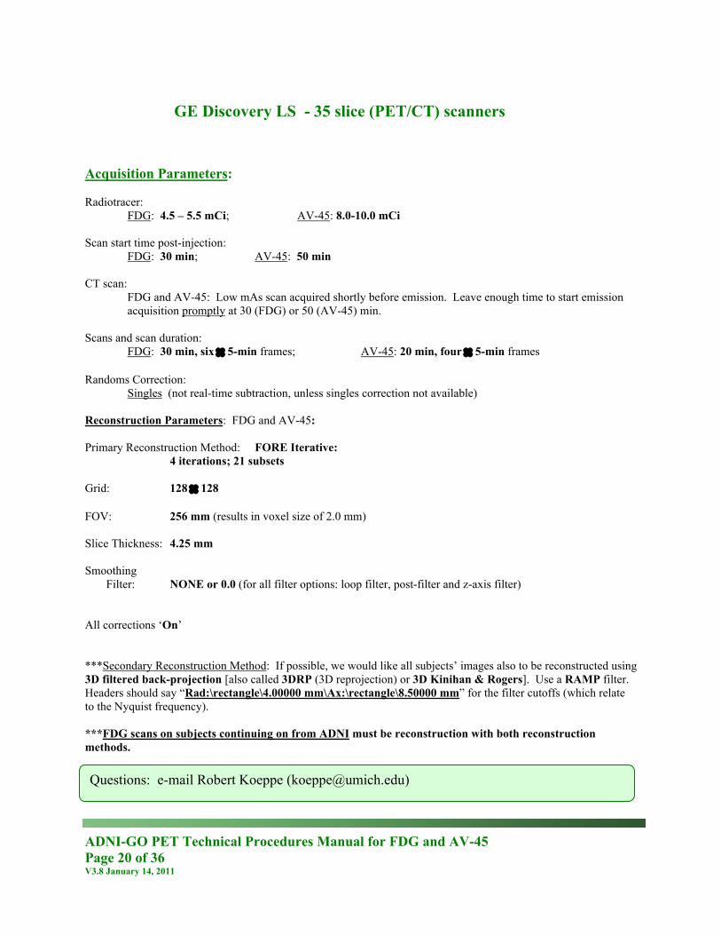
GE Discovery LS - 35 slice (PET/CT) scanners
Acquisition Parameters: Radiotracer:
FDG: 4.5 – 5.5 mCi; AV-45: 8.0-10.0 mCi Scan start time post-injection:
FDG: 30 min; AV-45: 50 min CT scan:
FDG and AV-45: Low mAs scan acquired shortly before emission. Leave enough time to start emission acquisition promptly at 30 (FDG) or 50 (AV-45) min.
Scans and scan duration:
FDG: 30 min, six × 5-min frames; AV-45: 20 min, four × 5-min frames Randoms Correction:
Singles (not real-time subtraction, unless singles correction not available) Reconstruction Parameters: FDG and AV-45: Primary Reconstruction Method: FORE Iterative: 4 iterations; 21 subsets Grid: 128 × 128 FOV: 256 mm (results in voxel size of 2.0 mm) Slice Thickness: 4.25 mm Smoothing
Filter: NONE or 0.0 (for all filter options: loop filter, post-filter and z-axis filter) All corrections ‘On’ ***Secondary Reconstruction Method: If possible, we would like all subjects’ images also to be reconstructed using 3D filtered back-projection [also called 3DRP (3D reprojection) or 3D Kinihan & Rogers]. Use a RAMP filter. Headers should say “Rad:\rectangle\4.00000 mm\Ax:\rectangle\8.50000 mm” for the filter cutoffs (which relate to the Nyquist frequency). ***FDG scans on subjects continuing on from ADNI must be reconstruction with both reconstruction methods.
Questions: e-mail Robert Koeppe ([email protected])
ADNI-GO PET Technical Procedures Manual for FDG and AV-45 Page 21 of 36 V3.8 January 14, 2011
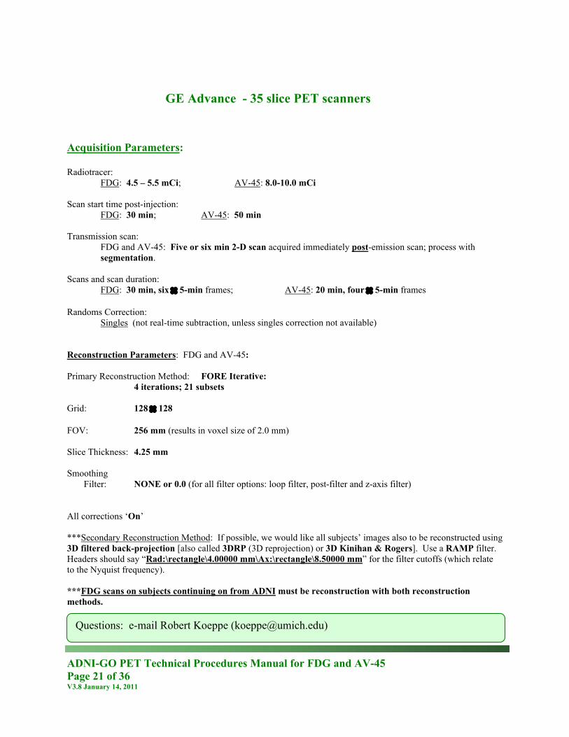
GE Advance - 35 slice PET scanners Acquisition Parameters: Radiotracer:
FDG: 4.5 – 5.5 mCi; AV-45: 8.0-10.0 mCi Scan start time post-injection:
FDG: 30 min; AV-45: 50 min Transmission scan:
FDG and AV-45: Five or six min 2-D scan acquired immediately post-emission scan; process with segmentation.
Scans and scan duration:
FDG: 30 min, six × 5-min frames; AV-45: 20 min, four × 5-min frames Randoms Correction:
Singles (not real-time subtraction, unless singles correction not available) Reconstruction Parameters: FDG and AV-45: Primary Reconstruction Method: FORE Iterative: 4 iterations; 21 subsets Grid: 128 × 128 FOV: 256 mm (results in voxel size of 2.0 mm) Slice Thickness: 4.25 mm Smoothing
Filter: NONE or 0.0 (for all filter options: loop filter, post-filter and z-axis filter) All corrections ‘On’ ***Secondary Reconstruction Method: If possible, we would like all subjects’ images also to be reconstructed using 3D filtered back-projection [also called 3DRP (3D reprojection) or 3D Kinihan & Rogers]. Use a RAMP filter. Headers should say “Rad:\rectangle\4.00000 mm\Ax:\rectangle\8.50000 mm” for the filter cutoffs (which relate to the Nyquist frequency). ***FDG scans on subjects continuing on from ADNI must be reconstruction with both reconstruction methods.
Questions: e-mail Robert Koeppe ([email protected])
ADNI-GO PET Technical Procedures Manual for FDG and AV-45 Page 22 of 36 V3.8 January 14, 2011
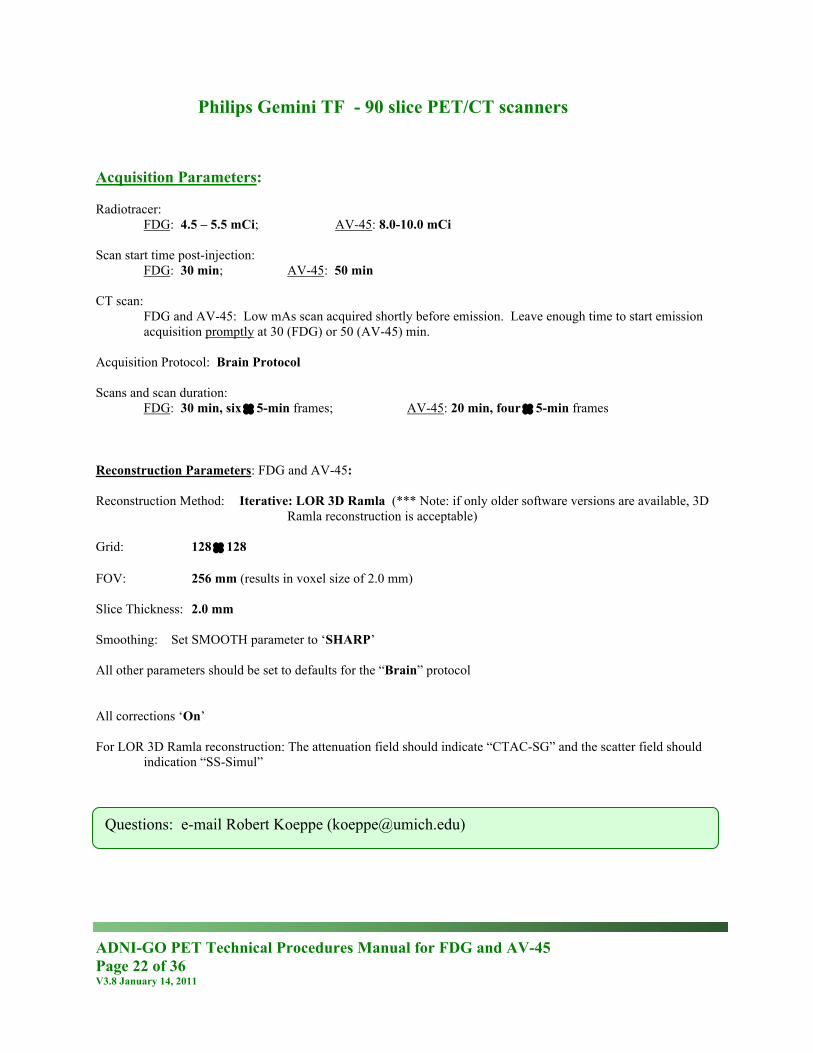
Philips Gemini TF - 90 slice PET/CT scanners Acquisition Parameters: Radiotracer:
FDG: 4.5 – 5.5 mCi; AV-45: 8.0-10.0 mCi Scan start time post-injection:
FDG: 30 min; AV-45: 50 min CT scan:
FDG and AV-45: Low mAs scan acquired shortly before emission. Leave enough time to start emission acquisition promptly at 30 (FDG) or 50 (AV-45) min.
Acquisition Protocol: Brain Protocol Scans and scan duration:
FDG: 30 min, six × 5-min frames; AV-45: 20 min, four × 5-min frames Reconstruction Parameters: FDG and AV-45: Reconstruction Method: Iterative: LOR 3D Ramla (*** Note: if only older software versions are available, 3D
Ramla reconstruction is acceptable) Grid: 128 × 128 FOV: 256 mm (results in voxel size of 2.0 mm) Slice Thickness: 2.0 mm Smoothing: Set SMOOTH parameter to ‘SHARP’ All other parameters should be set to defaults for the “Brain” protocol All corrections ‘On’ For LOR 3D Ramla reconstruction: The attenuation field should indicate “CTAC-SG” and the scatter field should
indication “SS-Simul”
Questions: e-mail Robert Koeppe ([email protected])
ADNI-GO PET Technical Procedures Manual for FDG and AV-45 Page 23 of 36 V3.8 January 14, 2011
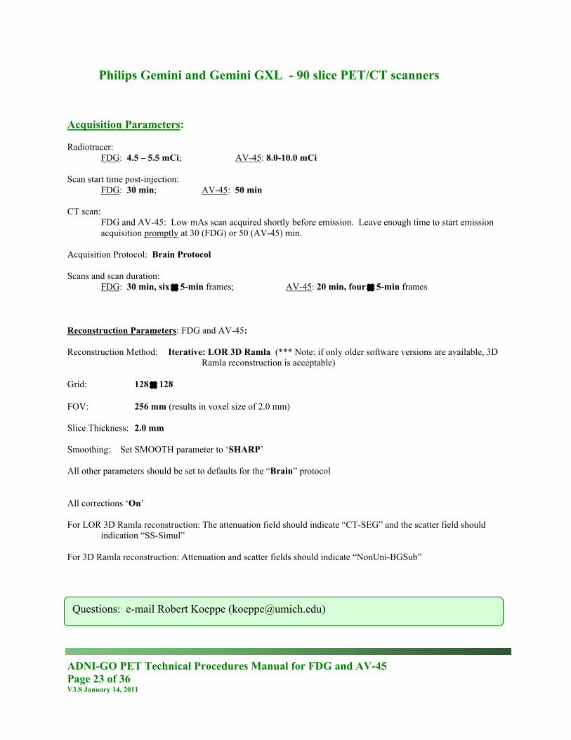
Philips Gemini and Gemini GXL - 90 slice PET/CT scanners Acquisition Parameters: Radiotracer:
FDG: 4.5 – 5.5 mCi; AV-45: 8.0-10.0 mCi Scan start time post-injection:
FDG: 30 min; AV-45: 50 min CT scan:
FDG and AV-45: Low mAs scan acquired shortly before emission. Leave enough time to start emission acquisition promptly at 30 (FDG) or 50 (AV-45) min.
Acquisition Protocol: Brain Protocol Scans and scan duration:
FDG: 30 min, six × 5-min frames; AV-45: 20 min, four × 5-min frames Reconstruction Parameters: FDG and AV-45: Reconstruction Method: Iterative: LOR 3D Ramla (*** Note: if only older software versions are available, 3D
Ramla reconstruction is acceptable) Grid: 128 × 128 FOV: 256 mm (results in voxel size of 2.0 mm) Slice Thickness: 2.0 mm Smoothing: Set SMOOTH parameter to ‘SHARP’ All other parameters should be set to defaults for the “Brain” protocol All corrections ‘On’ For LOR 3D Ramla reconstruction: The attenuation field should indicate “CT-SEG” and the scatter field should
indication “SS-Simul” For 3D Ramla reconstruction: Attenuation and scatter fields should indicate “NonUni-BGSub”
Questions: e-mail Robert Koeppe ([email protected])
ADNI-GO PET Technical Procedures Manual for FDG and AV-45 Page 24 of 36 V3.8 January 14, 2011
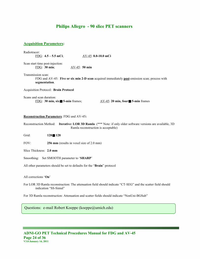
Philips Allegro - 90 slice PET scanners Acquisition Parameters: Radiotracer:
FDG: 4.5 – 5.5 mCi; AV-45: 8.0-10.0 mCi Scan start time post-injection:
FDG: 30 min; AV-45: 50 min Transmission scan:
FDG and AV-45: Five or six min 2-D scan acquired immediately post-emission scan; process with segmentation.
Acquisition Protocol: Brain Protocol Scans and scan duration:
FDG: 30 min, six × 5-min frames; AV-45: 20 min, four × 5-min frames Reconstruction Parameters: FDG and AV-45: Reconstruction Method: Iterative: LOR 3D Ramla (*** Note: if only older software versions are available, 3D
Ramla reconstruction is acceptable) Grid: 128 × 128 FOV: 256 mm (results in voxel size of 2.0 mm) Slice Thickness: 2.0 mm Smoothing: Set SMOOTH parameter to ‘SHARP’ All other parameters should be set to defaults for the “Brain” protocol All corrections ‘On’ For LOR 3D Ramla reconstruction: The attenuation field should indicate “CT-SEG” and the scatter field should
indication “SS-Simul” For 3D Ramla reconstruction: Attenuation and scatter fields should indicate “NonUni-BGSub”
Questions: e-mail Robert Koeppe ([email protected])
ADNI-GO PET Technical Procedures Manual for FDG and AV-45 Page 25 of 36 V3.8 January 14, 2011
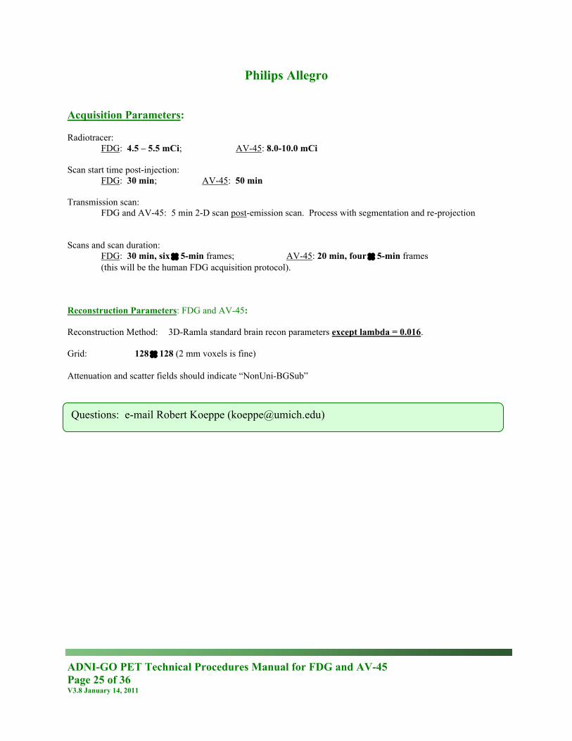
Philips Allegro Acquisition Parameters: Radiotracer:
FDG: 4.5 – 5.5 mCi; AV-45: 8.0-10.0 mCi Scan start time post-injection:
FDG: 30 min; AV-45: 50 min Transmission scan:
FDG and AV-45: 5 min 2-D scan post-emission scan. Process with segmentation and re-projection
Scans and scan duration:
FDG: 30 min, six × 5-min frames; AV-45: 20 min, four × 5-min frames (this will be the human FDG acquisition protocol).
Reconstruction Parameters: FDG and AV-45: Reconstruction Method: 3D-Ramla standard brain recon parameters except lambda = 0.016. Grid: 128 × 128 (2 mm voxels is fine) Attenuation and scatter fields should indicate “NonUni-BGSub”
Questions: e-mail Robert Koeppe ([email protected])
ADNI-GO PET Technical Procedures Manual for FDG and AV-45 Page 26 of 36 V3.8 January 14, 2011
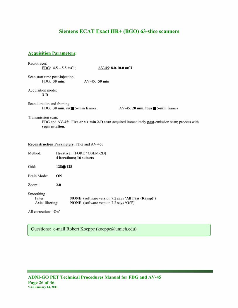
Siemens ECAT Exact HR+ (BGO) 63-slice scanners Acquisition Parameters: Radiotracer:
FDG: 4.5 – 5.5 mCi; AV-45: 8.0-10.0 mCi Scan start time post-injection:
FDG: 30 min; AV-45: 50 min Acquisition mode:
3-D Scan duration and framing:
FDG: 30 min, six × 5-min frames; AV-45: 20 min, four × 5-min frames Transmission scan:
FDG and AV-45: Five or six min 2-D scan acquired immediately post-emission scan; process with segmentation.
Reconstruction Parameters, FDG and AV-45: Method: Iterative: (FORE / OSEM-2D) 4 iterations; 16 subsets Grid: 128 × 128 Brain Mode: ON Zoom: 2.0 Smoothing
Filter: NONE (software version 7.2 says ‘All Pass (Ramp)’) Axial filtering: NONE (software version 7.2 says ‘Off’)
All corrections ‘On’
Questions: e-mail Robert Koeppe ([email protected])
ADNI-GO PET Technical Procedures Manual for FDG and AV-45 Page 27 of 36 V3.8 January 14, 2011
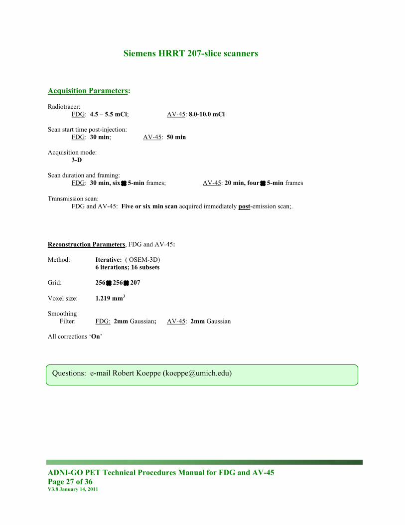
Siemens HRRT 207-slice scanners Acquisition Parameters: Radiotracer:
FDG: 4.5 – 5.5 mCi; AV-45: 8.0-10.0 mCi Scan start time post-injection:
FDG: 30 min; AV-45: 50 min Acquisition mode:
3-D Scan duration and framing:
FDG: 30 min, six × 5-min frames; AV-45: 20 min, four × 5-min frames Transmission scan:
FDG and AV-45: Five or six min scan acquired immediately post-emission scan;. Reconstruction Parameters, FDG and AV-45: Method: Iterative: ( OSEM-3D) 6 iterations; 16 subsets Grid: 256 × 256 × 207 Voxel size: 1.219 mm3 Smoothing
Filter: FDG: 2mm Gaussian; AV-45: 2mm Gaussian All corrections ‘On’
Questions: e-mail Robert Koeppe ([email protected])
ADNI-GO PET Technical Procedures Manual for FDG and AV-45 Page 28 of 36 V3.8 January 14, 2011
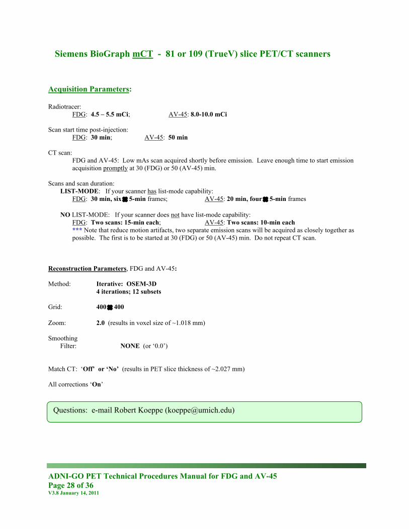
Siemens BioGraph mCT - 81 or 109 (TrueV) slice PET/CT scanners Acquisition Parameters: Radiotracer:
FDG: 4.5 – 5.5 mCi; AV-45: 8.0-10.0 mCi Scan start time post-injection:
FDG: 30 min; AV-45: 50 min CT scan:
FDG and AV-45: Low mAs scan acquired shortly before emission. Leave enough time to start emission acquisition promptly at 30 (FDG) or 50 (AV-45) min.
Scans and scan duration: LIST-MODE: If your scanner has list-mode capability:
FDG: 30 min, six × 5-min frames; AV-45: 20 min, four × 5-min frames NO LIST-MODE: If your scanner does not have list-mode capability:
FDG: Two scans: 15-min each; AV-45: Two scans: 10-min each *** Note that reduce motion artifacts, two separate emission scans will be acquired as closely together as
possible. The first is to be started at 30 (FDG) or 50 (AV-45) min. Do not repeat CT scan. Reconstruction Parameters, FDG and AV-45: Method: Iterative: OSEM-3D 4 iterations; 12 subsets Grid: 400 × 400 Zoom: 2.0 (results in voxel size of ~1.018 mm) Smoothing
Filter: NONE (or ‘0.0’) Match CT: ‘Off’ or ‘No’ (results in PET slice thickness of ~2.027 mm) All corrections ‘On’
Questions: e-mail Robert Koeppe ([email protected])
ADNI-GO PET Technical Procedures Manual for FDG and AV-45 Page 29 of 36 V3.8 January 14, 2011
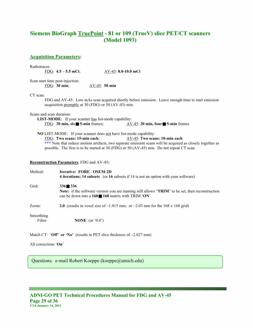
Siemens BioGraph TruePoint - 81 or 109 (TrueV) slice PET/CT scanners (Model 1093)
Acquisition Parameters: Radiotracer:
FDG: 4.5 – 5.5 mCi; AV-45: 8.0-10.0 mCi Scan start time post-injection:
FDG: 30 min; AV-45: 50 min CT scan:
FDG and AV-45: Low mAs scan acquired shortly before emission. Leave enough time to start emission acquisition promptly at 30 (FDG) or 50 (AV-45) min.
Scans and scan duration: LIST-MODE: If your scanner has list-mode capability:
FDG: 30 min, six × 5-min frames; AV-45: 20 min, four × 5-min frames NO LIST-MODE: If your scanner does not have list-mode capability:
FDG: Two scans: 15-min each; AV-45: Two scans: 10-min each *** Note that reduce motion artifacts, two separate emission scans will be acquired as closely together as
possible. The first is to be started at 30 (FDG) or 50 (AV-45) min. Do not repeat CT scan. Reconstruction Parameters, FDG and AV-45: Method: Iterative: FORE / OSEM-2D 4 iterations; 14 subsets (or 16 subsets if 14 is not an option with your software) Grid: 336 × 336
Note: if the software version you are running still allows “TRIM” to be set, then reconstruction can be down into a 168 × 168 matrix with TRIM ‘ON’
Zoom: 2.0 (results in voxel size of ~1.015 mm; or ~2.03 mm for the 168 × 168 grid) Smoothing
Filter: NONE (or ‘0.0’) Match CT: ‘Off’ or ‘No’ (results in PET slice thickness of ~2.027 mm) All corrections ‘On’
Questions: e-mail Robert Koeppe ([email protected])
ADNI-GO PET Technical Procedures Manual for FDG and AV-45 Page 30 of 36 V3.8 January 14, 2011
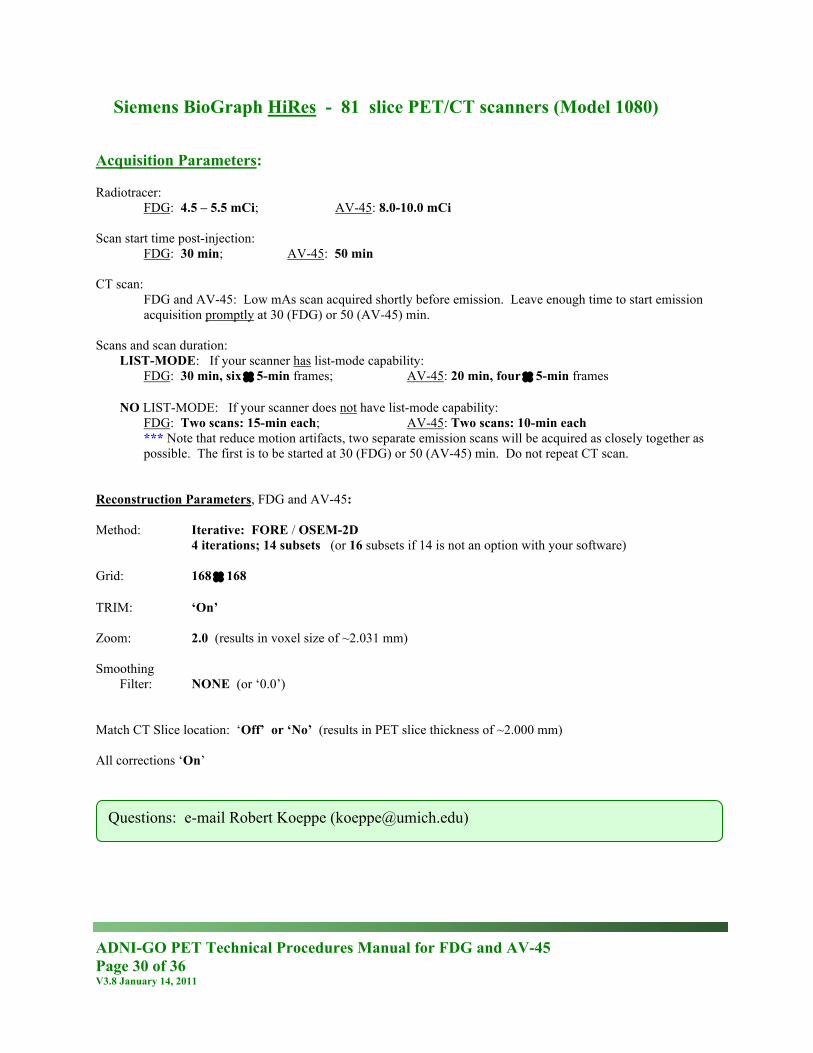
Siemens BioGraph HiRes - 81 slice PET/CT scanners (Model 1080) Acquisition Parameters: Radiotracer:
FDG: 4.5 – 5.5 mCi; AV-45: 8.0-10.0 mCi Scan start time post-injection:
FDG: 30 min; AV-45: 50 min CT scan:
FDG and AV-45: Low mAs scan acquired shortly before emission. Leave enough time to start emission acquisition promptly at 30 (FDG) or 50 (AV-45) min.
Scans and scan duration: LIST-MODE: If your scanner has list-mode capability:
FDG: 30 min, six × 5-min frames; AV-45: 20 min, four × 5-min frames NO LIST-MODE: If your scanner does not have list-mode capability:
FDG: Two scans: 15-min each; AV-45: Two scans: 10-min each *** Note that reduce motion artifacts, two separate emission scans will be acquired as closely together as
possible. The first is to be started at 30 (FDG) or 50 (AV-45) min. Do not repeat CT scan. Reconstruction Parameters, FDG and AV-45: Method: Iterative: FORE / OSEM-2D 4 iterations; 14 subsets (or 16 subsets if 14 is not an option with your software) Grid: 168 × 168 TRIM: ‘On’ Zoom: 2.0 (results in voxel size of ~2.031 mm) Smoothing
Filter: NONE (or ‘0.0’) Match CT Slice location: ‘Off’ or ‘No’ (results in PET slice thickness of ~2.000 mm) All corrections ‘On’
Questions: e-mail Robert Koeppe ([email protected])
ADNI-GO PET Technical Procedures Manual for FDG and AV-45 Page 31 of 36 V3.8 January 14, 2011
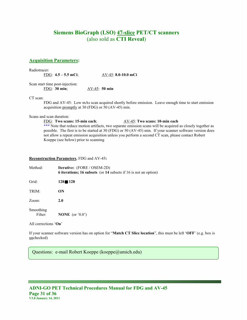
Siemens BioGraph (LSO) 47-slice PET/CT scanners (also sold as CTI Reveal)
Acquisition Parameters: Radiotracer:
FDG: 4.5 – 5.5 mCi; AV-45: 8.0-10.0 mCi Scan start time post-injection:
FDG: 30 min; AV-45: 50 min CT scan:
FDG and AV-45: Low mAs scan acquired shortly before emission. Leave enough time to start emission acquisition promptly at 30 (FDG) or 50 (AV-45) min.
Scans and scan duration:
FDG: Two scans: 15-min each; AV-45: Two scans: 10-min each *** Note that reduce motion artifacts, two separate emission scans will be acquired as closely together as
possible. The first is to be started at 30 (FDG) or 50 (AV-45) min. If your scanner software version does not allow a repeat emission acquisition unless you perform a second CT scan, please contact Robert Koeppe (see below) prior to scanning
Reconstruction Parameters, FDG and AV-45: Method: Iterative: (FORE / OSEM-2D) 6 iterations; 16 subsets (or 14 subsets if 16 is not an option) Grid: 128 × 128 TRIM: ON Zoom: 2.0 Smoothing
Filter: NONE (or ‘0.0’) All corrections ‘On’ If your scanner software version has on option for “Match CT Slice location”, this must be left ‘OFF’ (e.g. box is unchecked)
Questions: e-mail Robert Koeppe ([email protected])
ADNI-GO PET Technical Procedures Manual for FDG and AV-45 Page 32 of 36 V3.8 January 14, 2011
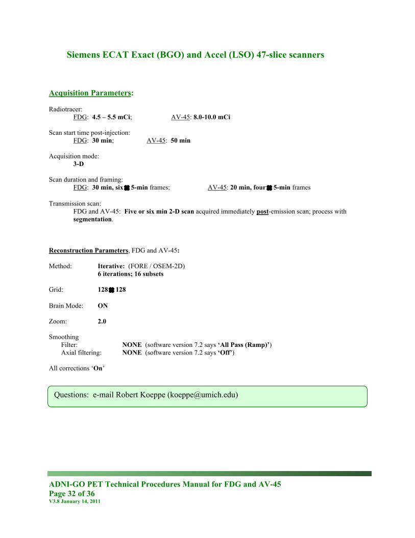
Siemens ECAT Exact (BGO) and Accel (LSO) 47-slice scanners Acquisition Parameters: Radiotracer:
FDG: 4.5 – 5.5 mCi; AV-45: 8.0-10.0 mCi Scan start time post-injection:
FDG: 30 min; AV-45: 50 min Acquisition mode:
3-D Scan duration and framing:
FDG: 30 min, six × 5-min frames; AV-45: 20 min, four × 5-min frames Transmission scan:
FDG and AV-45: Five or six min 2-D scan acquired immediately post-emission scan; process with segmentation.
Reconstruction Parameters, FDG and AV-45: Method: Iterative: (FORE / OSEM-2D) 6 iterations; 16 subsets Grid: 128 × 128 Brain Mode: ON Zoom: 2.0 Smoothing
Filter: NONE (software version 7.2 says ‘All Pass (Ramp)’) Axial filtering: NONE (software version 7.2 says ‘Off’)
All corrections ‘On’
Questions: e-mail Robert Koeppe ([email protected])
ADNI-GO PET Technical Procedures Manual for FDG and AV-45 Page 33 of 36 V3.8 January 14, 2011
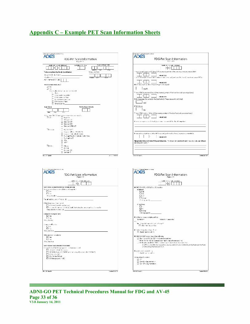
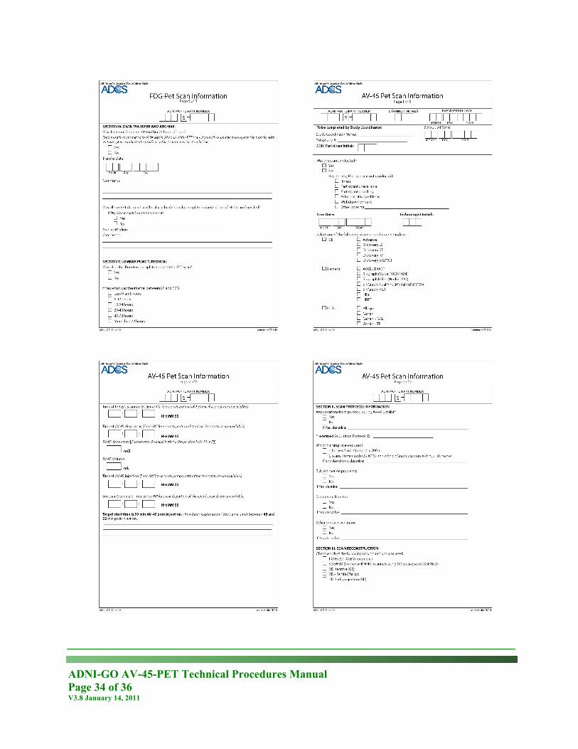
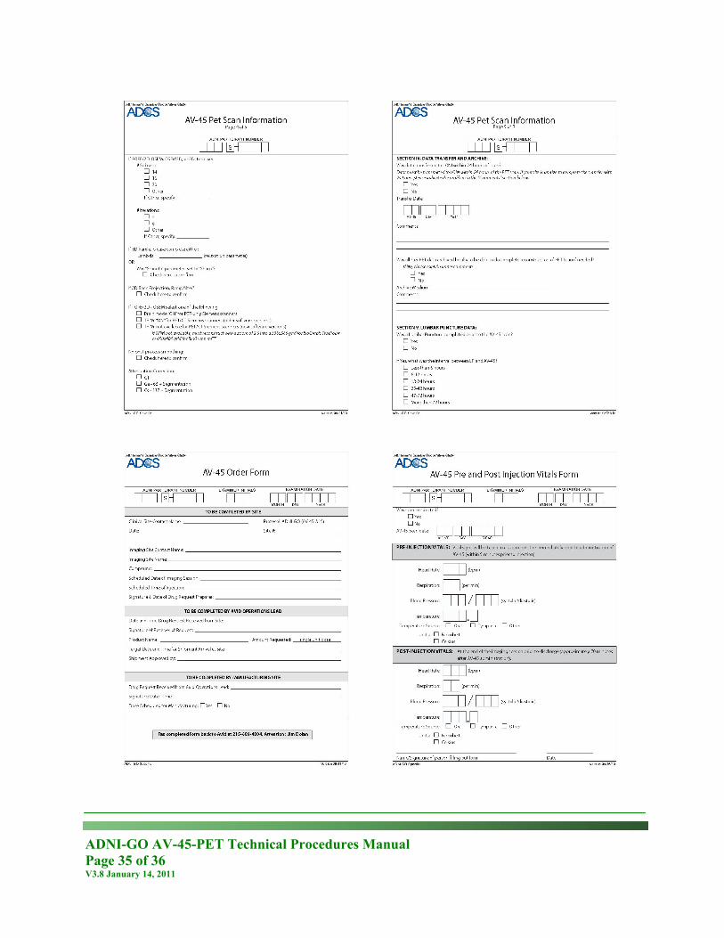
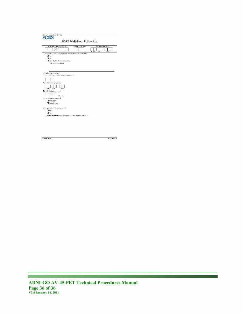
Appendix C – Example PET Scan Information Sheets
ADNI-GO AV-45-PET Technical Procedures Manual Page 34 of 36 V3.8 January 14, 2011
ADNI-GO AV-45-PET Technical Procedures Manual Page 35 of 36 V3.8 January 14, 2011
ADNI-GO AV-45-PET Technical Procedures Manual Page 36 of 36 V3.8 January 14, 2011
Related Documents


