Administration of M. leprae Hsp65 Interferes with the Murine Lupus Progression Eliana B. Marengo 1 , Luciana V. de Moraes 2 , Marcella Faria 3 , Beatriz L. Fernandes 3 , Luciana V. Carvalho 1 , Denise V. Tambourgi 1 , Luiz V. Rizzo 2 , Fernanda C. V. Portaro 1 , Anto ˆ nio Carlos M. Camargo 3 , Osvaldo A. Sant’Anna 1 * 1 Immunochemistry Laboratory, Instituto Butantan, Sa ˜o Paulo, Brazil, 2 Clinical Immunology Laboratory, Instituto de Cie ˆncias Biome ´dicas, University of Sa ˜o Paulo, Sa ˜o Paulo, Brazil, 3 Center for Applied Toxinology – CAT/CEPID, Sa ˜o Paulo, Brazil Abstract The heat shock protein [Hsp] family guides several steps during protein synthesis, are abundant in prokaryotic and eukaryotic cells, and are highly conserved during evolution. The Hsp60 family is involved in assembly and transport of proteins, and is expressed at very high levels during autoimmunity or autoinflammatory phenomena. Here, the pathophysiological role of the wild type [WT] and the point mutated K 409 A recombinant Hsp65 of M. leprae in an animal model of Systemic Lupus Erythematosus [SLE] was evaluated in vivo using the genetically homogeneous [NZBxNZW]F 1 mice. Anti-DNA and anti-Hsp65 antibodies responsiveness was individually measured during the animal’s life span, and the mean survival time [MST] was determined. The treatment with WT abbreviates the MST in 46%, when compared to non- treated mice [p,0.001]. An increase in the IgG2a/IgG1 anti-DNA antibodies ratio was also observed in animals injected with the WT Hsp65. Incubation of BALB/c macrophages with F 1 serum from WT treated mice resulted in acute cell necrosis; treatment of these cells with serum from K 409 A treated mice did not cause any toxic effect. Moreover, the involvement of WT correlates with age and is dose-dependent. Our data suggest that Hsp65 may be a central molecule intervening in the progression of the SLE, and that the point mutated K 409 A recombinant immunogenic molecule, that counteracts the deleterious effect of WT, may act mitigating and delaying the development of SLE in treated mice. This study gives new insights into the general biological role of Hsp and the significant impact of environmental factors during the pathogenesis of this autoimmune process. Citation: Marengo EB, de Moraes LV, Faria M, Fernandes BL, Carvalho LV, et al. (2008) Administration of M. leprae Hsp65 Interferes with the Murine Lupus Progression. PLoS ONE 3(8): e3025. doi:10.1371/journal.pone.0003025 Editor: Graham Pockley, University of Sheffield, United Kingdom Received May 8, 2008; Accepted August 1, 2008; Published August 21, 2008 Copyright: ß 2008 Marengo et al. This is an open-access article distributed under the terms of the Creative Commons Attribution License, which permits unrestricted use, distribution, and reproduction in any medium, provided the original author and source are credited. Funding: This work was supported by funds of FAPESP [Fundac ¸a ˜o de Amparo a ` Pesquisa do Estado de Sa ˜o Paulo] through the CAT/CEPID program. E. B. Marengo, L. V. Moraes and L. V. Carvalho are recipient of FAPESP fellowships. D. V. Tambourgi, L. V. Rizzo, A. C. M. Camargo and O.A. Sant’Anna are researchers of CNPq-Brazil. Competing Interests: The authors have declared that no competing interests exist. * E-mail: [email protected] Introduction The Hsp60 guides essential activities for cell homeostasis being highly conserved among organisms; in stress or inflammation conditions, they increase 4 to 5-fold in the cell subsequently undergoing autolysis returning to basal levels [1]. The distinct Hsp classes show extensive amino acid sequence similarities, from microbial to mammalian. The mycobacterium 65 kDa, a member of the Hsp60 family, shows an approximately 55% similarity/ identity with human Hsp60 [2,3]. The difference between Hsp60 and Hsp65 is striking in regions containing epitopes recognized by T cells of the vertebrate host and the cross-reactivity between these determinants is due to conserved regions across all organisms. Stress proteins of a wide variety of microorganisms have been found to stimulate an immune response in vertebrates [4,5], but the evolutionary consequences of this recognition are poorly understood. Published data show that Hsp65 is one of the major target for the immune response to pathogens [6,7]. The major sequence similarity among species renders the Hsp65 a potential inducer of immune responses to host self molecules that may lead to autoimmune phenomena. Due to high similarity inter-species in their sequence, it is suggested from a process of molecular mimicry, the participation of the Hsp60 in the modulation and etiology or pathogenesis of autoimmunities [8–11]. Pockley et al [12] indicated the presence of antibodies for Hsp70 family in healthy individuals, such as that of Stephanou et al [13] showing significant increase of anti-Hsp90 antibody titers in patients with SLE compared to healthy individuals. There are several studies that attempt to establish the relation between Hsp60 and inflammatory and proliferative responses. High anti-Hsp60/65 antibody titers were not restricted to disease patients and could also be detected during aging [12,14–16]. Recently, our group described that the recombinant Hsp65 from Mycobacterium leprae displays proteolytic activity towards oligopeptides. The amino acid sequence alignment of the M. leprae Hsp65 with the Escherichia coli HslVU protease suggested two putative threonine catalytic groups, one in the N-domain [Thr 136 , Lys 168 , Tyr 264 ] and the other in the C-domain [Thr 375 , Lys 409 , Ser 502 ]. Mutagenesis studies showed that these amino acid residues at the C-domain form the catalytic group that carries out the main proteolytic activity of this molecule [17]. Systemic lupus erythematosus [SLE] is the prototypic autoim- mune disease, influenced by a combination of genetic and environmental factors and characterized by a marked loss of tolerance to self antigens such as DNA, RNA and other nuclear PLoS ONE | www.plosone.org 1 August 2008 | Volume 3 | Issue 8 | e3025

Welcome message from author
This document is posted to help you gain knowledge. Please leave a comment to let me know what you think about it! Share it to your friends and learn new things together.
Transcript
Administration of M. leprae Hsp65 Interferes with theMurine Lupus ProgressionEliana B. Marengo1, Luciana V. de Moraes2, Marcella Faria3, Beatriz L. Fernandes3, Luciana V. Carvalho1,
Denise V. Tambourgi1, Luiz V. Rizzo2, Fernanda C. V. Portaro1, Antonio Carlos M. Camargo3, Osvaldo A.
Sant’Anna1*
1 Immunochemistry Laboratory, Instituto Butantan, Sao Paulo, Brazil, 2 Clinical Immunology Laboratory, Instituto de Ciencias Biomedicas, University of Sao Paulo, Sao
Paulo, Brazil, 3 Center for Applied Toxinology – CAT/CEPID, Sao Paulo, Brazil
Abstract
The heat shock protein [Hsp] family guides several steps during protein synthesis, are abundant in prokaryotic andeukaryotic cells, and are highly conserved during evolution. The Hsp60 family is involved in assembly and transport ofproteins, and is expressed at very high levels during autoimmunity or autoinflammatory phenomena. Here, thepathophysiological role of the wild type [WT] and the point mutated K409A recombinant Hsp65 of M. leprae in an animalmodel of Systemic Lupus Erythematosus [SLE] was evaluated in vivo using the genetically homogeneous [NZBxNZW]F1
mice. Anti-DNA and anti-Hsp65 antibodies responsiveness was individually measured during the animal’s life span, and themean survival time [MST] was determined. The treatment with WT abbreviates the MST in 46%, when compared to non-treated mice [p,0.001]. An increase in the IgG2a/IgG1 anti-DNA antibodies ratio was also observed in animals injected withthe WT Hsp65. Incubation of BALB/c macrophages with F1 serum from WT treated mice resulted in acute cell necrosis;treatment of these cells with serum from K409A treated mice did not cause any toxic effect. Moreover, the involvement ofWT correlates with age and is dose-dependent. Our data suggest that Hsp65 may be a central molecule intervening in theprogression of the SLE, and that the point mutated K409A recombinant immunogenic molecule, that counteracts thedeleterious effect of WT, may act mitigating and delaying the development of SLE in treated mice. This study gives newinsights into the general biological role of Hsp and the significant impact of environmental factors during the pathogenesisof this autoimmune process.
Citation: Marengo EB, de Moraes LV, Faria M, Fernandes BL, Carvalho LV, et al. (2008) Administration of M. leprae Hsp65 Interferes with the Murine LupusProgression. PLoS ONE 3(8): e3025. doi:10.1371/journal.pone.0003025
Editor: Graham Pockley, University of Sheffield, United Kingdom
Received May 8, 2008; Accepted August 1, 2008; Published August 21, 2008
Copyright: � 2008 Marengo et al. This is an open-access article distributed under the terms of the Creative Commons Attribution License, which permitsunrestricted use, distribution, and reproduction in any medium, provided the original author and source are credited.
Funding: This work was supported by funds of FAPESP [Fundacao de Amparo a Pesquisa do Estado de Sao Paulo] through the CAT/CEPID program. E. B.Marengo, L. V. Moraes and L. V. Carvalho are recipient of FAPESP fellowships. D. V. Tambourgi, L. V. Rizzo, A. C. M. Camargo and O.A. Sant’Anna are researchers ofCNPq-Brazil.
Competing Interests: The authors have declared that no competing interests exist.
* E-mail: [email protected]
Introduction
The Hsp60 guides essential activities for cell homeostasis being
highly conserved among organisms; in stress or inflammation
conditions, they increase 4 to 5-fold in the cell subsequently
undergoing autolysis returning to basal levels [1]. The distinct Hsp
classes show extensive amino acid sequence similarities, from
microbial to mammalian. The mycobacterium 65 kDa, a member
of the Hsp60 family, shows an approximately 55% similarity/
identity with human Hsp60 [2,3]. The difference between Hsp60
and Hsp65 is striking in regions containing epitopes recognized by
T cells of the vertebrate host and the cross-reactivity between these
determinants is due to conserved regions across all organisms.
Stress proteins of a wide variety of microorganisms have been
found to stimulate an immune response in vertebrates [4,5], but the
evolutionary consequences of this recognition are poorly understood.
Published data show that Hsp65 is one of the major target for the
immune response to pathogens [6,7]. The major sequence similarity
among species renders the Hsp65 a potential inducer of immune
responses to host self molecules that may lead to autoimmune
phenomena. Due to high similarity inter-species in their sequence, it
is suggested from a process of molecular mimicry, the participation
of the Hsp60 in the modulation and etiology or pathogenesis of
autoimmunities [8–11]. Pockley et al [12] indicated the presence of
antibodies for Hsp70 family in healthy individuals, such as that of
Stephanou et al [13] showing significant increase of anti-Hsp90
antibody titers in patients with SLE compared to healthy individuals.
There are several studies that attempt to establish the relation
between Hsp60 and inflammatory and proliferative responses. High
anti-Hsp60/65 antibody titers were not restricted to disease patients
and could also be detected during aging [12,14–16].
Recently, our group described that the recombinant Hsp65
from Mycobacterium leprae displays proteolytic activity towards
oligopeptides. The amino acid sequence alignment of the M.
leprae Hsp65 with the Escherichia coli HslVU protease suggested two
putative threonine catalytic groups, one in the N-domain [Thr136,
Lys168, Tyr264] and the other in the C-domain [Thr375, Lys409,
Ser502]. Mutagenesis studies showed that these amino acid residues
at the C-domain form the catalytic group that carries out the main
proteolytic activity of this molecule [17].
Systemic lupus erythematosus [SLE] is the prototypic autoim-
mune disease, influenced by a combination of genetic and
environmental factors and characterized by a marked loss of
tolerance to self antigens such as DNA, RNA and other nuclear
PLoS ONE | www.plosone.org 1 August 2008 | Volume 3 | Issue 8 | e3025
factors, followed by immune-mediated injury to multiple organs
[18]. The New Zealand Black [H–2d2] 6New Zealand White [H–
2z] [NZBxNZW]F1 hybrid mouse is genetically predisposed to
develop autoimmune disease, which resembles SLE in humans. The
evolution of this chronic inflammatory disease is characterized by an
abnormal polyclonal B cell activation with a elevated production of
antinuclear antibodies that include anti-double-stranded DNA,
lymphadenopathy, arthritis, hemolytic anemia, vasculitis and
glomerulonephritis [19,20]. DNA plays a central role in the
pathogenesis of SLE, serving as a target antigen of auto–antibodies
as well as a major component of immune complexes [21,22].
Whereas the origin of the autoantibodies in SLE has received intense
investigation, the mechanisms involved in increased anti-DNA titers
are not well understood. It has been suggested that accelerated
monocytes/macrophages apoptosis in SLE contributes to impaired
clearance of apoptotic cells and increases apoptotic material [23,24].
Similarly to humans, the experimental disease is most frequent and
severe in female mice and the pathologic features appear when these
animals are around 6-month-old and they usually evolve to death
around 10–12-month of age [25–27].
Motivated by the Ilya Prigogine theoretical proposition [28,29]
that living systems achieve ordered state from relatively disorga-
nized configurations by assembling a new type of dynamic state
defined as dissipative structures. In this study the purpose was to
establish an original approach whereas disequilibrium is induced
by passive administration of homologous proteins. The attempts of
the system to reestablish an equilibrium situation seem severely
compromised in [NZBxNZW]F1 autoimmunity prone mice. Thus,
the putative pathophysiological role of the wild type [WT] and the
one point mutated K409A recombinant Hsp65 [rHsp65] of M.
leprae in SLE was extensively evaluated in genetically homogeneous
[NZBxNZW]F1 mice treated at distinct ages. Clinical signs were
analyzed, including development of ascites, pile erection, lethargy,
anorexia, as well as the mean survival time. In addition, the
quantitative phenotypic traits of anti-DNA and anti-Hsp65
antibody responsiveness were individually determined during
one year of life span. The in vitro induction of apoptosis/necrosis
by serum from WT or K409A rHsp65 treated F1 mice were also
evaluated in normal macrophages. The results suggest the
involvement of the Hsp65 as a central molecule intervening in
the foundation and progression of the SLE syndrome. The relation
between phenotypes in untreated and WT rHsp65 treated
individuals, as the K409A mutant employed, gave new insights
into the general biological role of Hsp molecules, as potential
neutralizers of the great impact of environmental factors on SLE.
Results
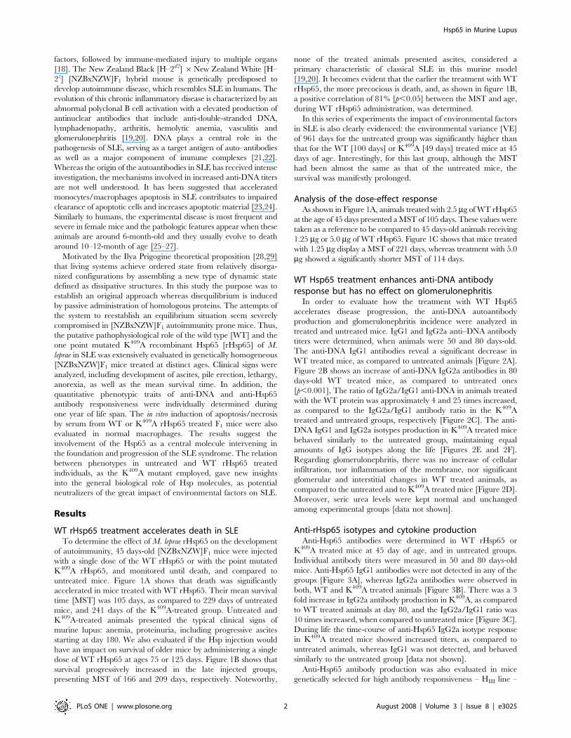
WT rHsp65 treatment accelerates death in SLETo determine the effect of M. leprae rHsp65 on the development
of autoimmunity, 45 days-old [NZBxNZW]F1 mice were injected
with a single dose of the WT rHsp65 or with the point mutated
K409A rHsp65, and monitored until death, and compared to
untreated mice. Figure 1A shows that death was significantly
accelerated in mice treated with WT rHsp65. Their mean survival
time [MST] was 105 days, as compared to 229 days of untreated
mice, and 241 days of the K409A-treated group. Untreated and
K409A-treated animals presented the typical clinical signs of
murine lupus: anemia, proteinuria, including progressive ascites
starting at day 180. We also evaluated if the Hsp injection would
have an impact on survival of older mice by administering a single
dose of WT rHsp65 at ages 75 or 125 days. Figure 1B shows that
survival progressively increased in the late injected groups,
presenting MST of 166 and 209 days, respectively. Noteworthy,
none of the treated animals presented ascites, considered a
primary characteristic of classical SLE in this murine model
[19,20]. It becomes evident that the earlier the treatment with WT
rHsp65, the more precocious is death, and, as shown in figure 1B,
a positive correlation of 81% [p,0.05] between the MST and age,
during WT rHsp65 administration, was determined.
In this series of experiments the impact of environmental factors
in SLE is also clearly evidenced: the environmental variance [VE]
of 961 days for the untreated group was significantly higher than
that for the WT [100 days] or K409A [49 days] treated mice at 45
days of age. Interestingly, for this last group, although the MST
had been almost the same as that of the untreated mice, the
survival was manifestly prolonged.
Analysis of the dose-effect responseAs shown in Figure 1A, animals treated with 2.5 mg of WT rHsp65
at the age of 45 days presented a MST of 105 days. These values were
taken as a reference to be compared to 45 days-old animals receiving
1.25 mg or 5.0 mg of WT rHsp65. Figure 1C shows that mice treated
with 1.25 mg display a MST of 221 days, whereas treatment with 5.0
mg showed a significantly shorter MST of 114 days.
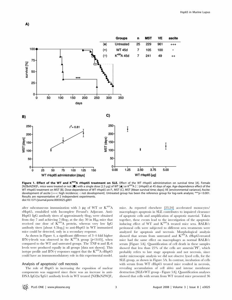
WT Hsp65 treatment enhances anti-DNA antibodyresponse but has no effect on glomerulonephritis
In order to evaluate how the treatment with WT Hsp65
accelerates disease progression, the anti-DNA autoantibody
production and glomerulonephritis incidence were analyzed in
treated and untreated mice. IgG1 and IgG2a anti–DNA antibody
titers were determined, when animals were 50 and 80 days-old.
The anti-DNA IgG1 antibodies reveal a significant decrease in
WT treated mice, as compared to untreated animals [Figure 2A].
Figure 2B shows an increase of anti-DNA IgG2a antibodies in 80
days-old WT treated mice, as compared to untreated ones
[p,0.001], The ratio of IgG2a/IgG1 anti-DNA in animals treated
with the WT protein was approximately 4 and 25 times increased,
as compared to the IgG2a/IgG1 antibody ratio in the K409A
treated and untreated groups, respectively [Figure 2C]. The anti-
DNA IgG1 and IgG2a isotypes production in K409A treated mice
behaved similarly to the untreated group, maintaining equal
amounts of IgG isotypes along the life [Figures 2E and 2F].
Regarding glomerulonephritis, there was no increase of cellular
infiltration, nor inflammation of the membrane, nor significant
glomerular and interstitial changes in WT treated animals, as
compared to the untreated and to K409A treated mice [Figure 2D].
Moreover, seric urea levels were kept normal and unchanged
among experimental groups [data not shown].
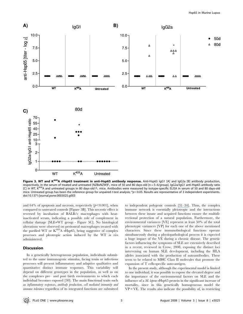
Anti-rHsp65 isotypes and cytokine productionAnti-Hsp65 antibodies were determined in WT rHsp65 or
K409A treated mice at 45 day of age, and in untreated groups.
Individual antibody titers were measured in 50 and 80 days-old
mice. Anti-Hsp65 IgG1 antibodies were not detected in any of the
groups [Figure 3A], whereas IgG2a antibodies were observed in
both, WT and K409A treated animals [Figure 3B]. There was a 3
fold increase in IgG2a antibody production in K409A, as compared
to WT treated animals at day 80, and the IgG2a/IgG1 ratio was
10 times increased, when compared to untreated mice [Figure 3C].
During life the time-course of anti-Hsp65 IgG2a isotype response
in K409A treated mice showed increased titers, as compared to
untreated animals, whereas IgG1 was not detected, and behaved
similarly to the untreated group [data not shown].
Anti-Hsp65 antibody production was also evaluated in mice
genetically selected for high antibody responsiveness – HIII line –
Hsp65 in Murine Lupus
PLoS ONE | www.plosone.org 2 August 2008 | Volume 3 | Issue 8 | e3025
after subcutaneous immunization with 5 mg of WT or K409A
rHsp65, emulsified with Incomplete Freund’s Adjuvant. Anti-
Hsp65 IgG antibody titers of approximately 6log2 were obtained
from day 7 and achieving 7-8log2 at the day 30 in HIII mice that
received one dose of K409A protein, whereas very low IgG
antibody titers [about 4.5log2] to anti-Hsp65 in WT immunized
mice could be detected, only in a secondary response.
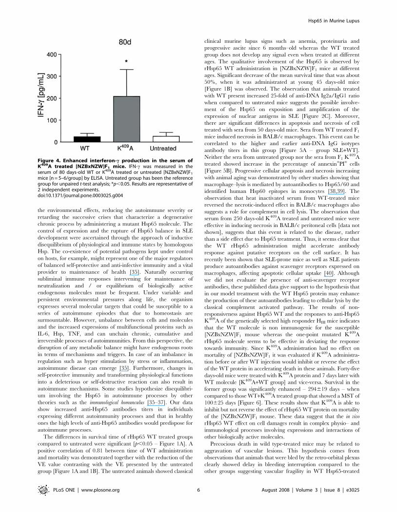
As shown in Figure 4, a significant difference of 3–4 fold higher
IFN-c-levels was observed in the K409A group [p,0.05], when
compared to the WT and untreated groups. The TNF-a and IL-6
levels were produced equally in all groups [data not shown]. This
isotype profile and IFN-c responses suggest that the K409A rHsp65
could have an immunomodulatory role in this experimental model.
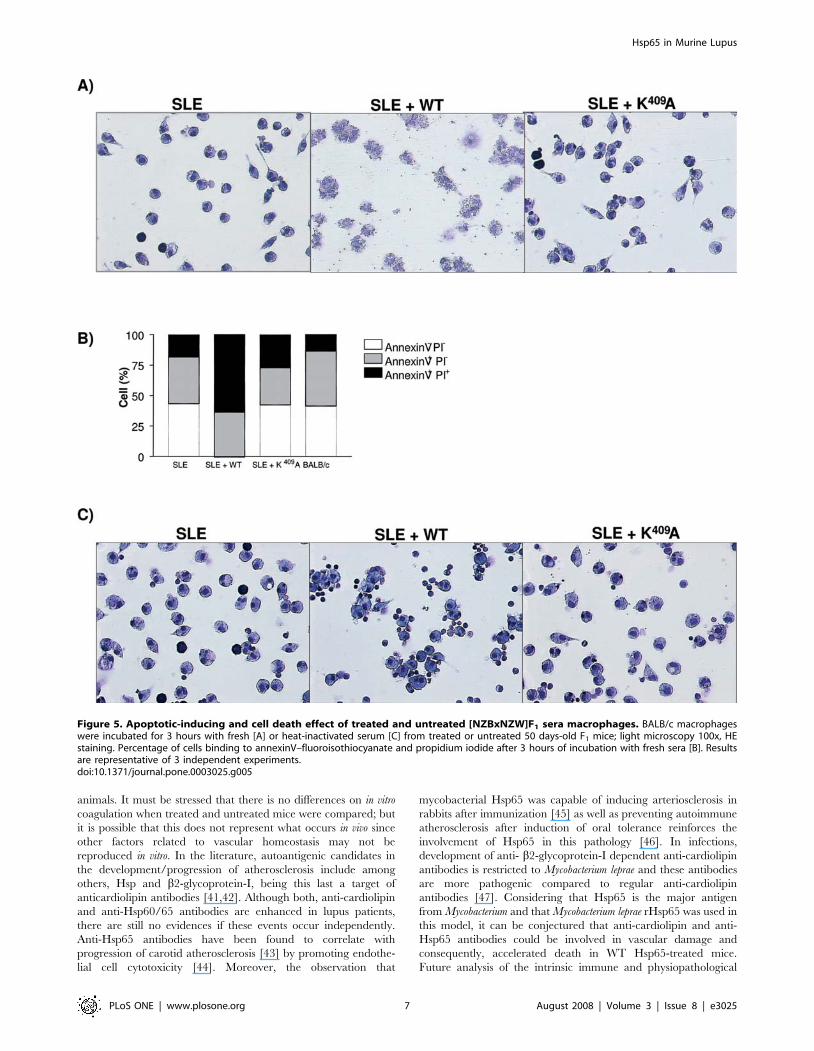
Analysis of apoptosis/ cell necrosisThe role of Hsp65 in increasing the exposition of nuclear
components was suggested since there was an increase in anti-
DNA IgG2a/IgG1 antibody levels in WT treated [NZBxNZW]F1
mice. As reported elsewhere [23,24] accelerated monocytes/
macrophages apoptosis in SLE contributes to impaired clearance
of apoptotic cells and amplification of apoptotic material. Taken
together, these events lead to the investigation of the apoptosis-
inducing effect of WT and K409A treated mice sera. BALB/c
peritoneal cells were subjected to different sera treatments were
analyzed for apoptosis and necrosis. Morphological analysis
showed that serum from untreated and K409A rHsp65-treated
mice had the same effect on macrophages as normal BALB/c
serum [Figure 5A]. Quantification of cell death in these samples
showed that less than 25% of the cells are annexin+PI+, which
probably refers to late stage apoptosis and not necrosis, since
under microscopic analysis we did not observe lysed cells, for the
SLE group, as shown in Figure 5A. In contrast, incubation of cells
with serum from WT rHsp65 treated mice resulted in necrosis,
revealing accumulation of cell debris and intense membrane
destruction [SLE+WT group - Figure 5A]. Quantification analyses
showed that cells with serum from WT treated mice presented 36
Figure 1. Effect of the WT and K409A rHsp65 treatment on SLE. Effect of the WT rHsp65 administration on survival time [A]. Female[NZBxNZW]F1 mice were treated or not [&] with a single dose [2.5 mg] of WT [m] or K409A [#]rHsp65 at 45 days of age. Age-dependence effect of theWT rHsp65 treatment on MST [B]. Dose-dependence of WT rHsp65 on F1 MST [C]. MST [Mean survival time; days]; VE [environmental variance]; Ascite:development of ascite [+++: high incidence; -: not development]. Untreated group has been the reference group for log-rank analysis; ***p,0.001.Results are representative of 2 independent experiments.doi:10.1371/journal.pone.0003025.g001
Hsp65 in Murine Lupus
PLoS ONE | www.plosone.org 3 August 2008 | Volume 3 | Issue 8 | e3025
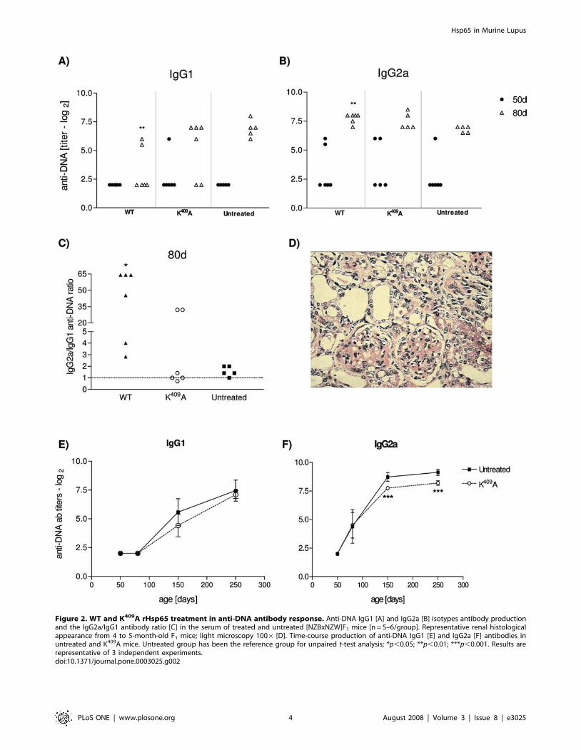
Figure 2. WT and K409A rHsp65 treatment in anti-DNA antibody response. Anti-DNA IgG1 [A] and IgG2a [B] isotypes antibody productionand the IgG2a/IgG1 antibody ratio [C] in the serum of treated and untreated [NZBxNZW]F1 mice [n = 5–6/group]. Representative renal histologicalappearance from 4 to 5-month-old F1 mice; light microscopy 1006 [D]. Time-course production of anti-DNA IgG1 [E] and IgG2a [F] antibodies inuntreated and K409A mice. Untreated group has been the reference group for unpaired t-test analysis; *p,0.05; **p,0.01; ***p,0.001. Results arerepresentative of 3 independent experiments.doi:10.1371/journal.pone.0003025.g002
Hsp65 in Murine Lupus
PLoS ONE | www.plosone.org 4 August 2008 | Volume 3 | Issue 8 | e3025
and 64% of apoptosis and necrosis, respectively [p,0.001], when
compared to untreated controls [Figure 5B]. This necrotic effect is
reversed by incubation of BALB/c macrophages with heat-
inactivated serum, indicating a possible role of complement in
cellular damage [SLE+WT group - Figure 5C]. No histological
alterations were observed on peritoneal macrophages treated with
the purified WT or K409A rHsp65, being suggestive of complex
processes and pleotropic action induced by the WT in vivo
administered.
Discussion
In a genetically heterogeneous population, individuals submit-
ted to the same immunogenic stimulus, facing toxin or infectious
processes will present inflammatory and adaptive qualitative and
quantitative distinct immune responses. This variability will
depend on different genotypes in the population, as well as on
the complexes pre– and post–birth environments to which each
individual becomes exposed [30]. The main functional traits such
as inflammatory responses, antibody production, cell mediated immunity and
immune tolerance regardless of its integrated functions are submitted
to independent polygenic controls [31–34]. Thus, the complex
immune network is essentially pleiotropic and the interactions
between these innate and acquired functions ensure the multidi-
rectional protection of a natural population. Furthermore, the
environmental variances [VE] represent at least 50% of the total
phenotypic variances [VP] for each one of the above mentioned
characters. Since these immunobiological functions operate
simultaneously during a physiopathological process it is expected
a huge impact of the VE during a chronic disease. The genetic
factors influencing the symptoms of SLE are extensively described
in a recent, reviewed in Crow, 2008, exposing the distinct loci
intervening on human SLE development, including the HLA
alleles associated with the production of autoantibodies. These
seem to be related to MHC Class II molecules that promote the
expansion of T cells-specific auto-antigen.
In the present study, although the experimental model is limited
to one individual, it was possible to expose the elevated degree and
the importance of the environmental factors on SLE and the
influence of a M. leprae rHsp65 protein in the significant increase of
mortality, since in this genetically homogeneous model the
VP = VE. The results also indicate the possibility of, in restricting
Figure 3. WT and K409A rHsp65 treatment in anti-Hsp65 antibody response. Anti-Hsp65 IgG1 [A] and IgG2a [B] antibody production,respectively, in the serum of treated and untreated [NZBxNZW]F1 mice of 50 and 80 days-old [n = 5–6/group]. IgG2a/IgG1 anti-Hsp65 antibody ratio[C] in WT, K409A and untreated groups in 80 days-old F1 mice. Antibodies were measured by isotype-specific ELISA in serum of 50 and 80 days-oldmice. Untreated group has been the reference group for unpaired t-test analysis; *p,0.05. Results are representative of 3 independent experiments.doi:10.1371/journal.pone.0003025.g003
Hsp65 in Murine Lupus
PLoS ONE | www.plosone.org 5 August 2008 | Volume 3 | Issue 8 | e3025
the environmental effects, reducing the autoimmune severity or
retarding the successive crises that characterize a degenerative
chronic process by administering a mutant Hsp65 molecule. The
control of expression and the rupture of Hsp65 balance in SLE
development were ascertained through the approach of inductive
disequilibrium of physiological and immune states by homologous
Hsp. The co-existence of potential pathogens kept under control
on hosts, for example, might represent one of the major regulators
of balanced self-protective and anti-infective immunity and a vital
provider to maintenance of health [35]. Naturally occurring
subliminal immune responses intervening for maintenance of
neutralization and / or equilibrium of biologically active
endogenous molecules must be frequent. Under variable and
persistent environmental pressures along life, the organism
expresses several molecular targets that could be susceptible to a
series of autoimmune episodes that due to homeostasis are
surmountable. However, unbalance between cells and molecules
and the increased expressions of multifunctional proteins such as
IL-6, Hsp, TNF, and can unchain chronic, cumulative and
irreversible processes of autoimmunities. From this perspective, the
disruption of any metabolic balance might have endogenous roots
in terms of mechanisms and triggers. In case of an imbalance in
regulation such as hyper stimulation by stress or inflammation,
autoimmune disease can emerge [35]. Furthermore, changes in
self-protective immunity and transforming physiological functions
into a deleterious or self-destructive reaction can also result in
autoimmune mechanisms. Some studies hypothesize disequilibri-
um involving the Hsp65 in autoimmune processes by other
theories such as the immunological homunculus [35–37]. Our data
show increased anti-Hsp65 antibodies titers in individuals
expressing different autoimmunity processes and that in healthy
ones the high levels of anti-Hsp65 antibodies would predispose for
autoimmune processes.
The differences in survival time of rHsp65 WT treated groups
compared to untreated were significant [p,0.05 – Figure 1A]. A
positive correlation of 0.81 between time of WT administration
and mortality was demonstrated together with the reduction of the
VE value contrasting with the VE presented by the untreated
group [Figure 1A and 1B]. The untreated animals showed classical
clinical murine lupus signs such as anemia, proteinuria and
progressive ascite since 6 months–old whereas the WT treated
group does not develop any signal even when treated at different
ages. The qualitative involvement of the Hsp65 is observed by
rHsp65 WT administration in [NZBxNZW]F1 mice at different
ages. Significant decrease of the mean survival time that was about
50%, when it was administrated at young 45 days-old mice
[Figure 1B] was observed. The observation that animals treated
with WT present increased 25-fold of anti-DNA Ig2a/IgG1 ratio
when compared to untreated mice suggests the possible involve-
ment of the Hsp65 on exposition and amplification of the
expression of nuclear antigens in SLE [Figure 2C]. Moreover,
there are significant differences in apoptosis and necrosis of cell
treated with sera from 50 days-old mice. Sera from WT treated F1
mice induced necrosis in BALB/c macrophages. This event can be
correlated to the higher and earlier anti-DNA IgG isotypes
antibody titers in this group [Figure 5A – group SLE+WT].
Neither the sera from untreated group nor the sera from F1 K409A
treated showed increase in the percentage of annexin+PI+ cells
[Figure 5B]. Progressive cellular apoptosis and necrosis increasing
with animal aging was demonstrated by other studies showing that
macrophage–lysis is mediated by autoantibodies to Hsp65/60 and
identified human Hsp60 epitopes in monocytes [38,39]. The
observation that heat inactivated serum from WT-treated mice
reversed the necrotic-induced effect in BALB/c macrophages also
suggests a role for complement in cell lysis. The observation that
serum from 250 days-old K409A treated and untreated mice were
effective in inducing necrosis in BALB/c peritoneal cells [data not
shown], suggests that this event is related to the disease, rather
than a side effect due to Hsp65 treatment. Thus, it seems clear that
the WT rHsp65 administration might accelerate antibody
response against putative receptors on the cell surface. It has
recently been shown that SLE-prone mice as well as SLE patients
produce autoantibodies against scavenger receptors expressed on
macrophages, affecting apoptotic cellular uptake [40]. Although
we did not evaluate the presence of anti-scavenger receptor
antibodies, these published data give support to the hypothesis that
in our model treatment with the WT Hsp65 protein may enhance
the production of these autoantibodies leading to cellular lysis by the
classical complement activated pathway. The results of non-
responsiveness against Hsp65 WT and the responses to anti-Hsp65
K409A of the genetically selected high responder HIII mice indicates
that the WT molecule is non immunogenic for the susceptible
[NZBxNZW]F1 mouse whereas the one-point mutated K409A
rHsp65 molecule seems to be effective in deviating the response
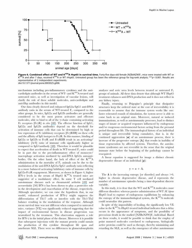
towards immunity. Since K409A administration had no effect on
mortality of [NZBxNZW]F1 it was evaluated if K409A administra-
tion before or after WT injection would inhibit or reverse the effect
of the WT protein in accelerating death in these animals. Forty-five
days-old mice were treated with K409A protein and 7 days later with
WT molecule [K409A+WT group] and vice-versa. Survival in the
former group was significantly enhanced – 294619 days – when
compared to those WT+K409A treated group that showed a MST of
100625 days [Figure 6]. These results show that K409A is able to
inhibit but not reverse the effect of rHsp65 WT protein on mortality
of the [NZBxNZW]F1 mouse. These data suggest that the in vivo
rHsp65 WT effect on cell damages result in complex physio– and
immunological processes involving expressions and interactions of
other biologically active molecules.
Precocious death in wild type-treated mice may be related to
aggravation of vascular lesions. This hypothesis comes from
observations that animals that were bled by the retro-orbital plexus
clearly showed delay in bleeding interruption compared to the
other groups suggesting vascular fragility in WT Hsp65-treated
Figure 4. Enhanced interferon-c production in the serum ofK409A treated [NZBxNZW]F1 mice. IFN-c was measured in theserum of 80 days-old WT or K409A treated or untreated [NZBxNZW]F1
mice [n = 5–6/group] by ELISA. Untreated group has been the referencegroup for unpaired t-test analysis; *p,0.05. Results are representative of2 independent experiments.doi:10.1371/journal.pone.0003025.g004
Hsp65 in Murine Lupus
PLoS ONE | www.plosone.org 6 August 2008 | Volume 3 | Issue 8 | e3025
animals. It must be stressed that there is no differences on in vitro
coagulation when treated and untreated mice were compared; but
it is possible that this does not represent what occurs in vivo since
other factors related to vascular homeostasis may not be
reproduced in vitro. In the literature, autoantigenic candidates in
the development/progression of atherosclerosis include among
others, Hsp and b2-glycoprotein-I, being this last a target of
anticardiolipin antibodies [41,42]. Although both, anti-cardiolipin
and anti-Hsp60/65 antibodies are enhanced in lupus patients,
there are still no evidences if these events occur independently.
Anti-Hsp65 antibodies have been found to correlate with
progression of carotid atherosclerosis [43] by promoting endothe-
lial cell cytotoxicity [44]. Moreover, the observation that
mycobacterial Hsp65 was capable of inducing arteriosclerosis in
rabbits after immunization [45] as well as preventing autoimmune
atherosclerosis after induction of oral tolerance reinforces the
involvement of Hsp65 in this pathology [46]. In infections,
development of anti- b2-glycoprotein-I dependent anti-cardiolipin
antibodies is restricted to Mycobacterium leprae and these antibodies
are more pathogenic compared to regular anti-cardiolipin
antibodies [47]. Considering that Hsp65 is the major antigen
from Mycobacterium and that Mycobacterium leprae rHsp65 was used in
this model, it can be conjectured that anti-cardiolipin and anti-
Hsp65 antibodies could be involved in vascular damage and
consequently, accelerated death in WT Hsp65-treated mice.
Future analysis of the intrinsic immune and physiopathological
Figure 5. Apoptotic-inducing and cell death effect of treated and untreated [NZBxNZW]F1 sera macrophages. BALB/c macrophageswere incubated for 3 hours with fresh [A] or heat-inactivated serum [C] from treated or untreated 50 days-old F1 mice; light microscopy 100x, HEstaining. Percentage of cells binding to annexinV–fluoroisothiocyanate and propidium iodide after 3 hours of incubation with fresh sera [B]. Resultsare representative of 3 independent experiments.doi:10.1371/journal.pone.0003025.g005
Hsp65 in Murine Lupus
PLoS ONE | www.plosone.org 7 August 2008 | Volume 3 | Issue 8 | e3025
mechanisms including pro-inflammatory cytokines and the anti-
cardiolipin antibodies in the serum of WT- and K409A-treated and
untreated mice, as well as investigation of vascular lesions, will
clarify the role of these soluble molecules, anti-cardiolipin and
anti-Hsp antibodies in this model.
Our data clearly showed and enhanced IgG2a/IgG1 anti-DNA
antibody ratio in the serum of WT-treated F1 compared to the
other groups. In mice, IgG2a and IgG2b antibodies are generally
considered to be the most potent activators and effectors
molecules, able to bind to all of the c-chain–containing activating
Fc receptors [FccR] in vitro [48]. The effector function of IgG1,
IgG2a and IgG2b antibodies depend on the threshold for
activation of immune cells that can be determined by high or
low expression of Fc inhibitory receptors [FccRIIB] on these cells
and the affinity of IgG isotypes to FccR. In this manner, binding of
IgG2a or IgG2b to FccR and FccRIIB result in an activating/
inhibitory [A/I] ratio of immune cells significantly higher as
compared to IgG1antibody [48]. Therefore it would be plausible
to expect that acceleration of death in WT-treated F1 mice could
be in part due to the pro-inflammatory effect of monocyte/
macrophage activation mediated by anti-DNA IgG2a autoanti-
bodies. On the other hand, the lack of effect of the K409A
administration in the mortality of F1 animals can be due to the
modulation of the anti-DNA IgG2a/IgG1 antibody ratio reflecting
in a more balanced activating/inhibitory ratio of immune cells by
IgG2a-FccR engagement. Moreover, as shown in Figure 4, higher
IFN-c levels in the serum of Hsp65 K409A treated mice are
suggestive of a modulatory effect. In some T cell mediated
autoimmune diseases such as rheumatoid arthritis [49] and
uveoretinitis [50] IFN-c has been shown to play a protective role
in the development and exacerbation of the disease, respectively.
Although speculative, we can raise the hypothesis that, in our
model, enhanced production of IFN-c might suppress the
differentiation of Th17 cells or interfere with the Th1/Th2
balance resulting in the modulation of the response. Although
mean survival time was not different when compared to untreated
mice, variance [VE] was significantly reduced suggesting that the
impact of environmental factors in disease progression was
neutralized by the treatment. This observation supports a role
for IFN-c in the initial phase of the disease. Moreover it is possible
that subsequent injections with the mutant protein could sustain
the production of this cytokine throughout life span and
ameliorate SLE. There were no differences in glomerulonephritis
analyses and seric urea levels between treated or untreated F1
groups of animals. All these data denote that although WT Hsp65
treatment enhances anti-DNA production and it does not reflect in
any kidney injury.
Finally, returning to Prigogine’s principle that dissipative
structures keep the ordered state at the cost of irreversibility it is
reasonable to assume that the immune system works like one:
faster reiterated rounds of stimulation, the system never is able to
come back to an original state. Moreover, natural or induced
immunizations, as well as autoimmunity processes, lead to distinct
stages of innate or acquired responses influenced by endogenous
and/or exogenous environmental factors acting from the prenatal
period throughout life. The immunological history of an individual
is unique and irreversible being cumulative, that is, in the
continued aggravation [a] of an autoimmune process, there is
increase of the progressive entropy [Di] that results in inability of
tissue regeneration by affected systems. Therefore, the autoim-
mune syndromes are not reversible in the sense that the original
immune state before the beginning of the pathology cannot be
reacquired.
A linear equation is suggested for image a distinct chronic
degenerative disease of an individual [y]:
y ~ a z Di
The D is the increasing entropy [or disorder] and always .0,
higher in chronic degenerative disease, and i represents the
number of autoimmune episodes being a time-dependent discrete
variable.
In this study, it is clear that the WT and K409A molecules cause
different disorders: whereas passive administration of WT M. leprae
Hsp65 lead to rupture of endogenous equilibrium by enhancing
the entropy of the immunobiological system, the K409A molecule
could neutralize this pattern.
In spite of the impossibility of healing, the significantly low VE
value in the K409A rHsp65 treated individual indicates that a delay
occurs in the appearance of symptoms, and the possibility of
precocious death in the studied [NZBxNZW]F1 individual. Based
on these results, it would be possible to think that the employ of
point mutated molecules correlated to endogenous, biologically
active proteins could be used as therapy, thereby minimizing and
retarding the SLE, as well as the emergence of other autoimmune
Figure 6. Combined effect of WT and K409A Hsp65 in survival time. Forty-five days-old female [NZBxNZW]F1 mice were treated with WT orK409A and after 7 days, received K409A or WT rHsp65. Untreated group has been the reference group for log-rank analysis; ***p,0.001. Results arerepresentative of 2 independent experiments.doi:10.1371/journal.pone.0003025.g006
Hsp65 in Murine Lupus
PLoS ONE | www.plosone.org 8 August 2008 | Volume 3 | Issue 8 | e3025
diseases, or reducing the progressive tissue damage and improving
life quality.
Materials and Methods
Expression of the recombinant M. leprae Hsp65 inEscherichia coli and purification
Clone pIL161, containing the coding sequence of the M. leprae
Hsp65, was a gift of Prof. Celio L. Silva from the University of Sao
Paulo, Ribeirao Preto, Brazil, and its mutated form, containing the
sequence of the proteolytically non active K409A form [17] were
amplified in E. coli DH5a cells. Expression of the recombinant
proteins WT and K409A rHsp65 was performed as described in
[17]. The recombinant proteins [WT or K409A rHsp65] present in
the bacterial extract pellet were solubilized with 6 M urea,
submitted to preparative SDS-PAGE and elution of the Hsp65
band from the polyacrylamide gel slice was performed to obtain
purified WT and K409A rHsp65 [51]. The homogeneity of the
recombinant M. leprae Hsp65 preparations was analyzed by
polyacrylamide gel electrophoresis [52] followed by silver staining
and submitted to peptide mass fingerprinting analyses, performed
as described in [53]. WT and K409A rHsp65 protein concentra-
tions were determined as previously described [54].
AnimalsMice were maintained at the animal facility of the Immuno-
chemistry Laboratory from Butantan Institute and caged and
handled under ethical conditions, according to international rules
of animal care by the International Animal Welfare Recommen-
dations [55]. New Zealand Black [NZB] female and New Zealand
White [NZW] male mice were obtained from the University of
Sao Paulo, Brazil. Parental lines were mated in our Laboratory to
produce the [NZBxNZW]F1 hybrids. After weaning, the F1
hybrids were housed in groups of four-six in plastic cages filled
with hardwood bedding, provided with water and rodent chow ad
libitum. The animals were kept in a room with controlled lighting
[12-h light/dark cycle], pressure, humidity and temperature
[2462uC]. Due to the fact that murine lupus is more prevalent
in females [56] only this gender was used in the present study. The
genetically selected high responder mice [HIII line] were bred in
the animal facility of the Immunochemistry Laboratory from
Butantan Institute [57]. Female HIII mice were immunized with
WT or K409A rHsp65 when 3-month-old.
Treatment with the WT and K409A rHsp65 moleculesFemale [NZBxNZW]F1 mice at the age of 45, 75 or 125 days
were inoculated i.p. with a single dose of 2.5 mg of WT or K409A
rHsp65 of M. leprae in 0.2 ml of phosphate buffer saline pH7.4
[PBS]. Untreated mice were treated with 0.2 ml of PBS. rHsp65
treated and untreated mice were periodically bled and the
individual serum samples stored at 220uC until titration of the
anti–DNA and anti–Hsp65 antibodies, which was performed by
ELISA. Animals were evaluated during 300 days of age or until
their natural death. For the longevity evaluations, mice were
periodically examined for clinical signs that include development
of ascites, lethargy, anorexia, and death.
Titration of anti-DNA antibodiesLevels of anti–DNA IgG and the IgG1 and IgG2a isotypes were
determined by ELISA. Briefly, 96–well microtiter plates [Nunc
MaxiSorpTM, Roskilde, Denmark] were coated with 1 mg/well of
native salmon sperm DNA [Sigma-Aldrich] diluted in 50 ml of
10 mM TRIS–HCl pH7.5, 1 mM EDTA overnight at 4uC. Plates
were washed three times with PBS containing 0.05% Tween 20
[PBS–T] and the wells were blocked with 1% PBS-bovine serum
albumin [PBS–BSA] at 37uC for 1h. To each well 100 ml of 1:16
diluted serum sample were added and incubated at 37uC for 1h.
Plates were washed again and wells were incubated with
peroxidase–labelled anti-mouse IgG [1:2500], IgG1 [1:1000] or
IgG2a [1:1000] at 37uC for 1h. After further washing 100 ml of
freshly substrate solution [containing 0.5mg/mL o–phenylenedi-
amine [Sigma Chemical Company, St. Louis, MO, USA] and
0.03% H2O2 in citrate/phosphate buffer at pH4.9] was added to
each well, and these were then incubated at 37uC until 10 min and
the reaction was ended with 50 ml of 0.2 M citric acid. The optical
density was measured at l490nm. The antibody titers were
expressed as log2 of the reciprocal serum dilution giving an
absorbance value of 20% of the saturation level.
Titration of anti-Hsp65 antibodiesSpecific IgG1 and IgG2a isotypes were detected with indirect
ELISA. Microplates [Nunc MaxiSorpTM] were coated with 1 mg/
well of Mycobacterium leprae WT rHsp65 diluted in 100 ml of 0.1 M
NaHCO3 pH9.6, overnight at 4uC. Further steps were performed
as described above. Serum was serially diluted starting at 1:16.
Anti–Hsp65 antibodies were measured in the serum of WT and
K409A rHsp65 treated mice. Titers were expressed as log2 of the
reciprocal serum dilution giving an absorbance value of 20% of
the saturation level.
IFN-c, TNF-a and IL-6 assaysLevels of IFN-c, TNF-a and IL-6 were determined in serum of
WT or K409A rHsp65 treated and untreated [NZBxNZW]F1 mice
by ELISA in 80 days-old. Cytokine measurements were performed
using BD OptEIA enzyme-linked immunosorbent assay sets [BD
Biosciences Pharmingen, SanDiego, CA, USA] according to the
manufacturer’s instructions.
Renal function and HistologyBlood urea nitrogen [BUN] measurements were performed in
duplicate by the urease method. Kidney specimens were fixed in
10% formalin and embedded in paraffin. After processing, 5 mm
tissue sections were stained with hematoxylin and eosin [HE] and
microscopic analysis was conducted by a trained pathologist.
Preparation of adherent peritoneal cell culturesPeritoneal cells were obtained from the peritoneal cavity of non–
immunized BALB/c mice. Cells were harvested in the absence of
any inflammatory stimulus. Approximately 5 ml of Dulbecco’s
modified Eagle’s medium [DMEM] [Cambrex, Verviers, Belgium]
were injected into the peritoneal cavity of the mouse and
subsequently collected. Cells were washed and transferred to
DMEM supplemented with 1025 M 2-mercaptoethanol [Sigma
Chemicals Co, St. Louis, MO, EUA] 2 mM L-glutamine, 0.1 mM
vitamins, 1 mM sodium pyruvate, 0.1 mM non-essential amino
acids and 100 mg/ml gentamicine all purchased from Gibco BRL
[Rockville, NY, USA]. Cells were counted, tested for viability with
Trypan blue and incubated [2.56105 cells] for 4 hours in a 8 well
chamber slide [Nalge Nunc International, IL, USA] at 37uC in an
atmosphere containing 5% CO2. Non-adherent cells were removed
by washing the wells with medium. Cells were incubated for 3 hours
with 20% serum obtained from female [NZBxNZW]F1 treated or
not with 2.5 mg of WT or K409A rHsp65 [this approach was
modified from [58]]. Normal BALB/c mouse serum [NMS] was
used as negative control. After incubation period medium was
withdrawn and slides were stained with hematoxilin and eosin using
the Protocol Hema3 kit [Biochemical Sciences, Swedesboro, NJ].
Hsp65 in Murine Lupus
PLoS ONE | www.plosone.org 9 August 2008 | Volume 3 | Issue 8 | e3025
Cell death evaluationBinding of annexinV–fluoroisothiocyanate [FITC] and propi-
dium iodide [PI] [BD Biosciences Pharmingen, SanDiego, CA,
USA] to the cells was used to detect viable, early apoptotic and late
apoptotic or necrotic cells by flow cytometry. Peritoneal cells
[56105] were cultured in 1 mL of complete medium containing
20% of mouse serum from WT or K409A rHsp65 treated
[NZBxNZW]F1 mice for 16 hours. After incubation cells were
washed in annexin buffer [10 mM Hepes, pH7.4, 150 mM NaCl,
5 mM KCl, 1 mM MgCl2, 1.8 mM CaCl2] and labeled with
annexinV–FITC [1:500] in 100 ml of annexin buffer for 20 minutes
at room temperature in the dark. Immediately before analysis cells
were stained with PI [1:100]. Cells were analyzed in a FACSCalibur
[CellQuest software] cell cytometer [BD Biosciences].
WT and K409A rHsp65 combined administrationFemale [NZBxNZW]F1 mice groups with 45 days-old were
inoculated i.p. with a single dose of 2.5 mg of WT or K409A rHsp65
of M. leprae in 0.2 ml of PBS. pH7.4. Seven days after, they received
more 2.5 mg of the opposite Hsp, i.p. WT+K409A and K409A+WT
groups were periodically bled; individual serum sample titration and
clinical signs were evaluated according to describe above.
Statistical analysisAll data are expressed as the mean [X] and standard deviation
[SD] or environmental variance [VE]. Statistical significance was
set at p,0.05 by the Unpaired t–test. The Kaplan-Meier plot for
survival was analyzed by log-rank test. The correlation coefficients
were determined by the linear regression of group data comparing
the mean survival time [MST] with age, dose or administration
period of WT or K409A rHsp65.
Acknowledgments
This work is dedicated to Guido Biozzi [1920–2006] and Luigi Cavalli-
Sforza.
Author Contributions
Conceived and designed the experiments: EBM LVM FCVP OAS.
Performed the experiments: EBM LVM LVC OAS. Analyzed the data:
EBM LVM MF BLF DVT OAS. Contributed reagents/materials/analysis
tools: LVM DVT LVR FCVP ACMC OAS. Wrote the paper: EBM LVM
BLF DVT LVR FCVP ACMC OAS.
References
1. Lindquist S (1986) The heat-shock response. Annu Rev Biochem 55:1151–1191.
2. Winfield JB (1989) Stress proteins, arthritis, and autoimmunity. Arthritis Rheum32: 1497–1504.
3. Jones DB, Hunter NR, Duff GW (1990) Heat-shock protein 65 as a beta cell
antigen of insulin-dependent diabetes. Lancet 336: 583–585.
4. Young RA (1990) Stress proteins and immunology. Annu Rev Immunol 8:
401–420.
5. Young RA, Elliott TJ (1989) Stress proteins, infection, and immune surveillance.Cell 59: 5–8.
6. Thole J, van Der Zee R (1990) The 65kD antigen: molecular studies on anubiquitous antigen. In: McFadden J, ed. Molecular Biology of the Mycobacteria.
London: Elsevier Science & Technology. 244 p.
7. Young D, Lathigra R, Hendrix R, Sweetser D, Young RA (1988) Stress proteins
are immune targets in leprosy and tuberculosis. Proc Natl Acad Sci U S A 85:4267–4270.
8. Cohen IR (1991) Autoimmunity to chaperonins in the pathogenesis of arthritis
and diabetes. Annu Rev Immunol 9: 567–589.
9. Kohm AP, Fuller KG, Miller SD (2003) Mimicking the way to autoimmunity: an
evolving theory of sequence and structural homology. Trends Microbiol 11:101–105.
10. Oldstone MB (1987) Molecular mimicry and autoimmune disease. Cell 50:
819–820.
11. Wick G, Perschinka H, Millonig G (2001) Atherosclerosis as an autoimmune
disease: an update. Trends Immunol 22: 665–669.
12. Pockley AG, Shepherd J, Corton JM (1998) Detection of heat shock protein 70(Hsp70) and anti-Hsp70 antibodies in the serum of normal individuals. Immunol
Invest 27: 367–377.
13. Stephanou A, Latchman DS, Isenberg DA (1998) The regulation of heat shock
proteins and their role in systemic lupus erythematosus. Semin Arthritis Rheum28: 155–162.
14. Pockley AG, Bulmer J, Hanks BM, Wright BH (1999) Identification of human
heat shock protein 60 (Hsp60) and anti-Hsp60 antibodies in the peripheral
circulation of normal individuals. Cell Stress Chaperones 4: 29–35.
15. Rea IM, McNerlan S, Pockley AG (2001) Serum heat shock protein and anti-heat shock protein antibody levels in aging. Exp Gerontol 36: 341–352.
16. Xu Q, Schett G, Perschinka H, Mayr M, Egger G, et al. (2000) Serum solubleheat shock protein 60 is elevated in subjects with atherosclerosis in a general
population. Circulation 102: 14–20.
17. Portaro FC, Hayashi MA, De Arauz LJ, Palma MS, Assakura MT, et al. (2002)The Mycobacterium leprae hsp65 displays proteolytic activity. Mutagenesis
studies indicate that the M. leprae hsp65 proteolytic activity is catalytically
related to the HslVU protease. Biochemistry 41: 7400–7406.
18. Tsao BP (2003) The genetics of human systemic lupus erythematosus. TrendsImmunol 24: 595–602.
19. Burnet FM, Holmes MC (1965) The natural history of the NZB/NZW F1
hybrid mouse: a laboratory model of systemic lupus erythematosus. Australas
Ann Med 14: 185–191.
20. Theofilopoulos AN, Dixon FJ (1985) Murine models of systemic lupuserythematosus. Adv Immunol 37: 269–390.
21. Hahn BH (1998) Antibodies to DNA. N Engl J Med 338: 1359–1368.
22. Pisetsky DS (1998) Antibody responses to DNA in normal immunity and
aberrant immunity. Clin Diagn Lab Immunol 5: 1–6.
23. Kaplan MJ (2004) Apoptosis in systemic lupus erythematosus. Clin Immunol112: 210–218.
24. Kaplan MJ, Lewis EE, Shelden EA, Somers E, Pavlic R, et al. (2002) Theapoptotic ligands TRAIL, TWEAK, and Fas ligand mediate monocyte death
induced by autologous lupus T cells. J Immunol 169: 6020–6029.
25. Ishikawa S, Akakura S, Abe M, Terashima K, Chijiiwa K, et al. (1998) A subsetof CD4+ T cells expressing early activation antigen CD69 in murine lupus:
possible abnormal regulatory role for cytokine imbalance. J Immunol 161:
1267–1273.
26. Putterman C, Naparstek Y (1994) Murine Models of Spontaneous Systemic
Lupus Erythematosus. In: Cohen IR, Miller A, eds. Autoimmune Models: AGuidebook. New York: Academic Press, Inc. pp 217–243.
27. Struhar D, Harbeck R, Cherniack R (1988) Elastic properties of the excised
lungs of NZB/W mice and their correlation with histopathologic changes. Lung166: 107–112.
28. Prigogine I (1978) Time, Structure, and Fluctuations. Science 201: 777–785.
29. Prigogine I (2003) Chemical kinetics and dynamics. Ann N Y Acad Sci 988:
128–132.
30. Mouton D, Sant’anna OA, Biozzi G (1988) Multigenic Control of Specific and Non-
specific Immunity in Mice. A Review. Livestock Production Sicence 20: 277–286.
31. Biozzi G, Mouton D, Sant’Anna OA, Passos HC, Gennari M, et al. (1979)Genetics of immunoresponsiveness to natural antigens in the mouse. Curr Top
Microbiol Immunol 85: 31–98.
32. da Silva AC, de Souza KW, Machado RC, da Silva MF, Sant’Anna OA (1998)Genetics of immunological tolerance: I. Bidirectional selective breeding of mice
for oral tolerance. Res Immunol 149: 151–161.
33. Ibanez OM, Stiffel C, Ribeiro OG, Cabrera WK, Massa S, et al. (1992) Genetics
of nonspecific immunity: I. Bidirectional selective breeding of lines of mice
endowed with maximal or minimal inflammatory responsiveness. Eur J Immunol22: 2555–2563.
34. Sant’Anna OA, Ferreira VC, Reis MH, Gennari M, Ibanez OM, et al. (1982)
Genetic parameters of the polygenic regulation of antibody responsiveness toflagellar and somatic antigens of salmonellae. J Immunogenet 9: 191–205.
35. Prohaszka Z, Fust G (2004) Immunological aspects of heat-shock proteins-theoptimum stress of life. Mol Immunol 41: 29–44.
36. Poletaev A, Osipenko L (2003) General network of natural autoantibodies as
immunological homunculus (Immunculus). Autoimmun Rev 2: 264–271.
37. Cohen IR (2007) Biomarkers, self-antigens and the immunological homunculus.J Autoimmun 29: 246–249.
38. Habich C, Kempe K, Burkart V, Van Der Zee R, Lillicrap M, et al. (2004)
Identification of the heat shock protein 60 epitope involved in receptor bindingon macrophages. FEBS Lett 568: 65–69.
39. Schett G, Metzler B, Mayr M, Amberger A, Niederwieser D, et al. (1997)Macrophage-lysis mediated by autoantibodies to heat shock protein 65/60.
Atherosclerosis 128: 27–38.
40. Wermeling F, Chen Y, Pikkarainen T, Scheynius A, Winqvist O, et al. (2007)Class A scavenger receptors regulate tolerance against apoptotic cells, and
autoantibodies against these receptors are predictive of systemic lupus. J Exp
Med 204: 2259–2265.
Hsp65 in Murine Lupus
PLoS ONE | www.plosone.org 10 August 2008 | Volume 3 | Issue 8 | e3025
41. Vaarala O, Alfthan G, Jauhiainen M, Leirisalo-Repo M, Aho K, et al. (1993)
Crossreaction between antibodies to oxidised low-density lipoprotein and to
cardiolipin in systemic lupus erythematosus. Lancet 341: 923–925.
42. George J, Harats D, Gilburd B, Afek A, Levy Y, et al. (1999) Immunolocal-
ization of beta2-glycoprotein I (apolipoprotein H) to human atherosclerotic
plaques: potential implications for lesion progression. Circulation 99:
2227–2230.
43. Xu Q, Willeit J, Marosi M, Kleindienst R, Oberhollenzer F, et al. (1993)
Association of serum antibodies to heat-shock protein 65 with carotid
atherosclerosis. Lancet 341: 255–259.
44. Schett G, Xu Q, Amberger A, Van der Zee R, Recheis H, et al. (1995)
Autoantibodies against heat shock protein 60 mediate endothelial cytotoxicity.
J Clin Invest 96: 2569–2577.
45. Xu Q, Dietrich H, Steiner HJ, Gown AM, Schoel B, et al. (1992) Induction of
arteriosclerosis in normocholesterolemic rabbits by immunization with heat
shock protein 65. Arterioscler Thromb 12: 789–799.
46. Harats D, Yacov N, Gilburd B, Shoenfeld Y, George J (2002) Oral tolerance
with heat shock protein 65 attenuates Mycobacterium tuberculosis-induced and
high-fat-diet-driven atherosclerotic lesions. J Am Coll Cardiol 40: 1333–1338.
47. Hojnik M, Gilburd B, Ziporen L, Blank M, Tomer Y, et al. (1994)
Anticardiolipin antibodies in infections are heterogenous in their dependency
on beta 2-glycoprotein I: analysis of anticardiolipin antibodies in leprosy. Lupus
3: 515–521.
48. Nimmerjahn F, Ravetch JV (2006) Fcgamma receptors: old friends and new
family members. Immunity 24: 19–28.
49. Kim EY, Chi HH, Bouziane M, Gaur A, Moudgil KD (2008) Regulation of
autoimmune arthritis by the pro-inflammatory cytokine interferon-gamma. ClinImmunol 127: 98–106.
50. Yoshimura T, Sonoda KH, Miyazaki Y, Iwakura Y, Ishibashi T, et al. (2008)
Differential roles for IFN-gamma and IL-17 in experimental autoimmuneuveoretinitis. Int Immunol 20: 209–214.
51. Pereira CM, Sattlegger E, Jiang HY, Longo BM, Jaqueta CB, et al. (2005)IMPACT, a protein preferentially expressed in the mouse brain, binds GCN1
and inhibits GCN2 activation. J Biol Chem 280: 28316–28323.
52. Laemmli UK (1970) Cleavage of structural proteins during the assembly of thehead of bacteriophage T4. Nature 227: 680–685.
53. Westermeier R, Naven T (2002) Proteomics in Practice: A Laboratory Manualof Proteome Analysis. Weinheim: John Wiley and Sons Ltd.. 342 p.
54. Bradford MM (1976) A rapid and sensitive method for the quantitation ofmicrogram quantities of protein utilizing the principle of protein-dye binding.
Anal Biochem 72: 248–254.
55. Giles AR (1987) Guidelines for the use of animals in biomedical research.Thromb Haemost 58: 1078–1084.
56. Helyer BJ, Howie JB (1963) Spontaneous auto-immune disease in NZB/BLmice. Br J Haematol 9: 119–131.
57. Siqueira M, Bandieri A, Reis MS, Sant’anna OA, Biozzi G (1976) Selective
breeding of mice for antibody responsiveness to flagellar and somatic antigens ofsalmonellae. Eur J Immunol 6: 241–249.
58. Bengtsson AA, Sturfelt G, Gullstrand B, Truedsson L (2004) Induction ofapoptosis in monocytes and lymphocytes by serum from patients with systemic
lupus erythematosus - an additional mechanism to increased autoantigen load?Clin Exp Immunol 135: 535–543.
Hsp65 in Murine Lupus
PLoS ONE | www.plosone.org 11 August 2008 | Volume 3 | Issue 8 | e3025
Related Documents