Copyright © 2017 Wound, Ostomy and Continence Nurses Society™. Unauthorized reproduction of this article is prohibited. Copyright © 2017 by the Wound, Ostomy and Continence Nurses Society™ JWOCN ¡ Month/Month 2017 1 J Wound Ostomy Continence Nurs. 2017;00(0):1-10. Published by Lippincott Williams & Wilkins Wound Care ABSTRACT PURPOSE: The purpose of this study was to compare the effect of standard wound care with adjunctive hyperbaric oxygen therapy (HBOT) to standard wound care alone on wound healing, markers of inflammation, glycemic control, amputation rate, survival rate of tissue, and health-related quality of life in patients with diabetic foot ulcers (DFUs). DESIGN: Prospective, randomized, open-label, controlled study. SUBJECTS AND SETTING: The sample comprised 38 patients with nonhealing DFUs who were deemed poor candidates for vascular surgery. Subjects were randomly allocated to an experimental group (standard care plus HBOT, n = 20) or a control group (standard care alone, n = 18). The study setting was a medical center in Kaohsiung City, Taiwan. METHODS: Hyperbaric oxygen therapy was administered in a hyperbaric chamber under 2.5 absolute atmospheric pressure for 120 minutes; subjects were treated 5 days a week for 4 consecutive weeks. Both groups received standard wound care including debridement of necrotic tissue, topical therapy for Wagner grade 2 DFUs, dietary control and pharmacotherapy to maintain optimal blood glucose levels. Wound physiological indices were measured and blood tests (eg, markers of inflammation) were undertaken. Health-related quality of life was measured using the Medical Outcomes Study 36-Item Short Form. RESULTS: Complete DFU closure was achieved in 5 patients (25%) in the HBOT group (n = 20) versus 1 participant (5.5%) in the routine care group (n = 18) ( P = .001). The amputation rate was 5% for the HBOT group and 11% for the routine care group ( χ 2 = 15.204, P = .010). The HBOT group showed statistically significant improvements in inflammation index, blood flow, and health-related quality of life from pretreatment to 2 weeks after the last therapy ended ( P < .05). Hemoglobin A 1c was significantly lower in the HBOT group following treatment ( P < .05) but not in the routine care group. CONCLUSIONS: Adjunctive HBOT improved wound healing in persons with DFU. Therapy also reduced the risk of amputation of the affected limb. We assert that at least 20 HBOT sessions are required to be effective. KEY WORDS: Clinical trials, Diabetes mellitus, Diabetic foot ulcers, Hyperbaric oxygen therapy, Nonhealing wounds, Nursing care, Quality of life. INTRODUCTION Foot ulcers and infections are prevalent in persons with dia- betes mellitus; reported prevalence rates vary from 1.7% to Chen-Yu Chen, MSN, RN, Hyperbaric Oxygen Therapy Center, Kaohsiung Chang Gung Memorial Hospital, Kaohsiung, Taiwan, R.O.C.; and Chang Gung University, College of Medicine, Taoyuan, Taiwan, R.O.C. Re-Wen Wu, MD, Department of Orthopaedic Surgery, Kaohsiung Chang Gung Memorial Hospital, Kaohsiung, Taiwan, R.O.C.; and Chang Gung University, College of Medicine, Taoyuan, Taiwan, R.O.C. Mei-Chi Hsu, PhD, RN, Department of Nursing, I-Shou University, Kaohsiung City, Taiwan, R.O.C. Ching-Jung Hsieh, MD, Department of Internal Medicine, Paochien Hospital, Ping Tung, Taiwan, R.O.C. Man-Chun Chou, MSN, RN, Kaohsiung Chang Gung Memorial Hospital, Kaohsiung, Taiwan, R.O.C. The authors declare that they have no conflict of interests. The funder had no role in the study design, data collection and analysis, decision to publish, or preparation of the manuscript. Correspondence: Mei-Chi Hsu, PhD, RN, Department of Nursing, I-Shou University, No. 8, Yida Rd, Jiaosu Village, Yanchao District, Kaohsiung City 82445, Taiwan ([email protected]). Adjunctive Hyperbaric Oxygen Therapy for Healing of Chronic Diabetic Foot Ulcers A Randomized Controlled Trial Chen-Yu Chen ¡ Re-Wen Wu ¡ Mei-Chi Hsu ¡ Ching-Jung Hsieh ¡ Man-Chun Chou DOI: 10.1097/WON.0000000000000374 25%. 1-3 Approximately 40% of patients with diabetic foot in- fections return to hospital for repeated treatment, and 1 in 6 of these individuals will die within 1 year. 4 e pathophysiology of diabetic foot ulcers (DFUs) is not entirely understood. In- dividuals with diabetes mellitus often have impaired leukotaxis and phagocytosis function that increase the likelihood of de- veloping a wound infection by 17-fold. 5 A nonhealing, infect- ed DFU damages both soft tissue and bone; 85% of individu- als who develop a DFU ultimately undergo amputation, and 68% will die within 5 years of amputation. 6-8 e severity of the ulcer, angiopathy, infection, and poor blood glucose con- trol are important predictors for diabetic foot amputation. 9-11 Hyperbaric oxygen therapy (HBOT) is used as adjuvant therapy in conjunction with topical and systemic thera- py frequently including debridement, recombinant human platelet-derived growth factor, or other skin substitutes in persons with nonhealing or deteriorating DFU. 12 Evidence concerning the efficacy of HBOT for healing DFUs is mixed. Some researchers report greater efficacy when HBOT is com- pared to sham or placebo treatment, 13-16 but others found no differences. 17-19 Hyperbaric oxygen therapy also promotes res- olution of infection in persons with DFU, and it reduces the likelihood of amputation. 14,15

Welcome message from author
This document is posted to help you gain knowledge. Please leave a comment to let me know what you think about it! Share it to your friends and learn new things together.
Transcript
Copyright © 2017 Wound, Ostomy and Continence Nurses Society™. Unauthorized reproduction of this article is prohibited.
Copyright © 2017 by the Wound, Ostomy and Continence Nurses Society™ JWOCN Month/Month 2017 1
J Wound Ostomy Continence Nurs. 2017;00(0):1-10.
Published by Lippincott Williams & Wilkins
Wound Care
ABSTRACT PURPOSE: The purpose of this study was to compare the effect of standard wound care with adjunctive hyperbaric oxygen
therapy (HBOT) to standard wound care alone on wound healing, markers of infl ammation, glycemic control, amputation rate,
survival rate of tissue, and health-related quality of life in patients with diabetic foot ulcers (DFUs).
DESIGN: Prospective, randomized, open-label, controlled study.
SUBJECTS AND SETTING: The sample comprised 38 patients with nonhealing DFUs who were deemed poor candidates for
vascular surgery. Subjects were randomly allocated to an experimental group (standard care plus HBOT, n = 20) or a control
group (standard care alone, n = 18). The study setting was a medical center in Kaohsiung City, Taiwan.
METHODS: Hyperbaric oxygen therapy was administered in a hyperbaric chamber under 2.5 absolute atmospheric pressure
for 120 minutes; subjects were treated 5 days a week for 4 consecutive weeks. Both groups received standard wound care
including debridement of necrotic tissue, topical therapy for Wagner grade 2 DFUs, dietary control and pharmacotherapy to
maintain optimal blood glucose levels. Wound physiological indices were measured and blood tests (eg, markers of infl ammation)
were undertaken. Health-related quality of life was measured using the Medical Outcomes Study 36-Item Short Form.
RESULTS: Complete DFU closure was achieved in 5 patients (25%) in the HBOT group (n = 20) versus 1 participant (5.5%) in
the routine care group (n = 18) ( P = .001). The amputation rate was 5% for the HBOT group and 11% for the routine care group
( χ 2 = 15.204, P = .010). The HBOT group showed statistically signifi cant improvements in infl ammation index, blood fl ow, and
health-related quality of life from pretreatment to 2 weeks after the last therapy ended ( P < .05). Hemoglobin A 1c
was signifi cantly
lower in the HBOT group following treatment ( P < .05) but not in the routine care group.
CONCLUSIONS: Adjunctive HBOT improved wound healing in persons with DFU. Therapy also reduced the risk of amputation
of the affected limb. We assert that at least 20 HBOT sessions are required to be effective.
KEY WORDS: Clinical trials , Diabetes mellitus , Diabetic foot ulcers , Hyperbaric oxygen therapy , Nonhealing wounds , Nursing
care , Quality of life .
INTRODUCTION
Foot ulcers and infections are prevalent in persons with dia-betes mellitus; reported prevalence rates vary from 1.7% to
Chen-Yu Chen, MSN, RN, Hyperbaric Oxygen Therapy Center, Kaohsiung
Chang Gung Memorial Hospital, Kaohsiung, Taiwan, R.O.C.; and Chang Gung
University, College of Medicine, Taoyuan, Taiwan, R.O.C.
Re-Wen Wu, MD, Department of Orthopaedic Surgery, Kaohsiung Chang
Gung Memorial Hospital, Kaohsiung, Taiwan, R.O.C.; and Chang Gung
University, College of Medicine, Taoyuan, Taiwan, R.O.C.
Mei-Chi Hsu, PhD , RN, Department of Nursing, I-Shou University, Kaohsiung
City, Taiwan, R.O.C.
Ching-Jung Hsieh, MD, Department of Internal Medicine, Paochien Hospital,
Ping Tung, Taiwan, R.O.C.
Man-Chun Chou, MSN, RN, Kaohsiung Chang Gung Memorial Hospital,
Kaohsiung, Taiwan, R.O.C.
The authors declare that they have no confl ict of interests.
The funder had no role in the study design, data collection and analysis,
decision to publish, or preparation of the manuscript.
Correspondence: Mei-Chi Hsu, PhD, RN, Department of Nursing, I-Shou
University, No. 8, Yida Rd, Jiaosu Village, Yanchao District, Kaohsiung City
82445, Taiwan ( [email protected] ).
Adjunctive Hyperbaric Oxygen Therapy for Healing of Chronic Diabetic Foot Ulcers A Randomized Controlled Trial Chen-Yu Chen Re-Wen Wu Mei-Chi Hsu Ching-Jung Hsieh Man-Chun Chou
DOI: 10.1097/WON.0000000000000374
25%. 1-3 Approximately 40% of patients with diabetic foot in-fections return to hospital for repeated treatment, and 1 in 6 of these individuals will die within 1 year. 4 Th e pathophysiology of diabetic foot ulcers (DFUs) is not entirely understood. In-dividuals with diabetes mellitus often have impaired leukotaxis and phagocytosis function that increase the likelihood of de-veloping a wound infection by 17-fold. 5 A nonhealing, infect-ed DFU damages both soft tissue and bone; 85% of individu-als who develop a DFU ultimately undergo amputation, and 68% will die within 5 years of amputation. 6-8 Th e severity of the ulcer, angiopathy, infection, and poor blood glucose con-trol are important predictors for diabetic foot amputation. 9-11
Hyperbaric oxygen therapy (HBOT) is used as adjuvant therapy in conjunction with topical and systemic thera-py frequently including debridement, recombinant human platelet-derived growth factor, or other skin substitutes in persons with nonhealing or deteriorating DFU. 12 Evidence concerning the effi cacy of HBOT for healing DFUs is mixed. Some researchers report greater effi cacy when HBOT is com-pared to sham or placebo treatment, 13-16 but others found no diff erences. 17-19 Hyperbaric oxygen therapy also promotes res-olution of infection in persons with DFU, and it reduces the likelihood of amputation. 14 , 15

Copyright © 2017 Wound, Ostomy and Continence Nurses Society™. Unauthorized reproduction of this article is prohibited.
2 JWOCN Month/Month 2017 www.jwocnonline.com
Despite mixed evidence concerning its effi cacy, HBOT has been advocated and adopted for DFU treatment in some wound care centers. Th e aim of this study was to compare the eff ect of HBOT plus standard wound care to standard wound care alone in the treatment of DFU. Outcome measures were wound size, amputation rate, survival rate of tissue in the aff ected limb, markers of infl ammation, glycemic control, bacterial colonization (distribution of microorganism culture isolated from the wound), and health-related quality of life (HRQOL).
METHODS
We conducted a prospective, randomized, controlled trial to evaluate the effi cacy of HBOT plus standard therapy to stan-dard therapy alone for treatment of DFUs. Data were collected from June 2011 to June 2013. Th e time frame for data col-lection was divided into following 4 segments: pretreatment (before fi rst administration of HBOT; T1), during treatment (at the 10th administration of HBOT; T2), posttreatment (at the 20th administration of HBOT; T3), and follow-up (2 weeks after the last therapy ended; T4). Th e study setting was a medical center in Kaohsiung City, Taiwan.
Inclusion criteria were (1) adults 20 years or older, (2) diag-nosis of diabetes mellitus, (3) nonhealing DFUs that had not achieved closure after at least 2 months and following treat-ment for at least 1 month, (3) Wagner wound classifi cation of grade 1, 2, and 3 ulcers, and (4) deemed appropriate for hospital admission because of skin ulcer and soft-tissue infec-tion. Exclusion criteria were (1) gangrene, (2) contraindication for HBOT such as untreated pneumothorax, active cancerous condition, chronic obstructive pulmonary disease, and pul-monary emphysema with retention of CO 2 , and (3) planned vascular surgical procedures or revascularization of the limb.
Study procedures were reviewed by the institution-al review board of the Chang Gung Medical Foundation (IRB1010507C; No. 100-0876B and 101-0507C). Th is trial was registered at https://clinicaltrials.gov/ under the identifi ca-tion code NCT02328508. We ensured patients’ rights through-out the study according to the ethical principles for medical research on human beings set out in the Declaration of Helsin-ki. All participants provided written informed consent prior to random allocation to the control or intervention group.
Sample Size Sample size was based on a power analysis using the Statistical Software Sample Power software, version 2.0. Th e power was set at 0.8 to limit the risk of committing a type II error to 20%, the α level was set at .05, and the covariate’s R 2 at 0.13. Th e sample size was calculated based on a previous clinical trial of professionally lead support group for Taiwanese people with schizophrenia in 4 waves of data collection (baseline, 10, 20, and 30 days of HBOT). 20 Th erefore, the eff ect size of co-variate adjustment in this study was set at 0.37, and the num-ber of samples for each group was calculated to be 20. Based on these calculations, a total of 40 patients would be needed for 4 waves of data collection in order to provide 80% power (2-sided P < .05) to detect statistically signifi cant diff erences ( P value of .05) between 2 groups, at moderate eff ect sizes of 0.68 and 0.70, respectively, and power of 0.8 to account for a 15% attrition rate. 21 , 22 In this study, 38 patients were selected and randomly allocated to 2 study groups.
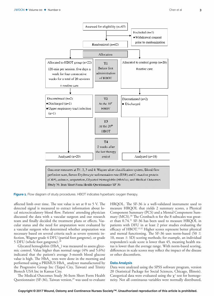
Study Procedures Patients were randomly allocated to HBOT plus routine care or routine care alone groups. Group allocation was randomly generated by a computer and sealed by the primary researcher in opaque, serially numbered envelopes. Another researcher enrolled participants and assigned participants to interven-tions. Patients in both the intervention (HBOT) and control (routine care only) groups were hospitalized during the study, which reduced variability in standard care ( Figure 1 ).
Standard care incorporated topical and systemic therapy for DFUs. It included maintaining good blood glucose control, offl oading, debridement of necrotic tissue, antibiotic therapy for management of diabetic foot infection, and topical dress-ings, depending on the type and grade of the ulcer. An array of wound dressings was used for topical therapy. For exam-ple, silver-impregnated dressings such as Flamazine ointment (silver sulfadiazine) were applied to Wagner grade 1 and 2 DFUs. Hydrocolloid dressings, topical amoxicillin, and hy-drogels were applied for Wagner grade 2 and 3 DFUs; wound dressings and topical piperacillin/ciprofl oxacin were used to manage Wagner grade 3 and 4 DFUs. Antibiotic therapy was driven by protocol and culture and sensitivity reports.
Hyperbaric oxygen therapy was delivered via a multiperson chamber. Patients allocated to the HBOT group were placed in a hyperbaric chamber daily 5 days per week for 4 consecu-tive weeks for a total of 20 sessions. Patients were treated with 2.5 absolute atmospheric pressure for 120 minutes. Th e time period of the intervention was approximately 1 month. Th e intervention program was carried out by the primary research team (C.-Y.C. and R-W.W.).
Outcome Measures We selected multiple outcome measures to evaluate the effi -cacy of HBOT as adjunctive therapy for patients with non-healing DFU. Th ey included wound physiological indices and blood biochemistry tests. Data were collected at 4 diff erent time frames from T1 to T4. Th e same outcome measures were collected at the same time frames for both groups. Wound as-sessment was based on the Wagner classifi cation system. 23 , 24 Th e Wagner grading system considers depth of penetration, the presence of underlying osteomyelitis, and the extent of tis-sue necrosis; ulcers are graded from 0 (preulcerous changes) to 5 (amputation required). Patient wounds classifi ed as Wagner grade 0 or 1 were considered healed. 25
Serum erythrocyte sedimentation rate (ESR) and C-reactive protein (CRP) were used to evaluate infl ammation. Erythro-cyte sedimentation rate was measured with an ESR instrument (model Bedia-15s; Becton, Dickinson and Company, Taipei City, Taiwan). C-reactive protein levels were measured via a Unicel automatic biochemistry analyzer (Beckman, Brea, California).
Foot ulcer microbiology and presence of infection were evaluated via wound cultures obtained using the aseptic swab technique 26 and were collected for anaerobic and aerobic bac-terial cultures. Antibiotic therapy was based on culture and sensitivity fi ndings.
Blood fl ow perfusion scan was used to show when blood fl ow started and to evaluate tissue survival rate 27 in the aff ected limb during the study period. Measurements were performed using the PeriScan PIM II Laser Doppler Perfusion Imager (Perimed, Beijing, China) by R-W.W. Th is system visualizes spatial blood perfusion by scanning across the tissue in the
Copyright © 2017 Wound, Ostomy and Continence Nurses Society™. Unauthorized reproduction of this article is prohibited.
JWOCN Volume 00 Number 0 Chen et al 3
aff ected limb over time. Th e test value is set at 0 to 5 V. Th e detected signal is measured to extract information about lo-cal microcirculatory blood fl ow. Patients’ attending physician discussed the data with a vascular surgeon and our research team and fi nally decided the treatment plans or eff ects. Vas-cular status and the need for amputation were evaluated by a vascular surgeon who determined whether amputation was necessary based on several criteria such as severe systemic in-fection, Wagner grade 4 DFU (partial foot gangrene), or grade 5 DFU (whole foot gangrene). 28
Glycated hemoglobin (HbA 1c ) was measured to assess glyce-mic control. Value higher than normal range (4% and 5.6%) indicated that the patient’s average 3-month blood glucose value is high. Th e HbA 1c tests were done in the morning and performed using a PRIMUS HbA 1c analyzer manufactured by the Progressive Group Inc (Taipei City, Taiwan) and Trinity Biotech USA Inc in Kansas City.
Th e Medical Outcomes Study 36-Item Short Form Health Questionnaire (SF-36), Taiwan version, 29 was used to evaluate
HRQOL. Th e SF-36 is a well-validated instrument used to measure HRQOL that yields 2 summary scores, a Physical Component Summary (PCS) and a Mental Component Sum-mary (MCS). 30 Th e Cronbach α for the 8 subscales was great-er than 0.74. 31 SF-36 has been used to measure HRQOL in patients with DFU in at least 2 prior studies evaluating the effi cacy of HBOT. 13 , 32 Higher scores represent better physical and mental functioning. Th e SF-36 uses norm-based (50 ± 10, mean ± SD) scoring methods; for example, an individual respondent’s scale score is lower than 45, meaning health sta-tus is lower than the average range. With norm-based scoring, diff erences in scale scores may refl ect the impact of the disease or other discomforts.
Data Analysis Data were analyzed using the SPSS software program, version 20 (Statistical Package for Social Sciences, Chicago, Illinois). Categorical data were evaluated using the χ 2 test for homoge-neity. Not all continuous variables were normally distributed,
Figure 1. Flow diagram of study procedures. HBOT indicates hyperbaric oxygen therapy.

Copyright © 2017 Wound, Ostomy and Continence Nurses Society™. Unauthorized reproduction of this article is prohibited.
4 JWOCN Month/Month 2017 www.jwocnonline.com
and they were analyzed using the Mann-Whitney U test. Specifi cally, the Mann-Whitney U test was used to evaluate diff erences in CRP, ESR, HbA 1c , and Doppler measurements. Kruskal-Wallis H tests analyzed changes in the CRP, ESR, HbA 1c , and Doppler measurements between the 2 groups be-fore and after the HBOT. Th e trends of change over time of the SF-36 were compared between groups by generalized es-timating equation (GEE) using the fi rst-order autoregressive (AR1) to handle repeated observations within the subject. A proper working correlation matrix when applying the GEE method. Statistical signifi cance was set at P < .05.
RESULTS
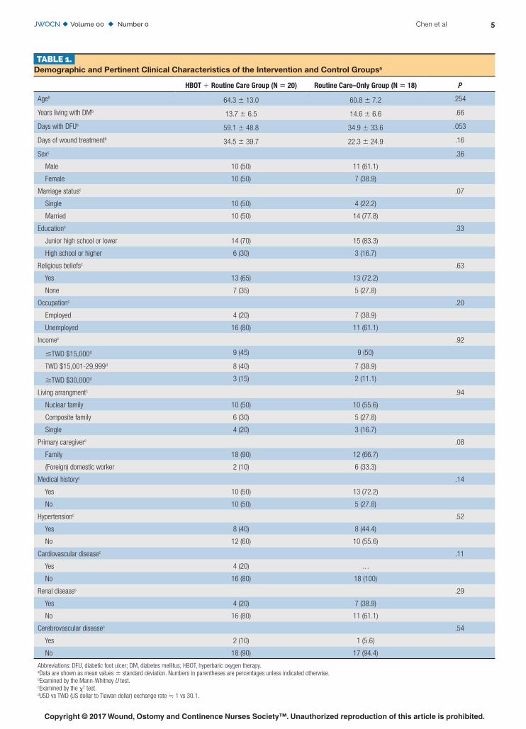
Th irty-eight patients completed the study, with 20 in the HBOT group and 18 in the routine care group. Th e mean age ± SD of patients in the HBOT and routine care groups was 64.3 ± 13 and 60.8 ± 7.2 years, respectively. Th e distri-butions of age were compatible between the 2 groups. Simi-larly, all other demographic variables of patients in the routine care group were compatible to those in the HBOT group ( P > .05). At baseline, no signifi cant diff erences in demographic and clinical characteristics were noted between the HBO and control groups following random allocation ( Table 1 ).
Baseline evaluation found no diff erences in wound severity between the groups ( χ 2 = 1.643, P = .200). When assessed at 2 weeks after the individual’s last therapy ended (T4), 3 patients in the HBOT group had Wagner grade 1 wounds, 7 had grade 2 wounds, 4 received skin grafts, 5 healed, and 1 underwent amputation. In contrast, 9 patients had Wagner grade 3 wounds, 3 had grade 2 wounds, 1 had grade 1 wound, 2 patients received skin grafts, 1 patient healed, and 2 patients underwent amputation. Th is diff erence in wound severity following treatment was statistically signifi cant ( χ 2 = 15.204, P = .010).
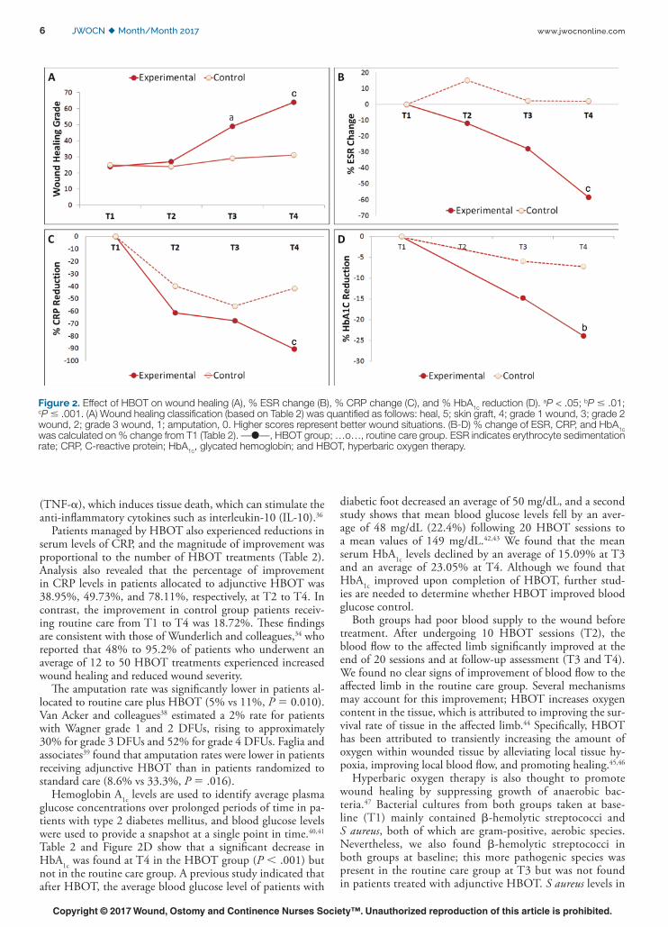
Wound-healing scores diff ered signifi cantly between the interventio and control groups at the 20th administration of HBOT (T3) to T4 ( Z = − 4.205, P = .038, Mann-Whitney U test). Wounds in the HBOT group began to show clear im-provements at T3. We also quantifi ed wound healing based on a score of 0 to 5, where a score of 5 indicated a healed wound, a score of 4 indicated wound managed by skin grafts, a score of 3 indicated a Wagner grade 1 wound, a score of 3 indicated a Wagner grade 2 wound, a score of 2 indicated a Wagner grade 3 wound, and a score of 0 indicated amputation; this scoring system is used in Figure 2 A to illustrate the eff ect of HBOT on ulcer healing.
Diff erences over time were analyzed by the Kruskal-Wal-lis H test; analysis revealed a signifi cant decrease between the ESR value at T4 versus T1 in the HBOT group ( Z = − 3.291, P < .001; Table 2 ). Th ere were no signifi cant diff erences in the routine care group ( Z = − 1.743, P > .05). Th e ESR values at T4 in the HBOT group were signifi cantly lower than those in the routine care group by using the Mann-Whitney U test ( Z = − 4.096, P < .05). Degree of changes in ESR values from T1 to T4 is shown in Figure 2 B.
Analysis also revealed signifi cantly diff erent CRP values be-tween T4 and T1 in the HBOT group (Kruskal-Wallis H test, Z = − 3.920, P < .05; Table 2 ). Participants in the HBOT group had signifi cantly lower CRP levels at T4 when compared to subjects in the routine care group ( Z = − 3.480, P < .001; Figure 2 C). We found a signifi cant decline in mean HbA 1c levels at T4 in the HBOT group (Kruskal-Wallis H tests, Z =
− 3.826, P < .001), but no diff erences were found in the rou-tine care group ( Table 2 ). Degree of changes in HbA 1c values at T1, T3, and T4 can be seen in Figure 2 D.
Doppler blood fl ow in the limb with a DFU signifi cant-ly increased at the 10th administration of HBOT (T2) and maintained that level ( Z = − 2.221, P < .05; Table 2 ). In contrast, no signifi cant change in blood fl ow was observed in the control group.
In the HBOT group, Proteus mirabilis ( P mirabilis ), Staph-ylococcus aureus ( S aureus ), and Morganella morganii appeared at T1. S aureus and P mirabilis persisted from T1 to T4. Th ere was a striking decrease in the number of pathogens of S aureus and P mirabilis per sample from 5 to 2 and 7 to 1, respective-ly. Morganella morganii disappeared at T2. In the routine care group, new species, Pseudomonas aeruginosa , appeared at T2 and showed an increase in the number of pathogens per sam-ple from 4 (T2) to 7 (T4).
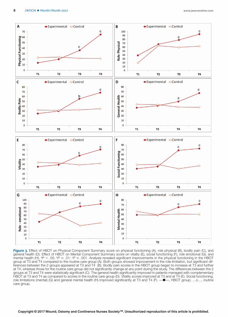
Scores from the SF-36 questionnaire were analyzed using the GEE, taking into account mean values and statistical-ly signifi cant diff erence between 2 groups for each subscale ( Figures 3 A-3H). Improvements were noted after 20 HBOT sessions (T3) and persisted at posttreatment follow-up (T4). Improvement was noted on both PCS and MCS. Using a mixed linear model analysis, the HBOT group ( F = 24.297, P < .001) had signifi cant progress in PCS at all time points, compared to the routine care group ( F = 1.661, P = .171), and in MCS ( F = 11.195, P < .001 vs F = 2.491, P = 0.052).
DISCUSSION
Findings from our study suggest that HBOT, when combined with standard care, alleviates infl ammatory indices of persons with nonhealing DFU. Specifi cally, we examined the eff ect of HBOT at 4 points in time; wounds in the routine care plus HBOT showed signs of wound healing after 10 treatments (T2), while wounds in the routine care group began to deteri-orate. As shown in Figure 2 A, the status of wounds in patients with HBOT was signifi cantly improved at the end of treat-ment (T3) and improved further at follow-up (T4), whereas DFUs in the routine care group showed little change through-out the study. Our results are consistent with prior studies re-porting that adjunctive HBOT reduced wound size. 13 , 33 , 34
Changes in ESR and serum CRP levels in patients with DFU are particularly relevant because of their utility in evalu-ating treatment effi cacy and likelihood of amputation. 1 , 15 , 35 , 36 We found that patients allocated to adjunctive HBOT experi-enced a reduction in the infl ammatory marker ESR ( Table 2 , Figures 2 B and 2C). Th ese changes were not seen or were less prominent in the routine care group, supporting fi ndings from others that HBOT can reduce infl ammation in diabet-ic foot wound tissue. 37 In type 2 diabetes, low-grade infl am-mation is also refl ected in serum CRP levels. 1 , 36 Natorska and colleagues 36 found that CRP concentrations in 40 diabetic and nondiabetic patients with aortic valve stenosis were 9.20 and 4.70 mg/L, respectively. Th e white blood cell concentration in blood and ESR have been found to be signifi cantly elevated in patients with DFU and infection requiring amputation. 35 Th e relationship between infl ammation and wound healing is not entirely understood; it may be attributable to the actions of several bioactive chemicals. Hyperbaric oxygen therapy has been shown to suppress multiple cytokines, such as interleu-kin-1 (IL-1) and interleukin-6 (IL-6), that trigger a local in-fl ammatory response and suppress tissue necrosis factor-alpha
Copyright © 2017 Wound, Ostomy and Continence Nurses Society™. Unauthorized reproduction of this article is prohibited.
JWOCN Volume 00 Number 0 Chen et al 5
TABLE 1. Demographic and Pertinent Clinical Characteristics of the Intervention and Control Groups a
HBOT + Routine Care Group (N = 20) Routine Care–Only Group (N = 18) P
Age b 64.3 ± 13.0 60.8 ± 7.2 .254
Years living with DM b 13.7 ± 6.5 14.6 ± 6.6 .66
Days with DFU b 59.1 ± 48.8 34.9 ± 33.6 .053
Days of wound treatment b 34.5 ± 39.7 22.3 ± 24.9 .16
Sex c .36
Male 10 (50) 11 (61.1)
Female 10 (50) 7 (38.9)
Marriage status c .07
Single 10 (50) 4 (22.2)
Married 10 (50) 14 (77.8)
Education c .33
Junior high school or lower 14 (70) 15 (83.3)
High school or higher 6 (30) 3 (16.7)
Religious beliefs c .63
Yes 13 (65) 13 (72.2)
None 7 (35) 5 (27.8)
Occupation c .20
Employed 4 (20) 7 (38.9)
Unemployed 16 (80) 11 (61.1)
Income c .92
≤ TWD $15,000 d 9 (45) 9 (50)
TWD $15,001-29,999 d 8 (40) 7 (38.9)
≥ TWD $30,000 d 3 (15) 2 (11.1)
Living arrangment c .94
Nuclear family 10 (50) 10 (55.6)
Composite family 6 (30) 5 (27.8)
Single 4 (20) 3 (16.7)
Primary caregiver c .08
Family 18 (90) 12 (66.7)
(Foreign) domestic worker 2 (10) 6 (33.3)
Medical history c .14
Yes 10 (50) 13 (72.2)
No 10 (50) 5 (27.8)
Hypertension c .52
Yes 8 (40) 8 (44.4)
No 12 (60) 10 (55.6)
Cardiovascular disease c .11
Yes 4 (20) …
No 16 (80) 18 (100)
Renal disease c .29
Yes 4 (20) 7 (38.9)
No 16 (80) 11 (61.1)
Cerebrovascular disease c .54
Yes 2 (10) 1 (5.6)
No 18 (90) 17 (94.4)
Abbreviations: DFU, diabetic foot ulcer; DM, diabetes mellitus; HBOT, hyperbaric oxygen therapy.
a Data are shown as mean values ± standard deviation. Numbers in parentheses are percentages unless indicated otherwise.
b Examined by the Mann-Whitney U test.
c Examined by the χ 2 test.
d USD vs TWD (US dollar to Tiawan dollar) exchange rate ≒ 1 vs 30.1.
Copyright © 2017 Wound, Ostomy and Continence Nurses Society™. Unauthorized reproduction of this article is prohibited.
6 JWOCN Month/Month 2017 www.jwocnonline.com
(TNF- α ), which induces tissue death, which can stimulate the anti-infl ammatory cytokines such as interleukin-10 (IL-10). 36
Patients managed by HBOT also experienced reductions in serum levels of CRP, and the magnitude of improvement was proportional to the number of HBOT treatments ( Table 2 ). Analysis also revealed that the percentage of improvement in CRP levels in patients allocated to adjunctive HBOT was 38.95%, 49.73%, and 78.11%, respectively, at T2 to T4. In contrast, the improvement in control group patients receiv-ing routine care from T1 to T4 was 18.72%. Th ese fi ndings are consistent with those of Wunderlich and colleagues, 34 who reported that 48% to 95.2% of patients who underwent an average of 12 to 50 HBOT treatments experienced increased wound healing and reduced wound severity.
Th e amputation rate was signifi cantly lower in patients al-located to routine care plus HBOT (5% vs 11%, P = 0.010). Van Acker and colleagues 38 estimated a 2% rate for patients with Wagner grade 1 and 2 DFUs, rising to approximately 30% for grade 3 DFUs and 52% for grade 4 DFUs. Faglia and associates 39 found that amputation rates were lower in patients receiving adjunctive HBOT than in patients randomized to standard care (8.6% vs 33.3%, P = .016).
Hemoglobin A 1c levels are used to identify average plasma glucose concentrations over prolonged periods of time in pa-tients with type 2 diabetes mellitus, and blood glucose levels were used to provide a snapshot at a single point in time. 40 , 41 Table 2 and Figure 2 D show that a signifi cant decrease in HbA 1c was found at T4 in the HBOT group ( P < .001) but not in the routine care group. A previous study indicated that after HBOT, the average blood glucose level of patients with
diabetic foot decreased an average of 50 mg/dL, and a second study shows that mean blood glucose levels fell by an aver-age of 48 mg/dL (22.4%) following 20 HBOT sessions to a mean values of 149 mg/dL. 42 , 43 We found that the mean serum HbA 1c levels declined by an average of 15.09% at T3 and an average of 23.05% at T4. Although we found that HbA 1c improved upon completion of HBOT, further stud-ies are needed to determine whether HBOT improved blood glucose control.
Both groups had poor blood supply to the wound before treatment. After undergoing 10 HBOT sessions (T2), the blood fl ow to the aff ected limb signifi cantly improved at the end of 20 sessions and at follow-up assessment (T3 and T4). We found no clear signs of improvement of blood fl ow to the aff ected limb in the routine care group. Several mechanisms may account for this improvement; HBOT increases oxygen content in the tissue, which is attributed to improving the sur-vival rate of tissue in the aff ected limb. 44 Specifi cally, HBOT has been attributed to transiently increasing the amount of oxygen within wounded tissue by alleviating local tissue hy-poxia, improving local blood fl ow, and promoting healing. 45 , 46
Hyperbaric oxygen therapy is also thought to promote wound healing by suppressing growth of anaerobic bac-teria. 47 Bacterial cultures from both groups taken at base-line (T1) mainly contained β -hemolytic streptococci and S aureus , both of which are gram-positive, aerobic species. Nevertheless, we also found β -hemolytic streptococci in both groups at baseline; this more pathogenic species was present in the routine care group at T3 but was not found in patients treated with adjunctive HBOT. S aureus levels in
Figure 2. Effect of HBOT on wound healing (A), % ESR change (B), % CRP change (C), and % HbA 1c
reduction (D). a P < .05; b P ≤ .01; c P ≤ .001. (A) Wound healing classifi cation (based on Table 2 ) was quantifi ed as follows: heal, 5; skin graft, 4; grade 1 wound, 3; grade 2 wound, 2; grade 3 wound, 1; amputation, 0. Higher scores represent better wound situations. (B-D) % change of ESR, CRP, and HbA
1c
was calculated on % change from T1 ( Table 2 ). —●—, HBOT group; …o…, routine care group. ESR indicates erythrocyte sedimentation rate; CRP, C-reactive protein; HbA
1c , glycated hemoglobin; and HBOT, hyperbaric oxygen therapy.
Copyright © 2017 Wound, Ostomy and Continence Nurses Society™. Unauthorized reproduction of this article is prohibited.
JWOCN Volume 00 Number 0 Chen et al 7
the wound can be used to diff erentiate grades of DFU, and its eradication has been associated with wound healing. 48 Two weeks following treatment (T4), we found decreased colony counts of S aureus in patients treated with HBOT. In contrast, control group subjects had increased colony counts of S aureus . We hypothesize that HBOT suppressed the growth of anaerobic bacteria and possibly impaired acti-vation of bacterial endotoxins. It has previously been shown that HBOT provides phagocytic leukocytes with 15 times more oxygen than required when digesting microorganisms, resulting in large amounts of free oxygen radicals that kill bacteria. 49 Our fi ndings are consistent with these results, in-dicating an antimicrobial function of HBOT. 1 , 17 , 50
Treatment signifi cantly improved HRQOL assessed by the SF-36v2 ( P < .01). Improvements in HRQOL may be at-tributed to progress toward ulcer healing and decreased emo-tional stress. Th e results of this study are consistent with those of Lin’s group 51 and Löndahl’s group, 13 who also reported im-
provement in quality of life following HBOT for treatment of DFUs.
Limitations Several elements of the design may limit generalizability of fi ndings. Subjects were enrolled for a single medical center, and follow-up did not continue until the DFU closed. Further investigations including multiple settings may be needed to confi rm the fi ndings in this study.
CONCLUSION
Findings from our study suggest that HBOT promoted DFU healing by increasing oxygen dispersion to damaged tissues, al-leviating infl ammation, and suppressing the growth of anaerobic bacteria. In addition, we found that HBOT reduced the risk of amputation of the aff ected limb and improved HRQOL. We rec-ommend administering at least 20 treatments to maximize the benefi cial eff ects of HBOT on patients with nonhealing DFU.
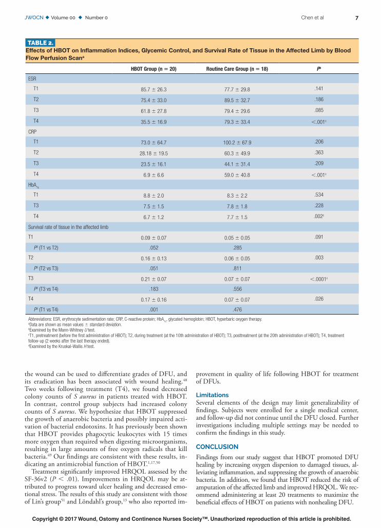
TABLE 2. Effects of HBOT on Inflammation Indices, Glycemic Control, and Survival Rate of Tissue in the Affected Limb by Blood Flow Perfusion Scan a
HBOT Group (n = 20) Routine Care Group (n = 18) P b
ESR
T1 85.7 ± 26.3 77.7 ± 29.8 .141
T2 75.4 ± 33.0 89.5 ± 32.7 .186
T3 61.8 ± 27.8 79.4 ± 29.6 .085
T4 35.5 ± 16.9 79.3 ± 33.4 < .001 c
CRP
T1 73.0 ± 64.7 100.2 ± 67.9 .206
T2 28.18 ± 19.5 60.3 ± 49.9 .363
T3 23.5 ± 16.1 44.1 ± 31.4 .209
T4 6.9 ± 6.6 59.0 ± 40.8 < .001 c
HbA 1c
T1 8.8 ± 2.0 8.3 ± 2.2 .534
T3 7.5 ± 1.5 7.8 ± 1.8 .228
T4 6.7 ± 1.2 7.7 ± 1.5 .002 c
Survival rate of tissue in the affected limb
T1 0.09 ± 0.07 0.05 ± 0.05 .091
P d (T1 vs T2) .052 .285
T2 0.16 ± 0.13 0.06 ± 0.05 .003
P d (T2 vs T3) .051 .811
T3 0.21 ± 0.07 0.07 ± 0.07 < .0001 c
P d (T3 vs T4) .183 .556
T4 0.17 ± 0.16 0.07 ± 0.07 .026
P d (T1 vs T4) .001 .476
Abbreviations: ESR, erythrocyte sedimentation rate; CRP, C-reactive protein; HbA 1c
, glycated hemoglobin; HBOT, hyperbaric oxygen therapy.
a Data are shown as mean values ± standard deviation.
b Examined by the Mann-Whitney U test.
c T1, pretreatment (before the fi rst administration of HBOT); T2, during treatment (at the 10th administration of HBOT); T3, posttreatment (at the 20th administration of HBOT); T4, treatment
follow-up (2 weeks after the last therapy ended).
d Examined by the Kruskal-Wallis H test.
Copyright © 2017 Wound, Ostomy and Continence Nurses Society™. Unauthorized reproduction of this article is prohibited.
8 JWOCN Month/Month 2017 www.jwocnonline.com
Figure 3. Effect of HBOT on Physical Component Summary score on physical functioning (A), role physical (B), bodily pain (C), and general health (D). Effect of HBOT on Mental Component Summary score on vitality (E), social functioning (F), role emotional (G), and mental health (H). a P < .05; b P ≤ .01; c P ≤ .001. Analysis revealed signifi cant improvements in the physical functioning in the HBOT group at T3 and T4 compared to the routine care group (A). Both groups showed improvement in the role limitation, but signifi cant dif-ferences between the 2 groups appeared at T3 and T4 (B). Bodily pain scores in the HBOT group began to increase at T3 and further at T4, whereas those for the routine care group did not signifi cantly change at any point during the study. The differences between the 2 groups at T3 and T4 were statistically signifi cant (C). The general health signifi cantly improved in patients managed with complementary HBOT at T3 and T4 as compared to scores in the routine care group (D). Vitality scores improved at T3 and at T4 (E). Social functioning, role limitations (mental) (G) and general mental health (H) improved signifi cantly at T3 and T4 (F). —●—, HBOT group; …o…, routine care group.
Copyright © 2017 Wound, Ostomy and Continence Nurses Society™. Unauthorized reproduction of this article is prohibited.
JWOCN Volume 00 Number 0 Chen et al 9
ACKNOWLEDGMENT
Th is study was partly supported by Chang Gung Hospital (research project CMRPG8A0341). Th e authors thank all the participants in this study for their hard work and dedication.
REFERENCES 1. Richard JL , Sotto A , Lavigne JP . New insights in diabetic foot infec-
tion . World J Diabetes . 2011 ; 2 ( 2 ): 24-32 .
2. Boulton AJ , Vileikyte L , Ragnarson-Tennvall G , Apelqvist J . The global
burden of diabetic foot disease . Lancet . 2005 ; 366 ( 9498 ): 1719-1724 .
3. Margolis DJ , Malay DS , Hoffstad OJ , et al. Incidence of Diabetic Foot Ulcer
and Lower Extremity Amputation Among Medicare Benefi ciaries, 2006
to 2008: Data Points #2 . Rockville, MD : Agency for Healthcare Research
and Quality ; 2011 . AHRQ Publication No. 10(11)-EHC009-1-EF.
4. Fincke BG , Miller DR , Turpin R . A classifi cation of diabetic foot in-
fections using ICD-9-CM codes: application to a large computerized
medical database . BMC Health Serv Res . 2010 ; 10 : 192 .
5. Chen FT , Chiu CC , Liu SC , Lee PY , Liao CC . Literature review and
case analysis on diabetic foot infection . Formosa J Clin Pharm .
1996 : 5 ( 1 ): 102-110 .
6. Boulton AJ , Armstrong DG , Albert SF , et al. Comprehensive foot ex-
amination and risk assessment: a report of the task force of the foot
care interest group of the American Diabetes Association, with en-
dorsement by the American Association of Clinical Endocrinologists .
Diabetes Care . 2008 ; 31 ( 8 ): 1679-1685 .
7. Singh N , Armstrong DG , Lipsky BA . Preventing foot ulcers in patients
with diabetes . JAMA . 2005 ; 293 ( 2 ): 217-228 .
8. Schofi eld CJ , Libby G , Brennan GM , et al. Mortality and hospitalization
in patients after amputation: a comparison between patients with and
without diabetes . Diabetes Care . 2006 ; 29 ( 10 ): 2252-2256 .
9. Miyajima S , Shirai A , Yamamoto S , Okada N , Matsushita T . Risk fac-
tors for major limb amputations in diabetic foot gangrene patients .
Diabetes Res Clin Pract . 2006 ; 71 ( 3 ): 272-279 .
10. Ghanassia E , Villon L , Thuan Dit Dieudonné JF , Boegner C , Avignon A ,
Sultan A . Long-term outcome and disability of diabetic patients hos-
pitalized for diabetic foot ulcers: a 6.5-year follow-up study . Diabetes
Care . 2008 ; 31 ( 7 ): 1288-1292 .
11. Nather A , Bee CS , Huak CY , et al. Epidemiology of diabetic foot prob-
lems and predictive factors for limb loss . J Diabetes Complications .
2008 ; 22 ( 2 ): 77-82 .
12. Steed DL , Attinger C , Colaizzi T , et al. Guidelines for the treatment of
diabetic ulcers . Wound Repair Regen . 2006 ; 14 ( 6 ): 680-692 .
13. Löndahl M , Landin-Olsson M , Katzman P . Hyperbaric oxygen thera-
py improves health-related quality of life in patients with diabetic and
chronic foot ulcer . Diabet Med . 2011 : 28 ( 2 ): 186-190 .
14. Tiaka EK , Papanas N , Manolakis AC , Maltezos E . The role of hy-
perbaric oxygen in the treatment of diabetic foot ulcers . Angiology .
2012 ; 63 ( 4 ): 302-314 .
15. Liu R , Li L , Yang M , Boden G , Yang G . Systematic review of the ef-
fectiveness of hyperbaric oxygenation therapy in the management of
chronic diabetic foot ulcers . Mayo Clin Proc . 2013 ; 88 ( 2 ): 166-175 .
16. Kalani M , Jörneskog G , Naderi N , Lind F , Brismar K . Hyperbaric ox-
ygen (HBO) therapy in treatment of diabetic foot ulcers. long-term
follow-up . J Diabetes Complications . 2002 ; 16 ( 2 ): 153-158 .
17. Margolis DJ , Gupta J , Hoffstad O , et al. Lack of effectiveness of
hyperbaric oxygen therapy for the treatment of diabetic foot ulcer
and the prevention of amputation: a cohort study . Diabetes Care .
2013 ; 36 ( 7 ): 1961-1966 .
18. Kranke P , Bennett M , Roeckl-Wiedmann I , Debus S . Hyperbaric
oxygen therapy for chronic wounds . Cochrane Database Syst Rev .
2004 ; 2 : CD004123 .
19. Fedorko L , Bowen JM , Jones W , et al. Hyperbaric oxygen therapy does
not reduce indications for amputation in patients with diabetes with
nonhealing ulcers of the lower limb: a prospective, double-blind, ran-
domized controlled clinical trial . Diabetes Care . 2016 ; 39 ( 3 ): 392-399 .
20. Yadav A , Pawar M , Garg R , Banerjee N . To compare the effects of
multiple sessions of hyperbaric oxygen therapy in neurological im-
provement in head injury patients: a prospective randomized trial . J
Neuroanaesth Crit Care . 2015 ; 2 : 110-113 .
21. Cohen J . A power primer . Psychol Bull . 1992 ; 12 : 155-159 .
22. Cohen J . Statistical Power Analysis for the Behavioral Sciences . 2nd
ed. Mahwah, NJ : Lawrence Erlbaum ; 1988 : 19-65 , 414.
23. Wagner FW Jr . The dysvascular foot: a system for diagnosis and
treatment . Foot Ankle . 1981 ; 2 : 64-122 .
24. Harris C , Bates-Jensen B , Parslow N , Raizman R , Singh M ,
Ketchen R . Bates-Jensen wound assessment tool: pictorial guide
validation project . J Wound Ostomy Continence Nurs . 2010 ; 37 ( 3 ):
253-259 .
25. Jeon BJ , Choi HJ , Kang JS , Tak MS , Park ES . Comparison of fi ve
systems of classifi cation of diabetic foot ulcers and predictive factors
for amputation . Int Wound J . 2017 ; 14 ( 3 ): 537-545 .
26. Bowler PG , Duerden BI , Armstrong DG . Wound microbiology and
associated approaches to wound management . Clin Microbiol Rev .
2001 ; 14 ( 2 ): 244-269 .
27. Ma L , Li P , Shi Z , Hou T , Chen X , Du J . A prospective, randomized,
controlled study of hyperbaric oxygen therapy: effects on healing and
oxidative stress of ulcer tissue in patients with a diabetic foot ulcer .
Ostomy Wound Manage . 2013 ; 59 ( 3 ): 18-24 .
28. Frykberg RG , Zgonis T , Armstrong DG , et al. Diabetic foot disor-
ders: a clinical practice guideline (2006 revision) . J Foot Ankle Surg .
2006 ; 45 ( 5 )( suppl ): S1-S66 .
29. Lu JF , Tseng HM , Tsai YJ . Assessment of health-related quality of life
in Taiwan (I): development and psychometric testing of SF-36 Taiwan
version . Taiwan J Public Health . 2003 ; 22 ( 6 ): 501-511 .
30. Tseng MY , Liang J , Shyu YI, et al. Effects of interventions on trajecto-
ries of health-related quality of life among older patients with hip frac-
ture: a prospective randomized controlled trial . BMC Musculoskelet
Disord . 2016 ; 17 : 114 .
31. Hsieh FT , Huang GS , Ko WJ , Lou MF . Health status and quality of
life of survivors of extra corporeal membrane oxygenation: a cross-
sectional study . J Adv Nurs . 2016 ; 72 ( 7 ): 1626-1637 .
32. Abidia A , Laden G , Kuhan G, et al. The role of hyperbaric oxy-
gen therapy in ischaemic diabetic lower extremity ulcers: a double
-blind randomised-controlled trial . Eur J Vasc Endovasc Surg .
2003 ; 25 ( 6 ): 513-518 .
33. Löndahl M , Katzman P , Nilsson A , Hammarlund C . Hyperbaric oxygen
therapy facilitates healing of chronic foot ulcers in patients with diabe-
tes . Diabetes Care . 2010 ; 33 ( 5 ): 998-1003 .
34. Wunderlich RP , Peters EJ , Lavery LA . Systemic hyperbaric oxygen
therapy: lower-extremity wound healing and the diabetic foot . Diabe-
tes Care . 2000 ; 23 ( 10 ): 1551-1555 .
35. Akinci B , Yener S , Yesil S , Yapar N , Kucukyavas Y , Bayraktar F . Acute
phase reactants predict the risk of amputation in diabetic foot infec-
tion . J Am Podiatr Med Assoc . 2011 : 101 ( 1 ): 1-6 .
36. Natorska J , Wypasek E , Grudzien G , et al. Does diabetes accelerate
the progression of aortic stenosis through enhanced infl ammatory re-
sponse within aortic valves? Infl ammation . 2012 ; 35 ( 3 ): 834-840 .
37. Karadurmus N , Sahin M , Tasci C , et al. Potential benefi ts of hyperbaric
oxygen therapy on atherosclerosis and glycaemic control in patients
with diabetic foot . Endokrynol Pol . 2010 ; 61 ( 3 ): 275-279 .
38. Van Acker K , De Block C , Abrams P , et al. The choice of diabet-
ic foot ulcer classifi cation in relation to the fi nal outcome . Wounds .
2002 ; 14 ( 1 ): 16-25 .
39. Faglia E , Favales F , Aldeghi A , et al. Adjunctive systemic hyper-
baric oxygen therapy in treatment of severe prevalently isch-
emic diabetic foot ulcer: a randomized study . Diabetes Care .
1996 ; 19 ( 12 ): 1338-1343 .
40. Gholap NN , Davies MJ , Mostafa SA , Khunti K . Diagnosing type 2
diabetes and identifying high-risk individuals using the new glycat-
ed haemoglobin (HbA 1c
) criteria . Br J Gen Pract . 2013 ; 63 ( 607 ): e165
-e167 .
41. John WG . UK Department of Health Advisory Committee on Diabe-
tes. Use of HbA 1c
in the diagnosis of diabetes mellitus in the UK. The
implementation of World Health Organization guidance 2011 . Diabet
Med . 2012 ; 29 ( 11 ): 1350-1357 .
42. Goldman RJ . Hyperbaric oxygen therapy for wound healing and limb
salvage: a systematic review . PMR . 2009 : 1 ( 5 ): 471-489 .
43. Huang CT , Huang YH , Chang LP . The effect of hyperbaric oxy-
genation on blood sugar level in DM patients . Chin J Occup Med .
2004 ; 11 ( 4 ): 235-242 .
44. Thom SR . Hyperbaric oxygen: its mechanisms and effi cacy . Plast Re-
constr Surg . 2011 ; 127 ( suppl 1 ): 131S-141S .
45. Kranke P , Bennett MH , Martyn-St James M , Schnabel A , Debus SE .
Hyperbaric oxygen therapy for chronic wounds . Cochrane Database
Syst Rev . 2012 ; 4 : CD004123 .
46. Zoranovic U , Jevtic M , Jovanovic M , Pucar D , Cizmic M . Hyperbaric
oxygenation effects determination in the therapy of chronic occlusive

Copyright © 2017 Wound, Ostomy and Continence Nurses Society™. Unauthorized reproduction of this article is prohibited.
10 JWOCN Month/Month 2017 www.jwocnonline.com
lower extremities arteries disease by the use of perfusion scintigraphy .
Vojnosanit Pregl . 2010 ; 67 ( 4 ): 279-285 .
47. Chen TL , Xu B , Liu JC , et al. Effects of hyperbaric oxygen on ag-
gressive periodontitis and subgingival anaerobes in Chinese patients .
J Indian Soc Periodontol . 2012 ; 16 ( 4 ): 492-497 .
48. Albert M . The role of hyperbaric oxygen therapy in wound healing .
Wound Care Can . 2008 ; 6 : 60-62 .
49. Jain KK . Textbook of Hyperbaric Medicine . 5th ed. Cambridge, MA :
Hogrefe & Huber Publishers ; 2009 .
50. Joseph WS , Lipsky BA . Medical therapy of diabetic foot infections .
J Vasc Surg . 2010 ; 52 ( 3 )( suppl ): 67S-71S .
51. Lin LC , Yau G , Lin TF , Lin TK , Tang YY , Wang KY . The effi cacy of hy-
perbaric oxygen therapy in improving the quality of life in patients with
problem wounds . J Nurs Res . 2006 ; 14 ( 3 ): 219-226 .
Related Documents