ADHESION PROPERTIES AND CELL SURFACE CHARACTERISTICS OF THE ENTOMOPATHOGENIC FUNGUS Beauveria bassiana: A LINK BETWEEN MORPHOLOGY AND VIRULENCE By DIANE J. HOLDER A DISSERTATION PRESENTED TO THE GRADUATE SCHOOL OF THE UNIVERSITY OF FLORIDA IN PARTIAL FULFILLMENT OF THE REQUIREMENTS FOR THE DEGREE OF DOCTOR OF PHILOSOPHY UNIVERSITY OF FLORIDA 2005

Welcome message from author
This document is posted to help you gain knowledge. Please leave a comment to let me know what you think about it! Share it to your friends and learn new things together.
Transcript

ADHESION PROPERTIES AND CELL SURFACE CHARACTERISTICS
OF THE ENTOMOPATHOGENIC FUNGUS Beauveria bassiana: A LINK BETWEEN MORPHOLOGY AND VIRULENCE
By
DIANE J. HOLDER
A DISSERTATION PRESENTED TO THE GRADUATE SCHOOL OF THE UNIVERSITY OF FLORIDA IN PARTIAL FULFILLMENT
OF THE REQUIREMENTS FOR THE DEGREE OF DOCTOR OF PHILOSOPHY
UNIVERSITY OF FLORIDA
2005

Copyright 2005
by
Diane J. Holder

This document is dedicated to my parents, my sister and her children

iv
ACKNOWLEDGMENTS
I would like to thank my family for always being there for me, and my major
advisor, Dr. Nemat Keyhani, without whom this would all have been impossible. I
would also like to thank all the members of Dr. Keyhani’s lab (past and present) for the
help, support and friendship that I will always treasure. I also would like to recognize the
other members of the department that helped me with both hands on and theoretical
applications, especially Waultraud Dunn, Donna Williams, Dr. Henry Aldridge, Louise
Monroe, members of Dr. James Preston’s lab, Dr. Keelnatham Shammungam’s lab, and
Dr. Julie Maupin’s lab. I would also like to acknowledge the students, staff and
professors in PERC, for their help and the use of their equipment.
Special thanks goes to Brett Kirkland, Dr. Eunmin Cho, and the myriad of
undergraduates who made this work possible. I thank all the members of my committee,
Dr. Nemat Keyhani, Dr. Peter Kima, Dr. Drion Boucias, Dr. Jeff Rollings and Dr. Julie
Maupin, who helped guide me through this work, generously giving of time, equipment
and advice. I cannot finish without thanking the rest of the staff, students and faculty of
the Department of Microbiology and Cell Science, for the simple day-to-day things that
made this project move forward as smoothly as possible and eventually come to an end.
And last but not least, thanks go to Hamlet, for just being you.

v
TABLE OF CONTENTS page
ACKNOWLEDGMENTS ................................................................................................. iv
LIST OF TABLES........................................................................................................... viii
LIST OF FIGURES ........................................................................................................... ix
ABSTRACT....................................................................................................................... xi
CHAPTER
1 LITERATURE REVIEW .............................................................................................1
Beauveria bassiana.......................................................................................................2 Biological Control ........................................................................................................7
Fungal Biocontrol Agents......................................................................................7 Beauveria bassiana as a Biocontrol Agent ...........................................................7
Factors Affecting Adhesion..........................................................................................8 Specific Binding ....................................................................................................9 Nonspecific Binding..............................................................................................9 Steric, Bridging and Depletion forces .................................................................10 Hydrophobic Interactions ....................................................................................11 Specific Molecules or Structures Involved in Fungal Adhesion .........................12
Molecular Biology ......................................................................................................14 Methods Used to Study Global Differential Gene Expression in Fungi .............14
Differential screening of cDNA libraries .....................................................15 Real Time RT-PCR .............................................................................................17
Objectives ...................................................................................................................18
2 ADHESION OF THE ENTOMOPATHOGENIC FUNGUS BEAUVERIA BASSIANA TO SUBSTRATA..................................................................................20
Introduction.................................................................................................................20 Materials and Methods ...............................................................................................21
Cultivation of Fungi ............................................................................................21 FITC-labeling of B. bassiana Cells .....................................................................22 Adhesion Assay ...................................................................................................22 Enzyme Treatments .............................................................................................23 Effect of pH Influence on Attachment ................................................................24
vi
Competition Assays.............................................................................................24 Contact Angle Determination..............................................................................24
Results.........................................................................................................................25 Discussion...................................................................................................................34
3 SURFACE CHARACTERISTICS OF THE ENTOMOPATHOGEN BEAUVERIA BASSIANA ............................................................................................38
Introduction.................................................................................................................38 Materials and Methods ...............................................................................................43
Atomic Force Microscopy...................................................................................44 Zeta Potential.......................................................................................................44 MATH Assay.......................................................................................................44 Contact Angle Determination..............................................................................45
Results.........................................................................................................................45 Atomic Force Microscopy...................................................................................45 Zeta Potential.......................................................................................................46 MATH Assay.......................................................................................................46 Contact Angles ....................................................................................................48
Discussion...................................................................................................................49
4 SUPPRESSIVE SUBTRACTION HYBRIDIZATION ANALYSIS OF BEAUVERIA BASSIANA GROWN ON INSECT CUTICLE ................................55
Introduction.................................................................................................................55 Virulence Factors.................................................................................................55 Adherence and Colonization factors ...................................................................56 Toxins ..................................................................................................................57 Molecules Involved in Evading Host Defences ..................................................58 Siderophores ........................................................................................................58 Molecules Involved in Toxin Transport ..............................................................58 Suppression Subtractive Hybridization ...............................................................59
Materials and methods................................................................................................60 Cultivation of Fungi ............................................................................................60
Cells grown in the presence of glucose (source of driver RNA)..................60 Cells grown on insect cuticle (source of tester RNA)..................................60
RNA Isolation......................................................................................................61 Supression Subtractive Hybridization .................................................................61
Results.........................................................................................................................61 Discussion...................................................................................................................65
5 MOLECULAR ANALYSIS OF TWO BEAUVERIA BASSIANA HYDROPHOBINS AND A HYDROPHOBIN LIKE PROTEIN .............................70
Introduction.................................................................................................................70 Materials and Methods ...............................................................................................73
Cultivation of Fungi ............................................................................................73

vii
RNA Isolation......................................................................................................74 Rodlet Layer Extraction ......................................................................................74 Mass Peptide Spectrometry (Peptide Fingerprinting) .........................................75 Reverse Transcriptase RT-PCR...........................................................................76
Results.........................................................................................................................77 Identification of a B. bassiana Cell Wall Hydrophobin ......................................77 Reverse Transcriptase RT-PCR...........................................................................77
Discussion...................................................................................................................80
6 GENERAL DISCUSSION .........................................................................................83
Statement of Hypotheses ............................................................................................83 Is There a Measurable Difference in The Cell Surface Characteristics of The
Single Cell Propagules of B. bassiana? .................................................................83 Visual Differences ...............................................................................................83 Differences in Hydrophobicity ............................................................................83 Differences in Effective Surface Charge.............................................................84 Cell Wall Proteins................................................................................................85 Conclusions .........................................................................................................86
Are there Differences in the Binding Properties of These Propagules, Which Can Be Related to the Cell Surface Characteristics? ....................................................86
Adhesion Profiles of Aerial Conidia, Submerged conidia and Blastospores ......86 Conclusion...........................................................................................................86
APPENDIX AGROBACTERIUM MEDIATED TRANSFORMATION OF BEAUVERIA BASSIANA ........................................................................................88
Introduction.................................................................................................................88 Materials and Methods ...............................................................................................89
Fungal Cultures ...................................................................................................89 Agrobacterium tumefaciens Cultivation..............................................................90 Transformation Procedure ...................................................................................90
Discussion...................................................................................................................93
LIST OF REFERENCES...................................................................................................96
BIOGRAPHICAL SKETCH ...........................................................................................105

viii
LIST OF TABLES
Table page 1-1 Classification B. bassiana ..........................................................................................3
2-1 Effects of various competitors and chemicals on B. bassiana cell-type adhesion...31
2-2 Effect of pH on B. bassiana cell-type adhesion .......................................................32
2-3 Effects of various enzymatic treatments on B. bassiana cell-type adhesion ...........33
3-1 Contact angle values for the three cell types for water, bromonapthalene and glycerol with calculated interfacial and polar free energy values. ...........................49
3-2 Advancing and receding water contact angle data with calculated surface energy values for three B. bassiana single cell propagules. ................................................50
4-1 Blast hits of sequence fragments from the SSH library to virulence factors and allergens. ..................................................................................................................63
5-1 Primer sequences and product size for the reverse transcriptase RT-PCR ..............76
5-2 mRNA abundance of bhd1, bhd2 in Beauveria bassiana single cell propagules. ...78
A-1 Putative transformants obtained from Agrobacterium tumefaciens mediated transformation of B. bassiana with selection markers for hygromycin B (hph) and neomycin (neo) resistance. ................................................................................93

ix
LIST OF FIGURES
Figure page 1-1 The life-cycle of a typical hyphomycete entomopathogen. .......................................6
2-1 Bright field and fluorescent microscopy of FITC-labeled B. bassiana aerial conidia, blastospores and submerged conidia ..........................................................25
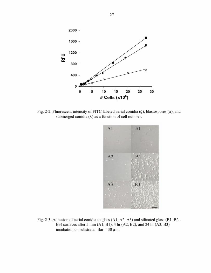
2-2 Fluorescent intensity of FITC labeled aerial conidia, blastospores, and submerged conidia as a function of cell number......................................................27
2-3 Adhesion of aerial conidia to glass and silinated glass surfaces. .............................27
2-4 Adhesion of blastospores to glass and silinated glass surfaces. ...............................28
2-5 Adhesion of submerged conidia to glass and silinated glass ...................................28
2-6 Saturation point of binding sites for B. bassiana aerial conidia, blastospores, and submerged conidia to microtiter plates ....................................................................29
2-7 Quantitative adhesion and influence of washing on adhesion of aerial conidia, blastospores, and submerged conidia to silinated F-200 (hydrophobic), F-200 (weakly polar), and F-600 (hydrophilic) microtiter plates. ......................................30
3-1 Contact angles (θ)are formed at the interface between the liquid, solid and gas boundaries of a droplet on the surface of interest.. ..................................................40
3-2 Atomic force micrographs of B. bassiana conidia, submerged conidia and blastospores ..............................................................................................................46
3-3 Zeta potential values for the three spore types as a function of pH. ........................47
3-4 Microbical adhesion to hydrocarbon.and hydrophobicity indices for B. bassiana aerial conidia, blastospores and submerged conidia. ...............................................47
4-1 Relative numbers of gene fragments representing functional groups present in the subtracted library................................................................................................62
4-2 B. bassiana H1 hydrophobin genomic sequence .....................................................66
4-3 Alignment of H1 and H2 with homologous hydrophobins ......................................67

x
4-4 Bootstrapped (1000) phylogenetic tree of class I and class II hydrophobins...........68
5-1 SDS –PAGE (10% polyacrylamide, Bis-Tris) Gel of SDS soluble/TFA insoluble cell wall proteins.. ....................................................................................................77
5-2 Mass spectroscopy data showing the mass of individual amino acids in one of the two main fragments from the 12 KDa trifluoroacetic acid soluble/sodium dodecyl sulfate insoluble B. bassiana cell wall protein. ..........................................79
5-3 Mass peptide fingerprinting results for two identifiable fragments of the 12 KDa trifluoroacetic acid soluble, sodium dodecyl sulfate insoluble B. bassiana cell wall protein...............................................................................................................80
5-4 Comparison of Bhd1 and Bhd2 and other hydrophobins consensus spacing for Class I and Class II hydrophobins............................................................................81

xi
Abstract of Dissertation Presented to the Graduate School of the University of Florida in Partial Fulfillment of the Requirements for the Degree of Doctor of Philosophy
ADHESION PROPERTIES AND CELL SURFACE CHARACTERISTICS OF THE ENTOMOPATHOGENIC FUNGUS Beauveria bassiana: A LINK BETWEEN
MORPHOLOGY AND VIRULENCE
By
Diane J. Holder
August 2005
Chair: Nemat O. Keyhani Major Department: Microbiology and Cell Science
The entomopathogen Beauveria bassiana produces three distinct in vitro, single
cell propagules: aerial conidia, blastospores and submerged conidia. Atomic force
microscopy (AFM) was used to visualize the surface of aerial conidia and confirmed the
presence of a rodlet layer that was absent from the surface of both blastospores, which
were smooth, and submerged conidia, which appeared coarse. Interfacial free energies of
interaction and hydrophobicity indicies, derived from contact angle data, and
hydrocarbon partitioning revealed differential properties of the three propagules
regarding cell surface hydrophobicity ranging from strongly hydrophobic (aerial conidia)
to hydrophilic (blastospores). Adhesion studies with fluorescently labeled cells
suggested that 1) aerial conidia bound better to hydrophobic surfaces, 2) blastospores
bound better to hydrophilic surfaces, and 3) submerged conidia bound equally well to
both types of surfaces.
xii
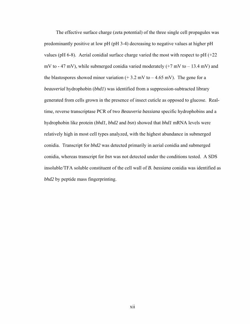
The effective surface charge (zeta potential) of the three single cell propagules was
predominantly positive at low pH (pH 3-4) decreasing to negative values at higher pH
values (pH 6-8). Aerial conidial surface charge varied the most with respect to pH (+22
mV to - 47 mV), while submerged conidia varied moderately (+7 mV to – 13.4 mV) and
the blastospores showed minor variation (+ 3.2 mV to – 4.65 mV). The gene for a
beauverial hydrophobin (bhd1) was identified from a suppression-subtracted library
generated from cells grown in the presence of insect cuticle as opposed to glucose. Real-
time, reverse transcriptase PCR of two Beauveria bassiana specific hydrophobins and a
hydrophobin like protein (bhd1, bhd2 and bsn) showed that bhd1 mRNA levels were
relatively high in most cell types analyzed, with the highest abundance in submerged
conidia. Transcript for bhd2 was detected primarily in aerial conidia and submerged
conidia, whereas transcript for bsn was not detected under the conditions tested. A SDS
insoluble/TFA soluble constituent of the cell wall of B. bassiana conidia was identified as
bhd2 by peptide mass fingerprinting.
1
CHAPTER 1 LITERATURE REVIEW
The earliest reports of a fungal entomopathogen, possibly the organism that would
come to be known as Beauveria bassiana (Balsamo) Vuillemin, came from China, as far
back as 2700 BC (79). It was not until 1835 that Agostino Bassi demonstrated that
Calcino, or White Muscardine, a disease that was devastating the Italian silkworm
industry, was contagious and caused by a parasitic fungus (63). Balsamo Crivelli
officially named the organism Botrytis paradoxica, eventually changing the name to
Botrytis bassiana to honor the man who first described it.
In 1912, Vuillemin, determined that there were enough features peculiar to Botrytis
bassiana to assign it to the new genus Beauveria (19). There now are multiple species in
the genus Beauveria Vuill. some of the most important ones are: B. bassiana, B.
brongniartii, and B. alba. B. bassiana and B. brogniartii well known entomopathogens
with a wide host range, including arthropods other than insects, are now being used as
biological control agents to control a variety of crop damaging insects. B. alba is mainly
isolated as an indoor contaminant and displays the lowest pathogenicity of these three
beauveria species (1). Due to the practical applications of fungal entomopathogens as
biological control agents, the biology (and to a lesser extent the molecular biology) of
these fungi has been the subject of much research.
Major efforts have been targeted towards isolation and characterization of strains
with high virulence, improved cost effectiveness, and to technologies that could be
applied to other economically important Ascomycetes. One of the most important steps in

2
the host-pathogen interaction is the initial attachment of the fungus to the host cuticle.
Modifying the formulation of commercial products, or of the fungus itself, namely to
improve targeting and attachment to the host cuticle, may lead to improvements in
infection rates, and host mortality, and hence the effectiveness of the biocontrol.
This dissertation examines the physiological and molecular determinants of B.
bassiana attachment. New techniques to evaluate these adhesion profiles were
developed, and a protein implicated in the adhesion process were isolated and
characterized.
Beauveria bassiana
All fungal phyla include species that are able to reproduce either sexually or
asexually. The production of multiple spore types increases the chances of survival
during adverse environmental conditions (1). These spore types can be produced in
response to environmental conditions, as well as at different times in the lifecycle and can
have different dispersal mechanisms.
In 2002, Huang et al. (47) identified Cordyceps bassiana as the ascomycote
teleomorph of B. bassiana. However, the organism is most frequently described and
identified in the anamorph stage and assigned to the Deuteromycota. Taxonomical
identification within the Deuteromycota relies heavily on physical characteristics such as
shape, size and color, as well as the manner in which the asexual spores, or conidia, are
produced.
It is a common, and often useful, practice to use separate nomenclatures for
different stages of the same species (Table 1-1), because most Ascomycetes produce
different spore types specific to the particular stage in the life cycle in which they find
themselves and in some species the sexual stage may occur as infrequently as once a year

3
(1). Predominant stages tend to be the most commonly described (the descriptions are
usually based morphological characteristics such as spore types); as a result connections
between different lifecycle stages within the same fungus are not always immediately
apparent. Fungi for which a sexual stage has not yet been identified are considered
second (deutero) class, imperfect fungi, and are currently assigned to the artificially
constructed phylum/class. Deuteromycota/Deuteromycetes (1).
Correct nomenclature involves assigning the fungus a holomorphic name once the
sexual and asexual stages have been officially linked and the organism is considered
“complete.” The whole organism, with all life cycles, is the holomorph; the sexual stage,
the teleomorph, and the asexual stage, the anamorph
Table 1-1. Classification B. bassiana Holomorph1 Anamorph
Kingdom Fungi Phylum Ascomycotina Subphylum Pezizomycotina Deuteromycota Class Sordariomycetes Hyphomycetes/Deuteromycetes Subclass Hypocreomycetidae Order Hypocreales Moniliales Family Clavicipitaceae Genus Cordyceps Beauveria Species bassiana bassiana
1 National Center for Biotechnology Information at the National Institutes of Health, Bethesda, MD
Species within the genus Beauveria are typically differentiated from other fungi by
morphological characteristics. They are filamentous fungi that produce colorless
(hyaline) aerial conidia from conidiogenous cells freely on the mycelia. This
characteristic places them within the moniliaceous (having hyaline conidia)
Hyphomycetes (19). Aerial conidia are initially produced as terminal swellings formed
on the neck of the conidiophore. The next conidium grows laterally, half way up the first
neck of the conidiophore, in another direction, and is pushed upwards by sympodial

4
growth (19). The resulting denticulate rachis, with denticles equally wide as the rachis, is
characteristic of Beauveria spp.
B. bassiana colonies grow relatively slowly and can appear powdery or wooly,
with colors ranging from white to yellow, and occasionally pinkish. Aerial hyphae are
septate, smooth, hyaline, and about 2 µm wide. Submerged hyphae are similarly
structured, but larger (1.5 – 3µm) (19). Conidiogenous cells, which arise from short
swollen stalk cells, are often found in dense clusters or whorls. They consist of a globose
base and the characteristic denticulate rachis. The aerial conidia are hyaline, smooth,
relatively thin walled and vary from being oval to spherical depending on the species, and
occasionally by cultural conditions (19, 47).
Typical of hyphomycete entomopathogens, B. bassiana invades through the host
cuticle, although as with other hyphomycetes, entry through the digestive tract is also
possible. The initial and crucial steps in the infection process are attachment to, and
penetration of, the host cuticle. Arthropod cuticles are complex structures, which in the
case of insects are composed of two main layers the epicuticle and the procuticle.
The epicuticle, a thin layer which overlays the procuticle, lacks chitin, but is
composed of sklerotinized proteins overlaid by a waxy layer containing fatty acids,
sterols, and lipids. The bulk of the cuticle, the procuticle, consists of chitin embedded in
a protein matrix (15, 40). Fungal entomopathogens use mechanical pressure and a
mixture of enzymes to penetrate and dissolve the insect cuticle (15). Although several
entomopathogens use swellings at the tip of the germ tube (appressoria) to generate
mechanical pressure and increase attachment to the insect cuticle, such structures are
rarely observed in B. bassiana. However, the battery of enzymes including proteases,
5
and chitinases produced by this entomopathogen are similar in nature to those produced
by other hyphomycete entomopathogens such as Metharhizium ansiopliae (6, 77).
Once the fungal hyphae reach the hemocoel, thin walled, yeast like, hyphal-bodies,
or blastospores, are generated and dispersed throughout the host (40). Host death appears
to result from a number of factors including production of toxins by the fungus, physical
obstruction of the circulatory system, invasion of organs, and nutrient depletion.
Upon host death, the parasite switches from yeast-like to hyphal growth invading
all the tissues of the host body, while attempting to reduce or eliminate competing
organisms with a variety of antimicrobial metabolites. The mummified corpse can
remain in the environment unchanged for months, but under favorable conditions the
hyphae emerge from within the corpse, sporulate and the resulting aerial conidia are
dispersed via air or water (40).
Beauveria sp. produce a number of metabolites some of which have cytotoxic
effects alexopoulos (1). These metabolites include beauvericin, bassianolide,
beauveriolides, bassianin, tenellin, and oosporein. Beauvericin and bassioanolide are
ionophores that differ in specificity for cations. Beauvericin, a hexadepsipeptide, has
antimicrobial activity against both gram-negative and gram-positive bacteria, is toxic to
brine shrimp with a LD50 of 2.8 µg ml-1 water, but has no demonstrated insecticidal
effects (81). Bassianolide, a cyclo-octadepsipeptide, also has antimicrobial effects and is
lethal to silkworm larvae at a concentration of 13 ppm (81).
Although beauveriolides are structurally related to beauvericin and bassioanolide,
they are not as well characterized, and their antimicrobial or insecticidal potential have
yet to be described (81). Namatame et al. (2004) have recently shown that beauveriolides

6
have an inhibitory effect on lipid drop formation in mouse erythrocytes and as a result
could be marketed as anti-cholesterol drugs. According to their data, beauveriolides have
few cytotoxic effects on mouse cells at levels up to 100 mg-1day-1.
The pigments, bassianin, tenellin, and oosporein are toxic to erythrocyte membrane
ATPases (50). Oosporein is also a denaturing agent and a potent antibiotic specific to
gram-positive organisms. The toxicity of these pigments towards insect host cells has not
been well defined (81).
Saprophytic cycle
Parasitic cycle
Germination (2)
Hyphal proliferation (3)
Spore dispersal (1)
Sporulation (7)
Hyphal proliferationthrough the host cuticle
(4)
Switch from hyphal growth toyeast like growth (5)
Hyphal growth outthrough the cuticle
(6) Epicutcle
Procuticle
HeomocelBlastospores
Conidia
Hypha
Fig. 1-1. The life-cycle of a typical hyphomycete entomopathogen. Aerial conidia
disperse and land on diverse substrata (1). If the conditions are suitable, the condia will germinate (2) and the hypahe will proliferate (3). If there is not a suitable host, once the nutrients are used up, the hyphae will generate conidiogenous cells, produce aerial conidia and the cycle will repeat (7). If there is a suitable host, the hyphae will proliferate over the surface of the cuticle (3) until a suitable entry point is found. The hyphae will then digest their way through the layers of the cuticle (4) and enter the hemocoel. Within the hemocoel, growth switches from hyphal to yeast-like (5), and yeast-like hyphal bodies (blastospores) proliferate throughout the host. When the host dies, the switch occurs again and the resulting hyphae grow out through the insect cuticle (6), upon emerging from the host the hyphae produce conidiogenous cells, and the cycle repeats (7).

7
Biological Control
Fungal Biocontrol Agents
Fungi are important in limiting insect populations in nature and over 700
entomopathogenic fungal species, predominantly hyphomycetes have been described.
The effective impact on host populations depends on host range and the ability to attack
specific life stages. These attributes vary greatly by species and in some cases by strain
(81). A number of commercial mycoinsecticidal preparations containing Verticillium
lecanii, Metarhizium anisopliae, Beauveria bassiana, B. brongniartii, M. flavoridiahave,
and Paecilomyces fumosoroseus have been developed and used with some success in
Holland, Australia, Brazil, Germany, France, the former USSR, the former
Czechoslovakia, Switzerland, Austria, Australia, and the UK (81). The targets of these
formulations are diverse, with whiteflies, aphids, coffee berry and corn borers,
grasshoppers, locusts, colorado beetles and cockchafers as the most targeted organisms.
Some commercial mycoinsecticides have been developed in the US, however
several factors have hampered widespread use including complex large-scale production
problems and potential loss of viability if precise production and storage conditions are
not met (89). Commercial mycoinsecticidal efficiency is sensitive to environmental
conditions, especially low humidity levels. Even in soil applications, where exposure to
ultraviolet radiation and desiccating conditions are limited, competition with soil
microbes can limit the efficiency of fungal based insecticides (89).
Beauveria bassiana as a Biocontrol Agent
Commercial insecticidal formulations based on B. bassiana have been developed
targeting diverse organisms including beetle larvae, plant hoppers on rice, grasshoppers,
whiteflies, and locusts. As with other commercial mycoinsecticides, despite the
8
advantages, including wide host ranges, low environmental persistence (desirable in
application practices), mass production, and a wider range of target hosts not effectively
targeted by other biocontrol agents (viruses, bacteria, nematodes etc.), there are still a
number of obstacles to widespread applications.
The highest success of commercial mycopesticides, including B. bassiana based
products, is in third world countries where lower production costs and fewer
governmental regulations make the products more cost effective (37). In the US
Companies such as Mycotech and Troy biosciences have developed and are marketing
Mycotrol® and Naturalis® which are B. bassiana based formulations.
Information relating to how different beauveria cell types initiate the infectious
processes, combined with the development of efficient methodologies to transform,
genetically manipulate, and characterize filamentous fungi at a molecular level, will
significantly affect the development of improved mycoinsecticides which in turn could
lead to increased use of these products.
Factors Affecting Adhesion
Intermolecular forces between biological molecules involve complex interactions.
In addition to the intermolecular effects of biological structures such as membranes, cell
wall structures, or the entire organism acting in a non-localized, non-specific manner,
interactions derived from the influence exerted by proteins, lipids, carbohydrates, and
other molecules that can have localized receptor-ligand binding in addition to
electrostatic and hydrophobic/hydrophilic interactions also need to be taken into account.
The parameters that influence adhesion interact with each other in non-linear manners
and can involve competing, synergistic or highly interdependent dynamic interactions.
9
Van der Waals forces, permanent diapole-diapole interactions, H-bonding,
hydration forces, hydrophobic interactions and entropy-driven forces mediate van der
Waals interactions. Some of these forces are short ranged and are primarily involved in
adhesion and binding, whereas long-range forces are involved in colloidal aggregation.
For descriptive purposes biological intermolecular reactions will be separated into two
main categories, non-specific and specific binding, although some processes may involve
both types of interactions intermolecular (59).
Specific Binding
Biospecific interactions are a subclass of highly complementary, non-covalent
bonds. They are characterized by the close chemical and geometric fit between ligand
and receptor binding pocket and entail relatively strong binding energies. These high
bond energies (1015 m-1 for biotin/avidin) (95) are highly dependent on very small
distances between the ligand and receptor.
Specific binding is dependent on distance and acts primarily when molecules are
very close to each other. The molecules are brought into close proximity by the action of
long-range forces, which guide the ligand into the specific receptor. Similar forces
govern both specific and non-specific biological interactions. Specific forces tend to be
attractive, relatively small and heterogeneous, whereas non-specific forces are usually
repulsive, influence large areas, and the interacting surfaces tend to be more
homogeneous intermolecular (59).
Nonspecific Binding
Non-specific interactions important for biological intermolecular forces include
electrostatic interactions, steric interactions, and hydrophobic interactions. Electrostatic

10
interactions can be further subdivided into van der Waals interactions, permanent
diapoles, and hydrogen bonding.
Electrostatic interactions are repulsive or attractive interactions that occur between
ions and charged surfaces. Van der Waals, diapole-diapole, and hydrogen bonds are
relatively weak attractive forces between temporary or permanent diapoles. At one point,
the definition of van der Waals forces encompassed all intermolecular forces, but
presently only London (dispersive) forces are included. London forces are transitory
forces generated when electron clouds of different molecules oscillate in unison creating
attractive forces between the molecules. These forces are transitory, aligned with each
other and are dependent on the size of the molecule, the larger the molecule the stronger
the force.
The diapoles in London forces are derived from non-polar molecules and are
induced and temporary in contrast interactions between compounds with permanent
diapoles (polar molecules) lead to increased adhesion and cohesion. This explains the
observed higher boiling points for polar liquids, as compared to non-polar liquids. If
hydrogen atoms are attached to small electronegative groups such as O, N, or F, the
diapoles generated are bigger and the resulting interactions (H-bonds) are proportionally
stronger.
Steric, Bridging and Depletion forces
Flexible polymer-like groups are present of the surfaces of many microbes; these
include polysaccharides, tethered ligands, or lipids, which mediate many biological
interactions. The interactions mediated by these groups can be repulsive if polymer-like
groups are tethered to the surface, coiled or do not interact strongly with each other, the
bulkier the groups, the stronger the repulsion. These polymers can initiate adhesion to

11
other surfaces via attractive forces if the ends and/or the whole length of the tethered
polymers are linked to functional groups, as the guest molecule approaches they can
initiate the binding process.
When polymers (such as polyethylene glycol, PEG) are free in solution they can
also mediate adhesion of biological surfaces by resulting in depletion forces arising from
the osmotic pressure between the bulk solution, and the zone between the two surfaces
devoid of polymer (depletion zone). Water is driven out of the depletion zone and into
the bulk solution thus forcing the two surfaces together. This is the principle behind
using PEG in cell fusions and transformations (59).
Hydrophobic Interactions
Hydrophobic interactions are considered to be the most important interactions of
molecules dissolved in highly polar liquids (87). It is considered the predominant
interaction mediating microbial binding to biosurfaces and Boucias et al. (1998) noted
that it probably was the predominant interaction mediating fungal adhesion to insect
cuticle. The actual mechanisms behind hydrophobic interactions are still not fully
understood, however, if a non-polar liquid is dissolved in polar liquid droplets of the
former will rapidly aggregate. This gives the appearance that non-polar molecules ‘fear
water, hence the name water fearing (hydrophobic) substances.
Hydrophobic interactions can be best described as the exclusion of hydrophobic
molecules from water resulting in the squeezing together effect of these surfaces.
Hydrophobic forces are relatively strong and long ranged, more so than other non-
specific intermolecular forces by a factor of 5-10 times (59).
The driving force behind hydrophobic interactions is the high free energy of
cohesion of water. According to Van Oss, Lifshitz-van der Waals (LW) forces have little

12
or no effect on hydrophobic interactions, because hydrophobic materials have apolar
(LW) surface tension components (γwLW) that are close to that of water (γw
LW of water @
20 0C = 21.8 mJ m-2, the γiLW of completely apolar materials range from 18 to 33 mJ/m2).
This results in LW free energies of attraction (∆GiwiLW) at close range between water and
the material, of 0 to –2.3 mJ/m-2. The Lewis acid-base surface energy components
(∆GiwiAB) are much larger (up to –102 mJ/m-2). Because the total interfacial energy is
equal to the combined values of the LW and AB components, the contribution from LW
tends to be very low (87).
Hydrophobic compounds do have an affinity for water with ∆Giw values of –40 to –
55 mJ/m2, but this affinity is not strong enough to overcome the high cohesive energy of
water. Hydrophilic substances, on the other hand, have higher affinities for water (∆Giw
values of – 113 to – 143 mJ/M2). Hydrophilic interactions are characterized by a net
repulsion between similar hydrophilic surfaces in water. This repulsion can occur only if
the surfaces have a stronger adhesion to water than the polar free energy of cohesion of
water (∆Gw = - 102 mJ/M2) (87).
Specific Molecules or Structures Involved in Fungal Adhesion
The forces regulating spore-cuticle initial interactions between entomopathogenic
fungi and hosts are preexisting involving various combinations of specific and non-
specific interactions. Specific interactions can involve: (1) the recognition of specific
carbohydrate groups present on the insect cuticle by lectins (carbohydrate binding
proteins), (2) protein-lipid binding, (3) protein-protein binding, and (4) other receptor-
ligand binding. Non-specific interactions are mediated by hydrophobic and/or
electrostatic interactions.

13
Freeze etched carbon replicas of the aerial conidial surfaces visualized with
electron microscopy (EM) (52, 68, 96), and more recently Atomic Force Microscopy
(AFM) (Kirkland & Keyhani, unpublished data), (27), have shown that hydrophobic
aerial conidia from various fungi possess a well organized, uncoated, outer layer of
hydrophobic rodlets. These rodlets are predominantly composed of hydrophobins small,
secreted, hydrophobic proteins characterized by eight conserved cysteine residues and the
ability to self-assemble into amphipathic membranes. These proteins are ubiquitous in
ascomycete and basidiomycete fungi, but have not yet been found in other phylogenetic
groups. They are involved in a variety of functions including spore coat protection,
escape of aerial structures from water, virulence, and signaling (90).
Most other fungal cell types, including hydrophilic conidiospores, either lack
hydrophobin layers, coat the hydrophobin layer with mucilage (obscuring visualization),
or possess very disorganized, uncoated rodlet layers (increasing organization, being
correlated with increasing hydrophobicity) (96).
For some hydrophilic fungi, preexisting mucilaginous coats or mucus released at
the time of cuticle contact mediate attachment to hosts. Fungi that produce motile
hydrophilic spores promote attachment by secreting adhesion vesicles from pseudopodia
when in contact with the insect cuticle; others release mucilage from germ tubes, or
appressoria, in order to consolidate adhesion (91).
Understanding the mechanisms that underlie aerial conidial adhesion to insect
cuticles and other surfaces is important, because this is the initial and thus a crucial step
in pathogenesis. Aside from aerial conidia, B. bassiana produces at least two other single
cell propagules that have the potential to initiate infection. Knowledge concerning the
14
biochemical and biophysical mechanisms that mediate the adhesive properties of aerial
conidia and other specialized cells can be a basis for making rational decisions on cell
formulations optimized towards specific targets. This information is important in
understanding the biology and ecology of B. bassiana, and can have an impact on
practical considerations for the use of B. bassiana as a biological control agent.
Molecular Biology
Methods Used to Study Global Differential Gene Expression in Fungi
Increasingly the information about the surface properties of fungi, including factors
affecting pathogenicity and virulence, which could lead to technologies that may improve
the efficacy of mycoinsecticides, will be found by analyzing the organisms at a molecular
level. Understanding which genes are important for the infection process may lead to the
development of commercial insecticidal products specifically designed to adhere better,
with higher infection rates and/or virulence, this in turn will decrease the infective doses
leading to more cost effective products.
Genes important for the infectious process are carefully regulated (either up or
down) when the parasite comes into contact with the host. Comparison of the
concentrations of individual mRNAs in samples obtained from cells, which have been
exposed to different conditions, or which have been genetically altered, can yield
valuable information about the spatial and temporal relationships of gene expression.
Isolation of differentially expressed transcripts can lead to the characterization of
regulatory networks and motifs (promoters and other cis elements). Comparative
analysis of transcript and proteome data can provide further information about how
protein levels are regulated.

15
There are a number of methods used to analyze differential expression of multiple
genes (transcriptome analysis). These techniques include array technology, serial
analysis of gene expression (SAGE), techniques based on amplified fragment length
polymorphism (AFLP), differential display, reverse transcription PCR (DDRT-PCR),
differential screening of cDNA libraries, and reverse transcriptase, real time PCR
(reverse transcriptase RT-PCR). Some of these methods are combined to improve upon
the limitations of the individual techniques.
Differential screening of cDNA libraries
Screening cDNA libraries is the classical method of isolating differentially
expressed cDNAs. Successful application of this method is limited unless the mRNA of
interest comprises at least 0.05% of the total RNA in one cell line, and less than 0.01% in
the other (55). Subtractive hybridization (SH) is specifically designed to remove
common expressed sequences, increasing the concentration of differentially expressed
sequences in the probe, and therefore the overall specificity of the procedure. SH
technology involves hybridizing cDNA from treated sample libraries (tester cDNA) to
cDNA from control sample libraries (driver cDNA). The DS, hybridized cDNA is
removed, leaving behind SS molecules representing genes that are expressed at a higher
level in the tester population.
A further improvement to this technology is Subtraction Suppressive Hybridization
(SSH); a PCR based technique that normalizes, subtracts, and amplifies message
expressed at higher levels in the tester cDNA library without the requirement of
physically separating DS cDNA from SS cDNA. Unlike SH, SSH does not require
multiple rounds of hybridization and the normalization and subtraction steps are
combined in a single reaction (23, 36, 61, 64).

16
cDNA from the experimental groups (tester), and from the subtracting cDNA
(driver) are digested with a four base cutter. Then tester cDNA is split equally into two
aliquots, which are then ligated to different adaptors. Each tester sample is mixed with
excess driver, denatured and allowed to anneal. Normalization (equalization) of
sequence abundance occurs during this step because high abundance sequences will self-
anneal faster than low abundances reducing the percent of high abundance differentially
expressed molecules in the SS cDNA pool. The tester populations are mixed together in
the prescence of excess driver, this further enriches for differentially expressed SS
cDNA, but it also allows the formation of hybrid molecules with different adapters at
each end (heterohybrids). The ends of the molecules are then filled in to generate primer-
annealing sites.
During the PCR amplification only these heterohybrids (tester1/tester2) are
exponentially amplified, whereas homohybrids (tester1/tester1, tester2/tester2 and
driver/driver) will not be amplified. The tester homoybrids have identical adapters with
long inverted terminal repeats that form self anneal at temperatures higher than the
annealing temperatures of the primers and forming panhandle structures which cannot be
amplified (Fig. 1-2).
Adapters
cDNA molecule
Fig 1-2. Panhandle structure due to annealing of identical adapters
Driver/driver homohybrids have no adapter and cannot be amplified. Driver/tester
hybrids will be amplified linearly, and none of the single stranded molecules will be
amplified because they don’t have adapters, or they don’t have primer-annealing sites.
17
The end result is amplified sequence fragments representing differentially expressed
sequences, normalized with respect to sequence abundance in the original sample. These
fragments can then be cloned into appropriate vectors for further analysis. This technique
was used to identify B. bassiana genes up regulated during the initial stages of
pathogenesis; it was hypothesized that if hydrophobins were important in the infection
process they might be upregulated at this stage.
Real Time RT-PCR
The polymerase chain reaction (PCR) has three basic reaction rates. Initially, when
reagents are not limiting, the reaction proceeds exponentially; eventually one or more
reagent becomes limiting and the reaction proceeds in a linear manner; and finally very
little new product is made and amplification rates reach a plateau.
Using PCR to accurately quantify DNA relies on collecting the data during the
exponential stage and having a large enough dynamic range (several orders of
magnitude). Real time PCR automates this procedure by automatically generating and
plotting data points from all samples during each amplification cycle, yielding a dynamic
range of about 107 fold. Data analysis, standard curve generation, and copy number
calculations are also automated. Converting mRNA to cDNA prior to PCR amplification
and amplifying specific genes within samples yields information about differential
abundance of mRNA species under different conditions.
Real time RT-PCR products are detected fluorescently. There are currently four
basic technologies used for this purpose: Taqman ® (Applied Biosystems), Molecular
beacons, Scorpions ®, and SYBR ® Green (Molecular probes). All these technologies,
except SYBR green, rely on Förster Resonance Energy Transfer (FRET) to quench
fluorescently labeled probes until they have hybridized with the template. This quenching

18
is accomplished by physically restraining the fluorophore in close proximity to the
quencher. Once the probe is amplified, the quencher and probe are separated by a variety
of processes including release of the fluorophor, and inhibition of hairpin loops present in
free unhybridized probes. Because fluorescence is dependent on template-probe
hybridization increases in the quantity of template results in equivalent increases in
fluorescence from cycle to cycle.
SYBR® green is a fluorescent dye which fluoresces strongly in the presence of
double stranded DNA. As with the other techniques, the level of fluorescence increases
proportionally to the amount of PCR product. SYBR-green based technologies tend to be
less expensive than probe based systems, but more sensitive to errors derived from the
amplification of non-specific products or the presence of primer dimers. Although the
costs associated with fluorescent probes tend to be higher, there is the added advantage of
being able to perform multiplex PCR; where DNA species within the same sample are
amplified and differentiated by labeling the probes with differently colored fluorescent
molecules.
Once differential abundance of message for specific genes is observed and
confirmed, the importance of these genes in the processes of interest needs to be verified.
This often requires targeted gene manipulation (knockout) of wild-type genes and
characterization of the resultant mutants.
Objectives
The objectives of this research include: (1) quantification of the relative
contributions of surface forces in the adhesion processes of Beauveria bassiana single
cell propagules to solid surfaces; (2) analysis of the hydrophobic and electrostatic cell
surface properties of B. bassiana single cell propagules; (3) the development of a

19
quantitative assay to measure microbial adhesion to surfaces; (4) molecular
characterization of proteins involved in B. bassiana adhesion to solid surfaces; and (5)
analysis of the mRNA levels of these molecules in different cell types and under varying
growth culture conditions.

20
CHAPTER 2 ADHESION OF THE ENTOMOPATHOGENIC FUNGUS BEAUVERIA BASSIANA
TO SUBSTRATA
Introduction
Under intensive study for use as a biopesticide, the entomopathogenic fungus
Beauveria bassiana displays a broad host range able to target a diverse number of
arthropod species. Strains of B. bassiana have been selected for control of insects and
other arthropods that act as disease vectors including mosquitoes and ticks (14, 54); crops
pests such as whiteflies, caterpillars, grasshoppers, and borers (12, 20, 53, 58, 100) ; and
even ecologically hazardous, invading pests such as fire ants and termites (11, 17). The
varied cuticles of these organisms represent the first barrier to the pathogen, and
attachment of fungal propagules to the cuticle is the initial event in establishing mycosis.
Air currents, dispersion via water droplets, as well as saprophytic growth over substrata
inhabited by insects are considered the major routes for contact of fungal spores with host
cuticles. Upon contact, fungal cells bind to the cuticle and initiate a developmental
program that includes the production of specialized infection structures such as germ
tubes and penetrant hyphae (9, 42). If the infection is successful, the fungus will grow
across the cuticle surface, penetrating the host cuticle to invade and proliferate within the
hemolymph, ultimately resulting in the death of the host.
Fungal cell attachment to cuticle may involve specific receptor-ligand and/or non-
specific hydrophobic and electrostatic mechanisms (9, 10, 25). A haploid anamorphic
fungus, B. bassiana, produces a number of mono-nucleated single cell types including

21
aerial conidia, blastospores, and submerged conidia that can be isolated from agar plates,
rich broth submerged cultures, and nutrient limited submerged cultures, respectively.
Although it is well known that culture conditions (and hence the cell type produced) can
affect successful virulence towards targeted hosts, little is known about the adhesion
process of B. bassiana cell types other than conidia. This report describes a quantitative
assay used to determine the binding qualities and adhesion substrata preferences of B.
bassiana aerial conidia, blastospores, and submerged conidia.
Materials and Methods
Cultivation of Fungi
Beauveria bassiana (ATCC 90517) was routinely grown on potato dextrose agar
(PDA) (4). Plates were incubated at 26oC for 10-14 days and aerial conidia were
harvested by flooding the plate with sterile dH2O. Conidial suspensions were filtered
through a single layer of Mira-cloth (Clabiochem, CA) and final spore concentrations
were determined by direct count using a hemocytometer. Blastospores were produced in
DifcoTM Sabouraud dextrose (Becton, Dickinson and Co., MD)+ 1-2% yeast extract
liquid broth cultures (SDY) using conidia harvested from plates to a final concentration
of 0.5-5 x 10⁵ conidia/ml as the inoculum. Cultures were grown for 3-4 days at 26oC with
aeration (150-200 rpm). Cultures were filtered (2x) through glass wool to remove
mycelia, and the concentration of blastospores determined by direct count. Submerged
conidia were produced in TKI broth using fructose as the carbon source as described by
Thomas et al. (1987). For all cell types, Mira-cloth (Clabiochem, CA) or glass wool
filtered cell suspensions were harvested by centrifugation (10,000xg, 15 min, 4oC),
washed two times with sterile dH2O, and resuspended to the desired concentration as

22
indicated (typically 107-108 cells/ml) in water for final dilution as required for the
experiment.
FITC-labeling of B. bassiana Cells
Fluorescein isothiocyanate (100 µl of 1mg/ml stock solution per ml of fungal cells)
was added to washed fungal cell-types (0.5-1x10⁸ cells/ml) resuspended in 50 mM
calcium carbonate buffer, pH 9.2. The reaction was incubated for 20 min in the dark,
after which the cells were extensively washed (4-5 times with volumes equal to the
original volume) with TB (50 mM Tris-HCl, pH 8). Final cell pellets were resuspended
in TB (pH 8) to the desired concentrations as indicated. Final single cell propagule
concentrations were checked by direct count using a hemocytometer.
Adhesion Assay
Two assays were used to assess adhesion to substrata. In the first (qualitative),
fungal cell suspensions (100 µl, 1-20 x 106 cells/ml) were spotted and incubated in slide
chambers (treated and untreated glass surfaces, Lab Tech chamber slide system, Nalgene
Nunc, Naperville, Il), at 25oC and 100% humidity for various periods of time. Adhesion
was assessed microscopically after a 1x wash or 3x wash in TB (pH 8). Digital images
were taken using a Nikon Optiphot-2 microscope with a digital camera. Adhesion was
also assessed quantitatively using FITC-labeled cells incubated in various black
microtiter plate test substrata. Fungal cell suspensions (100 µl, 1-20 x 106 cells/ml) were
placed in (black) microtiter plate wells and incubated at 25oC in the dark for various
periods of time. Unbound cells were removed by aspiration of the liquid from the wells
followed by up to 3 washes with 450 µl TB (pH 8). Fluorescence was measured using a
Spectra Max Gemini XS microplate fluorometer (Molecular Devices Corp., Sunnydale,

23
CA), Ex wavelength: 495 nm, Em: 530 nm, cutoff: 515 nm. For each experiment a
standard curve of fluorescent intensity versus cell number (as measured by direct count)
was prepared. Typically, the fluorescent intensity was measured before wash (total
number of cells) and after each wash. Weakly polar microtiter plates (Fluorotrac F200)
and hydrophilic microtiters plates (Fluorotrac F600) from Greiner Biotech (Longwood,
Fl) and were used unmodified. Hydrophobic substrata were prepared by addition of a
thin layer of silicone using Sigmacote (Sigma Corp., St. Louis, Missouri) onto glass
slides or into Fluorotrac F200 microtiter plate wells. Typically, substrata were treated up
to three times with Sigmacote and the treated plates or slides were placed in a fume hood
overnight to ensure solvent evaporation.
Enzyme Treatments
Aerial conidia, blastospores, and submerged conidia (0.5 – 1.0 x 10⁷ cells/ml) were
washed twice and resuspended in the manufacturer’s suggested enzyme reaction buffers.
Stock solutions (100 µl of 10 mg/ml) of amylase (Sigma, A6255), cellulase (Sigma,
C9422), or laminarinase (Sigma, L5272) in 0.01 M KPO4, pH 6.8 for α-amylase and pH
5.6 for cellulase and laminarinase, were added to 0.9 ml cells resuspended in the same
buffer. For protease treatments, 100 µl of a 10 mg/ml stock solution and 50 µl of a 1
mg/ml stock solution of Proteinase K (Sigma, p6911) and Pronase E (Stratagene, 300140)
respectively, in buffer (0.1 M Tris HCl, pH 7.8, 0.5% sodium dodecyl sulfate (SDS), and
1 mM CaCl2) were added to cells resuspended in the same buffer. Glycosidase reaction
mixtures were incubated for 4 hr at 250 C, and protease treatments were performed for 4
hr at 370 C. After incubation, cells were extensively washed in 50 mM calcium
bicarbonate buffer pH 9.2 (7-8 times, 1 ml each) by centrifugation (10,000 x g, 5 min).
24
Treated, washed cells were then FITC-labeled and used in adhesion assays as described
above.
Effect of pH Influence on Attachment
FITC-labeled cell types (0.5 – 1.0 x 10⁷ cell/ml) were washed twice and
resuspended in one of the following physiological buffers (0.1 M): acetate (pH 4 and 5),
MES (pH 6 and 7), HEPES (pH 7 and 8) and TB (pH 8) before being used in adhesion
assays. Control wells with cells suspended in TB (pH 8) were used to determine initial
cell concentrations due to the pH sensitivity of fluorescence intensity measurements.
Normalization due to pH effects on the FITC intensity was achieved by allowing adhered
cells (i.e. after the adhesion assay incubation and washing steps) to equilibrate in TB
buffer (pH 8) until the fluorescence intensity of signal of the cells stopped increasing.
Competition Assays
Cells were FITC labeled, and the final cell pellets resulting from the washing steps
of the labeling reaction were suspended in TB containing 0.3 M carbohydrate (added as a
competitor), 0.1% detergent (SDS, Tween 80 or CTAB), or 1 M NaCl solution. Cells
were immediately used in adhesion assays.
Contact Angle Determination
Contact angle measurements of the surfaces used to evaluate the adhesive
properties of the fungal cell types were determined using a Ramé-hart Model 500
Advanced Goniometer with automated drop dispenser and tilting plate running
DropImage Advanced software (Ramé-hart). Dynamic angle measurements were
determined just prior to movement of the water drop. Briefly, a 10 µl drop of sterile
water was placed onto the surface of the substrata to be tested. The stage and the camera
were tilted at 10oC increments until the drop was on the verge of movement. The leading
25
edge (dynamic) contact angle was determined from the last measurement taken prior to
drop movement.
Results
A quantitative assay was developed to measure the kinetics of fungal cell adhesion
to various substrata. Fungal cells chemically treated with the fluorescent reagent FITC,
appeared uniformly labeled, with clear halo rings defining the cell wall (Fig. 2-1).
Labeling of all three cell types, aerial conidia, blastospores, and submerged conidia
displayed a linear relationship between cell number (as measured by cell counting using a
hemocytometer) and fluorescence intensity (Fig. 2-2). Little variation was observed
within experiments (each point in Fig. 2-2 represents triplicate samples); however, some
variation (up to 2-fold in fluorescence intensity) was observed between separate FITC
labeling reactions for each experiment. Therefore, a standard curve of fluorescence
intensity versus cell number, as determined by cell counting, was determined and used
for each experiment.
Fig. 2-1. Bright field (A, B, C) and fluorescent microscopy (D, E, F) of FITC-labeled B.
bassiana aerial conidia (A, D), blastospores (B, E) and submerged conidia (C, F). Bar = 5 µm (A) and (B) and 10 µm (C).

26
In order to determine the effects of the labeling reaction on the adhesive qualities of
the cells, a series of preliminary qualitative experiments was performed using untreated
and silinized glass slides with both unlabeled (not shown) and labeled cells (Fig. 2-1). In
all instances no difference was observed using FITC-labeled or unlabeled cells (data not
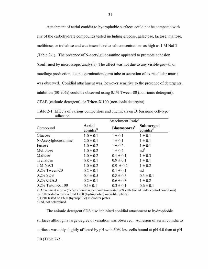
shown). These experiments demonstrated: (1) that aerial conidia were able to bind to
hydrophobic, but not hydrophilic surfaces (Fig. 2-3), (2) blastospores bound uniformly to
hydrophilic surfaces, but bound poorly to hydrophobic surfaces, forming small clumps on
the latter (Fig. 2-4), and (3) submerged conidia bound equally well to both hydrophilic
and hydrophobic surfaces, forming large clumps that appeared more evenly distributed
over time (Fig. 2-5). These patterns were identical between FITC-labeled and unlabeled
cells (data not shown).
For the quantitative assays, three types of black polystyrene based microtitre plates
with differing surface characteristics were used as substrata: (1) siliconized Fluorotrac-
F200, highly hydrophobic, (2) F200 untreated polystyrene surface, weakly polar, and (3)
F600, treated polystyrene, hydrophilic, polar surface containing hydroxyl, carbonyl, and
amino groups with a small net negative charge. Dynamic leading edge water droplet
contact angle (c∠a) measurements of the three substrata agreed with their decreasing
hydrophobicity. The silinated F-200 plates displayed a c∠a = 104.7o, the untreated F-200
plates a c∠a = 95.6o, and the F-600 plates a c∠a = 85.6o (contact angles for cleaned
polished glass, the glass chamber slides, and silinated glass were determined to be: 73.1,
87.4, and 109.7, respectively).
27
Fig. 2-2. Fluorescent intensity of FITC labeled aerial conidia (ζ), blastospores (µ), and submerged conidia (λ) as a function of cell number.
Fig. 2-3. Adhesion of aerial conidia to glass (A1, A2, A3) and silinated glass (B1, B2, B3) surfaces after 5 min (A1, B1), 4 hr (A2, B2), and 24 hr (A3, B3) incubation on substrata. Bar = 30 µm.

28
Fig. 2-4. Adhesion of blastospores to glass (A1, A2, A3) and silinated glass (B1, B2, B3) surfaces after 5 min (A1, B1), 4 hr (A2, B2), and 24 hr (A3, B3) incubation on substrata. Bar = 30 µm.
Fig. 2-5. Adhesion of submerged conidia to glass (A1, A2, A3) and silinated glass (B1, B2, B3) surfaces after 5 min (A1, B1), 4 hr (A2, B2), and 24 hr (A3, B3) incubation on substrata. Bar = 30 µm.
The number of binding sites per microtiter plate well was estimated at 4-8 x 10⁵
cells as determined by the saturation point derived from plots of the percent cell bound as
a function of cell concentration (Fig. 2-6). These data indicated that the linear range of

29
each cell type was similar, although the saturation point for each cell type varied from
approximately 25% of the aerial conidial cells able to bind per well, to greater than 70%
of the submerged conidia bound in wells using ~5 x 10⁵ cells/well. All subsequent
experiments were performed using cell concentrations within the linear range of the
attachment curve (2-5 x 10⁵ cells/well) (Fig. 2-6).
Quantitative adhesion assays were performed using aerial conidia, blastospores,
and submerged conidia on hydrophobic, weakly polar, and hydrophilic surfaces with
either 1 or 3 washes (Fig. 2-7). Aerial conidia bound rapidly and tightly to hydrophobic
surfaces, with no loss of cell binding by up to 10 washes with buffer (data not shown).
Aerial conidia bound poorly to weakly polar surfaces even after prolonged (24 hr)
exposure to the substrata. Interestingly, these cells bound weakly to hydrophilic surfaces
and were readily washed off indicating that this binding process might be biphasic, with
initial weak electrostatic binding.
Fig. 2-6. Saturation point of binding sites for B. bassiana aerial conidia (λ), blastospores (ν), and submerged conidia (O) to microtiter plates
bernie04
Oval
bernie04
Rectangle

30
In contrast, blastospores bound poorly to hydrophobic surfaces with no greater than
10% of the cells bound even after 24 hr. Blastopsores bound moderately to weakly polar
surfaces, with approximately 1-2 x 10⁵ cells bound/well (30% of 5 x 10⁵ cells used in the
assay) after 4 hr incubation. Blastospores bound more readily to hydrophilic surfaces,
with up to 50% of the cells bound within 30 min. Submerged conidia displayed the
broadest binding characteristics, adhering to all three surfaces, although with slightly
differing kinetics. On hydrophobic, weakly polar, as well as hydrophilic surfaces up to
60% of the cells used bound to the substrata within 4 hr, although in the case of the
hydrophobic and weakly polar surfaces, almost half of the bound cells could be removed
using three washes.
Fig. 2-7. Quantitative adhesion and influence of washing on adhesion of aerial conidia,
blastospores, and submerged conidia to silinated F-200 (hydrophobic), F-200 (weakly polar), and F-600 (hydrophilic) microtiter plates. Dark violet bars represent a single wash; maroon bars represent a triple wash.
31
Attachment of aerial conidia to hydrophobic surfaces could not be competed with
any of the carbohydrate compounds tested including glucose, galactose, lactose, maltose,
melibiose, or trehalose and was insensitive to salt concentrations as high as 1 M NaCl
(Table 2-1). The presence of N-acetylglucosamine appeared to promote adhesion
(confirmed by microscopic analysis). The affect was not due to any visible growth or
mucilage production, i.e. no germination/germ tube or secretion of extracellular matrix
was observed. Conidial attachment was, however sensitive to the presence of detergents,
inhibition (80-90%) could be observed using 0.1% Tween-80 (non-ionic detergent),
CTAB (cationic detergent), or Triton-X 100 (non-ionic detergent).
Table 2-1. Effects of various competitors and chemicals on B. bassiana cell-type adhesion
Attachment Ratioa
Compound Aerial conidiab Blastosporesc Submerged
conidiac
Glucose 1.0 ± 0.1 1 ± 0.1 1 ± 0.1 N-Acetylglucosamine 2.0 ± 0.1 1 ± 0.1 1 ± 0.1 Fucose 1.0 ± 0.2 1 ± 0.2 1 ± 0.1 Melibiose 1.0 ± 0.2 1 ± 0.2 ndd
Maltose 1.0 ± 0.2 0.1 ± 0.1 1 ± 0.3 Trehalose 0.8 ± 0.1 0.9 ± 0.1 1 ± 0.1 1 M NaCl 1.0 ± 0.2 0.9 ± 0.2 1 ± 0.2 0.2% Tween-20 0.2 ± 0.1 0.1 ± 0.1 nd 0.2% SDS 0.4 ± 0.5 0.8 ± 0.3 0.3 ± 0.1 0.2% CTAB 0.2 ± 0.1 0.6 ± 0.3 1 ± 0.2 0.2% Triton-X 100 0.1± 0.1 0.3 ± 0.1 0.6 ± 0.1 a) Attachment ratio = (% cells bound under condition tested)/(% cells bound under control conditions) b) Cells tested on siliconized F200 (hydrophobic) microtiter plates. c) Cells tested on F600 (hydrophilic) microtiter plates. d) nd, not determined
The anionic detergent SDS also inhibited conidial attachment to hydrophobic
surfaces although a large degree of variation was observed. Adhesion of aerial conidia to
surfaces was only slightly affected by pH with 30% less cells bound at pH 4.0 than at pH
7.0 (Table 2-2).
32
A unique feature of blastospore attachment was that adhesion could be competed
with maltose (Table 2-1). No other sugar tested had any affect on blastospore adhesion
nor did maltose affect conidial or submerged conidial adhesion. Blastospore attachment
was insensitive to salt (NaCl), SDS and CTAB, but was inhibited by Tween-20 and
Triton X-100. In contrast to the other cell types, adhesion of blastospores appeared to be
pH dependent with a 50% decrease in the number of cells bound when assays were
preformed at pH values of 4-5 compared to pH 7-8 (Table 2-2). Submerged conidia
behaved similar to aerial conidia except that N-acetylglucosamine did not increase the
number of cells bound and the presence of the detergents CTAB and Triton X-100 (Table
1) and changes in pH had only a minor affect on submerged conidial adhesion.
Table 2-2. Effect of pH on B. bassiana cell-type adhesion Attachment Ratioa
pH Aerial conidiab Blastosporesc Submerged
conidiac
4 0.7 ± 0.1 0.5 ± 0.3 0.8 ± 0.1 5 0.8 ± 0.1 0.5 ± 0.3 0.9 ± 0.1 6 0.8 ± 0.2 0.6 ± 0.4 0.8 ± 0.1 7 1.0 ± 0.2 0.9 ± 0.2 0.9 ± 0.3 8 1.0 ± 0.2 1.0 ± 0.1 0.9 ± 0.3
a) Attachment ratio = (% cells bound under condition tested)/(% cells bound under control conditions) b) Cells tested on siliconized F200 (hydrophobic) microtiter plates. c) Cells tested on F600 (hydrophilic) microtiter plates. d) nd, not determined
Removal of carbohydrates (maltose, glucose or glucuronic acid) from the cell
surface of aerial conidia using either α-amylase or laminarinase, but not cellulase resulted
in decreased conidial adhesion to hydrophobic surfaces but had no affect on conidial
adhesion to hydrophilic surfaces (Table 2-3). Treatment of blastospores with
glycosidases appeared to either slightly promote adhesion (α-amylase and to a lesser
extent cellulase treatment) to hydrophilic surfaces or not affect adhesion (laminarinase

33
and/or hydrophobic surface). Glycosidase treatment of submerged conidia resulted in a
25-50% decrease in adhesion to hydrophilic surfaces and hydrophobic surfaces. The only
other remarkable difference was noted in the adhesion of α-amylase, treated cells to
hydrophobic surfaces where a large variation in adhesion was noted.
Some differential effects were observed by protease treatment of the cell types
(Table 2-3). Aerial conidia treated with Pronase E displayed a greater than 50% loss of
adhesion to hydrophobic surfaces but no loss in adhesion to hydrophilic surfaces,
although a large variation was observed. This variation was between experiments, i.e.
different cell batches treated with the enzyme and may reflect surface heterogeneity or
accessibility of target substrates to the enzyme. Protease K treatment of aerial conidia
did not result in appreciable changes in adhesion. Similar treatment of blastspores with
proteases had no effect or resulted in an almost 2-fold apparent increase in adhesion.
Microscopic analysis (i.e. visual counting) of the number of cells bound indicated that
there did not appear to be an actual increase in the number of cells bound and instead
protease treatment appeared to increase the fluorescence intensity signal.
Table 2-3. Effects of various enzymatic treatments on B. bassiana cell-type adhesion Attachment Ratioa
Enzyme Aerial conidia hydrophobicb hydrophilicc
Blastospores
hydrophobic hydrophilic Submerged conidia hydrophobic hydrophilic
α-amylase 0.3 ± 0.1 1.0 ± 0.3 1.2 ± 0.2 1.5 ± 0.15 1.4 ± 0.2 0.5 ± 0.2 Cellulase 0.8 ± 0.2 1.1 ± 0.2 1.0 ± 0.7 1.3 ± 0.05 0.4 ± 0.2 0.6 ± 0.2 Laminarinase 0.3 ± 0.1 0.9 ± 0.3 0.9 ± 0.1 1.0 ± 0.2 0.4 ± 0.2 0.8 ± 0.2 Pronase E 0.4 ± 0.3 1.2 ± 0.3 0.9 ± 0.1 ndd 1.1 ± 0.2 1.1 ± .1 Protease K 0.9 ± 0.2 0.8 ± 0.1 1.0 ± 0.1 nd d ndd 1.0 ± 0.2
a) Attachment ratio = (% cells bound under condition tested)/(% cells bound under control conditions) b) Cells tested on siliconized F200 (hydrophobic) microtiter plates. c) Cells tested on F600 (hydrophilic) microtiter plates. d) Apparent increase (see text for details).

34
These were the only conditions tested in which the fluorescent signal was affected
by the treatment. In all other experiments, microscopic analysis was in agreement with
the fluorescent intensity measurements. Finally, little to no effect was observed for the
adhesion properties of protease treated submerged conidia.
Discussion
Conidial adhesion has been examined in a number of plant and insect pathogenic
fungi (9, 65). Adhesion of entomopathogenic fungi appears to involve an initial binding
interaction followed by a consolidation step, resulting in firm attachment to the cuticle (9,
33, 34). Similarly, studies on the phytopathogenic fungus Botrytis cinerea revealed a
two-stage adhesion process; immediate adhesion occurring upon hydration and
characterized by relatively weak attachment and a stronger delayed adhesion observed as
the conidia germinate (25, 26). Immediate adhesion of B. cinerea is passive
(nonmetabolic) and although no specific structures were visible on the conidia, adhesion
is characterized as dependent (in part) on hydrophobic interactions. Hydrophobic
interactions have also been implicated in the attachment of conidia of the insect-
pathogenic fungi Nomuraea rileyi, Metarhizium anisopliae, and B. bassiana to both host
and non-host cuticle preparations (9).
B. bassiana produces at least three single cell types that can be distinguished based
upon morphological and adhesive characteristics. Experiments qalitatively assessing
entomopathogenic fungal adhesion to various surfaces including insect cuticles (9, 10, 25,
26, 42) have almost exclusively addressed conidial binding to surfaces and have not
examined the adhesion properties of either blastospores or submerged conidia. Using a
quantitative adhesion assay our results demonstrate complex interactions between various
cell types and substrata with different surface properties. All three B. bassiana single cell

35
types studied (aerial conidia, blastospores, and submerged conidia) displayed different
adhesion properties that appeared to be mediated by different cell-specific mechanisms.
B. bassiana aerial conidia were able to bind both hydrophobic and hydrophilic
surfaces although adhesion to the latter was weak and the cells could readily be washed
off. Aerial conidia binding to hydrophobic surfaces could not be competed with any
carbohydrate tested, although addition of N-acetylglucosamine, the monomeric
constituent of chitin (the major carbohydrate polymer found in arthropod cuticles)
appeared to increase adhesion. The hydrophobic nature of B. bassiana conidial spores, as
well as those from other entomopathogens such as N. rileyi, M. anisopliae, and
Paecilomyces fumsoroseus, was correlated with the presence of an outer cell layer
comprised of rodlets or fascicles as visualized by electron microscopy boucias (9, 10).
These rodlet layers are presumably formed by assembly of specific proteins termed
hydrophobins, which in turn are thought to passively mediate adhesion to hydrophobic
surfaces (92, 96). Although our results are consistent with this model, they also indicate
that the interaction of aerial conidia with hydrophobic surfaces may be more complex.
Since amylase and laminarinase as well as protease treatments reduced adhesion (but
displayed no discernable effects on the rodlet layer, Kirkland, Holder, & Keyhani,
unpublished results), both carbohydrates on the cell surface as well as proteins may be
involved in mediating adhesion of aerial conidia.
In contrast, blastospores, cells lacking a visible rodlet layer (9, 10), bound poorly to
hydrophobic surfaces forming small aggregates or clumps, but displayed high binding to
hydrophilic substrata. Blastospores were also able to bind to weakly polar substrata,
although a greater incubation time compared to hydrophilic substrata was required.
36
Intriguingly, blastospore attachment could be specifically competed with maltose. No
other carbohydrate tested appeared to compete with adhesion of blastospores or the other
fungal cell types including trehalose the major carbohydrate constituent found in insect
hemolymph. In-vivo generated blastospores (distinct but similar to the rich broth
produced blastospores), produced during fungal proliferation in the insect hemolymph
after penetration of the cuticle, are able to evade recognition by insect hemocytes and
display altered membrane characteristics (48, 49, 70). However, the physiological
significance of potential maltose inhibition of adhesion of these cells is unclear.
Submerged conidia displayed the broadest binding characteristics of the B.
bassiana single cell types, capable of efficiently binding hydrophobic, weakly polar, as
well as hydrophilic surfaces. Spore tip or mucilage-covered appendages and adhesive
knobs have been implicated to mediate conidial adhesion of several fungi (8, 45, 85).
The mucosal coat of nematophagous fungi not only appears to mediate adhesion but is
attractive to host insects. A wide variety of arthropod mycopathogens appear to produce
exocellular mucilage during germ tube or appressorial formation (9). Similarly, the
hydrophilic nature of conidia of the Entomophthorales is thought to be mediated by a
mucilaginous coat released upon attachment to cuticle surfaces and that acts as a glue
mediating attachment (30, 57). Although blastospores and submerged conidia attached to
hydrophilic surfaces, no obvious mucilaginous coat was visible in either cell type. In
addidtion, scanning electron microscopy did not reveal any specific structures in conidia,
blastospores, or submerged conidia of B. bassiana that are predicted to be involved in
mediating adhesion (unpublished data). It is possible, however that extracellular matrix

37
components or mucilage located between the inner and outer wall that is not readily
detectable could be involved in mediating adhesion.
Although aerial conidia are considered easily dispersible via air currents, and are
more resistant to adverse environmental conditions such as desiccation and extreme
temperatures, submerged conidiation and blastospore formation may occur under a
variety of environmental conditions as well as during the host-pathogen interaction. The
ability to produce multiple cell types, with differing adhesive properties, may occur in
response to specific environmental conditions.
This may allow fungal cells to bind to a broad range of host targets and provide the
fungus with a means to adapt to substrata conditions (33, 34). It is unlikely, however that
alteration in adhesion can account for emergence of restricted host-range B. bassiana
strains, since these strains may have altered (cuticle degrading) enzyme production or
may be unable to penetrate and/or respond to surface cues of certain hosts, but may still
retain the means to initiate binding or adhesion interactions. Indeed, there is some
evidence that when entomopathogens specialize they loose structures rather than gain
them, It would be interesting to see whether there is any alteration in the adhesion
kinetics of general and specialized strains of B. bassiana.
Our data indicates that certain practical considerations should be taken into account
during application of B. bassiana. For instance, if blastospores were to be used,
formulations should probably avoid hydrophobic solutions. Instead aqueous or polar
liquids are recommended. Such formulations may prove to be more successful in the
biocontrol of certain hosts as compared to aerial conidia.
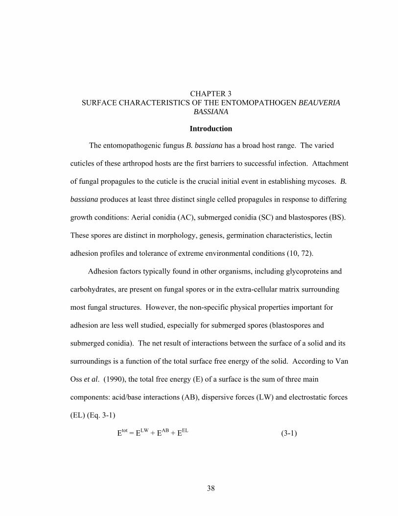
38
CHAPTER 3 SURFACE CHARACTERISTICS OF THE ENTOMOPATHOGEN BEAUVERIA
BASSIANA
Introduction
The entomopathogenic fungus B. bassiana has a broad host range. The varied
cuticles of these arthropod hosts are the first barriers to successful infection. Attachment
of fungal propagules to the cuticle is the crucial initial event in establishing mycoses. B.
bassiana produces at least three distinct single celled propagules in response to differing
growth conditions: Aerial conidia (AC), submerged conidia (SC) and blastospores (BS).
These spores are distinct in morphology, genesis, germination characteristics, lectin
adhesion profiles and tolerance of extreme environmental conditions (10, 72).
Adhesion factors typically found in other organisms, including glycoproteins and
carbohydrates, are present on fungal spores or in the extra-cellular matrix surrounding
most fungal structures. However, the non-specific physical properties important for
adhesion are less well studied, especially for submerged spores (blastospores and
submerged conidia). The net result of interactions between the surface of a solid and its
surroundings is a function of the total surface free energy of the solid. According to Van
Oss et al. (1990), the total free energy (E) of a surface is the sum of three main
components: acid/base interactions (AB), dispersive forces (LW) and electrostatic forces
(EL) (Eq. 3-1)
Etot = ELW + EAB + EEL (3-1)

39
Dispersive forces, acid/base interactions, and hydrophobic interactions can be
calculated from contact angle data and by using Young’s equation, whereas the effective
surface charge, or zeta potential, can be derived from the mobility (µ) of cells in an
electric field.
Spores with rodlet layers or rough surfaces are more hydrophobic than smooth
spores (39, 97). In our experiments atomic force microscopy (AFM) was used to
generate high resolution images of the surfaces of the three spore types produced by B.
bassiana. AFM is an example of proximity probe microscopy, where a cantilever, placed
close the surface of the sample, is scanned in a raster (back and forth in an ordered
manner) pattern to measure physical features such as height, elasticity, or hardness. A
laser directed towards the cantilever is deflected, and the deflections are recorded on a
positron-sensitive photodiode. As the probe follows the contours of the sample, it is
attracted, or repulsed by it. The instrument measures the force of these interactions, and
renders the output graphically. The resolution of the instrument, which depends on the
size of the probe, ranges from 10 pm to 125 µm; the vertical limit of the sample is 5 µm.
In addition to high-resolution cell surface imaging, AFM can also generate data
about surface biophysical properties. Two basic types of images are generated, height
and deflection. The former gives accurate information about cell surface roughness and
size, while the latter generates images with higher resolution for viewing ultra-structural
details. In addition to these images, AFM can be used to generate force curves that can
be used to measure local physical properties such as adhesion and elasticity.
AFM images can be correlated to electrostatic, or hydrophobic characteristics of the
different spore types. The hydrophobic character of the spores can be determined

40
indirectly, by observing how well the spores attach to different surfaces, or directly, by
quantitatively measuring hydrophobic indicies. Past research has shown that different
methodologies used to evaluate cell surface hydrophobicity do not always correlate well
with each other, and can yield statistically significant differences for similar cells even
from a single strain (2, 29, 46).
Phase exclusion assays, specifically microbial adhesion to hydrocarbons (MATH)
and contact angles, have been used in a number of biological systems to obtain
quantifiable hydrophobic indices. The advantages of this system include ease of use,
reasonable reproducibility, extensive use in the literature, and no requirement for costly
equipment. However other factors, including electrostatic interactions, temperature, and
pH, can affect the results of these experiments. Contact angle (CA) data is affected, to a
lesser degree than MATH assays by similar factors and by the requirement, when using
specific equipment that the cells remain flat, and are dry.
The advantage of CA data is the multitude of information that can be generated
from one set of data. To obtain contact angle measurements, a drop of liquid is deposited
onto a given solid an image of the drop is projected onto a screen and a tangent is drawn
close to the drop’s surface, at the liquid/solid/gas interphase. The angle at the
solid/liquid/gas interface (θ) is called the contact angle (CA) (Fig. 3-1).
Fig. 3-1. Contact angles (θ) are formed at the interface between the liquid, solid and gas
boundaries of a droplet on the surface of interest.
θ
Solid
Liquid
Gas

41
The contact angle (θ) is a measure of how well a liquid wets a surface. An angle of
00 indicates complete wetting, while an angle of 1800 indicates absolute non-wetting.
More generally when contact angles are < 900 the liquid is able to wet the surface to some
degree, and the surface is hydrophilic. Angles > 900 indicate that the surface is
hydrophobic. At 900 the surface energy of the solid is equal to the cohesive energy of
water. The surface energy parameters of the solid can be determined if multiple, fully
characterized liquids are used to obtain contact angles.
According to van Oss et al. (1990), this information can be used to obtain solid-
liquid-solid interfacial energy (∆Giwi), a measure of how quickly the surfaces will
aggregate in liquids, when the solute is water; this is a measure of the hydrophobic nature
of the surface. The surface energy parameters for the solid of interest are used to
calculate ∆Giwi by applying Young’s equation (Eq. 3-2) to the contact angles of multiple,
well-defined liquids present on its surface.
ϒSV – ϒSL = ϒVL cosθ (3−2)
Other surface energy parameters, such as the work of adhesion, the energy
associated with the adhesion of a solid and a liquid, also defined as the work required to
separate them (Eq. 3-2). The work of cohesion, the energy required to separate a liquid
into two parts, can also be calculated from contact angle data using the following
equations (Eq. 3-3 & 3-4).
Wa = ϒ (1 + cos θ) (3-3)
Wc = 2ϒ (3−4)
Where, Wa = work of adhesion, Wc = work of cohesion ϒ = surface tension (liquid)/surface energy (solid)

42
It is no longer acceptable to use a single, static contact angle measurement to
determine surface energy properties, or wet-ability. Dynamic contact angles are
measured either while the drop is actively moving (advancing and receding angles), by
gradually increasing the volume of the drop, or by tilting the surface until the drop moves
while continously measuring the advanced and receded angle. The advanced and receded
angles are defined as the angles obtained just prior to movement of the droplet. The
advanced/advancing angle is the angle that is formed just as the drop is about to move
over a dry surface. The receded/receding angle is the angle that is formed over already
wet surfaces. The advanced angle is less affected by hysteresis than the receded angle, so
it is the one most often reported (31, 86, 87).
Hysteresis, the difference between the advanced/advancing and the
receded/receding angles, is affected by surface homogeneities larger than approximately
10 nm and by hydrophilic and hydrophobic microenvironments on solid surfaces.
Hydrophobic micro domains are thought to have a breaking effect on the advanced/ing
angle, while hydrophilic domains appear to have a retarding effect on the receded/ing
angle. Sample roughness at the microscopic level increases hysteresis and has an effect
on hydrophobicity. Rough surfaces, that are otherwise identical to smooth surfaces, tend
to be more hydrophobic, especially if they already have hydrophobic characteristics
hysteresis (56).
While acid/base, dispersive and hydrophobic interactions can be calculated from
contact angle data, zeta potentials are the most effective way of determining the
electrostatic contribution. The movement of cells in an electrical field is directed by the
potential at the boundary between the cell and the ions tightly associated with it and the

43
surrounding medium. This potential is known as the zeta potential, and is a function of
the cell’s surface charge, absorbed layers on the surface of the cell and the medium in
which the cell is suspended. It is usually the same sign as the actual charge at the surface
of the cell. The zeta potential is determined by measuring the movement of the cells in
an electric field, or the particle mobility (velocity under unit field strength). There is a
simple relationship between particle mobility (µ) and zeta potential (in water @ 25oC
zeta potential = 12.85 µ).
Materials and Methods
Cultivation of Fungal Cells
B. bassiana (ATCC 90517) was routinely grown on potato dextrose agar (PDA).
Plates were incubated at 26o C for 10-15 days, and aerial conidia were harvested by
flooding the plate with sterile dH20. Conidial suspensions were filtered through several
layers of Mira, cloth and the final spore concentrations were determined by direct count
using a hemocytometer. Blastospores and submerged conidia were grown by inoculating
conidia harvested from plates to a final concentration of 0.5 to 5 x 10⁵ conidia/ml into
DifcoTM Sabouraud dextrose (Becton, Dickinson and Co., MD) with 1-2 % yeast extract
(SDY) and TK1 broth (83), with fructose as the carbon source, respectively.
Cultures, grown for 3 to 5 days at 26oC with aeration (150-200 rpm), were filtered
(2X) through glass wool to remove mycelia. Final filtered suspensions of the three cell
types were harvested by centrifugation (10,000 x g, 15 min, 40oC), washed twice with
sterile dH20, and resuspended to the desired concentrations in appropriate buffers. Pre-
centrifugation, post-centrifugation, and final spore concentrations were confirmed by
direct cell count (106-9 cells/ml).

44
Atomic Force Microscopy
Atomic force migrographs were generated using Digital Instruments’ Multimode
SPM Atomic force Microscope with a silica nitrate tipped cantilever. Data from the
micrographs was analyzed using Nanoscope SPM v.4.4 (Digital Images) and SPM Image
Magic demo v1.10 (Alexander Kryzhanosvsky, A.F. Loffe physio-technical institute, St.
Petersburg, Russia).
Zeta Potential
Aerial conidia, blastospores and submerged conidia were washed (3X) in water.
The rinsed pellets were resuspended in 1mM KCl (pH 3-10, adjusted just prior to running
the samples) to a concentration of 106 cfu/ml within 1 hr of running the samples on a
Zetaplus micro-electrophoretic unit (Brookhaven Instruments Cooperation) with native
software (PALS). Each cell type was run in triplicate. Ten microphoretic readings were
run for each triplicate for a total of 30 readings for each sample. The readings for the
triplicate runs were averaged, and the standard error of the mean was determined for each
sample.
MATH Assay
Cell surface hydrophobicity was determined essentially as described by Thomas et
al. (1986). Briefly aerial conidia, blastospores, and submerged conidia were washed into
PUM buffer (1.0 M sodium phosphate-urea-magnesium, pH 7.1). Fungal cell
suspensions were adjusted to an OD470 = 0.4 and dispensed (3 ml) into acid washed glass
tubes (12 x 75 mm). Hexadecane (300 µl) was added, each tube was vortexed (3 x 30 s)
and allowed to incubated at RT for 15 min before the hexadecane phase was carefully
removed and discarded. Tubes were cooled to 5oC and residual solidified hexadecane
was removed. The tubes were warmed to RT, and the absorbencies of the resultant cell

45
suspensions were determined at 470 nm. The hydrophobic index (HI) was calculated
using the following equation (Eq. 3-5):
HI = (initial OD470 – final OD470)/initial OD470 (3-5)
Contact Angle Determination
Contact angle measurements of the B. bassiana cell types were determined from
digital images of drops obtained using a Ramé-hart Instruments Co. (NJ) Model 500
Advanced goinometer (automated drop dispenser, tilting plate, digital camera, native
Dropimage Advanced software). Advancing angle measurements were determined just
prior to movement of the water drop. Briefly, a 10 µl drop of solution was placed onto
the surface of the substrata to be tested. The stage and the camera were tilted at 10
increments until the drop began to move. The leading edge (dynamic) contact angle was
determined as the angle obtained just prior to drop movement.
Interfacial energies of interaction were calculated using the Acid/Base tool of the
Dropimage Advance software. Water (polar), α-bromonapthalene (non-polar) and
glycerol (polar) were used to determine the free surface energy components of the solid.
Results
Atomic Force Microscopy
Fascicle bundles (a rodlet layer) were clearly visible on B. bassiana aerial conidia,
but were absent from both blastospores and submerged conidia (Fig 3-2.) The
blastospore surface appeared smooth, whereas the submerged conidial surface was rough
and a raised lip was apparent on most of the latter cells. No fascicles were visible on the
germ tubes of germinating aerial conidia (data not shown).

46
A B C
Fig. 3-2. Atomic force micrographs of B. bassiana A) conidia, B) submerged conidia and
C) blastospores. (bar A) 10 nm, B) 0.1 µm and C) 0.5 µm)
Zeta Potential
The surface charge of the three cell types decreased as the pH increased. The
isoelectric point for all cell types was PI4, except 16-day-old conidia, which was PI5.
Mean zeta potential values for aerial conidia, blastospores, and submerged conidia were
obtained over a pH range from 3-9 (Fig. 3-2). These values were obtained for a
minimum of 3 different samples; each sample was run at least 10 times. Aerial conidia
(16 and 20-day-old combined) had a positive zeta potential (22 ± 2 mV) that rapidly
became negative by pH 4-4.5, reaching a net negative surface charge of – 47 ± 4 mV at
pH 6-7. Blastospores and submerged conidia also displayed a net positive zeta potential
at low pH that decreased to – 4 ± 4 mV and – 13 ± 2 mV at pH 6-7, respectively (Fig. 4-
3).
MATH Assay
Hydrophobicity indices (HI) indicated that almost 90% (HI= 0.88) of the aerial
conidia partitioned into the hydrocarbon layer. Submerged conidia partitioned at an
intermediate level (HI = 0.7), and at least 60% of the blastospores (HI = 0.4) partitioned
preferentially in the aqueous phase. These values can be interpreted as relative
hydrophobicity. However, absolute determination of which cell types are definitely
hydrophobic and which are hydrophilic are not possible and the cutoff is often set
arbitrarily at 70% (HI = 0.7) (66).
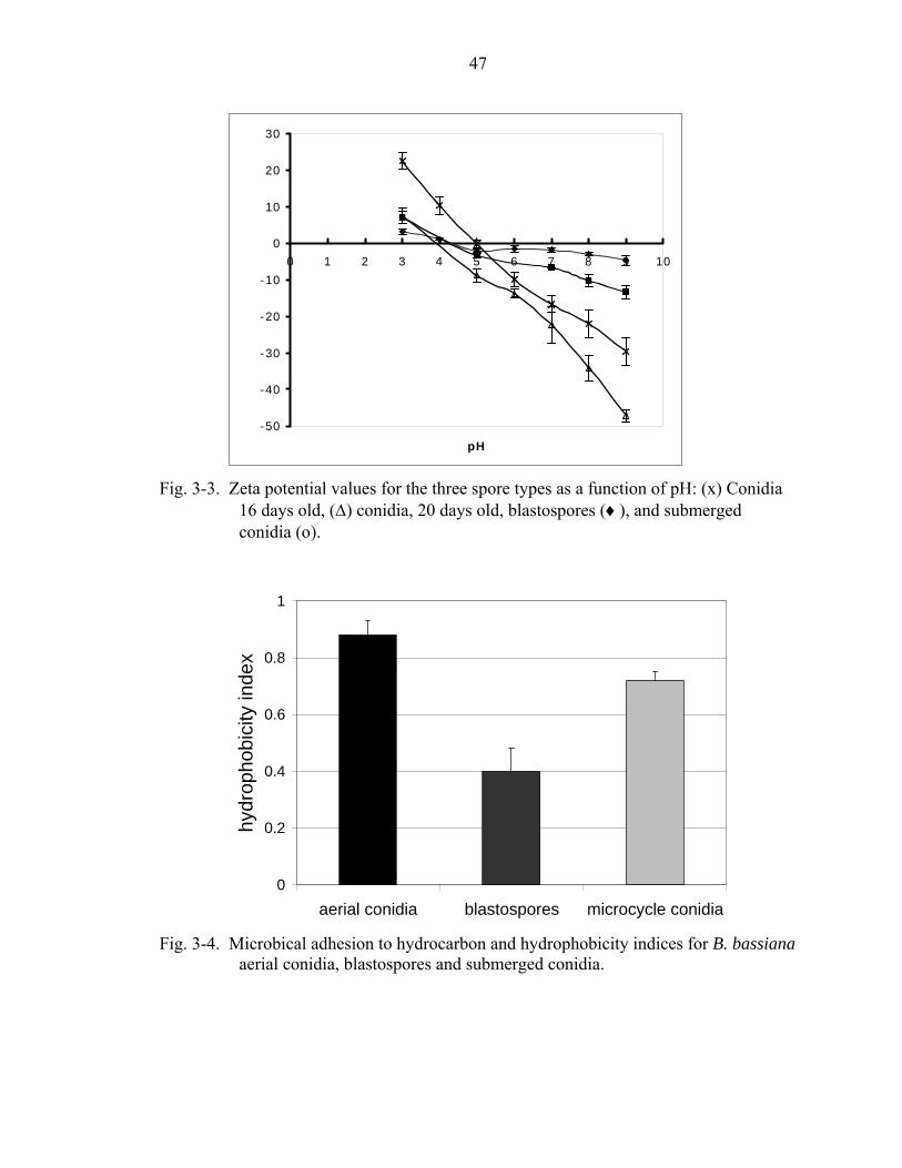
47
-50
-40
-30
-20
-10
0
10
20
30
0 1 2 3 4 5 6 7 8 9 10
pH
Fig. 3-3. Zeta potential values for the three spore types as a function of pH: (x) Conidia
16 days old, (∆) conidia, 20 days old, blastospores (♦), and submerged conidia (o).
0
0.2
0.4
0.6
0.8
1
aerial conidia blastospores microcycle conidia
hydr
opho
bici
ty in
dex
Fig. 3-4. Microbical adhesion to hydrocarbon and hydrophobicity indices for B. bassiana
aerial conidia, blastospores and submerged conidia.
bernie04
Rectangle

48
Contact Angles
A surface is considered wettable if the liquid tested forms angles smaller than 90o.
Water contact angles for the conidia of all three B. bassiana phenotypes were well over
90o (Table 3-1), while that of the submerged conidia, and the blastospores were
consistently below 900.
The propensity of two surfaces to adhere to each other in water is calculated by the
interfacial energy of interaction (∆GiwiIF) between the surfaces (in this case the surfaces
of two similar spores) in water. The original parental 90517 appeared to have two
distinct phenotypes, which once separated bred true (Puff, 90517p and Blue, 90517b).
The ∆GiwiIF of conidia, regardless of phenotype, was negative (90517 –34.8 mJ/m2,
90517p –22.3 mJ/m2, and 90517b –22.8 mJ/m2), indicating that these spores are
hydrophobic; whereas the submerged conidia and the blastospores, had ∆GiwiIF values
greater than 0 mJ/m2 (30.2 mJ/m2 to 46 mJ/m2 and for submerged conidia and 66.3 mJ/m2
to 75.2 mJ/m2 for blastospores) indicating that they were hydrophilic (Table 3-2).
The values for acid/base component of the interfacial energy, ∆GiwiAB for conidial
cells ranged from 23.9 mJ/m2 to 69.4 mJ/m2, are were below +102 mJ/m2 (∆GwAB)which
means they would be unable to overcome the cohesive energy of water dissolve in water.
The values for blastospores and submerged conidia were all greater than +102 mJ/m2;
specifically, 177.4 mJ/m2 to 191.6 mJ/m2 for the blastospores and, 171.5 mJ/m2 to 212
mJ/m2 for the submerged conidia indicating that these cells tend to disperse in water and
are therefore hydrophilic. For the ∆GiwiAB value to be greater than 102 mJ/m2, the
electron donor surface energy parameter (ϒS+) must be much lower than 25.5 mJ/m2 (ϒL
+
for water), often around zero, and the proton acceptor surface energy parameter (ϒL-)

49
must be much larger than 25.5 mJ/m2 (ϒL- for water). This was found to be true for the
blastospores and mycrocycle conidia, but not for the aerial conidia (Table 3-2).
Discussion
The surface properties of the three B. bassiana cell types, as measured by AFM,
contact angles, and the MATH assay, were markedly different. Freeze-etch, carbon-
platinum transmission electron microscopy has previously shown the presence of a
conidial rodlet layer on the dry aerial conidia of many hyphomycetes, including
Beauveria bassiana (5, 68, 97). In this dissertation Atomic Force Microscopy confirmed
the presence of this layer on the surface of our strain of B. bassiana. Aerial conidia were
shown to be highly hydrophobic, especially when compared to the other spore types
investigated.
Table 3-1. Contact angle values for the three cell types for water, bromonapthalene and glycerol with calculated interfacial and polar free energy values. Cell type mean Contact anglea Hydrophobicityb
Water α-Bromonapthalene Glycerol ∆GiwiAB ∆Giwi
IF 90517 Aerial Conidia 120.3±2.5c 66.2±0.5 112.7±0.9 47.2 -34.8 Blastospores 23.25±1.8 33.6±0.4 59.4±1.8 187.4 74.4 Submerged conidia 30.6±1.0 56.4±0.8 53.6±1.1 167.1 46.0 90517pd
Aerial Conidia 122.1± 0.6 47.2±0.6 108.2±1.3 49.3 -23.3 Blastospores 33.2±1.0 33.6±1.4 61.4±0.4 171.2 66.3 Submergd conidia 28.3±1.3 29.5±0.4 42.3±1.7 152.8 36.5 90517b Aerial Conidia 119.3±0.7 42.6±0.4 122.0±1.4 50.0 -23.8 Blastospores 23.9±0.8 39.5±0.5 68.9±0.9 183.7 75.2 Submerged conidia 28.6±1.0 38.8 ±2.7 40.8±1.7 147.3 30.2
a) Mean of advanced contact angles n = 2<x>8 different samples (>2 drops were used per sample) b) AB = acid/base, IF= interfacial energy. c) Advanced/receded angles were measured just before the drop started to roll after the stage was
tilted d) p (puff) and b (blue) two stable phenotypes, derived from the parent strain (90517)

50
Table 3-2. Advancing and receding water contact angle data with calculated surface energy values for three B. bassiana single cell propagules.
Cell type Polar component
ϒsAB ϒs
LW ϒstotal ϒι
+ ϒι−
(Polar)a (Dispersive) 90517 Aerial Conidia 9.7± 1.5 33.3±0.6 43.0±1.1 4.3±0.4 5.5±1.5 Blastospores 10.8±2.3 23.6±0.3 34.4±1.3 0.4±0.2 86.1±3.7 Submerged conidia 23.3±1.1 16.3±0.4 39.6±0.8 2.0±0.3 68.4±2.0 90517p Aerial Conidia 0.6± 0.01 33.3±0.3 33.9±0.1 9.4±0.5 6.0±0.6 Blastospores 3.2± 0.8 24.5±0.50 27.7±0.7 0.04±0.02 71.8±1.7 Submerged conidia 16.6±2.0 25.1±0.30 41.7±2.5 1.9±0.3 57.3±2.6 90517b Aerial Conidia 13.6±1.1 30.3±0.2 43.9±0.7 2.1±0.2 0.9±0.16 Blastospores 7.3± 1.4 21.7±0.6 29.0±1.0 0.16±0.1 82.7±2.5 Submerged conidia 24.5±0.9 24.7±0.4 49.2±0.8 2.8±0.4 53.2±1.7
a) AB = acid/base, s=solid, l= liquid gamma= surface energy components. b) p (puff) and b (blue) two individual phenotypes, genetically identical to the parent strain (90517).
In 1999, Jeffs et al. concluded that the hydrophilic spores had more electronegative
groups than the hydrophobic ones because amine modified, electropositive, polystyrene
beads attached better to hydrophilic aerial conidia than to hydrophobic aerial. Our results
suggest that the most hydrophobic spores, aerial conidia (based on zeta potential
measurements) have more electronegative groups than the hydrophilic ones, but that the
stronger, and thus predominant, interactions for these spores are hydrophobic and the
electronegativity of the spores probably has a limited on effect initial interactions,
although surface charge may be important for binding once the aerial conidia have made
contact.
Blastospores, the most hydrophillic spores, have fewer electronegative groups, and
electrostatic interactions appear to be the most important factors involved in surface
interactions for these cells. Submerged conidia are intermediate in both hydrophobicity
51
and electronegativity, indicating that for these cells both factors are likely to play an
important role in adhesion to solid surfaces, dispersion in liquids, and flocculation.
Previously, surface charge measurements of fungal spores often relied on indirect
techniques, or were carried out at a single pH value. While these experiments yielded
valuable information about the adhesion profiles of the spores, they often did not take
into account the interactions of competing and synergistic forces on the adhesion profiles.
As a result, indirect measurements of surface potential should not be the only criterion
used when making inferences about the actual electric potential at the cell surface of the
spores.
In these experiments, the electronegative character of aerial conidia increased
steadily with pH, reaching values as low as – 47 mJ/m2 at pH 8.0. St. Leger et al. (1989)
observed, that during fungal entomopathogenic infections, the pH of the insect cuticle
rises from 6.3 to 7.7 during the initial phases, indicating that conidia will be moderately
electronegative during the first stages of infection, but that they will become more
electronegative as the infection progresses.
Because hydrophobic interactions are stronger and act over much larger distances
than electrostatic forces, it is likely that the primary intermolecular interaction involved
in the initial adhesion process is hydrophobic in nature. It is equally probable that other
intermolecular interactions become relatively more important once the spore is in close
proximity with the insect cuticle. The variety of intermolecular forces are involved in the
adhesion process probably ensure that B. bassiana aerial conidia remain attached to the
cuticle until the germ tube penetrates, despite rapidly changing micro-environmental
conditions.

52
B. bassiana spores can vary widely in hydrophobicity, between strain as well as
within the same strain, depending on age of the cells and culture conditions. This
phenomena has been linked to possible specialization of the fungus towards different
strains (51). Rodlet layers, while important to the overall hydrophobicity of fungal
spores, are not the only cell surface features involved in hydrophobic interactions.
Removing these layers decreases, but does not eliminate the hydrophobic character of
Aspergillus fumigatus conidia, and has different effects on cell surface characteristics
such as surface charge and ability to bind to hydrocarbons on A. nidualns and A.
fumigatus (39).
Submerged conidia are similar in shape to aerial conidia, but are typically larger
and do not appear to posses a highly organized rodlet layer (9). These cells (when
imaged using AFM) appeared rough compared to blastospores whose surfaces appeared
smooth. Previous reports have shown that if these cells do posses a rodlet layer it is
highly disorganized (10, 83). Submerged conidia also had smaller absolute zeta potential
than aerial conidia at higher pH. Although the environmental condition that leads to the
formation of submerged conidia has not been well characterized, it has been observed
that these cells are able to initiate infection at rates equal to or higher than aerial conidia
(80, 83). Submerged conidia appear to be less tolerant to environmental stresses than
aerial conidia and may be a specialized strategy related to nutrient/host availability.
Blastospores, the smoothest of the three single cell propagules, did not posses a
rodlet layer and were the most hydrophillic of the spore types. Hyphal bodies present
inside the insect during infection are similar in shape and size to these spores but appear
to have much thinner cell walls than blastospores (71). The electrostatic properties of

53
blastospores did not vary as dramatically with pH as the other spore types, although
previously we have noticed that the binding of these cells to hydrophilic surfaces is
dependent on pH.
The ability of blastospores to attach under favorable conditions increased with
increasing pH until the isoelectric point was reached (around pH 4). Adhesion did not
however decrease with increasing pH >4.0 as expected, considering increased
electrostatic repulsion under these conditions. Instead, the adhesion rates continued to
increase as the ambient pH increased. As noted by Jeffs et al. (1999), this is probably to
due to other intermolecular factors stabilizing the adhesive process and minimizing the
electrostatic repulsion. This may also explain why these spores have moderate
electrostatic properties, compared with aerial conidia. Strong electronegative surface
charge and weak hydrophobicity may result in increased electrostatic repulsion leading to
decreased adhesion.
Both the MATH assay and contact angle data can be used to measure
hydrophobicity. However, they do not give exactly the same information. In the MATH
assay, the propensity of the spores to partition in one layer preferentially over another is
used to assess the hydrophobic nature of the spores. However, this assay does not take
into account other non-specific forces that may affect partitioning and as a result should
be used as a direct measure of the surface hydrophobicity of the cells. Contact angles can
give more precise determinations of the cell surface hydrophobicity (CSH); however the
cells are usually dry when tested and this needs be taken into account when considering
the results. Given these caveats, our results demonstrate that the three spore types are
very different in terms of their surface characteristics.

54
Previous studies had conflicting results regarding the hydrophobicity of submerged
conidia. Some reports noted that they were as hydrophobic as aerial conidia (83), but
Jeffs et al. (1999), using SAT, noted that blastospores were less hydrophobic than aerial
conidia. Our results appear to back up this last interpretation; however it should be noted
that there appears to be great heterogeneity between the cell surface characteristics of the
same type of spores from different B. bassiana strains. In addition, the hydrophobicity of
the spores can vary within a strain, depending on the age of the culture and the cultural
conditions.
This report also notes the importance of studying cell surface characteristics (CSC)
of any spore that might be considered for commercial applications, using a range of
biochemical parameters. The rational is that the microenvironment the spores are likely
to encounter on the surface of the insect cuticle may change rapidly as the infection
proceeds.
55
CHAPTER 4 SUPPRESSIVE SUBTRACTION HYBRIDIZATION ANALYSIS OF BEAUVERIA
BASSIANA GROWN ON INSECT CUTICLE
Introduction
Virulence Factors
Deuteromycete entomopathogenic fungi usually infect their arthropod hosts
through the cuticle. Infection is dependent on the fungus successfully adhering to and
penetrating through the host’s cuticle. Once inside the host the fungi must avoid being
destroyed by the immune response and in some cases have a means of exiting the host
and dispersing its spores into the environment so that the cycle can continue. The
molecular factors important to the infection process determine if an organism is
pathogenic and which hosts it can invade, these factors are collectively termed virulence
factors.
There are a number of definitions in the literature describing virulence factors.
Some of the most common of these definitions include: 1) components that when deleted
specifically impair virulence, but not the ability of the organism to grow, 2) products that
allow the organism to cause disease and 3) components of the pathogen that cause
damage to the host.
The factors involved in the pre-penetration and penetration steps are important for
initiation of infection, but are often redundant, or they work in tandem with other
virulence factors. This makes it difficult to define them as virulence factors under the
first definition because it might be difficult to show that virulence is decreased when

56
candidate genes are deleted. In the case of adhesion factors, while they are important in
initiating the infectious process, they often do not cause significant damage to the host
tissue, making it difficult to define under the third definition of virulence factors
requiring virulence factors to cause significant damage to host tissue.
The Edward Jenner institute for vaccine research lists the following groups of
virulence factors, based on definitions in current literature, as follows: 1) adherence and
colonization factors (e.g. adhesions, integrins, hydrophobins), 2) invasins (extracellular
enzymes involved in breaking down host defenses, including proteases and chitinases), 3)
toxins (e.g. cerato-plantanin and snodprot1), 4) molecules that permit the pathogen to
evade the host’s immune system, 5) siderophores, and 6) molecules involved in the
transport of toxins.
Adherence and Colonization factors
The adhesion of entomopathogens to the insect cuticle is mediated by specific and
non-specific factors including adhesins (e.g. hydrophobins), integrins (molecules
containing the tripeptide sequence arginine-glycine-aspartic acid (RGD) recognizable by
molecules on host cells, lipids, and polysaccharides (84).
Invasins
Entomopathogenic fungi secrete specific extracellular enzymes (invasions) during
pre-penetration and penetration events. The activity of infection specific proteases
increases in the presence of insect cuticle and secretion is often regulated by
environmental cues (pH and nutrients) that signal to the organism that it is on the cuticle
of a suitable host. Many entomopathogenic fungi secrete substilin-like proteases (e.g.
Pr1), and trypsin like proteases (e.g. Pr2) in response to environmental cues including
high pH and nutrient deprivation ph (78). St. Leger et al. (1998) examined pH dependent

57
secretion of M. anisopliae proteases and noted that these enzymes (Pr1, Pr2 and
metalloproteases) that are typically secreted in response to the presence of insect cuticle
and at higher pH (pH 8) than aspartyl proteases and amino pepdidases which are not
typically involved in cuticle degredation. The highest activity of chitinases is observed
when the fungi are exposed to chitin and is secreted later in the infection process than
infection related proteases (78). The expression of hydrophobins implicated in adhesion
and recognition of cuticle surfaces appears to be regulated by mechanisms and
environmental cues similar to those that regulate the serine proteases and
metalloproteases (78).
Toxins
Information relating to toxin production during infection comes mainly from the
study of plant pathogenic fungi. During the infection process these fungi employ similar
adhesion and penetration mechanisms as those observed in entomopathogenic fungi. The
wheat pathogen Phaeosphaeria nodorum produces a hydrophobin-like protein
(Snodprot1), which is secreted mainly during invasion. Although this protein
accumulates at the site of infection and brown lesions that progressively increased in size
can be observed at the site of application of drop difussates from phaeospheria nudorum
infected wheat leaves, the purified snodprot1 protein appears to have no specific
cytotoxic effects (44). Conversely, purified extracts of a protein (cerato-platanin) highly
homologus highly produced by the tabacco pathogen Ceratocystis fimbriata the causative
agent of canker stain, shown to cause necrosis in tabacco leaves (69). Snodprot-like
proteins are related to hydrophobins by n-terminal sequence homology, level of
hydrophobicity, numerous cysteines and size.

58
Enzymes of pathogenic fungi involved in the synthesis of small cyclic
polypeptides, or polyketide synthetases, are sometimes considered virulence factors.
Polyketides are secondary metabolites produced by a variety of organisms including,
fungi, plants, insects, and mollusks (60). Polyketides important for host-pathogen
interactions include antibiotics (e.g. erytrhomycin A), anti-tuberculosis compounds (e.g.
rifamycin B), anti-helmintic compounds (e.g. avermectin) and numerous
immunosuppressant agents including ripamycin polyketides.
Molecules Involved in Evading Host Defences
Successful entomopathogenic infections depend on the pathogen evading the host’s
immune response, and/or using this response to increase the spread of the organism
through the organism. Aspergillus spp. have numerous virulence factors, the ones that
have been implicated with evasion of the host’s immune system include the conidial
rodlet layers, conidial melanins, detoxifying systems for reactive oxygen species
(catalases and supre oxide dismutases) (73).
Siderophores
Iron is a catalytic cofactor in many redox reactions; as a result it is an essential
nutrient for many pathogenic fungi. Iron is relatively unavailable because it is typically
found in the insoluble ferric form (oxides and hydroxides) and within the host it is
usually bound to carrier proteins such as transferrin. Pathogenic organisms obtain iron
from scavenging systems such as high affinity iron permeases, siderophores (iron specific
chelators) or heam oxygenases which aquire iron from haem groups (67).
Molecules Involved in Toxin Transport
Shapiro-Ilan et al. (2002) examined the natural and induced variability of the
resistance of Beauveria bassiana strains to fungicidal agents. They noted that exposure

59
to fungicides increased virulence against insect hosts, and this effect was observed
without concurrent exposure of the fungicide to the insect. Indicating that being exposed
to fungicides, in a manner not well understood, increased the organism’s virulence (76).
Beauveria bassiana strains are major secretor of toxins. Transporters are required
to secrete these molecules and protect the organism from toxins produced by the host.
There are two major groups of multidrug transporters the major facilitator superfamily
(MFS) transporters and the ATP-binding cassette (ABC) transporters (21).
Suppression Subtractive Hybridization
Suppression subtractive hybridization (SSH) a technique developed by Diatchenko
et al. (1996), combines normalization and suppression into a single procedure. Within a
single subtractive cycle, SSH suppresses the amplification of a portion of abundantly
expressed sequences, normalizing the ratio of high and low abundance transcripts.
Differentially expressed transcripts are selectively amplified without the need to
physically separate single stranded (SS) cDNA from double stranded (DS) cDNA (22).
Many proteins and enzymes involved in pre-penetration, and penetration stages are
secreted in response to insect cuticle. The enzymes secreted at this stage are more
efficient in breaking down insect cuticle than similar extracellular enzymes produced by
non-pathogenic fungi, or by the entomopathogenic fungi growing saprophytically
enzymes (13). It has been relatively difficult to correlate individual enzymes with
virulence, or pathogenicity, and thus to conclusively defined them as virulence factors.
This is often due to the high redundancy of these enzymes, many different isoforms of the
same type of enzyme (e.g. Pr1 and pr1b, multiple chitinases, etc) that appear to replace
those in mutant entomopathogens lacking specific hydrolases (13).

60
Materials and methods
Cultivation of Fungi
Beauveria bassiana (ATCC 90517) was routinely grown on Luria Bertani (LB)
(Difco, MI) (4). Plates were incubated at 26oC for 10-14 days, and aerial conidia were
harvested by flooding the plate with sterile dH2O. Conidial suspensions were filtered
through a single layer of Mira-cloth (Clabiochem, CA) and resuspended in sterile
deionized water. Final spore concentrations were determined by direct count using a
hemocytometer. Czapek dox (Difco, MI) broth (24 ml) was inoculated with a loopful of
the colonies on the LB agar plates and incubated at 26oC with aeration (210-230 rpm).
Cells grown in the presence of glucose (source of driver RNA)
Minimal medium with 1% (w/v) glucose (0.4 g/l KH2PO4, 1.4 g/l Na2HPO4, 0.6 g/l
MgSO4.7H2O, 1.0 g/l KCl, 0.25 g/l NH4NO3, 0.01 mg/l FeSO4 and 10 g/l glucose) was
inoculated with 0.1 ml of the Czapek dox (24 ml) cultures (6 days). Cultures were grown
for 6 days at 26oC with aeration (210-230 rpm) and filtered through a 0.22 µm filter
(Corning, NY), to remove culture supernatant. The mycelial mat was washed (2x) with
water and then lyophilized.
Cells grown on insect cuticle (source of tester RNA)
The insect cuticles were removed from frozen (-20oC) mole crickets (Scapteriscus
vicinus). The cuticles were disinfected by soaking in them 30% ethanol (30 min),
followed by 30% cholorox (2 x 15 min). The cuticles were rinsed repeatedly with water
until there was no detectable chlorine odor.
Minimal medium with 1% (w/v) glucose (0.4 g/l KH2PO4, 1.4 g/l Na2HPO4, 0.6 g/l
MgSO4.7H2O, 1.0 g/l KCl, 0.25 g/l NH4NO3, 0.01 mg/l FeSO4 and 10 g/l sterilized
cuticle) was inoculated with 0.1 ml of the Czapek dox (24 ml) cultures (6 days). Cultures

61
were grown for 6 days at 26oC with aeration (210-230 rpm) and filtered through a 0.22
µm filter (Corning, NY), to remove culture supernatant. The mycelial mat was washed
(2x) with water and then lyophilized.
RNA Isolation
Fungal cells were frozen in liquid Nitrogen, or lyophilized and stored @ -700C until
used. Immediately prior to use the cells were crushed in liquid Nitrogen and total RNA
was extracted with RNAwizTM (Ambion, TX) per manufacturer’s instructions. Poly A
RNA was enriched using magnetic beads covalently attached to oligo-dT tails
(Dynabeads mRNA direct kit, Dynal, WI, Poly (A) Quick® mRNA isolation kit,
Stratagene CA, or Poly A tract® mRNA isolation Systems, Promega, WI).
Supression Subtractive Hybridization
The PCR-Select cDNA subtraction kit (Clontech, CA) used as per manufacture’s
instructions. The RNA purified from cells grown on glucose was designated as the driver
RNA, while the RNA from cells grown on insect cuticle, was designated as the tester
RNA. The amplified PCR product was cloned into Topo® TA (Invitrogen, CA) or
pGEM®-T easy (Promega, WI) cloning vectors.
Results
Aproximately 350 clones were sequenced and analyzed, resulting in 280 non-
redundant sequences. Of these, 57 % had no homology to known proteins, had weak
homology (expect value < 1e-5) to known proteins, or gave hits to hypothetical, or
proteins of unknown function. The remaining proteins were loosely grouped according
to function: (i) Enzymes (10%), (ii) Miscellaneous (proteins that did not fit neatly into
specific groupings (18%), (iii) signal transduction (6%), (iv) virulence factors (1%), (v)

62
allergens (2%), and (vi) ribosome/histone proteins (5%) (Fig. 4-1). The proteins with
weak homology to specific functional groups were listed accordingly in figure 4-1.
Signal transduction
6%
Virulence factors1%
Allergens2%
Other57%
Ribosome/histone5%
Enzyme10%
possbile ribosome/histone
1%
Hyoptheticalproteins
1%
pos. virulencefactors
1%
possbile signaltransduction
4%
possible allergens1%
possible enzymes5%
No significanthomolgy
24%
No Hits6%
possible misc.15%
Miscellaneous18%
Fig. 4-1. Relative numbers of gene fragments representing functional groups present in
the subtracted library, the smaller graph represents proteins with no homology to known proteins, strong homology to hypothetical proteins, or proteins with unknown function or with weak homology (expect value < 0.0001) to proteins with known functions.
Among the virulence factors, two fragments were sequenced that gave strong hits
(4e-7 and 2e-6) to a class I, rodlet layer hydrophobin belonging to the Aspergillus genus
(expect values for whole genes: 2e-36 A. fumigatus (rodA), 1e-34 A. nidulans (rodA) and
4e-30 Gibberella moniliformes (HYD1)). In addition to the hydrophobins, strong hits
were obtained to a tetracycline efflux protein (1e15) an allergen (9e-5) (Table 4-1). The
full expressed and genomic sequence for bhd1 was obtained (Keyhani, unpublished
results) (fig. 4-2) and this information was used to design the primers for the real time
RT-PCR.

63
Table 4-1. Blast hits of sequence fragments from the SSH library to virulence factors and allergens.
ID function e value type Known
virulence factor
accession #
2-4-G02.g-b1 angi_emeni antigen 1 precursor 9.00E-05 allergen emb|CAA90537.1|
1RCA-F05.g-b1 Tertracycline efflux protein 1.0E-15 antibiotic transport x emb|CAC28808|
2-4-E02.g-b1 rodl_emeni rodlet protein 2.00E-06 hydrophobin x sp|P28346|
1RCA-A06.g-b1 Rodl_emeni rodlet protein precursor 4.0E-07 hydrophobin x sp|P28346|
1RCA-B06.g-b1 STARP antigen 1.0E-06 invasin x emb|CAA82996| 2-4-A05.g-b1 aldehyde dehydrogenase 7.00E-55 metabolism gb|AAF82789.1| 2-3-H05.g-b1 aldehyde dehydrogenase 1.00E-20 metabolism sp|P41751| 2-4-E09.g-b1 aldehyde dehydrogenase 5.00E-13 metabolism emb|CAA5071.1| 2-4-E08.g-b1 cyanate lyase 5.00E-06 metabolism djb|BAB73248.1|
2-3-F01.g-b1 Fatty acid synthase (EC 2.3.1.85) 2.00E-16 metabolism sp|P15368|
1RCA-E02.g-b1 fructose 1-6 bisphosphate aldolase 1.00E-07 metabolisim dbj|BAB12232.1|
2-3-B11.g-b1 fructose 1-6 bisphosphate aldolase 3.00E-19 metabolism dbj|BAB12232.1|
2-3-B03.g-b1 fructose 1-6 bisphosphate aldolase 1.00E-09 metabolism dbj|BAB12232.1|
2-3-H12.g-b1 fructose 1-6 bisphosphate aldolase 9.00E-09 metabolism dbj|BAB12232.1|
1RCA-C10.g-b1 Fructose 1-6 bisphosphate aldolase 4.00E-08 metabolism dbj|BAB12232.1|
1RCA-H06.g-b1 glucose repressible gene 1.0E-16 metabolism emb|CAA32907.1| 2-3-C12.g-b1 Glyceraldehyde-3-phosphatase 2.00E-13 metabolism gb|AAK20083|
2-4-H05.g-b1 glyceraldehyde-3-phosphate dehydrogenase 6.00E-52 metabolism sp|P54117|
2-4-G09.g-b1 glyceraldehyde-3-phosphate dehydrogenase 2.00E-14 metabolism emb|CAA37943.1|
1RCA-A03.g-b1 homocytrate synthase 4.0E-53 metabolism gb|AAF66618.1| 2-4-D01.g-b1 nitrogen metabolite rpression 9.00E-08 metabolism x emb|CAA758663.1|
1RCA-D03.g-b1 nucleoside diphosphate kinase domain 5.6E-101 metabolism pfam dom PF00334
2-3-E07.g-b1 serine biosynthesis enzyme 3.00E-17 metabolism emb|CAA86096.1|
2-4-G10.g-b1 serine biosynthesis enzyme (EC 1.1.1.95) 3.00E-30 metabolism emb|CAA86096.1|
1RCA-A07.g-b1 sphingolipid boisynthesis protein 1.0E-12 metabolism ref|NP_014814.1|
1RCA-E08.g-b1 Sterol methyltransferase 3.00E-20 metabolism emb|BAB97289.1|
2-4-H01.g-b1 Succinate dehydrogenase (ubiquinone) 9.00E-40 metabolism gp|2801670|
1RCA-C04.g-b1 utp glucose phosphate urydyltransferase 9.0E-09 metabolism emb|CAA22857.1|
2-4-B08.g-b1 metallochaperone 7.00E-12 metal homoestasis x gi|13787053|
2-B11.g-b1 Cap20 2.00E-09 opacity factor x gb|AAA77678.1|

64
Table 4-1. Continued
ID function e value type Known virulence factor
accession #
1RCA-D12.g-b1 ATP synthase (EC 3.6.1.34) 2.00E-43 other sp|P23704| 1RCA-C08.g-b1 ATP synthase (EC 3.6.1.34) 7.00E-30 other sp|P37211| 1RCA-H10.g-b1 Canal 40S ribosomal protein 1.00E-68 other emb|CAA57542.1|
1RCA-G11.g-b1 chromosomal protein, non histone 8.00E-14 other emb|CAA33378.1|
2-4-C07.g-b1 Histone H2A 1.00E-32 other emb|CAA07351.1| 2-F01.g-b1 Histone H2B 1.00E-31 other gi|283352| 2-4-A08.g-b1 Histone H2B 7.00E-25 other sp|P37210| 2-4-E05.g-b1 histone H3 5.00E-19 other sp|P07041| 1RCA-E12.g-b1 Histone H4 1.00E-18 other emb|CAA25760.1|
2-4-F11.g-b1 leucyl-tRNA synthetase (EC 6.1.1.4) 1.00E-31 other sp|P10857|
2-4-H12.g-b1 leycyl-tRNA synthetase (EC 6.1.1.4) 2.00E-30 other sp|P10857|
2-4-H06.g-b1 lukemia related protein 1.00E-05 other ref|NP_065882.1|
2-4-F06.g-b1 Nuclear plasma membrane ATPase (proton pump) (EC 3.6.1.35)
3.00E-34 other emb|CAB91207.1|
2-4-G06.g-b1 nuclear transport factor 6.00E-19 other gb|AAK71467|
2-4-A11.g-b1 signal recognition particle receptor 6.00E-31 other ref|NP_010578.1|
2-4-D07.g-b1 Vacuolar ATP synthase 2.00E-14 other gb|AAA03446| 2-3-F06.g-b1 zinc finger protein 3.00E-09 other ref|NP_006620.1|
2-3-B09.g-b1 60s ribosomal protein 2.00E-29 protein metabolism
emb|CAB10813.1
2-3-C09.g-b1 ubiquitin like protein 3.00E-25 protein metabolism
gb|AAAF24230.1|
2-4-C10.g-b1 40s ribosomal protein 3.00E-13 protein synthesis emb|CAA33608|
2-3-C04.g-b1 60s ribosomal protein 4.00E-14 protein synthesis sp|Q9C0T1|
2-3-B10.g-b1 elongation factor 1 alpha 4.00E-40 protein synthesis sp|Q01765|
2-3-C11.g-b1 elongation factor 1 alpha 7.00E-35 protein synthesis gb|AAK49353.1|
2-D10.g-b1 elongation factor 2 1.00E-35 protein synthesis gb|AAK49353.1|
2-4-A12.g-b1 ribosomal protein 3.00E-58 protein synthesis emb|CAC28787.1|
2-3-D10.g-b1 ribosomal protein 5.00E-29 protein synthesis sp|P40910|
2-4-A10.g-b1 ribosomal protein 1.00E-18 protein synthesis emb|CAC18189.1|
2-3-D07.g-b1 ribosomal protein L30 3.00E-21 protein synthesis gb|AAK58056.1|
2-3-A02.g-b1 ribosomal protein L34 9.00E-14 protein synthesis gb|AAK58051|
2-4-D04.g-b1 translation elongation factor 6.00E-20 protein synthesis dbj|BAA11572.1|
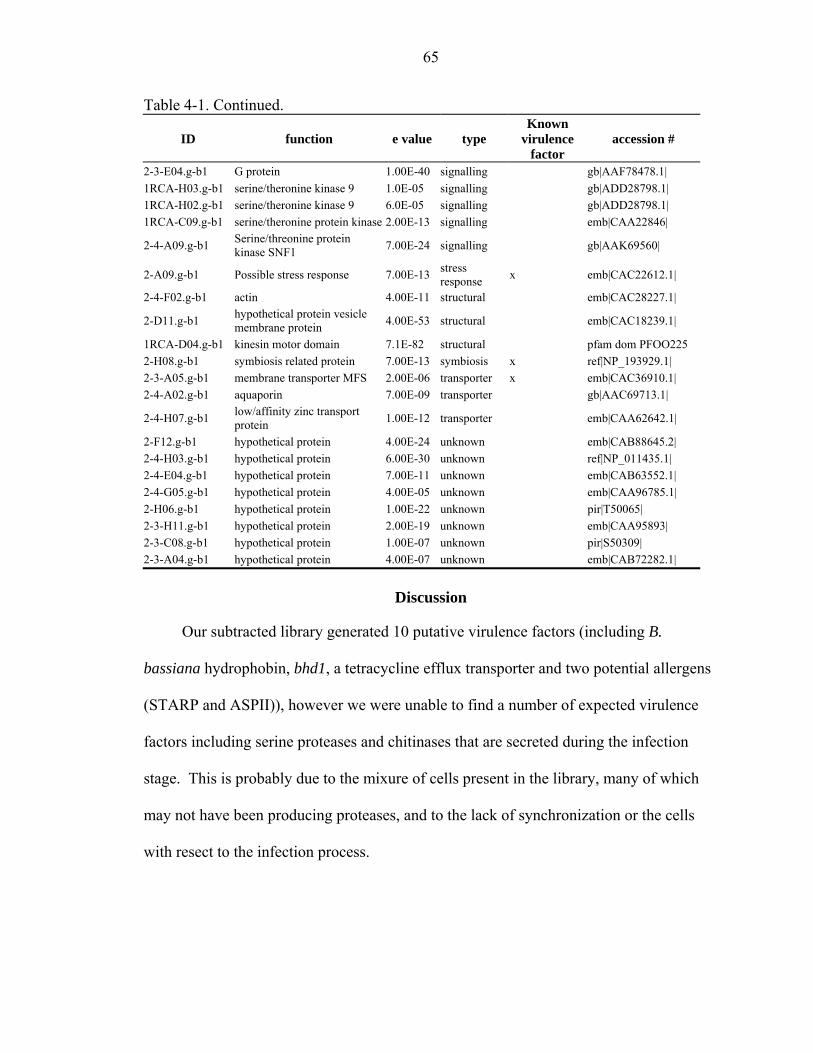
65
Table 4-1. Continued.
ID function e value type Known
virulence factor
accession #
2-3-E04.g-b1 G protein 1.00E-40 signalling gb|AAF78478.1| 1RCA-H03.g-b1 serine/theronine kinase 9 1.0E-05 signalling gb|ADD28798.1| 1RCA-H02.g-b1 serine/theronine kinase 9 6.0E-05 signalling gb|ADD28798.1| 1RCA-C09.g-b1 serine/theronine protein kinase 2.00E-13 signalling emb|CAA22846|
2-4-A09.g-b1 Serine/threonine protein kinase SNF1 7.00E-24 signalling gb|AAK69560|
2-A09.g-b1 Possible stress response 7.00E-13 stress response x emb|CAC22612.1|
2-4-F02.g-b1 actin 4.00E-11 structural emb|CAC28227.1|
2-D11.g-b1 hypothetical protein vesicle membrane protein 4.00E-53 structural emb|CAC18239.1|
1RCA-D04.g-b1 kinesin motor domain 7.1E-82 structural pfam dom PFOO225 2-H08.g-b1 symbiosis related protein 7.00E-13 symbiosis x ref|NP_193929.1| 2-3-A05.g-b1 membrane transporter MFS 2.00E-06 transporter x emb|CAC36910.1| 2-4-A02.g-b1 aquaporin 7.00E-09 transporter gb|AAC69713.1|
2-4-H07.g-b1 low/affinity zinc transport protein 1.00E-12 transporter emb|CAA62642.1|
2-F12.g-b1 hypothetical protein 4.00E-24 unknown emb|CAB88645.2| 2-4-H03.g-b1 hypothetical protein 6.00E-30 unknown ref|NP_011435.1| 2-4-E04.g-b1 hypothetical protein 7.00E-11 unknown emb|CAB63552.1| 2-4-G05.g-b1 hypothetical protein 4.00E-05 unknown emb|CAA96785.1| 2-H06.g-b1 hypothetical protein 1.00E-22 unknown pir|T50065| 2-3-H11.g-b1 hypothetical protein 2.00E-19 unknown emb|CAA95893| 2-3-C08.g-b1 hypothetical protein 1.00E-07 unknown pir|S50309| 2-3-A04.g-b1 hypothetical protein 4.00E-07 unknown emb|CAB72282.1|
Discussion
Our subtracted library generated 10 putative virulence factors (including B.
bassiana hydrophobin, bhd1, a tetracycline efflux transporter and two potential allergens
(STARP and ASPII)), however we were unable to find a number of expected virulence
factors including serine proteases and chitinases that are secreted during the infection
stage. This is probably due to the mixure of cells present in the library, many of which
may not have been producing proteases, and to the lack of synchronization or the cells
with resect to the infection process.

66
1 CTATAGGGCG AATTGGGCCC TCTAGATGCA TGCTCGAGCG GCCGCCAGTG 51 TGATGGATAT CTGCAGAATT CGCCCTTTCT TCACATCTCT TCCTAGTGAT 101 GTCCATGCAC CAGCCTACTG GATAAGACTG CCAATGGCGA CGCAAGGCAG 151 GGCAAGGTTG ACAAGACCAC CGTAAGCCTA ATTGTATCGT TAGATCCTGA 201 ACAGCAGTCA ATTAGAGACT TGATATAACT TACGACAGAG GGAGTGTTGT 251 GGCAGCAGGC CGCACGGTTG GTGCACTCGG ACTGGAGAAG GTTGCCGACA 301 GCGAGGACTG GAGACTTGTT AGTACTTGCT CAGCGACTTT ATCGACCACT 351 TGAGGCTTAC CATTGACGGG GATCTTGGTG CAAGTACCGA GAAGGTGGGA 401 AAGAAGACCA TCAGCACCGA CAGTGGCACC GAGCAGGCCG CCGAGGCCGG 451 TTTCGGGGGA GGAGTGCTCG ATCTTGGTCG TCTTCTCGTT GCAGCAGTAG 501 ATGGACTGGT CGGCACCGCA CCTGTCGCCA GCCTGGCCGA GAGTCATCTT 551 GTGCAGATCC TTTCCACCAT GGTGGCCGGA GGGAGCAGCA GTCACAGCGG 601 CGATGAGGGT GGTGATGGCA AGAGCGAAAC GCATGATGAC GGTATTGTTT 651 GTTTGGTTGA AGGATGTGGA AGAGGTGAAG GGCGAATTCC AGCACACTGG 701 CGGCCGTTAC TAGTGGATCC GAGCTCGGTA CCAAGCTTGG CGTAATCATG 751 GTCATAGCTG TTTCCTGTGT GAAATTGTTA TCCG Fig. 4-2. B. bassiana H1 hydrophobin genomic sequence. Expressed sequence in bold.
In our laboratory we have generated a series of B. bassiana stage and cell specific
cDNA libraries (Cho et al personal communication). From these libraries it has become
apparent that many of these proteins are expressed under conditions present in our driver
library, and that further analysis of subtracted libraries of purified cells from different
stages would give a better picture of the expression of putative virulence factors.
The fragments hydrophobin (bhd1) and other potential virulence factors found in
the SSH library were sequenced, cloned and the full-length sequences were obtained
using 5’prime RACE (Keyhani, unpublished results) when needed (some of the smaller
genes did not require additional analysis to generate full length cDNA sequences). PSI-
Blast searches indicated that the hydrophobin had the strongest homology to the RodA
proteins of A. fumigatus and A. nidulans, and a weaker homology to the HYD1 and
HYD2 proteins of Gibberella moniliformis (anamorph: Fusarium verticillioides).
However, Bootstrapped, phylogenetic analysis (Phylip 3.6, ClustalX, and phylodendron)
of class I and class II hydrophobins deposited in the NCBI protein database, from
Ascomycetes and the Basidiomycetes, places them closer to the F. verticilliodes
hydrophobins (figures 4-3 & 4-4).

67
Signal peptide
M._grisea_mpg1_class MFSLKTVVLALAAAAFVQAIP------------------------APGEG M._anisopliae_ssgA_precursor MF--KALIVALAAVAA--AIPTQ----------------------QPSSN B._bassiana_H2 MLA--TTIFATLLAL-AAAAPH-----------------------GPSHG A._nidulans_rodl_emeni MKFSIAAAVVAFAAS-VAALPPAHDSQFAGNGVGNK--GNSNVKFPVPEN A._fumigatus_rodl MKFSLSAAVLAFAVS-VAALP-QHDVNAAGNGVGNK--GNANVRFPVPDD A._fumigatus_Rodbp MKF--LAVVSLLAAT-ALALP-------------NA--GVVHPTFASADK G._moniliformis_H1_Fusarium MQY--MTIVAFLAAT-VAAGP------------------QIRAY-PSIDQ G._moniliformis_H2_Fusarium MYA--YTVIAFLAAS-VAAAG--------------------NG--PSISS B._bassiana_H1 MRF-ALAITTLIAA--VTAAP-----------------SGHHGG-KDLHK A._nidulans_DEWA_EMENI__class_ MRFI-VSLLAFTAAATATALP---------------ASAAKNAKLATSAA * . * M._grisea_mpg1_class PSVSMAQQKCGAEKVVSCCNSKELKNSKSGAEIPID-------------- M._anisopliae_ssgA_precursor ---EMN---CDSG--VYCCN--KVAQN-TGIVVPID-------------- B._bassiana_H2 PSVKTG-DICGNGNTMHCCNDESVTNKLTGPSVLSDL------------- A._nidulans_rodl_emeni VTVKQASDKCGDQAQLSCCN--KATYAGDTTTVDEGLLSGALSG--LIGA A._fumigatus_rodl ITVKQATEKCGDQAQLSCCN--KATYAGDVTDIDEGILAGTLKN--LIGG A._fumigatus_Rodbp YTLQQAQNKCGEHTTLSCCN--HVSKVGDTTAFNYGLLNGLLGN--AI-- G._moniliformis_H1_Fusarium ITVAQANNACGNNMQVTCCN--KVTNTPAGNAVGNGAG------------ G._moniliformis_H2_Fusarium LTVQQAANSCANGQSVYCCN--KTSNKPAGNSVGDGAG------------ B._bassiana_H1 MTLGQAGDRCGADQSIYCCN--EKTTKIEHSSPETGLGG-LLGATV---- A._nidulans_DEWA_EMENI__class_ FAKQAEGTTCNVG-SIACCNSPAETNNDSLLSGLLGAG--LLNGL----- * : *** . M._grisea_mpg1_class -----VLS----GECKNIPINILT--INQLIP--INNFCSDTVSCCSG-- M._anisopliae_ssgA_precursor -----ALS----STCG----DTLK--LVTVDA--LNDKCTSQTVCCNN-- B._bassiana_H2 -DLRKLLA----AECSPISVNVL---LNQLVP--IDNKCKQQSICCGE-- A._nidulans_rodl_emeni GSGAEGLG--LFDQCSKLDVAVLI--G---IQDLVNQKCKQNIACCQNSP A._fumigatus_rodl GSGTEGLG--LFNQCSNVDLQIPV-IGIP-IQALVNQKCKQNIACCQNSP A._fumigatus_Rodbp -SGPEGVG--ILSGCQKISVTALI--G---VDDLLNKQCQQNVACCQDNK G._moniliformis_H1_Fusarium --ILNNLS--LFDQCSKLDVNVLA-IA----NGLLNKECQANAACCQNSG G._moniliformis_H2_Fusarium --IANGLS--LFSQCSKVDVNVIA-IA----NNLLNKECQANAACCQDSP B._bassiana_H1 --GADGLLSHLLGTCTKIPVNVLA-VG----N-LLQSECTNRAACCHNTP A._nidulans_DEWA_EMENI__class_ -SGNTGSA------CAKASLIDQLGLLALVDHTEEGPVCKNIVACCPEG- * * ** M._grisea_mpg1_class -EQI-GLVNIQCTPILS------ M._anisopliae_ssgA_precursor -VQQNGLVNVACTPIDV------ B._bassiana_H2 -QKLNGLVNLGCTPITVLG---- A._nidulans_rodl_emeni SSADGNLIGVGLP-CVALGSIL- A._fumigatus_rodl SDASGSLIGLGLP-CIALGSIL- A._fumigatus_Rodbp SVATGGLINIATPACVALDSII- G._moniliformis_H1_Fusarium GSATGGLVNVALP-CIALSSLI- G._moniliformis_H2_Fusarium GTAAAGLVNAALP-CVAISNLV- B._bassiana_H1 SVAYGGLVNLALP-CVAIGSLIQ A._nidulans_DEWA_EMENI__class_ ---TTN--------CVA------ . : .
Fig. 4-3. Alignment of H1 and H2 with homologous hydrophobins (ClustalX) that gave
significant expect values in PSI blast. Signal peptide length was determined using SignalP.

68
0.1
Fig. 4-4. Bootstrapped (1000) phylogenetic tree of class I and class II hydrophobins using alignments generated with ClastalX and visualized with the online tree printer Phylodendron (http://iubio.bio.indiana.edu/treeapp/treeprint-form.html)
69
This is interesting because the F. vercticilliodes microconidia are hydrophilic (38)
and this organism is a plant pathogen with specificity towards corn and B. bassiana, an
entomopathogen with hydrophobic conidia, can infect corn asymptomatically (88). It is
possible that some of the same factors that allow F. verticilliodes to infect corn are
similar to those that allow B. bassiana to colonize the same organism.
It is possible that the expected genes were present in the subtracted library. The
fragments generated by the SSH procedure are quite small making it difficult to
determine if the fragments did not have strong hits because there were not matches, or if
they were to small to generate strong hits. Annotated information for fungal genomes
lags behind that for many other organisms, and as more annotated sequences are
deposited into the databases, more hits to named proteins is likely to occure.

70
CHAPTER 5 MOLECULAR ANALYSIS OF TWO BEAUVERIA BASSIANA HYDROPHOBINS
AND A HYDROPHOBIN LIKE PROTEIN
Introduction
Hydrophobins are small, cysteine rich, secreted proteins unique to fungi. Many
hydrophobins were initially identified from abundantly transcribed mRNA present during
sporulation, fruit body formation, and during pathogenic interactions with plants and
animals (52). These proteins play a critical role in numerous filamentous fungal processes
including; growth, conidial formation, conidial dispersion, aerial structure formation, and
attachment to (hydrophobic) surfaces, and pathogenicity (93).
Wessels et al. (1991) noted a positive correlation between Sc3 mRNA abundance
and the presence of aerial hyphae and fruiting bodies in Schizophyllum commune. They
estimated that by the fourth day of growth up to 8.1% of the protein synthesis activities in
the dikaryon (fruiting bodies are composed of dikaryon hyphae) is directed towards the
synthesis of hydrophobins.
Rodlet layers have been observed on conidial surfaces of numerous filamentous
fungi including, Neuorospora crassa, Aspergillus sp., Magnaporthe grisea,
Metharhizium. anisopliae and Beauveria bassiana. The presence of this rodlet layer
appears to increase the hydrophobic nature of the conidia. The better organized, the
fascicles, the more hydrophobic the spores. Hydrophilic conidia formed by fungi such as
Gibberella moniliformis, Verticillium lecanii and Botrytis spp. do not appear to form a

71
rodlet layer, and have additional structures that mediate attachment to surfaces including
mucosal coats, and adhesive knobs (94, 99).
Hydrophobins produced during sporulation are involved in the production of the
rodlet layer that has been observed on the surface of many hydrophobic conidia.
Deletion mutants lacking rodAp in A. nidulans and A. fumigatus (68) or mpg1 in M.
grisea (52) generated spores lacking the characteristic rodlet layer, and displaying altered
adhesion characteristics. The relative importance of the hydrophobins to rodlet layer
formation and adhesion properties may vary with species.
Paris et al. 2003 observed that both A. fumigatus and A. nidulans possess two
conidial hydrophobins RodAp (both sp.) RodB (A. fumigatus) and DewAp (A. nidulans).
Deleting rodAp resulted in loss of the conidial rodlet layer in A. fumigatus while deleting
rodBp had no effect on rodlet layer formation. In A. nidulans similar studies showed that
both rodAp and dewAp were required for conidial rodlet layer formation. Paris et al.
(2003) also noted that changes in cell surface characteristics were more profound in the
A. nidulans rodletless mutants than in the corresponding A. fumigatus mutants.
Unlike A. nidulans and A. fumigatus, the rodlet layer of B. bassiana, Neurospora
crassa and Magnaporthe grisea appear to posses a single hydrophobin (68). In M.
grisea, deleting mpg1 resulted in rodletless mutants similar to those observed for A.
fumigatus and A. nidulans (52). This protein was also linked to pathogenicity by the
observation that appropriate appressorium formation, in response to external surface cues,
was impaired in ∆mpg1 mutants (3).
All hydrophobins isolated to date self-assemble into 10 nm thick layers at
hydrophobic/hydrophilic interfaces. Based on the solubility characteristics of the layers

72
and hydropathy patterns of the amino acids, hydrophobins have been separated into two
classes (I and II) (96). Treatments with trifluoroacetic acid (TFA), or formic acid (FA)
are often required to dissolve class I layers which remain insoluble in 2 % sodium
dodecyl sulfate (SDS) at 1000C. In contrast, layers formed by class II hydrophobins are
more soluble and can be dissolved in 60 % ethanol and 2 % SDS.
Class I hydrophobins self assemble at hydrophilic/hydrophobic interfaces, forming
5-12 rodlets that bundle together into fascicles. The rodlets are made up of
protofilaments 2.5 nm in diameter. Thus far, similar bundles have not been observed in
the amphipathic layers formed by Class II hydrophobins (96). Atomic resolution of the
structure of HfbI and HfbII, hydrophobins from Trichoderma reesei, revealed that these
Class II hydrophobins form tetrameric supramolecules that assemble into crystalline
domains (43).
The rodlet layer, like the proteins from which it is composed, is amphipathic. When
coating the surface of fungal structures this layer arranges itself so that the hydrophilic
part of the membrane faces the cell wall of the fungus, while the hydrophobic side is
exposed to the environment. In A. nidulans and other fungi with hydrophobic conidia
this layer, especially when present on the surface of conidia, is thought to be important in
hydrophobic interactions between fungus and surfaces, however in the closely related
species, A. fumigatus adhesion to hydrophobic substrates is not completely lost in mutant
strains lacking the major spore coat hydrophobins (68).
There is little sequence homology between Class II hydrophobins, and even less for
Cass I hydrophobins, although all hydrophobins share eight cysteines that are
characteristic of these molecules. Complementation studies of rodlet hydrophobins from
73
closely related species, such as A. nidulans and A fumigatus, show that they often can
restore partial wild-type characteristics, however when the fungi that are not as closely
related, as is the case with M. griseae and A. nidulans, phenotypes may not be restored
unless the gene is introduced under the control of the promoter from the host organism’s
own hydrophobin suggesting that regulation may be more important than the
hydrophobin’s sequence in determining function (52, 97, 98).
In addition to being the major proteins in the rodlet layer of hydrophobic conidia,
hydrophobins are also found on the surface of hyphae and fruiting bodies. They are
highly surface active molecules and reduce the surface tension of water allowing the
fungus to form aerial structures (62). They have also been implicated in signaling (82)
and are important for mediating adhesion. Determining the mRNA levels of bhd1, bhd2
and bsn in different cell types and under different cultural conditions can be expected to
yield insights into their function.
In this study real-time reverse transcriptase PCR (real-time RT-PCR), currently the
most sensitive technique for mRNA detection was used to quantify the levels of
transcripts encoding the two hydrophobins (bhd1 and bhd2) and the hydrophobin like
protein (Bsn) in aerial conidia, submerged conidia and blastospores.
Materials and Methods
Cultivation of Fungi
Beauveria bassiana (ATCC 90517) was routinely grown on potato dextrose agar
(PDA) (4). Plates were incubated at 26oC for 10-14 days, and aerial conidia were
harvested by flooding the plate with sterile dH2O. Conidial suspensions were filtered
through a single layer of Mira-cloth (Clabiochem, CA), and final spore concentrations
were determined by direct count using a hemocytometer. Blastospores were produced in
74
DifcoTM Sabouraud dextrose (Becton, Dickinson and Co., MD) + 1-2% yeast extract
liquid broth cultures (SDY) using conidia harvested from plates to a final concentration
of 0.5 - 5 x 10⁵ conidia/ml as the inoculum. Cultures were grown for 3-4 days at 26oC
with aeration (150-200 rpm). Cultures were filtered (2x) through glass wool to remove
mycelia, and the concentration of blastospores was determined by direct count.
Submerged conidia were produced in TKI broth using fructose as the carbon source as
described (83). For all cell types, Mira-cloth (Clabiochem, CA) or glass wool filtered
cell suspensions were harvested by centrifugation (10,000 x g, 15 min, 4oC), washed two
times with sterile dH2O, and resuspended to the desired concentration as indicated
(typically 107-108 cells/ml). Cells producing oosporein were grown in DifcoTM
Sabouraud dextrose (Becton, Dickinson and Co., MD) + 1-2% yeast extract liquid broth
cultures (SDY) using conidia harvested from plates to a final concentration of 0.5-5 x 10⁵
conidia/ml as the inoculum until they started producing oosporein, and the mycelium
were obtained from unfiltered cultures used to grow blastospores.
RNA Isolation
Fungal cells were frozen in liquid Nitrogen, or lyophilized and stored @ -70oC until
used. Immediately prior to use the cells were crushed in liquid Nitrogen and total RNA
was extracted with RNAwizTM (Ambion, TX) per manufacturers instructions. Poly A
RNA was enriched using magnetic beads covalently attached to oligo-dT tails
(Dynabeads mRNA direct kit, Dynal, WI, Poly (A) Quick® mRNA isolation kit,
Stratagene CA, or Poly A tract® mRNA isolation Systems, Promega, WI).
Rodlet Layer Extraction
The rodlet layer proteins were removed from the surface of the spores as described
by Paris et al. (68). Briefly, aerial conidia, blastospores, and submerged conidia were
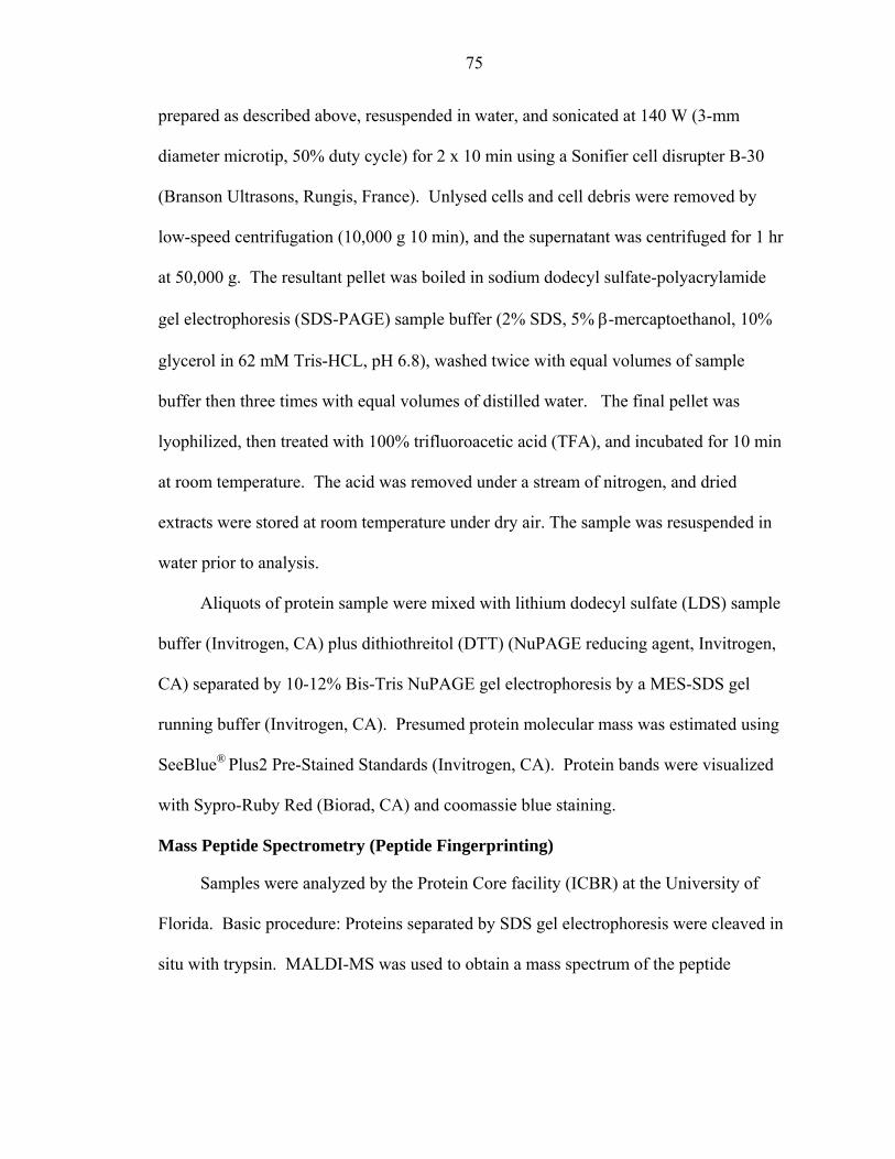
75
prepared as described above, resuspended in water, and sonicated at 140 W (3-mm
diameter microtip, 50% duty cycle) for 2 x 10 min using a Sonifier cell disrupter B-30
(Branson Ultrasons, Rungis, France). Unlysed cells and cell debris were removed by
low-speed centrifugation (10,000 g 10 min), and the supernatant was centrifuged for 1 hr
at 50,000 g. The resultant pellet was boiled in sodium dodecyl sulfate-polyacrylamide
gel electrophoresis (SDS-PAGE) sample buffer (2% SDS, 5% β-mercaptoethanol, 10%
glycerol in 62 mM Tris-HCL, pH 6.8), washed twice with equal volumes of sample
buffer then three times with equal volumes of distilled water. The final pellet was
lyophilized, then treated with 100% trifluoroacetic acid (TFA), and incubated for 10 min
at room temperature. The acid was removed under a stream of nitrogen, and dried
extracts were stored at room temperature under dry air. The sample was resuspended in
water prior to analysis.
Aliquots of protein sample were mixed with lithium dodecyl sulfate (LDS) sample
buffer (Invitrogen, CA) plus dithiothreitol (DTT) (NuPAGE reducing agent, Invitrogen,
CA) separated by 10-12% Bis-Tris NuPAGE gel electrophoresis by a MES-SDS gel
running buffer (Invitrogen, CA). Presumed protein molecular mass was estimated using
SeeBlue® Plus2 Pre-Stained Standards (Invitrogen, CA). Protein bands were visualized
with Sypro-Ruby Red (Biorad, CA) and coomassie blue staining.
Mass Peptide Spectrometry (Peptide Fingerprinting)
Samples were analyzed by the Protein Core facility (ICBR) at the University of
Florida. Basic procedure: Proteins separated by SDS gel electrophoresis were cleaved in
situ with trypsin. MALDI-MS was used to obtain a mass spectrum of the peptide
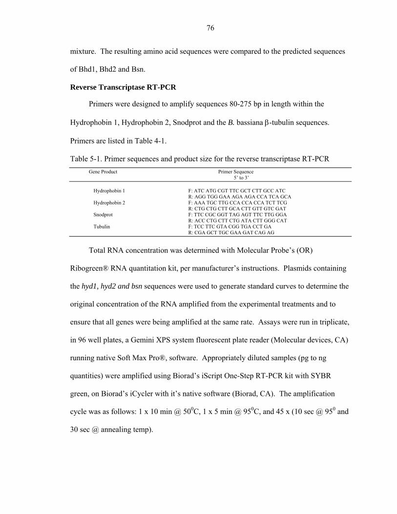
76
mixture. The resulting amino acid sequences were compared to the predicted sequences
of Bhd1, Bhd2 and Bsn.
Reverse Transcriptase RT-PCR
Primers were designed to amplify sequences 80-275 bp in length within the
Hydrophobin 1, Hydrophobin 2, Snodprot and the B. bassiana β-tubulin sequences.
Primers are listed in Table 4-1.
Table 5-1. Primer sequences and product size for the reverse transcriptase RT-PCR Gene Product Primer Sequence 5’ to 3’
Hydrophobin 1 F: ATC ATG CGT TTC GCT CTT GCC ATC R: AGG TGG GAA AGA AGA CCA TCA GCA Hydrophobin 2 F: AAA TGC TTG CCA CCA CCA TCT TCG R: CTG CTG CTT GCA CTT GTT GTC GAT Snodprot F: TTC CGC GGT TAG AGT TTC TTG GGA
R: ACC CTG CTT CTG ATA CTT GGG CAT Tubulin F: TCC TTC GTA CGG TGA CCT GA
R: CGA GCT TGC GAA GAT CAG AG
Total RNA concentration was determined with Molecular Probe’s (OR)
Ribogreen® RNA quantitation kit, per manufacturer’s instructions. Plasmids containing
the hyd1, hyd2 and bsn sequences were used to generate standard curves to determine the
original concentration of the RNA amplified from the experimental treatments and to
ensure that all genes were being amplified at the same rate. Assays were run in triplicate,
in 96 well plates, a Gemini XPS system fluorescent plate reader (Molecular devices, CA)
running native Soft Max Pro®, software. Appropriately diluted samples (pg to ng
quantities) were amplified using Biorad’s iScript One-Step RT-PCR kit with SYBR
green, on Biorad’s iCycler with it’s native software (Biorad, CA). The amplification
cycle was as follows: 1 x 10 min @ 500C, 1 x 5 min @ 950C, and 45 x (10 sec @ 950 and
30 sec @ annealing temp).
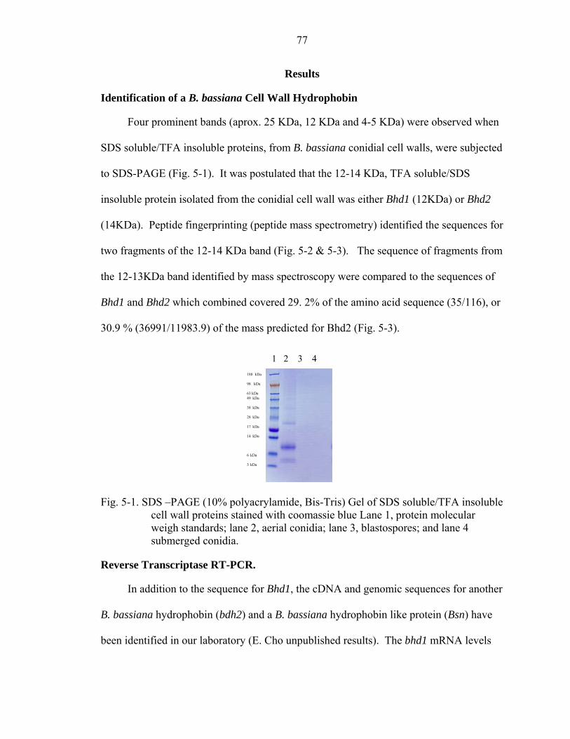
77
Results
Identification of a B. bassiana Cell Wall Hydrophobin
Four prominent bands (aprox. 25 KDa, 12 KDa and 4-5 KDa) were observed when
SDS soluble/TFA insoluble proteins, from B. bassiana conidial cell walls, were subjected
to SDS-PAGE (Fig. 5-1). It was postulated that the 12-14 KDa, TFA soluble/SDS
insoluble protein isolated from the conidial cell wall was either Bhd1 (12KDa) or Bhd2
(14KDa). Peptide fingerprinting (peptide mass spectrometry) identified the sequences for
two fragments of the 12-14 KDa band (Fig. 5-2 & 5-3). The sequence of fragments from
the 12-13KDa band identified by mass spectroscopy were compared to the sequences of
Bhd1 and Bhd2 which combined covered 29. 2% of the amino acid sequence (35/116), or
30.9 % (36991/11983.9) of the mass predicted for Bhd2 (Fig. 5-3).
Fig. 5-1. SDS –PAGE (10% polyacrylamide, Bis-Tris) Gel of SDS soluble/TFA insoluble
cell wall proteins stained with coomassie blue Lane 1, protein molecular weigh standards; lane 2, aerial conidia; lane 3, blastospores; and lane 4 submerged conidia.
Reverse Transcriptase RT-PCR.
In addition to the sequence for Bhd1, the cDNA and genomic sequences for another
B. bassiana hydrophobin (bdh2) and a B. bassiana hydrophobin like protein (Bsn) have
been identified in our laboratory (E. Cho unpublished results). The bhd1 mRNA levels
188 kDa
98 kDa
63 kDa49 kDa
38 kDa
28 kDa
17 kDa
14 kDa
6 kDa
3 kDa
1 2 3 4

78
were highest in the submerged conidia with a relative concentration (RC) of 41.77.
Aerial conidia and blastospores had much lower levels with RC 3.61 and 1.69
respectively.
The relative amount of bdh2 mRNA (a cell wall SDS insoluble/TFA soluble
protein) was much lower than that of bdh1 in all cell types (RC 1.69 - 41.77 for bdh2 and
RC 0.01-1.59 for bdh1). The highest abundance of bdh2 mRNA was found in aerial
conidia (RC 1.59) much lower levels were found submerged conidia (RC 0.18) and
blastospores (RC 0.01). The concentration of the tubulin mRNA was lower in conidial
cells ([tub]/[total RNA] = 0.001), when compared with the other two spore types
([tub]/[total RNA] = 0.02). We were unable to demonstrate high levels of expression in
any of the cell types tested; we also were unable to find significant expression of
Snodprot in any of the cell types analyzed (Table 5-2).
Table 5-2. mRNA abundance of bhd1, bhd2 in Beauveria bassiana single cell propagules.
Aerial conidia Submerged conidia Blastospores Total RNA 89.7 ng/mla 838.6 ng/ml 649 ng/ml [tub]b ng/ml 0.09 1.46 15.96 [bhd1] ng/ml 0.33 61.02 26.97 [bhd2] ng/ml 0.15 0.26 0.20 [bhd1]/[tub]c 3.61 41.77 1.69 [bhd2]/[tub] 1.59 0.18 0.01
a) Ribogreen assay (Molecular probes, Invitrogen, CA). b) tub (tubulin gene), bhd1 (Beauveria bassiana hydrophobin 1), and bhd2 (Beauveria bassiana hydrophobin 2). c) The concentrations were normalized using the concentration values obtained amplification of the tubulin genes.

79
Avg Mass: 11983.9 Seq # b y (+1) --- -- ------ ------ -- L 1 114.1 1385.8 13 T 2 215.1 1272.7 12 G 3 272.2 1171.6 11 P 4 369.2 1114.6 10 S 5 456.2 1017.6 9 V 6 555.3 930.5 8 L 7 668.4 831.5 7 S 8 755.4 718.4 6 D 9 870.5 631.3 5 L 10 983.5 516.3 4 D 11 1098.6 403.2 3 L 12 1211.7 288.2 2 R 13 1367.8 175.1 1 Fig. 5-2. Mass spectroscopy data showing the mass of individual amino acids in one of
the two main fragments from the 12 KDa trifluoroacetic acid soluble/sodium dodecyl sulfate insoluble B. bassiana cell wall protein.

80
Coverage: 13/116 = 11.2% by amino acid count, 1385.6/11983.9 = 11.6% by mass
Hydrophobin sequence H2 MLATTIFATL LALAAAAPHG PSHGPSVKTG DICGNGNTMH CCNDESVTNK LTGPSVLSDL DLRKLLAAEC SPISVNVLLN QLVPIDNKCK QQSICCGEQK LNGLVNLGCT PITVLG
PepStat LTGPSVLSDLDLR position: 51 - 63
Avg Mass: 11983.9 Coverage: 22/116 = 19.0% by amino acid count,
2313.5/11983.9 = 19.3% by mass Hydrophobin sequence H2 MLATTIFATL LALAAAAPHG PSHGPSVKTG DICGNGNTMH CCNDESVTNK LTGPSVLSDL DLRKLLAAEC SPISVNVLLN QLVPIDNKCK QQSICCGEQK LNGLVNLGCT PITVLG
PepStat TGDICGNGNTMHCCNDESVTNK position: 29 - 50 Fig. 5-3. Mass peptide fingerprinting results for two identifiable fragments of the 12
KDa trifluoroacetic acid soluble, sodium dodecyl sulfate insoluble B. bassiana cell wall protein confirming that this protein is the Bhd2 hydrophobin.
Discussion
Fragments of the hydrophobin gene (bhd1) present in the SSH library (chapter 4)
were sequenced, cloned and were ultimately found to collectively represent the full-
length sequence of the gene (Keyhani, unpublished results). Since then we have
identified sequences for two additional proteins Bhd2 and Bsn from phage display
libraries enriched for phage displaying proteins that bound to carbohydrates (E. Cho,
unpublished results). Blast searches (NCBI) indicated that these proteins are similar
(38% and 51% identity, respectively) to Mpg1 hydrophobin of Magnaporthe grisea (E
value 9e-11) and the Neurospora crassa Snodprot1 toxin (E value 2e-35). Based on the
number of sequences between the cysteines, both Bhd1 and Bhd2 are class I
hydrophobins (fig. 5-3) (38).
Our previous results (Chapter 3) suggest that B. bassiana conidia are hydrophobic,
and a rodlet layer is clearly visible by AFM on the surface (Chapter 2). Previous research

81
(7) of the B. bassiana rodlet layer appeared to indicate that it contains a single cell wall
hydrophobin (10-14 KDa protein), however we have identified the genes for at least two
hydrophobins (Bhd1 and Bhd2) and a hydrophobin like protein (Bsn).
Class I: -C-X5-8-CC-X17-39-C-X8-23-C-X5-6-CC-X6-18-C-X2-13 H1 7 27 18 5 12 7 H2 7 37 17 5 17 8 Class II: -C-X9-10-CC-X11 -C-X16 -C-X6-9-CC-X6-18-C-X2-13 Fig. 5-4. Comparison of Bhd1 and Bhd2 and other hydrophobins (A) consensus spacing
for Class I and Class II hydrophobins as suggested by Fuchs et al. (38).
At least four major bands were observed when TFA soluble, SDS insoluble
proteins were extracted from aerial conidial cell walls (Figure 5-1). Based on reports in
the literature discussing B. bassiana cell wall proteins that were insoluble in hot SDS, but
soluble in TFA or FA hydrophobin (7, 68), we selected the band with a molecular weight
of approximately 10-14 KDa (based on SDS page anlaysis) for further analysis because
we considered it the best candidate for the cell wall hydrophobin.
Mass peptide spectrometry analysis of the 10-14 KDa band, when compared to the
sequence of the two hydrophobins and the hydrophobin like protein, identified the protein
as Bhd2. These preliminary results indicate that Bhd2 is a component of the B. bassiana
cell wall. Aside from aerial conidia B. bassiana produces the other two single cell
propagules, blastospores and submerged conidia. These cells do not appear have a
visible rodlet layer and SDS insoluble, TFA soluble proteins were not isolated from
purified cultures of these cells. However, real-time RT-PCR on RNA isolated from these
cells indicated that transcripts corresponding to bhd1 and bhd2 could be detected in pure
cultures of these cell types, although levels of bdh2 were low in all cell types.
It is likely that conidia are not metabolically active; as reflected by the relatively
low levels of tubulin amplified by real-time RT-PCR (Table 5-2). Alternatively, the low

82
levels of tubulin could be explained by low expression of tubulin alone, with other genes
being expressed at relatively higher levels. Using other housekeeping genes, or purifying
and quantifying poly A RNA will determine which of these explanations is the most
likely.
There are a number of possibilities explaining the low levels of bhd2 RNA
observed in the cell types analyzed. It is possible that bhd2 is only produced in
sporulating cultures, specifically in cells responsible for producing conidia
(conidiophores) and was therefore not expressed by any of the cell types tested. In
contrast bhd1 was present in almost all the cultures tested at a relatively high abundance.
This protein while not being recruited to the single cell propagule cell walls may instead
be secreted into the medium. Similar to other hydrophobins which are not recruited to
conidial cell walls it may function as a surfactant, coat surfaces acting as a lubricant, or
function as an adhesin directing hyphal attachment and growth.
Bsn, like bhd2, is likely produced under specific conditions not examined in these
studies. Based on homology to proteins produced by Pisolithus microcarpus and
Ceratocystis fimbriata. Homologous proteins are produced by these organisms are
expressed by vegetative mycelium, when the expression levels of other hydrophobins or
carbon availability is low. Putative functions for the product of this gene may include
host cell toxicity (28, 69).

83
CHAPTER 6 GENERAL DISCUSSION
Statement of Hypotheses
The work in this provides scientific data to examining the validity of the following
hypotheses: 1) that there is a measurable difference in the cell surface characteristics of
the single cell propagules of Beauveria bassiana, and 2) there are differences in the
binding properties of these propagules which can be related to the cell surface
characteristics.
Is There a Measurable Difference in The Cell Surface Characteristics of The Single Cell Propagules of B. bassiana?
Visual Differences
Visual inspection of the single cell propagules using light or atomic force
microscopy reveals that the morphology and the surfaces of B. bassiana aerial conidia,
submerged conidia and blastospores are different. Aerial conidia are spherical, 2-4 µm in
diameter, hyaline and covered with a distinct rodlet layer. Submerged conidia are similar
in shape to the aerial conidia but are larger (3-5 µm in diameter), and while the surface
appears rough, there was no visible evidence of a rodlet layer in those cells. Blastospores
are rod shaped and larger than the other spores at 8-12 µm in length, there is no visible
rodlet layer on their surface, which is smooth (35, 83).
Differences in Hydrophobicity
Interfacial free energies of interaction derived from contact angle data and percent
retention of the spores in the hydrocarbon phase (MATH) assay, both confirmed that the

84
conidia have negative interfacial free energies of interaction indicating that they would be
unable to overcome the high cohesive force of water, and with almost 88 % retention in
the hydrocarbon layer, it is clear that these spores are highly hydrophobic. Submerged
conidia were less moderately hydrophilic, with slightly positive interfacial free energies
of interaction and 74 % retention in the hydrocarbon layer. Blastospores with strongly
negative interfacial free energies of interaction and low retention (40%) in the
hydrocarbon layers were the most hydrophilic.
Differences in Effective Surface Charge
Effective surface charge (zeta potential) of the three spores was similar in sign
(positive at low pH and negative at high pH) as pH changes from 3-9. All of the spores
had the same isoelectric point (pH 4), except for conidia that were 16-days-old, the
isoelectric point for these cells was pH 5. Conidia registered the greatest magnitude of
charge of the three spore types, (zeta potential: -29 mV, day 16 and – 47 mV, day 20) at
very high pH (pH 8) and at low pH (pH 3) for 16-day-old aerial conidia (zeta potential:
22 mV. At low pH (ph 3) the zeta potential for all the spores, except for the 16-day-old
conidia, was between 3-8 mV. The magnitude of charge for submerged conidia at high
pH was intermediate with a zeta potential of -10 mV, and that for the blastospores was
the lowest with a zeta potential of – 5 mV. The overall change in zeta potential with
respect to pH for the blastospores was 7 mV, while that for the submerged conidia was –
20 mV, and that of the aerial conidia (16 and 20 days combined) was 69 mV.
The conidia that were the most hydrophobic had the highest charge, this could be
indicative that: 1) electrostatic repulsion is less of a factor for the more hydrophobic cells
because hydrophobic forces are stronger and act over a longer range, 2) that the surface to
which aerial conidia bind to tend to be less electronegative at higher pH than aerial
85
conidia (this is true for insect cuticle; many of the proteins in the insect cuticle tend to be
positively charged at higher pH), whereas the surfaces that the blastospores and
microccyle conidia bind to tend to be less more at higher pH than either of those cell
types, or that 3) aerial conidia are designed to adhere to hydrophobic surfaces, using the
surface of water as a dispersion mechanism, whereas blastospores and submerged conidia
are designed to be resuspended in aqueous solutions for longer periods of time.
Cell Wall Proteins
Sodium dodecyl sulfate (SDS) insoluble, trifluoroacetic acid (TFA) soluble
proteins were extracted from the cell wall of conidia, but not from the cell walls of
microcycleconidia, these proteins often belong to a class of small, cysteine rich, secreted
fungal proteins called hydrophobins (99). The genes for two hydrophobins and one
hydrophobin like protein specific to B. bassiana have been identified, cloned and
sequenced in our laboratory. The bhd1 sequence was identified from gene fragments
obtained from a SSH library designed to find virulence factors upregulated when the
organism is grown in the presence of chitin, or insect cuticle. The other two genes were
identified by E. Cho in this laboratory from phage display libraries designed to enrich for
carbohydrate binding proteins. It has been noted, but never conclusively proven that
hydrophobins poses lectin like qualities hydrophobin (93, 99).
The amino acid sequences of the two hydrohobins (Bhd1 and Bhd2) were
compared to the sequences of fragments obtained by running peptide fingerprinting on a
12 KDa SDS insoluble, TFA soluble protein isolated from the cell wall of the conidia.
The sequence of two of the fragments matched at least 29% of the sequence of Bhd2. To
determine the expression patterns of all three genes, real-time reverse transcriptase PCR
(real time RT-PCR), was performed on the three single cell propagules and vegetative

86
mycelia from cells grown on chitin or in sabauroud dextrose broth (SDY) (1-2% YE).
bhd1 was expressed in all cell types, bhd2 was expressed mostly in cells exposed to
chitin, and submerged conidia and bsn was not expressed in any of the cell types tested.
Conclusions
From these results we can conclude that there are measurable differences in the
surface characteristics of the three cell types with regards to morphology, surface charge,
hydrophobic characteristics, and cell wall protein composition.
Are there Differences in the Binding Properties of These Propagules, Which Can Be Related to the Cell Surface Characteristics?
Adhesion Profiles of Aerial Conidia, Submerged conidia and Blastospores
Our fluorescent cell, microtitre plate adhesion assay determined that b. bassiana
aerial conidia bound predominantly to hydrophobic surfaces (silinized polystyrene), and
weakly to hydrophilic surfaces (polystyrene, factory-treated to be more hydrophilic).
Submerged conidia bound well to either surface, while blastospores bound predominantly
to the more hydrophilic surfaces. This correlates well with the hydrophobic character of
each spore type, however treating with amylase, laminarinase and protease could reduce
the binding of aerial conidia to hydrophobic surfaces, and treatment with maltose reduced
blastospores adhesion to hydrophilic surfaces, indicating that factors other than
hydrophobicity are implicated in spore adhesion.
Conclusion
The surface characteristics of B. bassiana single cell propagules are consistent with
the adhesion profiles on hydrophilic, and hydrophobic surfaces. However, it is clear that
other the adhesion properties of the spores are a result of the interactions of short and
long range intermolecular forces. These intermolecular forces are consistent with the
87
distinct cell wall characteristics of the three cell types, however it is probable that other
features not yet identified are contributing to the adhesion characteristics of the cell types
Confirmation of the role of the Bhd2 hydrophobin in the adhesion profile of B.
bassiana conidia and the function of the other two B. bassiana specific proteins (Bhd1
and Bsn) will require generating transformants lacking Bhd2. The Agrobacterium
tumefaciens mediated transformation procedure, with minor modifications, will be used
for this purpose.
Finally it should be noted that the adhesion profiles and the cell surface
characteristics unique to B. bassiana must be important in the ability of the fungus to
successfully function as an entomopathogen. Although the different propagules have
different surface characteristics and adhesion profiles, all are able to successfully initiate
infection, the main observable difference between the cells, with respect to use in
mycoinsecticidal preparations, is the resistance of aerial conidia to environmental stresses
such as UV light and desiccation spores, this difference has previously been correlated to
the presence of the rodlet layer on the surface of the aerial conidia (80)

88
APPENDIX AGROBACTERIUM MEDIATED TRANSFORMATION OF BEAUVERIA
BASSIANA
Introduction
Little known concerning the regulation of molecules and metabolic pathways
involved in fungal pathogen-host interactions particularly for the entomopathogen. This
is primarily due to the lack of efficient transformation systems for filamentous fungi.
Development of efficient transformation systems for filamentous fungi have been limited
to several species particularly those related to human health. Successful genetic
manipulation of fungi, as with any organism, requires a plasmid mediated transformation
system involving the entry and replication, or integration of foreign DNA (75) Once
DNA has been successfully introduced into the fungal cells its fate varies depending on
the host. DNA usually integrates into the chromosomes; autonomously replicating
plasmids are rare in most filamentous fungi.
There are a number of transformation strategies currently being used with mixed
degrees of success. The earliest, and still widely used, transformation method for
filamentous fungi is polyethylene glycol (PEG) mediated transformation of fungal
protoplasts. More recently developed methods sometimes used in conjunction with the
generation of protoplasts; include lithium acetate treatment, Agrobacterium tumefaciens
mediated transformation (ATMT), electroporation, restriction enzyme mediated
transformation (REMI), and biolistic transformation (75). Many of these methods,
89
simplify transformation procedures, reduce variability, and/or increase transformation
efficiencies.
Agrobacterium tumefaciens mediated transformation (ATMT) is routinely used for
the genetic modification, primarily of dicotyledon plant species. Agrobacterium
tumefaciens causes crown gall in plants by inducing tumor formation, and the over-
production of specific nutrients (opines) within the host cell. This is accomplished via
the transfer of part of its Ti plasmid DNA to the host genome. The transferred DNA (T-
DNA) contains genes encoding auxins, cytokinins and opines; the remainder of the
plasmid, and part of the bacterial genome, codes for proteins involved in the detection of
acetosyringone, a substance released by wounded plants. Excision, transfer and
integration of the T-DNA, into the host genome are not dependent on DNA present in the
T-DNA region, which can be artificially modified without affecting the transformation
process.
In 1998, de Groot and Gouka demonstrated that efficient transfer of T-DNA to
filamentous fungi, specifically to the fungus Aspergillus awamori, was possible, and that
transformation frequencies were increased up to 600 fold compared to traditional
methodologies (18, 41). Transformation by the same group of other fungi, including
Aspergillus niger, Fusarium venetatum and Trichoderma reesei, Colletotrichum
gloeosporioides, Neurospora crassa and Agaricus bisporus (a mushroom), demonstrated
that this system that could be adapted for a variety of filamentous fungi (18).
Materials and Methods
Fungal Cultures
Beauveria bassiana (ATCC 90517) was routinely grown on potato dextrose agar
(PDA) (4). Plates were incubated at 26oC for 10-14 days, and aerial conidia were
90
harvested by flooding the plate with sterile dH2O. Conidial suspensions were filtered
through a single layer of Mira-cloth (Clabiochem, CA), and final spore concentrations
were determined by direct count using a hemocytometer.
Blastospores were produced in DifcoTM Sabouraud dextrose (Becton, Dickinson
and Co., MD) + 1-2% yeast extract liquid broth cultures (SDY) using conidia harvested
from plates to a final concentration of 0.5 - 5 x 10⁵ conidia/ml as the inoculum. Cultures
were grown for 3-4 days at 26oC with aeration (150-200 rpm). Cultures were filtered
(2x) through glass wool to remove mycelia, and the concentration of blastospores was
determined by direct count.
For all cell types, Mira-cloth (Clabiochem, CA) or glass wool filtered cell
suspensions were harvested by centrifugation (10,000 x g, 15 min, 4oC), washed two
times with sterile dH2O, and resuspended to the desired concentration as indicated
(typically 107-108 cells/ml).
Agrobacterium tumefaciens Cultivation
The transformation was performed as described by Covert et al. (2001) with the
following modifications. Conidia (104-7 cfu/ml) were mixed in equal amounts with an
overnight Luria Bertani (LB, Difco, MI) broth (40 µg/ml ampicillin and 50 µg/ml
kanamycin) culture of Agrobacterium tumefaciens (AGL1). The A. tumefaciens culture
was in diluted to O.D. 0.15 with induction medium (16) supplemented with
acetosyringone (IMAS) and allowed to grow for 4 hrs to reach an O.D. of 0.6-0.8.
Transformation Procedure
Then 50-200 µl of the Agrobacterium and B. bassiana cultures were mixed and
plated onto sterile cellophane overlaid onto IMAS plates. The plates were incubated for
3 days at 20oC and the filters were transferred to the Czapek-dox (CZD) (Difco, MI)

91
plates supplemented with the appropriate antibiotic (1000 µg/ml hygromycin B with 350
µg/ml of cefotaxime, or 750 µg/ml G418 with 350 µg/ml cefotaxime).
The fresh plates were incubated at 200 C for 4 to 10 days until isolated colonies
were visible. Sensitivity to the various antibiotics tested, was affected by the salt
concentration of the medium as well as the pH. It was determined that LB and the M-100
media did not have sufficient buffering capacity. CZD agar gave the best results for
sensitivity for all antibiotics tested, and buffering capacity (results not shown).
Putative transformants were restreaked onto selective media and then inoculated
into SDY (1-2% YE) broth. PCR reactions were run on purified genomic DNA
(GenElute Plant Genomic DNA Miniprep Kit, Sigma-Aldrich, MO) of the putative
transformants, with primers designed to amplify genes (e.g. hph) present in the T-DNA,
on the wild type (WT) chromosome (aspfII).
Results
Agrobacterium tumefaciens-Mediated Transformation
ATMT with pPK2 resulted in at least 526 colonies able to grow on the selective
media, however the background of wild type colonies (growth of colonies on control
plates w/o acetosyringone) was sometimes very high. Colony PCR of the initial
transformants obtained using pPK2 showed that many of the colonies in the presence of
Agrobacterium (at least 66%) contained the hph gene (Fig. A-1), which was not
amplified from any of the control colonies.

92
20001500
1000
70050040030020010050
1 2 3 4 5 6 7 8 9 10 11 12 13 14 15 16
B 1 2 3 4 5 6 7 8 9 10 11 12 13 14 15 16 17 18 19 20
2000
1500
1000
700
50040030020010050
A
Fig. A-1. PCR analysis of putative Beauveria bassiana transformants: A) lanes 4, 7, 10,
13, and 16 positive control for Beauveria (AspfII), lanes 3, 6, 9, 12, and 15 amplification of the hph gene B) lanes 3, 5, 8, 11, 14,17 and 20 positive control for Beauveria (AspfII), lanes 4, 6, 9, 12, 16, 18, and 19 amplification of the hph gene
Transformation with pPk2 derived plasmids containing the neomycin
phosphotransferase gene (neor) or neor combined with the gene for green fluorescent
protein gfp produced hundreds (715) of putative transformants. Many of these (198/717)
failed to grow when subcultured to fresh selective media unless the concentration of the
antibiotic was reduced in half and none were confirmed to have the neor gene (Table A-
1).
ATMT with our strain of B. bassiana, using hph as the marker, on average
produced more colonies (including many background colonies) from blastospores than
using neor (≥ 250 colonies/103 cells vs. 83.2 ± 24. 8 colonies/103 cells n=4 plates) (Table
A-1). Azaserine, a purine synthesis inhibitor slightly increased the number of putative
transformants (gfp-neo) from an average of 16 ± 4.24 colonies/102 cells to 32.5± 17.6
colonies/102 cells (n=2 plates). Blastospores, regardless of selectivity gene, produced

93
more putative transformants (≥ 250 colonies/ 104) on average, than conidia (e.g. 87.7 ±
90.81 colonies/ 104 cells) (Table A-1).
Table A-1. Putative transformants obtained from Agrobacterium tumefaciens mediated transformation of B. bassiana with selection markers for hygromycin B (hph) and neomycin (neo) resistance.
Resistance marker
Conidia 103 a
Conidia 104
Conidia 105
Blastospores 102
Blastospores 103
Blastospores 104
hphb -c
- -
8d, 10 200,20028, 80
-,- TNTC,TNTC
TNTC
- TNTC TNTC
Neor 0e
0 - -
0 0 - -
0 0 -
-
19 0 0 0
90f, 74f
96, 102
37g, 100g
TNTC - - -
gfp-Neor - - - 13, 19 20g, 45g
100 TNTCf
- -
a. Conidia were obtained from 4-7 day old plates, blastospores were obtained by growing Bb in SAB dextrose broth with 2% yeast extract for 3 days.
b. Beauveria bassiana strain 90517 was transformed with plasmids containing the hph gene, Neor and gfp-Neor markers. c. Not done d. Replicate 1, replicate 2 TNTC = to numerous to count e. No colonies were recovered from plates using conidia and the neomycin resistance marker f. Unable to unable to subculture colonies on media with 750µg/ml of G418, only on media with 350µg/ml of G418. g. Beauveria was grown with Azaserine added to the media. All other experiments either had TNTC beauveria colonies on
all plates, or Agrobacterium over growth
Discussion
An Agrobacterium tumefaciens-mediated transformation (ATMT) protocol was
developed for use with B. bassiana. This procedure will eventually be used to conduct
targeted mutagenesis of the hydrophobins (bhd1 and bhd2), the hydrophobin like protein
(bsn), and other genes identified in the suppression subtraction hybridization library of
genes upregulated in the presence of insect cuticle.
Covert et al. (2001) described a method for the ATMT of Fusarium circinatum.
The protocol and plasmid (pPK2) used in those experiments were obtained from Dr. S.
Covert at the University of Georgia. The A. tumefaciens strain AGL1, derived from
EHA101, was obtained from the American type culture collection (ATTC). The ATMT

94
procedure described in Covert et al. (2001) was used with B. bassiana with the following
modifications: 1) the selective medium was changed from M100 to Czapek-dox, 2) the
selective marker in pPK2 is the hygromycin phosphotransferase gene (hph). Eventually,
due to B. bassiana’s resistance to hygromycin at levels of 1000 µg/ml, other selective
markers were required, and the appropriate plasmids containing genes conferring
resistance to Genticin (G418), and Zeocin were obtained, and the time of co-cultivation
was increased from 2 days to 3 days.
Other researchers have adapted ATMT for use with B. bassiana The B. bassiana
strain used by Fang et al. (2003) was also highly resistant to hygromycin (up to 2 mg/ml).
This resistance appears to be strain dependent however; dos Reis et al. (2004) found their
strain to be sensitive to concentrations of hygromycin as low as 600 µg/ml transformation
(24, 32).
Although we successfully obtained putative transformants using the hph gene, we
were unable to prevent overgrowth of background colonies on control co-cultivation
plates without A. tumefaciens or, plates without acetosyringone, even at high
concentrations (100 µg/ml) of hygromycin B. Changing the selective marker from hph to
neor increased the number of putative transformants, and reduced the number of
background colonies. However, these putative transformants failed to retain resistance to
the selective marker when subcultured, although colony PCR indicated that the neor gene
was present in most of these colonies.
Using blastospores during the co-cultivation yielded more transformants than using
conidia and this is probably due to the faster germination rate (Keyhani, unpublished
results) of the blastospores compared to conidia. Roberts et al. (2003) reported that

95
deleting Saccharomyces cerevisiae genes involved made the yeast supersensitive to
transformation with Agrobacterium tumefaciens. They noted that the magnitude of this
sensitivity was large, up to three orders of magnitude. The same group showed that plant
cells were also supersensitive to transformation when treated with purine inhibitors,
including azaserine (74). We used azaserine in our experiments but did not note the same
supersensitivity. There was a modest increase in the number of putative transformants
obtained in the presence of azaserine, but that increase was not three orders of magnitude
larger.
This technique will be used for targeted mutagenesis of the hydrophobins, and
other genes that were identified using the subtractive suppression hybridization procedure
to find genes that were potentially upregulated when the fungus was growing on insect
cuticle.

96
LIST OF REFERENCES
1. Alexopoulos, C. J., C. W. Mims, and M. Blackwell. 1996. Introductory mycology, Fourth ed. John Wiley & Sons, New York, NY.
2. Balebona, M. C., M. A. Morinigo, and J. J. Borrego. 2001. Hydrophobicity and adhesion to fish cells and mucus of Vibrio strains isolated from infected fish. Int. Microbiol. 4:21-26.
3. Beckerman, J. L., and D. J. Ebbole. 1996. MPG1, a gene encoding a fungal hydrophobin of Magnaporthe grisea, is involved in surface recognition. Mol. Plant. Microbe Interact. 9:450-6.
4. Beever, R. E., and E. G. Bollard. 1970. The nature of the stimulation of fungal growth by potato extract. J. Gen. Microbiol. 60:273-279.
5. Bell-Pedersen, D., J. C. Dunlap, and J. J. Loros. 1992. The Neurospora circadian clock-controlled gene, ccg-2, is allelic to eas and encodes a fungal hydrophobin required for formation of the conidial rodlet layer. Genes Dev. 6:2382-94.
6. Bidochka, M. J., and G. G. Khachatourians. 1990. Identification of Beauveria bassiana extracellular protease as a virulence factor in pathogenicity toward the migratory grasshopper Melanoplus sanguinipes. Journal of Invertebrate Pathology 56.
7. Bidochka, M. J., R. J. St Leger, L. Joshi, and D. W. Roberts. 1995. An inner cell wall protein (cwp1) from conidia of the entomopathogenic fungus Beauveria bassiana. Microbiology 141:1075-80.
8. Bird, A. F., and A. C. McKay. 1987. Adhesion of conidia of the fungus Dilophosphora alopecuri to the cuticle of the nematode Anguina agrostes, the vector in annual ryegrass toxicity. Int. J. Parasitol. 17:1239-1247.
9. Boucias, D., and J. Pendland. 1991. Attachment of mycopathogens to cuticle. In G. T. Cole and H. C. Hoch (ed.), The fungal spore and disease initiation in plants and animals. Plenum Press, New York, NY.
10. Boucias, D. G., J. C. Pendland, and J. P. Latge. 1988. Nonspecific Factors Involved in Attachment of Entompathogenic Deuteromycetes to Host Insect Cuticle. Applied and Environmental Microbiology 54:1795-1805.
97
11. Broome, J. R., P. P. Sikorowski, and B. R. Norment. 1976. A mechanism of pathogenicity of Beauveria bassiana on larvae of the imported fire ant, Solenopsis richteri. J. Invertebr. Pathol. 28:87-91.
12. Brownbridge, M., S. Costa, and S. T. Jaronski. 2001. Effects of in vitro passage of Beauveria bassiana on virulence to Bemisia argentifolii. J. Invertebr. Pathol. 77:280-3.
13. Charnley, A. K., and R. St Leger. 1991. The role of cuticle degrading enzymes in fungal pathogenesis of insects, p. 267-286. In G. T. Cole and H. C. Hoch (ed.), The Fungal Spore and Disease Initiation. Plenum Press, New York, NY.
14. Clark, T. B., W. R. Kellen, T. Fukuda, and J. E. Lindegren. 1968. Field and laboratory studies on the pathogenicity of the fungus Beauveria bassiana to three genera of mosquitoes. J. Invertebr. Pathol. 11:1-7.
15. Clarkson, J. M., and A. K. Charnley. 1996. New insights into the mechanisms of fungal pathogenesis in insects. Trends Microbiol. 4:197-203.
16. Covert, S. F., P. Kapoor, M. Lee, B. A., and C. J. Narin. 2001. Agrobacterium tumefaciens mediated transformation of fusarium circinatum. Mycol. Res. 105:259-264.
17. Culliney, T. W., and J. K. Grace. 2000. Prospects for the biological control of subterranean termites (Isoptera: rhinotermitidae), with special reference to coptotermes formosanus. Bull Entomol Res 90:9-21.
18. de Groot, M. J. A., P. Bundock, and A. G. Beijersbergen. 1998. Agrobacterium tumefaciens-mediated transformation of filamentous fungi. Nat. Biotechnol. 16:839-842.
19. De Hoog, G. S. 1972. The genera Beauveria, Isaria, Tritirachium and Acrodontium. Studies in Mycology 1:1-41.
20. De la Rosa, W., R. Alatorre, J. F. Barrera, and C. Toreillo. 2000. Effect of Beauveria bassiana and Metarhizium anisopliae (Deuteromycetes) upon the coffee berry borer (Coleoptera: Scolytidae) under field conditions. J. Econ. Entomol. 93:1409-14.
21. Del Sorbo, G., H. Schoonbeek, and M. De Waard. 2000. Fungal transporters involved in efflux of natural toxic compounds and fungicides. Fungal. Genet. Biol. 30:1-15.
22. Diatchenko, L., Y.-F. C. Lau, A. P. Campbell, A. Chenchik, F. Moqadam, B. Huang, S. Lukyanov, K. Lukyanov, N. Gurskaya, E. D. Sverdlov, and P. D. Siebert. 1996. Suppression subtractive hybridization: A method for generating differentially regulated or tissue-specific cDNA probes and libraries. Proceedings of the National Academy of Sciences of the United States of America. 93.

98
23. Diatchenko, L., S. Lukyanov, Y. F. Lau, and P. D. Siebert. 1999. Suppression subtractive hybridization: a versatile method for identifying differentially expressed genes. Methods Enzymol. 303:349-80.
24. dos Reis, M. C., H. M. Pelegrinelli Fungaro, R. T. Delgado Duarte, L. Furlaneto, and M. C. Furlaneto. 2004. Agrobacterium tumefaciens-mediated genetic transformation of the entomopathogenic fungus Beavueria bassiana. J. Microbiol. Methods 58:1197-1202.
25. Doss, R. P., S. W. Potter, G. A. Chastagner, and J. K. Christian. 1993. Adhesion of nongerminated Botrytis cinerea conidia to several substrata. Appl. Environ. Microbiol. 59:1786-1791.
26. Doss, R. P., S. W. Potter, A. H. Soeldner, J. K. Christian, and L. E. Fukunaga. 1995. Adhesion of germlings of Botrytis cinerea. Appl. Environ. Microbiol. 61:260-5.
27. Dufrene, Y. F. 2002. Atomic force microscopy, a powerful tool in microbiology. Journal of Bacteriology 184:205-213.
28. Duplessis, S., P.-E. Courty, D. Tagu, and F. Martin. 2005. Transcript patterns associated with ectomycorrhiza development in Eucalyptus globulus and Pisolithus microcarpus. New Phytol. 165:599-611.
29. Dynesen, J., and J. Nielsen. 2003. Surface hydrophobicity of Aspergillus nidulans conidiospores and its role in pellet formation. Biotechnol. Prog. 19:1049-1052.
30. Eilenberg, J., J. Bresciani, and J. P. Latge. 1986. Ultrastructural studies of primary spore formation and discharge in the genus Entomophtora. J. Invertebr. Pathol. 48:318-324.
31. Emerson, R. J. t., and T. A. Camesano. 2004. Nanoscale investigation of pathogenic microbial adhesion to a biomaterial. Appl. Environ. Microbiol. 70:6012-22.
32. Fang, W., Y. Zhang, X. Zheng, H. Duan, Y. Li, and Y. Pei. 2004. Agrobacterium tumefaciens-mediated transformation of Beauveria bassiana using a herbicide resistance tene as a selection marker. J. Invertebr. Pathol. 85:18-24.
33. Fargues, J. 1984. Adhesion of the fungal spore to the insect cucticle in relation to pathogenicity, p. 90-110. In D. W. Roberts and J. R. Aist (ed.), Infection Processes of Fungi. Rockefeller Foundation Conference Reports, New York, NY.
34. Fargues, J., and G. Remaudiere. 1977. Considerations on the specificity of entomopathogenic fungi. Mycopathologia 62:31-37.

99
35. Fargues, J., N. Smits, C. Vidal, A. Vey, F. Vega, G. Mercadier, and P. Quimby. 2002. Effect of liquid culture media on morphology, growth, propagule production, and pathogenic activity of the hyphomycete, Metarhizium flavoviride. Mycopathologia 154:127-138.
36. Fernandez, D., P. Santos, C. Agostini, M.-C. Bon, A.-S. Petitot, M. C. Silva, L. Guerra-GuimarÃEs, A. N. A. Ribeiro, X. Argout, and M. Nicole. 2004. Coffee (Coffea arabica L.) genes early expressed during infection by the rust fungus (Hemileia vastatrix). Molecular Plant Pathology 5:515-525.
37. Frederici, B. A. 1999. A perspective on pathogens as biological control agents for insect pests., p. 517-548. In T. W. Fisher and T. S. Bellows (ed.), Handbook of biological control. Academic Press, San Diego, CA.
38. Fuchs, U., K. J. Czymmek, and J. A. Sweigard. 2004. Five hydrophobin genes in Fusarium verticillioides include two required for microconidial chain formation. Fungal Genet. Biol. 41:852-64.
39. Girardin, H., S. Paris, J. Rault, M.-N. Bellon-Fontaine, and J.-P. Latge. 1999. The role of the rodlet structure on the physicochemical properties of Aspergillus conidia. Letters in Applied Microbiology 29:364-369.
40. Goettel, M. S., and G. D. Inglis. 1997. Fungi: Hyphomycetes. In L. A. Lacey (ed.), Manual of techniques in insect pathology. Academic press, New York, NY.
41. Gouka, R. J., C. Gerk, P. J. Hooykaas, P. Bundock, W. Musters, C. T. Verrips, and M. J. de Groot. 1999. Transformation of Aspergillus awamori by Agrobacterium tumefaciens-mediated homologous recombination. Nat. Biotechnol. 17:598-601.
42. Hajek, A. E., and C. C. Eastburn. 2003. Attachment and germination of Entomophaga maimaiga conidia on host and non-host larval cuticle. J. Invertebr. Pathol. 82:12-22.
43. Hakanpaa, J., A. Paananen, S. Askolin, T. Nakari-Setala, T. Parkkinen, M. Penttila, M. B. Linder, and J. Rouvinen. 2004. Atomic resolution structure of the HFBII hydrophobin, a self-assembling amphiphile. J. Biol. Chem. 279:534-9.
44. Hall, N., J. P. R. Keon, and J. A. Hargreaves. 1999. A homologue of a gene implicated in the virulence of human fungal diseases is present in a plant fungal pathogen and is expressed during infection. Physiological and Molecular Plant Pathology 55:69-73.
45. Hamer, J. E., R. J. Howard, F. G. Chumley, and B. Valent. 1988. A mechanism for surface attachment in spores of a plant pathogenic fungus. Science 239:288-290.
100
46. Hazen, K. C., and W. G. LeMelle. 1990. Improved assay for surface hydrophobic avidity of Candida albicans cells. Appl. Environ. Microbiol. 56:1974-1976.
47. Huang, B., L. Chun-ru, L. Zhen-gang, F. Mei-zhen, and Z. Li. 2002. Molecular identification of the telomorph of Beauveria bassiana. Mycotaxon 81:229-236.
48. Hung, C. Y., and D. G. Boucias. 1992. Influence of Beauveria bassiana on the cellular defense response of the Beet Armyworm, Spodoptera exigua. J. Invertebr. Pathol. 60:152-158.
49. Hung, S. Y., D. G. Boucias, and A. J. Vey. 1993. Effect of Beauveria bassiana and Candida albicans on the cellular defense response of Spodoptera exigua. J. Invertebr. Pathol. 61:179-87.
50. Jeffs, L. B., and G. G. Khachatourians. 1997. Toxic properties of Beauveria pigments on erythrocyte membranes. Toxicon 35:1351-1356.
51. Jeffs, L. B., L. J. Xavier, R. E. Matai, and G. G. Khachatourians. 1999. Relationships between fungal spore morphologies and surface properties for entomopathogenic members of the genera Beauveria, Metarhizium, Paecilomyces, Tolypocladium, and Verticilium. Can. J. Microbiol. 45:936-948.
52. Kershaw, M. J., G. Wakley, and N. J. Talbot. 1998. Complementation of the Mpg1 mutant phenotype in Magnaporthe grisea reveals functional relationships between fungal hydrophobins. EMBO J. 17:3838-3849.
53. Khachatourians, G. G. 1992. Virulence of five Beauveria strains, Paecilomyces farnosus, and Verticillium lecanii against the migratory grasshopper, Melanoplus sanguinipes. J. Invertebr. Pathol. 59:212-214.
54. Kirkland, B. H., G. S. Westwood, and N. O. Keyhani. 2004. Pathogenicity of entomopathogenic fungi Beauveria bassiana and Metarhizium anisopliae to Ixodidae tick species Dermacentor variabilis, Rhipicephalus sanguineus, and Ixodes scapularis. J. Med. Entomol. 41:705-11.
55. Kuhn, E. 2001. From library screening to Microarray technology: strategies to determine gene expression profiles and to identify differentially regulated genes in plants. Annals of Botany 87:139-155.
56. Lam, C. N., R. L. Wu, M. L. Hair, and A. W. Neumann. 2002. Study of the advancing and receding contact angles: liquid sorption as a cause of contact angle hysterisis. Adv. Colloid. Interface Sci. 25:169-191.
57. Latge, J. P., M. Monsigny, and M. C. Prevost. 1988. Visualization of exocellular lectins in the entomopathogenic fungus Conidiobolus obscurus. J. Histochem. Cytochem. 36:1419-24.

101
58. Leathers, T. D., and S. C. Gupta. 1993. Susceptability of the Eastern Tent Caterpillar (Malacosma americanum) to the Entomogenous Fungus Beauveria bassiana. Journal of Invertebrate Pathology 61:217-219.
59. Leckband, D., and J. Israelachivili. 2001. Intermolecular forces in biology. Quarterly reviews of Biophysics 34:105-267.
60. Lee, T., S. H. Yun, K. T. Hodge, R. A. Humber, S. B. Krasnoff, G. B. Turgeon, O. C. Yoder, and D. M. Gibson. 2001. Polyketide synthase genes in insect- and nematode-associated fungi. Appl. Microbiol. Biotechnol. 56:181-7.
61. Liang, L., Y. Q. Ding, and Y. M. Shi. 2003. Suppression subtractive hybridization and its application in study of tumors. Ai Zheng 22:997-1000.
62. Martin, G. G., G. C. Cannon, and C. L. McCormick. 2000. Sc3p hydrophobin organization in aqueous media and assembly onto surfaces as mediated by the associated polysaccharide schizophyllan. Biomacromolecules 1:49-60.
63. McCoy, C. W., R. A. Samson, and D. Boucias. 1988. Entomogenous fungi, p. 151-236. In C. M. Ignoffo and N. B. Mandava (ed.), Handbook of Natural Pesticides. CRC Press, Boca Raton, FL.
64. Morales, P., C. F. Thurston, N. Osherov, J. Mathew, A. Romans, and G. S. May. 2003. Efficient isolation of genes differentially expressed on cellulose by suppression subtractive hybridization in Agaricus bisporus. Mycological Research 107.
65. Nicholson, R. L., and L. Epstein. 1991. Adhesion of fungi to the plant surface: prerequisites for pathogenesis, p. 3-23. In G. T. Cole and H. C. Hoch (ed.), Fungal spore and disease initiation in plants and animals. Plenum Press, New York, NY.
66. Nostro, A., M. A. Cannatelli, G. Crisafi, A. D. Musolino, F. Procopio, and V. Alonzo. 2004. Modifications of hydrophobicity, in vitro adherence and cellular aggregation of Streptococcus mutans by Helichrysum italicum extract. Lett. Appl. Microbiol. 38:423-427.
67. Nyilasi, I., T. Papp, M. Tako, E. Nagy, and C. Vagvolgyi. 2005. Iron gathering of opportunistic pathogenic fungi. A mini review. Acta Microbiol. Immunol. Hung. 52:185-197.
68. Paris, S., J. P. Debeaupuis, R. Crameri, M. Carey, F. Charles, M. C. Prevost, C. Schmitt, B. Philippe, and J. P. Latge. 2003. Conidial hydrophobins of Aspergillus fumigatus. Appl. Environ. Microbiol. 69:1581-8.
69. Pazzagli, L., G. Cappugi, G. Manao, G. Camici, A. Santini, and A. Scala. 1999. Purification, characterization, and amino acid sequence of cerato-platanin, a new phytotoxic protein from Ceratocystis fimbriata f. sp. platani. J. Biol. Chem. 274:24959-64.

102
70. Pendland, J. C., S. Y. Hung, and D. G. Boucias. 1993. Evasion of host defense by in vivo-produced protoplast-like cells of the insect mycopathogen Beauveria bassiana. J. Bacteriol. 175:5962-9.
71. Pendland, J. C., S. Y. Hung, and D. G. Boucias. 1993. Evasion of host defense by in vivo-produced protoplast-like cells of the insect mycopathogen Beauveria bassiana. J. Bacteriol. 175:5962-5969.
72. Quintela, E. D., and C. W. McCoy. 1998. Conidial attachment of Metarhizium anisopliae and Beauveria bassiana to the larval cuticle of Diaprepes abbreviatus (Coleoptera: Curculionidae) treated with imidacloprid. Journal of Invertebrate Pathology 72:220-230.
73. Rementeria, A., N. Lopez-Molina, A. Ludwig, A. Vivanco, J. Bikandi, J. Ponton, and J. Garaizar. 2005. Genes and molecules involved in Aspergillus fumigatus virulence. Rev. Iberoam. Micol. 22:1-23.
74. Roberts, R. L., M. Metz, D. E. Monks, M. L. Mullaney, T. Hall, and E. W. Nester. 2003. Purine synthesis and increased Agrobacterium tumefaciens transformation of yeast and plants. Proceedings of the National Academy of Sciences 100:6634-6639.
75. Ruiz-Diez, B. 2002. Strategies for the transformation of filamentous fungi. Journal of Applied Microbiology 92:189-195.
76. Shapiro-Ilan, D. I., C. C. Reilly, M. W. Hotchkiss, and B. W. Wood. 2002. The potential for enhanced fungicide resistance in Beauveria bassiana through strain discovery and artificial selection. Journal of Invertebrate Pathology 81:86-93.
77. St Leger, R., A. K. Charnley, and R. M. Cooper. 1986. Cuticle degrading enzymes of entomopathogenic fungi; mechanisms of interaction between pathogen enzymes and insect cuticle. J. Invertebr. Pathol. 47:295-302.
78. St Leger, R. J., L. Joshi, and D. Roberts. 1998. Ambient pH is a major determinant in the expression of cuticle- degrading enzymes and hydrophobin by Metarhizium anisopliae. Appl. Environ. Microbiol. 64:709-13.
79. Steinhaus, E. A. 1956. Microbial control-The emergence of an idea: A brief history of insect pathology through the nineteenth century. Hilgardia 26:107-160.
80. Stephan, D., and G. Zimmermann. 1998. Development of a spray-drying technique for submerged spores of entomopathogenic fungi. Biocontrol Science and Technology 8:3-11.
81. Strasser, H., D. Abendstein, H. Stuppner, and T. M. Butt. 2000. Monitoring the distribution of secondary metabolites produced by the entomogenous fungus Beauveria bronniartii with particular reference to oosporein. Mycology Research 104:1227-1233.

103
82. Talbot, N. J., M. J. Kershaw, G. E. Wakley, O. De Vries, J. Wessels, and J. E. Hamer. 1996. MPG1 Encodes a Fungal Hydrophobin Involved in Surface Interactions during Infection-Related Development of Magnaporthe grisea. Plant Cell 8:985-999.
83. Thomas, K. C., G. G. Khachatourians, and I. W.M. 1987. Production and properties of Beauveria bassiana conidia cultivated in submerged culture. Can. J. Microbiol 33:12-20.
84. Tucker, S. L., and N. J. Talbot. 2001. Surface attachment and pre-penetration stage development by plant pathogenic fungi. Annu. Rev. Phytopathol. 39:385-417.
85. van den Boogert, P. H., J. Dijksterhuis, H. Velvis, and M. Veenhuis. 1992. Adhesive knob formation by conidia of the nematophagous fungus Drechmeria coniospora. Antonie Van Leeuwenhoek 61:221-9.
86. van Oss, C. J. 1990. Aspecific and specific intermolecular interactions in aqueous media. J. Mol. Recognit. 3:128-136.
87. van Oss, C. J. 1995. Hydrophobic, hydrophillic and other interactions in epitope-paratope binding. Molecular Immunology 32:199-211.
88. Wagner, B. L., and L. C. Lewis. 2000. Colonization of corn, Zea mays, by the entomopathogenic fungus Beauveria bassiana. Appl. Environ. Microbiol. 66:3468-73.
89. Weinzeril, R., and T. Henn. 1997. Microbial insecticides, Circular 1295, vol. 1. Office of Agricultural Entomology University of Illinois at Urbana-Champaign, Urbana-Champain, Ill.
90. Wessels, J., O. De Vries, S. A. Asgeirsdottir, and F. Schuren. 1991. Hydrophobin Genes Involved in Formation of Aerial Hyphae and Fruit Bodies in Schizophyllum. Plant Cell 3:793-799.
91. Wessels, J. G. 1999. Fungi in their own right. Fungal Genet. Biol. 27:134-45.
92. Wessels, J. G. 2000. Hydrophobins, unique fungal proteins. Mycologist 14:153-159.
93. Wessels, J. G. 1997. Hydrophobins: proteins that change the nature of the fungal surface. Adv. Microb. Physiol. 38:1-45.
94. Wessels, J. G. H. 1999. Fungi in their own right. Fungal Genetics and Biology 27:134-145.
95. Wong, J., A. Chikoti, and V. T. Moy. 1999. Direct force measurements of the streptavidin-biotin interaction. Biomolecular Engineering 16:45-55.
104
96. Wosten, H. A. 2001. Hydrophobins: multipurpose proteins. Annu. Rev. Microbiol. 55:625-46.
97. Wosten, H. A., and M. L. de Vocht. 2000. Hydrophobins, the fungal coat unravelled. Biochim. Biophys. Acta. 1469:79-86.
98. Wosten, H. A., F. H. Schuren, and J. G. Wessels. 1994. Interfacial self-assembly of a hydrophobin into an amphipathic protein membrane mediates fungal attachment to hydrophobic surfaces. EMBO J. 13:5848-54.
99. Wosten, H. A. B. 2001. Hydrophobins: multipurpose proteins. Annu. Rev. Microbiol. 55:625-646.
100. Wraight, S. P., R. I. Carruthers, C. A. Bradley, S. T. Jaronski, L. A. Lacey, P. Wood, and S. Galaini-Wraight. 1998. Pathogenicity of the entomopathogenic fungi Paecilomyces spp. and Beauveria bassiana against the silverleaf whitefly, Bemisia argentifolii. J. Invertebr. Pathol. 71:217-26.
105
BIOGRAPHICAL SKETCH
Diane’s first school was a small British school in Rome, Italy called the Junior
English School of Rome. The school only accommodated students up to Form 1 (age 10)
so I transferred to a second school; St. Georges English school also located in Rome.
After completing her O and A levels in 1986, she applied to the biology program at the
University of Maryland at College park (UMCP). By 1991 she had completed her
bachelor’s degree in Biology, and she continued on to complete a Master’s in
Microbiology at UMCP (presently the department of molecular and cell biology) under
the guidance of Dr. S. Joseph. During this period she was involved in a project USDA
funded project to develop a detection system for Salmonella sp. in meat products. She
obtained her Master’s degree in 1995, and continued to work at the same department for a
year as a faculty assistant. She subsequently worked as a supervisor for the quality
control laboratory at Allen’s Family Foods. In 2001 after having successfully petitioned
for a national interest waiver based on her Master’s work allowing her to self sponsor her
permanent immigration application, she decided to continue her education and applied to
the doctoral program at Florida Institute of technology, in Melbourne Fl, after one year
she transferred to the doctoral program in Microbiology and Cell Science at the
University of Florida at Gainesville, Florida where she has been working on the analysis
of cell surface properties of the entomopathogenic fungus Beauveria bassiana under the
guidance of Dr. N. O. Keyhani.
Related Documents