Adhesion and proliferation of skeletal muscle cells on single layer poly(lactic acid) ultra-thin films Leonardo Ricotti & Silvia Taccola & Virginia Pensabene & Virgilio Mattoli & Toshinori Fujie & Shinji Takeoka & Arianna Menciassi & Paolo Dario Published online: 16 June 2010 # Springer Science+Business Media, LLC 2010 Abstract An increasing interest in bio-hybrid systems and cell-material interactions is evident in the last years. This leads towards the development of new nano-structured devices and the assessment of their biocompatibility. In the present study, the development of free-standing single layer poly(lactic acid) (PLA) ultra-thin films is described, together with the analysis of topography and roughness properties. The biocompatibility of the PLA films has been tested in vitro, by seeding C2C12 skeletal muscle cells, and thus assessing cells shape, density and viability after 24, 48 and 72 h. The results show that free-standing flexible PLA nanofilms represent a good matrix for C2C12 cells adhesion, spreading and proliferation. Early differentiation into myotubes is also allowed. The biocompatibility of the novel ultra-thin films as substrates for cell growth promotes their application in the fields of regenerative medicine, muscle tissue engineering, drug delivery, and—in general— in the field of bio-hybrid devices. Keywords Nanofilms . Biocompatibility . Poly(lactic acid) . Skeletal muscle cells . C2C12 . Bio-hybrid devices 1 Introduction In the last 10 years microorganisms, muscle cells and cardiomyocytes have been utilized as drivers for cell-based actuators, which consist of a rigid or flexible artificial substrate in the micrometer scale actuated by the natural energy and motion of the cells. A typical feature of all cell- based bio-microactuators is that they are self-actuated, wireless mechanochemical transducers requiring no exter- nally coupled energy sources, unlike conventional actua- tors. A challenging feature is the complete integration of a cell-based actuator, as its energy source is represented by the chemical energy of the nutrients, “embedded” in the cellular aqueous environment. Cells and micromechanical systems can be combined to develop microdevices (Tanaka et al. 2007a (I)), such as transport systems, rotating or oscillating structures, swimming or walking robots and microfluidic pumps (Weibel et al. 2005; Hiratsuka et al. 2006; Herr and Dennis 2004; Xi et al. 2005; Tanaka et al. 2006). Consequently, artificial substrates for bio-actuated systems find application not only in tissue engineering, but also as novel substrate concepts to enhance microactuator performances and usability. Most systems in literature use cardiomyocytes or cardiomyocytes sheets as actuating element, because these cells contract spontaneously, thus representing intrinsically pulsatile microactuators. The regular and robust pulsatile phenotype of these cells allows the realization of interesting L. Ricotti : S. Taccola : A. Menciassi : P. Dario Scuola Superiore Sant’Anna, Piazza Martiri della Libertà, 33, 56127 Pisa, Italy V. Pensabene : V. Mattoli : A. Menciassi : P. Dario Italian Institute of Technology (IIT), Via Morego, 30, 16163 Genova, Italy T. Fujie : S. Takeoka Faculty of Science and Engineering, Waseda Universtiy (TWIns), 2-2 Wakamatsu-cho, Shinjuku, Tokyo 162-8480, Japan L. Ricotti (*) CRIM & ARTS Lab—Scuola Superiore Sant’Anna, Viale Rinaldo Piaggio, 34, 56025 Pontedera (PI), Italy e-mail: [email protected] Biomed Microdevices (2010) 12:809–819 DOI 10.1007/s10544-010-9435-0

Welcome message from author
This document is posted to help you gain knowledge. Please leave a comment to let me know what you think about it! Share it to your friends and learn new things together.
Transcript
Adhesion and proliferation of skeletal muscle cells on singlelayer poly(lactic acid) ultra-thin films
Leonardo Ricotti & Silvia Taccola & Virginia Pensabene &
Virgilio Mattoli & Toshinori Fujie & Shinji Takeoka &
Arianna Menciassi & Paolo Dario
Published online: 16 June 2010# Springer Science+Business Media, LLC 2010
Abstract An increasing interest in bio-hybrid systems andcell-material interactions is evident in the last years. Thisleads towards the development of new nano-structureddevices and the assessment of their biocompatibility. In thepresent study, the development of free-standing single layerpoly(lactic acid) (PLA) ultra-thin films is described,together with the analysis of topography and roughnessproperties. The biocompatibility of the PLA films has beentested in vitro, by seeding C2C12 skeletal muscle cells, andthus assessing cells shape, density and viability after 24, 48and 72 h. The results show that free-standing flexible PLAnanofilms represent a good matrix for C2C12 cellsadhesion, spreading and proliferation. Early differentiationinto myotubes is also allowed. The biocompatibility of thenovel ultra-thin films as substrates for cell growth promotestheir application in the fields of regenerative medicine,
muscle tissue engineering, drug delivery, and—in general—in the field of bio-hybrid devices.
Keywords Nanofilms . Biocompatibility . Poly(lacticacid) . Skeletal muscle cells . C2C12 . Bio-hybrid devices
1 Introduction
In the last 10 years microorganisms, muscle cells andcardiomyocytes have been utilized as drivers for cell-basedactuators, which consist of a rigid or flexible artificialsubstrate in the micrometer scale actuated by the naturalenergy and motion of the cells. A typical feature of all cell-based bio-microactuators is that they are self-actuated,wireless mechanochemical transducers requiring no exter-nally coupled energy sources, unlike conventional actua-tors. A challenging feature is the complete integration of acell-based actuator, as its energy source is represented bythe chemical energy of the nutrients, “embedded” in thecellular aqueous environment. Cells and micromechanicalsystems can be combined to develop microdevices (Tanakaet al. 2007a (I)), such as transport systems, rotating oroscillating structures, swimming or walking robots andmicrofluidic pumps (Weibel et al. 2005; Hiratsuka et al.2006; Herr and Dennis 2004; Xi et al. 2005; Tanaka et al.2006). Consequently, artificial substrates for bio-actuatedsystems find application not only in tissue engineering, butalso as novel substrate concepts to enhance microactuatorperformances and usability.
Most systems in literature use cardiomyocytes orcardiomyocytes sheets as actuating element, because thesecells contract spontaneously, thus representing intrinsicallypulsatile microactuators. The regular and robust pulsatilephenotype of these cells allows the realization of interesting
L. Ricotti : S. Taccola :A. Menciassi : P. DarioScuola Superiore Sant’Anna,Piazza Martiri della Libertà, 33,56127 Pisa, Italy
V. Pensabene :V. Mattoli :A. Menciassi : P. DarioItalian Institute of Technology (IIT),Via Morego, 30,16163 Genova, Italy
T. Fujie : S. TakeokaFaculty of Science and Engineering, Waseda Universtiy (TWIns),2-2 Wakamatsu-cho, Shinjuku,Tokyo 162-8480, Japan
L. Ricotti (*)CRIM & ARTS Lab—Scuola Superiore Sant’Anna,Viale Rinaldo Piaggio, 34,56025 Pontedera (PI), Italye-mail: [email protected]
Biomed Microdevices (2010) 12:809–819DOI 10.1007/s10544-010-9435-0
devices (Morishima et al. 2006; Tanaka et al. 2007b (II);Kim et al. 2006). The controllability of the system, in termsof frequency, amplitude and period of the stimuli, can beachieved using not-spontaneous contractile cell lines, suchas C2C12 murine myoblasts (Blau et al. 1983), which is acommon model for muscle biology and regeneration studies(Bach et al. 2004). Both in vitro and in vivo, thedifferentiation process and the following new muscleformation require that mononucleated muscle precursorcells proliferate onto the substrate, fuse together into multi-nucleated syncytia, and finally differentiate into elongatedstructures, called myotubes.
After their differentiation in myotubes, it is possible tostimulate the contractile activity of C2C12 cells by usingexternal electrical pulses, as described by Fujita (Fujita etal. 2007) on a porous membrane-based cell culture device.Typically, the electrical stimulation of cultured muscle cellson a dish is performed using a pair of stimulating electrodesplaced at both side of the chamber (Sathaye et al. 2006;Wilson et al. 2007; Vandenburgh et al. 2008), but by usingrigid substrates the muscle contraction is obviously isomet-ric, as the substrate cannot deform under the relativelysmall force generated by the cells. The orientation of thestimulating electrical field respect to the myotubes axisplays also an important role (Ishibashi et al. 2008) and, ingeneral, the contraction depends on the pulse frequency andreflects the anisotropy of the electric field. Yamasakiquantitatively investigated the effect of electrical pulsefrequency on the excitability of myotubes (Yamasaki et al.2009). The obtained results lead towards the realization ofcontrollable muscle-actuated devices based on C2C12skeletal muscle cells.
Coupling cells with a flexible substrate is a complexissue, regulated by a precise balance between the specificcell behavior and the chemical, physical and mechanicalproperties of the substrate. The first event occurring when acell contacts a material is adhesion. Several workshighlighted the possibility to tune this phase by modifyingthe substrate, adding chemical and physical cues whichreplicate the natural features of the extracellular matrix(Hubbell 1999). Concerning the surface morphology, it hasbeen demonstrated that cells adhesion is very sensitive tovariations in nanometer-scale topography (Raffa et al.2007). An example is given by Washburn (Washburn etal. 2004) indicating that nanoroughness values rangingfrom 0.5 to 13 nm influence the adhesion of MC3T3-E1osteoblastic cells on substrates coated with poly(L-lacticacid) (PLLA) films. In addition, controlling the surfaceproperties of the material is of crucial importance, as theycan also influence proliferation, differentiation and spread-ing (Shin et al. 2003). In particular, within a tissue or in invitro cultures, cells grow not only by applying forces, butalso responding through cytoskeleton organization to the
resistance deriving from normal tissue matrix, syntheticsubstrate, or even adjacent cells (Discher et al. 2005). Thisis particularly true for contractile cells, such as differenti-ated muscle cells, whose function is also related to thestiffness of the matrix. Myotubes ultimately transmitactomyosin contractions through their attachments to thematrix, and it has been demonstrated that initial myoblastadhesion, proliferation, and subsequent myoblast differen-tiation of skeletal muscle cells into myotubes dependintimately on optimal outside-in signaling of matrixelasticity (Engler et al. 2004).
The contraction of tissue cells was already observed onfree-standing micrometric polydimethylsyloxane (PDMS)flexible films (Feinberg et al. 2007), due to the transmissionof the traction forces exerted by the cell phenotype to thematrix, occurring primarily through specialized adhesionand cytoskeletal linkages. The cellular forces producesubstrate displacement, but only if allowed by the appro-priate mechanical properties of the substrate. Furthermorethe role of thin film mechanical properties (with filmthickness in the micrometric range) on cell differentiationhas been elucidated using multilayer films made of poly(L-lisine)/Hyaluronan (PLL/HA) over a large range of filmstiffness (Ren et al. 2008). However, the use of free-standing growth scaffold with nanometric thickness has stillto be investigated, as their use as flexible supports for cell-based actuators.
Polymeric single and multilayer ultra-thin films (alsocalled “nanofilms”) were recently developed and soonafter they drew the attention of many research groups(Tang et al. 2006). They can be prepared by spin coatedassisted deposition, first delivering a sacrificial layerwhich can be dissolved in water or in a specific solvent,thus enabling the detachment of the film from the substrateand the manipulation of the nanofilm in liquid. By spincoating deposition, plenty of polymers can be used, byobtaining biocompatible and resistant sheets (Fujie et al.2007). Thanks to the controllability of the thickness,which is related to the production components, to the kindof polymer and it is also linearly dependent on the numberof layers, nanofilms show ultra-thin thickness, large area,and elastic modules up to the GPa (Vendamme et al.2006).
We investigate the behavior of a single layer ultra-thinfilm as substrate for cell-based devices, in order to identifya scaffold able to easily follow the contraction and therelative movement induced by the cells cultured anddifferentiated on its surface. The study of adhesion andproliferation is obviously a prerequisite to cell differentia-tion, and we approach these specific aspects in this work.We first characterized single layer PLA nanofilms, in termsof thickness and roughness affecting the mechanical andstructural properties. We also evaluated the elastic modulus
810 Biomed Microdevices (2010) 12:809–819
of the nanofilms. We report our results as regards C2C12cells cultured on the nanofilms, without any surface coatingor functionalization, thus evaluating short and medium termbiocompatibility, cell adhesion, proliferation and observingearly differentiation into myotubes.
2 Materials and methods
2.1 Fabrication and characterization of PLA nanofilms
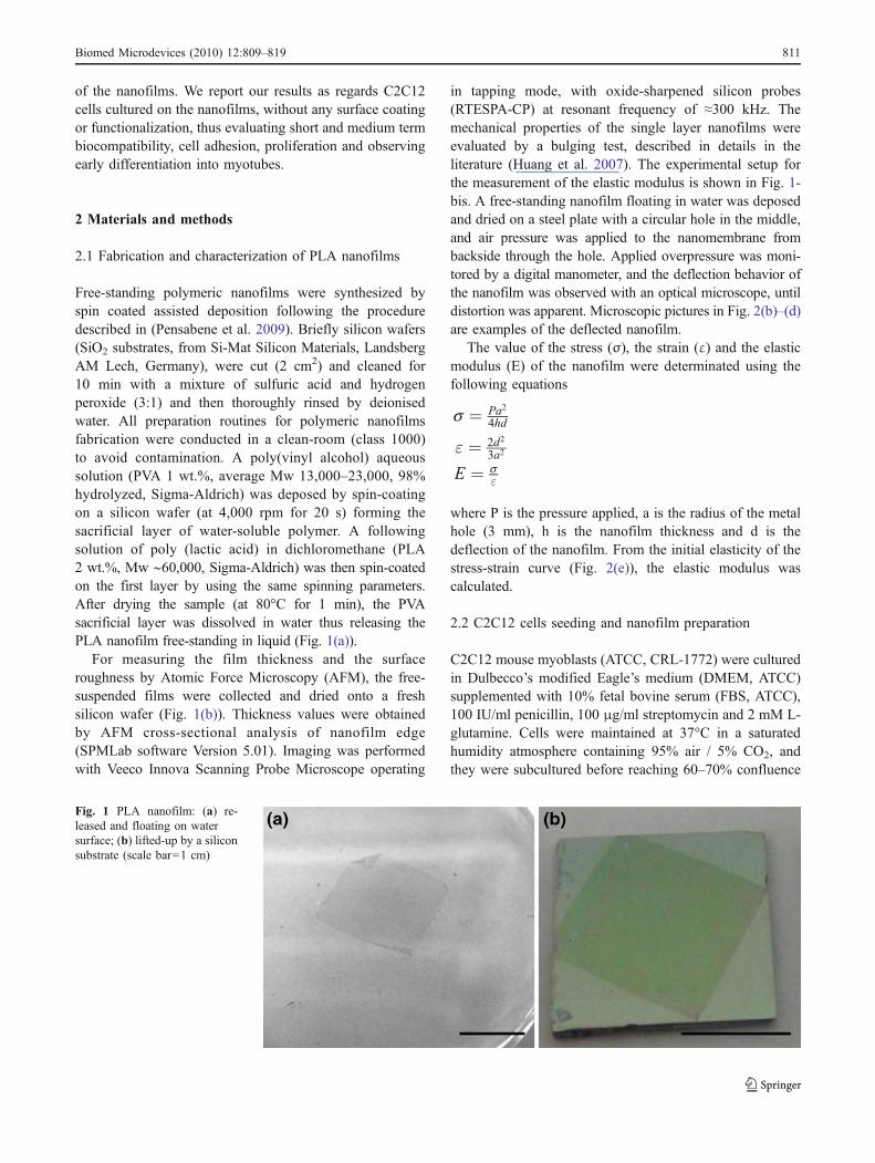
Free-standing polymeric nanofilms were synthesized byspin coated assisted deposition following the proceduredescribed in (Pensabene et al. 2009). Briefly silicon wafers(SiO2 substrates, from Si-Mat Silicon Materials, LandsbergAM Lech, Germany), were cut (2 cm2) and cleaned for10 min with a mixture of sulfuric acid and hydrogenperoxide (3:1) and then thoroughly rinsed by deionisedwater. All preparation routines for polymeric nanofilmsfabrication were conducted in a clean-room (class 1000)to avoid contamination. A poly(vinyl alcohol) aqueoussolution (PVA 1 wt.%, average Mw 13,000–23,000, 98%hydrolyzed, Sigma-Aldrich) was deposed by spin-coatingon a silicon wafer (at 4,000 rpm for 20 s) forming thesacrificial layer of water-soluble polymer. A followingsolution of poly (lactic acid) in dichloromethane (PLA2 wt.%, Mw ∼60,000, Sigma-Aldrich) was then spin-coatedon the first layer by using the same spinning parameters.After drying the sample (at 80°C for 1 min), the PVAsacrificial layer was dissolved in water thus releasing thePLA nanofilm free-standing in liquid (Fig. 1(a)).
For measuring the film thickness and the surfaceroughness by Atomic Force Microscopy (AFM), the free-suspended films were collected and dried onto a freshsilicon wafer (Fig. 1(b)). Thickness values were obtainedby AFM cross-sectional analysis of nanofilm edge(SPMLab software Version 5.01). Imaging was performedwith Veeco Innova Scanning Probe Microscope operating
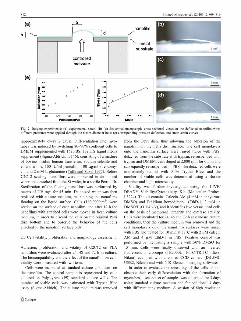
in tapping mode, with oxide-sharpened silicon probes(RTESPA-CP) at resonant frequency of ≈300 kHz. Themechanical properties of the single layer nanofilms wereevaluated by a bulging test, described in details in theliterature (Huang et al. 2007). The experimental setup forthe measurement of the elastic modulus is shown in Fig. 1-bis. A free-standing nanofilm floating in water was deposedand dried on a steel plate with a circular hole in the middle,and air pressure was applied to the nanomembrane frombackside through the hole. Applied overpressure was moni-tored by a digital manometer, and the deflection behavior ofthe nanofilm was observed with an optical microscope, untildistortion was apparent. Microscopic pictures in Fig. 2(b)–(d)are examples of the deflected nanofilm.
The value of the stress (σ), the strain (ε) and the elasticmodulus (E) of the nanofilm were determinated using thefollowing equations
s ¼ Pa2
4hd
" ¼ 2d2
3a2
E ¼ s"
where P is the pressure applied, a is the radius of the metalhole (3 mm), h is the nanofilm thickness and d is thedeflection of the nanofilm. From the initial elasticity of thestress-strain curve (Fig. 2(e)), the elastic modulus wascalculated.
2.2 C2C12 cells seeding and nanofilm preparation
C2C12 mouse myoblasts (ATCC, CRL-1772) were culturedin Dulbecco’s modified Eagle’s medium (DMEM, ATCC)supplemented with 10% fetal bovine serum (FBS, ATCC),100 IU/ml penicillin, 100 μg/ml streptomycin and 2 mM L-glutamine. Cells were maintained at 37°C in a saturatedhumidity atmosphere containing 95% air / 5% CO2, andthey were subcultured before reaching 60–70% confluence
Fig. 1 PLA nanofilm: (a) re-leased and floating on watersurface; (b) lifted-up by a siliconsubstrate (scale bar=1 cm)
Biomed Microdevices (2010) 12:809–819 811
(approximately every 2 days). Differentiation into myo-tubes was induced by switching 80–90% confluent cells toDMEM supplemented with 1% FBS, 1% ITS liquid mediasupplement (Sigma-Aldrich, I3146), consisting of a mixtureof bovine insulin, human transferrin, sodium selenite andethanolamine, 100 IU/ml penicillin, 100 μg/ml streptomy-cin and 2 mM L-glutamine (Yaffe and Saxel 1977). BeforeC2C12 seeding, nanofilms were immersed in de-ionizedwater and detached from the Si wafer, in a sterile Petri dish.Sterilization of the floating nanofilms was performed bymeans of UV rays for 45 min. Deionized water was thenreplaced with culture medium, maintaining the nanofilmsfloating on the liquid surface. Cells (160,000/cm2) wereseeded on the surface of each nanofilm, and after 12 h thenanofilms with attached cells were moved in fresh culturemedium, in order to discard the cells on the original Petridish bottom and to observe the behavior of the cellsattached to the nanofilm surface only.
2.3 Cell vitality, proliferation and morphology assessment
Adhesion, proliferation and vitality of C2C12 on PLAnanofilms were evaluated after 24, 48 and 72 h in culture.The biocompatibility and the effect of the nanofilm on cellsvitality were measured with two tests.
Cells were incubated at standard culture conditions onthe nanofilm. The control sample is represented by cellscultured on Polystyrene (PS) standard culture wells. Thenumber of viable cells was estimated with Trypan Blueassay (Sigma-Aldrich). The culture medium was removed
from the Petri dish, thus allowing the adhesion of thenanofilm on the Petri dish surface. The cell monolayersonto the nanofilm surface were rinsed twice with PBS,detached from the substrate with trypsin, re-suspended withtrypsin and DMEM, centrifuged at 2,000 rpm for 6 min andsubsequently re-suspended in PBS. The detached cells wereimmediately stained with 0.4% Trypan Blue, and thenumber of viable cells was determined using a Burkerchamber and light microscopy.
Vitality was further investigated using the LIVE/DEAD® Viability/Cytotoxicity Kit (Molecular Probes,L3224). The kit contains Calcein AM (4 mM in anhydrousDMSO) and Ethidium homodimer-1 (EthD-1, 2 mM inDMSO/H2O 1:4 v/v), and it identifies live versus dead cellson the basis of membrane integrity and esterase activity.Cells were incubated for 24, 48 and 72 h at standard cultureconditions, then the culture medium was removed and thecell monolayers onto the nanofilms surfaces were rinsedwith PBS and treated for 10 min at 37°C with 2 µM calceinAM and 4 µM EthD-1 in PBS. Positive control wasperformed by incubating a sample with 50% DMSO for15 min. Cells were finally observed with an invertedfluorescent microscope (TE2000U, FITC-TRITC filters,Nikon) equipped with a cooled CCD camera (DS-5MCUSB2, Nikon) and with NIS Elements imaging software.
In order to evaluate the spreading of the cells and toobserve their early differentiation with the formation ofmyotubes, a second set of samples was cultivated for 3 daysusing standard culture medium and for additional 4 dayswith differentiating medium. A session of high resolution
Fig. 2 Bulging experiments; (a) experimental setup; (b)–(d) Sequential macroscopic cross-sectional views of the deflected nanofilm whendifferent pressures were applied through the 6 mm diameter hole; (e) corresponding pressure-deflection and stress-strain curves
812 Biomed Microdevices (2010) 12:809–819
imaging was performed, analyzing cell shape and spreadingonto the nanofilm surface with Atomic Force Microscopy(AFM) and Focused Ion Beam (FIB) microscopy.
For AFM analysis, the presence and the shape ofelongated myotubes were evaluated 7 days after from cellseeding. The culture medium was removed, allowing theadhesion of the nanofilm on the bottom of the Petri dish.The nanofilm surfaces covered by cell monolayers wererinsed twice with deionized water and 10 µl of 2% formalinsolution were added. The samples were maintained at 4°Cfor 15 min, and afterwards on ice, with the Petri dish openin order to dry up, for 20 min.
As regards FIB imaging, we chose a sample cultured for3 days after cell seeding, in order to evaluate cell spreadingand shape. Samples were prepared and fixed following thesame procedure selected for AFM analysis and a final goldlayer (20 nm) was deposed on the dried sample by chemicalvapour deposition (RF Sputtering Sistec, model DCC 150)in order to have a conductive sample. The conductivecoating enhances the contrast and brightness of the sample.In this work a FIB system FEI 200 (Focused Ion Beam forlocalized milling and deposition) was used, delivering a30 keV beam of gallium ions (Ga+), with variable beamcurrents, ranging from 1 pA up to 11 nA.
3 Results
3.1 Nanofilm characterization
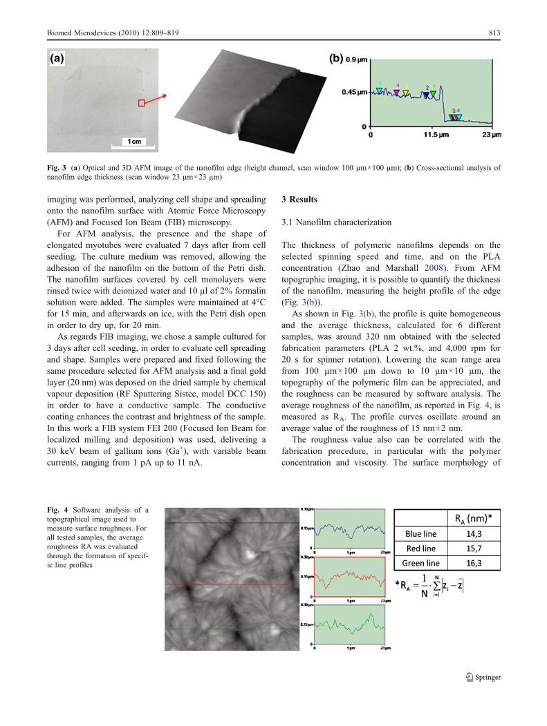
The thickness of polymeric nanofilms depends on theselected spinning speed and time, and on the PLAconcentration (Zhao and Marshall 2008). From AFMtopographic imaging, it is possible to quantify the thicknessof the nanofilm, measuring the height profile of the edge(Fig. 3(b)).
As shown in Fig. 3(b), the profile is quite homogeneousand the average thickness, calculated for 6 differentsamples, was around 320 nm obtained with the selectedfabrication parameters (PLA 2 wt.%, and 4,000 rpm for20 s for spinner rotation). Lowering the scan range areafrom 100 µm×100 µm down to 10 µm×10 µm, thetopography of the polymeric film can be appreciated, andthe roughness can be measured by software analysis. Theaverage roughness of the nanofilm, as reported in Fig. 4, ismeasured as RA. The profile curves oscillate around anaverage value of the roughness of 15 nm±2 nm.
The roughness value also can be correlated with thefabrication procedure, in particular with the polymerconcentration and viscosity. The surface morphology of
Fig. 3 (a) Optical and 3D AFM image of the nanofilm edge (height channel, scan window 100 µm×100 µm); (b) Cross-sectional analysis ofnanofilm edge thickness (scan window 23 µm×23 µm)
Fig. 4 Software analysis of atopographical image used tomeasure surface roughness. Forall tested samples, the averageroughness RA was evaluatedthrough the formation of specif-ic line profiles
Biomed Microdevices (2010) 12:809–819 813
the PLA nanofilm in Fig. 4 showed remarkably crystallinestructure of the assembled polymer.
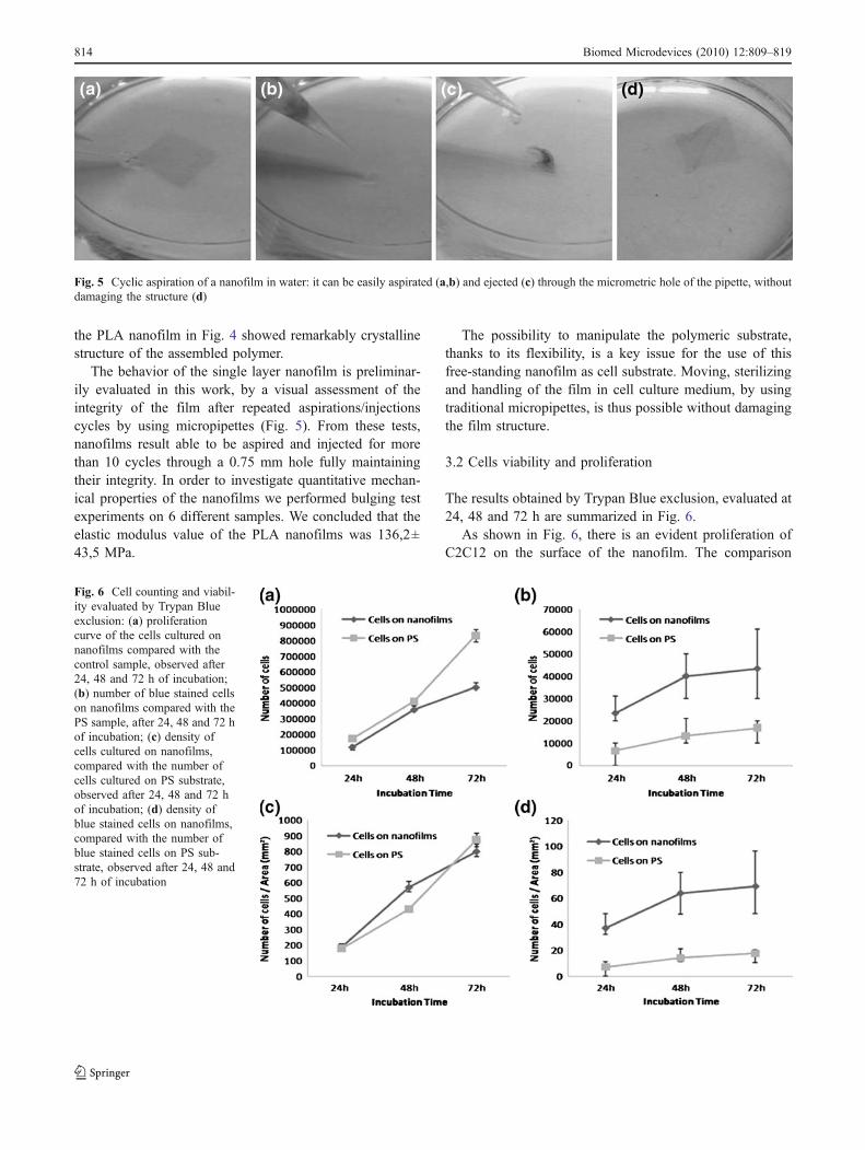
The behavior of the single layer nanofilm is preliminar-ily evaluated in this work, by a visual assessment of theintegrity of the film after repeated aspirations/injectionscycles by using micropipettes (Fig. 5). From these tests,nanofilms result able to be aspired and injected for morethan 10 cycles through a 0.75 mm hole fully maintainingtheir integrity. In order to investigate quantitative mechan-ical properties of the nanofilms we performed bulging testexperiments on 6 different samples. We concluded that theelastic modulus value of the PLA nanofilms was 136,2±43,5 MPa.
The possibility to manipulate the polymeric substrate,thanks to its flexibility, is a key issue for the use of thisfree-standing nanofilm as cell substrate. Moving, sterilizingand handling of the film in cell culture medium, by usingtraditional micropipettes, is thus possible without damagingthe film structure.
3.2 Cells viability and proliferation
The results obtained by Trypan Blue exclusion, evaluated at24, 48 and 72 h are summarized in Fig. 6.
As shown in Fig. 6, there is an evident proliferation ofC2C12 on the surface of the nanofilm. The comparison
Fig. 5 Cyclic aspiration of a nanofilm in water: it can be easily aspirated (a,b) and ejected (c) through the micrometric hole of the pipette, withoutdamaging the structure (d)
Fig. 6 Cell counting and viabil-ity evaluated by Trypan Blueexclusion: (a) proliferationcurve of the cells cultured onnanofilms compared with thecontrol sample, observed after24, 48 and 72 h of incubation;(b) number of blue stained cellson nanofilms compared with thePS sample, after 24, 48 and 72 hof incubation; (c) density ofcells cultured on nanofilms,compared with the number ofcells cultured on PS substrate,observed after 24, 48 and 72 hof incubation; (d) density ofblue stained cells on nanofilms,compared with the number ofblue stained cells on PS sub-strate, observed after 24, 48 and72 h of incubation
814 Biomed Microdevices (2010) 12:809–819
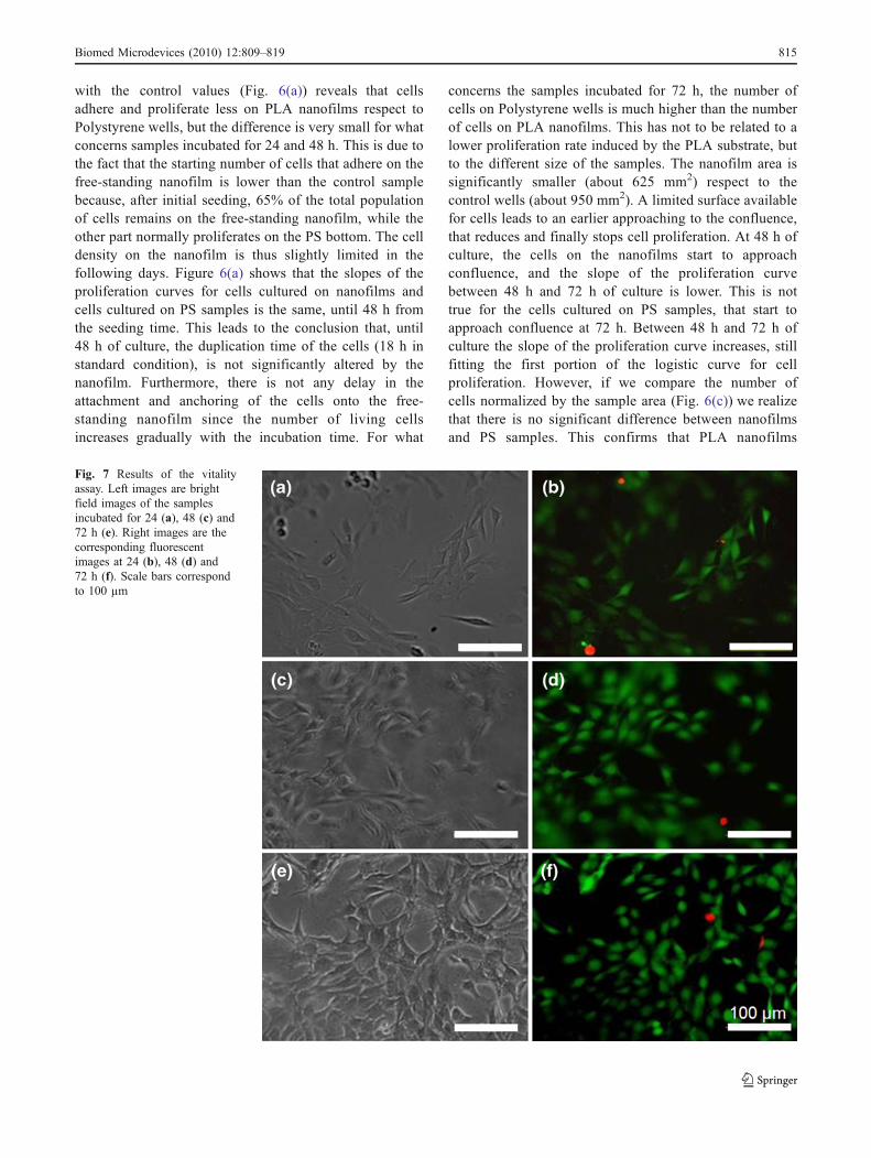
with the control values (Fig. 6(a)) reveals that cellsadhere and proliferate less on PLA nanofilms respect toPolystyrene wells, but the difference is very small for whatconcerns samples incubated for 24 and 48 h. This is due tothe fact that the starting number of cells that adhere on thefree-standing nanofilm is lower than the control samplebecause, after initial seeding, 65% of the total populationof cells remains on the free-standing nanofilm, while theother part normally proliferates on the PS bottom. The celldensity on the nanofilm is thus slightly limited in thefollowing days. Figure 6(a) shows that the slopes of theproliferation curves for cells cultured on nanofilms andcells cultured on PS samples is the same, until 48 h fromthe seeding time. This leads to the conclusion that, until48 h of culture, the duplication time of the cells (18 h instandard condition), is not significantly altered by thenanofilm. Furthermore, there is not any delay in theattachment and anchoring of the cells onto the free-standing nanofilm since the number of living cellsincreases gradually with the incubation time. For what
concerns the samples incubated for 72 h, the number ofcells on Polystyrene wells is much higher than the numberof cells on PLA nanofilms. This has not to be related to alower proliferation rate induced by the PLA substrate, butto the different size of the samples. The nanofilm area issignificantly smaller (about 625 mm2) respect to thecontrol wells (about 950 mm2). A limited surface availablefor cells leads to an earlier approaching to the confluence,that reduces and finally stops cell proliferation. At 48 h ofculture, the cells on the nanofilms start to approachconfluence, and the slope of the proliferation curvebetween 48 h and 72 h of culture is lower. This is nottrue for the cells cultured on PS samples, that start toapproach confluence at 72 h. Between 48 h and 72 h ofculture the slope of the proliferation curve increases, stillfitting the first portion of the logistic curve for cellproliferation. However, if we compare the number ofcells normalized by the sample area (Fig. 6(c)) we realizethat there is no significant difference between nanofilmsand PS samples. This confirms that PLA nanofilms
Fig. 7 Results of the vitalityassay. Left images are brightfield images of the samplesincubated for 24 (a), 48 (c) and72 h (e). Right images are thecorresponding fluorescentimages at 24 (b), 48 (d) and72 h (f). Scale bars correspondto 100 µm
Biomed Microdevices (2010) 12:809–819 815
represent a good surface for C2C12 anchoring, spreadingand proliferation.
This is also confirmed by the results shown in Fig. 6(b) and(d). Dead and apoptotic cells number does not increase in adramatic way on the nanofilm samples with the incubationtime, and it is comparable with the results on Polystyrenesamples. The mortality of cells on nanofilms is higher, butthis can be due to the stresses that cells feel during the initialoperations of manipulation and seeding of the nanofilms.
Figure 7 summarizes results obtained with LIVE/DEAD®assay. The green stained cells are live cells attached to thenanofilms, while the red colored cells are the apoptotic ones.The limited number of dead and apoptotic cells (whichreaches a percentage of 7% at 72 h) confirms the vitality ofcells attached on the nanofilm surface.
3.3 Cells attachment and spreading
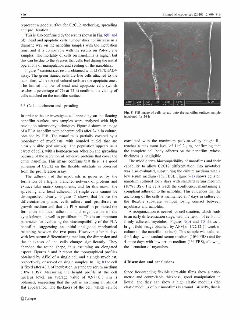
In order to better investigate cell spreading on the floatingnanofilm surface, two samples were analyzed with highresolution microscopy techniques. Figure 8 shows an imageof a PLA nanofilm with adherent cells after 24 h in culture,obtained by FIB. The nanofilm is partially covered by amonolayer of myoblasts, with rounded nuclei that areclearly visible (red arrows). The population appears as acarpet of cells, with a homogeneous adhesion and spreadingbecause of the secretion of adhesive proteins that cover theentire nanofilm. This image confirms that there is a goodadhesion of C2C12 on the flexible substrate as observedfrom the proliferation assay.
The adhesion of the myoblasts is governed by theformation of a highly branched network of proteins andextracellular matrix components, and for this reason thespreading and focal adhesion of single cells cannot bedistinguished clearly. Figure 7 shows that before thedifferentiation phase, cells adhere and proliferate ingrowth medium and that the PLA nanofilm promoted theformation of focal adhesions and organization of thecytoskeleton, as well as proliferation. This is an importantparameter for evaluating the biocompatibility of the PLAnanofilm, suggesting an initial and good mechanicalmatching between the two parts. However, after 4 dayswith low serum differentiating medium, the dimension andthe thickness of the cells change significantly. Theyabandon the round shape, thus assuming an elongatedaspect. Figures 8 and 9 report the topographical profilesobtained by AFM of a single cell and a single myoblast,respectively, observed on single samples. In Fig. 8 the cellis fixed after 48 h of incubation in standard serum medium(10% FBS). Measuring the height profile at the cellnucleus level, an average value of 0,97±0,3 µm isobtained, suggesting that the cell is assuming an almostflat appearance. The thickness of the cell, which can be
correlated with the maximum peak-to-valley height Rt,reaches a maximum level of 1±0.2 µm, confirming thatthe complete cell body adheres on the nanofilm, whosethickness is negligible.
The middle term biocompatibility of nanofilms and theircapability to allow C2C12 differentiation into myotubeswas also evaluated, substituting the culture medium with alow serum medium (1% FBS). Figure 9(a) shows cells onnanofilm cultured for 7 days with standard serum medium(10% FBS). The cells reach the confluence, maintaining acompliant adhesion to the nanofilm. This evidences that theanchoring of the cells is maintained at 7 days in culture onthe flexible substrate without losing contact betweenmyoblasts and nanofilm.
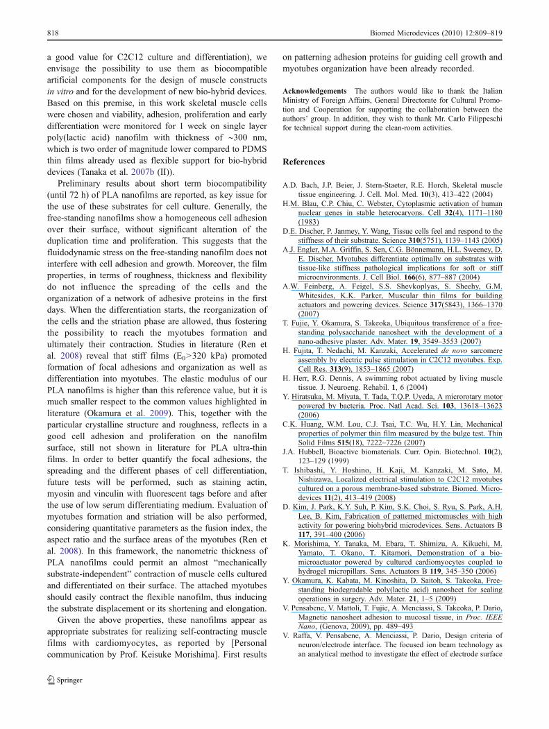
A reorganization is needed for cell striation, which leadsto an early differentiation stage, with the fusion of cells intofirmly adherent myotubes. Figures 9(b) and 10 shows abright field image obtained by AFM of C2C12 (1 week ofculture on the nanofilm surface). This sample was culturedfor 3 days with standard serum medium (10% FBS) and for4 more days with low serum medium (1% FBS), allowingthe formation of myotubes.
4 Discussion and conclusions
Since free-standing flexible ultra-thin films show a nano-metric and controllable thickness, good manipulation inliquid, and they can show a high elastic modulus (theelastic modulus of our nanofilms is around 136 MPa, that is
Fig. 8 FIB image of cells spread onto the nanofilm surface; sampleincubated for 24 h
816 Biomed Microdevices (2010) 12:809–819
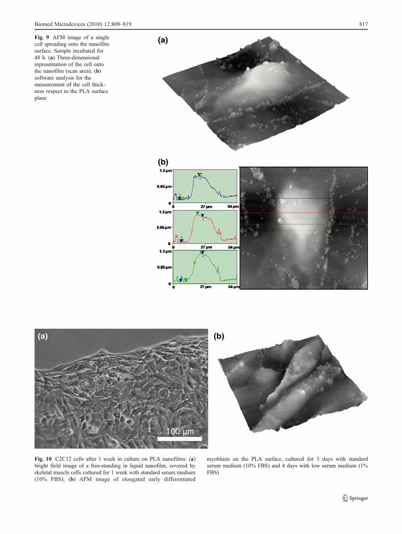
Fig. 9 AFM image of a singlecell spreading onto the nanofilmsurface. Sample incubated for48 h. (a) Three-dimensionalrepresentation of the cell ontothe nanofilm (scan area); (b)software analysis for themeasurement of the cell thick-ness respect to the PLA surfaceplane
Fig. 10 C2C12 cells after 1 week in culture on PLA nanofilms: (a)bright field image of a free-standing in liquid nanofilm, covered byskeletal muscle cells cultured for 1 week with standard serum medium(10% FBS); (b) AFM image of elongated early differentiated
myoblasts on the PLA surface, cultured for 3 days with standardserum medium (10% FBS) and 4 days with low serum medium (1%FBS)
Biomed Microdevices (2010) 12:809–819 817
a good value for C2C12 culture and differentiation), weenvisage the possibility to use them as biocompatibleartificial components for the design of muscle constructsin vitro and for the development of new bio-hybrid devices.Based on this premise, in this work skeletal muscle cellswere chosen and viability, adhesion, proliferation and earlydifferentiation were monitored for 1 week on single layerpoly(lactic acid) nanofilm with thickness of ∼300 nm,which is two order of magnitude lower compared to PDMSthin films already used as flexible support for bio-hybriddevices (Tanaka et al. 2007b (II)).
Preliminary results about short term biocompatibility(until 72 h) of PLA nanofilms are reported, as key issue forthe use of these substrates for cell culture. Generally, thefree-standing nanofilms show a homogeneous cell adhesionover their surface, without significant alteration of theduplication time and proliferation. This suggests that thefluidodynamic stress on the free-standing nanofilm does notinterfere with cell adhesion and growth. Moreover, the filmproperties, in terms of roughness, thickness and flexibilitydo not influence the spreading of the cells and theorganization of a network of adhesive proteins in the firstdays. When the differentiation starts, the reorganization ofthe cells and the striation phase are allowed, thus fosteringthe possibility to reach the myotubes formation andultimately their contraction. Studies in literature (Ren etal. 2008) reveal that stiff films (E0>320 kPa) promotedformation of focal adhesions and organization as well asdifferentiation into myotubes. The elastic modulus of ourPLA nanofilms is higher than this reference value, but it ismuch smaller respect to the common values highlighted inliterature (Okamura et al. 2009). This, together with theparticular crystalline structure and roughness, reflects in agood cell adhesion and proliferation on the nanofilmsurface, still not shown in literature for PLA ultra-thinfilms. In order to better quantify the focal adhesions, thespreading and the different phases of cell differentiation,future tests will be performed, such as staining actin,myosin and vinculin with fluorescent tags before and afterthe use of low serum differentiating medium. Evaluation ofmyotubes formation and striation will be also performed,considering quantitative parameters as the fusion index, theaspect ratio and the surface areas of the myotubes (Ren etal. 2008). In this framework, the nanometric thickness ofPLA nanofilms could permit an almost “mechanicallysubstrate-independent” contraction of muscle cells culturedand differentiated on their surface. The attached myotubesshould easily contract the flexible nanofilm, thus inducingthe substrate displacement or its shortening and elongation.
Given the above properties, these nanofilms appear asappropriate substrates for realizing self-contracting musclefilms with cardiomyocytes, as reported by [Personalcommunication by Prof. Keisuke Morishima]. First results
on patterning adhesion proteins for guiding cell growth andmyotubes organization have been already recorded.
Acknowledgements The authors would like to thank the ItalianMinistry of Foreign Affairs, General Directorate for Cultural Promo-tion and Cooperation for supporting the collaboration between theauthors’ group. In addition, they wish to thank Mr. Carlo Filippeschifor technical support during the clean-room activities.
References
A.D. Bach, J.P. Beier, J. Stern-Staeter, R.E. Horch, Skeletal muscletissue engineering. J. Cell. Mol. Med. 10(3), 413–422 (2004)
H.M. Blau, C.P. Chiu, C. Webster, Cytoplasmic activation of humannuclear genes in stable heterocaryons. Cell 32(4), 1171–1180(1983)
D.E. Discher, P. Janmey, Y. Wang, Tissue cells feel and respond to thestiffness of their substrate. Science 310(5751), 1139–1143 (2005)
A.J. Engler, M.A. Griffin, S. Sen, C.G. Bönnemann, H.L. Sweeney, D.E. Discher, Myotubes differentiate optimally on substrates withtissue-like stiffness pathological implications for soft or stiffmicroenvironments. J. Cell Biol. 166(6), 877–887 (2004)
A.W. Feinberg, A. Feigel, S.S. Shevkoplyas, S. Sheehy, G.M.Whitesides, K.K. Parker, Muscular thin films for buildingactuators and powering devices. Science 317(5843), 1366–1370(2007)
T. Fujie, Y. Okamura, S. Takeoka, Ubiquitous transference of a free-standing polysaccharide nanosheet with the development of anano-adhesive plaster. Adv. Mater. 19, 3549–3553 (2007)
H. Fujita, T. Nedachi, M. Kanzaki, Accelerated de novo sarcomereassembly by electric pulse stimulation in C2C12 myotubes. Exp.Cell Res. 313(9), 1853–1865 (2007)
H. Herr, R.G. Dennis, A swimming robot actuated by living muscletissue. J. Neuroeng. Rehabil. 1, 6 (2004)
Y. Hiratsuka, M. Miyata, T. Tada, T.Q.P. Uyeda, A microrotary motorpowered by bacteria. Proc. Natl Acad. Sci. 103, 13618–13623(2006)
C.K. Huang, W.M. Lou, C.J. Tsai, T.C. Wu, H.Y. Lin, Mechanicalproperties of polymer thin film measured by the bulge test. ThinSolid Films 515(18), 7222–7226 (2007)
J.A. Hubbell, Bioactive biomaterials. Curr. Opin. Biotechnol. 10(2),123–129 (1999)
T. Ishibashi, Y. Hoshino, H. Kaji, M. Kanzaki, M. Sato, M.Nishizawa, Localized electrical stimulation to C2C12 myotubescultured on a porous membrane-based substrate. Biomed. Micro-devices 11(2), 413–419 (2008)
D. Kim, J. Park, K.Y. Suh, P. Kim, S.K. Choi, S. Ryu, S. Park, A.H.Lee, B. Kim, Fabrication of patterned micromuscles with highactivity for powering biohybrid microdevices. Sens. Actuators B117, 391–400 (2006)
K. Morishima, Y. Tanaka, M. Ebara, T. Shimizu, A. Kikuchi, M.Yamato, T. Okano, T. Kitamori, Demonstration of a bio-microactuator powered by cultured cardiomyocytes coupled tohydrogel micropillars. Sens. Actuators B 119, 345–350 (2006)
Y. Okamura, K. Kabata, M. Kinoshita, D. Saitoh, S. Takeoka, Free-standing biodegradable poly(lactic acid) nanosheet for sealingoperations in surgery. Adv. Mater. 21, 1–5 (2009)
V. Pensabene, V. Mattoli, T. Fujie, A. Menciassi, S. Takeoka, P. Dario,Magnetic nanosheet adhesion to mucosal tissue, in Proc. IEEENano, (Genova, 2009), pp. 489–493
V. Raffa, V. Pensabene, A. Menciassi, P. Dario, Design criteria ofneuron/electrode interface. The focused ion beam technology asan analytical method to investigate the effect of electrode surface
818 Biomed Microdevices (2010) 12:809–819
morphology on neurocompatibility. Biomed. Microdevices 9(3),371–383 (2007)
K. Ren, T. Crouzier, C. Roy, C. Picart, Polyelectrolyte multilayer filmsof controlled stiffness modulate myoblast cells differentiation.Adv. Funct. Mater. 18(9), 1378–1389 (2008)
A. Sathaye, N. Bursac, S. Sheehy, L. Tung, Electrical pacingcounteracts instrinsic shortening of action potential duration ofneonatal rat ventricular cells in culture. J. Mol. Cell. Cardiol. 41,633–641 (2006)
H. Shin, S. Jo, A.G. Mikos, Review: biomimetic materials for tissueengineering. Biomaterials 24, 4352–4364 (2003)
Y. Tanaka, K. Morishima, T. Shimizu, A. Kikuchi, M. Yamato, T.Okano, T. Kitamori, Demonstration of a PDMS-based bio-microactuator using cultured cardiomyocytes to drive polymermicropillars. Lab Chip 6, 230–235 (2006)
Y. Tanaka, K. Sato, T. Shimizu, M. Yamato, T. Okano, T. Kitamori,Biological cells mon microchips: new technologies and applica-tions. Biosens. Bioelectron. 23, 449–458 (2007a)
Y. Tanaka, K. Sato, T. Shimizu, M. Yamato, T. Okano, T. Kitamori, Amicro-spherical heart pump powered by cultured cardiomyocytes.Lab Chip 7(2), 207–212 (2007b)
Z. Tang, Y. Wang, P. Podsiadlo, N.A. Kotov, Biomedical applicationsof Layer-by-Layer assembly: from biomimetics to tissue engi-neering. Adv. Mater. 18(24), 3203–3224 (2006)
H. Vandenburgh, J. Shansky, F. Benesch-Lee, V. Barbata, J. Reid, L.Thorrez, R. Valentini, G. Crawford, Drug-screening platform
based on the contractility of tissue-engineered muscle. MuscleNerve 37, 438–447 (2008)
R. Vendamme, S.Y. Onoue, A. Nakao, T. Kunitake, Robust free-standing nanomembranes of organic/inorganic interpenetratingnetworks. Nat. Mater. 5(6), 494–501 (2006)
N.R. Washburn, K.M. Yamada, C.G. Simon, S.B. Kennedy, E.J. Amis,High-throughput investigation of osteoblast response to polymercrystallinity: influence of nanometer-scale roughness on prolifer-ation. Biomaterials 25(7–8), 1215–1224 (2004)
D.B. Weibel, P. Garstecki, D. Ryan, W.R. DiLuzio, M. Mayer, J.E.Seto, G.M. Whitesides, Microoxen: microorganisms to movemicroscale loads. Proc. Natl. Acad. Sci. 102, 11963–11967(2005)
K. Wilson, P. Molnar, J. Hickman, Integration of functional myotubeswith a Bio-MEMS device for non-invasive interrogation. LabChip 7, 920–922 (2007)
J. Xi, J.J. Schmidt, C.D. Montemagno, Self-assembled microdevicesdriven by muscle. Nat. Mater. 4, 180–184 (2005)
D. Yaffe, O. Saxel, A myogenic cell line with altered serum requirementsfor differentiation. Differentiation 7(3), 159–166 (1977)
K. Yamasaki, H. Hayashi, K. Nishiyama, H. Kobayashi, S. Uto, H.Kondo, S. Hashimoto, T. Fujisato, Control of myotube contrac-tion using electrical pulse stimulation for bio-actuator. J. Artif.Organs 12(2), 131–137 (2009)
Y. Zhao, J.S. Marshall, Spin coating of a colloidal suspension. Phys.Fluids 20(4), 043302 (2008)
Biomed Microdevices (2010) 12:809–819 819
Related Documents