Adherence of Candida albicans to Mammalian Cells Adherence of Candida albicans to Mammalian Cells In Vitro: Nutritional Influences Richard Calderone*, Elena Enache, Tara Eskandari, Elsa Wadsworth and Joy Sturtevant Summary Georgetown University School of Medicine, Department of Microbiology and Immunology, Washington, DC 20007, USA Recognition of host cells by Candida albicans has been suggested as essential to the establishment of the organism in tissues. The fact that Candida has been shown thus far to possess a number of adhesins is not surprising considering its ability to colonize and invade a variety of human tissues and mucosal surfaces. One of the Candida adhesins has been characterized as a 60 kDa mannoprotein (MP60). Mutants lacking or with reduced expression of MP60 are also avirulent in animal models of endocarditis, vaginitis and, additionally, while they are able to colonize the gut, cannot invade systemically. The MP60 appears to have both RGD and non-RGD-ligand recognition sites. A closer analysis of the MP60 mutants indicates a correlation between putative mutations in the RGD or non - RGD sites and the inability to cause specific infections. While the mutant studies have provided correlative data, a complete dissection of MP60 function can only be determined by gene knockout studies. To this end, the isolation of the MP60-encoding gene has been attempted using either the human homologue of the B-Iymphocyte CR2 gene to probe Candida libraries or degenerate oligonu- cleotides constructed from the MP60 amino acid sequence. Both approaches have yielded limited success. Adherence of the organism to a variety of host cells can be significantly increased if the organism is grown in 500mM galactose vs. 50mM glucose. Currently, we are using a human eoso- phageal cell line to study the influence of galactose on the adherence of C. albicans. Western blot analyses of cell wall extracts from galactose or glucose grown cells have been performed. A glycosylated protein of approximately 190kDa is observed only in galactose-grown cells. To identify transcripts specific for galactose-grown cells, we have begun studies using RT-PCR differential display. PCR products specific from cells grown in galactose have been subcloned and are currently being studied. This approach may be useful in identifying a variety of galactose-specific genes, including wall proteins which may have an adhesin function. Key words: Candida albicans; adhereuce; mammalian cells; 60-KDa mannoprotein; RGD recognition site Introduction Recognition of host cells by Candida albicans like other microorganisms is thought to involve the interaction of complimentary cell surface molecules (7,8,11-13,17,28). These interactions can be either protein-protein or protein-carbohydrate; in the latter category, fucosyl or amino sugar residues of epithelial cell oligosaccharides are recognized by cell surface lectin-like proteins of C. albicans (15,16,38,42). Protein-protein interactions are thought to involve ,B-integrin- like candidal proteins and arginine-glycine-aspartic acid (RGD) residues of mammalian cell ligands (7,28). * Correspondence: Professor Richard Calderone, Ph.D. Department of Microbiology, Georgetown University School of Medicine, 3900 Reservoir Road NW Washington, DC 20007-2197, USA Phone: + 1-202-687-1151 Fax: + 1-202-687-1800 73

Welcome message from author
This document is posted to help you gain knowledge. Please leave a comment to let me know what you think about it! Share it to your friends and learn new things together.
Transcript
Adherence of Candida albicans to Mammalian Cells
Adherence of Candida albicans to Mammalian Cells In Vitro: Nutritional Influences
Richard Calderone*, Elena Enache, Tara Eskandari, Elsa Wadsworth and Joy Sturtevant
Summary
Georgetown University School of Medicine, Department of Microbiology and Immunology, Washington, DC 20007,
USA
Recognition of host cells by Candida albicans has been suggested as essential to the establishment of the organism in tissues. The fact that Candida has been shown thus far to possess a number of adhesins is not surprising considering its ability to colonize and invade a variety of human tissues and mucosal surfaces. One of the Candida adhesins has been characterized as a 60 kDa mannoprotein
(MP60). Mutants lacking or with reduced expression of MP60 are also avirulent in animal models of endocarditis, vaginitis and, additionally, while they are able to colonize the gut, cannot invade systemically. The MP60 appears to have both RGD and non-RGD-ligand recognition sites. A closer analysis of the MP60 mutants indicates a correlation between putative mutations in the RGD or non - RGD sites and the inability to cause specific infections. While the mutant studies have provided correlative data, a complete dissection of MP60 function can only be determined by gene knockout studies. To this end, the isolation of the MP60-encoding gene has been attempted using either the human homologue of the B-Iymphocyte CR2 gene to probe Candida libraries or degenerate oligonucleotides constructed from the MP60 amino acid sequence. Both approaches have yielded limited success. Adherence of the organism to a variety of host cells can be significantly increased if the organism is grown in 500mM galactose vs. 50mM glucose. Currently, we are using a human eosophageal cell line to study the influence of galactose on the adherence of C. albicans. Western blot analyses of cell wall extracts from galactose or glucose grown cells have been performed. A glycosylated protein of approximately 190kDa is observed only in galactose-grown cells. To identify transcripts specific for galactose-grown cells, we have begun studies using RT-PCR differential display. PCR products specific from cells grown in galactose have been subcloned and are currently being studied. This approach may be useful in identifying a variety of galactose-specific genes, including wall proteins which may have an adhesin function.
Key words: Candida albicans; adhereuce; mammalian cells; 60-KDa mannoprotein; RGD recognition site
Introduction Recognition of host cells by Candida albicans like other microorganisms is thought to involve the
interaction of complimentary cell surface molecules (7,8,11-13,17,28). These interactions can be either protein-protein or protein-carbohydrate; in the latter category, fucosyl or amino sugar residues of epithelial cell oligosaccharides are recognized by cell surface lectin-like proteins of C. albicans (15,16,38,42). Protein-protein interactions are thought to involve ,B-integrin- like candidal proteins and arginine-glycine-aspartic acid (RGD) residues of mammalian cell ligands (7,28).
* Correspondence: Professor Richard Calderone, Ph.D. Department of Microbiology, Georgetown University School of Medicine, 3900 Reservoir Road NW Washington, DC 20007-2197, USA Phone: + 1-202-687-1151 Fax: + 1-202-687-1800
73

Calderone et al.
Because C. albicans can colonize and invade a number of mammalian cells and tissues, each with a specific set of cell surface ligands, it is not surprising that the organism would appear to possess a repetoire of cell surface adhesins which promote its colonization. Because of this complexity, there have been several attempts to categorize the adhesins of C. albicans according to the type of host cell
(epithelial vs. endothelial), the nature of the intereaction (protein-p"rotein or protein-carbohydrate), as well as the type of candidial adhesin (mannan or mannoprotein) . By and large, the candida I adhesins have been identified as mannoproteins with binding domains that recognize oligosaccharides or peptides (7,28) residues of host cell ligands, as stated above. Several examples of the candidal adhesins will be discussed below.
Of particular interest, more recently, investigations have centered upon the isolation of genes which encode recognition proteins of C. albicans. Their role in virulence can be directly studied using gene disruption techniques which circumvent problems associated with diploidy of the organism, such as an inability to utilize classical genetic approaches to study gene function. Unfortunately, while highly desired, studies of this nature are few in number. This manuscript will also focus upon some of those studies.
The C. albicans Epithelial Cell Lectins. 1. Fucosyl/NAGA lectin adhesin.
Adherence of C. albicans to human buccal (HBEC) and vaginal epithelial (HVEC) cells was first reported by Lee and King (30). Their study was also the first to indicate a correlation between an adherence phenotype and virulence in that only the most pathogenic species of Candida could adhere. More extensive studies of the candidal cell surface adhesin which recognizes HBEC has been reported by Douglas and co-workers (15,16,36-38,42) . Among their many observations, they concluded that: 1. galactose-grown yeast cells adhere significantly higher than glucose-grown organisms; 2. an extracellular, polymeric material (EP) could be recovered from culture supernatants of galactose-grown yeasts which was rich in adhesin activity; 3. if coated on acrylic, EP could increase the binding of poorly- adherent strains of the organism; 4. the galactose effect could only be observed with strains of C. albicans from active infections; 5. these same strains when grown in D-galactose resulted in a significantly higher mortality in an animal model; 6. adherence of yeast cells to HBEC was inhibited by fucose or amino sugars and the inhibition was strain-specific and effective only when the sugar was preincubated with the yeast cells and not HBEC. Conversely, when HBEC was preincubated with lectins which recognize fucose, yeast cells were unable to bind to HBEC. The latter two observations indicate that yeast cells possess a lectin-like protein which recognizes either fucosyl - or glucosamine-containing oligosaccharides of epithelial cells. Of all the strains tested, most were inhibited from adhering to HBEC by fucose or fucose-binding lectins and, thus, express a fucosyl - type lectin.
Purification of the candidallectin was accomplished by a combination of affinity (concanavalin A) and ion exchange chromatography and removal of oligosaccharides by glycosidases (42). The active material was proteinaceous and recognition of HBEC was unaffected by removal of the mannan. As an additional step in purification of this HBEC adhesin, following N-glycanase, papain and dilute alkali treatments of EP, protein fragments were isolated by affinity adsorption with the trisaccharide determinant of the H (type 2) blood group antigen which terminates in a residue of L-fucose (42). Again, the active material isolated following affinity purification was devoid of carbohydrate and had an adhesin activity which was greater than 200-fold that of the starting material (EP).
2. The fimbrial adhesin. Yu et al (44- 46) have described surface structures of C. albicans which resemble bacterial
74

Adherence of Candida albicans to Mammalian Cells
fimbriae. Since fimbriae confer adhesin functions in other organisms, these investigators purified the candidal fimbriae in order to determine their function in the adherence of C. albicans to HBEC. Fimbriae were obtained by gentle homogenization of yeast cells grown on Sabouraud agar, followed by centrifugation to remove whole cells and filtration. This preparation, termed crude fimbriae (CF) was subsequently fractionated by size exclusion-high performance liquid chromatography (46). The active material isolated by this technique (enriched fimbriae, EF) had a molecular mass of 66 kD and was composed of approximately 85% carbohydrate and 15% protein (46). Antibodies prepared against EF confirmed the molecular mass of the mannoprotein and the cell surface location of the fimbriae. Purified fimbriae bound to HBEC in a saturable and concentration-dependent manner and also inhibited the adherence of yeast cells when pre-incubated with HBEC.
In attempting to identify the ligand of HBEC recognized by the fimbrial protein, an in vitro binding assay was established in which fimbriae were incubated with several glycolipids which had been resolved by thin layer chromatography (44) . Of the glycolipids tested, fimbriae bound to asialo -GM1 and asialo-GM2 but not monosialo-GM1 or other glycolipids tested. The disaccharide, ,BGaIN Ac (1-4) ,BGal, is thought to be the minimal structural requirement for a number of asialo-GM1 -adhesins. When conjugated to BSA and immobilized on microtiter plates, fimbriae bound to this disaccharide in a saturable and concentration-dependent manner, and, when preincubated with fimbriae, asialo-GM1 as well as a methyl ester of the disaccharide (mentioned above) inhibited binding of the fimbriae to HBEC. On the other hand, binding to HBEC was not inhibited by those glycolipids which did not bind fimbriae in thin layer chromatography assays.
The asialo-GM1 receptor is used by several microorganisms including Pseudomonas aeruginosa (45). The availability of a number of antibodies reactive with the pseudomonal pilin, asialo-GM1
adhesin at and near the ligand binding domain allowed Yu et al. to determine the similarity of the candidal ligand-binding domain with that of P. aeruginosa (45). Agglutination of yeast cells was observed with antibodies to the P. aeruginosa whole pi lin or to the asialo-GM1 binding domain of the pilin adhesin. Conversely, a candidal antibody also agglutinated P. aeruginosa whole cells demonstrating the cross-reactivity of epitopes between these diverse organisms. Other anti-pseudomonal antibodies directed to other regions of the pilin protein failed to agglutinate whole yeast cells indicating that the conservation among these proteins may be limited to specific and functional domains of the proteins.
3. Nutritional Influences on the expression of the candidal lectin. As stated above, Douglas and co-workers (36-38) have shown that yeast cells of C. albicans
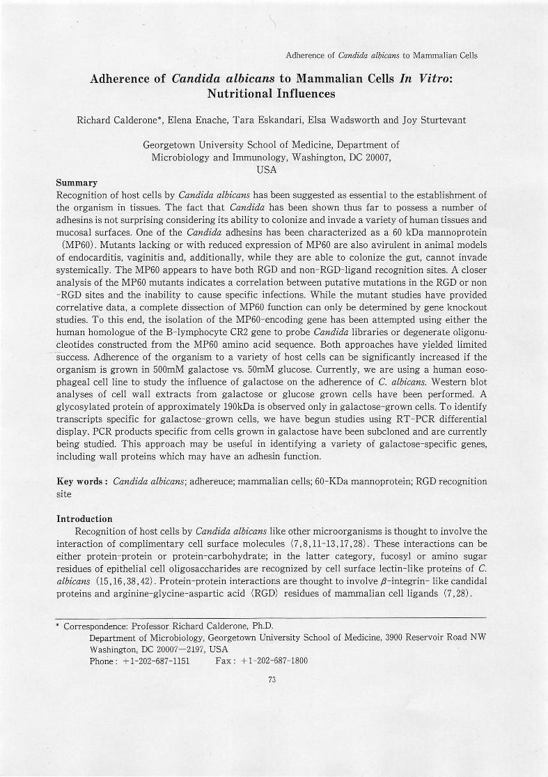
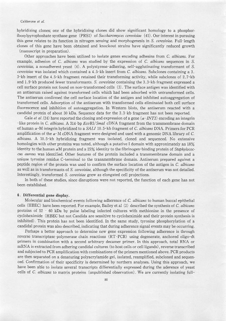
adhere to HBEC a greater extent when grown in 50 mM galactose in comparison to concentrations of glucose. We have observed a similar effect of galactose on the adherence of the organism to a human esophageal cell line. These cells were chosen for study because the esophagus ·is a target for C. albicans in HIV + / AIDS patients (31). Orignially described by Stoner et al. (39), the esophageal cell line (HET1-A) used in our studies has been transformed with SV-40 but is not tumorigenic, retains the typical array of epithelial cell antigens and displays a polarized growth in culture. We demonstrated that adherence to HET1-A was significantly increased when yeast cells were grown overnight in 500mM galact?se (Fig. 1, Table 1) incomparison to 500mM glucose (Enache et al ., Microbiology, manuscript in press). Adherence of the organism to HET1-A cells was blocked by amino sugars but not by fucose. While strain specificity of C. albicans has been observed in regard to recognition of specific sugar residues of glycosides of HBEC (fucose or NAGA) , all strains of C. albicans tested in this study for adherence to HET1-A cells were inhibited by amino sugars but not fucose, galactose, or mannose. Following growth of yeast cells in 500 mM galactose or glucose for 48 hr, cell wall proteins were extracted and analyzed by SDS-PAGE and Western blot. We observed a 190 kDa mannoprotein only in galactose-grown cells (Fig. 2). This protein has been purified,
75

Calderone et at.
although its relationship to an adhesin function is not yet known. We are currently sequencing the protein.
60
w50 () z40 w ffi30
520 <I: ~10
Adherence to HET1-A Cells
30 60 90 180 MINUTES
o GALACTOSE I GLUCOSE
Fig.1. Adherence of C. albicans to HETI-A cells over time. Yeast cells were grown overnight in either 500mM D-galactose or D-glucose, washed and added to HETI-A cell monolayers.
Table 1. The Effect of Sugars on the Adherence of Candida albicans to Human Esophageal Cells (HETI -A)
% of HETl-A with Adhering yeasts
Carbon source
Glucose
Galactose Trehalose Maltose Xylose
p=.0003, galactose vs. glucose
11. 7
31.1 14.5 23.4 30.0
± 4
± 7*
± 5
± 3
± 2
Yeasts cells were grown overninght in each of the sugars liste
above, standardized and added to HETl-A cells. Adherence was
then determined.
Adherence of C. albicans to HETI-A cells has also been studied by light and electron microscopy. By using scanning and transmission EM, we observed that HETI-A cells phagocytized both yeast cells and germinating yeast cells. Pseudopodia of HETI-A cells appeared to form in response to candida I cells (Fig. 3) . Also, extensive membrane folding (apparently of HETI-A origin) appeared in regions of close contact between the cells.
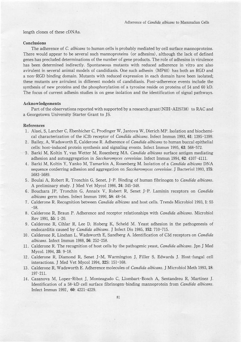
We used reverse transcriptase-polymerase chain reaction (RT -peR) to analyze differential gene transcription in galactose or glucose grown C. albicans . Initial experiments revealed several galactose-specific transcripts, which currently are the subject of study in the laboratory (Fig. 4).
76

Adherence of Candida albicans to Mammalian Cells
Fig. 2. Western blot analyses of glucose- (lane 1) and galactose- (lane 2) grown yeast cells of C. albicans. Cells grown under each condition were washed and extracted with beta-mercaptoethanol. Electrophoresed proteins were transferred to nitrocellulose membranes "and reacted with a polyclonal antiserum raised in infected rabbits. Reactive proteins were detected using a peroxidase-conjugated, goat anti-rabbit IgG. A mannoprotein of 190 kD is observed only in galactose grown cells.
Fig. 3. Transmission electron micrograph of HETI-A cells infected with yeast cells of C. albicans, 90 min post-infection. Note the pseudopodia of the HETI-A cells and what would appear to be a phagocytic process.(reproduced by permission of the Society for General Microbiology)
77

Calderone et at.
6
:::l '---1 .::> 2---1 -' « .J « C)(S(!3(!J
III-...
~ ,.. . ,._ -~ ..... := ~
Fig. 4. Reverse transcriptase-polymerase chain reaction (RT-peR) of glucose (GLU) vs. galactose (GAL) -grown C. albicans. RNA was isolated from each cell population and reverse transcribed. cDN As were peR-amplified using degenerate, anchored ·oligo dT primers and an arbitary decamer. peR products were then electrophoresed. In this figure, only oligo-dTG (G) and oligo-dTe (e) reactions from two representative experiments are shown.
4 . The endothelial adhesin (s) . A number of laboratories have described the expression of cell surface ,B-integrin-like proteins
in C. albicans by demonstrating that antibodies to the mammalian family of integrin proteins cross -react with candidal proteins (28). Also demonstrated has been the binding of yeast, pseudohyphal or hyphal forms of the organism to sheep erythrocytes conjugated with a complement conversion product of C3 (iC3b) or several endothelial extracellular matrix proteins (ECM), each of which contains RGD sequences typically recognized by ,B-integrin proteins (10,20,25-28,32 ,33,35,40,43).
Purification of a C. albicans 60 kDa mannoprotein (MP60), which recognizes both RGD (iC3b) and non-RGD (C3d) containing ligands, by ion exchange and ligand affinity chromatography, has been reported (10,35,43). This protein is expressed in vivo, as determined by MP60-induced proliferation of lymphocytes from infected animals (23) and by immunoelectron microscopic tagging of MP60 from tissues of infected animals (29).
Other investigators have reported that a protein (s) of similar molecular mass (60 kDa) binds ECM proteins such as laminin, fibrinogen and fibronectin (5,6,32). In comparison, a protein of 58 kD molecular mass, which appears to be different immunologically from the MP60, has demonstrated fibrinogen-binding activity (14) . A cytosolic protein of 165 kD from C. albicans is recognized by an antibody to a human ,B-integrin protein (28). The relationship of this protein to a cell surface adhesin is unknown. Alaei et al . (1) also have shown that a 42 kDa mannoprotein from C. albicans also has an integrin-like activity; further, their data indicate that other reactive proteins exist which probably represent various glycosylated forms of the 42 kDa protein.
Spontaneous mutants with reduced expression of MP60 adhere less readily to HBEC, vaginal epithelial cells and fibrin-platelet clots formed in vitro (7,9). Correlated with an impaired adherence of the mutants is their reduced infectivity in animal models of endocarditis and, vaginitis (9,34) .
78

Adherence of Candida albicans to Mammalian Cells
Table 2. Expression of ligand binding activity (RGD or non-RGD) in mutants of Candida albicans: relationship to virulence
Strain* wt-4918 M-IO P
' A.
MP60 Ligands Virulence Non-RGD RGD Systemic Vaginitis
A. A. Vir Vir -- --A. Vir Avir --
A. Avir Vir
MP60 protein with binding domains (A.) for Non-RGD and RGD containing ligands
*strains m-10 and P are spontaneously derived and are resistan to cerulenin (m-10) or clotrimazole {P}
Further, one of these mutants is able to colonize murine gut tissue but is unable to invade systemically from that site (19) .
The MP60 mutants, upon closer examination, can be placed in two categories; those with reduced expression of the non-RGD binding domain (such as C. albicans strain P) are able to cause rat vaginitis (18) but are unable to cause systemic infection in animals when inoculated by the intravenous route (22) , On the other hand, a mutant with impaired binding of RGD ligands (c. albicans m-l0) does not cause vaginitis (34) but can invade systemically. These data indicate a correlation between the ability of strains to invade specific tissues and the presence/ absence of specific ligand binding activities (Table 2).
Another association between the expression of ,B-integr-ins in C. albicans and virulence is the observation that pretreatment of mice with RGD-containing peptides prevents colonization of tissues in animals subsequently infected (33). The mutant studies (described above) and ligand protection experiments are only correlative and do not uneqivocally establish the ,B- integrin-like protein of C. albicans as a virulence factor.
5. Molecular characterization of the C. albicans adhesins The diploid genome of C. albicans has complicated the isolation of specific mutations. As stated
above, while non-adhering, avirulent, spontaneously-derived mutants exist for study, one cannot be certain that these strains represent only specific mutations. For this reason, the preferred approach to obtaining mutations in a specific gene is to construct knockout strains through sequential transformation with a disruption casette [His-Ura- His] inserted within a known gene sequence (21). Recovery of transformants, disrupted in one allele by homologous recomb'ination between the disrupted gene and a wild type allele (heterozygotes) , is followed by a second transformation (as described above). Doubly disrupted transformants are confirmed by hybridizations; their phenotype then can be compared to parental cells. This procedure obviously requires that the gene in question has been isolated and sequenced. In regard to genes which encode adhesin proteins in C. albicans, there has been far more failures than successes. We have used heterologous sequences as probes of C. albicans genomic libraries but have been unable to isolate any clones which resemble adhesin proteins; for example, genomic clones of C. albicans which hybridized to a human B-cell complement receptor type 2 (CR2) cDNA were homologous instead with a family of ATP-binding cassette genes
(ABC) associated with transport functions in a variety of organisms (manuscript in preparation). We have also constructed degenerate oligonucleotides (from peptide sequences of the MP60) for use as probes of candidal libraries (4l). Again, we were unable to identify specific sequences from
79
Calderone et al.
hybridizing clones; one of the hybridizing clones did show significant homology to a phosphoribosylpyrophoshate synthase gene (PRS1) of Saccharomyces cerevisiae (4l). Our interest in pursuing. this gene relates to its function in nitrogen sensing and morphogenesis in S. ceresiviae. Full-length clones of this gene have been obtained and knockout strains have significantly reduced growth
(manuscript in preparation) . Other apprqaches have been utilized to isolate genes encoding adhesins from C. albicans. For
example, adhesion of C. albicans was studied by the expression of C. albicans sequences in S. cerevisiae, a nonadherent yeast (4). A polystyrene-adhering, self-agglutinating transformant of S. cerevisiae was isolated which contained a 4. 5-kb insert from C. albicans. Subclones containing a 3. 3-kb insert of the 4. 5-kb fragment retained their transforming activity, while subclones of 2. 7-kb and 1.9-kb produced fewer transformants. S. cerevisiae containing the 3.3-kb fragment expressed a cell surface protein not found on non-transformed cells (3) . The surface antigen was identified with an antiserum raised against transformed cells which had been adsorbed with untransformed cells. The antiserum confirmed the cell surface location of the antigen and inhibited aufoaggregation of transformed cells. Adsorption of the antiserum with transformed cells eliminated both cell surface fluorescence and inhibition of autoaggregation. In Western blots, the antiserum reacted with a candidal protein of about 30 kDa. Sequence data for the 3.3-kb fragment has not been reported.
Gale et al. (24) have reported the cloning and expression of a gene (a-INTi) encoding an integrin -like protein in C. albir:;ans. A 314-bp EcoRI/ SmaI cDNA fragment from the transmembrane domain of human a-M integrin hybridized to a SALI 10 .5-kb fragment of C. albicans DNA. Primers for PCR amplification of the a-M cDNA fragment were designed and used with a genomic DNA library of C. albicans. A 10. 5-kb hybridizing fragment was isolated, cloned and sequenced. No extensive homologies with other proteins was noted, although a putative I domain with approximately an 18% identity to the human aM protein and a 25% identity to the fibrinogen-binding protein of Staphylococcus aureus was identified. Other features of the protein included a transmembrane domain and a unique tyrosine residue C-terminal to the transmembrane domain. Antiserum prepared against a peptide region of the protein was used to confirm the surface location of the antigen in C. albicans as well as in transformants of S. cerevisiae, although the specificity of the antiserum was not detailed. Interestingly, transformed S. cerevisiae grew as elongated cell projections.
In both of these studies, since disruptions were not reported, the function of each gene has not been established.
6. Differential gene display. Molecular and biochemical events following adherence of C. albicans to human buccal epithelial
cells (HBEC) have.been reported. For example, Bailey et al (2) described the synthesis of C. albicans proteins of 52 - 60 kDa by pulse labeling infected cultures with methionine in the presence of cycloheximide (HBEC but not Candida are sensitive to cycloheximide and their protein synthesis is inhibited) . This protein has not been identified. In the same study, tyrosine phosphorylation of a candidal protein was also described, indicating that during adherence signal events may be occurring.
Perhaps a better approach to determine new gene expression following adherence is through reverse transcriptase-polymerase chain reactions (RT - PCR) using degenerate, anchored oligo-dt primers in combination with a second arbitrary decamer primer. In this approach, total RNA or mRNA is extracted from adhering candidal cultures (to host cells or cell ligands) , reverse transcribed and subjected to PCR amplification with combinations of the primers mentioned above. PCR products are then separated on a denaturing polyacrylamide gel, isolated, reamplified, subcloned and sequenced. Confirmation of their specificity is determined by northern analyses. Using this approach, we have been able to isolate several transcripts differentially expressed during the aderence of yeast cells of C. albicans to matrix proteins (unpublished observation) . We are currently isolating full-
80
Adherence of Candida albicans to Mammalian Cells
length clones of these cDNAs.
Conclusions The adherence of C. albicans to human cells is probably mediated by cell surface mannoproteins.
There would appear to be several such mannoproteins (or adhesins), although the lack of defined genes has precluded determinations of the number of gene products. The role of adhesins in virulence has been determined indirectly. Spontaneous mutants with reduced adherence in vitro are also avirulent in several animal models of candidiasis. One such adhesin (MP60) has both an RGD and a non-RGD binding domain. Mutants with reduced expression in each domain have been isolated; these mutants are avirulent in different models of candidiasis. Post-adherence events include the synthesis of new proteins and the phosphorylation of a tyrosine reside on proteins of 54 and 60 kD. The focus of current adhesin studies is on gene isolation and the identification of signal pathways.
Acknowledgements Part of the observations reported with supported by a research grant (NIH -AI25738) to RAC and
a Georgetown University Starter Grant to JS.
References 1. Alaei, S, Larcher C, Ebenbicher C, Prodinger W, J antova W, Dierich MP. Isolation and biochemi
cal characterization of the iC3b receptor of Candida albicans. Infect Immun 1993, 61: 1395-1399. 2. Bailey, A, Wadsworth E, Calderone R. Adherence of Candida albicans to human buccal epithelial
cells: host-induced protein synthesis and signaling events. Infect Immun 1995, 63: 568-572 . . - -So Barki M, Koltin Y, van Wetter M, Rosenberg MA. Candida albicans surface antigen mediating
adhesion and autoaggregation in Saccharomyces cerevisiae. Infect Immun 1994, 62: 4107-411l. 4. Barki M, Koltin Y, Yanko M, Tamarkin A, Rosenberg M. Isolation of a Candida albicans DNA
sequence conferring adhesion and aggregation on Saccharomyces cerevisiae. J Bacteriol1993, 175: 5683-5689.
5. Boulai A, Robert R, Tronchin G, Senet, J-P. Binding of human fibrinogen to Candida albicans. A preliminary study. J Med Vet Mycol 1986, 24: 345-348.
6. Bouchara JP, Tronchin G, Annaix V, Robert R, Senet J-P. Laminin receptors on Candida albicans germ tubes. Infect Immun 1990, 58: 48-54.
7. Calderone R. Recognition between Candida albicans and host cells. Trends Microbiol 1993, 1: 55 -58.
8. Calderone R, Braun P. Adherence and receptor relationships with Candida albicans. Microbiol Rev 1991, 55: 1-20.
9. Calderone R, Cihlar R, Lee D, Hoberg K, ScheId M. Yeast adhesion in the pathogenesis of endocarditis caused by Candida albicans. J Infect Dis 1985, 152: 7l0-7l5.
10. Calderone R, Linehan L, Wadsworth E, Sandberg A. Identification of C3d receptors on Candida albicans. Infect Irhmun 1988, 56: 252-258.
11 . Calderone R. The recognition of host cells by the pathogenic yeast, Candida albicans. J pn J Med Mycol. 1994, 35: 9-18.
12. Calderone R, Diamond R, Senet J - M, Warmington J, Filler S, Edwards J. Host-fungal cell interactions. J Med Vet Mycol 1994, 32S: 151-168.
13. Calderone R, Wadsworth E. Adherence molecules of Candida albicans. J Microbiol Meth 1993, 18: 197-21l.
14 . Casanova M, Lopez-Ribot J, Monteagudo C, Llombart-Bosch A, Sentandreu R, Martinez J. Identification of a 58-kD cell surface fibrinogen-binding mannoprotein from Candida albicans . Infect Immun 1992, 60: 4221-4229.
81

Calderone et at.
15. Critchley lA, Douglas LJ. Role of glycosides as epithelial cell receptors for Candida albicans. J Gen Microbiol 1987, 133: 637-643.
16 . Critchley lA, Douglas LJ. Isolation and partial characterization of an adhesin from Candida albicans. J Gen Microbiol 1987, 133: 629-636.
17. Cutler JE. Putative virulence factors of Candida albicans. Annu Rev Microbiol1991, 45: 187-218. 18 . DeBernar:disF, Cassone A, Sturtevant], Calderone RExpression of. Candida albicans SAPI and
SAP2 in experimental vaginitis. Infect Imrnun 1995, 63: 1887-1892. 19. DeRepentigny L, Phaneuf M, Mathieu L-G. Gastrointestinal colonization and systemic dissemina·
tion by Candida albicans. Infect Immun 1992, 60: 4907-4914. 20. Edwards J, Gaither T, 0' Shea], Rotrosen D, Lawley T, Frank M, Green I. Expression of specific
binding sites on Candida with functional and antigenic characteristics of human complement receptors. J Immunol 1986, 137: 3577-3583.
21 . Fonzi W, Irwin M. Isogenic strain construction and gene mapping in Candida albicans. Genetics 1993, 134: 712-728.
22. Franzke S, Calderone R, Schaller K. Isolation of avirulent clones of Candida albicans with reduced ability to recognize the CR2 ligand C3d. Infect Immun 1993, 61: 2662-2669.
23. Fukayama M, Wadsworth E, Calderone R Expression of the C3d-binding protein (CR2) from Candida albicans during experimental candidiasis as measured by lymphoblastogenesis. Infect Immun 1992, 60: 8-12.
24. Gale C, Finkel D, Tao N, Meinke M, McClellan M, Olson], Kendrick K, Hostetter M. Cloning and expression of a gene encoding an integrin-like protein in Candida albicans. Proc Natl Acad Sci 1996, 3: 357-361.
25. Gilmore B, Retsinas E, Lorenz], Hostetter M. An iC3b receptor on Candida albicans: structure, function and correlates for pathogenicity. J Infect Dis 1988, 157: 38-46.
26. Gustafson K, Verceletti G, Bendei C, Hostetter M. Molecular mimicry in Candida albicans: role of an integrin analogue in adhesion of the yeast to human endothelium. J Clin Invest 1991, 87: 1896-1902.
27. Heidenreich F, Dierich M. Candida albicans and Candida stellatoidea, in contrast other Candida species, bind iC3b and C3d but not C3b. Infect Immun 1985, 50: 598-600.
28 . Hostetter M. Adhesins and ligands involved in the interaction of Candida spp with epithelial and endothelial surfaces. Clin Microbiol Rev 1994, 7: 29-42.
29. Kanbe T, Li R -K, Wadsworth E, Calderone R, Cutler J. Evidence for expression of the C3d receptor of Candida albicans in vitro and in vivo obtained by immunofluorescent and immunoelectron microscopy. Infect Immun 1991, 59: 1832-1838.
30. King RD, Lee JC, and Morris AL. Adherence of Candida albicans and other Candida species to mucosal epithelial cells. Infect Imrnun 1962, 27: 667-674.
31. Klein RS, Harris CA, Small CB, Moll B, Lesser M, Friedland GH. Oral candidiasis in high risk patients as the initial manifestation of acquired immunodeficiency syndrome. N Engl J Med 1984, 311: 354-358.
32 . Klotz, SA. Adherence of Candida albicans to components of the subendothelial extracellular matrix. FEMS Microbiol Lett 1990, 68:249-254.
33. Klotz SA, Smith RL, Stewart BW. Effect of an arginine-glycine-aspartic acid-containing peptide on hematogenous candidal infections in rabbits. Infect Immun 1992, 36: 132-136.
34. Lehrer N, Segal E, Cihlar R, and Calderone R Pathogenesis of vaginal candidiasis: studies with a mutant which has reduced ability to adhere in vitro. J Med Vet Mycol 1986, 24: 127-131.
35. Linehan L, Wadsworth E, Calderone R Candida albicans C3d receptor, isolated using a monoclonal antibody. Infect Immun 1988, 56: 1981-1986.
36. McCourtie J, Douglas LJ. Relationship between cell surface composition of Candida albicans and
82
Adherence of Candida albicans to Mammalian Cells
adherence to acrylic after growth on different carbon sources. Infect Immun 1981. 32: 1234-124l. 37. McCourtie J. Douglas LJ. Relationship between cell surface composition. adherence and viru
lence of Candida albicans. Infect Immun 1984. 45: 6-12. 38. McCourtie J. Douglas LJ. Extracellular polymer of Candida albicans: isolation. analysis and role
in adherence. J Gen Microbiol 1985. 131: 495-503. 39. Stoner GD. Kaighn ME. Reddel RR. Resau JH. Bowman D. Naito. Z. Matsukura N. You M. Galati
AJ. Curtis CC. Establishment and characterization of SV -40 T-antigen immortalized human esophageal epithelial cells. Cancer Res '1991. 51: 365-37l.
40. Ollert, MW, Wadsworth E, Calderone R. Reduced expression of the functionally active complement receptor for iC3b but not for C3d on an avirulent mutant of Candida albicans. Infect Immun 1990. 58: 909-913.
41. Payne T, Calderone R. Isolation of phospho ribosyl pyrophosphate synthetase (PRS1) gene from Candida albicans. Yeast. 1995, 11: 1295-1302.
42. Tosh FD, Douglas LJ. Characterization of a fucoside-binding adhesin of Candida albicans. Infect Immun 1992. 60: 4734-4739.
43. Wadsworth E, Calderone R. Analysis of mannoproteins from blastoconidia and hyphae of Candida albicans. with a common epitope recognized by anti-complement receptor type 2 antibodies. Infect Immun 1993. 61: 4675-468l.
44. Yu L, Lee K, Hodges R. Paranchych W. Irvin RT. Adherence of Pseudomonas aeruginosa and Candida albicans to glycosphingolipid (asialo-GMl) receptors is achieved by a conseerved receptor-binding domain present on their adhesins. Infect Immun 1994. 64: 5210-5219.
45. Yu L, Lee K, Sheth H, Lane-Hall- P, Srivastava G, Hindsgaul 0, Paranchych W, Hodges R, Irvin RT. Fimbria-mediated adherence of Candida albicans to glycosphingolipid receptors on human buccal epithelial cells. Infect Immun 1994. 62: 2843-2848.
46. Yu, L. Lee K. Ens K, Doig, p. Carpenter M. Staddon W, Hodges R, Paranchych W, Irvin RT. Partial characterization of a Candida albicans fimbrial adhesin. Infect Immun 1994. 62: 2834-2842.
83
Related Documents