1 Adenylyl cyclase/cAMP system involvement in the antiangiogenic effect of somatostatin in the retina. Results from transgenic mice. Chiara Ristori 1 , Maria Enrica Ferretti 2 , Barbara Pavan 2 , Franco Cervellati 2 , Giovanni Casini 3 , Elisabetta Catalani 3 , Massimo Dal Monte 1 , Carla Biondi 2 . 1 Dipartimento di Biologia - Università di Pisa - Pisa, Italy 2 Dipartimento di Biologia ed Evoluzione - Università di Ferrara - Ferrara, Italy. 3 Dipartimento di Scienze Ambientali - Università della Tuscia - Viterbo, Italy. Running head: Involvement of AC system in SRIF antiangiogenic action in hypoxic mouse retina. Corresponding author: Carla Biondi, Dipartimento di Biologia ed Evoluzione – Università di Ferrara – Via L. Borsari, 46 – 44100 Ferrara, Italy – Phone: +39 0532 455482 – FAX: +39 0532 207143 – e-mail: [email protected]

Welcome message from author
This document is posted to help you gain knowledge. Please leave a comment to let me know what you think about it! Share it to your friends and learn new things together.
Transcript

1
Adenylyl cyclase/cAMP system involvement in the antiangiogenic effect of somatostatin
in the retina. Results from transgenic mice.
Chiara Ristori1, Maria Enrica Ferretti
2, Barbara Pavan
2, Franco Cervellati
2, Giovanni
Casini3, Elisabetta Catalani
3, Massimo Dal Monte
1, Carla Biondi
2.
1Dipartimento di Biologia - Università di Pisa - Pisa, Italy
2Dipartimento di Biologia ed Evoluzione - Università di Ferrara - Ferrara, Italy.
3Dipartimento di Scienze Ambientali - Università della Tuscia - Viterbo, Italy.
Running head: Involvement of AC system in SRIF antiangiogenic action in hypoxic mouse
retina.
Corresponding author: Carla Biondi, Dipartimento di Biologia ed Evoluzione – Università
di Ferrara – Via L. Borsari, 46 – 44100 Ferrara, Italy – Phone: +39 0532 455482 – FAX: +39
0532 207143 – e-mail: [email protected]

2
Abstract
Neoangiogenesis is a response to retinal hypoxia that is inhibited by somatostatin (SRIF)
through its subtype 2 receptor (sst2). Using a mouse model of hypoxia-induced retinopathy,
we investigated the possibility that inhibition of adenylyl cyclase (AC) is involved in SRIF
anti-angiogenic actions. Hypoxia increased AC responsiveness in wild type (WT) retinas and
in retinas lacking sst2, but not in sst2-overexpressing retinas. Hypoxia also altered AC
isoform expression, but with different patterns depending on sst2 expression level. Among the
nine AC isoforms, AC VII isoform mRNA and protein resulted the most affected. Indeed, in
hypoxia AC VII expression was significantly enhanced in WT retinas and it was further
increased in sst2-lacking retinas, but not in retinas overexpressing sst2. These data suggest an
involvement of AC/cAMP in mediating both hypoxia-evoked retinal neoangiogenesis and
SRIF protective actions. The AC VII isoform is a candidate to a main role in these
mechanisms.
Key words: somatostatin; adenylyl cyclase system; hypoxia; mouse retina; transgenic mice.

3
Introduction
Proliferative retinopathy is characterized by the generation of proangiogenic factors
resulting in excessive proliferation of new blood vessels within the retina [1,2]. Other
molecules, acting as antiangiogenic factors, can regulate the angiogenic process [1]. Several
lines of experimental and clinical evidence indicate that somatostatin (SRIF) and its analogues
may act as antiangiogenic factors [3-7].
Although the mechanisms underlying SRIF antiangiogenic actions remain to be
elucidated, it has been proposed that this peptide acts both directly, by inhibiting endothelial
cell proliferation, and indirectly through a reduction of the release of proangiogenic factors,
such as vascular endothelial growth factor (VEGF) [8-10].
SRIF binds to five different subtypes of heterotrimeric G-protein coupled receptors,
which have been cloned and termed sst1 through sst5 [11]. The transduction pathways
influenced by SRIF receptors have been poorly characterized in mammalian retinas, however
an inhibition of adenylyl cyclase (AC) activity by SRIF has been described in the mouse
retina [12], indicating that retinal pathways activated by SRIF are likely to be the same as
those of other nervous or non-nervous tissues. Indeed, although each receptor subtype can
influence different signal transduction pathways, all five receptors are functionally coupled to
AC inhibition [13] leading to a reduction of intracellular adenosine 3’,5’cyclic AMP (cAMP).
Nine AC isoforms (AC I-IX) have been cloned [14] and all, with the exception of AC
VII, have been reported in mammalian retinas [15,16]. Taken together, these observations
indicate that AC inhibition may be implicated in SRIF antiangiogenic actions in the retina.
Consistently, activation of cAMP pathway has been reported to result in proangiogenic effects
in a variety of experimental models [17-20].
Among SRIF receptors, sst2 is likely to be responsible for SRIF antiangiogenic actions
in the retina. Indeed, sst2 preferring agonists inhibit neovascularization in proliferative

4
diabetic retinopathy [6,7] and counteract growth factor-induced proliferation of bovine retinal
endothelial cells under hypoxic conditions [3].
We have recently employed transgenic mice to investigate the involvement of sst2 in
mediating SRIF antiangiogenic actions. In particular, sst1 knockout (KO) mice are a model of
sst2 overexpression [22-24], while sst2 KO mice are a model of null sst2 expression. Using
retinas from these mice in a model of hypoxia-induced retinopathy, we have demonstrated
that neovascularization and VEGF expression significantly increase in the absence of sst2
[21], while VEGF expression is significantly reduced when sst2 is overexpressed [21]. In the
present work, using sst1 or sst2 KO mice and the model of hypoxia-induced retinopathy, we
investigated the hypothesis of a correlation between sst2 expression, AC activity and AC
isoform expression, supporting a role for the AC/cAMP system in mediating SRIF
antiangiogenic effects through sst2.
Experimental Procedure
Animals
Experiments were performed on retinas from normoxic and hypoxic mice of WT
(C57BL/6) and sst1 or sst2 KO strains of both sexes. sst1 and sst2 KO mice were generated as
previously reported [25,26]. Hypoxic mice were obtained utilizing the hypoxia-induced
retinopathy model described by Smith et al. [27]. Briefly, the model was produced in newborn
mouse pups at postnatal day (PD)7 by exposure to 75% oxygen for five days and subsequent
recovery in a room-air environment. After the transfer of pups to a normoxic condition (PD
12), the developing relative retinal hypoxia led to induction of abnormal vasoproliferation that
reached a maximum at PD 17. Mice kept in room air were used as controls (normoxia). At PD

5
17, normoxic and hypoxic mice were sacrificed and retinas were dissected, as described [21].
Mice were nursed in the animal house of the Department of Biology at the University of Pisa
and maintained in controlled environment with food and water ad libitum. All the animal
studies were performed according to the guidelines and approval of the ARVO Statement for
the Use of Animals in Ophthalmic and Vision Research and in compliance with the Italian law
on animal care N°116/1992 and the EEC/609/86. All efforts were made to reduce the number
of animals used.
Membrane preparations
Normoxic and hypoxic retinas, dissected from WT and transgenic mice at PD 17, were
teased from the pigment epithelium and maintained in ice-cold buffer (10 mM Hepes/Tris,
10% sucrose, pH 7.4). Retinas (12 per vial) were then frozen in liquid nitrogen and stored at -
80°C. The thawed retinas were homogenized using a Dounce homogenizer in the same ice-
cold buffer. The homogenate was centrifuged at 1,000 g for 10 min, the supernatant was
aspirated and stored. The pellet was re-suspended, re-homogenized, and centrifuged as above.
The two supernatants were combined and centrifuged at 11,000 g for 20 min. The pellet was
re-suspended in 30 ml of ice-cold buffer without sucrose and centrifuged at 27,000 g for 10
min. Finally, the pellet was re-suspended in the same buffer supplemented with the protease
inhibitors aprotinin (20 μg/ml) and leupeptin (20 μg/ml) and used immediately. All steps of
the above procedure were performed at 4°C. Protein concentration was determined according
to Bradford [28], using bovine serum albumin as a standard.
Measurements of AC activity
AC activity was measured as previously reported [12]. Briefly, the standard assay

6
mixture (0.4 ml/tube) contained: 75 mM Hepes/Tris, 1 mM MgSO4, 10 μM 3-isobutyl-1-
methylxanthine (IBMX), 500 μM GTP, 1 mM EGTA, pH 7.4, plus 50 μg of membrane
protein/tube and test substances, when indicated. Membranes were preincubated for 20 min at
0°C, the reaction was then initiated by the addition of 500 μM ATP, carried out for 10 min at
30°C and stopped by 2 min of boiling. After centrifugation of samples, cAMP was measured
on the clear supernatant, according to the method of Brown et al. [29]. Each experiment was
performed in duplicate. The data were expressed as pmol cAMP/mg protein/10 min.
RNA preparation, cDNA synthesis, and real time PCR analysis
Total RNA was extracted from 6 retinas of PD 17 normoxic and hypoxic WT and KO
mice, using the RNeasy Mini Kit (Qiagen, Valencia, CA), according to the manufacturer’s
recommended procedure. RNA samples were treated with DNase to remove traces of genomic
DNA, solubilized in RNase-free water and quantified by spectrophotometric analysis. The
samples were checked for integrity using formaldehyde denaturing RNA gel electrophoresis
(1.2%) before proceeding with the further real time PCR analysis. cDNA synthesis was
performed with 1 g of total RNA using iScript cDNA Synthesis Kit (Bio-Rad, Hercules, CA,
USA) in a final volume of 20 l, according to the manufacturer’s protocol. Real time PCR
was performed using SYBR Green on MiniOpticon System (Bio-Rad). The final reaction
mixture contained 1 l of cDNA, 300 nM of each primer, 7.5 l of iQ SYBR green Supermix
(Bio-Rad), and RNase-free water to complete the reaction mixture volume to 15 l. Primer
sequences are shown in Table I. Negative control reactions were set up as above without any
template cDNA. All reactions were run as triplicates. The PCR was performed with hot-start
denaturation step at 95°C for 3 min, and then was carried out for 40 cycles at 95°C for 10 sec
and 58°C for 20 sec. The fluorescence was read during the reaction, allowing a continuous
monitoring of the amount of PCR product. Dissociation curve analysis was performed for

7
quality control with data collection from a subsequent temperature ramp from 65°C to 95°C.
Real time PCR products were analyzed on a GelStar (Cambrex, East Rutherford, NJ)-
containing 3% agarose gel to verify the correct product sizes. Products identity was confirmed
by automated DNA sequencing (ABI PRISM 3130 Applera, Applied Biosystems). The
efficiency of target amplification had values close to 100% for all primer combinations. The
comparative Ct (threshold cycle) method normalized to cyclophilin B, a stable mRNA in our
experimental conditions, was used to analyze relative changes in gene expression as
previously described (amount of target = 2- Ct
) [32].
Immunohistochemistry
The eyes were removed from normoxic and hypoxic WT and KO animals at PD 17
and immersion fixed in 2% paraformaldehyde in 0.1 M phosphate buffer (PB), pH 7.4, for 2
h. The fixed eyes were transferred to 25% sucrose in 0.1 M PB and stored at 4°C. The retinas
were cut with the whole eye in a plane perpendicular to the vitreal surface at 10 μm with a
cryostat. The sections were mounted onto gelatin-coated slides and stored at -20°C. For
immunohistochemical detection of AC VII, sections from normoxic as well as hypoxic WT
and KO retinas were washed in 0.1 M PB and incubated overnight at 4°C in AC VII
antibodies (sc-25501) diluted 1:400 in 0.1 M PB containing 1% Triton X-100. Following
washes in 0.1 M PB, the sections were incubated in secondary antibody conjugated with
Alexa Fluor 546 at a dilution of 1:200 in 0.1 M PB containing 0.5% Triton X-100 for 1-2
hours at room temperature (RT). Then, the slides were coverslipped in a 0.1 M PB-glycerin
mixture. In control experiments, where the primary antibody was omitted, no unspecific
staining was observed in any retinal layer. Immunofluorescence images were acquired with a
laser confocal scanning microscope (Leica Microsystems Heidelberg GmbH, Mannheim,

8
Germany). The digital images were sized and saved at a minimum of 300 dpi using Adobe
Photoshop (Adobe Systems, Mountain View, CA).
Statistical analysis
Data were examined by unpaired t test or ANOVA, followed by Dunnett’s comparison
post test, as appropriate. Differences were considered statistically significant at p < 0.05. The
software PRISM (version 4.0, Graph Pad Software Inc., San Diego, CA) was used. All data
are reported as means ± SEM.
Chemicals
[8-3H]adenosine 3’-5’-cyclic phosphate (specific activity 24 Ci/mmol) was from
Amersham Biosciences, GE Healthcare UK Ltd, Little Chalfont, England. Aprotinin,
leupeptin, IBMX, GTP, cAMP, ATP, EGTA, FSK, SRIF and epinephrine were from Sigma, St.
Louis, MO. Octreotide (OCTR) was provided from Novartis, Basel, Switzerland. AC VII
antibody (sc-25501) was from Santa Cruz Biotechnology, Santa Cruz, CA. Secondary
antibody conjugated with Alexa Fluor 546 was from Molecular Probes, Eugene, OR.
Results
Adenylyl cyclase activity
AC activity measured on membrane preparations is reported in Table II. In normoxic
retinas, AC basal activity was similar in WT and sst1 KO, but it was significantly reduced in
sst2 KO retinas (-21%). The enzyme was potently stimulated by FSK and it was also activated

9
by epinephrine, whose receptors, positively coupled to AC, are expressed in the rodent retina
[33]. FSK-stimulated AC activity was insensitive to SRIF or OCTR in WT and in sst2 KO
retinas while, as expected [12], SRIF or OCTR greatly reduced FSK-stimulated AC activity (-
47% and -51%, respectively) in sst1 KO retinas. FSK and epinephrine concentration (1 μM)
was chosen on the basis of dose-response curves (data not shown). SRIF and OCTR were
utilized at 1 μM, according to our previous results [12]. Overall, the observations in normoxic
retinas were in agreement with those reported by Pavan et al. [12].
In hypoxic retinas, AC basal activity was significantly enhanced with respect to
normoxic in sst2 KO retinas (+65%). In addition, the enzyme responsiveness to FSK and
epinephrine was significantly increased both in WT (+26%, +34%, respectively) and in sst2
KO (+90%, +158%, respectively), but not in sst1 KO retinas. Similar to normoxic, also in
hypoxic retinas SRIF or OCTR did not affect FSK-stimulated AC activity in WT or in sst2
KO retinas. In contrast, in sst1 KO hypoxic retinas both SRIF and OCTR greatly decreased
FSK-stimulated AC activity (-48%, -56%, respectively) and the recorded values were not
different from those in normoxic retinas.
In summary, the hypoxic treatment caused a massive increase of FSK- or epinephrine-
stimulated AC activity both in WT and in sst2 KO retinas. In addition, in these retinas FSK-
stimulated AC activity was not influenced by either SRIF or OCTR. In contrast, FSK- or
epinephrine-stimulated AC activity in sst1 KO retinas subjected to hypoxic treatment tended
to remain similar to that in normoxic retinas, and the inhibition of the FSK-stimulated AC
activity by SRIF or OCTR persisted.
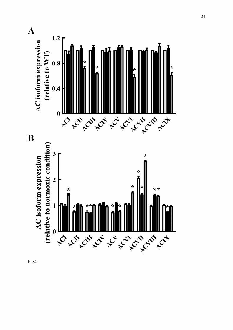
Adenylyl cyclase isoform mRNA expression
In order to find possible correlates of AC activity changes in the different experimental
conditions described above, a real time PCR analysis was performed to determine the

10
expression levels of AC isoforms in normoxic and in hypoxic mouse retinas. Preliminary
experiments performed on PD 17 WT normoxic mice showed that all AC isoform (AC I-IX)
transcripts are expressed in the retina (Fig. 1).
The relative levels of all transcripts in normoxic and hypoxic conditions normalized to
cyclophilin B were evaluated (Fig. 2). As shown in Fig. 2A, in normoxic retinas the genetic
deletion of sst1 did not alter AC isoform expression pattern versus WT. In contrast, when
compared to WT, in sst2 KO retinas a statistically significant reduction of the mRNAs for
some AC isoforms (AC II, III, VI and IX, -29%, -37%, -43%, -40%, respectively) was
detected.
The analysis of retinas in hypoxic conditions (Fig. 2B) revealed a significantly
different expression pattern of AC isoforms. In particular, in WT retinas mRNA expression of
AC II, III and V significantly decreased (-25%, -27%, -28%, respectively), whereas AC VII
mRNA expression highly increased (+103%) with respect to normoxic retinas. In sst1 KO
retinas, AC III and IX mRNAs significantly decreased (-30% and -27%, respectively),
whereas AC VII and VIII mRNA expression significantly increased (+41% and +38%,
respectively). In sst2 KO retinas, AC V mRNA underwent a statistically significant decrease
(-24%), whereas a statistically significant increase of mRNAs for AC I, VI, VII and VIII was
observed (+41%, +48%, +169%, and +34%, respectively).
In summary, the hypoxic treatment induced significant changes in the pattern of AC
isoform expression in the retinas of all three mouse strains. The most apparent modifications
were at the level of AC VII, whose expression was greatly increased in all strains, although
the enhancement in sst1 KO retinas was significantly (?) less marked than in WT or in sst2
KO retinas.

11
Immunohistochemistry
As shown by the real time PCR results, the AC VII isoform appears to be the most
affected by hypoxic treatment. This AC isoform has not been reported previously in
mammalian retinas, therefore we performed an immunohistochemical analysis to localize this
AC isoform and support the real time PCR data. In addition, although immunohistochemistry
is not a reliable quantitative technique, we checked whether the hypoxia-induced alterations
could be observed in immunostained retinal sections.
As shown in Fig. 3A, the pattern of AC VII immunofluorescence in normoxic WT
retinas was characterized by punctate staining, which was mainly confined to the
photoreceptor outer segments, the outer plexiform layer (OPL), the inner plexiform layer and
the ganglion cell layer. Similar patterns of AC VII immunoreactivity could be observed in
normoxic sst1 KO and sst2 KO retinas. However, as shown in Fig. 3B-D, these patterns
appeared to be differentially affected by hypoxic treatment. Indeed, a slight but evident
increase of AC VII immunofluorescence intensity was observed in hypoxic WT retinas with
respect to their normoxic controls, particularly in the OPL (Fig. 3B). In contrast, no apparent
differences were detected between normoxic and hypoxic sst1 KO retinas (Fig. 3C). Finally,
in sst2 KO retinas a considerable increase of AC VII immunofluorescence intensity could be
seen after hypoxic treatment in all retinal layers containing AC immunoreactivity (Fig. 3D).
Together, these changes in AC VII immunofluorescence were in line with those in AC
VII mRNA expression observed with real time PCR.
Discussion
In the present study, we have provided evidence of correlations between hypoxic
conditions, levels of sst2 expression and alterations of the AC/cAMP system, suggesting an
involvement of this transduction pathway in sst2-mediated antiangiogenic actions of SRIF.

12
These data also confirm and expand previous studies regarding AC/cAMP system modulation
by SRIF and in the mouse retina [12].
In addition to WT retinas, we have utilized retinas from transgenic mice were sst2 are
either overexpressed and overfunctional (the sst1 KO retinas) [12,23], or absent (the sst2 KO
retinas). These genetically modified strains represent useful experimental models to
investigate the role of such receptor in mediating SRIF responses. A central part of our study
has been concerned with measurements of AC activity in response to SRIF or OCTR. To
detect the effects of these substances, AC was stimulated by FSK. Indeed, as previously
reported in the retina as well as in other different tissues, SRIF effects on AC activity become
well evident when this activity is sustained by stimulators, among which FSK [12,34,35].
SRIF and the AC/cAMP system in normoxic retinas
The data of AC activity in normoxic retinas are in line with those of our previous
studies [12]. Basal AC activity was similar in WT and sst1 KO retinas, whereas a reduction
was observed in sst2 KO retinas. AC responsiveness to stimulators such as FSK or
epinephrine was similar in all strains, however AC inhibition by SRIF or OCTR was only
observed in sst1 KO retinas. As previously reported, this effect is likely to be due to a better
coupling between sst2 and Gαo, since both sst2 and G protein expressions are increased in
sst1 KO retinas [12,23].
Our data show that the mRNAs of all nine AC isoforms are present in the different
strains, however AC VII isoform has not been reported previously in mammalian retinas
[15,16]. The lack of evidence of AC VII mRNA in previous studies may be due to different
species used as experimental models, or to different experimental protocols. To our
knowledge, this is the first report of AC VII in the mouse retina both at the mRNA and at the
protein level, which substantiates the presence of AC VII in the mouse retina.

13
The AC mRNA expression patterns were not different in sst1 KO with respect to WT
retinas, whereas a reduction of expression was evident for several AC isoform mRNAs in sst2
KO retinas. These results indicate that the pattern of AC isoform expression is highly
influenced by sst2 deletion, but not sst1 deletion with consequent sst2 overexpression. At
present, it is difficult to explain why an alteration of sst expression results in a change of the
availability of AC system components. Our previous observations showed an increased
expression of Gαo in mouse retinas with sst1 deletion [12]. In addition, it has been clearly
demonstrated that an altered functionality of Gαi/o-coupled receptors, sst included, may
induce modifications of the expression of various AC transduction system components,
including catalytic subunits [36]. The decreased expression of some AC isoforms in sst2 KO
retinas may explain the decrease of enzyme basal activity with respect to WT retinas.
SRIF and the AC/cAMP system in hypoxic retinas
Concerning the data on AC activity in hypoxic conditions, the most evident finding is
that the hypoxic treatment significantly increased AC responsiveness to FSK and epinephrine
in WT and in sst2 KO retinas. In the latter, an enhancement of the enzyme activity was also
evident in basal conditions. It is important to note that, in contrast, stimulated AC activity was
not significantly affected in the presence of sst2 overexpression, as in sst1 KO retinas.
Moreover, while SRIF or OCTR treatment was ineffective in WT or in sst2 KO retinas, it
decreased AC activity in sst1 KO retinas to levels that were not significantly different from
those measured in similarly treated sst1 KO normoxic retinas. Together, these observations
indicate that overexpression of sst2, as in sst1 KO retinas, counteracts the effects of hypoxia
on AC activity, while lack of sst2 amplifies these effects.
The analysis of AC isoform mRNA expression in hypoxic WT retinas showed a
decrease of AC II, III, and V expression, but a great enhancement of AC VII mRNA with

14
respect to normoxic WT retinas. Alterations of AC isoform expression in response to oxygen
deficiency have been observed in different tissues [37,38]. In sst1 or sst2 KO retinas, the
combination of hypoxia and sst2 expression level resulted in strain-specific changes.
Noteworthy, AC VII mRNA expression increased in all strains, but to a lesser extent in sst1
KO and to a higher extent in sst2 KO retinas, if compared to the enhancement in WT. These
changes in AC VII mRNA expression were substantiated by our immunofluorescence data
showing concordant variations in immunofluorescence intensity. At present we have no
plausible explanation for these changes observed in sst1 and sst2 KO retinas in hypoxic
conditions. However, further alterations of sst2 expression can be excluded, since we
previously demonstrated that hypoxia does not alter per se sst2 expression [21].
It has been demonstrated that the AC VII isoform is highly responsive to stimulatory
agents, and insensitive to all known inhibitory regulators, Gαi/o included [39]. Therefore, its
enhanced expression could be mostly responsible for the observed in vitro increase in AC
responsiveness to stimulators. Since it has been reported that hypoxia increases the release of
AC stimulatory agents, such as catecholamines and adenosine, as well as their receptor
expression [40-42], AC VII isoform may play a central role in the enhancement of cAMP in
these conditions.
The increased cAMP level in hypoxia is likely to be instrumental to the induction of
neoangiogenesis and the increased expression of proangiogenic factors such as VEGF, that we
have observed recently in all three strains, in the same experimental model. In particular,
compared to WT retinas, both angiogenesis and VEGF expression are increased in sst2 KO,
while VEGF expression is decreased in sst1 KO retinas [21]. Together, these findings suggest
that an enhanced somatostatinergic function at sst2 protects against the production of
proangiogenic cAMP, while sst2 deletion results in an opposite condition.

15
Conclusion
In conclusion, we have investigated the AC/cAMP transduction pathway in an in vivo
model of retinal hypoxia using retinas from WT mice and from mice with over- or null sst2
expression. We observed changes of both AC activity and expression in response to hypoxia.
Most importantly, these changes could be correlated with different levels of sst2 expression,
as the patterns of AC activity or AC isoform expression were different in WT, sst1 KO or sst2
KO retinas. These data provide evidence in support of an involvement of the AC/cAMP
system in mediating not only hypoxia-evoked retinal neoangiogenesis, but also SRIF
protective actions. The AC VII isoform is a candidate to a main role in these mechanisms.
Acknowledgements
This work was supported by the Italian Ministry of University and Research (MUR,
PRIN, grant 2005052312) and the Fondazione Cassa di Risparmio di Cento.
We thank G. Bertolini (University of Pisa, Italy) for assistance with mouse colonies.

16
References
1. Campochiaro PA (2004) Ocular neovascularisation and excessive vascular
permeability. Expert Opin Biol Ther 4:1395-1402
2. Gariano RF, Gardner TW (2005) Retinal angiogenesis in development and disease.
Nature 438:960-966
3. Baldysiak-Figiel A, Lang GK, Kampmeier J et al (2004) Octreotide prevents growth
factor-induced proliferation of bovine retinal endothelial cells under hypoxia. J
Endocrinol 180:417-424
4. Grant MB, Mames RN, Fitzgerald C et al (2000) The efficacy of octreotide in the
therapy of severe nonproliferative and early proliferative diabetic retinopathy: a
randomized controlled study. Diabetes Care 23:504-509
5. Simo R, Lecube A, Sararols L et al (2002) Deficit of somatostatin-like
immunoreactivity in the vitreous fluid of diabetic patients: possible role in the
development of proliferative diabetic retinopathy. Diabetes Care 25:2282-2286
6. Davis MI, Wilson SH, Grant MB (2001) The therapeutic problem of proliferative
diabetic retinopathy: targeting somatostatin receptors. Horm Metab Res 33:295-299
7. Grant MB, Caballero S (2002) Somatostatin analogues as drug therapies for
retinopathies. Drugs Today 38:783-791
8. Garcia de la Torre N, Wass JA, Turner HE (2002) Antiangiogenic effects of
somatostatin analogues. Clin Endocrinol 57:425-441
9. Sall JW, Klisovic DD, O'Dorisio MS et al (2004) Somatostatin inhibits IGF-1
mediated induction of VEGF in human retinal pigment epithelial cells. Exp Eye Res
79:465-476
10. Dasgupta P (2004) Somatostatin analogues: multiple roles in cellular proliferation,

17
neoplasia, and angiogenesis. Pharmacol Ther 102:61-85
11. Weckbecker G, Lewis I, Albert R et al (2003) Opportunities in somatostatin research:
biological, chemical and therapeutic aspects. Nat Rev Drug Discov 2:999-1017
12. Pavan B, Fiorini S, Dal Monte M et al (2004) Somatostatin coupling to adenylyl
cyclase activity in the mouse retina. Naunyn-Schmiedebergs Arch Pharmacol 370:91-
98
13. Olias G, Viollet C, Kusserow H et al (2004) Regulation and function of somatostatin
receptors. J Neurochem 89:1057-1091
14. Hanoune J, Defer N (2001) Regulation and role of adenylyl cyclase isoforms. Annu
Rev Pharmacol Toxicol 41:145-174
15. Beitz E, Volkel H, Guo Y et al (1998) Adenylyl cyclase type 7 is the predominant
isoform in the bovine retinal pigment epithelium. Acta Anat 162:157-162
16. Abdel-Majid RM, Tremblay F, Baldridge WH (2002) Localization of adenylyl cyclase
proteins in the rodent retina. Brain Res Mol Brain Res 101:62-70
17. Amano H, Ando K, Minamida S et al (2001) Adenylate cyclase/protein kinase A
signaling pathway enhances angiogenesis through induction of vascular endothelial
growth factor in vivo. Jpn J Pharmacol 87:181-188
18. Sakurai S, Alam S, Pagan-Mercado G et al (2002) Retinal capillary pericyte
proliferation and c-Fos mRNA induction by prostaglandin D2 through the cAMP
response element. Invest Ophthalmol Vis Sci 43:2774-2781
19. Casibang M, Purdom S, Jakowlew S et al (2001) Prostaglandin E2 and vasoactive
intestinal peptide increase vascular endothelial cell growth factor mRNAs in lung
cancer cells. Lung Cancer 31:203-212
20. Schwarz N, Renshaw D, Kapas S et al (2006) Adrenomedullin increases the
expression of calcitonin-like receptor and receptor activity modifying protein 2 mRNA

18
in human microvascular endothelial cells. J Endocrinol 190:505-514
21. Dal Monte M, Cammalleri M, Martini D et al (2007) Anti-angiogenic role of
somatostatin receptor 2 in a model of hypoxia-induced neovascularization in the
retina: results from transgenic mice. Invest Ophthalmol Vis Sci 48:3480-3489
22. Dal Monte M, Petrucci C, Cozzi A et al (2003) Somatostatin inhibits potassium
evoked glutamate release by activation of the sst2 somatostatin receptor in the mouse
retina. Naunyn- Schmiedebergs Arch Pharmacol 367:188-192
23. Dal Monte M, Petrucci C, Vasilaki A et al (2003) Genetic deletion of somatostatin
receptor 1 alters somatostatinergic transmission in the mouse retina.
Neuropharmacology 45:1080-1092
24. Casini G, Dal Monte M, Petrucci C et al (2004) Altered morphology of rod bipolar cell
axonal terminals in the retinas of mice carrying genetic deletion of somatostatin
subtype receptor 1 or 2. Eur J Neurosci 19:43-54
25. Kreienkamp HJ, Akgun E, Baumeister H et al (1999) Somatostatin receptor subtype 1
modulates basal inhibition of growth hormone release in somatotrophs. FEBS Lett
462:464-466
26. Allen JP, Hathway GJ, Clarke NJ et al (2003) Somatostatin receptor 2 knockout/lacZ
knockin mice show impaired motor coordination and reveal sites of somatostatin
action within the striatum. Eur J Neurosci 17:1881-1895
27. Smith LE, Wesolowski E, McLellan A et al (1994) Oxygen-induced retinopathy in the
mouse. Invest Ophthalmol Vis Sci 35:101-111
28. Bradford MM (1976) A rapid and sensitive method for the quantitation of microgram
quantities of protein utilizing the principle of protein-dye binding. Anal Biochem
72:248-254
29. Brown BL, Ekins RP, Albano JD (1972) Saturation assay for cyclic AMP using

19
endogenous binding protein. Adv Cyclic Nucleotide Res 2:25-40
30. Wang X, Seed B (2003) A PCR primer bank for quantitative gene expression analysis.
Nucleic Acids Res 31:e154
31. Rozen S, Skaletsky H (2000) Primer3 on the WWW for general users and for biologist
programmers. Methods Mol Biol 132:365-386
32. Livak KJ, Schmittgen TD (2001) Analysis of relative gene expression data using real-
time quantitative PCR and the 2(-Delta Delta C(T)) method. Methods 25:402-408.
33. Tsai WH, Koh SW, Puro DG (1987) Epinephrine regulates cholinergic transmission
mediated by rat retinal neurons in culture. Neuroscience 22:675-680
34. Colas B, Valencia AM, Prieto JC et al (1992) Somatostatin binding and modulation of
adenylate cyclase in ovine retina membranes. Mol Cell Endocrinol 88:111-117
35. Masmoudi O, Gandolfo P, Tokay T et al (2005) Somatostatin down-regulates the
expression and release of endozepines from cultured rat astrocytes via distinct receptor
subtypes. J Neurochem 94:561-571
36. Watts VJ, Neve KA (2005) Sensitization of adenylate cyclase by Galpha i/o-coupled
receptors. Pharmacol Ther 106:405-421
37. Takeo S, Niimura M, Miyake-Takagi K et al (2003) A possible mechanism for
improvement by a cognition-enhancer nefiracetam of spatial memory function and
cAMP-mediated signal transduction system in sustained cerebral ischaemia in rats. Br
J Pharmacol 138:642-654
38. Zhao Y, Xu D, Quaegebeur JM et al (2002) Expression of adenylyl cyclase V/VI
mRNA and protein is upregulated in cyanotic infant human myocardium. Pediatr
Cardiol 23:536-541.
39. Cooper DM, Crossthwaite AJ (2006) Higher-order organization and regulation of
adenylyl cyclases. Trends Pharmacol Sci 27:426-431

20
40. Palmer GC (1985) Cyclic nucleotides in stroke and related cerebrovascular disorders.
Life Sci 36:1995-2006
41. Rocha-Singh KJ, Honbo NY, Karliner JS (1991) Hypoxia and glucose independently
regulate the beta-adrenergic receptor-adenylate cyclase system in cardiac myocytes. J
Clin Invest 88:204-213
42. Kuroko Y, Yamazaki T, Tokunaga N et al (2007) Cardiac epinephrine synthesis and
ischemia-induced myocardial epinephrine release. Cardiovasc Res 74:438-444

21
Captions to the Figures
Fig. 1. AC isoform mRNA expression in PD 17 WT normoxic retinas. Total RNA was
extracted from the retinas, reverse transcribed, and real time PCR performed with isoform-
specific primers as described in Materials and Methods. M: DNA Marker 50 bp ladder; Lane
1: AC type I; Lane 2: AC type II; Lane 3: AC type III; Lane 4: AC type IV; Lane 5: AC type
V; Lane 6: AC type VI; Lane 7: AC type VII; Lane 8: AC type VIII; Lane 9: AC type IX.
Fig. 2. AC isoform mRNA expression by real time PCR in WT (white bars), sst1 KO (black
bars) and sst2 KO (grey bars) mouse retinas.
A: AC isoforms expression level in normoxic conditions Each panel shows the ratio between
optical density (OD) of AC mRNA isoform and cyclophilin B mRNA in normoxic (white bar)
and hypoxic (black bar) retinas from the three mouse strains. Representative agarose gel
electrophoresis of PCR for AC isoforms and relative control loading cyclophilin B (Cyc)
amplicons is shown in the top of each panel. Data are means ± SEM of at least six
independent experiments. ◦ p < 0.05 vs WT normoxic (ANOVA, followed by Dunnett's post
test); * p < 0.05 vs relative normoxic condition (unpaired t test); § p < 0.05 vs WT hypoxic
(ANOVA, followed by Dunnett's post test).
Fig. 3. A: pattern of AC VII immunofluorescence in a WT, normoxic mouse retina. punctate
immunolabeling was densely distributed to the photoreceptor outer segments (POS) and it
was evident in the outer plexiform layer (OPL), in the inner plexiform layer (IPL) and in the
ganglion cell layer (GCL). B-D: AC VII immunostaining patterns in WT (B), sst1 KO (C) and
sst2 KO (D) retinas in normoxic (left half of each panel) or in hypoxic (right half of each
panel) conditions. A detectable increase of immunofluorescence intensity can be appreciated

22
after hypoxic treatment in WT retinas; no changes are seen in sst1 KO retinas; an evident
increase is seen in sst2 KO retinas. All images are single confocal optical sections. INL, inner
nuclear layer; ONL, outer nuclear layer. Scale bars: 20 μm.

23
Fig.1
24
Fig.2

25
Fig.3
Related Documents