University of Pennsylvania University of Pennsylvania ScholarlyCommons ScholarlyCommons Publicly Accessible Penn Dissertations 2018 Adenovirus Strategies To Regulate The Association Of Cellular Adenovirus Strategies To Regulate The Association Of Cellular Proteins With Viral Genomes Proteins With Viral Genomes Neha J. Pancholi University of Pennsylvania, [email protected] Follow this and additional works at: https://repository.upenn.edu/edissertations Part of the Cell Biology Commons, and the Virology Commons Recommended Citation Recommended Citation Pancholi, Neha J., "Adenovirus Strategies To Regulate The Association Of Cellular Proteins With Viral Genomes" (2018). Publicly Accessible Penn Dissertations. 2992. https://repository.upenn.edu/edissertations/2992 This paper is posted at ScholarlyCommons. https://repository.upenn.edu/edissertations/2992 For more information, please contact [email protected].

Welcome message from author
This document is posted to help you gain knowledge. Please leave a comment to let me know what you think about it! Share it to your friends and learn new things together.
Transcript
University of Pennsylvania University of Pennsylvania
ScholarlyCommons ScholarlyCommons
Publicly Accessible Penn Dissertations
2018
Adenovirus Strategies To Regulate The Association Of Cellular Adenovirus Strategies To Regulate The Association Of Cellular
Proteins With Viral Genomes Proteins With Viral Genomes
Neha J. Pancholi University of Pennsylvania, [email protected]
Follow this and additional works at: https://repository.upenn.edu/edissertations
Part of the Cell Biology Commons, and the Virology Commons
Recommended Citation Recommended Citation Pancholi, Neha J., "Adenovirus Strategies To Regulate The Association Of Cellular Proteins With Viral Genomes" (2018). Publicly Accessible Penn Dissertations. 2992. https://repository.upenn.edu/edissertations/2992
This paper is posted at ScholarlyCommons. https://repository.upenn.edu/edissertations/2992 For more information, please contact [email protected].
Adenovirus Strategies To Regulate The Association Of Cellular Proteins With Viral Adenovirus Strategies To Regulate The Association Of Cellular Proteins With Viral Genomes Genomes
Abstract Abstract Successful viral propagation relies on the careful regulation of cellular proteins. Controlling the cellular proteins that interact with viral genomes is an important regulatory strategy, since these interactions control a myriad of processes relevant to viral infection. Nuclear replicating DNA viruses face an especially difficult challenge, as their genomes are accessible to DNA-binding proteins that can promote or impair viral processes. Understanding the manipulation of host proteins associated with viral genomes provides insight into the role of cellular proteins in viral infection and provides targets for anti-viral therapeutics. Furthermore, these interactions can provide insight into the regulation of fundamental cellular processes, and have broader implications in understanding viral or cellular evolution. Here, we employed different strategies to understand how interactions with viral genomes are regulated. We studied adenovirus, a DNA virus that replicates in the nucleus, where its linear double-stranded DNA genome is accessible to nuclear DNA-binding proteins. First, we utilized evolutionary diverse adenovirus serotypes with distinct tissue tropisms to study interactions with known anti- viral proteins within the cellular DNA damage response (DDR). This project demonstrated that serotypes across the adenovirus family target DDR proteins, but do so with varying success. Some serotypes completely overcome inhibitory effects of the DDR, while other serotypes fail to do so. Further analysis demonstrated differences in the mechanisms used to target the DDR. Findings from this project showed that comparison of diverse adenovirus serotypes can provide mechanistic insight, and these findings may have broader implications in understanding tissue tropism and viral evolution. In the second project, we used proteomics to identify host proteins associated with viral genomes and uncovered a novel role for the histone-like viral protein VII in regulating these interactions. We found that protein VII promotes association of cellular proteins involved in transcription, splicing, and mRNA export. Furthermore, we found that protein VII suppresses the well characterized anti-viral interferon response. Together, our results demonstrate that defining interactions of cellular proteins with viral genomes is a useful strategy to identify cellular proteins that promote or impair viral processes and to understand viral mechanisms used to regulate their association with viral genomes.
Degree Type Degree Type Dissertation
Degree Name Degree Name Doctor of Philosophy (PhD)
Graduate Group Graduate Group Cell & Molecular Biology
First Advisor First Advisor Matthew D. Weitzman
Keywords Keywords Adenovirus, DNA damage response, Interferon, iPOND, Protein VII
Subject Categories Subject Categories Cell Biology | Virology
This dissertation is available at ScholarlyCommons: https://repository.upenn.edu/edissertations/2992
ADENOVIRUS STRATEGIES TO REGULATE THE ASSOCIATION OF
CELLULAR PROTEINS WITH VIRAL GENOMES
Neha J. Pancholi
A DISSERTATION
in
Cell and Molecular Biology
Presented to the Faculties of the University of Pennsylvania
in
Partial Fulfillment of the Requirements for the
Degree of Doctor of Philosophy
2018
Supervisor of Dissertation
_________________________
Matthew D. Weitzman, Ph.D.
Professor of Microbiology and Pathology and Laboratory Medicine
Graduate Group Chairperson
__________________________
Daniel S. Kessler, Ph.D.
Associate Professor of Cell and Developmental Biology
Dissertation Committee
Eric J. Brown, Ph.D., Associate Professor of Cancer Biology
Paul M. Lieberman, Ph.D., Professor of Gene Expression and Regulation
Susan R. Weiss, Ph.D., Professor of Microbiology
Jianxin You, Ph.D., Associate Professor of Microbiology
ADENOVIRUS STRATEGIES TO REGULATE THE ASSOCIATION OF CELLULAR PROTEINS
WITH VIRAL GENOMES
COPYRIGHT
2018
Neha Jayesh Pancholi
This work is licensed under the Creative Commons Attribution- NonCommercial-ShareAlike 3.0 License To view a copy of this license, visit
https://creativecommons.org/licenses/by-nc-sa/3.0/us/

iii
ACKNOWLEDGMENTS
I would like to thank my advisor, Matt Weitzman, for his mentorship throughout the past
five and half years. His high expectations and critiques motivated me to keep improving,
and his guidance has allowed me to grow as a scientist, public speaker, and writer. The
challenge to think independently, ask meaningful questions, and drive a research project
has been at many points daunting and frustrating, but Matt’s trust, encouragement, and
willingness to step in when needed gave me the confidence and skills to tackle this
challenge.
I would also like to thank the past and present members of the Weitzman lab. Life in the
lab would not have been nearly as wonderful without their support and friendship. I thank
them for all of the scientific discussions and for helping me put out fires, both
metaphorically and literally. In particular, I am incredibly grateful to have shared my time
in the lab with Daphne Avgousti and Emigdio Reyes. So much of my scientific growth is
due to my conversations and collaborations with them. I thank them for sharing their
expertise and for their perpetual willingness to offer advice on anything and everything.
I would also like to thank my committee members, Eric Brown, Paul Lieberman, Susan
Weiss, and Jianxin You, for their input on my projects throughout the years.
I am also grateful for the friends I have made through CAMB. From the early years spent
exploring the city, studying for prelims, and playing dodgeball to the more recent
weddings, thesis defenses, and wine nights, it has been wonderful sharing both my
scientific and personal lives with them.
I would like to thank my parents, grandparents, and brother for their love and support
throughout my life and for instilling in me the importance of education. I am extremely
grateful to spend my life with my husband Andrew, who supports my goals and shares
my love of learning, and with our cats Jack and Sophie, who support my head while I
sleep and share my love of eating.
iv
ABSTRACT
ADENOVIRUS STRATEGIES TO REGULATE THE ASSOCIATION OF
CELLULAR PROTEINS WITH VIRAL GENOMES
Neha J. Pancholi
Matthew D. Weitzman
Successful viral propagation relies on the careful regulation of cellular proteins.
Controlling the cellular proteins that interact with viral genomes is an important
regulatory strategy, since these interactions control a myriad of processes relevant to
viral infection. Nuclear replicating DNA viruses face an especially difficult challenge, as
their genomes are accessible to DNA-binding proteins that can promote or impair viral
processes. Understanding the manipulation of host proteins associated with viral
genomes provides insight into the role of cellular proteins in viral infection and provides
targets for anti-viral therapeutics. Furthermore, these interactions can provide insight into
the regulation of fundamental cellular processes, and have broader implications in
understanding viral or cellular evolution. Here, we employed different strategies to
understand how interactions with viral genomes are regulated. We studied adenovirus, a
DNA virus that replicates in the nucleus, where its linear double-stranded DNA genome
is accessible to nuclear DNA-binding proteins. First, we utilized evolutionary diverse
adenovirus serotypes with distinct tissue tropisms to study interactions with known anti-
viral proteins within the cellular DNA damage response (DDR). This project
demonstrated that serotypes across the adenovirus family target DDR proteins, but do
so with varying success. Some serotypes completely overcome inhibitory effects of the
DDR, while other serotypes fail to do so. Further analysis demonstrated differences in
the mechanisms used to target the DDR. Findings from this project showed that
v
comparison of diverse adenovirus serotypes can provide mechanistic insight, and these
findings may have broader implications in understanding tissue tropism and viral
evolution. In the second project, we used proteomics to identify host proteins associated
with viral genomes and uncovered a novel role for the histone-like viral protein VII in
regulating these interactions. We found that protein VII promotes association of cellular
proteins involved in transcription, splicing, and mRNA export. Furthermore, we found
that protein VII suppresses the anti-viral interferon response. Together, our results
demonstrate that defining interactions of cellular proteins with viral genomes is a useful
strategy to identify cellular proteins that promote or impair viral processes and to
understand viral mechanisms used to regulate their association with viral genomes.
vi
TABLE OF CONTENTS
ACKNOWLEDGMENTS ..................................................................................... III
ABSTRACT ......................................................................................................... IV
LIST OF TABLES ............................................................................................... IX
LIST OF ILLUSTRATIONS .................................................................................. X
CHAPTER 1: INTRODUCTION .......................................................................... 1
Virus-host interactions .................................................................................................................. 1
Viruses must regulate protein-DNA interactions ........................................................................ 1
Adenovirus ..................................................................................................................................... 2 Adenovirus family and classification ............................................................................................ 3 Viral capsid structure and core proteins ....................................................................................... 3 Viral entry ..................................................................................................................................... 4 Adenovirus genome and gene expression................................................................................... 5 Viral DNA replication and viral replication centers ..................................................................... 12 Virion assembly and release ...................................................................................................... 14
Adenovirus manipulation of cellular processes that respond to viral DNA .......................... 15 DNA damage response .............................................................................................................. 15 Interferon response .................................................................................................................... 23
Thesis goals ................................................................................................................................. 27
Figures .......................................................................................................................................... 29
CHAPTER 2: SEROTYPE-SPECIFIC RESTRICTION OF WILD-TYPE ADENOVIRUSES BY THE CELLULAR MRE11-RAD50-NBS1 COMPLEX .... 35
Introduction .................................................................................................................................. 35
Materials and Methods ................................................................................................................ 37 Cell lines ..................................................................................................................................... 37 Plasmids and transfections ........................................................................................................ 38 Viruses and infections ................................................................................................................ 38 Antibodies and inhibitors ............................................................................................................ 38 Immunoblotting ........................................................................................................................... 39 Immunofluorescence .................................................................................................................. 39 Virus genome accumulation by quantitative PCR ...................................................................... 40
Results .......................................................................................................................................... 40 Effect of adenovirus infection on MRN protein levels and localization ...................................... 40 ATM is activated during infection with multiple serotypes ......................................................... 42
vii
MRN impairs DNA replication for Ad9 and Ad12 serotypes ...................................................... 43 ATM does not impair Ad9 or Ad12 ............................................................................................. 45 Degradation of MRN by Ad12 occurs similarly to Ad5 ............................................................... 46 MRN colocalizes with E4orf3 and PML during Ad9 infection ..................................................... 46 Ad9-E4orf3 is not sufficient to alter MRN localization ................................................................ 47 Single residue site-directed mutagenesis does not affect mislocalization by Ad9-E4orf3 ......... 47 Divergent Nbs1 proteins from non-human primates impair E4-deleted Ad5 ............................. 48
Table 2.1 ........................................................................................................................................ 50
Figures .......................................................................................................................................... 51
Discussion .................................................................................................................................... 64
CHAPTER 3: EXAMINING THE ROLE OF ADENOVIRUS CORE PROTEIN VII IN REGULATING PROTEINS ASSOCIATED WITH VIRAL GENOMES .......... 68
Introduction .................................................................................................................................. 69
Materials and Methods ................................................................................................................ 69 Cell lines ..................................................................................................................................... 69 Viruses and infections ................................................................................................................ 70 Isolation of proteins on nascent DNA ......................................................................................... 70 Visualization of EdU-labeled DNA ............................................................................................. 72 Immunoprecipitation ................................................................................................................... 73 Deletion of protein VII by TAT-Cre ............................................................................................. 74 Immunofluorescence, immunoblotting, and antibodies .............................................................. 74 Quantitative PCR ....................................................................................................................... 75 Interferon stimulation .................................................................................................................. 75
Results .......................................................................................................................................... 76 Identification of proteins associated with adenovirus DNA by iPOND ....................................... 76 Comparison of viral and host iPOND proteomes reveals novel roles for host proteins in adenovirus replication ................................................................................................................ 78 Comparison of iPOND proteomes of wild-type and mutant viruses reveals targets of specific viral proteins ............................................................................................................................... 80 Core viral protein VII manipulates host chromatin ..................................................................... 82 Protein VII sequesters HMGB proteins in cellular chromatin ..................................................... 83 Conservation of protein VII’s effect on cellular chromatin and HMGB1 ..................................... 84 Protein VII deletion during infection ........................................................................................... 85 Protein VII interacts with cellular proteins enriched on viral genomes ...................................... 86 iPOND analysis of wild-type and protein VII-deleted genomes ................................................. 87 Protein VII deletion affects association of RNA and DNA processing proteins with viral genomes .................................................................................................................................... 89 Protein VII suppresses interferon signaling ............................................................................... 92
Tables and Figures ...................................................................................................................... 96
Discussion .................................................................................................................................. 130
CHAPTER 4: DISCUSSION ............................................................................ 137
Summary ..................................................................................................................................... 137
viii
Future directions ........................................................................................................................ 138 How does Ad9 mislocalize MRN? ............................................................................................ 138 How does protein VII suppress IFN levels? ........................................................................... 140 Does protein VII bind RNA? ..................................................................................................... 142
Significance ................................................................................................................................ 143 Common cellular obstacles to adenoviruses ........................................................................... 143 Resources to define interactions with host proteins ................................................................ 144 Insights into tissue and species tropism .................................................................................. 145
Conclusion .................................................................................................................................. 146
Figures ........................................................................................................................................ 148
BIBLIOGRAPHY .............................................................................................. 151

ix
LIST OF TABLES
Table 2.1: Summary of MRN degradation and mislocalization during adenovirus infection.
Table 3.1: Viral proteins identified by iPOND-MS.
Table 3.2: Proteins enriched on wild-type viral genomes.
Table 3.3: Proteins enriched on protein VII-deleted viral genomes.
x
LIST OF ILLUSTRATIONS
Figure 1.1: Adenovirus capsid and core proteins.
Figure 1.2: Adenovirus genome and transcription units.
Figure 1.3: Viral replication centers.
Figure 1.4: Adenovirus manipulates several steps of the DNA damage response.
Figure 1.5: Overview of interferon signaling.
Figure 2.1: Cytopathic effect (CPE) during infection with multiple adenovirus serotypes.
Figure 2.1: Cytopathic effect (CPE) during infection with multiple adenovirus serotypes.
Figure 2.2: Effect of adenovirus infection on MRN protein levels.
Figure 2.3: Effect of adenovirus infection on MRN localization.
Figure 2.4: ATM is activated during infection with multiple serotypes.
Figure 2.5: MRN impairs Ad9 and Ad12 replication.
Figure 2.6: ATM does not impair Ad9 or Ad12.
Figure 2.7: Ad12 E1b55K and E4orf6 are sufficient to degrade MRN.
Figure 2.8: MRN colocalizes with E4orf3 and PML during Ad9 infection.
Figure 2.9: Ad9-E4orf3 is not sufficient to alter MRN localization.
Figure 2.10: Effect of R105I mutation in Ad9-E4orf3.
Figure 2.11: Adenovirus replication is not affected by species-specific sequence variation in Nbs1.
Figure 3.1: iPOND identifies proteins associated with viral genomes.
Figure 3.2: Comparison of viral and host proteomes reveals novel roles for host proteins in adenovirus replication.
Figure 3.3: Comparison of wild-type and mutant viral proteomes reveals targets of specific viral proteins.
Figure 3.4: Core viral protein VII manipulates host chromatin.
Figure 3.5: Protein VII sequesters HMGB proteins in cellular chromatin.
Figure 3.6: Conservation of protein VII’s effect on cellular chromatin and HMGB1.
Figure 3.7: Protein VII deletion by Lox-Cre system.
Figure 3.8: Protein VII interacts with HMGB1 and cellular proteins enriched on viral genomes.
Figure 3.9: Protein VII is deleted without a dramatic effect on viral replication.
Figure 3.10: High reproducibility between iPOND replicates.
Figure 3.11: Protein VII deletion does not dramatically affect viral proteins associated with viral genomes.
Figure 3.12: Protein VII deletion significantly alters cellular proteins associated with viral genomes.
Figure 3.13: Localization of identified proteins during wild-type Ad5 infection.
Figure 3.14: Changes to cellular protein localization are dependent on protein VII.
Figure 3.15: Protein VII is not sufficient to alter protein localization and does not interact with identified proteins during infection.
Figure 3.16: Effect of protein VII on the interferon response.
Figure 3.17: Effect of protein VII on IFN is independent of protein VII’s effect on the cell cycle.
Figure 3.18: HMGB1 may contribute to protein VII-mediated IFN suppression.
Figure 4.1: Ad9-E1b55K is sufficient to alter localization of MRN components.
Figure 4.2: Potential post-translational modifications on E4orf3.
Figure 4.3: Viral RNA and protein VII have similar localization patterns.
1
CHAPTER 1:
Introduction
A portion of this chapter has been previously published in:
Pancholi, N.J., A.M. Price, and M.D. Weitzman, Take your PIKK: tumour viruses
and DNA damage response pathways. Philos Trans R Soc Lond B Biol Sci,
2017. 372(1732).
Virus-host interactions
As obligate intracellular pathogens, viruses must manipulate the host cell environment in
favor of viral replication. Such manipulation can have dire consequences for cellular
processes, thus cells have evolved mechanisms to defend against viruses by impairing
viral replication. Studying virus-host interactions is crucial to identifying the cellular
obstacles that defend against viruses and the mechanisms by which viruses evade
cellular defenses. This information provides potential targets for anti-viral therapies, and
provides insight into basic cellular processes and viral and host evolution. Studying the
interactions between viruses and cells has also been instrumental in dissecting
fundamental cellular pathways (Berk, 2005; Daugherty & Malik, 2012), as the cellular
proteins that viruses target are often regulatory nodes in cellular signaling pathways.
Virus-host interactions have therefore also been important resources to uncover key
regulatory mechanisms of cellular processes.
Viruses must regulate protein-DNA interactions
One of the multiple ways that viruses manipulate cellular environments to promote viral
replication is through regulation of protein-DNA interactions, as these interactions control
several critical processes, including DNA replication, gene expression, and the interferon
2
response. In the case of nuclear-replicating DNA viruses, cellular DNA-binding proteins
that usually interact with cellular DNA can often recognize and associate with viral DNA.
This can be beneficial or detrimental to viral replication, depending on the function of the
cellular DNA-binding protein. For example, recognition by DNA replication or
transcription enzymes can benefit the virus, but recognition by DNA sensors that activate
immune signaling can impair viral replication and spread. Therefore, viruses must tightly
regulate the cellular proteins that interact with viral genomes. Viruses must recruit
cellular proteins to their genomes that aid DNA replication and transcription of viral
genes, but must evade interaction with cellular proteins that can trigger anti-viral
processes. My thesis work examined how viruses regulate these interactions with
cellular DNA-binding proteins. To address this topic, we studied adenovirus, which is a
nuclear-replicating DNA virus that has been a historically useful model to study viral
manipulation of cellular processes. In Chapter 2, we demonstrate how diverse
adenoviruses evade recognition by a previously defined anti-viral DNA-binding protein
complex. In Chapter 3, we utilize proteomics to identify host proteins that interact with
viral DNA and describe how a viral DNA-binding protein may regulate these interactions.
Adenovirus
Adenovirus (Ad) was originally isolated from pediatric adenoid tissue in 1953 (Rowe,
Huebner, Gilmore, Parrott, & Ward, 1953) and has proven to be an especially powerful
model to study basic cellular processes (Berk, 2005). Interest in understanding the
interaction of Ad with the cell expanded following the 1962 observation that rodents
infected with Ad developed tumors (Trentin, Yabe, & Taylor, 1962). Later research
showed that Ad proteins transform human cells in culture and seize control of the cell
cycle (Endter & Dobner, 2004). There are many benefits to using Ad to study cellular
pathways: Ad propagates well in cell culture, and the life cycle of the virus has been well
3
characterized. Here, I will first describe the Ad family and the viral life cycle before
discussing strategies used by adenovirus to manipulate cellular processes that are
activated by interactions between host proteins and viral DNA.
Adenovirus family and classification
Ad is a non-enveloped DNA virus that replicates in the nucleus of host cells (Berk,
2013). At least fifty-six human types comprise the Ad family, and there are additional
Ads that infect other vertebrate species (Berk, 2013). Human Ad types were originally
classified by serology and hemagglutination assays (Berk, 2013), and have therefore
been referred to as “serotypes” historically. Ads have more recently been categorized by
genome similarity (Davison, Benko, & Harrach, 2003); however, I will continue to use the
term “serotype” here due to convention in the literature. Human serotypes are classified
into seven subgroups, A-G, and cause a variety of illnesses (Berk, 2013). There is a
moderate level of conservation between subgroups, and all serotypes examined to date
have similar genome structure and express homologous proteins (Berk, 2013; Davison
et al., 2003).
Viral capsid structure and core proteins
The adenovirus capsid is an icosehdral structure composed of the major capsid proteins,
hexon, penton, and fiber (H. Liu et al., 2010; Maizel, White, & Scharff, 1968; Reddy,
Natchiar, Stewart, & Nemerow, 2010; van Oostrum & Burnett, 1985). Hexon proteins
form the icosahedral structure, and penton is found at each vertex. Fiber proteins form
shafts that protrude from the vertices and play an important role in binding to the viral
receptor (Philipson, Lonberg-Holm, & Pettersson, 1968). The minor capsid proteins are
IIIa, VIII, and IX, and these stabilize interactions between hexon proteins (H. Liu et al.,
2010; Reddy et al., 2010). Many viral proteins are also found inside the capsid, and
4
these proteins are referred to as “core proteins” (Figure 1.1). Proteins VII, V, and are
small, basic core proteins that interact with the viral genome and likely contribute to
condensation (C. W. Anderson, Young, & Flint, 1989; Chatterjee, Vayda, & Flint, 1986;
Russell, Laver, & Sanderson, 1968). Protein VII is the major core protein, as it is the
most abundant, with over 800 copies found in each virion (Chelius et al., 2002; van
Oostrum & Burnett, 1985). In addition, a single terminal protein (TP) is found at the 5’
end of each DNA strand (Smart & Stillman, 1982; Van der Vliet, 1995), where it serves
as the primer for DNA replication during infection (Van der Vliet, 1995). Protein VI
associates with both hexon and protein V to tether the capsid to the interior DNA-protein
core (Reddy et al., 2010). Additionally, the virion contains the viral protease, which
cleaves viral proteins during entry (Cotten & Weber, 1995; Greber, Webster, Weber, &
Helenius, 1996) and during the final stages of packaging (Weber, 2007). The virion also
contains protein IVa2, which aids packaging by binding the packaging sequence in the
viral genome (Ostapchuk, Yang, Auffarth, & Hearing, 2005). Adenovirus virions do not
contain any cellular proteins.
Viral entry
Adenoviruses enter the cell through receptor-mediated endocytosis. Adenoviruses within
subgroups A, C, D, E, and F utilize the coxsackie and adenovirus receptor (CAR)
(Bergelson et al., 1997), while subgroup B adenoviruses use CD46 as a receptor
(Gaggar, Shayakhmetov, & Lieber, 2003). Entry is initiated by the binding of the capsid
fiber protein with the cellular receptor (Philipson et al., 1968), and virions enter the cell in
endosomes (Chardonnet & Dales, 1970; Greber, Willetts, Webster, & Helenius, 1993).
Acidification of the endosome results in activation of the viral protease, which cleaves
protein VI (Greber et al., 1996). Cleavage of protein VI is required for the complete
disassembly of viral particles that occurs at the nuclear membrane and for DNA import
5
into the nucleus (Cotten & Weber, 1995; Greber et al., 1996). Upon acidification and
endosome lysis, viral particles are released into the cytosol and transported on
microtubules to the nucleus through interactions between hexon and dynein proteins
(Dales & Chardonnet, 1973; Greber et al., 1993).
Interaction of the viral particles with the nuclear pore complex is required to trigger final
disassembly (Greber et al., 1997), likely as an attempt to prevent detection by
cytoplasmic DNA sensors. DNA import into the nucleus is aided by the interaction of
protein VII with the cellular transportin protein (Hindley, Lawrence, & Matthews, 2007).
Most capsid and core proteins remain associated with the nuclear pore complexes
(Greber et al., 1997), but protein VII and terminal proteins remain bound to viral
genomes as they enter the nucleus (Greber et al., 1997). Nuclear Ad genomes are then
transcribed and replicated to generate viral progeny.
Adenovirus genome and gene expression
Ad genomes are linear double-stranded DNA and range in size from 25-45 kb (Davison
et al., 2003). The ends of the viral genome are inverted terminal repeat sequences that
contain the origins of replication. There are five early transcription units (E1A, E1B, E2,
E3, and E4), four intermediate transcription units (IX, IVa2, L4 intermediate, and E2
late), and one late transcription unit (major late unit) (Figure 1.2). The intermediate and
late transcription units are expressed after the onset of viral DNA replication.
Ad expresses early proteins to establish a cellular environment conducive to viral
replication and late proteins to form viral particles. Early proteins are expressed from
genomic regions E1-E4, each of which expresses multiple proteins through alternative
splicing of transcripts (Berget, Moore, & Sharp, 1977) (Figure 1.2). E1 and E4 proteins
manipulate the cellular environment to promote viral processes, E2 expresses proteins
6
involved in viral DNA replication, and E3 expresses proteins to suppress the host innate
immune response. Early proteins are expressed before the onset of viral DNA
replication. Initiation of DNA replication marks the transition into the late stage of
infection, when the intermediate and late transcription units are transcribed.
Early proteins
E1A from the E1 genomic region is the first viral protein to be transcribed, due its strong
enhancer (Hearing & Shenk, 1983; Nevins, Ginsberg, Blanchard, Wilson, & Darnell,
1979). The E1A transcript produces two proteins, called large and small E1A. Large E1A
is a transactivator and stimulates transcription of E1A and the other early transcription
units by recruiting host transcription enzymes to viral genomes (Pelka et al., 2009;
Winberg & Shenk, 1984). In addition to promoting viral transcription, both small and
large E1A manipulate the cell cycle in order to promote entry into S phase so that viral
DNA replication can occur. This is achieved through interaction between E1A and the
cellular retinoblastoma (Rb) family of proteins. E1A binding to Rb releases E2F
transcription factors, allowing them to activate transcription of genes required for
progression into S phase (Bagchi, Raychaudhuri, & Nevins, 1990). The E1 region also
encodes two proteins expressed from the E1B transcription unit: E1b55K and E1b19K.
These proteins regulate apoptosis and the cell cycle in order to promote viral replication.
Together with E1A, E1B proteins can transform human cells in culture, and E1 proteins
are the transformative agents of the widely used 293 cells (Endter & Dobner, 2004).
E1b19K is a viral mimic of the anti-apoptotic MCL-1 protein (Cuconati & White, 2002),
whose degradation induces apoptosis. As an MCL-1 mimic, E1b19K is able to prevent
apoptosis even when MCL-1 has been degraded (Cuconati & White, 2002). E1b19K
inhibits apoptosis by binding the cellular BAK and BAX proteins, preventing their
interaction and pro-apoptotic activity (Cuconati & White, 2002). E1b55K regulates
7
cellular function partially through its ability to target cellular proteins for proteasome-
mediated degradation (Baker, Rohleder, Hanakahi, & Ketner, 2007; Cheng et al., 2011;
Dallaire, Blanchette, Groitl, Dobner, & Branton, 2009; Forrester et al., 2011; Harada,
Shevchenko, Shevchenko, Pallas, & Berk, 2002; Orazio, Naeger, Karlseder, &
Weitzman, 2011; Querido, Blanchette, et al., 2001; Querido et al., 1997; Schwartz et al.,
2008; Steegenga, Riteco, Jochemsen, Fallaux, & Bos, 1998; Stracker, Carson, &
Weitzman, 2002). E1b55K interacts with the viral E4orf6 protein (expressed from the E4
region), which recruits cellular proteins to form a VHL-like E3 ubiquitin ligase (Harada et
al., 2002; Querido, Blanchette, et al., 2001; Querido, Morrison, et al., 2001). E1b55K is
thought to provide substrate specificity to the ubiquitin ligase (Berk, 2005; Blackford &
Grand, 2009; Schwartz et al., 2008), which targets proteins involved in a myriad of
cellular processes, including the DNA damage response and cell cycle control (Baker et
al., 2007; Berk, 2005; Blackford & Grand, 2009; Forrester et al., 2011; Orazio et al.,
2011; Querido et al., 1997; Stracker et al., 2002). Activity of the E1b55K-E4orf6 ubiquitin
ligase is required for optimal viral replication, protein expression, and export of viral
mRNA (Blackford & Grand, 2009; Blanchette et al., 2008; Halbert, Cutt, & Shenk, 1985;
Lakdawala et al., 2008). In addition to targeting proteins for degradation, E1b55K
suppresses the activity of the tumor suppressor p53 by directly binding its transcriptional
activation domains (Sarnow, Ho, Williams, & Levine, 1982). This inhibits p53-mediated
transcriptional activation of cellular genes that promote cell cycle arrest.
The E2 region expresses proteins involved in replication of the viral genome. Activation
of the E2 transcription unit is mediated by the cellular E2F proteins (SivaRaman &
Thimmappaya, 1987), which function during S phase. This ensures that expression of
viral DNA replication proteins occurs only after cells have entered S phase (Berk, 2013).
E2 expresses the viral DNA polymerase (Ad Pol), pre-terminal protein (pTP), and the
8
single-stranded DNA binding protein (DBP). pTP associates with the 5’ ends of newly
replicated viral genomes and functions as a protein primer for DNA replication by
providing the 5’ hydroxyl group necessary for elongation (Smart & Stillman, 1982; Van
der Vliet, 1995). DBP binds and stabilizes single-stranded DNA intermediates produced
during replication (Van der Vliet, 1995; van der Vliet & Levine, 1973), and also promotes
strand separation (Dekker et al., 1997; Van der Vliet, 1995).
The E4 region expresses seven proteins: orf1, orf2, orf3, orf3/4, orf4, orf6, and orf6/7.
These proteins are involved in regulation of several different cellular processes,
including transcription, translation, apoptosis, mRNA splicing, protein stability, and DNA
damage responses, among others (reviewed in (Tauber & Dobner, 2001; Weitzman,
2005). While deletion or mutation of individual E4 orfs only moderately affects viral
replication (Halbert et al., 1985), deletion of both E4orf6 and E4orf3 or the entire E4
region results in a dramatic reduction of viral growth (Bridge & Ketner, 1989; Huang &
Hearing, 1989; Lakdawala et al., 2008; Weiden & Ginsberg, 1994). Therefore, E4orf3
and E4orf6 are considered redundant in promoting optimal lytic viral replication (Bridge &
Ketner, 1989; Huang & Hearing, 1989). E4orf3 and E4orf6 each manipulate cellular
proteins in multiple ways in order to evade anti-viral cellular pathways. E4orf3 forms
characteristic nuclear track structures (Carvalho et al., 1995; Doucas et al., 1996; Ou et
al., 2012) and disrupts PML nuclear bodies into nuclear tracks (Carvalho et al., 1995;
Doucas et al., 1996). E4orf3 can promote viral replication by mislocalizing cellular
proteins to these nuclear tracks in order to sequester them away from viral genomes
(Bridges, Sohn, Wright, Leppard, & Hearing, 2016; Reyes et al., 2017; Stracker et al.,
2002). In addition to mislocalization to E4orf3-PML tracks, E4orf3 recruits cellular
proteins involved in translation inhibition and mRNA degradation to perinuclear
aggresomes to prevent inhibition of viral protein synthesis (Greer, Hearing, & Ketner,
9
2011). E4orf3 also suppresses expression of p53-responsive genes through H3K9
methylation of p53 target gene promoters (Soria, Estermann, Espantman, & O'Shea,
2010) and has been demonstrated to suppress interferon signaling (Ullman & Hearing,
2008; Ullman, Reich, & Hearing, 2007). E4orf6 also manipulates cellular proteins to
evade anti-viral pathways. E4orf6, together with E1b55K, promotes degradation of
several cellular proteins (described in E1b55K section above) to promote viral
replication, mRNA export, and protein synthesis (Blackford & Grand, 2009; Blanchette et
al., 2008; Halbert et al., 1985; Lakdawala et al., 2008). In addition, E4orf6 has been
shown to interact with and inhibit tumor suppressors p53 and p73 independently of
E1b55K (Dobner, Horikoshi, Rubenwolf, & Shenk, 1996; Higashino, Pipas, & Shenk,
1998; Steegenga, Shvarts, Riteco, Bos, & Jochemsen, 1999).
Early viral proteins manipulate the cell to promote viral replication and to express the
viral proteins necessary to replicate the viral genome. Thus, once early proteins are
expressed, the viral genome is replicated and expression of late viral genes begins.
Late proteins
Late viral genes are expressed from a single transcription unit under the control of the
major late promoter (MLP) (Shaw & Ziff, 1980). The major late transcript is processed by
alternative splicing and alternative poly(A) usage to produce five families of late
transcripts (L1-L5). Further processing of each family generates at least 14 late mRNAs.
Viral DNA replication is a prerequisite to activation of the MLP (Thomas & Mathews,
1980), ensuring that late proteins are not expressed until they are needed to package
replicated viral genomes. Late proteins largely form the viral capsid and are involved in
DNA compaction and packaging. Functions of these proteins are described in the Viral
10
capsid structure and core proteins and Viral assembly and release sections of this
chapter.
Protein VII
Protein VII is a late protein expressed from the L2 region that has important roles at
several stages of infection, including viral entry (Greber et al., 1997), evasion of the DNA
damage response (Karen & Hearing, 2011), viral transcription (Komatsu, Haruki, &
Nagata, 2011; Matsumoto, Nagata, Ui, & Hanaoka, 1993; Okuwaki & Nagata, 1998), and
DNA condensation (Johnson et al., 2004). As an incoming viral protein, it is present even
before de novo viral protein synthesis (J. Chen, Morral, & Engel, 2007; Karen & Hearing,
2011), and new copies are produced during the late stage of infection (Xue, Johnson,
Ornelles, Lieberman, & Engel, 2005). As a result, protein VII is present throughout
infection. Protein VII is produced as a pre-cursor protein, and the pro-peptide sequence
is cleaved by the viral protease in the final stage of packaging (C. W. Anderson, Baum,
& Gesteland, 1973). The mature cleaved protein is found in viral particles and on
incoming genomes. Protein VII is the major core protein, with over 800 copies found in
each viral particle (van Oostrum & Burnett, 1985). This small, basic protein associates
with and condenses viral genomes (Chatterjee et al., 1986; Russell et al., 1968), and
contributes to nuclear entry of the genome (Hindley et al., 2007). While other core
proteins remain cytoplasmic (Greber et al., 1997), protein VII enters the nucleus in
association with viral genomes (Greber et al., 1997) and has been suggested to protect
incoming viral genomes from detection by DNA damage machinery before early viral
gene expression (Karen & Hearing, 2011). Surprisingly, deletion of protein VII does not
preclude packaging of the viral genome into capsids (Ostapchuk et al., 2017). This
suggests that other core proteins are redundant with protein VII for condensing DNA to
be packaged. While production of viral particles is not affected by protein VII deletion,
11
the viruses that are produced in the absence of protein VII are non-infectious
(Ostapchuk et al., 2017). Protein VII-deleted viruses are unable to escape from
endosomes during the initial steps of infection (Ostapchuk et al., 2017). This defect
raises the possibility that protein VII contributes to endosomal escape. However, the
defect could also be an indirect consequence of the ineffective protein VI cleavage that
was observed in the absence of protein VII (Ostapchuk et al., 2017) since protein VI
plays an important role in endosomal escape (Cotten & Weber, 1995; Greber et al.,
1996). Furthermore, it is possible that the protein-VII-deleted virus particles are
structurally distinct from wild-type viruses, and viral entry could be affected by any
structural abnormalities.
Protein VII has been described to impact viral transcription, but there are conflicting
reports as to its role. While protein VII-mediated DNA condensation is beneficial for
packaging genomes into capsids, it does not allow for efficient transcription of viral
genes (Matsumoto et al., 1993; Okuwaki & Nagata, 1998). Therefore, it would be
expected that protein VII is displaced to promote active transcription. There is some
evidence of gradual protein VII dissociation before the onset of transcription (Haruki,
Okuwaki, Miyagishi, Taira, & Nagata, 2006; Komatsu et al., 2011). However, protein VII
is also detected on viral genomes during later stages as well (Chatterjee et al., 1986;
Reyes et al., 2017; Xue et al., 2005). Since cellular histones interact with adenoviral
DNA during infection (Giberson, Davidson, & Parks, 2012; Komatsu & Nagata, 2012), it
is likely that some protein VII dissociates from genomes to make room for histones to
bind. The protein VII that remains associated with viral genomes is likely remodeled to
regulate the timing of viral genes (Giberson et al., 2012). Consistent with this theory,
protein VII was found associated with the major late promoter but not with the E1A
promoter at 6 hours post-infection (Haruki, Gyurcsik, Okuwaki, & Nagata, 2003), when
12
late transcription has not yet begun. Furthermore, protein VII interacts with the cellular
chromatin remodeling protein SET (also known as template activating factor 1) (Haruki
et al., 2003; Haruki et al., 2006; Komatsu et al., 2011; Matsumoto et al., 1993; Xue et al.,
2005). SET promotes viral replication and early viral gene expression by increasing DNA
accessibility (Matsumoto et al., 1993). Deletion of SET results in a moderate decrease in
viral gene expression and replication (Haruki et al., 2006). Despite the negative impact
that protein VII-mediated DNA condensation has on transcription, it appears that protein
VII is also capable of activating transcription in in vitro assays (Komatsu et al., 2011).
Furthermore, protein VII has been suggested to recruit the viral transactivator protein
E1A to viral DNA (Johnson et al., 2004). While some data suggest that protein VII must
be removed before transcription can begin, other data suggest that transcription is
actually required for protein VII dissociation (J. Chen et al., 2007). The impact of protein
VII on viral transcription remains unclear, as does the timing and extent of dissociation
from viral genomes.
Viral DNA replication and viral replication centers
Adenovirus DNA replication relies on three viral proteins from the E2 region: pre-
terminal protein (pTP), DNA-binding protein (DBP), and the adenovirus DNA polymerase
(Ad Pol) (Van der Vliet, 1995). The functions of these proteins are described in the
Adenovirus genome and gene expression section of this chapter. Several cellular
proteins, such as topoisomerase I, contribute to adenovirus DNA replication (Reyes et
al., 2017; Van der Vliet, 1995). Cellular helicases are not required for adenovirus DNA
replication because of the strand separating function of DBP (Dekker et al., 1997;
Dekker et al., 1998). Replication occurs in two rounds to duplicate the viral genome. In
the first round of replication, only one of the two DNA strands serves as the template,
and the second strand is displaced as the nascent DNA strand is elongated (Van der
13
Vliet, 1995). Therefore, the first round of replication produces one double-stranded viral
genome and a displaced DNA strand. The displaced strand circularizes by self-
annealing through the complementary inverted terminal ends found at each end of the
DNA strand, generating a panhandle structure (Van der Vliet, 1995). The annealed
portion of the panhandle has the same sequence and structure as the replication origin
of the viral genome. This allows replication initiation to occur through the same
mechanism as the first round of replication. By the end of the second round of
replication, two complete viral genomes have been produced.
Adenovirus DNA replication occurs in structures called viral replication centers (VRCs).
VRCs have been visualized in multiple ways: by immunofluorescence of single-stranded
DNA-binding proteins viral DBP or cellular RPA32 (Evans & Hearing, 2005; Pombo,
Ferreira, Bridge, & Carmo-Fonseca, 1994; Stracker et al., 2002; Stracker et al., 2005),
by incorporation of nucleotide analogs and subsequent visualization (Pombo et al., 1994;
Reyes et al., 2017), and by in situ hybridization using probes specific to the viral genome
(Pombo et al., 1994; Puvion-Dutilleul & Puvion, 1990a, 1990b; Weitzman, Fisher, &
Wilson, 1996). Representative images of VRCs from multiple adenovirus serotypes are
shown in Figure 1.3. The structure of VRCs changes throughout the course of infection.
VRCs begin as small foci that enlarge as replication produces more genomes, and the
sites of single-stranded DNA eventually become donut-shaped (Pombo et al., 1994;
Puvion-Dutilleul & Puvion, 1990a, 1990b). At very late stages of infection, VRCs
disassemble and can be seen as clusters of irregularly shaped aggregates. As viral
genomes replicate, newly synthesized double-stranded viral genomes are displaced to
the periphery of single-stranded DNA accumulation sites (Pombo et al., 1994; Puvion-
Dutilleul & Puvion, 1990a). Viral transcription of late genes occurs at the periphery of
VRCs, using the displaced genomes as templates (Pombo et al., 1994).
14
Virion assembly and release
Viral DNA replication and late gene expression result in accumulation of capsid proteins
and viral genomes that are assembled into viral particles. Once translated, the major
core proteins – hexon, penton, and fiber – form distinct fragments of the capsid in the
cytoplasm (Horwitz, Scharff, & Maizel, 1969; Velicer & Ginsberg, 1970). These
fragments are hexon trimers, which form the faces of the icosahedral capsid, and penton
capsomers, which are complexes of penton and fiber shafts (Horwitz et al., 1969; Velicer
& Ginsberg, 1970). Hexon trimers and penton capsomers are then imported into the
nucleus, where they associate to form the pro-capsid and where viral genomes are
packaged. Packaging of the viral genome requires seven AT-rich packaging sequences
located at the left end of the genome (Hearing, Samulski, Wishart, & Shenk, 1987;
Ostapchuk & Hearing, 2005), which are bound by the viral proteins IVa2, L4-22K, and
L1-52/55K (Ostapchuk & Hearing, 2005; Ostapchuk et al., 2005). IVa2 associates with
viral genomes and pro-capsids (Christensen et al., 2008), and using its ATPase activity
(Koonin, Senkevich, & Chernos, 1993), IVa2 works as an ATP-dependent motor to
encapsidate viral genomes (Ostapchuk & Hearing, 2005). Core viral proteins associated
with viral genomes are packaged as pre-cursors, and their pro-peptide sequences are
cleaved by the viral protease to generate mature core proteins in the final steps of virion
assembly (C. W. Anderson et al., 1973; Freimuth & Anderson, 1993). Cleavage by the
viral protease is required for stability and infectivity of the virions (Ostapchuk & Hearing,
2005).
Viral particles are released upon cellular lysis. Adenovirus increases cellular
susceptibility to lysis by disrupting cellular integrity through viral protease-dependent
cleavage of a cellular cytokeratin (P. H. Chen, Ornelles, & Shenk, 1993). Cell death and
lysis at the end of the viral replication cycle result from accumulation of the viral E3 11.6
15
kDa protein (Tollefson, Ryerse, Scaria, Hermiston, & Wold, 1996; Tollefson, Scaria, et
al., 1996), which has been referred to as the viral death protein due to its induction of
cell death. Released viral particles spread to uninfected cells to begin another round of
viral infection. Viral dissemination is facilitated by degradation of integrin 3 (Dallaire et
al., 2009) and disruption of tight junctions (Latorre et al., 2005; Walters et al., 2002).
Adenovirus manipulation of cellular processes that respond to viral DNA
DNA damage response
Maintenance of cellular genome integrity is paramount to preventing cellular
transformation. Thus, cells have a plethora of mechanisms in place to preserve genome
integrity. The pathways activated by DNA damage to protect genome integrity are
collectively called the DNA damage response (DDR), and they function to sense and
repair damage in cellular DNA (reviewed in (Ciccia & Elledge, 2010; Harper & Elledge,
2007; Jackson & Bartek, 2009; Polo & Jackson, 2011)). The DDR also responds to
viruses, which trigger DDR activation through several means (Luftig, 2014). For
example, viral genomes and replication intermediates may activate the DDR due to their
resemblance to damaged DNA structures. In addition, rapid viral DNA replication may
cause replication stress or errors that trigger the DDR. Viral inactivation of cell cycle
checkpoints may also allow mutations to accumulate in cellular DNA. Activation of the
DDR during infection can have a myriad of consequences for virus replication, and
several viruses therefore manipulate the DDR to promote infection (Hollingworth &
Grand, 2015; Lilley, Schwartz, & Weitzman, 2007; Luftig, 2014; Ryan, Hollingworth, &
Grand, 2016; Turnell & Grand, 2012).
Cellular genomes are damaged on average 100,000 times per day (Ciccia & Elledge,
2010). Sources of damage include exogenous assaults such as radiation, and
16
endogenous events such as replication fork collapse and DNA replication errors. DNA
damage occurs in multiple forms, including mismatched base pairs, pyrimidine dimers,
replication stress, and single-strand or double-strand DNA breaks (Ciccia & Elledge,
2010). Unchecked DNA damage has dramatic effects on cells since the accumulation of
mutations and DNA breaks can lead to cell death, chromosomal translocations, and
oncogenesis.
The DDR is a network of signal transduction pathways that respond to DNA damage.
Signaling is mediated by serine/threonine kinases within the PIKK family and the
downstream proteins that are activated (Ciccia & Elledge, 2010; Harper & Elledge, 2007;
Jackson & Bartek, 2009; Polo & Jackson, 2011). DDR signaling leads to arrest of the cell
cycle to allow recruitment of proteins to repair the damaged DNA (Ciccia & Elledge,
2010; Harper & Elledge, 2007; Jackson & Bartek, 2009; Polo & Jackson, 2011).
Alternatively, signaling can induce apoptosis to eradicate the damaged cell. The DDR is
activated by recognition of DNA damage via proteins called “sensors.” Sensors bind
DNA at the site of damage and recruit PIKK “transducers.” Transducers in turn activate
multiple downstream “effectors” to amplify signaling that mediates DNA repair and cell
cycle arrest at the G1/S, intra-S, and G2/M checkpoints (Ciccia & Elledge, 2010; Harper
& Elledge, 2007; Jackson & Bartek, 2009; Polo & Jackson, 2011). Effectors include
tumor suppressors, which halt cell division by activating cell cycle checkpoints or
apoptosis. Loss or inhibition of tumor suppressors can lead to unregulated cellular
proliferation and transformation. Viruses regulate the DDR through manipulation of
proteins at all three stages of the DDR.
The primary transducers of the DDR are ataxia telangiectasia mutated (ATM), ataxia
telangiectasia and Rad3 related (ATR), and DNA-dependent protein kinase (DNA-PK).
17
ATM, ATR, and DNA-PK are all members of the PIKK family and have similar domain
structures, including kinase and protein-binding domains (Bakkenist & Kastan, 2004;
Lovejoy & Cortez, 2009). The specific PIKK activated depends on the type of DNA
damage encountered. ATM and DNA-PK respond to double-strand DNA breaks (DSBs),
while ATR responds to replication stress and single-stranded DNA (ssDNA) (Bakkenist &
Kastan, 2004; Lovejoy & Cortez, 2009). The specific proteins activated in each pathway
are illustrated in Figure 1.4. Briefly, the MRE11-RAD50-NBS1 complex (MRN) senses
DSBs and promotes activation of ATM (Carson et al., 2003; Lee & Paull, 2005). ATM is
activated by auto-phosphorylation and through interactions with TIP60 (Bakkenist &
Kastan, 2003; Y. Sun, Jiang, Chen, Fernandes, & Price, 2005), and activated ATM then
phosphorylates downstream effectors to amplify signaling. Effectors include histone H2A
variant H2AX (H2AX when phosphorylated), NBS1, BRCA1, CHK2, and p53 (Banin et
al., 1998; Burma, Chen, Murphy, Kurimasa, & Chen, 2001; Cortez, Wang, Qin, &
Elledge, 1999; Lim et al., 2000; Matsuoka, Huang, & Elledge, 1998; Rogakou, Boon,
Redon, & Bonner, 1999). BRCA1 and RAD51 are required for repair of DSBs by
homologous recombination during S-phase, and CHK2 and p53 activate the G1/S, intra-
S, and G2/M checkpoints (Banin et al., 1998; Hirao et al., 2000; Kastan & Bartek, 2004;
Matsuoka et al., 1998). Another repair pathway for DSBs is non-homologous end joining
(NHEJ), which requires DNA-PK activity. The Ku complex senses DSBs and recruits the
catalytic subunit of DNA-PK (DNA-PKcs) (Ciccia & Elledge, 2010). The Ku-DNA-PKcs
complex recruits XRCC4 and DNA ligase IV to join broken ends (Nick McElhinny,
Snowden, McCarville, & Ramsden, 2000). Accumulation of ssDNA at resected DSBs
and replication forks promotes activation of the ATR pathway. Exposed ssDNA is coated
and protected by RPA, which recruits ATR through the ATR binding partner ATRIP (Zou
& Elledge, 2003). The ATR activator TOPBP1 is recruited by interacting with the 9-1-1
18
complex (RAD9, RAD1, HUS1) (Delacroix, Wagner, Kobayashi, Yamamoto, & Karnitz,
2007). ATR activation signals through downstream effectors CHK1 and p53 to cause cell
cycle arrest at the G2/M and intra-S checkpoints or apoptosis (Kastan & Bartek, 2004).
Since cell cycle arrest or cell death could limit viral replication, viruses employ multiple
strategies to misregulate the cell cycle, most notably through inactivation of tumor
suppressors p53 and RB (Endter & Dobner, 2004; Howley & Livingston, 2009; Jha,
Banerjee, & Robertson, 2016; Moody & Laimins, 2010; Pipas, 2009). Misregulation of
the cell cycle via disruption of tumor suppressors is a significant contributor to
transformation by tumor viral oncoproteins (Endter & Dobner, 2004; Howley &
Livingston, 2009; Jha et al., 2016; Moody & Laimins, 2010; Pipas, 2009).
The intricate relationship between viruses and the DDR has been extensively
demonstrated with adenovirus serotype 5 (Ad5). All three of the PIKKs are targeted by
adenoviral proteins, and these interactions revealed principles that have since been
extended to other viruses. Adenovirus has a linear, double-stranded DNA genome, and
one of the first indications that the DDR responded to adenovirus was the observation
that infection with genetic mutants of Ad5 resulted in fusion of viral genomes into
concatemers (Weiden & Ginsberg, 1994). This observation led to the hypothesis that the
blunt, double-stranded DNA ends of the Ad5 viral genome are recognized as DNA
breaks. Several DDR proteins are necessary for concatemer formation, supporting a role
for the DNA repair machinery (Boyer, Rohleder, & Ketner, 1999; Stracker et al., 2002).
This was the first demonstration that the cellular DDR recognizes and acts on viral DNA.
While the DDR responds to mutant Ad5 infection, wild-type Ad5 infection does not
produce concatemers (Carson et al., 2003; Stracker et al., 2002; Weiden & Ginsberg,
1994), indicating that Ad5 evades the DDR. Inactivation of DDR components is critical
for efficient Ad5 replication (Boyer et al., 1999; Evans & Hearing, 2005; Gautam &
19
Bridge, 2013; Lakdawala et al., 2008; Shah & O'Shea, 2015), suggesting a role for the
DDR in restricting adenoviral replication.
MRN
The cellular MRE11, RAD50, and NBS1 proteins comprise the MRN complex (MRN),
which can act as a sensor of double-strand DNA breaks (Figure 1). Ad5 regulates MRN
localization and protein levels to minimize the impacts of host detection of viral DNA.
During Ad5 infection, early viral proteins both degrade MRN and mislocalize MRN into
nuclear tracks and perinuclear aggresomes (Araujo, Stracker, Carson, Lee, & Weitzman,
2005; Cheng et al., 2011; Evans & Hearing, 2003, 2005; Forrester et al., 2011; Karen,
Hoey, Young, & Hearing, 2009; Ou et al., 2012; Shah & O'Shea, 2015; Stracker et al.,
2002). The MRN proteins become immobilized, preventing localization to Ad5 replication
centers (Carson et al., 2009; Stracker et al., 2002). Mislocalization is also necessary for
SUMOylation of MRE11 and NBS1 by an early viral protein, although the consequences
of SUMOylation are unclear (Sohn & Hearing, 2012).
Evading MRN appears to be important for the Ad5 life cycle. In the absence of MRN
mislocalization or degradation, MRN is present at viral replication centers where it
associates with viral DNA in an NBS1-dependent manner (Mathew & Bridge, 2007,
2008; Stracker et al., 2002). Ad5 mutants unable to target MRN are severely impaired in
viral DNA replication, late protein expression, and virion production (Evans & Hearing,
2005; Lakdawala et al., 2008; Mathew & Bridge, 2007). Although MRN is required for
concatemers (Stracker et al., 2002), MRN can also impair viral replication independently
of concatemer formation (Evans & Hearing, 2003; Lakdawala et al., 2008; Mathew &
Bridge, 2007). Loss of MRN rescues replication of Ad5 mutants that neither mislocalize
nor degrade MRN (Lakdawala et al., 2008; Mathew & Bridge, 2007). Ad5 mutants that

20
target MRN by only one of these mechanisms are not impaired for viral replication,
demonstrating that each mechanism is sufficient to evade MRN (Lakdawala et al., 2008).
There are several potential models for MRN restriction of adenovirus replication. One
model is that MRE11 removes the viral terminal protein (TP) from the 5’ ends of the
adenovirus genome through its nuclease activity. TP provides the 3’ hydroxyl group to
initiate DNA replication and may protect viral DNA from digestion, so its removal would
have a profound effect on adenovirus replication. This model is supported by the loss of
DNA sequences at concatemer junctions and the requirement for MRE11 exonuclease
activity for concatemer formation (Karen et al., 2009; Stracker et al., 2002; Weiden &
Ginsberg, 1994). Alternatively, recruitment of DDR proteins to viral DNA could physically
obstruct the interaction of viral and cellular replication proteins with viral genomes (Karen
& Hearing, 2011). A third model is that MRN indirectly impairs replication through
activation of downstream ATM signaling, which is supported by enhanced viral
replication during ATM inhibition (Gautam & Bridge, 2013; Shah & O'Shea, 2015). While
it is clear that MRN is a major obstacle for Ad5 replication, the mechanism by which it
restricts replication requires further study.
ATM
Observations from Ad5 were the first to demonstrate that MRN promotes ATM activation
in response to viruses and cellular double-strand breaks in mammalian cells (Carson et
al., 2003). Since MRN is the sensor that activates ATM signaling, it would be expected
that MRN targeting by Ad5 abrogates ATM activation. Multiple groups have observed
that degradation of MRN by wild-type Ad5 can prevent activation of ATM or downstream
substrates at viral replication centers (Carson et al., 2003; Gautam & Bridge, 2013; Shah
& O'Shea, 2015). Ad5 also employs means to prevent ATM activation before viral
21
proteins are expressed. Protein VII, a viral core protein bound to incoming viral DNA, is
negatively correlated with phosphorylated ATM on mutant Ad5 genomes early during
infection (Karen & Hearing, 2011). This suggests a role for protein VII in preventing DDR
recognition of incoming viral genomes. Furthermore, protein VII is sufficient to suppress
DDR signaling in response to breaks in the cellular genome (Cheng et al., 2013). The
effect of protein VII on the DDR in response to cellular and viral genomes may depend
on its interaction with the cellular SET/TAF1 protein (Cheng et al., 2013). Another
mechanism by which Ad5 may regulate ATM activation is through degradation of the
ATM activator TIP60 (Gupta, Jha, Engel, Ornelles, & Dutta, 2013). Together, these
studies demonstrate multiple ways that Ad5 infection can affect ATM activation and
signaling.
While there is consensus that ATM is not activated during early infection or at wild-type
Ad5 replication centers, some findings demonstrate pan-nuclear distribution of activated
ATM late in infection (Shah & O'Shea, 2015), suggesting MRN-independent ATM
activation during virus infection. This is consistent with the reported phosphorylation of
the ATM substrate KAP1 and replication-dependent widespread H2AX during wild-type
Ad5 infection (Forrester et al., 2011; Nichols, Schaack, & Ornelles, 2009; Shah &
O'Shea, 2015). KAP1 phosphorylation is also seen during infection with other Ad
serotypes (Forrester et al., 2011).
The effect of ATM activation on Ad5 infection may vary between cell types and stages of
the viral life cycle. When viral replication was measured by quantitative PCR in ATM
hypomorphic fibroblasts, ATM loss did not enhance replication of an Ad5 mutant unable
to target MRN (Lakdawala et al., 2008). However, when viral replication was measured
by dot blot hybridization in transformed cell lines, increased viral DNA from the mutant
22
Ad5 was observed when ATM was inhibited or depleted (Gautam & Bridge, 2013). In
primary lung epithelial cells, ATM has distinct effects on Ad5 at different stages of
replication (Shah & O'Shea, 2015). An Ad5 mutant incapable of targeting MRN was
impaired by ATM activation at replication centers early during infection in small airway
epithelial cells (Shah & O'Shea, 2015). In these cells, wild-type Ad5 avoided ATM
activation at replication centers by targeting MRN and progressed to late infection when
diffuse ATM activation occurred. Inhibition of ATM kinase activity during wild-type Ad5
infection does not affect replication in transformed or primary cells (Gautam & Bridge,
2013; Shah & O'Shea, 2015). Together, these findings suggest that ATM does not impair
wild-type Ad5 but may inhibit replication of specific Ad5 mutants in various cellular
settings.
ATR
ATR signaling is also abrogated during adenovirus infection. While ATR is generally
associated with prolonged exposure of ssDNA due to replication stress, double-strand
breaks can induce ssDNA exposure and subsequent ATR activation due to MRE11-
mediated resection at broken ends (Jazayeri et al., 2006). Ad5 mutants that do not target
MRN induce robust activation of ATR signaling (Carson et al., 2009; Carson et al.,
2003), which could occur due to replication intermediates or resection at genome ends.
ATR activation is prevented during infection with wild-type Ad5 due to MRN degradation
and mislocalization (Carson et al., 2003; Forrester et al., 2011). ATR and several
downstream proteins are found at viral replication centers (Blackford et al., 2008; Carson
et al., 2009) but ATR does not appear to affect Ad5 replication (Gautam & Bridge, 2013;
Lakdawala et al., 2008; Shah & O'Shea, 2015). Adenovirus serotype 12 (Ad12) inhibits
ATR through degradation of the ATR regulator TOPBP1 (Blackford et al., 2010).
Interestingly, Ad12 does not mislocalize MRN and therefore does not inhibit ATR
23
through this mechanism (Stracker et al., 2005). It appears that Ad12 and Ad5 employ
distinct mechanisms to inhibit ATR, while some other adenovirus serotypes induce
robust ATR signaling (Forrester et al., 2011). Inactivation of ATR by adenoviruses may
simply be a downstream consequence of MRN manipulation, or it may be specifically
targeted to promote some undetermined aspect of the life cycle.
DNA-PK
The formation of adenoviral genome concatemers requires DNA-PK and NHEJ proteins
to ligate DNA ends, and correlates with decreased late protein expression and DNA
packaging (Boyer et al., 1999; Jayaram & Bridge, 2005). Adenovirus proteins overcome
these limitations by disabling the DNA-PK pathway. All adenovirus serotypes examined
to date degrade DNA Ligase IV (Baker et al., 2007; Cheng et al., 2011; Forrester et al.,
2011), and Ad5 early proteins also interact with DNA-PK to inhibit its functions (Boyer et
al., 1999).
Adenovirus has been a powerful model to uncover fundamental principles of virus-host
interactions, including interactions with the DDR. Studies with Ad5 were the first to
demonstrate that the host DDR responds to viral DNA. In the case of Ad5, DDR proteins
seem to be inhibitory, and Ad5 thus disables DDR pathways to overcome anti-viral
defense and promote viral replication.
Interferon response
The innate immune response serves as a frontline of defense against invading
pathogens and is critical to preventing viral spread. The detection of pathogen
associated molecular patterns (PAMPs) triggers signaling that leads to production of
interferon proteins and extracellular release of cytokines. These events lead to the
synthesis of several anti-viral proteins and the recruitment of innate immune cells.
24
Therefore, suppression of interferon signaling is crucial to ensure success of viral
replication. Interferon signaling in response to viruses is most often activated upon
detection of viral genomes or viral nucleic acids by cellular DNA or RNA sensors
(Barbalat, Ewald, Mouchess, & Barton, 2011; Barber, 2011; Keating, Baran, & Bowie,
2011). Detection of viral DNA by DNA sensors leads to activation of the ‘stimulator of
interferon genes’ protein (STING) (Ishikawa & Barber, 2008; Ishikawa, Ma, & Barber,
2009; Jin et al., 2008; W. Sun et al., 2009; Zhong et al., 2008). The TANK-binding
kinase-1 (TBK-1) is subsequently recruited to STING, where it phosphorylates STING
(S. Liu et al., 2015) and the interferon regulatory factor-3 (IRF3) (Fitzgerald et al., 2003;
Sharma et al., 2003). Phosphorylated IRF3 dimerizes and complexes with the CBP/p300
acetyltransferase (R. Lin, Heylbroeck, Pitha, & Hiscott, 1998; Sato, Tanaka, Hata, Oda,
& Taniguchi, 1998; Wathelet et al., 1998; Yoneyama et al., 1998). The IRF3-CBP/p300
complex translocates to the nucleus to activate transcription of IFNwhich is then
secreted from the cell (R. Lin et al., 1998; Sato et al., 1998; Wathelet et al., 1998;
Yoneyama et al., 1998). Binding of IFNto a cell surface receptor activates autocrine
and paracrine signaling that triggers transcriptional activation of hundreds of interferon-
stimulated genes (ISGs) to combat viral infection in infected cells and to prevent
infection of neighboring cells (De Andrea, Ravera, Gioia, Gariglio, & Landolfo, 2002;
Haller, Kochs, & Weber, 2006). Binding of IFN to the cellular receptor activates JAK-
STAT signaling, resulting in phosphorylation of STAT-1 and STAT-2 (De Andrea et al.,
2002; Haller et al., 2006). Phosphorylated STAT proteins recruit IRF9, and the resulting
complex is called the IFN-stimulated gene factor 3 (ISGF3) (De Andrea et al., 2002;
Haller et al., 2006). ISGF3 complexes translocate to the nucleus, where they activate
expression of ISGs by binding IFN stimulated response elements (ISRE) found in the
promoters of these genes (De Andrea et al., 2002; Haller et al., 2006). Interferon
25
signaling is depicted in Figure 1.5. The proteins encoded by ISGs challenge viral
replication in several ways, including inhibition of viral transcription, degradation of viral
nucleic acids, manipulation of the cell cycle, and recruitment of immune cells (De Andrea
et al., 2002). The IFN response is therefore an important cellular defense against viral
infection.
In order to establish successful viral replication, adenovirus employs multiple
mechanisms to dismantle IFN signaling at various steps of the IFN pathway. The first of
these methods to be defined was inhibition of the RNA-activated protein kinase (PKR) by
a viral non-coding RNA called viral associated RNA, or VA-RNA I (Kitajewski, Schneider,
Safer, Munemitsu, et al., 1986; Kitajewski, Schneider, Safer, & Shenk, 1986; Mathews &
Shenk, 1991; Thimmappaya, Weinberger, Schneider, & Shenk, 1982). PKR exists as an
inactive monomer in the absence of infection. During viral infection, PKR can recognize
double-stranded RNA species generated by viral transcription. Recognition of dsRNA
leads to dimerization, autophosphorylation, and activation of PKR (Cole, 2007; Dey et
al., 2005; F. Zhang et al., 2001). In addition, PKR is an ISG and is therefore upregulated
in response to IFN signaling (Mathews & Shenk, 1991). Activated PKR phosphorylates
eIF-2, which results in inhibition of translation as a method to block viral protein
synthesis. During adenovirus infection, VA-RNA I binds PKR and prevents its activation
to avoid phosphorylation of eIF-2a and subsequent translational inhibition (Kitajewski,
Schneider, Safer, Munemitsu, et al., 1986; Thimmappaya et al., 1982). Another strategy
used by adenovirus to lessen the impact of the IFN response is through E1A-mediated
suppression of ISG expression (K. P. Anderson & Fennie, 1987; Fonseca et al., 2012).
Infection of an E1A-deleted mutant into cells pre-treated with IFN resulted in dramatically
reduced viral yield compared to wild-type virus, which is refractory to IFN treatment.
Furthermore, this effect was found to be independent of the effects of VA-RNA I on PKR
26
(K. P. Anderson & Fennie, 1987), demonstrating distinct mechanisms used by
adenovirus to evade IFN signaling. The ability of E1A to subvert effects of IFN signaling
is dependent on N-terminal binding to hBre1, a cellular E3 ubiquitin ligase that is
responsible for the transcription activating monoubiquitination of histone H2B at ISGs
(Fonseca et al., 2012). Another viral protein expressed from the E1 region of the
genome, E1b55K, has also been shown to suppress transcription of ISGs (Chahal, Qi, &
Flint, 2012; Miller, Rickards, Mashiba, Huang, & Flint, 2009). By comparing microarray
analyses from cells infected with either wild-type or E1b55K-deleted virus, it was found
that the absence of E1b55K during infection resulted in higher levels of ISG transcripts
(Miller et al., 2009). This suggested that E1b55K suppresses ISG expression. Consistent
with this finding, E1b55K-deleted viruses were significantly impaired when cells were
pre-treated with IFN. In the presence of IFN treatment, E1b55K-deleted viruses had
lower viral yields and failed to form viral replication centers (Chahal et al., 2012). These
effects were found to be independent of other known E1b55K functions, such as
association with E4orf6, prevention of apoptosis, and localization to PML tracks (Chahal
et al., 2012). Together, these findings demonstrated that both E1A and E1b55K can
suppress ISG expression. The mechanism by which E1b55K regulates ISG expression
remains to be uncovered, but E1A has been shown to regulate transcription of ISGs
through histone PTMs. While E1A and E1b55K each regulate ISG expression, their
effects do not appear to be redundant, as deletion of either E1A or E1b55K results in
increased sensitivity to IFN treatment (K. P. Anderson & Fennie, 1987; Chahal et al.,
2012; Fonseca et al., 2012; Miller et al., 2009). It is possible that the effects of E1A and
E1b55K are cell-type dependent, or that they regulate distinct ISGs. An additional
mechanism suggested to counter IFN is through E4orf3-mediated disruption of PML
bodies (Ullman & Hearing, 2008; Ullman et al., 2007). E4orf3-deleted viruses were
27
shown to be defective for replication in the presence of IFN (Ullman et al., 2007).
However, E1A levels were also decreased under these conditions (Ullman et al., 2007).
Since E1A contributes to suppression of the IFN response (K. P. Anderson & Fennie,
1987; Fonseca et al., 2012), the inability of an E4orf3-deleted virus to overcome the IFN
response could be an indirect effect of decreased E1A levels. As a result, the role for
E4orf3 in suppressing the IFN response during infection remains unclear. The authors
also demonstrated that expression of an E4orf3 mutant unable to disrupt PML bodies did
not rescue the defect of the E4orf3-deleted virus in the presence of IFN (Ullman et al.,
2007), supporting their conclusion that disruption of PML is necessary for adenovirus to
overcome IFN. Furthermore, PML depletion restored replication of the E4orf3-deleted
virus in IFN-treated cells (Ullman & Hearing, 2008). Depletion of cellular DAXX, which is
found in PML bodies, similarly rescued the replication defect of E4orf3-deleted virus in
the presence of IFN (Ullman & Hearing, 2008). These data suggest that E4orf3
reorganization of PML bodies allows adenovirus to overcome effects of IFN by inhibiting
the interferon-induced proteins, PML and DAXX. However, the decreased E1A levels
observed during infection with the E4orf3-deleted virus in IFN-treated cells confound
interpretation of these findings. Together, these works demonstrate that adenovirus has
evolved to suppress multiple steps of the IFN pathway, including ISG expression
(through E1A and E1b55K-mediated effects on transcription) and ISG activity (through
VA-RNA inhibition of PKR and E4orf3-mediated disruption of PML and DAXX).
Thesis goals
The adenovirus life cycle relies on the careful regulation of cellular proteins with viral
genomes. As described in this chapter, viral proteins recruit transcription factors,
chromatin remodeling complexes, and topoisomerase to promote viral DNA replication
and transcription. At the same time, adenoviruses prevent association of anti-viral
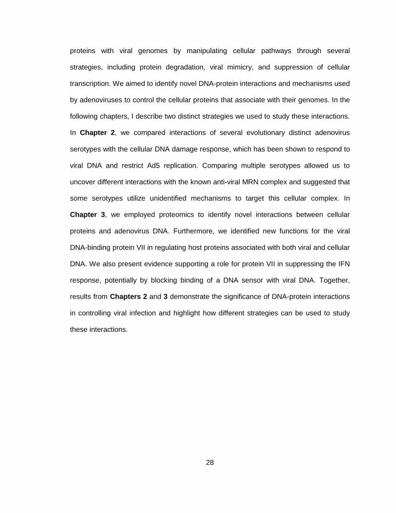
28
proteins with viral genomes by manipulating cellular pathways through several
strategies, including protein degradation, viral mimicry, and suppression of cellular
transcription. We aimed to identify novel DNA-protein interactions and mechanisms used
by adenoviruses to control the cellular proteins that associate with their genomes. In the
following chapters, I describe two distinct strategies we used to study these interactions.
In Chapter 2, we compared interactions of several evolutionary distinct adenovirus
serotypes with the cellular DNA damage response, which has been shown to respond to
viral DNA and restrict Ad5 replication. Comparing multiple serotypes allowed us to
uncover different interactions with the known anti-viral MRN complex and suggested that
some serotypes utilize unidentified mechanisms to target this cellular complex. In
Chapter 3, we employed proteomics to identify novel interactions between cellular
proteins and adenovirus DNA. Furthermore, we identified new functions for the viral
DNA-binding protein VII in regulating host proteins associated with both viral and cellular
DNA. We also present evidence supporting a role for protein VII in suppressing the IFN
response, potentially by blocking binding of a DNA sensor with viral DNA. Together,
results from Chapters 2 and 3 demonstrate the significance of DNA-protein interactions
in controlling viral infection and highlight how different strategies can be used to study
these interactions.
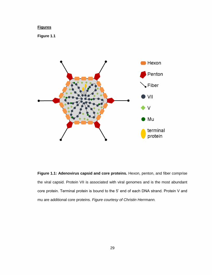
29
Figures
Figure 1.1
Figure 1.1: Adenovirus capsid and core proteins. Hexon, penton, and fiber comprise
the viral capsid. Protein VII is associated with viral genomes and is the most abundant
core protein. Terminal protein is bound to the 5’ end of each DNA strand. Protein V and
mu are additional core proteins. Figure courtesy of Christin Herrmann.
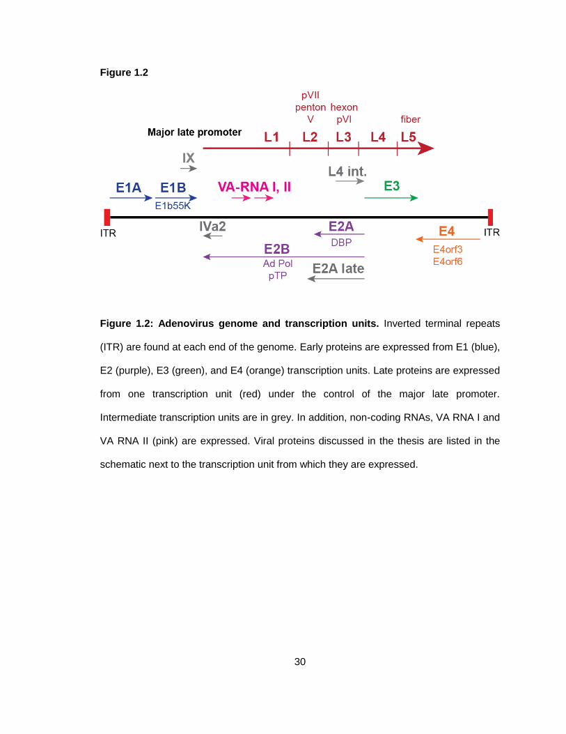
30
Figure 1.2
Figure 1.2: Adenovirus genome and transcription units. Inverted terminal repeats
(ITR) are found at each end of the genome. Early proteins are expressed from E1 (blue),
E2 (purple), E3 (green), and E4 (orange) transcription units. Late proteins are expressed
from one transcription unit (red) under the control of the major late promoter.
Intermediate transcription units are in grey. In addition, non-coding RNAs, VA RNA I and
VA RNA II (pink) are expressed. Viral proteins discussed in the thesis are listed in the
schematic next to the transcription unit from which they are expressed.
31
Figure 1.3
Figure 1.3: Viral replication centers. (A) Images of viral replication centers from
adenovirus-infected cells. DBP is a viral DNA replication protein that accumulates at
sites of single-stranded viral DNA and marks viral replication centers. RPA32 is a cellular
single-stranded DNA-binding protein and also marks viral replication centers. EdU is a
thymidine analog that is incorporated into replicating DNA. Colocalization of DBP,
RPA32, and EdU demonstrate that any of these methods can be used to visualize viral
replication centers. (B) Representative images from different stages of Ad5 infection.
Viral replication centers change as infection progresses.
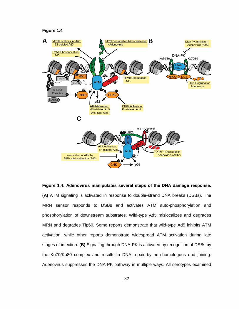
32
Figure 1.4
Figure 1.4: Adenovirus manipulates several steps of the DNA damage response.
(A) ATM signaling is activated in response to double-strand DNA breaks (DSBs). The
MRN sensor responds to DSBs and activates ATM auto-phosphorylation and
phosphorylation of downstream substrates. Wild-type Ad5 mislocalizes and degrades
MRN and degrades Tip60. Some reports demonstrate that wild-type Ad5 inhibits ATM
activation, while other reports demonstrate widespread ATM activation during late
stages of infection. (B) Signaling through DNA-PK is activated by recognition of DSBs by
the Ku70/Ku80 complex and results in DNA repair by non-homologous end joining.
Adenovirus suppresses the DNA-PK pathway in multiple ways. All serotypes examined
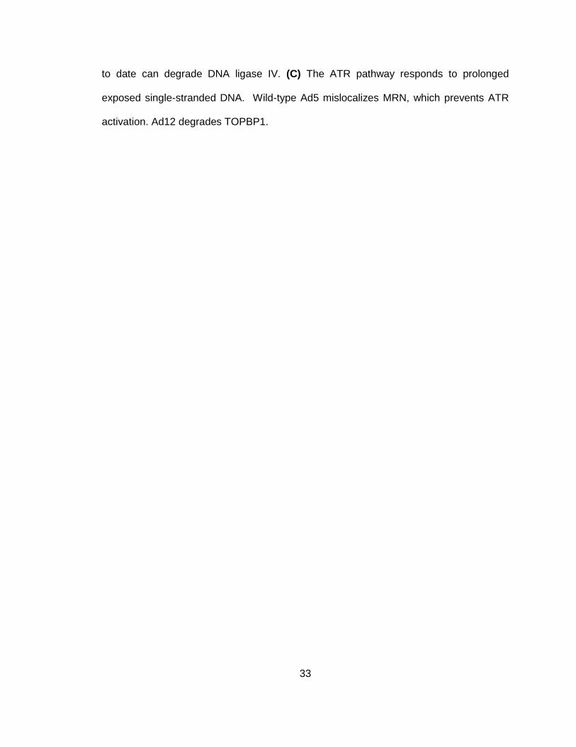
33
to date can degrade DNA ligase IV. (C) The ATR pathway responds to prolonged
exposed single-stranded DNA. Wild-type Ad5 mislocalizes MRN, which prevents ATR
activation. Ad12 degrades TOPBP1.
34
Figure 1.5
Figure 1.5: Overview of interferon signaling. Cytoplasmic DNA sensors recognize
viral DNA, which leads to activation of STING and interferon regulatory factor 3 (IRF3).
IRF3 translocates to the nucleus where it activates transcription of IFN. Newly
synthesized IFN protein is released from the infected cell and binds cellular receptors
on the infected and adjacent cells. This activates JAK-STAT signaling and expression of
interferon-stimulated genes in infected and adjacent cells. The protein products of ISGs
impair viral processes through multiple mechanisms.
35
CHAPTER 2:
Serotype-specific restriction of wild-type adenoviruses
by the cellular Mre11-Rad50-Nbs1 complex
Portions of this chapter are currently in press:
Pancholi, N.J. and Weitzman, M.D. Serotype-specific restriction of wild-type
adenoviruses by the cellular Mre11-Rad50-Nbs1 complex. Virology. (in press)
A figure from this chapter has been previously published in:
Lou, D. I.*, Kim, E. T.*, Meyerson, N. R., Pancholi, N. J., Mohni, K. N., Enard,
D., . . . Sawyer, S. L. (2016). An Intrinsically Disordered Region of the DNA
Repair Protein Nbs1 Is a Species-Specific Barrier to Herpes Simplex Virus 1 in
Primates. Cell Host Microbe, 20(2), 178-188.
Introduction
It has been well established that the DDR is an obstacle for wild-type Ad5 replication and
that Ad5 employs redundant mechanisms to evade its negative effects (see Chapter 1).
However, there has been relatively little research into the interactions between other
adenovirus serotypes and the DDR. Analysis of known Ad5 degradation substrates
during infection with other Ad serotypes revealed that all serotypes examined to date
lead to degradation of DNA ligase IV (Forrester et al., 2011). In contrast, some serotypes
appear not to degrade MRN, p53, or integrin 3, and only Ad12 has been found to
degrade the DDR regulatory protein TOPBP1 (Blackford et al., 2010; Bridges et al.,
2016; Cheng et al., 2011; Forrester et al., 2011). Interestingly, substrate degradation by
non-Ad5 serotypes does not always correlate with interaction with E1b55K (Cheng et al.,
36
2013), suggesting that degradation of host proteins by adenoviruses may be regulated in
additional unknown ways. Furthermore, infection with some serotypes does not result in
MRN mislocalization to tracks or to aggressomes (Blanchette, Wimmer, Dallaire, Cheng,
& Branton, 2013; Forrester et al., 2011; Stracker et al., 2005). These findings
demonstrate that although the ability to evade recognition by MRN is critical for optimal
wild-type Ad5 replication, this may not necessarily be representative across the whole
adenovirus family.
Since cellular restriction factors can influence tissue tropism and virulence, we reasoned
that there may be differences among serotypes in their ability to overcome MRN
inhibition. While previous studies have demonstrated that some serotypes do not
degrade or mislocalize MRN, it remains unknown how the different interactions with
MRN impact virus replication. Given the importance of inactivating MRN and
downstream responses during wild-type Ad5 replication, it is possible that virulence
and/or tissue tropism of adenoviruses are partially influenced by their potential to evade
inhibition by MRN. Furthermore, it is unclear whether MRN targeting by other serotypes
is accomplished by the analogous viral proteins as Ad5. Here, we examined more
closely the fate of MRN during infection with multiple adenovirus serotypes representing
several subgroups, and we determined the impact on wild-type viral DNA replication.
Consistent with previous reports (Cheng et al., 2011; Forrester et al., 2011), we
identified serotypes that target MRN through both degradation and mislocalization, and
other serotypes incapable of one or both of these mechanisms. We found that serotypes
Ad9 and Ad12 can target MRN by mislocalization or degradation but are still impaired for
DNA replication, demonstrating differences between these serotypes and Ad5. By
examining the viral proteins that target MRN, we found that Ad9-E4orf3 alone is not
sufficient to induce MRN mislocalization even though it is observed during Ad9 infection,
37
suggesting that MRN mislocalization by Ad9 may be regulated through additional viral
mechanisms. This work adds to our growing understanding of adenoviruses and the
DDR, and suggests that diverse strategies have evolved across the adenovirus family to
overcome MRN during wild-type virus infections.
Materials and Methods
Cell lines
U2OS were purchased from the American Tissue Culture Collection. Immortalized NBS
cells (ILB1) transduced to express Nbs1 or empty vector were previously described
(Cerosaletti et al., 2000; Kraakman-van der Zwet et al., 1999). Immortalized A-T cells
(AT22IJE-T) and matched cells complemented with ATM as previously described were
gifts from Y. Shiloh (Ziv et al., 1997; Ziv et al., 1989). All cells were maintained in
Dulbecco modified Eagle medium (Corning MT10-013-CV) supplemented with 10% fetal
bovine serum and 1% penicillin-streptomycin (Invitrogen 15140122) at 37°C in a
humidified incubator with 5% CO2. Acceptor cells for the generation of doxycycline-
inducible cell lines were provided by E. Makeyev and were used as previously described
(Khandelia, Yap, & Makeyev, 2011). Briefly, FLAG-Ad9-E4orf3 was PCR amplified from
the pL2-FLAG-Ad9-E4orf3 plasmid described below and inserted into the inducible
plasmid backbone. The inducible plasmid containing FLAG-Ad9-E4orf3 was transfected
into U2OS acceptor cells together with a plasmid expressing the Cre recombinase.
Recombined clones were selected with 1 g/mL Puromycin. Cells were induced with 0.2
g/mL doxycycline for 24 hours to express FLAG-Ad9-E4orf3. Expression was confirmed
by immunoblot and immunofluorescence. Inducible cells were maintained in medium
supplemented with tetracycline-free fetal bovine serum.
38
Plasmids and transfections
The Ad9-E4orf3 cDNA was obtained from cells infected with Ad9, PCR amplified, and
cloned into the pL2-FLAG plasmid backbone (described in (Stracker et al., 2005)).
Transfections were performed using the standard protocol for Lipofectamine 2000
(Invitrogen).
Viruses and infections
Wild-type Ad5, Ad2, Ad4, Ad9, Ad12, and Ad35 were purchased from American Tissue
Culture Collection. Mutant Ad5 viruses dl1004, dl110, and dl1006 were previously
described (Babiss & Ginsberg, 1984; Bridge & Ketner, 1989) and were gifts from G.
Ketner and D. Ornelles. Wild-type Ad5, Ad2, Ad4, Ad9, Ad12, Ad35, dl110, and dl1006
were propagated on 293 cells. The E4-deleted virus dl1004 was propagated on W162
cells. All viruses were purified by two sequential rounds of ultracentrifugation of cesium
chloride gradients and stored in 40% glycerol at -20°C. Viral titers were determined by
plaque assay on 293 cells. Infections were carried out by standard protocols using a
multiplicity of infection of 20 (Ad5 wild-type and mutants, Ad2, Ad4, Ad12, Ad35) or 50
(Ad9). Viruses were diluted in Dulbecco modified Eagle medium supplemented with 2%
fetal bovine serum and 1% penicillin-streptomycin and added to cell monolayers. Cells
were incubated with the virus for 2 hours at 37°C before supplementing infection
medium with medium containing 10% fetal bovine serum.
Antibodies and inhibitors
Primary antibodies to cellular proteins were purchased from commercial sources: Mre11
(Novus NB100-142), Rad50 (GeneTex [13B3] GTX70228), Nbs1 (Novus NB100-143),
ATM pS1981 (Epitomics 2152-1 and Abcam [EP1890Y] ab81292), ATM (Abcam [Y170]
ab32420 and Epitomics 1549-1), Actin (Sigma a5441), RPA32 (Abcam ab2175 and
39
Bethyl A300-244A), PML (Santa Cruz [PG-M3] sc-966), and FLAG (Sigma F3165 and
F7425). Primary antibodies to adenoviral proteins DBP and E4orf3 were gifts from A.
Levine and T. Dobner, respectively. Horseradish peroxidase-conjugated secondary
antibodies for immunoblotting were purchased from Jackson Laboratories. Fluorophore-
conjugated secondary antibodies for immunofluorescence were purchased from Life
Technologies. The ATM kinase inhibitor KU55933 was purchased from Abcam. The
proteasome inhibitor MG132 was purchased from Sigma-Aldrich.
Immunoblotting
Immunoblot analysis was carried out using standard methods. Briefly, protein samples
were prepared in lithium dodecyl sulfate loading buffer (NuPage) with 10% dithiothreitol
and boiled. Equal amounts of protein were separated by electrophoresis. Proteins were
transferred to nitrocellulose membranes (GE Healthcare Amersham) and blocked in 5%
milk in tris buffered saline with Tween (TBST). Proteins were detected by enhanced
chemiluminescence (Thermo Scientific) on film (HyBlot CL) or on a Syngene G-Box.
Immunofluorescence
Cells were plated on glass coverslips. Cells were washed with phosphate buffered saline
(PBS) and fixed with cold 4% paraformaldehyde for 15 minutes. Cells were
permeabilized for ten minutes with 0.5% Triton X-100 and coverslips were blocked for 1
hour with 3% bovine serum albumin (BSA) in PBS, incubated with each primary antibody
diluted in 3% BSA for one hour, and incubated with a mixture of secondary antibodies
and 4,6-diamidino-2-phenylindole (DAPI) in 3% BSA for one hour. Coverslips were
mounted onto glass slides using ProLong Gold AntiFade Reagent (Life Technologies)
and fluorescence was visualized using a Zeiss LSM 710 confocal microscope. Images
were processed using ImageJ and Adobe Creative Suite 6.
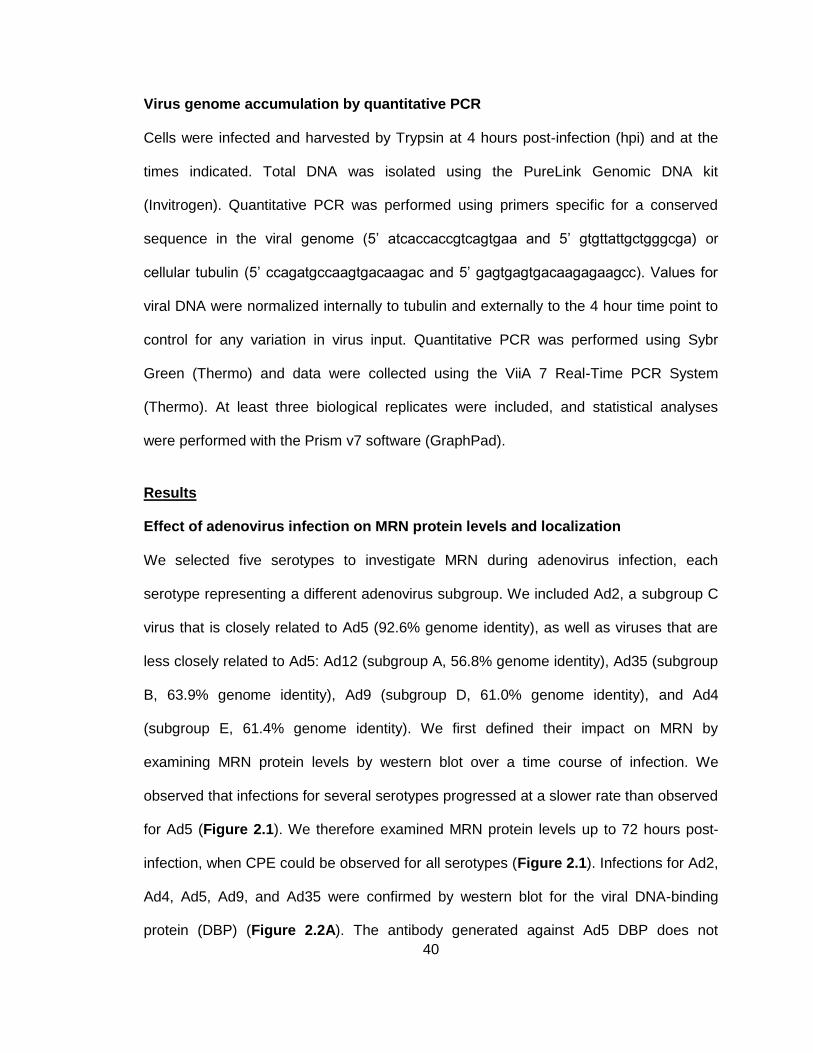
40
Virus genome accumulation by quantitative PCR
Cells were infected and harvested by Trypsin at 4 hours post-infection (hpi) and at the
times indicated. Total DNA was isolated using the PureLink Genomic DNA kit
(Invitrogen). Quantitative PCR was performed using primers specific for a conserved
sequence in the viral genome (5’ atcaccaccgtcagtgaa and 5’ gtgttattgctgggcga) or
cellular tubulin (5’ ccagatgccaagtgacaagac and 5’ gagtgagtgacaagagaagcc). Values for
viral DNA were normalized internally to tubulin and externally to the 4 hour time point to
control for any variation in virus input. Quantitative PCR was performed using Sybr
Green (Thermo) and data were collected using the ViiA 7 Real-Time PCR System
(Thermo). At least three biological replicates were included, and statistical analyses
were performed with the Prism v7 software (GraphPad).
Results
Effect of adenovirus infection on MRN protein levels and localization
We selected five serotypes to investigate MRN during adenovirus infection, each
serotype representing a different adenovirus subgroup. We included Ad2, a subgroup C
virus that is closely related to Ad5 (92.6% genome identity), as well as viruses that are
less closely related to Ad5: Ad12 (subgroup A, 56.8% genome identity), Ad35 (subgroup
B, 63.9% genome identity), Ad9 (subgroup D, 61.0% genome identity), and Ad4
(subgroup E, 61.4% genome identity). We first defined their impact on MRN by
examining MRN protein levels by western blot over a time course of infection. We
observed that infections for several serotypes progressed at a slower rate than observed
for Ad5 (Figure 2.1). We therefore examined MRN protein levels up to 72 hours post-
infection, when CPE could be observed for all serotypes (Figure 2.1). Infections for Ad2,
Ad4, Ad5, Ad9, and Ad35 were confirmed by western blot for the viral DNA-binding
protein (DBP) (Figure 2.2A). The antibody generated against Ad5 DBP does not
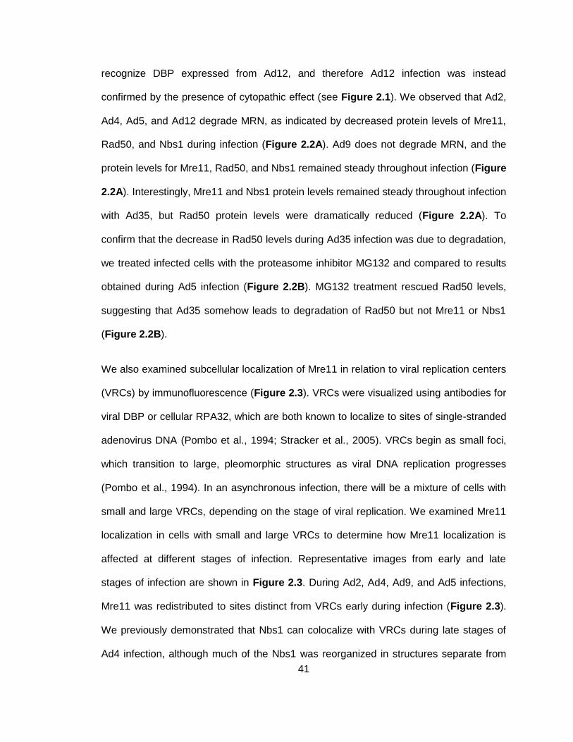
41
recognize DBP expressed from Ad12, and therefore Ad12 infection was instead
confirmed by the presence of cytopathic effect (see Figure 2.1). We observed that Ad2,
Ad4, Ad5, and Ad12 degrade MRN, as indicated by decreased protein levels of Mre11,
Rad50, and Nbs1 during infection (Figure 2.2A). Ad9 does not degrade MRN, and the
protein levels for Mre11, Rad50, and Nbs1 remained steady throughout infection (Figure
2.2A). Interestingly, Mre11 and Nbs1 protein levels remained steady throughout infection
with Ad35, but Rad50 protein levels were dramatically reduced (Figure 2.2A). To
confirm that the decrease in Rad50 levels during Ad35 infection was due to degradation,
we treated infected cells with the proteasome inhibitor MG132 and compared to results
obtained during Ad5 infection (Figure 2.2B). MG132 treatment rescued Rad50 levels,
suggesting that Ad35 somehow leads to degradation of Rad50 but not Mre11 or Nbs1
(Figure 2.2B).
We also examined subcellular localization of Mre11 in relation to viral replication centers
(VRCs) by immunofluorescence (Figure 2.3). VRCs were visualized using antibodies for
viral DBP or cellular RPA32, which are both known to localize to sites of single-stranded
adenovirus DNA (Pombo et al., 1994; Stracker et al., 2005). VRCs begin as small foci,
which transition to large, pleomorphic structures as viral DNA replication progresses
(Pombo et al., 1994). In an asynchronous infection, there will be a mixture of cells with
small and large VRCs, depending on the stage of viral replication. We examined Mre11
localization in cells with small and large VRCs to determine how Mre11 localization is
affected at different stages of infection. Representative images from early and late
stages of infection are shown in Figure 2.3. During Ad2, Ad4, Ad9, and Ad5 infections,
Mre11 was redistributed to sites distinct from VRCs early during infection (Figure 2.3).
We previously demonstrated that Nbs1 can colocalize with VRCs during late stages of
Ad4 infection, although much of the Nbs1 was reorganized in structures separate from
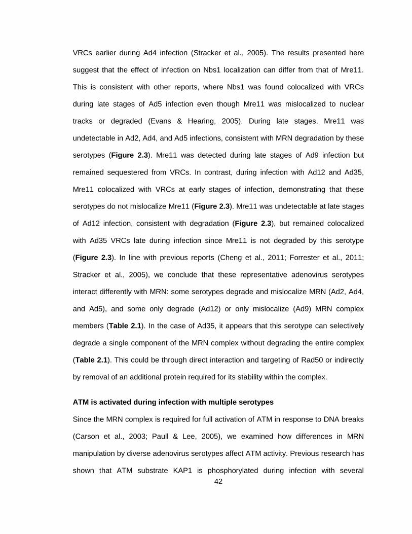
42
VRCs earlier during Ad4 infection (Stracker et al., 2005). The results presented here
suggest that the effect of infection on Nbs1 localization can differ from that of Mre11.
This is consistent with other reports, where Nbs1 was found colocalized with VRCs
during late stages of Ad5 infection even though Mre11 was mislocalized to nuclear
tracks or degraded (Evans & Hearing, 2005). During late stages, Mre11 was
undetectable in Ad2, Ad4, and Ad5 infections, consistent with MRN degradation by these
serotypes (Figure 2.3). Mre11 was detected during late stages of Ad9 infection but
remained sequestered from VRCs. In contrast, during infection with Ad12 and Ad35,
Mre11 colocalized with VRCs at early stages of infection, demonstrating that these
serotypes do not mislocalize Mre11 (Figure 2.3). Mre11 was undetectable at late stages
of Ad12 infection, consistent with degradation (Figure 2.3), but remained colocalized
with Ad35 VRCs late during infection since Mre11 is not degraded by this serotype
(Figure 2.3). In line with previous reports (Cheng et al., 2011; Forrester et al., 2011;
Stracker et al., 2005), we conclude that these representative adenovirus serotypes
interact differently with MRN: some serotypes degrade and mislocalize MRN (Ad2, Ad4,
and Ad5), and some only degrade (Ad12) or only mislocalize (Ad9) MRN complex
members (Table 2.1). In the case of Ad35, it appears that this serotype can selectively
degrade a single component of the MRN complex without degrading the entire complex
(Table 2.1). This could be through direct interaction and targeting of Rad50 or indirectly
by removal of an additional protein required for its stability within the complex.
ATM is activated during infection with multiple serotypes
Since the MRN complex is required for full activation of ATM in response to DNA breaks
(Carson et al., 2003; Paull & Lee, 2005), we examined how differences in MRN
manipulation by diverse adenovirus serotypes affect ATM activity. Previous research has
shown that ATM substrate KAP1 is phosphorylated during infection with several
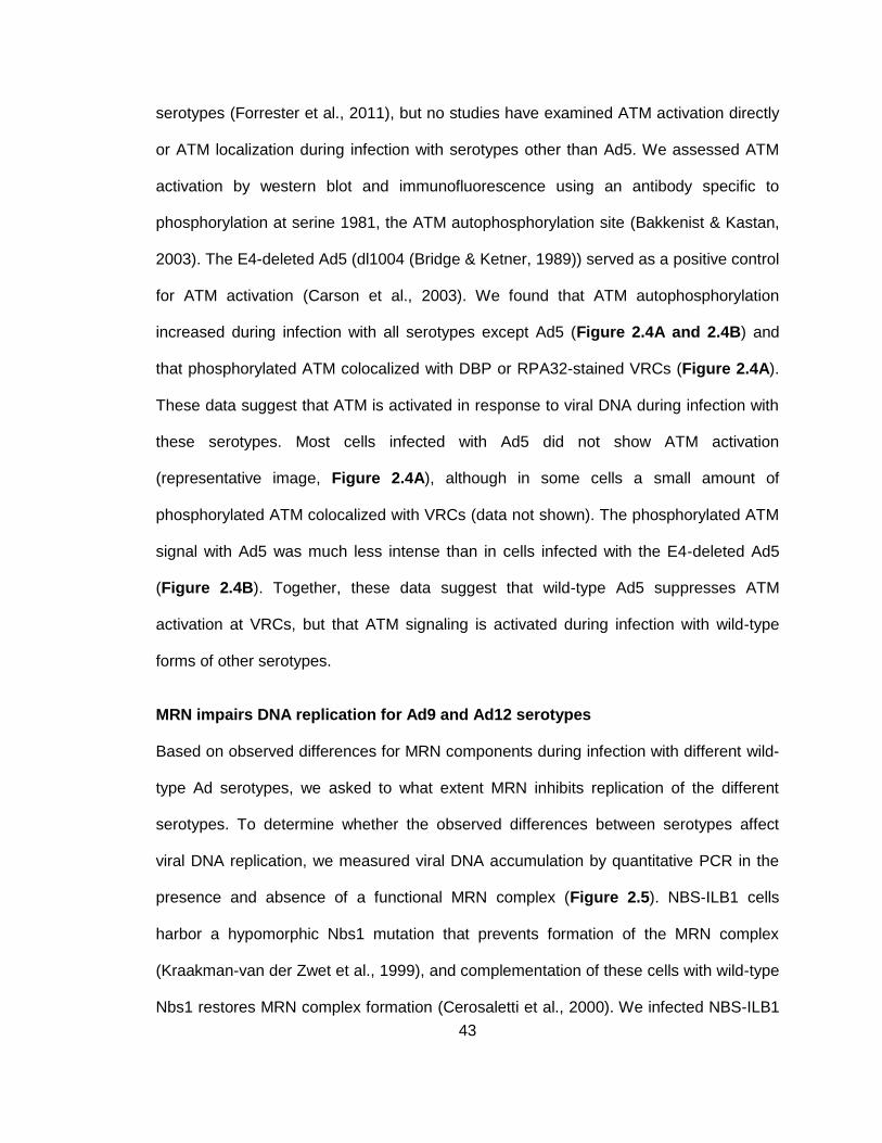
43
serotypes (Forrester et al., 2011), but no studies have examined ATM activation directly
or ATM localization during infection with serotypes other than Ad5. We assessed ATM
activation by western blot and immunofluorescence using an antibody specific to
phosphorylation at serine 1981, the ATM autophosphorylation site (Bakkenist & Kastan,
2003). The E4-deleted Ad5 (dl1004 (Bridge & Ketner, 1989)) served as a positive control
for ATM activation (Carson et al., 2003). We found that ATM autophosphorylation
increased during infection with all serotypes except Ad5 (Figure 2.4A and 2.4B) and
that phosphorylated ATM colocalized with DBP or RPA32-stained VRCs (Figure 2.4A).
These data suggest that ATM is activated in response to viral DNA during infection with
these serotypes. Most cells infected with Ad5 did not show ATM activation
(representative image, Figure 2.4A), although in some cells a small amount of
phosphorylated ATM colocalized with VRCs (data not shown). The phosphorylated ATM
signal with Ad5 was much less intense than in cells infected with the E4-deleted Ad5
(Figure 2.4B). Together, these data suggest that wild-type Ad5 suppresses ATM
activation at VRCs, but that ATM signaling is activated during infection with wild-type
forms of other serotypes.
MRN impairs DNA replication for Ad9 and Ad12 serotypes
Based on observed differences for MRN components during infection with different wild-
type Ad serotypes, we asked to what extent MRN inhibits replication of the different
serotypes. To determine whether the observed differences between serotypes affect
viral DNA replication, we measured viral DNA accumulation by quantitative PCR in the
presence and absence of a functional MRN complex (Figure 2.5). NBS-ILB1 cells
harbor a hypomorphic Nbs1 mutation that prevents formation of the MRN complex
(Kraakman-van der Zwet et al., 1999), and complementation of these cells with wild-type
Nbs1 restores MRN complex formation (Cerosaletti et al., 2000). We infected NBS-ILB1
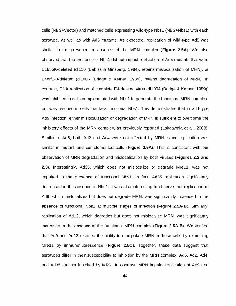
44
cells (NBS+Vector) and matched cells expressing wild-type Nbs1 (NBS+Nbs1) with each
serotype, as well as with Ad5 mutants. As expected, replication of wild-type Ad5 was
similar in the presence or absence of the MRN complex (Figure 2.5A). We also
observed that the presence of Nbs1 did not impact replication of Ad5 mutants that were
E1b55K-deleted (dl110 (Babiss & Ginsberg, 1984), retains mislocalization of MRN), or
E4orf1-3-deleted (dl1006 (Bridge & Ketner, 1989), retains degradation of MRN). In
contrast, DNA replication of complete E4-deleted virus (dl1004 (Bridge & Ketner, 1989))
was inhibited in cells complemented with Nbs1 to generate the functional MRN complex,
but was rescued in cells that lack functional Nbs1. This demonstrates that in wild-type
Ad5 infection, either mislocalization or degradation of MRN is sufficient to overcome the
inhibitory effects of the MRN complex, as previously reported (Lakdawala et al., 2008).
Similar to Ad5, both Ad2 and Ad4 were not affected by MRN, since replication was
similar in mutant and complemented cells (Figure 2.5A). This is consistent with our
observation of MRN degradation and mislocalization by both viruses (Figures 2.2 and
2.3). Interestingly, Ad35, which does not mislocalize or degrade Mre11, was not
impaired in the presence of functional Nbs1. In fact, Ad35 replication significantly
decreased in the absence of Nbs1. It was also interesting to observe that replication of
Ad9, which mislocalizes but does not degrade MRN, was significantly increased in the
absence of functional Nbs1 at multiple stages of infection (Figure 2.5A-B). Similarly,
replication of Ad12, which degrades but does not mislocalize MRN, was significantly
increased in the absence of the functional MRN complex (Figure 2.5A-B). We verified
that Ad9 and Ad12 retained the ability to manipulate MRN in these cells by examining
Mre11 by immunofluorescence (Figure 2.5C). Together, these data suggest that
serotypes differ in their susceptibility to inhibition by the MRN complex. Ad5, Ad2, Ad4,
and Ad35 are not inhibited by MRN. In contrast, MRN impairs replication of Ad9 and
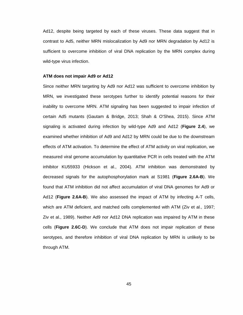
45
Ad12, despite being targeted by each of these viruses. These data suggest that in
contrast to Ad5, neither MRN mislocalization by Ad9 nor MRN degradation by Ad12 is
sufficient to overcome inhibition of viral DNA replication by the MRN complex during
wild-type virus infection.
ATM does not impair Ad9 or Ad12
Since neither MRN targeting by Ad9 nor Ad12 was sufficient to overcome inhibition by
MRN, we investigated these serotypes further to identify potential reasons for their
inability to overcome MRN. ATM signaling has been suggested to impair infection of
certain Ad5 mutants (Gautam & Bridge, 2013; Shah & O'Shea, 2015). Since ATM
signaling is activated during infection by wild-type Ad9 and Ad12 (Figure 2.4), we
examined whether inhibition of Ad9 and Ad12 by MRN could be due to the downstream
effects of ATM activation. To determine the effect of ATM activity on viral replication, we
measured viral genome accumulation by quantitative PCR in cells treated with the ATM
inhibitor KU55933 (Hickson et al., 2004). ATM inhibition was demonstrated by
decreased signals for the autophosphorylation mark at S1981 (Figure 2.6A-B). We
found that ATM inhibition did not affect accumulation of viral DNA genomes for Ad9 or
Ad12 (Figure 2.6A-B). We also assessed the impact of ATM by infecting A-T cells,
which are ATM deficient, and matched cells complemented with ATM (Ziv et al., 1997;
Ziv et al., 1989). Neither Ad9 nor Ad12 DNA replication was impaired by ATM in these
cells (Figure 2.6C-D). We conclude that ATM does not impair replication of these
serotypes, and therefore inhibition of viral DNA replication by MRN is unlikely to be
through ATM.
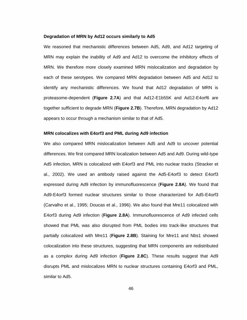
46
Degradation of MRN by Ad12 occurs similarly to Ad5
We reasoned that mechanistic differences between Ad5, Ad9, and Ad12 targeting of
MRN may explain the inability of Ad9 and Ad12 to overcome the inhibitory effects of
MRN. We therefore more closely examined MRN mislocalization and degradation by
each of these serotypes. We compared MRN degradation between Ad5 and Ad12 to
identify any mechanistic differences. We found that Ad12 degradation of MRN is
proteasome-dependent (Figure 2.7A) and that Ad12-E1b55K and Ad12-E4orf6 are
together sufficient to degrade MRN (Figure 2.7B). Therefore, MRN degradation by Ad12
appears to occur through a mechanism similar to that of Ad5.
MRN colocalizes with E4orf3 and PML during Ad9 infection
We also compared MRN mislocalization between Ad5 and Ad9 to uncover potential
differences. We first compared MRN localization between Ad5 and Ad9. During wild-type
Ad5 infection, MRN is colocalized with E4orf3 and PML into nuclear tracks (Stracker et
al., 2002). We used an antibody raised against the Ad5-E4orf3 to detect E4orf3
expressed during Ad9 infection by immunofluorescence (Figure 2.8A). We found that
Ad9-E4orf3 formed nuclear structures similar to those characterized for Ad5-E4orf3
(Carvalho et al., 1995; Doucas et al., 1996). We also found that Mre11 colocalized with
E4orf3 during Ad9 infection (Figure 2.8A). Immunofluorescence of Ad9 infected cells
showed that PML was also disrupted from PML bodies into track-like structures that
partially colocalized with Mre11 (Figure 2.8B). Staining for Mre11 and Nbs1 showed
colocalization into these structures, suggesting that MRN components are redistributed
as a complex during Ad9 infection (Figure 2.8C). These results suggest that Ad9
disrupts PML and mislocalizes MRN to nuclear structures containing E4orf3 and PML,
similar to Ad5.
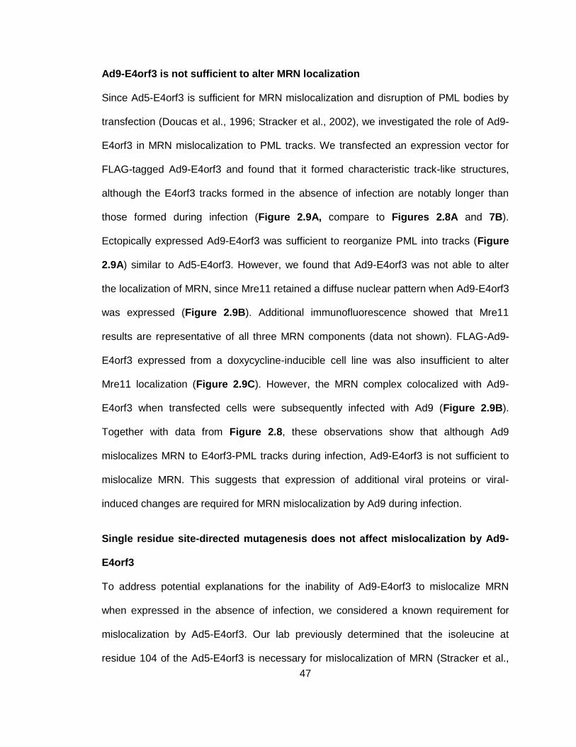
47
Ad9-E4orf3 is not sufficient to alter MRN localization
Since Ad5-E4orf3 is sufficient for MRN mislocalization and disruption of PML bodies by
transfection (Doucas et al., 1996; Stracker et al., 2002), we investigated the role of Ad9-
E4orf3 in MRN mislocalization to PML tracks. We transfected an expression vector for
FLAG-tagged Ad9-E4orf3 and found that it formed characteristic track-like structures,
although the E4orf3 tracks formed in the absence of infection are notably longer than
those formed during infection (Figure 2.9A, compare to Figures 2.8A and 7B).
Ectopically expressed Ad9-E4orf3 was sufficient to reorganize PML into tracks (Figure
2.9A) similar to Ad5-E4orf3. However, we found that Ad9-E4orf3 was not able to alter
the localization of MRN, since Mre11 retained a diffuse nuclear pattern when Ad9-E4orf3
was expressed (Figure 2.9B). Additional immunofluorescence showed that Mre11
results are representative of all three MRN components (data not shown). FLAG-Ad9-
E4orf3 expressed from a doxycycline-inducible cell line was also insufficient to alter
Mre11 localization (Figure 2.9C). However, the MRN complex colocalized with Ad9-
E4orf3 when transfected cells were subsequently infected with Ad9 (Figure 2.9B).
Together with data from Figure 2.8, these observations show that although Ad9
mislocalizes MRN to E4orf3-PML tracks during infection, Ad9-E4orf3 is not sufficient to
mislocalize MRN. This suggests that expression of additional viral proteins or viral-
induced changes are required for MRN mislocalization by Ad9 during infection.
Single residue site-directed mutagenesis does not affect mislocalization by Ad9-
E4orf3
To address potential explanations for the inability of Ad9-E4orf3 to mislocalize MRN
when expressed in the absence of infection, we considered a known requirement for
mislocalization by Ad5-E4orf3. Our lab previously determined that the isoleucine at
residue 104 of the Ad5-E4orf3 is necessary for mislocalization of MRN (Stracker et al.,
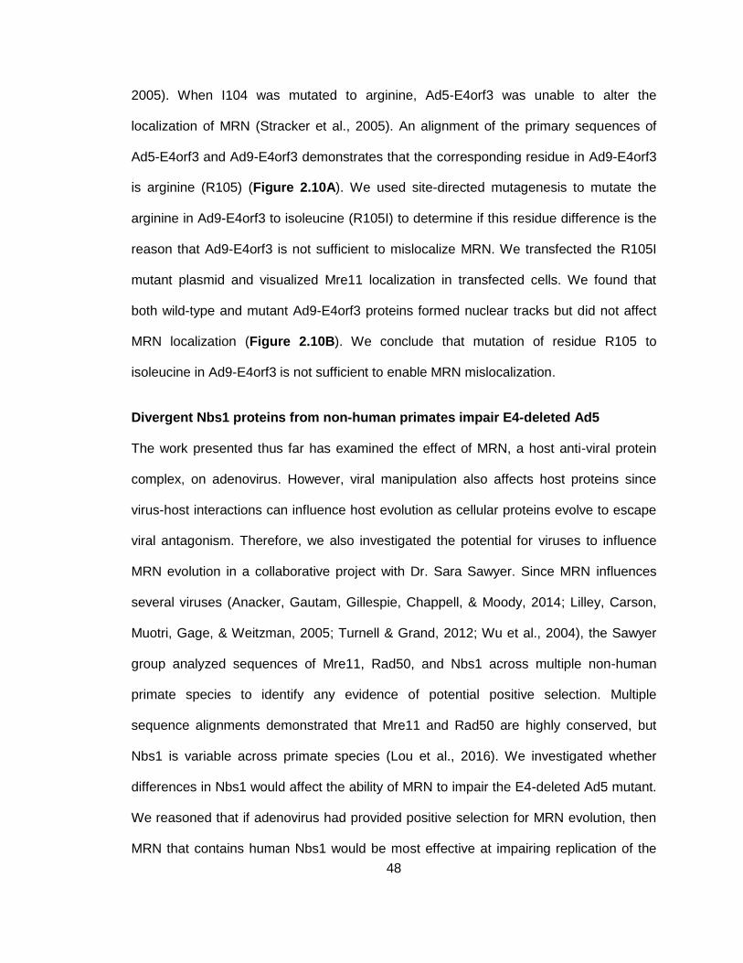
48
2005). When I104 was mutated to arginine, Ad5-E4orf3 was unable to alter the
localization of MRN (Stracker et al., 2005). An alignment of the primary sequences of
Ad5-E4orf3 and Ad9-E4orf3 demonstrates that the corresponding residue in Ad9-E4orf3
is arginine (R105) (Figure 2.10A). We used site-directed mutagenesis to mutate the
arginine in Ad9-E4orf3 to isoleucine (R105I) to determine if this residue difference is the
reason that Ad9-E4orf3 is not sufficient to mislocalize MRN. We transfected the R105I
mutant plasmid and visualized Mre11 localization in transfected cells. We found that
both wild-type and mutant Ad9-E4orf3 proteins formed nuclear tracks but did not affect
MRN localization (Figure 2.10B). We conclude that mutation of residue R105 to
isoleucine in Ad9-E4orf3 is not sufficient to enable MRN mislocalization.
Divergent Nbs1 proteins from non-human primates impair E4-deleted Ad5
The work presented thus far has examined the effect of MRN, a host anti-viral protein
complex, on adenovirus. However, viral manipulation also affects host proteins since
virus-host interactions can influence host evolution as cellular proteins evolve to escape
viral antagonism. Therefore, we also investigated the potential for viruses to influence
MRN evolution in a collaborative project with Dr. Sara Sawyer. Since MRN influences
several viruses (Anacker, Gautam, Gillespie, Chappell, & Moody, 2014; Lilley, Carson,
Muotri, Gage, & Weitzman, 2005; Turnell & Grand, 2012; Wu et al., 2004), the Sawyer
group analyzed sequences of Mre11, Rad50, and Nbs1 across multiple non-human
primate species to identify any evidence of potential positive selection. Multiple
sequence alignments demonstrated that Mre11 and Rad50 are highly conserved, but
Nbs1 is variable across primate species (Lou et al., 2016). We investigated whether
differences in Nbs1 would affect the ability of MRN to impair the E4-deleted Ad5 mutant.
We reasoned that if adenovirus had provided positive selection for MRN evolution, then
MRN that contains human Nbs1 would be most effective at impairing replication of the
49
human E4-deleted Ad5. We used human NBS-ILB1 cells (described above)
complemented with Nbs1 from human, gibbon, or siamang. We infected NBS-ILB1 and
complemented cells with the E4-deleted Ad5 mutant and measured viral DNA
accumulation by quantitative PCR (Figure 2.11). As expected, cells complemented with
human Nbs1 dramatically impaired the E4-deleted Ad5 (Figure 2.11). We found that
replication of the E4-deleted Ad5 was suppressed to a similar level in cells
complemented with gibbon or siamang Nbs1 (Figure 2.11). These data demonstrate
that the differences between human, gibbon, and siamang Nbs1 proteins do not affect
the ability of MRN to impair replication of Ad5. Since the observed differences between
human and non-human primate Nbs1 proteins do not confer an advantage in
suppressing human adenovirus, the observed sequence variability of Nbs1 between
these primate species is unlikely to have been selected for by adenovirus.
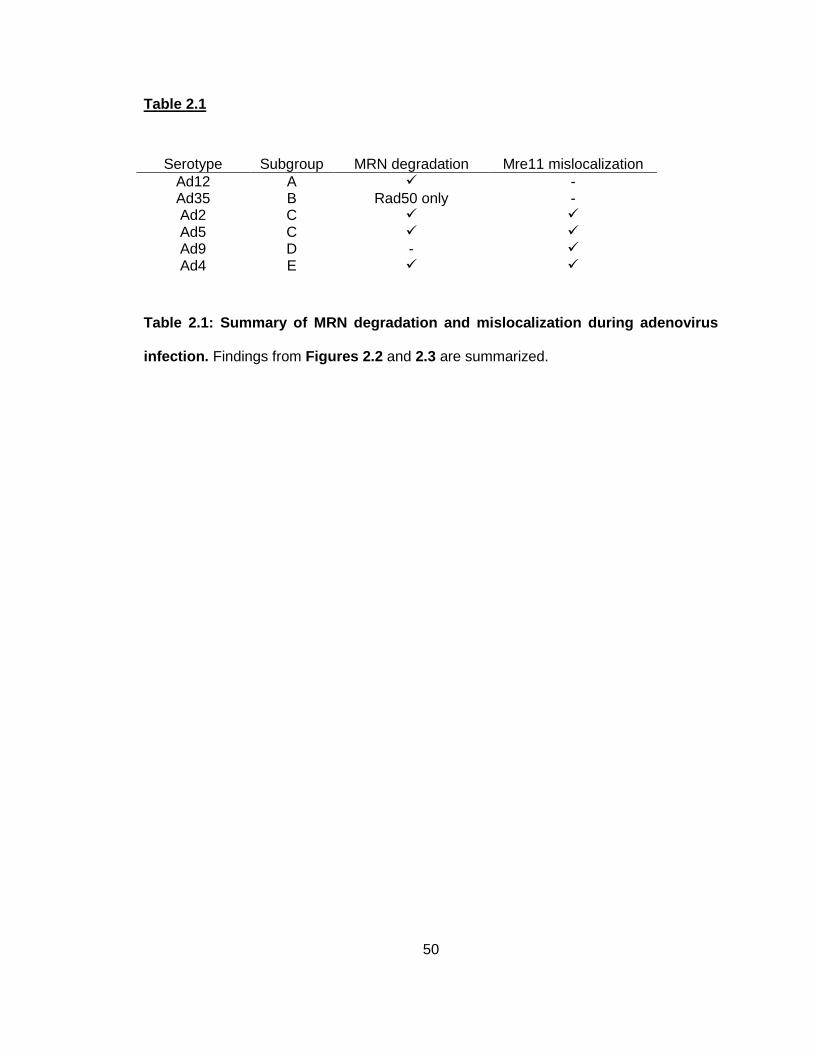
50
Table 2.1
Table 2.1: Summary of MRN degradation and mislocalization during adenovirus
infection. Findings from Figures 2.2 and 2.3 are summarized.
Serotype Subgroup MRN degradation Mre11 mislocalization
Ad12 A - Ad35 B Rad50 only - Ad2 C
Ad5 C
Ad9 D -
Ad4 E
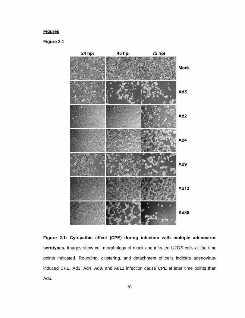
51
Figures
Figure 2.1
Figure 2.1: Cytopathic effect (CPE) during infection with multiple adenovirus
serotypes. Images show cell morphology of mock and infected U2OS cells at the time
points indicated. Rounding, clustering, and detachment of cells indicate adenovirus-
induced CPE. Ad2, Ad4, Ad9, and Ad12 infection cause CPE at later time points than
Ad5.
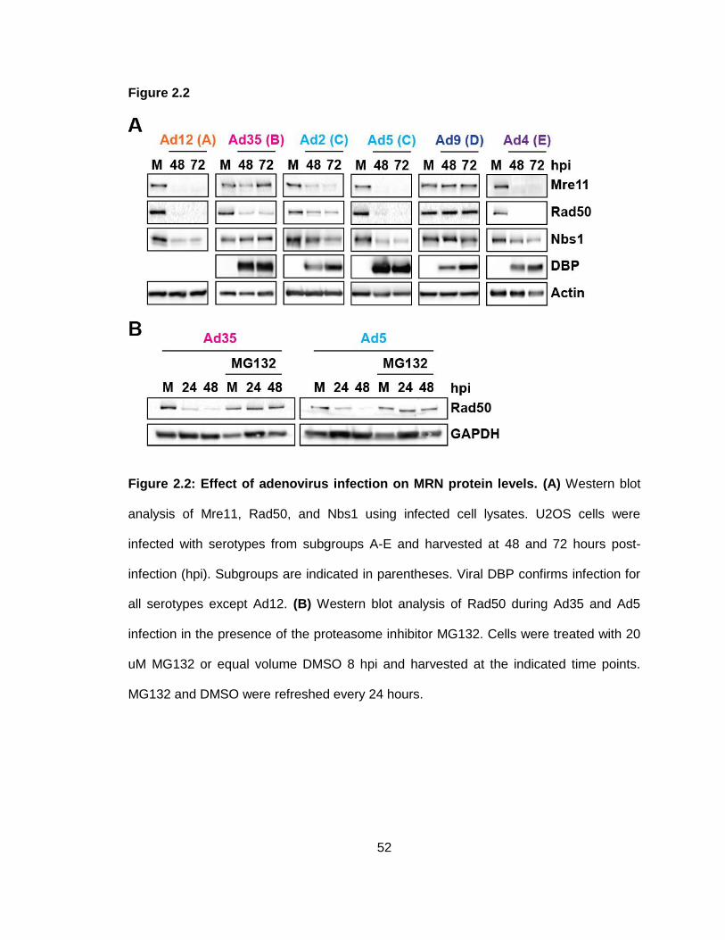
52
Figure 2.2
Figure 2.2: Effect of adenovirus infection on MRN protein levels. (A) Western blot
analysis of Mre11, Rad50, and Nbs1 using infected cell lysates. U2OS cells were
infected with serotypes from subgroups A-E and harvested at 48 and 72 hours post-
infection (hpi). Subgroups are indicated in parentheses. Viral DBP confirms infection for
all serotypes except Ad12. (B) Western blot analysis of Rad50 during Ad35 and Ad5
infection in the presence of the proteasome inhibitor MG132. Cells were treated with 20
uM MG132 or equal volume DMSO 8 hpi and harvested at the indicated time points.
MG132 and DMSO were refreshed every 24 hours.
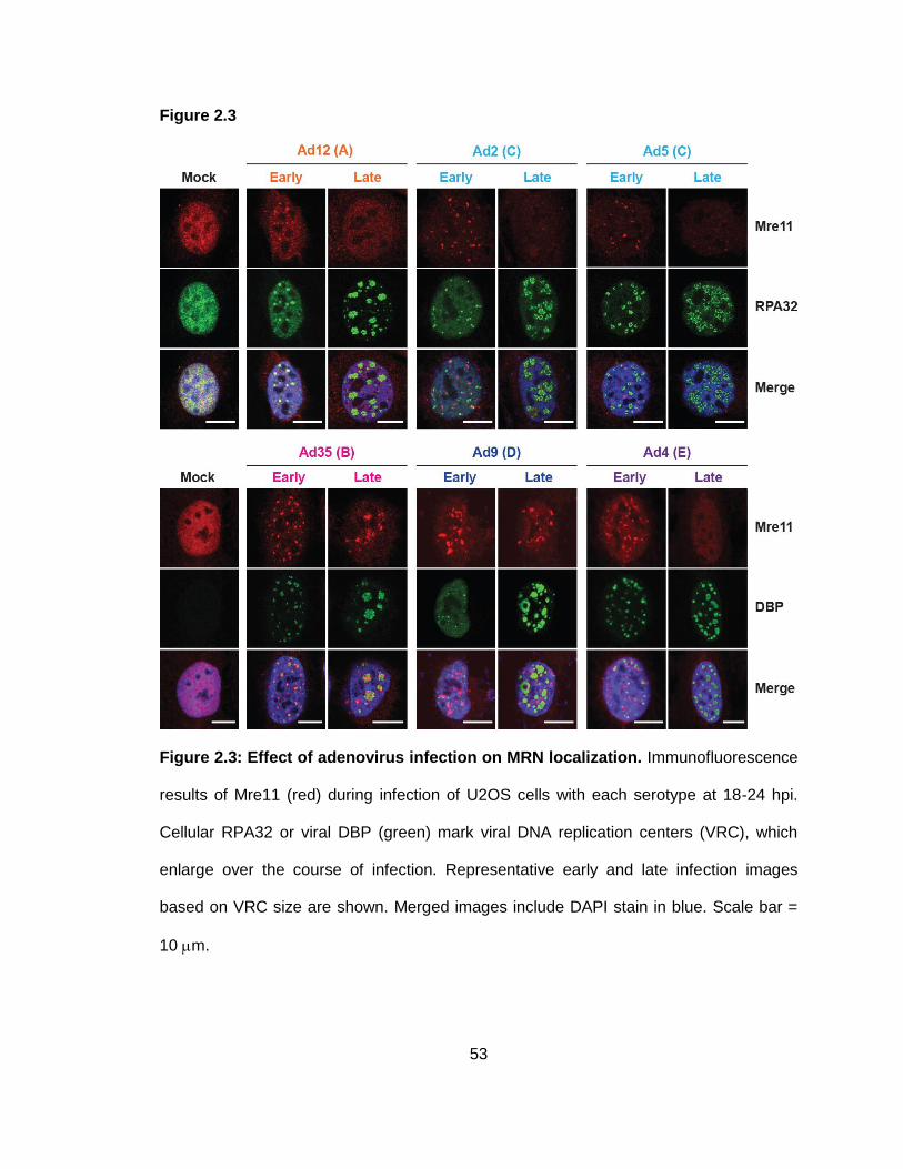
53
Figure 2.3
Figure 2.3: Effect of adenovirus infection on MRN localization. Immunofluorescence
results of Mre11 (red) during infection of U2OS cells with each serotype at 18-24 hpi.
Cellular RPA32 or viral DBP (green) mark viral DNA replication centers (VRC), which
enlarge over the course of infection. Representative early and late infection images
based on VRC size are shown. Merged images include DAPI stain in blue. Scale bar =
10 m.
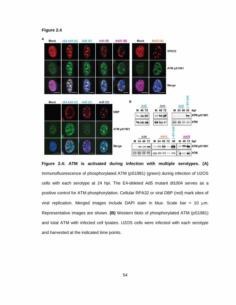
54
Figure 2.4
Figure 2.4: ATM is activated during infection with multiple serotypes. (A)
Immunofluorescence of phosphorylated ATM (pS1981) (green) during infection of U2OS
cells with each serotype at 24 hpi. The E4-deleted Ad5 mutant dl1004 serves as a
positive control for ATM phosphorylation. Cellular RPA32 or viral DBP (red) mark sites of
viral replication. Merged images include DAPI stain in blue. Scale bar = 10 m.
Representative images are shown. (B) Western blots of phosphorylated ATM (pS1981)
and total ATM with infected cell lysates. U2OS cells were infected with each serotype
and harvested at the indicated time points.
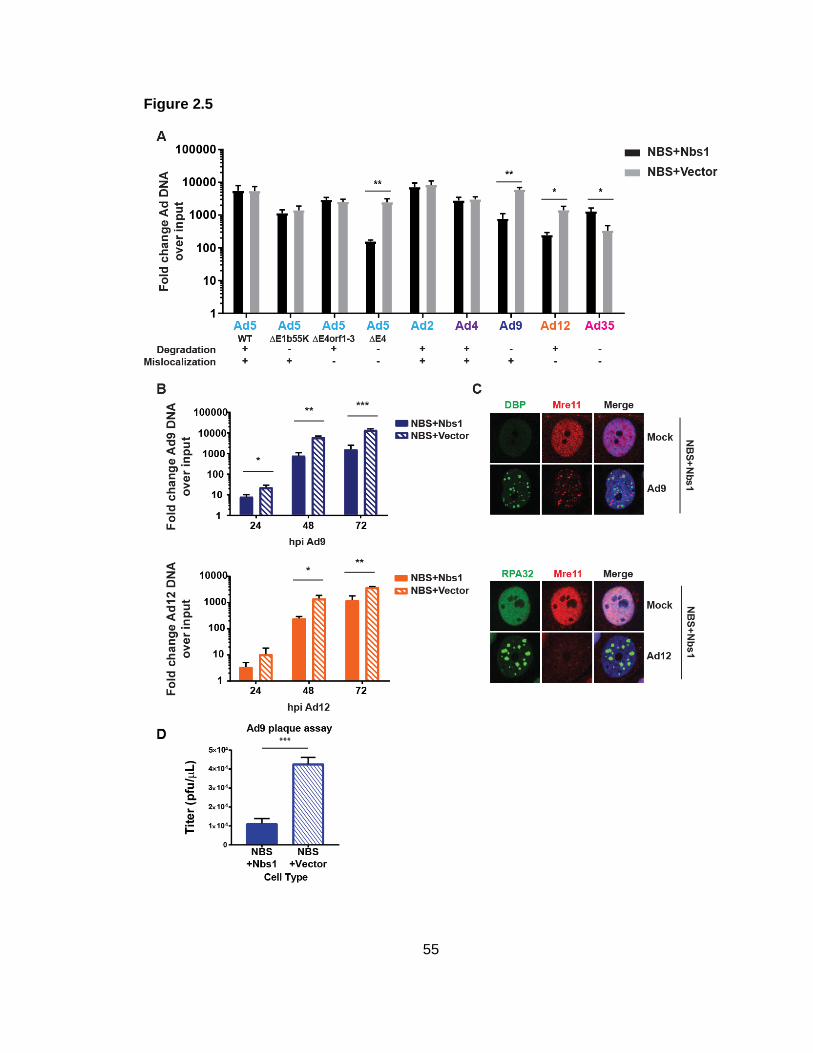
55
Figure 2.5
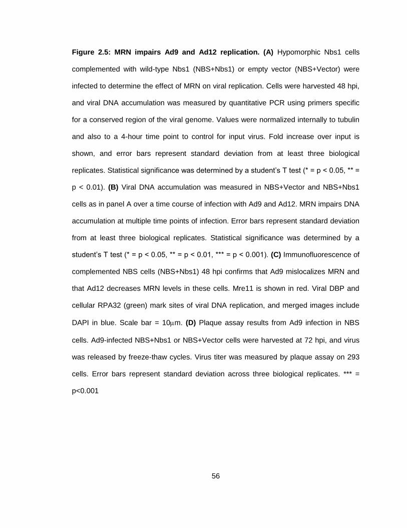
56
Figure 2.5: MRN impairs Ad9 and Ad12 replication. (A) Hypomorphic Nbs1 cells
complemented with wild-type Nbs1 (NBS+Nbs1) or empty vector (NBS+Vector) were
infected to determine the effect of MRN on viral replication. Cells were harvested 48 hpi,
and viral DNA accumulation was measured by quantitative PCR using primers specific
for a conserved region of the viral genome. Values were normalized internally to tubulin
and also to a 4-hour time point to control for input virus. Fold increase over input is
shown, and error bars represent standard deviation from at least three biological
replicates. Statistical significance was determined by a student’s T test (* = p < 0.05, ** =
p < 0.01). (B) Viral DNA accumulation was measured in NBS+Vector and NBS+Nbs1
cells as in panel A over a time course of infection with Ad9 and Ad12. MRN impairs DNA
accumulation at multiple time points of infection. Error bars represent standard deviation
from at least three biological replicates. Statistical significance was determined by a
student’s T test (* = p < 0.05, ** = p < 0.01, *** = p < 0.001). (C) Immunofluorescence of
complemented NBS cells (NBS+Nbs1) 48 hpi confirms that Ad9 mislocalizes MRN and
that Ad12 decreases MRN levels in these cells. Mre11 is shown in red. Viral DBP and
cellular RPA32 (green) mark sites of viral DNA replication, and merged images include
DAPI in blue. Scale bar = 10m. (D) Plaque assay results from Ad9 infection in NBS
cells. Ad9-infected NBS+Nbs1 or NBS+Vector cells were harvested at 72 hpi, and virus
was released by freeze-thaw cycles. Virus titer was measured by plaque assay on 293
cells. Error bars represent standard deviation across three biological replicates. *** =
p<0.001
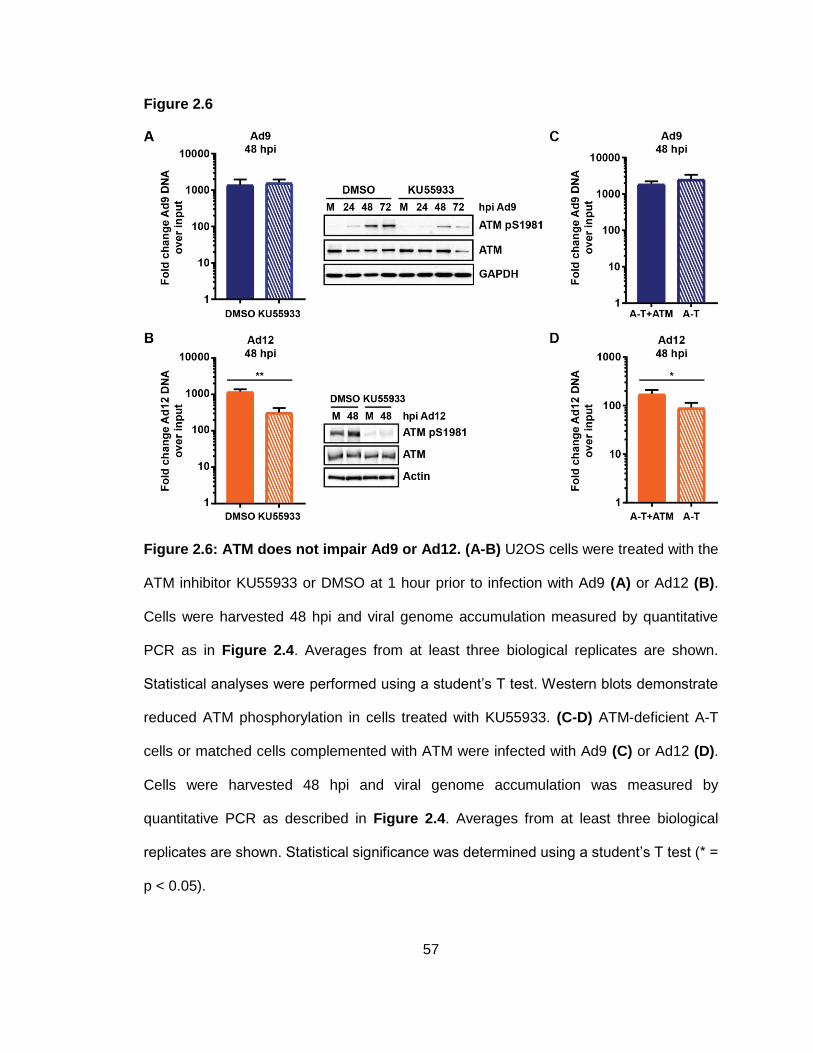
57
Figure 2.6
Figure 2.6: ATM does not impair Ad9 or Ad12. (A-B) U2OS cells were treated with the
ATM inhibitor KU55933 or DMSO at 1 hour prior to infection with Ad9 (A) or Ad12 (B).
Cells were harvested 48 hpi and viral genome accumulation measured by quantitative
PCR as in Figure 2.4. Averages from at least three biological replicates are shown.
Statistical analyses were performed using a student’s T test. Western blots demonstrate
reduced ATM phosphorylation in cells treated with KU55933. (C-D) ATM-deficient A-T
cells or matched cells complemented with ATM were infected with Ad9 (C) or Ad12 (D).
Cells were harvested 48 hpi and viral genome accumulation was measured by
quantitative PCR as described in Figure 2.4. Averages from at least three biological
replicates are shown. Statistical significance was determined using a student’s T test (* =
p < 0.05).
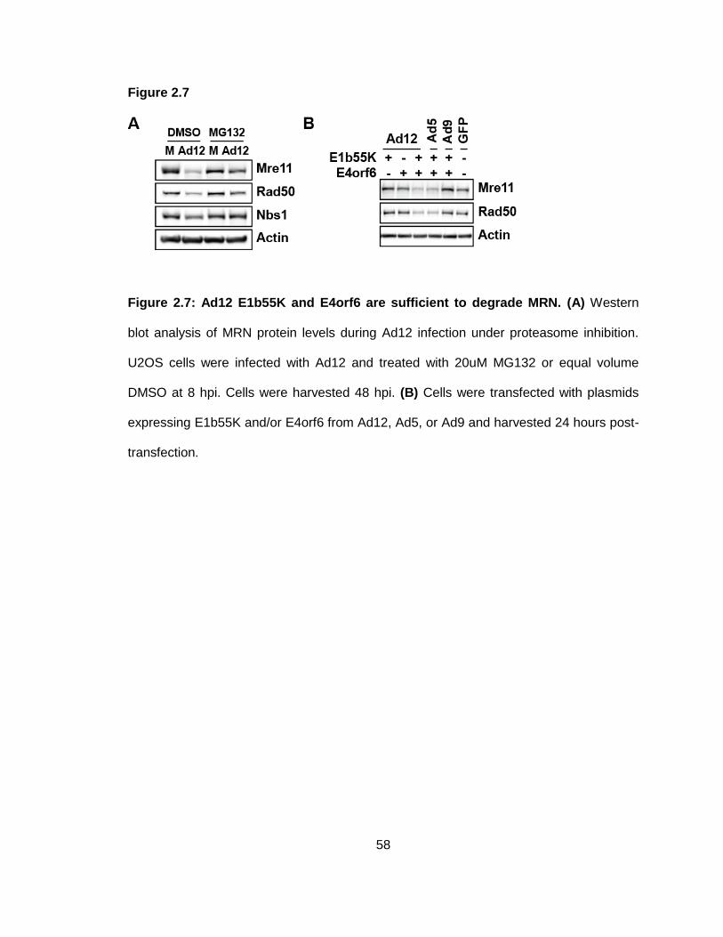
58
Figure 2.7
Figure 2.7: Ad12 E1b55K and E4orf6 are sufficient to degrade MRN. (A) Western
blot analysis of MRN protein levels during Ad12 infection under proteasome inhibition.
U2OS cells were infected with Ad12 and treated with 20uM MG132 or equal volume
DMSO at 8 hpi. Cells were harvested 48 hpi. (B) Cells were transfected with plasmids
expressing E1b55K and/or E4orf6 from Ad12, Ad5, or Ad9 and harvested 24 hours post-
transfection.
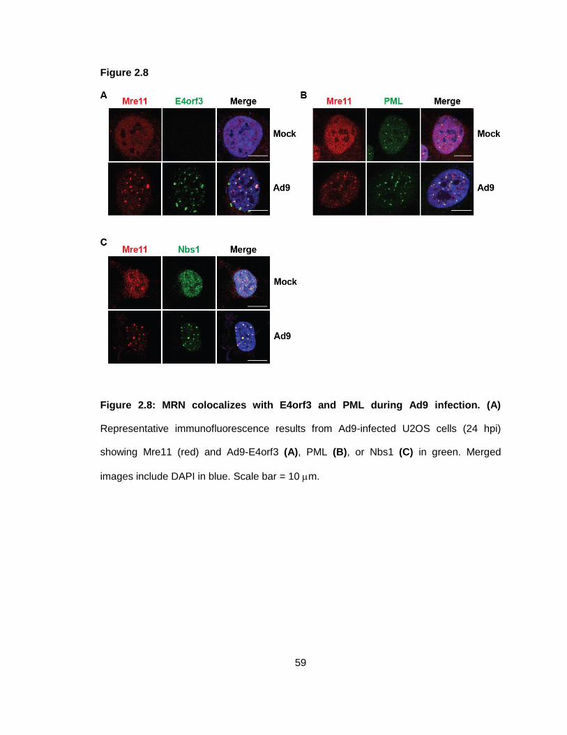
59
Figure 2.8
Figure 2.8: MRN colocalizes with E4orf3 and PML during Ad9 infection. (A)
Representative immunofluorescence results from Ad9-infected U2OS cells (24 hpi)
showing Mre11 (red) and Ad9-E4orf3 (A), PML (B), or Nbs1 (C) in green. Merged
images include DAPI in blue. Scale bar = 10 m.
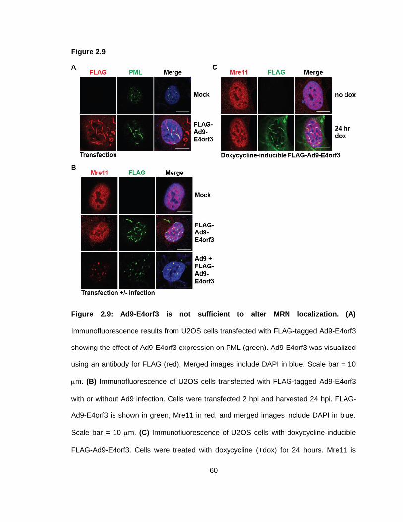
60
Figure 2.9
Figure 2.9: Ad9-E4orf3 is not sufficient to alter MRN localization. (A)
Immunofluorescence results from U2OS cells transfected with FLAG-tagged Ad9-E4orf3
showing the effect of Ad9-E4orf3 expression on PML (green). Ad9-E4orf3 was visualized
using an antibody for FLAG (red). Merged images include DAPI in blue. Scale bar = 10
m. (B) Immunofluorescence of U2OS cells transfected with FLAG-tagged Ad9-E4orf3
with or without Ad9 infection. Cells were transfected 2 hpi and harvested 24 hpi. FLAG-
Ad9-E4orf3 is shown in green, Mre11 in red, and merged images include DAPI in blue.
Scale bar = 10 m. (C) Immunofluorescence of U2OS cells with doxycycline-inducible
FLAG-Ad9-E4orf3. Cells were treated with doxycycline (+dox) for 24 hours. Mre11 is

61
shown in red, FLAG-Ad9-E4orf3 in green, and merged images include DAPI in blue.
Scale bar = 10 m.
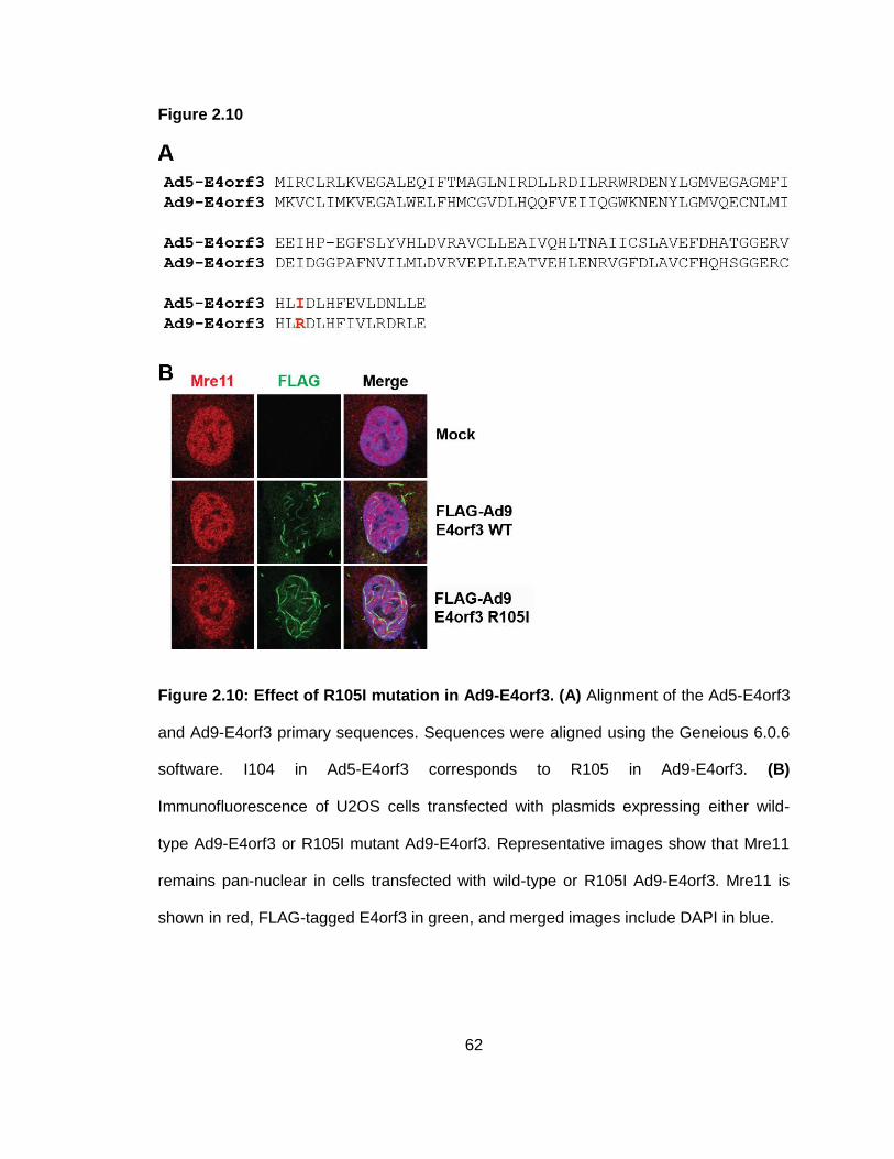
62
Figure 2.10
Figure 2.10: Effect of R105I mutation in Ad9-E4orf3. (A) Alignment of the Ad5-E4orf3
and Ad9-E4orf3 primary sequences. Sequences were aligned using the Geneious 6.0.6
software. I104 in Ad5-E4orf3 corresponds to R105 in Ad9-E4orf3. (B)
Immunofluorescence of U2OS cells transfected with plasmids expressing either wild-
type Ad9-E4orf3 or R105I mutant Ad9-E4orf3. Representative images show that Mre11
remains pan-nuclear in cells transfected with wild-type or R105I Ad9-E4orf3. Mre11 is
shown in red, FLAG-tagged E4orf3 in green, and merged images include DAPI in blue.
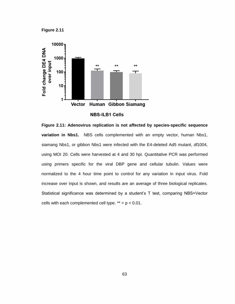
63
Figure 2.11
Figure 2.11: Adenovirus replication is not affected by species-specific sequence
variation in Nbs1. NBS cells complemented with an empty vector, human Nbs1,
siamang Nbs1, or gibbon Nbs1 were infected with the E4-deleted Ad5 mutant, dl1004,
using MOI 20. Cells were harvested at 4 and 30 hpi. Quantitative PCR was performed
using primers specific for the viral DBP gene and cellular tubulin. Values were
normalized to the 4 hour time point to control for any variation in input virus. Fold
increase over input is shown, and results are an average of three biological replicates.
Statistical significance was determined by a student’s T test, comparing NBS+Vector
cells with each complemented cell type. ** = p < 0.01.
64
Discussion
Cellular proteins can serve as obstacles to virus infection, and viruses have therefore
evolved strategies to overcome these intrinsic defenses. Extensive work from our lab
and others has demonstrated that proteins within the DDR can inhibit adenovirus DNA
replication, late protein production, and viral propagation. In particular, the MRN complex
has been suggested to impair viral replication both directly and indirectly through
downstream responses (Evans & Hearing, 2005; Lakdawala et al., 2008; Mathew &
Bridge, 2007; Shah & O'Shea, 2015; Stracker et al., 2002). The multiple ways that wild-
type Ad5 targets the MRN complex have presumably evolved to overcome this inhibition.
Previous work has demonstrated that adenovirus serotypes differ in their interactions
with MRN and other proteins in the DDR network (Blanchette et al., 2013; Cheng et al.,
2011; Cheng et al., 2013; Forrester et al., 2011; Stracker et al., 2005). In this study, we
further examined the relationship between MRN and serotypes across the adenovirus
family, with representatives from different adenovirus subgroups (A-E). We found that
adenovirus serotypes in different subgroups could target MRN complex proteins,
suggesting that MRN is a ubiquitous obstacle to viral DNA replication across the
adenovirus family. We specifically asked whether adenovirus serotypes differed in their
susceptibility to MRN inhibition and found that unlike Ad5, some serotypes are unable to
overcome impairment by MRN. Previous work demonstrated that MRN can impair
mutants of Ad5 (subgroup C) and Ad4 (subgroup E) that cannot target MRN (Evans &
Hearing, 2005; Lakdawala et al., 2008; Mathew & Bridge, 2007). Here, we demonstrate
that MRN can also restrict replication of wild-type serotypes from subgroup A (Ad12) and
subgroup D (Ad9) (Figure 2.5). We were surprised to find that even though Ad9 can
redistribute MRN away from viral replication centers, wild-type Ad9 genome levels were
significantly reduced in the presence of functional MRN complex (Figure 2.5). This
65
suggests that mislocalization by Ad9 is not sufficient to overcome inhibition by the MRN
complex. Results with Ad12 were also unexpected, since MRN significantly impaired
wild-type Ad12, despite being degraded during infection. The subgroup B serotype Ad35
did not degrade or mislocalize Mre11, similar to prior findings with other subgroup B
serotypes, Ad7 and Ad11 (Forrester et al., 2011). However, another study demonstrated
that transfection with E1b55K and E4orf6 from subgroup B serotypes Ad16 and Ad34
leads to a decrease in Mre11 levels (Cheng et al., 2011), raising the possibility that
interactions with MRN could vary even within a subgroup. While Ad35 did not degrade or
mislocalize Mre11, it did result in Rad50 degradation, demonstrating that this serotype
can target a single component of the complex. Surprisingly, degradation of Rad50 did
not affect Mre11 or Nbs1 levels, nor did it affect Mre11 localization to VRCs.
Interestingly, wild-type Ad35 DNA replication appeared to be enhanced in the presence
of MRN formation (Figure 2.5). It is possible that Ad35 prevents inhibition of DNA
replication by MRN through its degradation of Rad50. However, this alone would not be
expected to evade inhibition by Mre11, which localizes to Ad35 VRCs (Figure 2.3) and
has been suggested to impair adenovirus replication through its nuclease activity
(Stracker et al., 2002; Weiden & Ginsberg, 1994). Therefore, it is possible that Ad35
evades inhibition by Mre11 through an alternative, undefined mechanism. Results with
Ad35 raise the possibility that Ad35 could even exploit Mre11 or Nbs1 to benefit viral
replication, and these observations merit further investigation.
While previous studies have demonstrated that MRN can inhibit replication of mutants of
Ad5 that do not manipulate MRN (Evans & Hearing, 2005; Lakdawala et al., 2008;
Mathew & Bridge, 2007), we demonstrate for the first time that MRN can inhibit
replication of wild-type viruses Ad9 and Ad12 despite the fact that Ad9 and Ad12 alter
MRN localization or protein levels. We explored the role of ATM to determine if inhibition
66
could be through downstream signaling, since ATM can inhibit certain Ad5 mutants
(Gautam & Bridge, 2013; Shah & O'Shea, 2015). We first investigated how infection with
each of these serotypes affects ATM signaling. As previously reported (Carson et al.,
2003), wild-type Ad5 limited ATM activation at VRCs, but infection with the E4-deleted
Ad5 mutant dl1004 resulted in robust ATM activation at VRCs (Figure 2.4). ATM was
activated and colocalized with VRCs during infection with all other serotypes examined
(Figure 2.4). These data indicate that the ATM activation observed during infection with
these serotypes is in response to viral DNA or replication, rather than the global ATM
activation sometimes observed during Ad5 infection (Shah & O'Shea, 2015). Since all
but one of the serotypes we studied can target MRN through either degradation or
mislocalization, ATM activation at VRCs indicates that either (1) ATM is activated
independently of MRN during these infections, or (2) there is sufficient residual MRN at
VRCs to activate ATM. Pan-nuclear MRN-independent ATM activation has been
observed during late stages of wild-type Ad5 infection (Shah & O'Shea, 2015), but MRN
is required for ATM activation at Ad5 VRCs (Carson et al., 2003; Shah & O'Shea, 2015).
Therefore, we expect that the ATM activation at VRCs is due to residual MRN at VRCs.
Furthermore, since MRN inhibited wild-type Ad9 and Ad12, it is likely that there is some
MRN at VRCs of these two serotypes. We found that ATM did not impair replication of
Ad9 or Ad12 (Figure 2.6), excluding the possibility that MRN inhibition of these
serotypes is through downstream ATM signaling.
Since wild-type Ad9 and Ad12 viruses did not overcome MRN inhibition of viral DNA
replication, we investigated whether mislocalization or degradation by these serotypes
occurred through mechanisms different than Ad5. We reasoned that different
mechanisms could render these serotypes less effective at evading MRN recognition
and overcoming inhibition of viral DNA replication. We found that MRN colocalizes with
67
Ad9-E4orf3 and PML in nuclear tracks during infection (Figure 2.8), similar to MRN
localization during Ad5 infection (Stracker et al., 2002). However, unlike Ad5, we found
that Ad9-E4orf3 alone was not able to alter MRN localization, even though it was
sufficient to disrupt PML bodies (Figure 2.9). This difference could explain the inability of
Ad9 to overcome MRN. It is possible that during Ad9 infection another viral protein is
responsible for MRN mislocalization, either in conjunction with E4orf3 or by itself. The
responsible Ad9 protein may only partially sequester MRN from VRCs, allowing
sufficient MRN to accumulate at viral DNA and impair virus replication. Another
possibility is that Ad9 infection promotes changes to the cellular environment, to MRN, or
to E4orf3 that facilitate mislocalization. For example, there could be post-translational
modifications to Ad9-E4orf3 or to MRN that occur during infection and promote E4orf3
interaction with MRN. Such a requirement could delay mislocalization until after some
MRN had already associated with Ad9 VRCs and inhibited replication. Since Ad12 was
also inhibited by MRN during infection despite degradation of MRN proteins, we further
examined MRN degradation but were unable to identify any differences between Ad5
and Ad12 degradation in this study (Figure 2.7). Previous work has suggested that the
ubiquitin ligase formed by Ad12-E1b55K and Ad12-E4orf6 utilizes Cullin 2, in contrast to
the Cullin 5 used by Ad5 (Cheng et al., 2011). It is possible that this difference renders
Ad12 degradation less effective at overcoming MRN, or that differences in degradation
substrates between Ad12 and Ad5 (Blackford et al., 2010; Cheng et al., 2011; Forrester
et al., 2011) create distinct cellular environments that influence MRN function. Together,
our results demonstrate that interactions of adenovirus serotypes with the cellular MRN
complex vary across the viral family. These results may lead to a better understanding of
MRN targeting mechanisms, tissue tropism, or viral evolution. The broader implications
of this work will be discussed in Chapter 4.
68
CHAPTER 3:
Examining the role of adenovirus core protein VII
in regulating proteins associated with viral genomes
Some data from this chapter have been previously published in:
1. Reyes, E. D., Kulej, K., Pancholi, N. J., Akhtar, L. N., Avgousti, D. C., Kim, E. T.,
. . . Weitzman, M. D. (2017). Identifying host factors associated with DNA
replicated during virus infection. Mol Cell Proteomics.
doi:10.1074/mcp.M117.067116
2. Avgousti, D. C., Herrmann, C., Kulej, K., Pancholi, N. J., Sekulic, N., Petrescu,
J., . . . Weitzman, M. D. (2016). A core viral protein binds host nucleosomes to
sequester immune danger signals. Nature, 535(7610), 173-177.
This chapter incorporates several collaborative projects. As a result, some figures from
this chapter were generated by others in the lab and are credited in the figure legends.
Work on chromatin manipulation by protein VII was driven by Daphne Avgousti.
Adaptation of iPOND to identify cellular proteins on viral genomes was driven by
Emigdio Reyes. Mass spectrometry was performed by Kasia Kulej and the CHOP
Proteomics Core. Proteomic analyses were performed by Kasia Kulej and Joseph
Dybas.
69
Introduction
Successful viral replication and propagation require the careful regulation of the cellular
proteins that interact with viral DNA to allow viruses to recruit beneficial host proteins,
while preventing association of anti-viral factors. In this chapter, I describe how we used
proteomics to identify cellular proteins associated with viral genomes and how we
explored the role of a viral protein in regulating these interactions. This work began as
two separate projects in the lab to which I had the opportunity to contribute. The goal of
the first project was to identify the host proteins on viral genomes during infection using
a technique previously used to isolate proteins interacting with cellular DNA. The second
project examined how a histone-like adenovirus protein manipulates the composition of
cellular chromatin. Findings from these projects suggested that this histone-like viral
protein could influence the association of cellular proteins with adenoviral genomes,
which I then explored using the techniques I had learned through my involvement in both
projects. Here, I will briefly describe the findings of these projects and how we identified
novel functions for a core viral protein.
Materials and Methods
Cell lines
A549, U2OS, 293, mouse embryonic fibroblasts (MEF), hamster kidney cels (HaK), and
small airway epithelial cells (SAECs) were purchased from the American Tissue Culture
Collection (ATCC). 293 cells engineered to constitutively express Cre recombinase (293-
Cre) were a gift from P. Hearing. RA3331 FA-P cells (SLX4-deficient fibroblasts) and
matched complemented cells have been previously described (Kim et al., 2011) and
were gifts A. Smorgorzewska. HMGB1 knockout cells have been previously described
(Avgousti et al., 2017). Most cells were maintained in medium supplemented with 10%
70
fetal bovine serum and 1% penicillin-streptomycin (Invitrogen 15140122) at 37°C in a
humidified incubator with 5% CO2. Immortalized RA3331 FA-P cells (SLX4-deficient
fibroblasts) and matched cells expressing wild-type SLX4 were cultured in DMEM
supplemented with 15% FBS, 100 U/ml penicillin, 100 U/ml streptomycin, and non-
essential amino acids (Thermo Scientifc). Acceptor cells for the generation of
doxycycline-inducible cell lines were provided by E. Makeyev and were used as
previously described (Khandelia et al., 2011).
Viruses and infections
Ad5 and HSV-1 were purchased from ATCC. Flox-VII Ad5 was a gift from P. Hearing.
MAV-1 was a gift from K. Spindler. Infections were carried out by standard protocols.
Wild-type and flox-VII infections were carried out at multiplicity of infection 10 or 20.
HSV-1 infections were carried out with an MOI of 3, and MAV-1 infections with an MOI of
1. For most infections, viruses were diluted in medium supplemented with 2% fetal
bovine serum and 1% penicillin-streptomycin and added to cell monolayers. For iPOND
experiments, viruses were diluted in serum-free medium containing 1% penicillin-
streptomycin. Cells were incubated with virus for 2 hours at 37°C before supplementing
infection medium with medium containing 10% fetal bovine serum.
Isolation of proteins on nascent DNA
Cell culture: Eight confluent 15-cm plates (approximately 1.6x10⁸ cells) were used for
each sample. For adenovirus infections, cells were pulsed 24 hours post-infection with
10 mM EdU for 15 minutes at 37C. At the end of the pulse, media was aspirated and
cells were fixed by adding 10 mL of 1% paraformaldehyde and incubating for 20 minutes
at room temperature. Crosslinking was quenched by adding 1 mL of 1.25 M glycine.
Cells were harvested by scraping. Four plates per sample were combined into a single
71
50 mL conical tube. Cells were pelleted by centrifugation at 900xg for 5 minutes at 4C.
Cell pellets were washed twice by resuspending in 20 mL PBS. After the last wash,
supernatants were removed, and cell pellets were frozen in liquid nitrogen.
Permeabilization: Frozen cell pellets were thawed on ice and resuspended in 8 mL of
permeabilization buffer (PBS+0.25% Triton X-100). Cells were centrifuged at 900xg for 5
minutes at 4C. Pellets were resuspended in 4 mL PBS+0.5% BSA and transferred to 15
mL conical tubes. Cell pellets were washed once more with 4 mL PBS.
Click reactions: Click reactions were prepared in the dark by adding reagents in the
following order: 4.35 mL PBS, 0.05 mL Biotin Azide (stock concentration 1 mM), 0.5 mL
sodium ascorbate (stock concentration 100 mM, freshly prepared), and 0.1 mL copper
sulfate (stock concentration 100 mM). Volumes are per sample and were adjusted
accordingly to make master mixes for multiple samples. For “no biotin” controls, 0.05 mL
DMSO were added instead of biotin azide. Cells were resuspended in 4 mL click
reaction (+/- biotin azide) and incubated for 2 hours by rotating in the dark at room
temperature. Cells were then centrifuged for 5 minutes, 900xg, 4C. Pellets were washed
once with 4 mL PBS+0.5% BSA and then once with 4 mL of PBS. Supernatants were
removed by aspiration to ensure optimal removal of supernatant.
Lysis and capture: Cells were lysed by resuspending in 0.5 mL cold NLB buffer+0.5%
Triton X-100 (NLB buffer: 20 mM HEPES pH 7.9, 400 mM NaCl, 1 mM EDTA, 10%
glycerol) supplemented with protease inhibitors and 1 mM DTT. Cells were sonicated
using a Bioruptor for 20 minutes at 4C with 30 second on/off intervals. Sonication was
performed at high intensity. Sonicated samples were transferred to 1.5 mL
microcentrifuge tubes and cleared by centrifuging at maximum speed (15000-18000xg),
15 minutes, 4C in a tabletop microcentrifuge. Transfer cleared lysates to fresh tubes.
72
Remove 50 uL of each sample for input. Add 120 uL streptavidin magnetic beads (pre-
washed, 3x, 1mL NLB buffer+0.5% Triton X-100) (Dynabeads M-280, Invitrogen) to each
sample. Rotate samples with beads overnight, 4C, in the dark.
Sequential wash steps: 1) Wash beads with 1 mL NLB buffer+0.5% Triton X-100,
rotating for 5 minutes at room temperature. 2) Wash beads with 1 mL 1 M NaCl, rotating
for 10 minutes, room temperature. 3) Wash beads 4x with 1 mL IC wash buffer (20 mM
HEPES pH 7.4, 110 mM KOAc, 2 mM MgCl2, 0.1% Tween-20, 0.1% Triton-X 100, 150
mM NaCl) by rotating 5 minutes each time at room temperature. Transfer beads to fresh
tube after third wash step. 4) Wash beads 1x in 1 mL PBS by rotating for 5 minutes at
room temperature.
Elution: Resuspend beads of one of two tubes per sample in 60 L 1X lithium dodecyl
sulfate (LDS) buffer (Invitrogen) supplemented with 10% DTT. Elute proteins by boiling
at 95C for 10 minutes. Transfer supernatant to second tube of the same tube and repeat
boiling step. Transfer supernatant to fresh tubes. Reverse crosslinks by incubating
samples at 70C overnight.
Visualization of EdU-labeled DNA
Cells seeded on coverslips were pulsed for 15 minutes with 10 mM EdU at 37C. Pulsed
cells were fixed in 4% paraformaldehyde. Cells were permeabilized by incubating in
PBS+1% Triton X-100 for 30 minutes at room temperature. Click reaction mixes were
prepared as follows per coverslip: 427.5 uL PBS, 12.5 uL AlexaFluor 488 azide (Thermo
Scientific) (stock concentration 1 mM, 50 uL sodium ascorbate (stock concentration 100
mM), and 10 uL copper sulfate (stock concentration 100 mM). Cells were incubated with
click reaction for 1 hour, rocking, room temperature, in the dark. From this point onward,
all steps were performed in the dark. After click reaction incubation, cells were washed
73
with PBS. Cells were then blocked in 3% BSA and immunofluorescence was carried
using standard protocols.
Immunoprecipitation
Anti-HA immunoprecipitation: Two 15-cm plates (approximately 4x10⁷ cells) were used
per sample. VII-HA expression was induced by addition of 0.2 ug/mL doxycycline every
day for 4 days. Cells were harvested after 4 complete days of induction, and cells were
frozen in liquid nitrogen. Cell pellets were thawed on ice and resuspended in 500 uL IC
buffer (20 mM HEPES pH 7.4, 110 mM KOAc, 2 mM MgCl2, 0.1% Tween-20, 0.1%
Triton-X 100, 150 mM NaCl) supplemented with protease inhibitors (Roche) and
transferred to microcentrifuge tubes. Cells were incubated on ice for ten minutes,
vortexing every few minutes. After incubating, 5 L Benzonase nuclease
(Novagen/Millipore) was added to each sample, and samples were incubated on ice for
1 hour. Cells were then sonicated for 5 minutes, 30 seconds on/off intervals, 4C, at the
highest intensity. Samples were cleared by centrifugation at maximum speed (15000-
18000xg), 4C, 15 minutes. Supernatants were transferred to fresh tubes. 50 L were
removed for input. 50 L pre-washed anti-HA beads (Thermo Fisher) were added to
each sample. Samples were incubated for 1 hour at 4C, rotating. Beads were then
washed 3x, each time with 1 mL IC wash buffer supplemented with protease inhibitors
by rotating for 5 minutes at 4C. Proteins were eluted by resuspending beads in 100 L
HA peptide (Thermo Fisher) and incubating at 37C with shaking for 20 minutes.
Supernatants were transferred to new tubes.
Anti-VII immunoprecipitation: Two 15-cm plates (approximately 4x10⁷ cells) were used
per sample. A549 cells were infected with wild-type Ad5 and harvested at 24 hours post-
infection. Lysis and Benzonase treatment were carried out exactly as described above
74
for anti-HA IP. After clearing lysates and removing input, 50 L anti-VII hybridoma
supernatant (gift from H. Wodrich) were added to each sample. Samples were incubated
for 2 hours, 4C, rotating. 50 L pre-washed protein G beads (Dynabeads Thermo
Scientific 10004D) were then added to samples and returned to 4C for overnight
incubation with rotation. The next day, beads were washed 3x, each time with 1 mL IC
wash buffer supplemented with protease inhibitors by rotating for 5 minutes at 4C.
Proteins were eluted by boiling at 95C for 10 minutes in 100 L 1X LDS sample buffer
(Invitrogen) with 10% DTT. Supernatants were transferred to new tubes.
Deletion of protein VII by TAT-Cre
A549 cells were incubated with 0.5-1.5 mg/mL purified TAT-Cre in minimal volume
OPTI-MEM (Thermo Scientific) for 1 hour prior to infection. Control cells were incubated
with equal volume 50% glycerol in OPTI-MEM. After 1 hour, OPTI-MEM + TAT-
Cre/glycerol was removed, but cells were not washed before adding infection mix.
Infections were then carried out as usual with flox-VII virus at MOI 10.
Immunofluorescence, immunoblotting, and antibodies
Immunofluorescence and immunoblotting were performed as described in Chapter 2.
Primary antibodies to cellular proteins were purchased from commercial sources:
HMGB1 (Abcam), GFP (Abcam and Millipore), FMR1 (Sigma and Millipore), POLR2E
(Sigma), RBM8A (Novus), RNMTL1 (Novus), SLTM (Novus), SNRPE (Abcam), SRP14
(Abcam), RecQL (Santa Cruz, H-110), FUBP1 (Abcam), SPATA5 (Abcam), Cre
(Millipore), SRSF1 (Thermo), SLX4 (Novus and Abnova), Flag (Sigma), Actin (Sigma),
TCOF (Sigma), GAPDH (GeneTex), TFII-I (Santa Cruz), Rad50 (GeneTex), DDX21
(Abcam), SART1 (Abcam), TRRAP (Abcam), PML (Santa Cruz, PG-M3), histone H1
(Abcam), HA (Covance and Santa Cruz), histone H3 (Millipore), Tubulin (Santa Cruz),
75
Emerin (Abcam), NUP160 (Abcam), IDH3A (Thermo), phospho-STAT1 (Abcam), STAT1
(Santa Cruz). Primary antibodies to viral proteins were gifts: DBP (A. Levine), protein VII
(L. Gerace and H. Wodrich), late proteins (J. Wilson).
Quantitative PCR
Quantitative PCR to measure viral DNA accumulation was performed as described in
Chapter 2. For reverse transcription quantitative PCR (RT-PCR), RNA was isolated from
cells using the RNeasy Micro kit (Qiagen 74004). Reverse transcription was carried out
with 0.5-1 ug of RNA using the High Capacity RNA-to-cDNA kit (Thermo Fisher Scientific
4387406). Quantitative PCR was carried out using the standard procedure for Sybr
Green (Thermo). Primers: HMGB1 (5’ TAACTAAACATGGGCAAAGGAG and 5’
TAGCAGACATGGTCTTCCAC), protein VII (5’ GCGGGTATTGTCACTGTGC and 5’
CACCCAATACACGTTGCCC), ISG15 (5’ CAGATCACCCAGAAGATCGG and 5’
GCCCTTGTTATTCCTCACCA), MX2 (5’ CACATCCATATTTCAGAGTTCTCC and 5’
GGTGGCTCTCCCTTATTTGTC), NfkB (5’ CTAGCACAAGGAGACATGAAACAG and 5’
CCAGAGACCTCATAGTTGTCCA), and IFN(5’ CAGCATCTGCTGGTTGAAGA and 5’
CTAGCACAAGGAGACATGAAACAG).
Interferon stimulation
For stimulation by DNA, cells were transfected with 1 ug/mL poly(dA:dT)/LyoVec
(Invivogen tlrl-patc) by adding to regular growth medium. Cells were collected at
indicated time points (8 hours post-stimulation for RT-PCR; 6, 12, or 24 hours post-
stimulation for western blot). For treatment of cells with ectopic interferon, cells were
treated with 1000 units/mL universal type I interferon (PBL Assay Science) and collected
24 hours post-treatment.
76
Results
Identification of proteins associated with adenovirus DNA by iPOND
In order to identify novel host factors associated with viral DNA, we adapted a technique
previously used to isolate and identify proteins that interact with cellular replicating DNA
(Sirbu et al., 2013). This technique, called Isolation of Proteins on Nascent DNA, or
iPOND, relies on the selective labeling of nascent DNA with the nucleoside analog 5-
ethynyl-2’-deoxyuridine (EdU), which is incorporated into actively replicating DNA (Salic
& Mitchison, 2008). Since adenovirus infection results in suppression of cellular DNA
replication in favor of viral DNA replication (Halbert et al., 1985), we reasoned that
pulsing infected cells with EdU would allow for selective labeling of adenoviral DNA over
cellular DNA. To test this, we pulsed infected cells with EdU and visualized EdU by
immunofluorescence (Figure 3.1A). In uninfected cells, EdU was distributed throughout
the nucleus marking sites of replicated cellular DNA (Figure 3.1A), as has been
previously reported (Leonhardt et al., 2000; Nakamura, Morita, & Sato, 1986; Salic &
Mitchison, 2008). In contrast, EdU was found in distinct structures resembling viral
replication centers (VRCs) when infected cells were pulsed 24 hours post-infection
(Figure 3.1A). Colocalization of EdU with the viral DNA-binding protein, DBP, confirmed
that EdU was found at VRCs (Figure 3.1A), demonstrating that EdU is preferentially
incorporated into viral DNA during adenovirus infection. We therefore utilized the iPOND
protocol in order to identify proteins associated with EdU-labeled adenoviral DNA
(illustrated in Figure 3.1B). We pulsed infected and mock cells for 15 minutes at 24 hpi.
Due to asynchronous replication origin firing and the length of the pulse, EdU was
incorporated into replicated DNA throughout the viral genome, rather than strictly at
replication forks. This allowed us to identify proteins associated with viral DNA at
multiple stages of infection, rather than only those involved in active DNA replication.
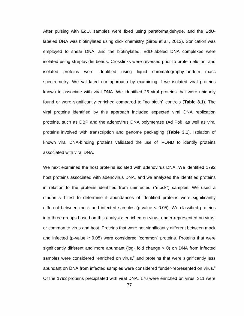
77
After pulsing with EdU, samples were fixed using paraformaldehyde, and the EdU-
labeled DNA was biotinylated using click chemistry (Sirbu et al., 2013). Sonication was
employed to shear DNA, and the biotinylated, EdU-labeled DNA complexes were
isolated using streptavidin beads. Crosslinks were reversed prior to protein elution, and
isolated proteins were identified using liquid chromatography-tandem mass
spectrometry. We validated our approach by examining if we isolated viral proteins
known to associate with viral DNA. We identified 25 viral proteins that were uniquely
found or were significantly enriched compared to “no biotin” controls (Table 3.1). The
viral proteins identified by this approach included expected viral DNA replication
proteins, such as DBP and the adenovirus DNA polymerase (Ad Pol), as well as viral
proteins involved with transcription and genome packaging (Table 3.1). Isolation of
known viral DNA-binding proteins validated the use of iPOND to identify proteins
associated with viral DNA.
We next examined the host proteins isolated with adenovirus DNA. We identified 1792
host proteins associated with adenovirus DNA, and we analyzed the identified proteins
in relation to the proteins identified from uninfected (“mock”) samples. We used a
student’s T-test to determine if abundances of identified proteins were significantly
different between mock and infected samples (p-value < 0.05). We classified proteins
into three groups based on this analysis: enriched on virus, under-represented on virus,
or common to virus and host. Proteins that were not significantly different between mock
and infected (p-value ≥ 0.05) were considered “common” proteins. Proteins that were
significantly different and more abundant (log2 fold change > 0) on DNA from infected
samples were considered “enriched on virus,” and proteins that were significantly less
abundant on DNA from infected samples were considered “under-represented on virus.”
Of the 1792 proteins precipitated with viral DNA, 176 were enriched on virus, 311 were
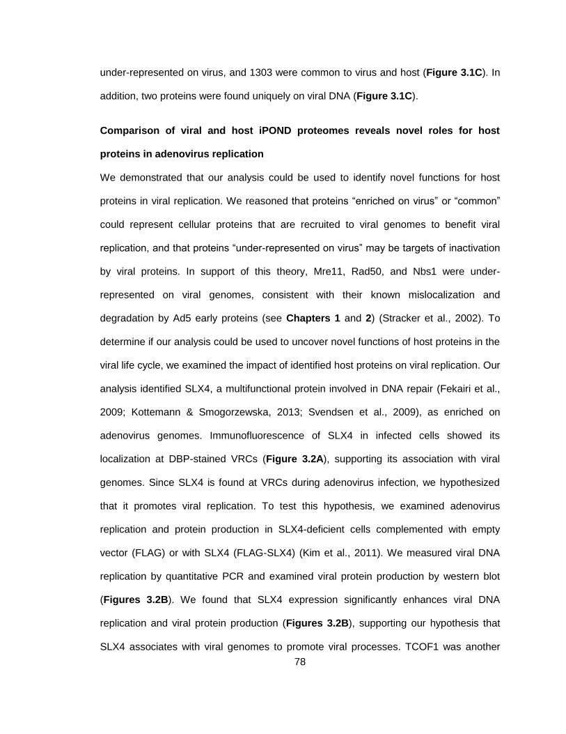
78
under-represented on virus, and 1303 were common to virus and host (Figure 3.1C). In
addition, two proteins were found uniquely on viral DNA (Figure 3.1C).
Comparison of viral and host iPOND proteomes reveals novel roles for host
proteins in adenovirus replication
We demonstrated that our analysis could be used to identify novel functions for host
proteins in viral replication. We reasoned that proteins “enriched on virus” or “common”
could represent cellular proteins that are recruited to viral genomes to benefit viral
replication, and that proteins “under-represented on virus” may be targets of inactivation
by viral proteins. In support of this theory, Mre11, Rad50, and Nbs1 were under-
represented on viral genomes, consistent with their known mislocalization and
degradation by Ad5 early proteins (see Chapters 1 and 2) (Stracker et al., 2002). To
determine if our analysis could be used to uncover novel functions of host proteins in the
viral life cycle, we examined the impact of identified host proteins on viral replication. Our
analysis identified SLX4, a multifunctional protein involved in DNA repair (Fekairi et al.,
2009; Kottemann & Smogorzewska, 2013; Svendsen et al., 2009), as enriched on
adenovirus genomes. Immunofluorescence of SLX4 in infected cells showed its
localization at DBP-stained VRCs (Figure 3.2A), supporting its association with viral
genomes. Since SLX4 is found at VRCs during adenovirus infection, we hypothesized
that it promotes viral replication. To test this hypothesis, we examined adenovirus
replication and protein production in SLX4-deficient cells complemented with empty
vector (FLAG) or with SLX4 (FLAG-SLX4) (Kim et al., 2011). We measured viral DNA
replication by quantitative PCR and examined viral protein production by western blot
(Figures 3.2B). We found that SLX4 expression significantly enhances viral DNA
replication and viral protein production (Figures 3.2B), supporting our hypothesis that
SLX4 associates with viral genomes to promote viral processes. TCOF1 was another
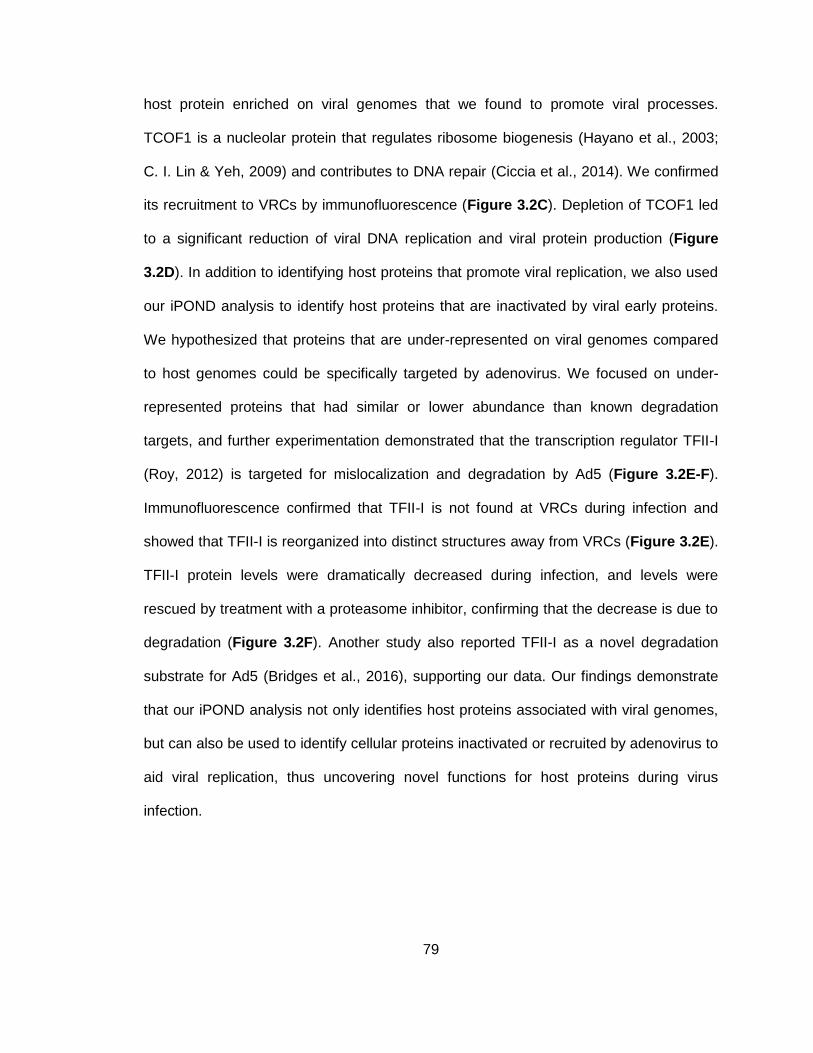
79
host protein enriched on viral genomes that we found to promote viral processes.
TCOF1 is a nucleolar protein that regulates ribosome biogenesis (Hayano et al., 2003;
C. I. Lin & Yeh, 2009) and contributes to DNA repair (Ciccia et al., 2014). We confirmed
its recruitment to VRCs by immunofluorescence (Figure 3.2C). Depletion of TCOF1 led
to a significant reduction of viral DNA replication and viral protein production (Figure
3.2D). In addition to identifying host proteins that promote viral replication, we also used
our iPOND analysis to identify host proteins that are inactivated by viral early proteins.
We hypothesized that proteins that are under-represented on viral genomes compared
to host genomes could be specifically targeted by adenovirus. We focused on under-
represented proteins that had similar or lower abundance than known degradation
targets, and further experimentation demonstrated that the transcription regulator TFII-I
(Roy, 2012) is targeted for mislocalization and degradation by Ad5 (Figure 3.2E-F).
Immunofluorescence confirmed that TFII-I is not found at VRCs during infection and
showed that TFII-I is reorganized into distinct structures away from VRCs (Figure 3.2E).
TFII-I protein levels were dramatically decreased during infection, and levels were
rescued by treatment with a proteasome inhibitor, confirming that the decrease is due to
degradation (Figure 3.2F). Another study also reported TFII-I as a novel degradation
substrate for Ad5 (Bridges et al., 2016), supporting our data. Our findings demonstrate
that our iPOND analysis not only identifies host proteins associated with viral genomes,
but can also be used to identify cellular proteins inactivated or recruited by adenovirus to
aid viral replication, thus uncovering novel functions for host proteins during virus
infection.
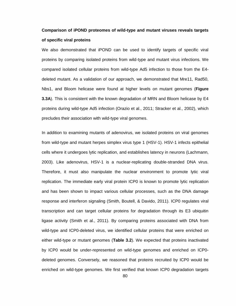
80
Comparison of iPOND proteomes of wild-type and mutant viruses reveals targets
of specific viral proteins
We also demonstrated that iPOND can be used to identify targets of specific viral
proteins by comparing isolated proteins from wild-type and mutant virus infections. We
compared isolated cellular proteins from wild-type Ad5 infection to those from the E4-
deleted mutant. As a validation of our approach, we demonstrated that Mre11, Rad50,
Nbs1, and Bloom helicase were found at higher levels on mutant genomes (Figure
3.3A). This is consistent with the known degradation of MRN and Bloom helicase by E4
proteins during wild-type Ad5 infection (Orazio et al., 2011; Stracker et al., 2002), which
precludes their association with wild-type viral genomes.
In addition to examining mutants of adenovirus, we isolated proteins on viral genomes
from wild-type and mutant herpes simplex virus type 1 (HSV-1). HSV-1 infects epithelial
cells where it undergoes lytic replication, and establishes latency in neurons (Lachmann,
2003). Like adenovirus, HSV-1 is a nuclear-replicating double-stranded DNA virus.
Therefore, it must also manipulate the nuclear environment to promote lytic viral
replication. The immediate early viral protein ICP0 is known to promote lytic replication
and has been shown to impact various cellular processes, such as the DNA damage
response and interferon signaling (Smith, Boutell, & Davido, 2011). ICP0 regulates viral
transcription and can target cellular proteins for degradation through its E3 ubiquitin
ligase activity (Smith et al., 2011). By comparing proteins associated with DNA from
wild-type and ICP0-deleted virus, we identified cellular proteins that were enriched on
either wild-type or mutant genomes (Table 3.2). We expected that proteins inactivated
by ICP0 would be under-represented on wild-type genomes and enriched on ICP0-
deleted genomes. Conversely, we reasoned that proteins recruited by ICP0 would be
enriched on wild-type genomes. We first verified that known ICP0 degradation targets
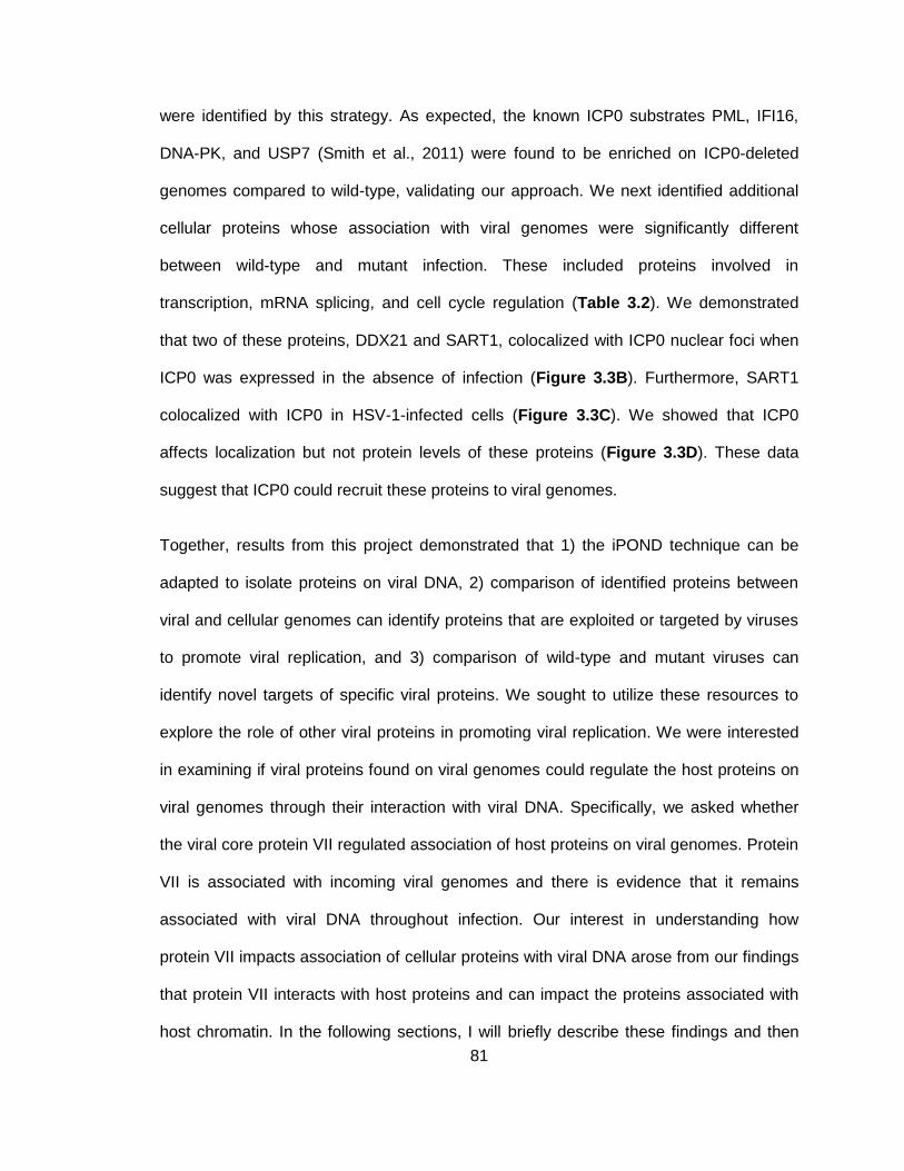
81
were identified by this strategy. As expected, the known ICP0 substrates PML, IFI16,
DNA-PK, and USP7 (Smith et al., 2011) were found to be enriched on ICP0-deleted
genomes compared to wild-type, validating our approach. We next identified additional
cellular proteins whose association with viral genomes were significantly different
between wild-type and mutant infection. These included proteins involved in
transcription, mRNA splicing, and cell cycle regulation (Table 3.2). We demonstrated
that two of these proteins, DDX21 and SART1, colocalized with ICP0 nuclear foci when
ICP0 was expressed in the absence of infection (Figure 3.3B). Furthermore, SART1
colocalized with ICP0 in HSV-1-infected cells (Figure 3.3C). We showed that ICP0
affects localization but not protein levels of these proteins (Figure 3.3D). These data
suggest that ICP0 could recruit these proteins to viral genomes.
Together, results from this project demonstrated that 1) the iPOND technique can be
adapted to isolate proteins on viral DNA, 2) comparison of identified proteins between
viral and cellular genomes can identify proteins that are exploited or targeted by viruses
to promote viral replication, and 3) comparison of wild-type and mutant viruses can
identify novel targets of specific viral proteins. We sought to utilize these resources to
explore the role of other viral proteins in promoting viral replication. We were interested
in examining if viral proteins found on viral genomes could regulate the host proteins on
viral genomes through their interaction with viral DNA. Specifically, we asked whether
the viral core protein VII regulated association of host proteins on viral genomes. Protein
VII is associated with incoming viral genomes and there is evidence that it remains
associated with viral DNA throughout infection. Our interest in understanding how
protein VII impacts association of cellular proteins with viral DNA arose from our findings
that protein VII interacts with host proteins and can impact the proteins associated with
host chromatin. In the following sections, I will briefly describe these findings and then
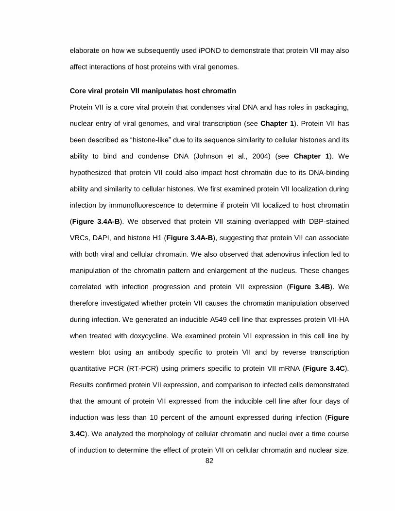
82
elaborate on how we subsequently used iPOND to demonstrate that protein VII may also
affect interactions of host proteins with viral genomes.
Core viral protein VII manipulates host chromatin
Protein VII is a core viral protein that condenses viral DNA and has roles in packaging,
nuclear entry of viral genomes, and viral transcription (see Chapter 1). Protein VII has
been described as “histone-like” due to its sequence similarity to cellular histones and its
ability to bind and condense DNA (Johnson et al., 2004) (see Chapter 1). We
hypothesized that protein VII could also impact host chromatin due to its DNA-binding
ability and similarity to cellular histones. We first examined protein VII localization during
infection by immunofluorescence to determine if protein VII localized to host chromatin
(Figure 3.4A-B). We observed that protein VII staining overlapped with DBP-stained
VRCs, DAPI, and histone H1 (Figure 3.4A-B), suggesting that protein VII can associate
with both viral and cellular chromatin. We also observed that adenovirus infection led to
manipulation of the chromatin pattern and enlargement of the nucleus. These changes
correlated with infection progression and protein VII expression (Figure 3.4B). We
therefore investigated whether protein VII causes the chromatin manipulation observed
during infection. We generated an inducible A549 cell line that expresses protein VII-HA
when treated with doxycycline. We examined protein VII expression in this cell line by
western blot using an antibody specific to protein VII and by reverse transcription
quantitative PCR (RT-PCR) using primers specific to protein VII mRNA (Figure 3.4C).
Results confirmed protein VII expression, and comparison to infected cells demonstrated
that the amount of protein VII expressed from the inducible cell line after four days of
induction was less than 10 percent of the amount expressed during infection (Figure
3.4C). We analyzed the morphology of cellular chromatin and nuclei over a time course
of induction to determine the effect of protein VII on cellular chromatin and nuclear size.
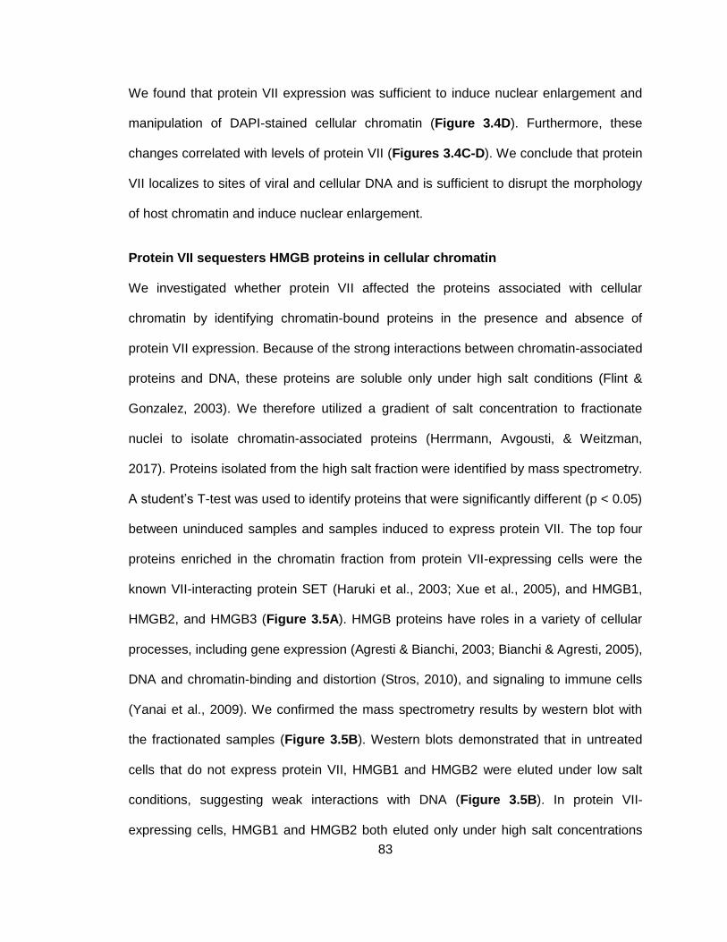
83
We found that protein VII expression was sufficient to induce nuclear enlargement and
manipulation of DAPI-stained cellular chromatin (Figure 3.4D). Furthermore, these
changes correlated with levels of protein VII (Figures 3.4C-D). We conclude that protein
VII localizes to sites of viral and cellular DNA and is sufficient to disrupt the morphology
of host chromatin and induce nuclear enlargement.
Protein VII sequesters HMGB proteins in cellular chromatin
We investigated whether protein VII affected the proteins associated with cellular
chromatin by identifying chromatin-bound proteins in the presence and absence of
protein VII expression. Because of the strong interactions between chromatin-associated
proteins and DNA, these proteins are soluble only under high salt conditions (Flint &
Gonzalez, 2003). We therefore utilized a gradient of salt concentration to fractionate
nuclei to isolate chromatin-associated proteins (Herrmann, Avgousti, & Weitzman,
2017). Proteins isolated from the high salt fraction were identified by mass spectrometry.
A student’s T-test was used to identify proteins that were significantly different (p < 0.05)
between uninduced samples and samples induced to express protein VII. The top four
proteins enriched in the chromatin fraction from protein VII-expressing cells were the
known VII-interacting protein SET (Haruki et al., 2003; Xue et al., 2005), and HMGB1,
HMGB2, and HMGB3 (Figure 3.5A). HMGB proteins have roles in a variety of cellular
processes, including gene expression (Agresti & Bianchi, 2003; Bianchi & Agresti, 2005),
DNA and chromatin-binding and distortion (Stros, 2010), and signaling to immune cells
(Yanai et al., 2009). We confirmed the mass spectrometry results by western blot with
the fractionated samples (Figure 3.5B). Western blots demonstrated that in untreated
cells that do not express protein VII, HMGB1 and HMGB2 were eluted under low salt
conditions, suggesting weak interactions with DNA (Figure 3.5B). In protein VII-
expressing cells, HMGB1 and HMGB2 both eluted only under high salt concentrations
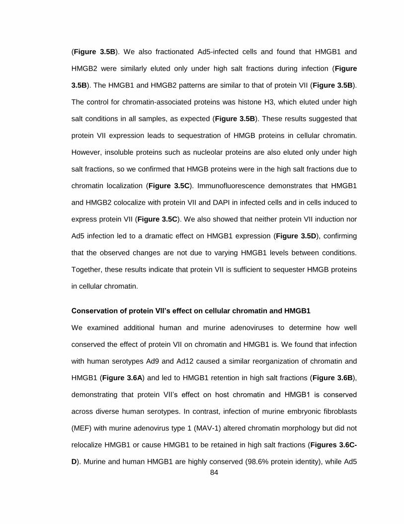
84
(Figure 3.5B). We also fractionated Ad5-infected cells and found that HMGB1 and
HMGB2 were similarly eluted only under high salt fractions during infection (Figure
3.5B). The HMGB1 and HMGB2 patterns are similar to that of protein VII (Figure 3.5B).
The control for chromatin-associated proteins was histone H3, which eluted under high
salt conditions in all samples, as expected (Figure 3.5B). These results suggested that
protein VII expression leads to sequestration of HMGB proteins in cellular chromatin.
However, insoluble proteins such as nucleolar proteins are also eluted only under high
salt fractions, so we confirmed that HMGB proteins were in the high salt fractions due to
chromatin localization (Figure 3.5C). Immunofluorescence demonstrates that HMGB1
and HMGB2 colocalize with protein VII and DAPI in infected cells and in cells induced to
express protein VII (Figure 3.5C). We also showed that neither protein VII induction nor
Ad5 infection led to a dramatic effect on HMGB1 expression (Figure 3.5D), confirming
that the observed changes are not due to varying HMGB1 levels between conditions.
Together, these results indicate that protein VII is sufficient to sequester HMGB proteins
in cellular chromatin.
Conservation of protein VII’s effect on cellular chromatin and HMGB1
We examined additional human and murine adenoviruses to determine how well
conserved the effect of protein VII on chromatin and HMGB1 is. We found that infection
with human serotypes Ad9 and Ad12 caused a similar reorganization of chromatin and
HMGB1 (Figure 3.6A) and led to HMGB1 retention in high salt fractions (Figure 3.6B),
demonstrating that protein VII’s effect on host chromatin and HMGB1 is conserved
across diverse human serotypes. In contrast, infection of murine embryonic fibroblasts
(MEF) with murine adenovirus type 1 (MAV-1) altered chromatin morphology but did not
relocalize HMGB1 or cause HMGB1 to be retained in high salt fractions (Figures 3.6C-
D). Murine and human HMGB1 are highly conserved (98.6% protein identity), while Ad5
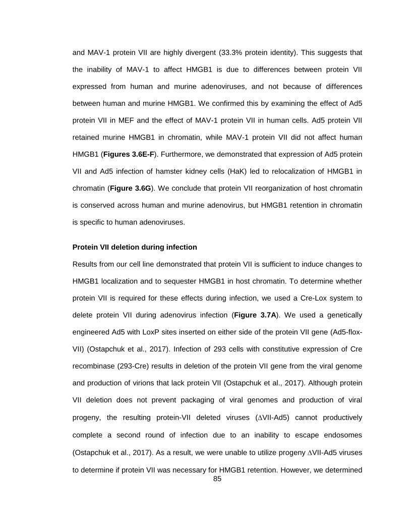
85
and MAV-1 protein VII are highly divergent (33.3% protein identity). This suggests that
the inability of MAV-1 to affect HMGB1 is due to differences between protein VII
expressed from human and murine adenoviruses, and not because of differences
between human and murine HMGB1. We confirmed this by examining the effect of Ad5
protein VII in MEF and the effect of MAV-1 protein VII in human cells. Ad5 protein VII
retained murine HMGB1 in chromatin, while MAV-1 protein VII did not affect human
HMGB1 (Figures 3.6E-F). Furthermore, we demonstrated that expression of Ad5 protein
VII and Ad5 infection of hamster kidney cells (HaK) led to relocalization of HMGB1 in
chromatin (Figure 3.6G). We conclude that protein VII reorganization of host chromatin
is conserved across human and murine adenovirus, but HMGB1 retention in chromatin
is specific to human adenoviruses.
Protein VII deletion during infection
Results from our cell line demonstrated that protein VII is sufficient to induce changes to
HMGB1 localization and to sequester HMGB1 in host chromatin. To determine whether
protein VII is required for these effects during infection, we used a Cre-Lox system to
delete protein VII during adenovirus infection (Figure 3.7A). We used a genetically
engineered Ad5 with LoxP sites inserted on either side of the protein VII gene (Ad5-flox-
VII) (Ostapchuk et al., 2017). Infection of 293 cells with constitutive expression of Cre
recombinase (293-Cre) results in deletion of the protein VII gene from the viral genome
and production of virions that lack protein VII (Ostapchuk et al., 2017). Although protein
VII deletion does not prevent packaging of viral genomes and production of viral
progeny, the resulting protein-VII deleted viruses (VII-Ad5) cannot productively
complete a second round of infection due to an inability to escape endosomes
(Ostapchuk et al., 2017). As a result, we were unable to utilize progeny VII-Ad5 viruses
to determine if protein VII was necessary for HMGB1 retention. However, we determined
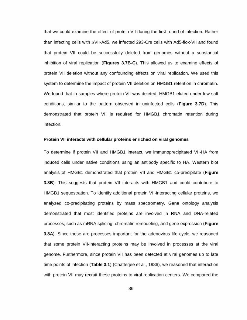
86
that we could examine the effect of protein VII during the first round of infection. Rather
than infecting cells with VII-Ad5, we infected 293-Cre cells with Ad5-flox-VII and found
that protein VII could be successfully deleted from genomes without a substantial
inhibition of viral replication (Figures 3.7B-C). This allowed us to examine effects of
protein VII deletion without any confounding effects on viral replication. We used this
system to determine the impact of protein VII deletion on HMGB1 retention in chromatin.
We found that in samples where protein VII was deleted, HMGB1 eluted under low salt
conditions, similar to the pattern observed in uninfected cells (Figure 3.7D). This
demonstrated that protein VII is required for HMGB1 chromatin retention during
infection.
Protein VII interacts with cellular proteins enriched on viral genomes
To determine if protein VII and HMGB1 interact, we immunoprecipitated VII-HA from
induced cells under native conditions using an antibody specific to HA. Western blot
analysis of HMGB1 demonstrated that protein VII and HMGB1 co-precipitate (Figure
3.8B). This suggests that protein VII interacts with HMGB1 and could contribute to
HMGB1 sequestration. To identify additional protein VII-interacting cellular proteins, we
analyzed co-precipitating proteins by mass spectrometry. Gene ontology analysis
demonstrated that most identified proteins are involved in RNA and DNA-related
processes, such as mRNA splicing, chromatin remodeling, and gene expression (Figure
3.8A). Since these are processes important for the adenovirus life cycle, we reasoned
that some protein VII-interacting proteins may be involved in processes at the viral
genome. Furthermore, since protein VII has been detected at viral genomes up to late
time points of infection (Table 3.1) (Chatterjee et al., 1986), we reasoned that interaction
with protein VII may recruit these proteins to viral replication centers. We compared the
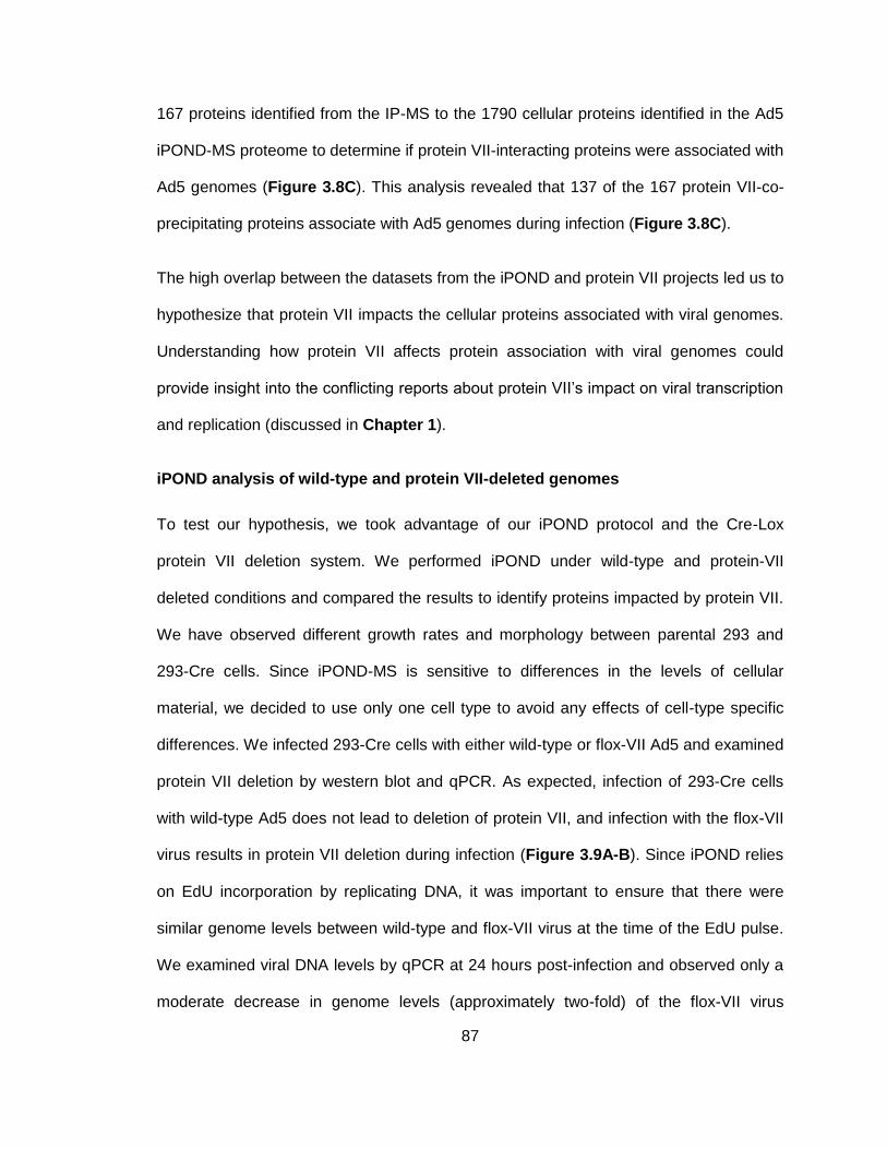
87
167 proteins identified from the IP-MS to the 1790 cellular proteins identified in the Ad5
iPOND-MS proteome to determine if protein VII-interacting proteins were associated with
Ad5 genomes (Figure 3.8C). This analysis revealed that 137 of the 167 protein VII-co-
precipitating proteins associate with Ad5 genomes during infection (Figure 3.8C).
The high overlap between the datasets from the iPOND and protein VII projects led us to
hypothesize that protein VII impacts the cellular proteins associated with viral genomes.
Understanding how protein VII affects protein association with viral genomes could
provide insight into the conflicting reports about protein VII’s impact on viral transcription
and replication (discussed in Chapter 1).
iPOND analysis of wild-type and protein VII-deleted genomes
To test our hypothesis, we took advantage of our iPOND protocol and the Cre-Lox
protein VII deletion system. We performed iPOND under wild-type and protein-VII
deleted conditions and compared the results to identify proteins impacted by protein VII.
We have observed different growth rates and morphology between parental 293 and
293-Cre cells. Since iPOND-MS is sensitive to differences in the levels of cellular
material, we decided to use only one cell type to avoid any effects of cell-type specific
differences. We infected 293-Cre cells with either wild-type or flox-VII Ad5 and examined
protein VII deletion by western blot and qPCR. As expected, infection of 293-Cre cells
with wild-type Ad5 does not lead to deletion of protein VII, and infection with the flox-VII
virus results in protein VII deletion during infection (Figure 3.9A-B). Since iPOND relies
on EdU incorporation by replicating DNA, it was important to ensure that there were
similar genome levels between wild-type and flox-VII virus at the time of the EdU pulse.
We examined viral DNA levels by qPCR at 24 hours post-infection and observed only a
moderate decrease in genome levels (approximately two-fold) of the flox-VII virus
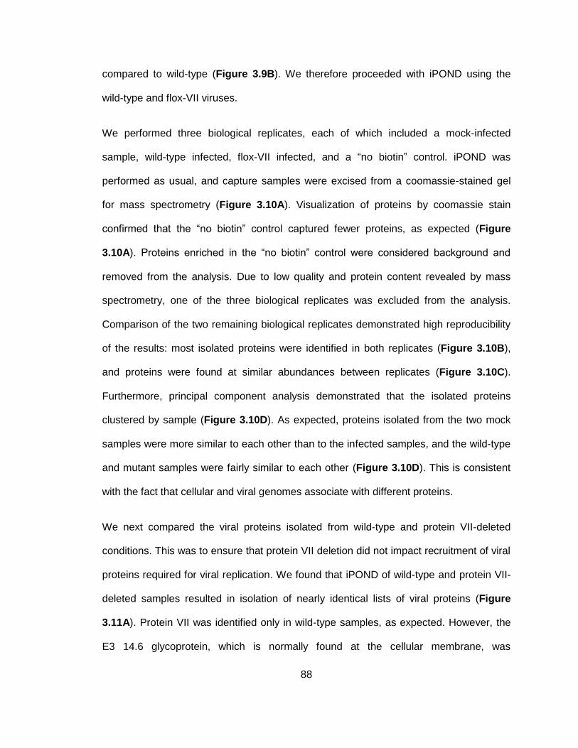
88
compared to wild-type (Figure 3.9B). We therefore proceeded with iPOND using the
wild-type and flox-VII viruses.
We performed three biological replicates, each of which included a mock-infected
sample, wild-type infected, flox-VII infected, and a “no biotin” control. iPOND was
performed as usual, and capture samples were excised from a coomassie-stained gel
for mass spectrometry (Figure 3.10A). Visualization of proteins by coomassie stain
confirmed that the “no biotin” control captured fewer proteins, as expected (Figure
3.10A). Proteins enriched in the “no biotin” control were considered background and
removed from the analysis. Due to low quality and protein content revealed by mass
spectrometry, one of the three biological replicates was excluded from the analysis.
Comparison of the two remaining biological replicates demonstrated high reproducibility
of the results: most isolated proteins were identified in both replicates (Figure 3.10B),
and proteins were found at similar abundances between replicates (Figure 3.10C).
Furthermore, principal component analysis demonstrated that the isolated proteins
clustered by sample (Figure 3.10D). As expected, proteins isolated from the two mock
samples were more similar to each other than to the infected samples, and the wild-type
and mutant samples were fairly similar to each other (Figure 3.10D). This is consistent
with the fact that cellular and viral genomes associate with different proteins.
We next compared the viral proteins isolated from wild-type and protein VII-deleted
conditions. This was to ensure that protein VII deletion did not impact recruitment of viral
proteins required for viral replication. We found that iPOND of wild-type and protein VII-
deleted samples resulted in isolation of nearly identical lists of viral proteins (Figure
3.11A). Protein VII was identified only in wild-type samples, as expected. However, the
E3 14.6 glycoprotein, which is normally found at the cellular membrane, was
89
unexpectedly isolated from protein VII-deleted samples. All other viral proteins, including
the DNA replication proteins DBP, Ad Pol, and pTP, were found under both wild-type
and VII-deleted conditions. Furthermore, viral proteins were found at similar abundances
in both conditions (Figure 3.11B). We conclude that protein VII deletion does not
dramatically affect the association of viral proteins. Importantly, association of viral DNA
replication proteins at similar levels suggests that deletion of protein VII does not impact
DNA replication, consistent with genome quantification from Figure 3.9B. As a result,
any changes to cellular protein association with genomes can be attributed to protein VII
deletion, rather than changes to DNA replication or other viral proteins.
Protein VII deletion affects association of RNA and DNA processing proteins with
viral genomes
We used a student’s T-test to identify cellular proteins differentially regulated between
wild-type and protein VII-deleted viruses. We reasoned that proteins significantly
(p<0.05) more enriched on wild-type virus represent proteins that could be recruited by
protein VII. Conversely, proteins that are significantly (p<0.05) more enriched on viral
DNA in the absence of protein VII represent proteins that do not associate as efficiently
with viral genomes when protein VII is present. We found that 97 proteins were
differentially regulated when protein VII was deleted (Figure 3.12). Thirty-two proteins
were significantly more abundant on genomes during wild-type infection, and 65 proteins
were significantly more abundant when protein VII was deleted (Figure 3.12). As a
control, we examined the effect of protein VII deletion on SET, a cellular protein known
to interact with protein VII and localize to viral genomes (Haruki et al., 2003; Haruki et
al., 2006) (see Chapter 1). We found that SET was isolated by iPOND under wild-type
conditions, but not when protein VII was deleted, validating our approach. We next
examined the functions of proteins enriched on wild-type and found that several of the
90
proteins most upregulated on wild-type genomes (log2 fold change > 1) are involved in
DNA or RNA-related processes. These processes include mRNA splicing and export,
DNA replication, and transcriptional regulation. Since these processes are important for
the adenovirus life cycle, our findings suggest that protein VII promotes the association
of proteins that contribute to viral replication and gene expression. Functions for
identified proteins are summarized in Table 3.2.
We examined localization of proteins enriched on wild-type genomes by
immunofluorescence of infected A549 cells. Consistent with iPOND-MS results, we
observed that RecQL1 and SRP14 co-localize with sites of viral DNA replication, as
marked by viral DBP (Figure 3.13). We also found that FUBP1 and SPATA5 were found
surrounding DBP-marked viral replication centers (Figure 3.13). This localization pattern
is similar to that of viral RNA and sites of late viral transcription (Pombo et al., 1994),
suggesting a role for these proteins in viral transcription. In fact, FUBP1 has been
suggested to recruit E1A and promote adenovirus transcription (unpublished data
presented at 2016 DNA Tumour Virus meeting, P.Pelka). Importantly, the iPOND
protocol does not include an RNA digestion step. Therefore, it is possible to isolate RNA-
interacting proteins through interactions of RNA with DNA. This likely explains the
isolation of host proteins involved in processes such as transcription and mRNA splicing.
We next examined localization in the absence of protein VII to determine if localization to
VRCs and viral transcription sites is dependent on protein VII. Immunofluorescence of
293 cells is difficult due to their small size and tendency to detach from coverslips.
Therefore, we optimized a system to delete protein VII in A549 cells by treating cells with
purified Cre protein prior to infection with the flox-VII virus. Cre was tagged with a
fragment of the HIV-1 TAT protein, which enhances cellular uptake of Cre (Peitz,
91
Pfannkuche, Rajewsky, & Edenhofer, 2002). We demonstrated deletion of protein VII in
infected cells pre-treated with TAT-Cre (Figure 3.14A-B). Similar to results in the 293
system, we found that protein VII was deleted without a substantial effect on viral
replication (Figure 3.14A-B) or on the identified cellular proteins (Figure 3.14C). We
next examined how protein VII deletion affects localization of FUBP1, which was
enriched on wild-type viral genomes (Figure 3.12) and redistributed during wild-type Ad5
infection (Figure 3.13). We first confirmed that TAT-Cre treatment had minimal impact
on infection efficiency (Figure 3.14C, DBP panel), and effectively deleted protein VII
(Figure 3.14C, VII panel). Next, we quantified cells with FUBP1 relocalization in control
and TAT-Cre-treated cells (Figure 3.14C, FUBP1 panel). We found a dramatic
decrease in the proportion of cells showing changes to FUBP1 when infected cells were
pre-treated with TAT-Cre. This suggests that protein VII deletion prevents the
relocalization of FUBP1 observed during wild-type Ad5 infection, validating our iPOND
results.
In order to gain more insight into the mechanism by which protein VII promotes the
observed changes, we examined whether protein VII is sufficient to induce the
localization changes to RecQL1, SRP14, FUBP1, and SPATA5 during infection. We
expressed GFP-tagged protein VII from a replication incompetent adenovirus vector.
Expression of protein VII was not sufficient to alter localization of these proteins (Figure
3.15A), indicating that additional viral proteins or processes are required. We also
examined whether proteins enriched on wild-type genomes interact with protein VII
during infection. We performed immunoprecipitation with wild-type Ad5-infected cells
using an antibody specific to protein VII. We did not detect interaction of these proteins
with protein VII during infection (Figure 3.15B). Together, results from Figure 3.15
indicate that localization changes to host proteins are unlikely to be through active
92
recruitment by protein VII. It is possible that protein VII instead induces changes to viral
DNA condensation or manipulates cellular pathways in such a way that promotes
localization of host proteins with viral genomes.
Protein VII suppresses interferon signaling
We reasoned that proteins enriched on protein VII-deleted genomes could provide
insight into cellular pathways targeted by protein VII. The cellular proteins TRIM25 and
UBR4 were enriched on protein VII-deleted genomes and have both been implicated in
the interferon response (Martin-Vicente, Medrano, Resino, Garcia-Sastre, & Martinez,
2017; Morrison et al., 2013) (Table 3.3). We therefore investigated whether protein VII
impacts this anti-viral pathway. We hypothesized that protein VII association with cellular
chromatin may affect expression of interferon stimulated genes (ISGs) through effects
on transcriptional regulation or DNA accessibility. To test this hypothesis, we examined
whether protein VII deletion affected ISG expression. We deleted protein VII by pre-
treatment of cells with TAT-Cre, infected cells with flox-VII virus, isolated RNA, and
performed RT-PCR using primers specific to ISG15 (Figure 3.16A). RT-PCR results
demonstrate that deletion of protein VII does not affect expression of this ISG compared
to wild-type infection (Figure 3.16A). Furthermore, we saw that infection did not lead to
a dramatic increase in ISG15 expression, likely due to the actions of other viral proteins.
Since multiple early viral proteins are known to suppress the interferon pathway (see
Chapter 1), effects of protein VII could be masked due to redundancy with early
proteins. Therefore, we explored the role of protein VII in the absence of infection to
avoid redundancy with early viral proteins. We examined ISG expression in response to
type I IFN treatment in cells expressing protein VII (Figure 3.16B). Again, we found that
protein VII did not affect ISG expression in response to ectopic IFN treatment. This
suggested that protein VII does not influence ISG expression downstream of IFN.
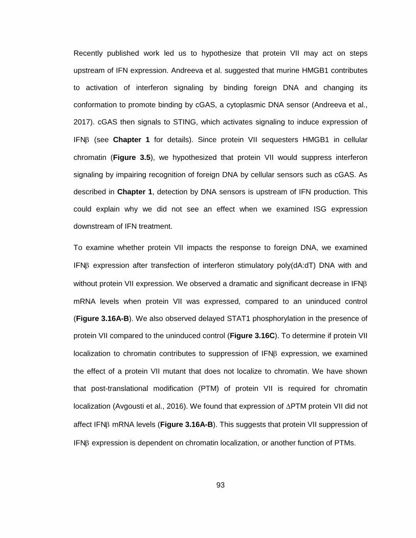
93
Recently published work led us to hypothesize that protein VII may act on steps
upstream of IFN expression. Andreeva et al. suggested that murine HMGB1 contributes
to activation of interferon signaling by binding foreign DNA and changing its
conformation to promote binding by cGAS, a cytoplasmic DNA sensor (Andreeva et al.,
2017). cGAS then signals to STING, which activates signaling to induce expression of
IFN (see Chapter 1 for details). Since protein VII sequesters HMGB1 in cellular
chromatin (Figure 3.5), we hypothesized that protein VII would suppress interferon
signaling by impairing recognition of foreign DNA by cellular sensors such as cGAS. As
described in Chapter 1, detection by DNA sensors is upstream of IFN production. This
could explain why we did not see an effect when we examined ISG expression
downstream of IFN treatment.
To examine whether protein VII impacts the response to foreign DNA, we examined
IFN expression after transfection of interferon stimulatory poly(dA:dT) DNA with and
without protein VII expression. We observed a dramatic and significant decrease in IFN
mRNA levels when protein VII was expressed, compared to an uninduced control
(Figure 3.16A-B). We also observed delayed STAT1 phosphorylation in the presence of
protein VII compared to the uninduced control (Figure 3.16C). To determine if protein VII
localization to chromatin contributes to suppression of IFN expression, we examined
the effect of a protein VII mutant that does not localize to chromatin. We have shown
that post-translational modification (PTM) of protein VII is required for chromatin
localization (Avgousti et al., 2016). We found that expression of PTM protein VII did not
affect IFN mRNA levels (Figure 3.16A-B). This suggests that protein VII suppression of
IFN expression is dependent on chromatin localization, or another function of PTMs.
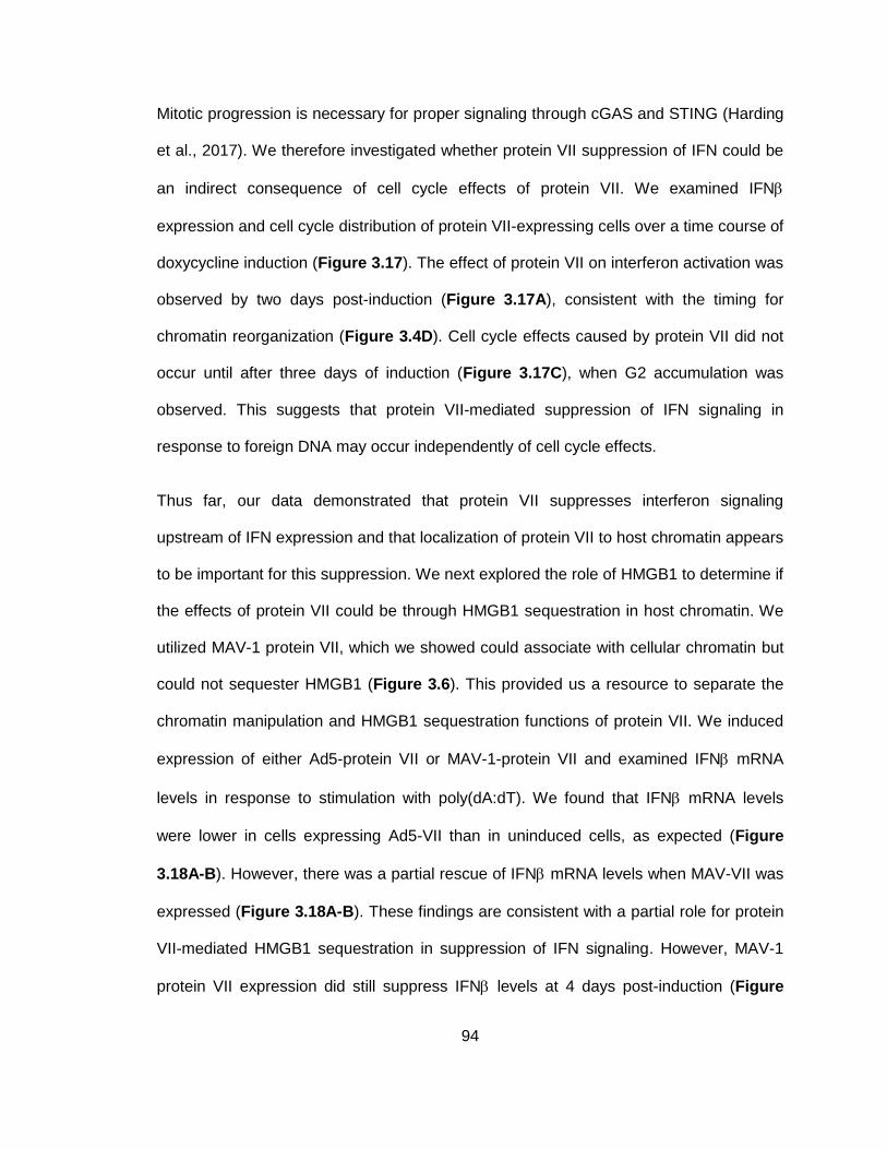
94
Mitotic progression is necessary for proper signaling through cGAS and STING (Harding
et al., 2017). We therefore investigated whether protein VII suppression of IFN could be
an indirect consequence of cell cycle effects of protein VII. We examined IFN
expression and cell cycle distribution of protein VII-expressing cells over a time course of
doxycycline induction (Figure 3.17). The effect of protein VII on interferon activation was
observed by two days post-induction (Figure 3.17A), consistent with the timing for
chromatin reorganization (Figure 3.4D). Cell cycle effects caused by protein VII did not
occur until after three days of induction (Figure 3.17C), when G2 accumulation was
observed. This suggests that protein VII-mediated suppression of IFN signaling in
response to foreign DNA may occur independently of cell cycle effects.
Thus far, our data demonstrated that protein VII suppresses interferon signaling
upstream of IFN expression and that localization of protein VII to host chromatin appears
to be important for this suppression. We next explored the role of HMGB1 to determine if
the effects of protein VII could be through HMGB1 sequestration in host chromatin. We
utilized MAV-1 protein VII, which we showed could associate with cellular chromatin but
could not sequester HMGB1 (Figure 3.6). This provided us a resource to separate the
chromatin manipulation and HMGB1 sequestration functions of protein VII. We induced
expression of either Ad5-protein VII or MAV-1-protein VII and examined IFN mRNA
levels in response to stimulation with poly(dA:dT). We found that IFN mRNA levels
were lower in cells expressing Ad5-VII than in uninduced cells, as expected (Figure
3.18A-B). However, there was a partial rescue of IFN mRNA levels when MAV-VII was
expressed (Figure 3.18A-B). These findings are consistent with a partial role for protein
VII-mediated HMGB1 sequestration in suppression of IFN signaling. However, MAV-1
protein VII expression did still suppress IFN levels at 4 days post-induction (Figure
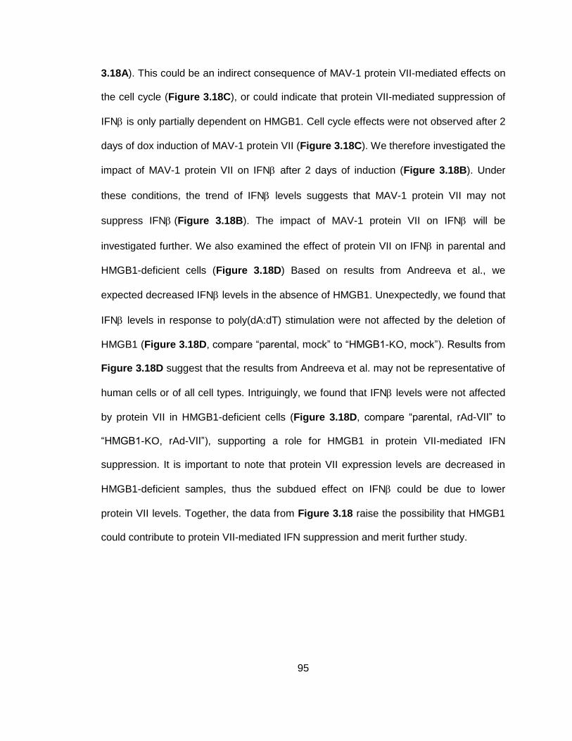
95
3.18A). This could be an indirect consequence of MAV-1 protein VII-mediated effects on
the cell cycle (Figure 3.18C), or could indicate that protein VII-mediated suppression of
IFN is only partially dependent on HMGB1. Cell cycle effects were not observed after 2
days of dox induction of MAV-1 protein VII (Figure 3.18C). We therefore investigated the
impact of MAV-1 protein VII on IFN after 2 days of induction (Figure 3.18B). Under
these conditions, the trend of IFN levels suggests that MAV-1 protein VII may not
suppress IFN(Figure 3.18B). The impact of MAV-1 protein VII on IFN will be
investigated further. We also examined the effect of protein VII on IFN in parental and
HMGB1-deficient cells (Figure 3.18D) Based on results from Andreeva et al., we
expected decreased IFN levels in the absence of HMGB1. Unexpectedly, we found that
IFN levels in response to poly(dA:dT) stimulation were not affected by the deletion of
HMGB1 (Figure 3.18D, compare “parental, mock” to “HMGB1-KO, mock”). Results from
Figure 3.18D suggest that the results from Andreeva et al. may not be representative of
human cells or of all cell types. Intriguingly, we found that IFN levels were not affected
by protein VII in HMGB1-deficient cells (Figure 3.18D, compare “parental, rAd-VII” to
“HMGB1-KO, rAd-VII”), supporting a role for HMGB1 in protein VII-mediated IFN
suppression. It is important to note that protein VII expression levels are decreased in
HMGB1-deficient samples, thus the subdued effect on IFN could be due to lower
protein VII levels. Together, the data from Figure 3.18 raise the possibility that HMGB1
could contribute to protein VII-mediated IFN suppression and merit further study.
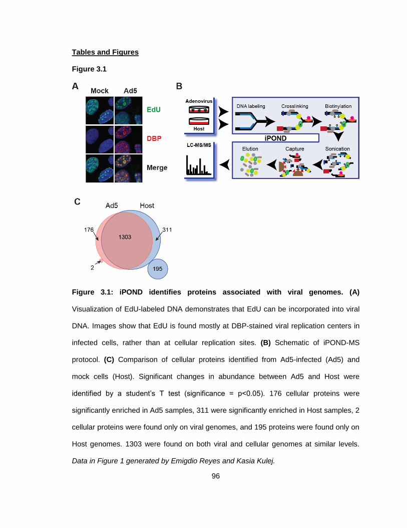
96
Tables and Figures
Figure 3.1
Figure 3.1: iPOND identifies proteins associated with viral genomes. (A)
Visualization of EdU-labeled DNA demonstrates that EdU can be incorporated into viral
DNA. Images show that EdU is found mostly at DBP-stained viral replication centers in
infected cells, rather than at cellular replication sites. (B) Schematic of iPOND-MS
protocol. (C) Comparison of cellular proteins identified from Ad5-infected (Ad5) and
mock cells (Host). Significant changes in abundance between Ad5 and Host were
identified by a student’s T test (significance = p<0.05). 176 cellular proteins were
significantly enriched in Ad5 samples, 311 were significantly enriched in Host samples, 2
cellular proteins were found only on viral genomes, and 195 proteins were found only on
Host genomes. 1303 were found on both viral and cellular genomes at similar levels.
Data in Figure 1 generated by Emigdio Reyes and Kasia Kulej.
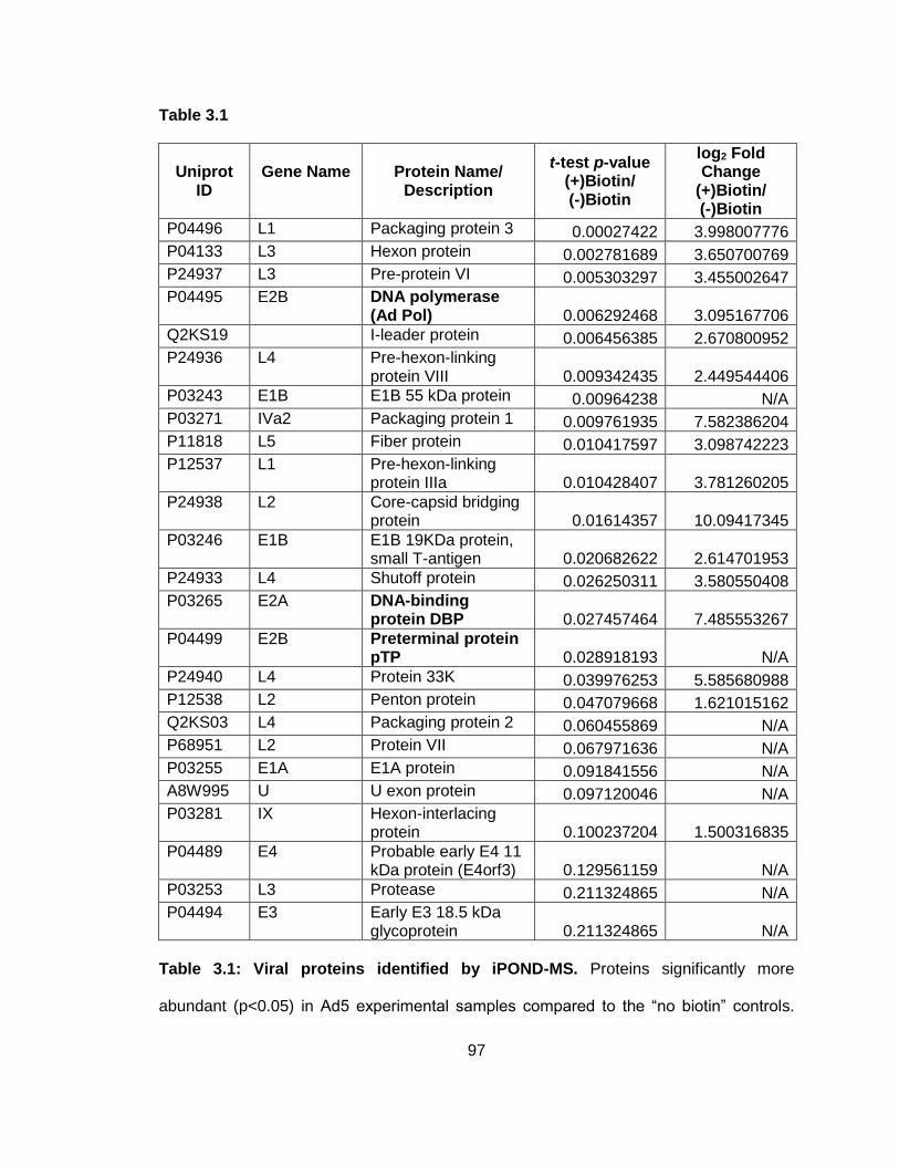
97
Table 3.1
Uniprot
ID
Gene Name
Protein Name/
Description
t-test p-value (+)Biotin/ (-)Biotin
log2 Fold Change
(+)Biotin/ (-)Biotin
P04496 L1 Packaging protein 3 0.00027422 3.998007776
P04133 L3 Hexon protein 0.002781689 3.650700769
P24937 L3 Pre-protein VI 0.005303297 3.455002647
P04495 E2B DNA polymerase (Ad Pol) 0.006292468 3.095167706
Q2KS19 I-leader protein 0.006456385 2.670800952
P24936 L4 Pre-hexon-linking protein VIII 0.009342435 2.449544406
P03243 E1B E1B 55 kDa protein 0.00964238 N/A
P03271 IVa2 Packaging protein 1 0.009761935 7.582386204
P11818 L5 Fiber protein 0.010417597 3.098742223
P12537 L1 Pre-hexon-linking protein IIIa 0.010428407 3.781260205
P24938 L2 Core-capsid bridging protein 0.01614357 10.09417345
P03246 E1B E1B 19KDa protein, small T-antigen 0.020682622 2.614701953
P24933 L4 Shutoff protein 0.026250311 3.580550408
P03265 E2A DNA-binding protein DBP 0.027457464 7.485553267
P04499 E2B Preterminal protein pTP 0.028918193 N/A
P24940 L4 Protein 33K 0.039976253 5.585680988
P12538 L2 Penton protein 0.047079668 1.621015162
Q2KS03 L4 Packaging protein 2 0.060455869 N/A
P68951 L2 Protein VII 0.067971636 N/A
P03255 E1A E1A protein 0.091841556 N/A
A8W995 U U exon protein 0.097120046 N/A
P03281 IX Hexon-interlacing protein 0.100237204 1.500316835
P04489 E4 Probable early E4 11 kDa protein (E4orf3) 0.129561159 N/A
P03253 L3 Protease 0.211324865 N/A
P04494 E3 Early E3 18.5 kDa glycoprotein 0.211324865 N/A
Table 3.1: Viral proteins identified by iPOND-MS. Proteins significantly more
abundant (p<0.05) in Ad5 experimental samples compared to the “no biotin” controls.

98
Viral proteins involved in viral DNA replication are in bold. Data in Table 3.1 generated
by Emigdio Reyes.
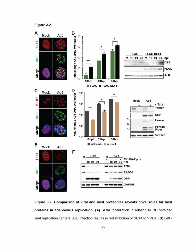
99
Figure 3.2
Figure 3.2: Comparison of viral and host proteomes reveals novel roles for host
proteins in adenovirus replication. (A) SLX4 localization in relation to DBP-stained
viral replication centers. Ad5 infection results in redistribution of SLX4 to VRCs. (B) Left -

100
Viral DNA accumulation in SLX4-deficient cells and matched cells complemented with
FLAG-tagged SLX4. There is increased viral DNA accumulation in SLX4-expressing
cells. Right - Western blot confirms expression of FLAG-SLX4 and demonstrates
increased viral DBP levels in SLX4-expressing cells. (C) TCOF1 localization in relation
to DBP-stained VRCs. Ad5 infection results in redistribution of TCOF1 from nucleoli to
sites surrounding VRCs. (D) Effect of TCOF1 depletion on viral DNA accumulation.
siRNA-mediated depletion of TCOF1 results in significantly decreased viral DNA levels.
Western confirms TCOF1 knockdown and demonstrates decreased early (DBP) and late
(hexon, penton, fiber) viral protein levels. (E) TFII-I localization in infected cells in
relation to DBP-marked VRCs. Ad5 infection leads to redistribution of TFII-I from a pan-
nuclear distribution to foci that do not colocalize with VRCs. (F) Western blot
demonstrating proteasome-dependent decrease of TFII-I during Ad5 infection.
Treatment with the proteasome inhibitors MG132 and epoxomicin rescues TFII-I levels.
Rad50 is a known Ad5 degradation substrate and serves as a control for degradation.
Panels A, C, E, and F by Emigdio Reyes and Lisa Akhtar.
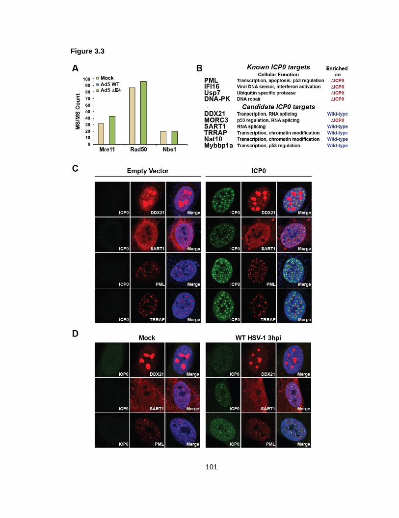
101
Figure 3.3
102
Figure 3.3: Comparison of wild-type and mutant viral proteomes reveals targets of
specific viral proteins. (A) Raw spectral count data of Mre11, Rad50, and Nbs1 from
iPOND-MS of mock, wild-type Ad5, and E4-deleted Ad5 samples. As expected, Mre11,
Rad50, and Nbs1 are isolated with replicated DNA from mock and E4-deleted samples,
but are not detected in wild-type Ad5 samples. This is consistent with the known
degradation of MRN during wild-type Ad5 infection, and the known association of MRN
with E4-deleted VRCs. (B) iPOND-MS with wild-type and ICP0-deleted HSV-1
demonstrates that known ICP0 degradation targets are enriched on ICP0-deleted
genomes. Additional cellular proteins were found enriched on wild-type or ICP0-deleted
genomes and represent proteins potentially regulated by ICP0. (C) Immunofluorescence
analysis of cellular proteins identified in B in cells transfected with an ICP0-expression
vector. DDX21, SART1, and PML colocalize with ICP0, while TRRAP does not. (D)
Immunofluorescence analysis of cellular proteins identified in B in mock and HSV-1
infected cells. SART1 and PML colocalize with ICP0 during infection. Results from C and
D are consistent with a role for ICP0 in affecting localization of these cellular proteins.
Data in panel B generated by Emigdio Reyes.
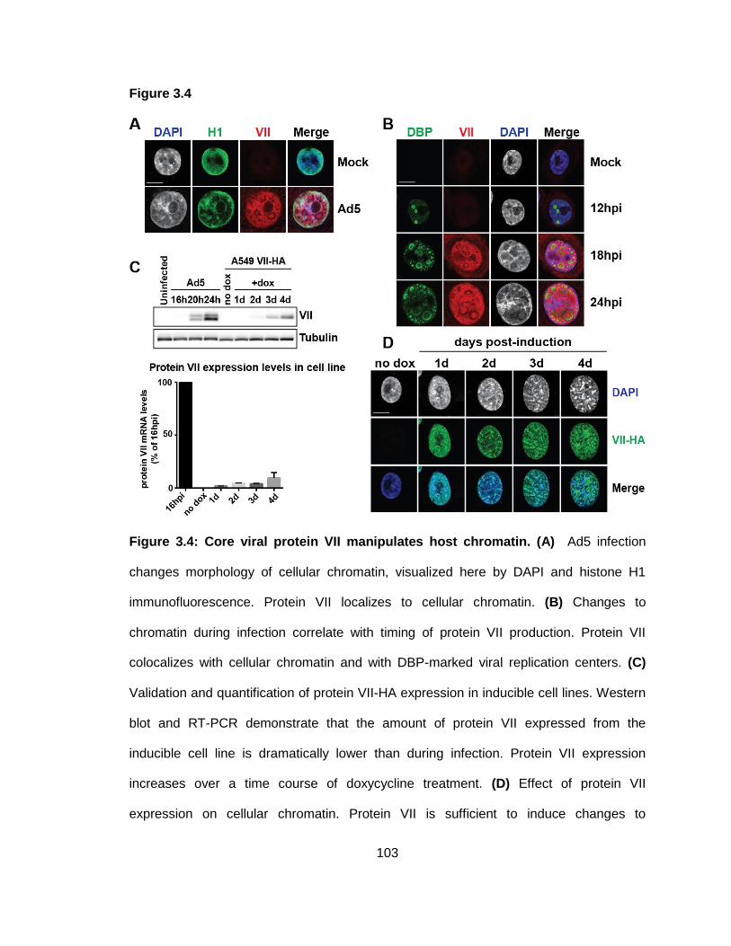
103
Figure 3.4
Figure 3.4: Core viral protein VII manipulates host chromatin. (A) Ad5 infection
changes morphology of cellular chromatin, visualized here by DAPI and histone H1
immunofluorescence. Protein VII localizes to cellular chromatin. (B) Changes to
chromatin during infection correlate with timing of protein VII production. Protein VII
colocalizes with cellular chromatin and with DBP-marked viral replication centers. (C)
Validation and quantification of protein VII-HA expression in inducible cell lines. Western
blot and RT-PCR demonstrate that the amount of protein VII expressed from the
inducible cell line is dramatically lower than during infection. Protein VII expression
increases over a time course of doxycycline treatment. (D) Effect of protein VII
expression on cellular chromatin. Protein VII is sufficient to induce changes to

104
appearance of host chromatin, represented by DAPI here. Changes to DAPI correlate
with increasing protein VII levels (see panel C). Panels A, B, and D by Daphne Avgousti.
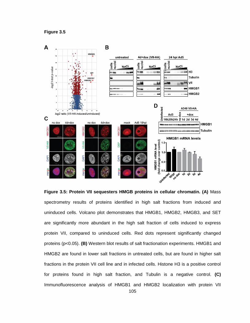
105
Figure 3.5
Figure 3.5: Protein VII sequesters HMGB proteins in cellular chromatin. (A) Mass
spectrometry results of proteins identified in high salt fractions from induced and
uninduced cells. Volcano plot demonstrates that HMGB1, HMGB2, HMGB3, and SET
are significantly more abundant in the high salt fraction of cells induced to express
protein VII, compared to uninduced cells. Red dots represent significantly changed
proteins (p<0.05). (B) Western blot results of salt fractionation experiments. HMGB1 and
HMGB2 are found in lower salt fractions in untreated cells, but are found in higher salt
fractions in the protein VII cell line and in infected cells. Histone H3 is a positive control
for proteins found in high salt fraction, and Tubulin is a negative control. (C)
Immunofluorescence analysis of HMGB1 and HMGB2 localization with protein VII
106
expression and Ad5 infection. Expression of protein VII is sufficient to relocalize to
HMGB1 and HMGB2 to DAPI-stained cellular DNA and protein VII. Ad5 infection
induces reorganization of HMGB1 to cellular chromatin, similar to protein VII localization.
(D) HMGB1 levels during infection and in the presence of protein VII. Western blot and
RT-PCR analysis demonstrate that neither protein VII expression nor Ad5 infection
results in dramatic changes to HMGB1 levels. Panels A-C by Daphne Avgousti and
Christin Herrmann. Proteomic analysis in panel A by Kasia Kulej.
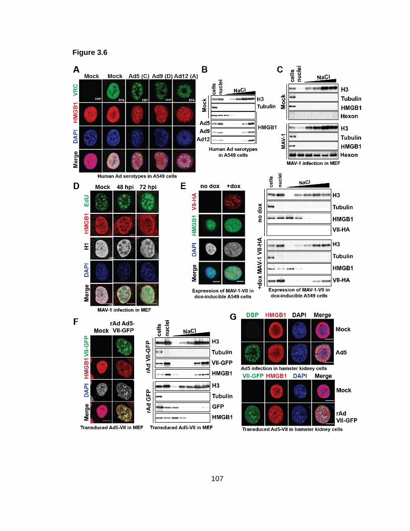
107
Figure 3.6
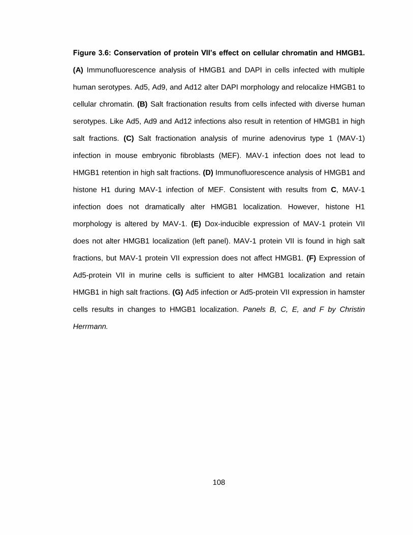
108
Figure 3.6: Conservation of protein VII’s effect on cellular chromatin and HMGB1.
(A) Immunofluorescence analysis of HMGB1 and DAPI in cells infected with multiple
human serotypes. Ad5, Ad9, and Ad12 alter DAPI morphology and relocalize HMGB1 to
cellular chromatin. (B) Salt fractionation results from cells infected with diverse human
serotypes. Like Ad5, Ad9 and Ad12 infections also result in retention of HMGB1 in high
salt fractions. (C) Salt fractionation analysis of murine adenovirus type 1 (MAV-1)
infection in mouse embryonic fibroblasts (MEF). MAV-1 infection does not lead to
HMGB1 retention in high salt fractions. (D) Immunofluorescence analysis of HMGB1 and
histone H1 during MAV-1 infection of MEF. Consistent with results from C, MAV-1
infection does not dramatically alter HMGB1 localization. However, histone H1
morphology is altered by MAV-1. (E) Dox-inducible expression of MAV-1 protein VII
does not alter HMGB1 localization (left panel). MAV-1 protein VII is found in high salt
fractions, but MAV-1 protein VII expression does not affect HMGB1. (F) Expression of
Ad5-protein VII in murine cells is sufficient to alter HMGB1 localization and retain
HMGB1 in high salt fractions. (G) Ad5 infection or Ad5-protein VII expression in hamster
cells results in changes to HMGB1 localization. Panels B, C, E, and F by Christin
Herrmann.
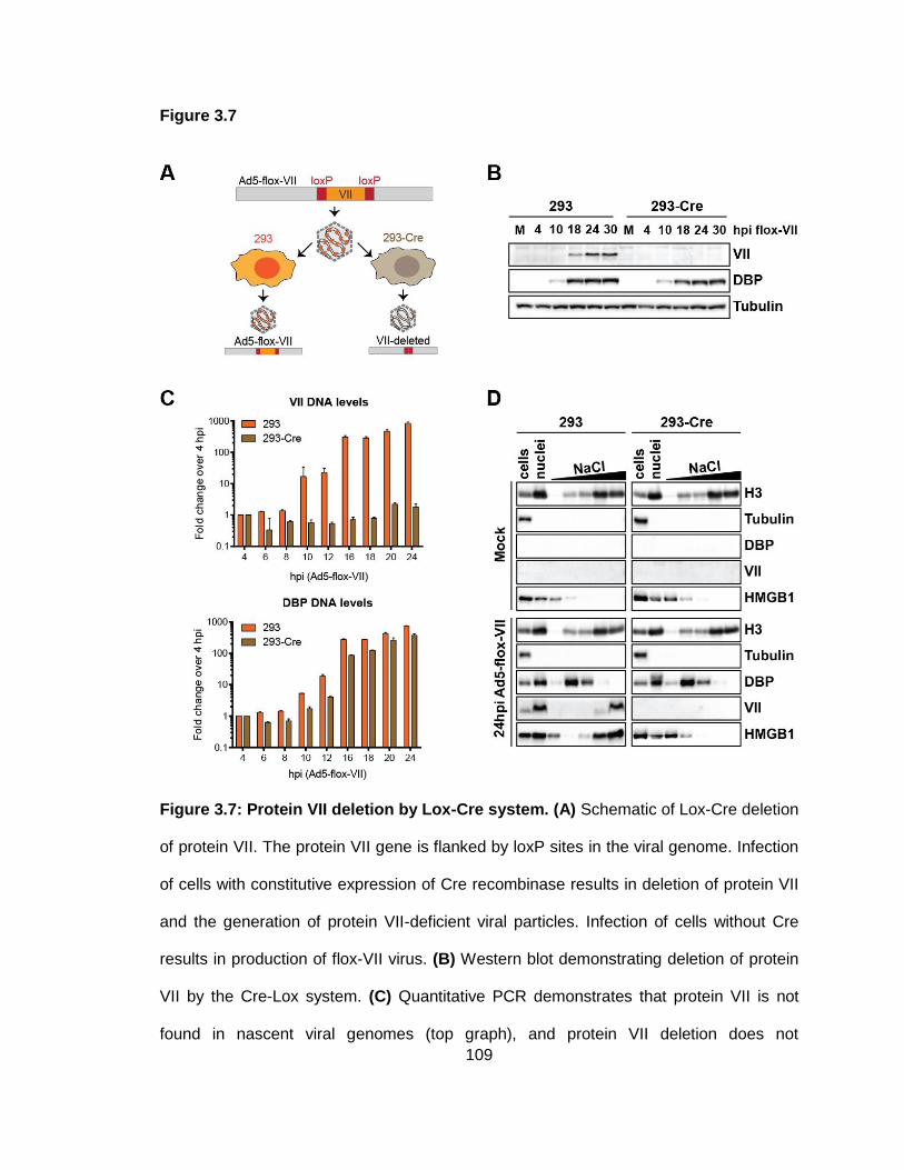
109
Figure 3.7
Figure 3.7: Protein VII deletion by Lox-Cre system. (A) Schematic of Lox-Cre deletion
of protein VII. The protein VII gene is flanked by loxP sites in the viral genome. Infection
of cells with constitutive expression of Cre recombinase results in deletion of protein VII
and the generation of protein VII-deficient viral particles. Infection of cells without Cre
results in production of flox-VII virus. (B) Western blot demonstrating deletion of protein
VII by the Cre-Lox system. (C) Quantitative PCR demonstrates that protein VII is not
found in nascent viral genomes (top graph), and protein VII deletion does not
110
dramatically affect viral DNA accumulation (bottom graph). (D) Salt fractionation of Cre
cells infected with flox-VII virus to assess the effect of protein VII deletion on HMGB1
retention in high salt fraction. HMGB1 is not retained in high salt fractions when protein
VII is deleted. Panel D by Christin Herrmann.
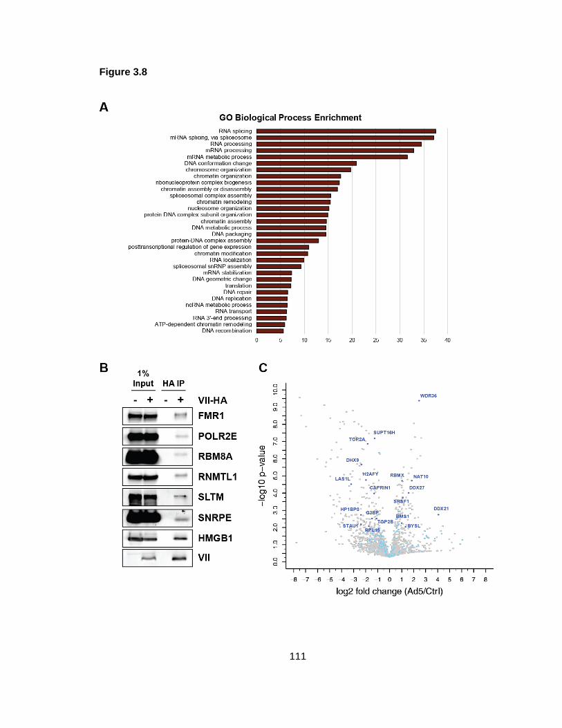
111
Figure 3.8
112
Figure 3.8: Protein VII interacts with HMGB1 and cellular proteins enriched on viral
genomes. (A) Gene ontology analysis of cellular proteins that co-precipitate with
ectopically expressed protein VII. X-axis is –log10 p-value. (B) Western blots confirm IP-
MS results and demonstrate that several proteins with RNA and DNA-related functions
co-precipitate with protein VII. IP-Western also demonstrates that HMGB1 co-
precipitates with protein VII. (C) Volcano plot of Ad5 iPOND results with protein VII-
interacting proteins highlighted. Blue dots of any shade represent proteins identified in
both iPOND-MS and VII IP-MS. Dark blue dots represent proteins significantly enriched
on mock or Ad5 iPOND proteomes. Data in panels A and C generated by Daphne
Avgousti and Emigdio Reyes. Proteomic analyses by Kasia Kulej and Joseph Dybas.
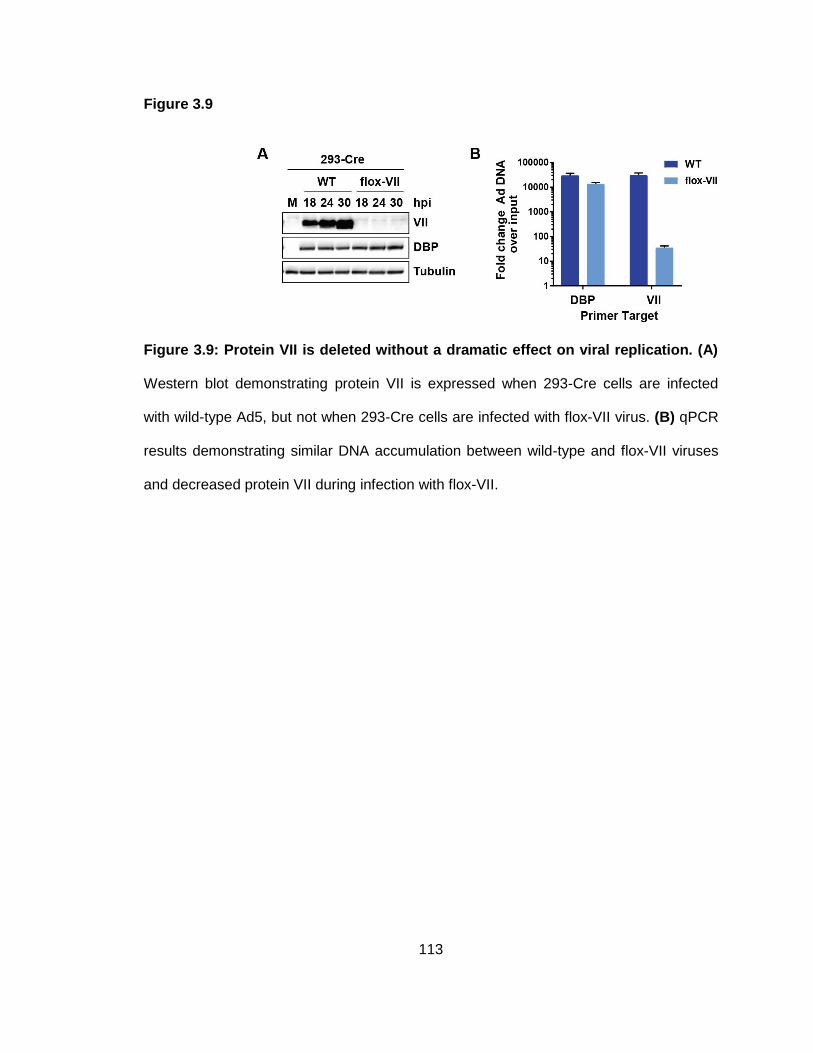
113
Figure 3.9
Figure 3.9: Protein VII is deleted without a dramatic effect on viral replication. (A)
Western blot demonstrating protein VII is expressed when 293-Cre cells are infected
with wild-type Ad5, but not when 293-Cre cells are infected with flox-VII virus. (B) qPCR
results demonstrating similar DNA accumulation between wild-type and flox-VII viruses
and decreased protein VII during infection with flox-VII.
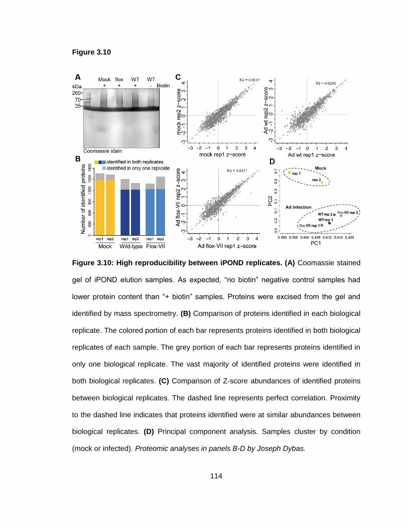
114
Figure 3.10
Figure 3.10: High reproducibility between iPOND replicates. (A) Coomassie stained
gel of iPOND elution samples. As expected, “no biotin” negative control samples had
lower protein content than “+ biotin” samples. Proteins were excised from the gel and
identified by mass spectrometry. (B) Comparison of proteins identified in each biological
replicate. The colored portion of each bar represents proteins identified in both biological
replicates of each sample. The grey portion of each bar represents proteins identified in
only one biological replicate. The vast majority of identified proteins were identified in
both biological replicates. (C) Comparison of Z-score abundances of identified proteins
between biological replicates. The dashed line represents perfect correlation. Proximity
to the dashed line indicates that proteins identified were at similar abundances between
biological replicates. (D) Principal component analysis. Samples cluster by condition
(mock or infected). Proteomic analyses in panels B-D by Joseph Dybas.
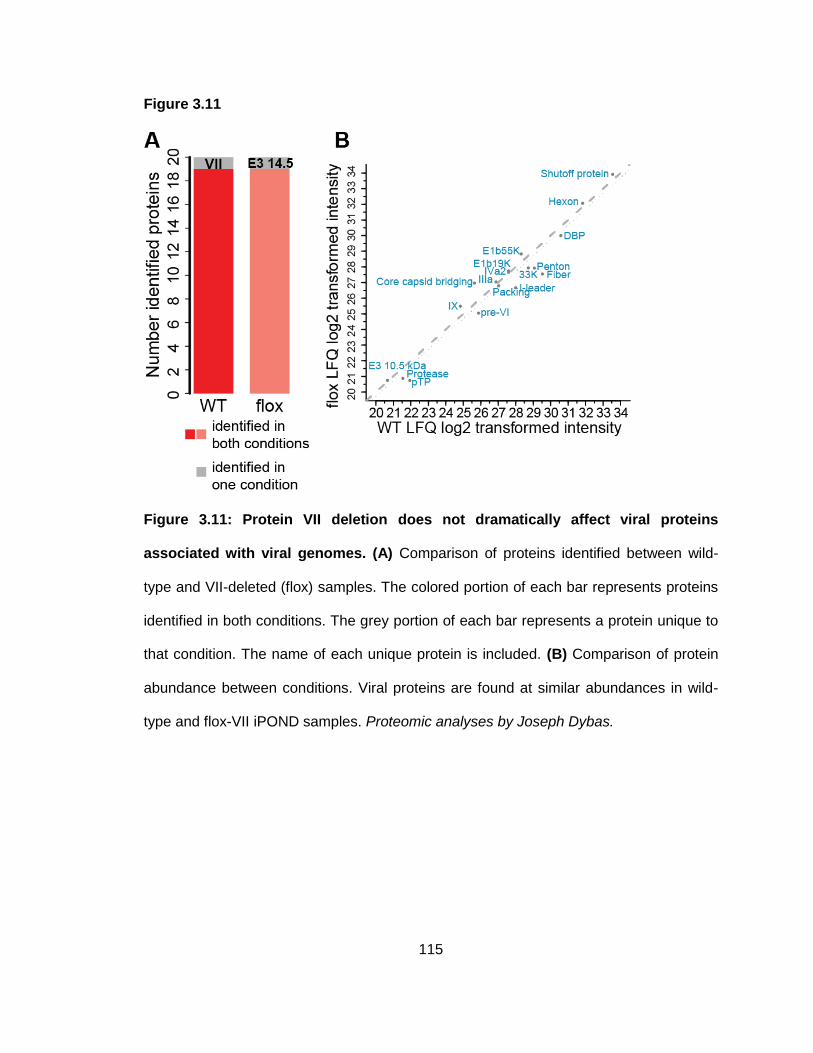
115
Figure 3.11
Figure 3.11: Protein VII deletion does not dramatically affect viral proteins
associated with viral genomes. (A) Comparison of proteins identified between wild-
type and VII-deleted (flox) samples. The colored portion of each bar represents proteins
identified in both conditions. The grey portion of each bar represents a protein unique to
that condition. The name of each unique protein is included. (B) Comparison of protein
abundance between conditions. Viral proteins are found at similar abundances in wild-
type and flox-VII iPOND samples. Proteomic analyses by Joseph Dybas.
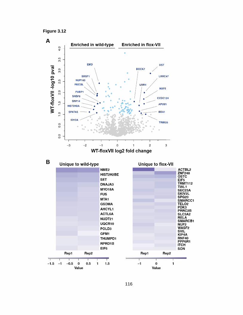
116
Figure 3.12
117
Figure 3.12: Protein VII deletion significantly alters cellular proteins associated
with viral genomes. (A) Volcano plot demonstrates that several cellular proteins are
significantly enriched on either wild-type or protein VII-deleted (flox-VII) genomes. Blue
dots represent proteins significantly enriched (p<0.05), and dark blue dots are those
proteins with fold change > 2. (B) Heat maps of proteins identified in only wild-type or
protein VII-deleted (flox-VII) iPOND samples. SET was found on only wild-type
genomes, consistent with the known role of protein VII in recruiting SET to viral
genomes. Proteomic analyses by Joseph Dybas.
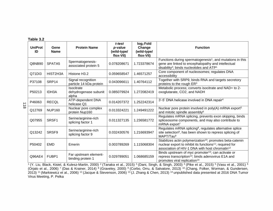
118
Table 3.2
UniProt ID
Gene Name
Protein Name
t-test p-value
(wild-type/ flox-VII)
log2 Fold Change
(wild-type/ flox-VII)
Function
Q8NB90 SPATA5 Spermatogenesis-associated protein 5
0.078208671 1.723378674 Functions during spermatogenesis1, and mutations in this gene are linked to encephalopathy and intellectual disability2; binds nucleotides and ATP1
Q71DI3 HIST2H3A Histone H3.2 0.059658547 1.46571257 Core component of nucleosomes; regulates DNA accessibility
P37108 SRP14 Signal recognition particle 14 kDa protein
0.043096611 1.40764112 Together with SRP9, binds RNA and targets secretory proteins to the rough ER3
P50213 IDH3A Isocitrate dehydrogenase subunit alpha
0.085079924 1.272082419 Metabolic process; converts isocitrate and NAD+ to 2-oxoglutarate, CO2, and NADH
P46063 RECQL ATP-dependent DNA helicase Q1
0.014207372 1.252242314 3’-5’ DNA helicase involved in DNA repair4
Q12769 NUP160 Nuclear pore complex protein Nup160
0.013324221 1.249491222 Nuclear pore protein involved in poly(A) mRNA export5 and mitotic spindle assembly6
Q07955 SRSF1 Serine/arginine-rich splicing factor 1
0.011327135 1.236581772 Regulates mRNA splicing, prevents exon skipping, binds spliceosome components, and may also contribute to mRNA export7
Q13242 SRSF9 Serine/arginine-rich splicing factor 9
0.032430576 1.216693947 Regulates mRNA splicing8, regulates alternative splice site selection8, has been shown to repress splicing of MAPT/Tau9
P50402 EMD Emerin 0.003789269 1.115068304 Stabilizes actin polymerization10; promotes beta-catenin nuclear export to inhibit its functions11; required for association of HIV-1 DNA with host chromatin12
Q96AE4 FUBP1 Far upstream element-binding protein 1
0.029789051 1.068685159 Binds upstream of myc promoter13; can activate or repress transcription13; binds adenovirus E1A and promotes viral replication14
1 (Y. Liu, Black, Kisiel, & Kulesz-Martin, 2000) 2 (Tanaka et al., 2015) 3 (Dani, Singh, & Singh, 2003) 4 (Pike et al., 2015) 5 (Vasu et al., 2001) 6
(Orjalo et al., 2006) 7 (Das & Krainer, 2014) 8 (Graveley, 2000) 9 (Corbo, Orru, & Salvatore, 2013) 10 (Chang, Folker, Worman, & Gundersen, 2013) 11 (Markiewicz et al., 2006) 12 (Jacque & Stevenson, 2006) 13 (J. Zhang & Chen, 2013) 14 unpublished data presented at 2016 DNA Tumor Virus Meeting, P. Pelka
119
Table 3.2: Proteins enriched on wild-type viral genomes. A student’s T test was used to identify proteins significantly more
abundant on viral genomes during wild-type infection when compared to flox-VII infection. Proteins that were significant (p<0.05)
and had a fold change in abundance > 2 are shown here.
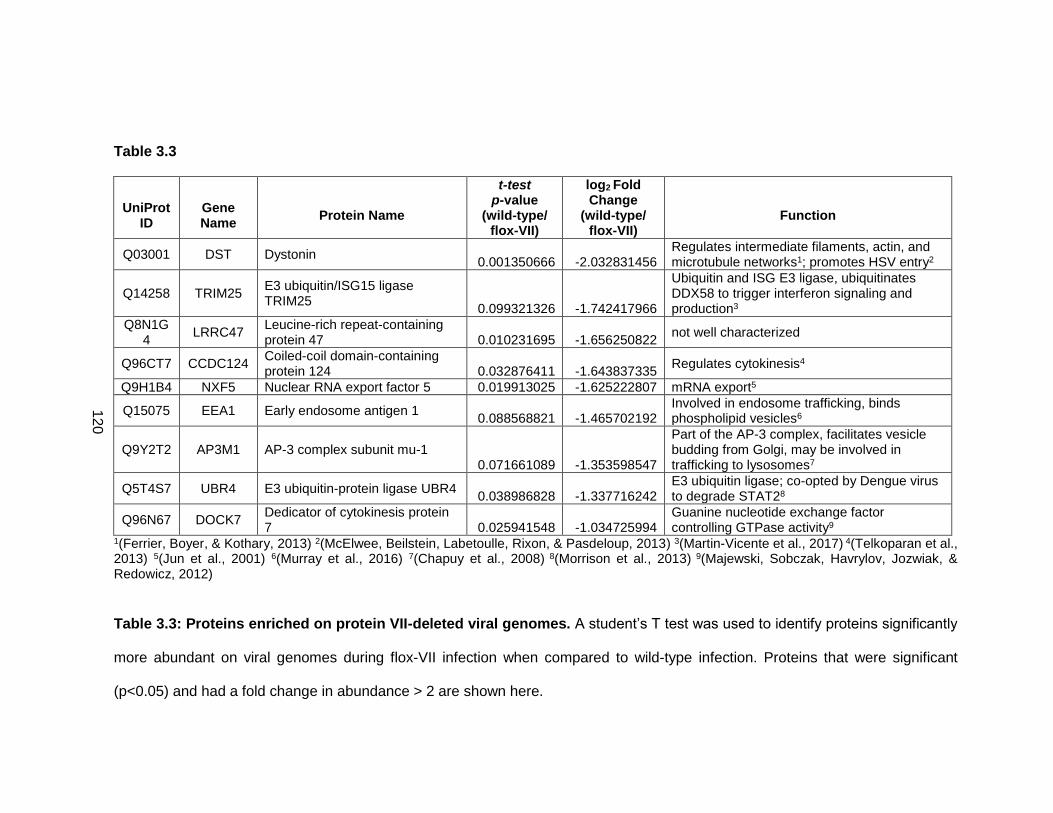
120
Table 3.3
UniProt
ID
Gene Name
Protein Name
t-test p-value
(wild-type/ flox-VII)
log2 Fold Change
(wild-type/ flox-VII)
Function
Q03001 DST Dystonin 0.001350666 -2.032831456
Regulates intermediate filaments, actin, and microtubule networks1; promotes HSV entry2
Q14258 TRIM25 E3 ubiquitin/ISG15 ligase TRIM25
0.099321326 -1.742417966
Ubiquitin and ISG E3 ligase, ubiquitinates DDX58 to trigger interferon signaling and production3
Q8N1G4
LRRC47 Leucine-rich repeat-containing protein 47 0.010231695 -1.656250822
not well characterized
Q96CT7 CCDC124 Coiled-coil domain-containing protein 124 0.032876411 -1.643837335
Regulates cytokinesis4
Q9H1B4 NXF5 Nuclear RNA export factor 5 0.019913025 -1.625222807 mRNA export5
Q15075 EEA1 Early endosome antigen 1 0.088568821 -1.465702192
Involved in endosome trafficking, binds phospholipid vesicles6
Q9Y2T2 AP3M1 AP-3 complex subunit mu-1 0.071661089 -1.353598547
Part of the AP-3 complex, facilitates vesicle budding from Golgi, may be involved in trafficking to lysosomes7
Q5T4S7 UBR4 E3 ubiquitin-protein ligase UBR4 0.038986828 -1.337716242
E3 ubiquitin ligase; co-opted by Dengue virus to degrade STAT28
Q96N67 DOCK7 Dedicator of cytokinesis protein 7 0.025941548 -1.034725994
Guanine nucleotide exchange factor controlling GTPase activity9
1(Ferrier, Boyer, & Kothary, 2013) 2(McElwee, Beilstein, Labetoulle, Rixon, & Pasdeloup, 2013) 3(Martin-Vicente et al., 2017) 4(Telkoparan et al., 2013) 5(Jun et al., 2001) 6(Murray et al., 2016) 7(Chapuy et al., 2008) 8(Morrison et al., 2013) 9(Majewski, Sobczak, Havrylov, Jozwiak, & Redowicz, 2012)
Table 3.3: Proteins enriched on protein VII-deleted viral genomes. A student’s T test was used to identify proteins significantly
more abundant on viral genomes during flox-VII infection when compared to wild-type infection. Proteins that were significant
(p<0.05) and had a fold change in abundance > 2 are shown here.
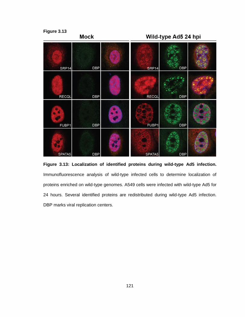
121
Figure 3.13
Figure 3.13: Localization of identified proteins during wild-type Ad5 infection.
Immunofluorescence analysis of wild-type infected cells to determine localization of
proteins enriched on wild-type genomes. A549 cells were infected with wild-type Ad5 for
24 hours. Several identified proteins are redistributed during wild-type Ad5 infection.
DBP marks viral replication centers.
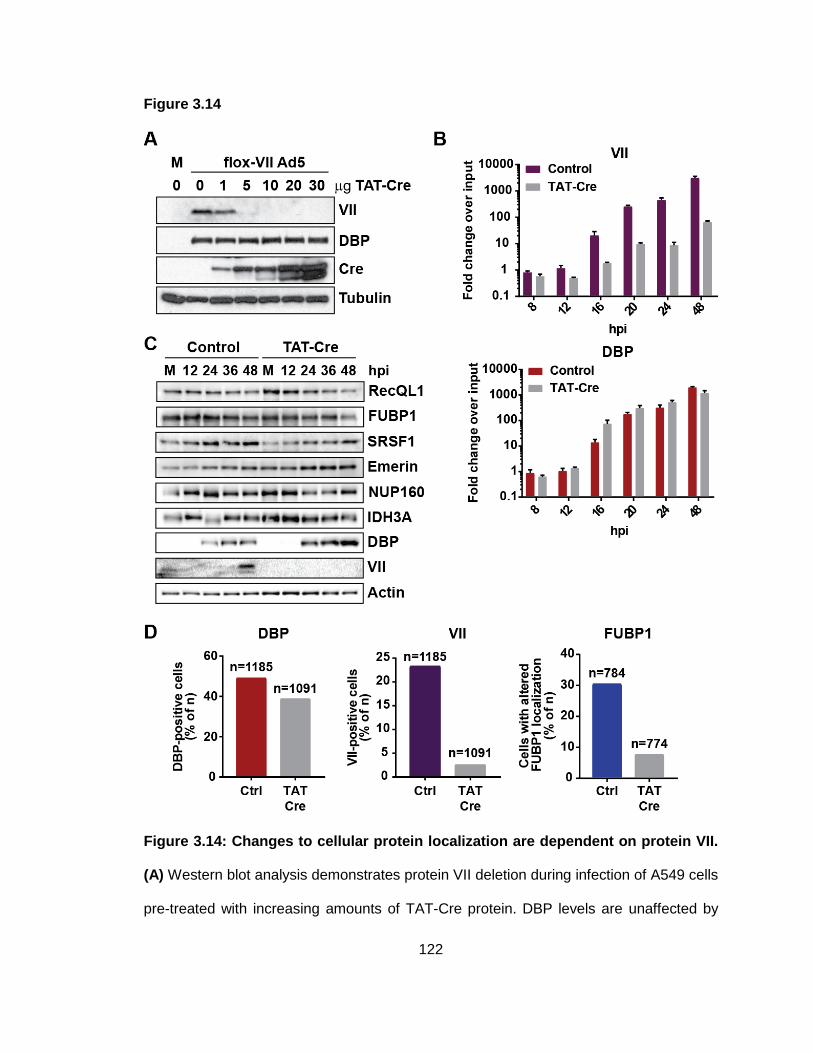
122
Figure 3.14
Figure 3.14: Changes to cellular protein localization are dependent on protein VII.
(A) Western blot analysis demonstrates protein VII deletion during infection of A549 cells
pre-treated with increasing amounts of TAT-Cre protein. DBP levels are unaffected by
123
TAT-Cre treatment or protein VII deletion. (B) A549 cells in 12-well plates were pre-
treated with 15 g TAT-Cre and infected with flox-VII at MOI 10. Cells were collected at
the indicated time points, DNA was isolated, and qPCR was performed using primers
specific to protein VII or DBP. qPCR results demonstrate a decrease in genomes
containing protein VII, but no effect on total genome accumulation. (C) Western blot
analysis of protein levels during infection in control or TAT-Cre treated cells. Cells were
treated as described in B. TAT-Cre treatment results in protein VII deletion, but does not
dramatically affect levels of cellular proteins. (D) Quantification of immunofluorescence
results. A549 cells in 12-well plates were pre-treated with 45 g TAT-Cre or treated with
50% glycerol as a control. Cells were infected with flox-VII virus at MOI 10 and collected
for immunofluorescence after 24 hours of infection. Quantification of DBP-positive cells
demonstrates that TAT-Cre treatment has only a minimal effect on infection efficiency
but has a dramatic impact on protein VII expression. Quantification of FUBP1
localization pattern demonstrates an approximately 3-fold decrease in the proportion of
total cells exhibiting changes to FUBP1 localization. “n” is the number of total cells
counted.
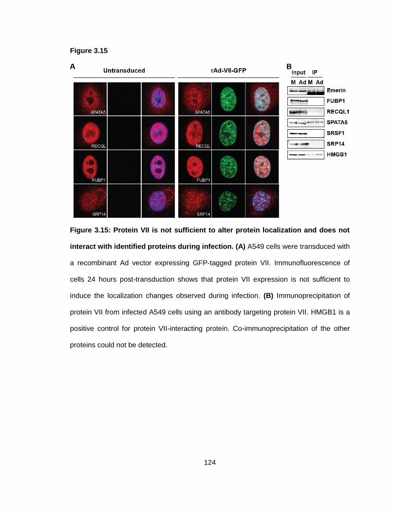
124
Figure 3.15
Figure 3.15: Protein VII is not sufficient to alter protein localization and does not
interact with identified proteins during infection. (A) A549 cells were transduced with
a recombinant Ad vector expressing GFP-tagged protein VII. Immunofluorescence of
cells 24 hours post-transduction shows that protein VII expression is not sufficient to
induce the localization changes observed during infection. (B) Immunoprecipitation of
protein VII from infected A549 cells using an antibody targeting protein VII. HMGB1 is a
positive control for protein VII-interacting protein. Co-immunoprecipitation of the other
proteins could not be detected.
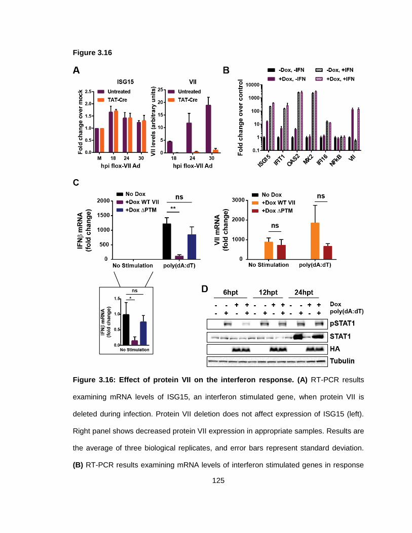
125
Figure 3.16
Figure 3.16: Effect of protein VII on the interferon response. (A) RT-PCR results
examining mRNA levels of ISG15, an interferon stimulated gene, when protein VII is
deleted during infection. Protein VII deletion does not affect expression of ISG15 (left).
Right panel shows decreased protein VII expression in appropriate samples. Results are
the average of three biological replicates, and error bars represent standard deviation.
(B) RT-PCR results examining mRNA levels of interferon stimulated genes in response
126
to ectopic treatment with type I IFN. NfkB serves as a negative control since its
expression is upstream of IFN expression, and VII verifies expression in appropriate
samples. Values are normalized to the parental, untreated sample. Type I IFN treatment
increases ISG expression, as expected. Protein VII expression does not impact ISG
expression in response to IFN treatment. Results are the average of three biological
replicates, and error bars represent standard deviation. (C) RT-PCR results showing the
effect of protein VII expression on IFN mRNA levels. A549 cells were induced for 4
days to express wild-type or PTM protein VII. Cells were transfected with poly(dA:dT)
DNA and harvested 8 hours post-transfection. IFN levels were measured by RT-PCR.
Wild-type protein VII expression suppresses IFN mRNA levels in unstimulated and
poly(dA:dT) stimulated cells. Results are the average of three biological replicates, and
error bars represent standard deviation. * = p<0.05; ** = p < 0.01; ns = not significant.
Right panel confirms protein VII expression in appropriate samples. (D) Western blot
analysis of STAT1 phosphorylation in response to poly(dA:dT) stimulation in uninduced
and induced cells. At 6 hours post-transfection of poly(dA:dT) DNA, STAT1
phosphorylation is dramatically decreased in protein VII-expressing cells compared to
uninduced controls.
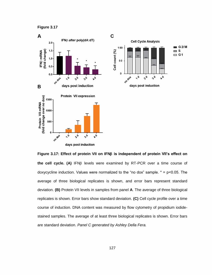
127
Figure 3.17
Figure 3.17: Effect of protein VII on IFN is independent of protein VII’s effect on
the cell cycle. (A) IFN levels were examined by RT-PCR over a time course of
doxycycline induction. Values were normalized to the “no dox” sample. * = p<0.05. The
average of three biological replicates is shown, and error bars represent standard
deviation. (B) Protein VII levels in samples from panel A. The average of three biological
replicates is shown. Error bars show standard deviation. (C) Cell cycle profile over a time
course of induction. DNA content was measured by flow cytometry of propidium iodide-
stained samples. The average of at least three biological replicates is shown. Error bars
are standard deviation. Panel C generated by Ashley Della Fera.
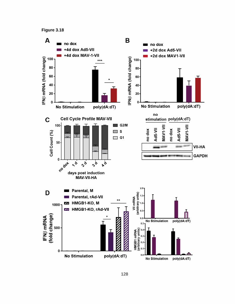
128
Figure 3.18
129
Figure 3.18: HMGB1 may contribute to protein VII-mediated IFN suppression. (A)
IFN mRNA levels in cells expressing protein VII from Ad5 or MAV-1 after 4 days of
induction. The average of three biological replicates is shown, and error bars show
standard deviation. * = p<0.05; *** = p<0.001. IFN levels are significantly higher in cells
expressing MAV-1 protein VII than Ad5 protein VII. (B) As in A, but with only 2 days of
dox induction. Western blot (bottom) confirms protein VII expression. (C) Cell cycle
profile of cells expressing MAV-1 protein VII over a time course of dox induction. DNA
content was measured by flow cytometry of propidium iodide stained cells. MAV-1 VII
expression results in accumulation of cells in G2/M after 3 days of dox treatment. The
average of three biological replicates is shown. Error bars are standard deviation. (D)
Left - The effect of protein VII on IFN mRNA levels was measured in wild-type and
HMGB1-deleted cells. Right – protein VII and HMGB1 expression. The average of three
biological replicates is shown. Error bars show standard deviation. * = p<0.05; ** =
p<0.01. Panel C generated by Ashley Della Fera.
130
Discussion
In this chapter, we demonstrate the power of a proteomics approach to identify novel
host factors associated with viral genomes and to identify novel targets of specific viral
proteins. We found that comparing the cellular proteins associated with viral DNA to
those associated with cellular DNA can be used to identify proteins that are targeted or
harnessed by viruses to promote viral processes. For example, we used this strategy to
identify TCOF1 and SLX4 as cellular proteins recruited by Ad5 to enhance viral
replication and TFII-I as a cellular protein that is targeted for degradation by Ad5 (Figure
3.2). Furthermore, we demonstrated that comparing host proteins associated with wild-
type and mutant viral genomes can be used to understand how specific viral proteins
manipulate or exploit cellular proteins. In Figure 3.3A, we demonstrated that comparison
of proteins associated with wild-type and E4-deleted Ad5 identified known E4 targets,
which validated our approach. We then compared wild-type and ICP0-deleted HSV-1
proteomes and identified potential ICP0 targets (Figure 3.3B). We conclude that iPOND-
MS is a valuable resource to identify strategies used by viruses to regulate interactions
of cellular proteins with viral genomes.
We also identified novel functions for a viral DNA-binding protein in influencing
interactions on viral and cellular genomes. We found that this small basic core protein,
called protein VII, is found at both viral and cellular genomes during infection and is
sufficient to alter the proteins associated with host chromatin. We identified the HMGB
family proteins as targets of protein VII and demonstrated that protein VII is necessary
and sufficient to sequester HMGB proteins in cellular chromatin. These data suggested
that manipulation of proteins in cellular chromatin could be a previously unexplored
strategy used by adenovirus to manipulate cellular processes. Interestingly, protein VII
produced by murine adenovirus localizes to chromatin and manipulates chromatin
131
structure, but does not sequester HMGB1. This suggests that murine HMGB1 may not
impact MAV-1 replication, or MAV-1 may employ a different strategy to manipulate or
harness HMGB1 function. It is possible that MAV-1 protein VII sequesters different
cellular proteins in chromatin to promote viral replication. It would be interesting to
identify the proteins targeted by MAV-1 protein VII to gain insight into the effects of MAV-
1 protein VII localization to chromatin.
The impact of protein VII on proteins associated with cellular chromatin led us to
investigate whether protein VII could also affect which cellular proteins associate with
viral genomes. By comparing protein VII-interacting proteins with those identified on
adenovirus genomes by iPOND, we found that several protein VII-interacting proteins
are associated with viral genomes during infection (Figure 3.8C). We therefore
hypothesized that protein VII regulates interactions of cellular proteins with viral
genomes. We utilized the iPOND strategies we had optimized to test this hypothesis.
Confirming our hypothesis, iPOND analysis of wild-type and protein VII-deleted viruses
identified several cellular proteins that are significantly enriched on viral genomes under
either wild-type or protein VII-deleted conditions. We predicted that protein VII would
recruit cellular proteins that promote viral processes, while preventing association with
anti-viral proteins. Consistent with this prediction, we found that proteins involved in DNA
replication, transcription, RNA splicing, and mRNA export were significantly more
abundant on viral genomes in the presence of protein VII. We observed that several of
these proteins localize to sites of viral DNA replication or transcription (Figure 3.13),
consistent with their association with isolated viral genomes by iPOND. Furthermore, we
demonstrated that this localization was dependent on protein VII for at least one of the
identified proteins (Figure 3.14). Future experiments will examine localization of other
identified proteins when protein VII is deleted. While several of the proteins enriched on
132
wild-type genomes co-precipitate with ectopically expressed protein VII by IP-MS, we did
not detect interaction with protein VII during Ad5 infection. Furthermore, expression of
protein VII was not sufficient to alter localization of these proteins. These data suggest
that the identified cellular proteins may not be actively recruited by protein VII. Instead,
changes to DNA conformation or accessibility may promote association of these cellular
proteins with viral genomes.
There are conflicting reports as to the effect of protein VII on viral transcription (see
Chapter 1). While some evidence suggests that protein VII-mediated DNA condensation
impairs DNA accessibility for transcription (Matsumoto et al., 1993; Okuwaki & Nagata,
1998), other reports demonstrate enhanced transcription when protein VII is added to in
vitro transcription assays (Komatsu et al., 2011). Our results indicate that protein VII
enhances the association of replication and transcription proteins with viral genomes.
This would suggest that protein VII promotes viral DNA replication and transcription. It is
important to note that our results do not allow us to determine where on the viral genome
protein VII or cellular proteins are associated. Therefore, it is possible that protein VII
and identified cellular proteins do not occupy the same regions of the genome. Protein
VII could be reorganized to condense certain regions of the genome, while
decondensing other regions to be more accessible to cellular proteins such as those we
found to be associated with viral genomes. Thus, it is possible that protein VII inhibits
transcription of some genes through DNA condensation, while promoting transcription of
genes that it does not occupy by allowing association of cellular transcription proteins
through an undefined mechanism. Curiously, we did not observe a dramatic effect on
viral DNA replication or viral protein levels when protein VII was deleted. One possible
explanation for this observation is the presence of incoming protein VII. Infection of 293-
Cre cells with flox-VII virus results in deletion of the protein VII gene from viral genomes
133
during infection, resulting in dramatically reduced levels of protein VII. However,
genomes of flox-VII virus are still packaged with protein VII, and these enter the nucleus
with viral genomes. Incoming protein VII may be sufficient to promote localization of
transcription and DNA replication proteins to early viral replication centers. Early
localization of these proteins to viral replication centers may allow these proteins to stay
in proximity to nascent viral genomes as infection progresses, even in the absence of de
novo protein VII synthesis. In such a scenario, decreased protein VII levels would lead to
significantly lower abundance of these cellular proteins on viral genomes since de novo
protein VII would not be present to promote higher levels of these proteins at viral
replication centers. However, the amount of these cellular proteins recruited early during
infection may be sufficient to allow replication and transcription to occur at near wild-type
levels. An alternative explanation could be that other cellular proteins that are not
regulated by protein VII are redundant for the functions of those proteins that are
significantly lower when protein VII is deleted.
We also examined proteins enriched on protein VII-deleted viral genomes to identify
pathways potentially targeted by protein VII. UBR4 and TRIM25 were significantly
enriched on protein VII-deleted genomes and are known to be involved in the interferon
pathway (Martin-Vicente et al., 2017; Morrison et al., 2013). We therefore investigated
whether protein VII impacted interferon signaling. We found that protein VII expression
led to significantly decreased levels of IFN mRNA in response to stimulation by
poly(dA:dT) transfection, but did not affect mRNA levels of ISGs in response to
stimulation by type I interferon. The effect of protein VII, therefore, must be upstream of
IFN production. Since HMGB1 has been suggested to promote detection of
cytoplasmic DNA by cellular sensors (Andreeva et al., 2017), we hypothesized that
protein VII-mediated sequestration of HMGB1 to host chromatin could prevent IFN
134
signaling by preventing recognition of foreign DNA. Consistent with this hypothesis, we
found that HMGB1 and localization of protein VII to chromatin may contribute to
suppression of IFN in response to poly(dA:dT) stimulation. However, we found that
protein VII expression also led to decreased IFN mRNA in unstimulated cells. This
suggests that the effect of protein VII may not be specific to poly(dA:dT) stimulation or
detection of foreign DNA. The effects of protein VII could instead be through changes to
the DNA conformation of the IFN locus, or through recruitment of transcriptional
regulators such as HMGB1. It is possible that protein VII recruits HMGB1 to repress
transcription of IFN. This is consistent with the observed increase in IFN levels in the
absence of HMGB1 (Figure 3.18D). Together, our results suggest that protein VII
suppresses IFN mRNA levels through a mechanism consistent with chromatin
localization and HMGB1. The details of this mechanism require further study (see
Chapter 4), but these data raise the possibility that protein VII could suppress host
defenses by targeting the anti-viral interferon response.
Protein VII suppression of interferon signaling represents a previously unidentified
mechanism used by adenovirus to evade this anti-viral pathway. As described in
Chapter 1, several early adenovirus proteins and VA-RNA contribute to evasion of
interferon-stimulated genes. This is the first demonstration of a late adenovirus protein in
suppressing interferon. It is interesting to speculate on the reasons a late viral protein
would need to target interferon. By the time de novo protein VII is expressed, viral DNA
replication and transcription have already initiated. Thus, suppression of interferon at this
stage would not be required for DNA replication or viral protein expression. This is
consistent with our observations that viral DNA replication is not dramatically affected by
protein VII deletion during infection (Figures 3.7, 3.9, and 3.14). Protein VII’s effect on
135
interferon may instead be required for proper viral spread. IFNis released from cells
and activates interferon signaling in neighboring cells through paracrine signaling. This
establishes anti-viral environments in activated cells that could prevent infection by
released viral particles. During late stages of infection, the virus is preparing to be
released from the cell. It would be beneficial for the virus to prevent interferon activation
in neighboring cells to allow for optimal viral spread. This may be especially important at
late stages of infection, when large amounts of accumulated viral DNA and protein could
lead to interferon activation. Therefore, the benefit of protein VII-mediated IFN
suppression may not be on viral processes within the infected cell, but rather through
promoting viral spread.
For this project, we focused our experiments on the host proteins that are involved in
processes known to be manipulated by adenovirus, such as transcription, splicing, and
interferon signaling. However, our iPOND analysis also identified proteins involved in
protein trafficking, vesicle budding, cytoskeletal organization, and metabolic processes
as differentially regulated by protein VII (Tables 3.2 and 3.3). This raises the possibility
that protein VII could manipulate these processes either directly or indirectly and could
thereby regulate cellular integrity.
Together, results from this chapter demonstrate that identifying cellular proteins
associated with adenovirus genomes can uncover host factors that facilitate or hinder
viral replication. Furthermore, comparing proteins associated with viral genomes during
infection with wild-type or mutant viruses can reveal novel targets and functions of
specific viral proteins. Here, we found that protein VII deletion affects the association of
cellular proteins with both viral and cellular genomes. Our data suggest that protein VII
may promote association of transcription, splicing, and RNA export proteins with viral
136
genomes, while suppressing anti-viral responses. Results from this chapter contribute to
our growing understanding of protein VII’s impact on multiple viral and cellular
processes, likely through regulating DNA-protein interactions.
137
CHAPTER 4:
Discussion
Summary
Successful viral propagation relies on manipulation of cellular proteins and pathways to
establish a cellular environment conducive to viral replication. Defining the mechanisms
underlying viral manipulation and understanding the outcomes of such manipulation
contribute to our comprehension of viral life cycles, as well as fundamental cellular
processes. Moreover, studying virus-host interactions can lead to improved strategies for
anti-viral therapeutics and viral vectors for gene therapy. Viruses utilize a myriad of
strategies to manipulate host cells in order to hijack cellular processes that benefit
viruses, and suppress or redirect those that impair viral growth. My thesis work focused
on understanding how adenovirus manipulates association of cellular proteins with viral
genomes. As a nuclear replicating DNA virus, adenovirus genomes are accessible to
cellular DNA-binding proteins, and adenovirus must therefore carefully regulate which
cellular proteins interact with them. In each chapter of this thesis, I discussed strategies
we used to understand how adenoviruses evade association of anti-viral cellular proteins
with viral genomes and how they promote recruitment of beneficial cellular proteins.
These approaches uncovered previously unidentified targets of viral manipulation and
mechanisms used by viruses to either target or exploit cellular proteins. In Chapter 2, I
described how comparison of evolutionary diverse adenovirus serotypes revealed
differences in the ways that viruses target a previously identified intrinsic defense. In
Chapter 3, I described how comparing the proteins associated with viral and cellular
genomes identified novel targets of viral manipulation and identified cellular proteins that
are exploited by adenovirus. Furthermore, we demonstrated that comparing proteins
between wild-type and mutant viral genomes identifies proteins manipulated by specific
138
viral proteins. These projects build on our knowledge of adenovirus and contribute to
understanding diverse mechanisms used by viruses to manipulate host cells. Our
interpretations are summarized in the discussion section of each respective chapter.
Here, I will discuss future directions to build on this work and the broader implications of
these findings.
Future directions
How does Ad9 mislocalize MRN?
Are additional viral proteins required?
In Chapter 2, we demonstrated that Ad9 infection results in mislocalization of MRN to
E4orf3-PML tracks, but expression of Ad9-E4orf3 is not sufficient to affect MRN
localization. This raises the question of what exactly changes during infection to allow for
MRN mislocalization. One possibility is that another viral protein contributes to
mislocalization. This protein could work together with E4orf3 to target MRN, or it may be
sufficient to mislocalize MRN. A potential candidate that we have begun to explore is the
Ad9-E1b55K protein. Studies with Ad5 have demonstrated that E1b55K is found at
several locations in the cell during Ad5 infection, including colocalized with E4orf3-PML
tracks. We reasoned that Ad9-E1b55K may share this localization and could recruit
MRN to these tracks. We therefore investigated the effect of Ad9-E1b55K on MRN
localization. We expressed HA-tagged Ad9-E1b55K and observed localization of MRN
by immunofluorescence. Unlike Ad5-E1b55K, which is cytoplasmic in the absence of
E4orf3 or E4orf6, Ad9-E1b55K is found in the nucleus in track-like structures (Figure
4.1). Intriguingly, we found that transfection of Ad9-E1b55K was sufficient to reorganize
MRN from a pan-nuclear distribution to track-like structures that colocalized with Ad9-
E1b55K (Figure 4.1). Initially, this suggested that Ad9-E1b55K could be sufficient to
mislocalize MRN to E4orf3 tracks. However, when we co-transfected Ad9-E1b55K and
139
Ad9-E4orf3, we found that these proteins do not colocalize (Figure 4.1). It appears that
Ad9-E1b55K can reorganize MRN but cannot recruit it to E4orf3-PML tracks. This raises
several questions about how MRN is targeted by viral proteins during Ad9 infection.
Future experiments should investigate the requirements for MRN mislocalization further.
For example, do Ad9-E1b55K and Ad9-E4orf3 colocalize during infection? If we find that
these viral proteins co-localize during infection, this would suggest that changes induced
during infection allow Ad9-E1b55K to localize with Ad9-E4orf3-PML tracks. Localization
of Ad5-E1b55K to PML is regulated by SUMOylation of E1b55K. Ad9-E1b55K
localization may be similarly regulated. It is possible that Ad9-E1b55K is sufficient to
interact with MRN but requires SUMOylation to localize to PML tracks during infection.
The observation that Ad9-E1b55K is sufficient to alter MRN localization suggests that
Ad9-E1b55K may interact with MRN. This should be determined by co-
immunoprecipitation and in vitro studies. If Ad9-E1b55K can interact with MRN, this
raises the question of why Ad9 does not degrade MRN. E1b55K has long been
considered the substrate recognition component of the ubiquitin ligase formed during
adenovirus infection. It is possible that interaction of Ad9-E1b55K with MRN components
precludes interaction with E4orf6 or cellular components of the ubiquitin ligase due to
structural changes to Ad9-E1b55K. The interaction of Ad9-E1b55K with ubiquitin ligase
proteins and with MRN may be mutually exclusive. This could be investigated by
sequential co-immunoprecipitation studies to determine whether the E1b55K that co-
precipitates with E4orf6 is associated with MRN components.
Are post-translational modifications required?
In addition to exploring the role of additional viral proteins, we have also considered the
potential role of post-translational modifications (PTMs) on E4orf3 in MRN
140
mislocalization. We hypothesized that PTMs could occur on Ad9-E4orf3 during Ad9
infection but not when Ad9-E4orf3 is expressed alone, and that these PTMs could
enable MRN mislocalization during Ad9 infection. To test this hypothesis, we generated
plasmids expressing FLAG-tagged E4orf3 from each of the serotypes in our study. We
omitted Ad2-E4orf3, as it is almost identical to Ad5-E4orf3 (99.1%). We transfected the
E4orf3 plasmids individually or combined with infection with each respective adenovirus
serotype. Immunoblotting of transfected and/or infected samples resulted in FLAG-
E4orf3 bands at the expected molecular weight of approximately 11 kDa (Figure 4.2).
We observed higher molecular weight bands (approximately 20 kDa) for samples
expressing Ad5 and Ad9-E4orf3 (Figure 4.2), which could represent post-translationally
modified E4orf3. Intriguingly, the higher molecular weight band in samples expressing
Ad9-E4orf3 intensifies during Ad9 infection (Figure 4.2). This may represent a post-
translational modification that increases upon Ad9 infection and could explain why MRN
is mislocalized during infection. Excision of these gel bands and identification of PTMs
by mass spectrometry would be an interesting future direction. Identified PTMs could be
tested by mutating the modified site in E4orf3 to determine if this affects its ability to alter
MRN localization.
How does protein VII suppress IFN levels?
In Chapter 3, we found that ectopic expression of protein VII leads to reduced IFN
mRNA levels and delayed downstream phosphorylation of STAT1. The mechanism by
which protein VII suppresses interferon signaling remains unclear and merits further
investigation. First, it should be determined whether reduced IFN mRNA levels are
caused by suppression of transcription or by mRNA instability/degradation. To test this,
luciferase assays testing activity of the IFN promoter in the presence and absence of

141
ectopic protein VII expression should be performed. In addition, the phosphorylation
status of interferon regulatory factors 3 and 9 (IRF3 and IRF9) should be examined by
western blot, since their activation is required for IFN expression. These experiments
will demonstrate whether protein VII affects IFNtranscriptional activation. To test
whether protein VII affects mRNA stability, nascent IFN transcription should be
inhibited by treating cells with the transcription inhibitor actinomycin D. The turnover rate
of IFN transcripts should be measured and compared between control cells and cells
expressing protein VII. Together, these experiments would determine whether the effect
on IFN mRNA is upstream or downstream of transcription.
We observed that the effects of MAV-1 protein VII, which does not affect HMGB1, are
less dramatic than those of Ad5 protein VII (Figure 3.18A-B). Furthermore, we found
that ectopic protein VII expression did not affect IFN levels in HMGB1 knockout cells
(Figure 3.18D), though this could be due to the decreased protein VII levels in HMGB1
knockout cells (Figure 3.18D). These observations indicate that HMGB1 could
contribute to protein VII-mediated suppression of IFN. However, it remains unclear at
which step of the interferon pathway protein VII and HMGB1 would be involved. Our
initial hypothesis was that protein VII-mediated HMGB1 sequestration could prevent
recognition of viral DNA by the cytoplasmic DNA sensor cGAS. This was based on the
recently published finding that HMGB1 could promote cGAS activation in mouse cells by
altering DNA conformation (Andreeva et al., 2017). Two observations from our
experiments suggest this hypothesis may be incorrect. The first is that IFN levels are
decreased in protein VII-expressing cells in the absence of stimulation by poly(dA:dT)
DNA (Figure 3.16C). This indicates that the effect of protein VII may not be specific to
detection of foreign DNA by sensors like cGAS. The second observation is that deletion
142
of HMGB1 leads to a rescue in IFN levels (Figure 3.18D). This demonstrates that the
protein VII-mediated suppression of IFN is relieved in the absence of HMGB1. If
HMGB1 were responsible for promoting IFN activation through cGAS detection, then
HMGB1 deletion would not rescue IFN levels. Therefore, it appears that HMGB1 may
actually repress IFNand protein VII may harness HMGB1 function rather than
inactivating it. This may represent a difference between mouse and human HMGB1,
since HMGB1 was shown to promote IFN activation in mouse cells (Andreeva et al.,
2017). Human HMGB1 is a known transcriptional regulator (Bianchi & Agresti, 2005);
therefore, it is possible that protein VII targets HMGB1 to the IFN gene locus to repress
transcription. To test this, chromatin immunoprecipitation studies with protein VII and
HMGB1 should be performed to determine if these proteins are found at genomic
regions that would regulate expression IFN.
Does protein VII bind RNA?
We identified several cellular proteins involved in RNA splicing and export as dependent
on protein VII for association with viral genomes (Table 3.2). Since our iPOND protocol
does not include RNA digestion, it is possible that these proteins are isolated due to
interactions of RNA with EdU-labeled DNA. In addition, we found that a large portion of
protein VII interacting proteins are involved in RNA processes (Figure 3.8A-B). This
leads us to hypothesize that protein VII could bind viral RNA and influence RNA
processes, such as splicing and mRNA export. Consistent with this hypothesis, we have
observed that the localization pattern of protein VII resembles that of viral RNA (Figure
4.3). Future experiments will test this hypothesis through several experiments. First,
fluorescent in situ hybridization (FISH) coupled with protein VII immunofluorescence
would demonstrate whether protein VII localizes to sites of viral RNA. Second, we will
143
determine whether protein VII associates with viral RNA by performing RNA
immunoprecipitation from infected samples. If protein VII co-precipitates viral RNA, this
would indicate that it can associate with RNA either directly or indirectly. To determine
whether protein VII can directly bind RNA, we will perform RNA electrophoretic mobility
shift assay (RNA EMSA) using purified protein VII. Protein VII interaction with viral RNA
would raise the possibility that protein VII can influence viral processes such as splicing
and mRNA export by promoting association of relevant cellular proteins. These
experiments would contribute to our growing understanding of protein VII functions.
Significance
Common cellular obstacles to adenoviruses
In each chapter of this thesis, we identified cellular proteins that are targeted by
serotypes across the adenovirus family. In Chapter 2, we demonstrated that several
serotypes target MRN by degradation, mislocalization, or by both mechanisms. In
Chapter 3, we demonstrated that several serotypes sequester HMGB1 in cellular
chromatin. Conservation across human adenovirus serotypes suggests that targeting
MRN and sequestering HMGB1 to host chromatin serve important functions during
human adenovirus infection. These observations also raise the possibility that MRN and
HMGB1 provided selective pressure for adenovirus evolution, since diverse serotypes all
evolved to target these proteins. Our finding that different adenovirus serotypes utilize
distinct mechanisms to target MRN further supports the idea that MRN provided
selective pressure for adenovirus evolution since this implies that serotypes separately
evolved to target the same cellular complex. Consistent with these theories, we found
that MRN can impair adenovirus replication and identified roles for HMGB1 in anti-viral
processes. Using an in vivo lipopolysaccharide (LPS) lung injury model, we showed that
protein VII expression in mouse lungs resulted in reduced HMGB1 secretion and
144
reduced neutrophil infiltration in response to LPS stimulation (data not shown) (Avgousti
et al., 2016). This demonstrated that sequestration of HMGB1 by protein VII could allow
adenovirus to inhibit recruitment of immune cells. In Chapter 3, we demonstrated that
protein VII can suppress interferon signaling, through a mechanism that may be
dependent on HMGB1 and localization to chromatin (Figures 3.16-3.18). As evasion of
interferon and innate immunity is critical to viral success in an in vivo setting, these
functions could explain the conservation of protein VII-mediated HMGB1 sequestration
among human adenoviruses. Together, our findings demonstrate how studying
interactions of host proteins with multiple adenoviruses can be used to identify important
cellular obstacles.
Resources to define interactions with host proteins
We identified differences in the ways that viral proteins from different adenoviruses
interact with cellular proteins. These proteins provide valuable resources that can be
used in future studies to define the requirements for interaction with host proteins. For
example, in Chapter 3, we demonstrated that Ad5 protein VII sequesters HMGB1 to
cellular chromatin. However, protein VII expressed from murine adenovirus MAV-1
localizes to chromatin but does not sequester HMGB1 in chromatin. Comparison of
protein sequences between human and murine adenoviruses would provide insight into
the residues or domains required for chromatin localization, as these would be expected
to be present in both human and murine adenovirus protein VII. Conversely, sequences
present in human Ad protein VII but not in MAV-1 protein VII are potential HMGB1-
interacting motifs. In a similar manner, results from Chapter 2 could be used to identify
requirements for interaction with MRN. We identified serotypes that cannot target MRN
through either mislocalization or degradation, and comparison with serotypes that do
degrade or mislocalize MRN could identify residues important for MRN targeting.
145
Interestingly, Ad9 mislocalizes MRN to E4orf3-PML tracks during infection, but
expression of Ad9-E4orf3 is not sufficient to alter MRN localization. It is possible that
another Ad9 viral protein is required to target MRN, in which case it would be interesting
to determine whether this protein shares any motifs with Ad5-E4orf3 that could be
required to mislocalize MRN. Another possibility is the potential role of post-translational
modifications (PTMs) on E4orf3 or MRN that could be required for MRN mislocalization.
Identifying PTMs on Ad9-E4orf3 and MRN components in the presence and absence of
infection would reveal whether Ad9-E4orf3 or MRN is differentially modified during
infection. Understanding the requirements for adenovirus proteins to target MRN or
HMGB1 could provide information to identify novel MRN or HMGB1-interacting proteins.
Cellular proteins or proteins expressed from other viruses could be examined to
determine if they contain MRN or HMGB1-interacting sequences identified from studying
adenovirus proteins.
Insights into tissue and species tropism
In Chapter 2, we used a single cell type in each experiment to examine serotypes with
diverse tissue tropisms. This experimental design allowed us to uncover differences in
interactions with MRN between these serotypes that may not be observed in their
natural cell types. It is possible that Ad9 and Ad12, which respectively cause
conjunctivitis and gastrointestinal disorder, are able to escape MRN inhibition in
conjunctival or gastrointestinal cells but not in the fibroblasts or osteosarcoma epithelial
cells used in our experiments (Cerosaletti et al., 2000; Kraakman-van der Zwet et al.,
1999). This could be due to unidentified differences in MRN levels, regulation, or activity
between cell types. Ad9 and Ad12 could potentially be used to uncover differences
between MRN from different cell types. It is possible that MRN provided selective
pressure for adenovirus evolution. Given the negative impact of MRN on adenovirus
146
replication, differences in tissue tropism between human adenovirus serotypes could be
partially due to an inability to evade MRN-mediated restriction in certain cell types. It
would also be interesting to examine the potential of HMGB1 to serve as a restriction
factor determining host tropism. Murine adenoviruses do not replicate efficiently in
human cells (Hartley & Rowe, 1960; Nguyen et al., 1999), indicating that murine
adenoviruses may fail to overcome a cellular obstacle. Interestingly, we found that MAV-
1 protein VII does not sequester HMGB1 to cellular chromatin. Since our data suggest
that HMGB1 sequestration allows human adenoviruses to suppress interferon, the
inability of MAV-1 protein VII to target HMGB1 could prevent or suppress the efficiency
of MAV-1 infection in human cells. This could influence host tropism, promoting MAV-1
infection of murine cells over human cells. To test this hypothesis, MAV-1 replication
could be examined in HMGB1-deleted human cells to determine if HMGB1 deletion
enhances MAV-1 replication. It is important to note that human and murine HMGB1 have
nearly identical protein sequences, so any differences in blocking viral infection would
indicate different cellular regulation of HMGB1 between human and mouse cells. It
would be interesting to examine whether murine HMGB1 is involved in immune signaling
and if MAV-1 employs different mechanisms to target HMGB1 in mouse cells. Together,
our results indicate that comparing adenoviruses with different tissue and species
tropism can identify potential barriers to cross-species or cross-tissue replication. This
information could be used to design adenovirus vectors for gene therapy targeted to
specific tissues.
Conclusion
Together, the work from this thesis demonstrates that adenoviruses utilize several
different strategies to regulate interactions of cellular proteins with viral genomes in order
to promote viral processes. We conclude that studying interactions of host proteins with

147
viral genomes can provide insight into virus-host interactions. Defining these interactions
has broader implications for understanding cellular processes, developing anti-viral
therapeutics or gene therapy vectors, and in understanding viral evolution.
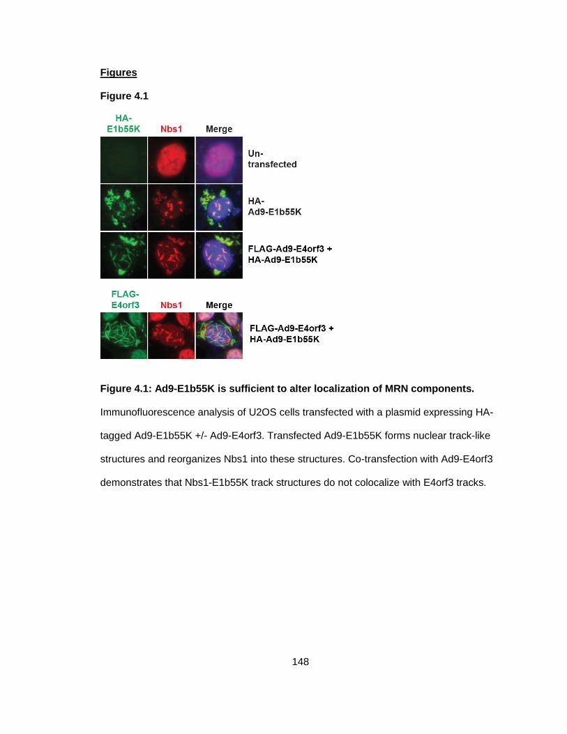
148
Figures
Figure 4.1
Figure 4.1: Ad9-E1b55K is sufficient to alter localization of MRN components.
Immunofluorescence analysis of U2OS cells transfected with a plasmid expressing HA-
tagged Ad9-E1b55K +/- Ad9-E4orf3. Transfected Ad9-E1b55K forms nuclear track-like
structures and reorganizes Nbs1 into these structures. Co-transfection with Ad9-E4orf3
demonstrates that Nbs1-E1b55K track structures do not colocalize with E4orf3 tracks.
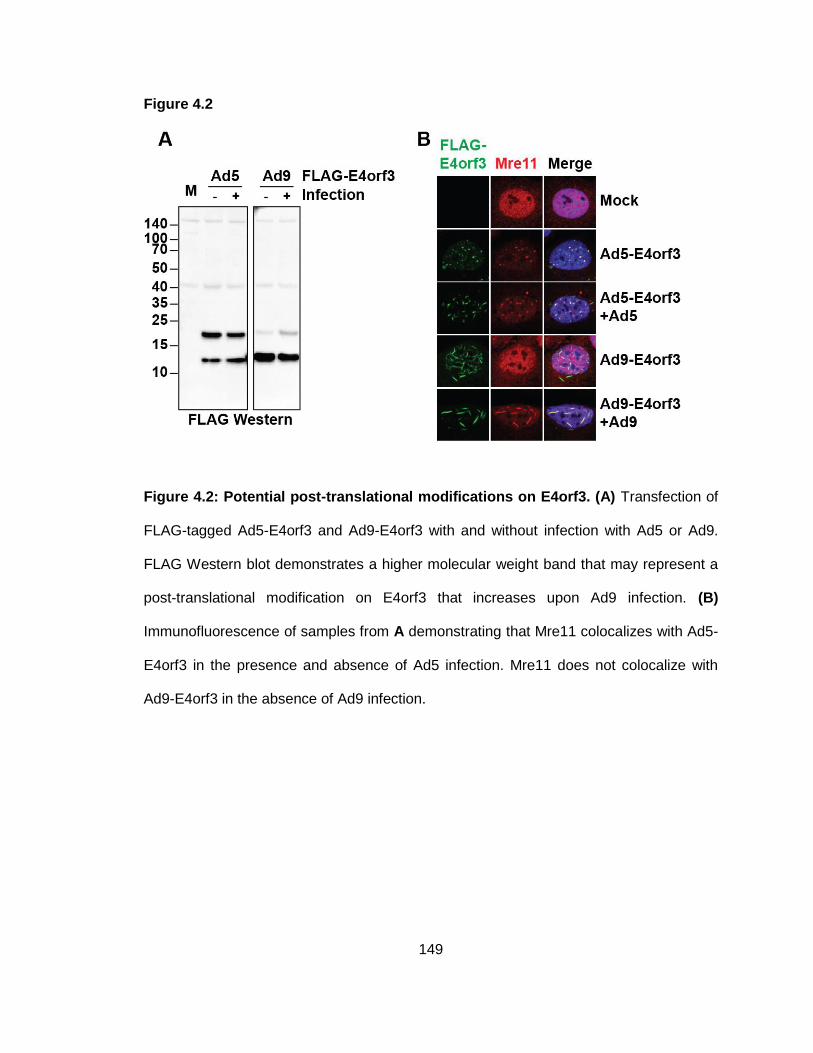
149
Figure 4.2
Figure 4.2: Potential post-translational modifications on E4orf3. (A) Transfection of
FLAG-tagged Ad5-E4orf3 and Ad9-E4orf3 with and without infection with Ad5 or Ad9.
FLAG Western blot demonstrates a higher molecular weight band that may represent a
post-translational modification on E4orf3 that increases upon Ad9 infection. (B)
Immunofluorescence of samples from A demonstrating that Mre11 colocalizes with Ad5-
E4orf3 in the presence and absence of Ad5 infection. Mre11 does not colocalize with
Ad9-E4orf3 in the absence of Ad9 infection.
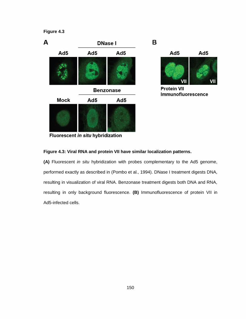
150
Figure 4.3
Figure 4.3: Viral RNA and protein VII have similar localization patterns.
(A) Fluorescent in situ hybridization with probes complementary to the Ad5 genome,
performed exactly as described in (Pombo et al., 1994). DNase I treatment digests DNA,
resulting in visualization of viral RNA. Benzonase treatment digests both DNA and RNA,
resulting in only background fluorescence. (B) Immunofluorescence of protein VII in
Ad5-infected cells.
151
BIBLIOGRAPHY
Agresti, A., & Bianchi, M. E. (2003). HMGB proteins and gene expression. Curr Opin Genet Dev, 13(2), 170-178.
Anacker, D. C., Gautam, D., Gillespie, K. A., Chappell, W. H., & Moody, C. A. (2014). Productive replication of human papillomavirus 31 requires DNA repair factor Nbs1. J Virol, 88(15), 8528-8544. doi: 10.1128/JVI.00517-14
Anderson, C. W., Baum, P. R., & Gesteland, R. F. (1973). Processing of adenovirus 2-induced proteins. J Virol, 12(2), 241-252.
Anderson, C. W., Young, M. E., & Flint, S. J. (1989). Characterization of the adenovirus 2 virion protein, mu. Virology, 172(2), 506-512.
Anderson, K. P., & Fennie, E. H. (1987). Adenovirus early region 1A modulation of interferon antiviral activity. J Virol, 61(3), 787-795.
Andreeva, L., Hiller, B., Kostrewa, D., Lassig, C., de Oliveira Mann, C. C., Jan Drexler, D., . . . Hopfner, K. P. (2017). cGAS senses long and HMGB/TFAM-bound U-turn DNA by forming protein-DNA ladders. Nature, 549(7672), 394-398. doi: 10.1038/nature23890
Araujo, F. D., Stracker, T. H., Carson, C. T., Lee, D. V., & Weitzman, M. D. (2005). Adenovirus type 5 E4orf3 protein targets the Mre11 complex to cytoplasmic aggresomes. J Virol, 79(17), 11382-11391. doi: 10.1128/JVI.79.17.11382-11391.2005
Avgousti, D. C., Della Fera, A. N., Otter, C. J., Herrmann, C., Pancholi, N. J., & Weitzman, M. D. (2017). Adenovirus core protein VII down-regulates the DNA damage response on the host genome. J Virol. doi: 10.1128/JVI.01089-17
Avgousti, D. C., Herrmann, C., Kulej, K., Pancholi, N. J., Sekulic, N., Petrescu, J., . . . Weitzman, M. D. (2016). A core viral protein binds host nucleosomes to sequester immune danger signals. Nature, 535(7610), 173-177. doi: 10.1038/nature18317
Babiss, L. E., & Ginsberg, H. S. (1984). Adenovirus type 5 early region 1b gene product is required for efficient shutoff of host protein synthesis. J Virol, 50(1), 202-212.
Bagchi, S., Raychaudhuri, P., & Nevins, J. R. (1990). Adenovirus E1A proteins can dissociate heteromeric complexes involving the E2F transcription factor: a novel mechanism for E1A trans-activation. Cell, 62(4), 659-669.
Baker, A., Rohleder, K. J., Hanakahi, L. A., & Ketner, G. (2007). Adenovirus E4 34k and E1b 55k oncoproteins target host DNA ligase IV for proteasomal degradation. J Virol, 81(13), 7034-7040. doi: 10.1128/JVI.00029-07
152
Bakkenist, C. J., & Kastan, M. B. (2003). DNA damage activates ATM through intermolecular autophosphorylation and dimer dissociation. Nature, 421(6922), 499-506. doi: 10.1038/nature01368
Bakkenist, C. J., & Kastan, M. B. (2004). Initiating cellular stress responses. Cell, 118(1), 9-17. doi: 10.1016/j.cell.2004.06.023
Banin, S., Moyal, L., Shieh, S., Taya, Y., Anderson, C. W., Chessa, L., . . . Ziv, Y. (1998). Enhanced phosphorylation of p53 by ATM in response to DNA damage. Science, 281(5383), 1674-1677.
Barbalat, R., Ewald, S. E., Mouchess, M. L., & Barton, G. M. (2011). Nucleic acid recognition by the innate immune system. Annu Rev Immunol, 29, 185-214. doi: 10.1146/annurev-immunol-031210-101340
Barber, G. N. (2011). Innate immune DNA sensing pathways: STING, AIMII and the regulation of interferon production and inflammatory responses. Curr Opin Immunol, 23(1), 10-20. doi: 10.1016/j.coi.2010.12.015
Bergelson, J. M., Cunningham, J. A., Droguett, G., Kurt-Jones, E. A., Krithivas, A., Hong, J. S., . . . Finberg, R. W. (1997). Isolation of a common receptor for Coxsackie B viruses and adenoviruses 2 and 5. Science, 275(5304), 1320-1323.
Berget, S. M., Moore, C., & Sharp, P. A. (1977). Spliced segments at the 5' terminus of adenovirus 2 late mRNA. Proc Natl Acad Sci U S A, 74(8), 3171-3175.
Berk, A. J. (2005). Recent lessons in gene expression, cell cycle control, and cell biology from adenovirus. Oncogene, 24(52), 7673-7685. doi: 10.1038/sj.onc.1209040
Berk, A. J. (2013). Adenoviridae. In D. M. Knipe, Howley, P.M. (Ed.), Fields Virology (6th ed., Vol. 2, pp. 1704-1731). Philadelphia, PA: Lippincott Williams & Wilkins.
Bianchi, M. E., & Agresti, A. (2005). HMG proteins: dynamic players in gene regulation and differentiation. Curr Opin Genet Dev, 15(5), 496-506. doi: 10.1016/j.gde.2005.08.007
Blackford, A. N., Bruton, R. K., Dirlik, O., Stewart, G. S., Taylor, A. M., Dobner, T., . . . Turnell, A. S. (2008). A role for E1B-AP5 in ATR signaling pathways during adenovirus infection. J Virol, 82(15), 7640-7652. doi: 10.1128/JVI.00170-08
Blackford, A. N., & Grand, R. J. (2009). Adenovirus E1B 55-kilodalton protein: multiple roles in viral infection and cell transformation. J Virol, 83(9), 4000-4012. doi: 10.1128/JVI.02417-08
Blackford, A. N., Patel, R. N., Forrester, N. A., Theil, K., Groitl, P., Stewart, G. S., . . . Turnell, A. S. (2010). Adenovirus 12 E4orf6 inhibits ATR activation by promoting TOPBP1 degradation. Proc Natl Acad Sci U S A, 107(27), 12251-12256. doi: 10.1073/pnas.0914605107
153
Blanchette, P., Kindsmuller, K., Groitl, P., Dallaire, F., Speiseder, T., Branton, P. E., & Dobner, T. (2008). Control of mRNA export by adenovirus E4orf6 and E1B55K proteins during productive infection requires E4orf6 ubiquitin ligase activity. J Virol, 82(6), 2642-2651. doi: 10.1128/JVI.02309-07
Blanchette, P., Wimmer, P., Dallaire, F., Cheng, C. Y., & Branton, P. E. (2013). Aggresome formation by the adenoviral protein E1B55K is not conserved among adenovirus species and is not required for efficient degradation of nuclear substrates. J Virol, 87(9), 4872-4881. doi: 10.1128/JVI.03272-12
Boyer, J., Rohleder, K., & Ketner, G. (1999). Adenovirus E4 34k and E4 11k inhibit double strand break repair and are physically associated with the cellular DNA-dependent protein kinase. Virology, 263(2), 307-312. doi: 10.1006/viro.1999.9866
Bridge, E., & Ketner, G. (1989). Redundant control of adenovirus late gene expression by early region 4. J Virol, 63(2), 631-638.
Bridges, R. G., Sohn, S. Y., Wright, J., Leppard, K. N., & Hearing, P. (2016). The Adenovirus E4-ORF3 Protein Stimulates SUMOylation of General Transcription Factor TFII-I to Direct Proteasomal Degradation. MBio, 7(1), e02184-02115. doi: 10.1128/mBio.02184-15
Burma, S., Chen, B. P., Murphy, M., Kurimasa, A., & Chen, D. J. (2001). ATM phosphorylates histone H2AX in response to DNA double-strand breaks. J Biol Chem, 276(45), 42462-42467. doi: 10.1074/jbc.C100466200
Carson, C. T., Orazio, N. I., Lee, D. V., Suh, J., Bekker-Jensen, S., Araujo, F. D., . . . Weitzman, M. D. (2009). Mislocalization of the MRN complex prevents ATR signaling during adenovirus infection. EMBO J, 28(6), 652-662. doi: 10.1038/emboj.2009.15
Carson, C. T., Schwartz, R. A., Stracker, T. H., Lilley, C. E., Lee, D. V., & Weitzman, M. D. (2003). The Mre11 complex is required for ATM activation and the G2/M checkpoint. EMBO J, 22(24), 6610-6620. doi: 10.1093/emboj/cdg630
Carvalho, T., Seeler, J. S., Ohman, K., Jordan, P., Pettersson, U., Akusjarvi, G., . . . Dejean, A. (1995). Targeting of adenovirus E1A and E4-ORF3 proteins to nuclear matrix-associated PML bodies. J Cell Biol, 131(1), 45-56.
Cerosaletti, K. M., Desai-Mehta, A., Yeo, T. C., Kraakman-Van Der Zwet, M., Zdzienicka, M. Z., & Concannon, P. (2000). Retroviral expression of the NBS1 gene in cultured Nijmegen breakage syndrome cells restores normal radiation sensitivity and nuclear focus formation. Mutagenesis, 15(3), 281-286.
Chahal, J. S., Qi, J., & Flint, S. J. (2012). The human adenovirus type 5 E1B 55 kDa protein obstructs inhibition of viral replication by type I interferon in normal human cells. PLoS Pathog, 8(8), e1002853. doi: 10.1371/journal.ppat.1002853
154
Chang, W., Folker, E. S., Worman, H. J., & Gundersen, G. G. (2013). Emerin organizes actin flow for nuclear movement and centrosome orientation in migrating fibroblasts. Mol Biol Cell, 24(24), 3869-3880. doi: 10.1091/mbc.E13-06-0307
Chapuy, B., Tikkanen, R., Muhlhausen, C., Wenzel, D., von Figura, K., & Honing, S. (2008). AP-1 and AP-3 mediate sorting of melanosomal and lysosomal membrane proteins into distinct post-Golgi trafficking pathways. Traffic, 9(7), 1157-1172. doi: 10.1111/j.1600-0854.2008.00745.x
Chardonnet, Y., & Dales, S. (1970). Early events in the interaction of adenoviruses with HeLa cells. I. Penetration of type 5 and intracellular release of the DNA genome. Virology, 40(3), 462-477.
Chatterjee, P. K., Vayda, M. E., & Flint, S. J. (1986). Identification of proteins and protein domains that contact DNA within adenovirus nucleoprotein cores by ultraviolet light crosslinking of oligonucleotides 32P-labelled in vivo. J Mol Biol, 188(1), 23-37.
Chelius, D., Huhmer, A. F., Shieh, C. H., Lehmberg, E., Traina, J. A., Slattery, T. K., & Pungor, E., Jr. (2002). Analysis of the adenovirus type 5 proteome by liquid chromatography and tandem mass spectrometry methods. J Proteome Res, 1(6), 501-513.
Chen, J., Morral, N., & Engel, D. A. (2007). Transcription releases protein VII from adenovirus chromatin. Virology, 369(2), 411-422. doi: 10.1016/j.virol.2007.08.012
Chen, P. H., Ornelles, D. A., & Shenk, T. (1993). The adenovirus L3 23-kilodalton proteinase cleaves the amino-terminal head domain from cytokeratin 18 and disrupts the cytokeratin network of HeLa cells. J Virol, 67(6), 3507-3514.
Cheng, C. Y., Gilson, T., Dallaire, F., Ketner, G., Branton, P. E., & Blanchette, P. (2011). The E4orf6/E1B55K E3 ubiquitin ligase complexes of human adenoviruses exhibit heterogeneity in composition and substrate specificity. J Virol, 85(2), 765-775. doi: 10.1128/JVI.01890-10
Cheng, C. Y., Gilson, T., Wimmer, P., Schreiner, S., Ketner, G., Dobner, T., . . . Blanchette, P. (2013). Role of E1B55K in E4orf6/E1B55K E3 ligase complexes formed by different human adenovirus serotypes. J Virol, 87(11), 6232-6245. doi: 10.1128/JVI.00384-13
Christensen, J. B., Byrd, S. A., Walker, A. K., Strahler, J. R., Andrews, P. C., & Imperiale, M. J. (2008). Presence of the adenovirus IVa2 protein at a single vertex of the mature virion. J Virol, 82(18), 9086-9093. doi: 10.1128/JVI.01024-08
Ciccia, A., & Elledge, S. J. (2010). The DNA damage response: making it safe to play with knives. Mol Cell, 40(2), 179-204. doi: 10.1016/j.molcel.2010.09.019
Ciccia, A., Huang, J. W., Izhar, L., Sowa, M. E., Harper, J. W., & Elledge, S. J. (2014). Treacher Collins syndrome TCOF1 protein cooperates with NBS1 in the DNA
155
damage response. Proc Natl Acad Sci U S A, 111(52), 18631-18636. doi: 10.1073/pnas.1422488112
Cole, J. L. (2007). Activation of PKR: an open and shut case? Trends Biochem Sci, 32(2), 57-62. doi: 10.1016/j.tibs.2006.12.003
Corbo, C., Orru, S., & Salvatore, F. (2013). SRp20: an overview of its role in human diseases. Biochem Biophys Res Commun, 436(1), 1-5. doi: 10.1016/j.bbrc.2013.05.027
Cortez, D., Wang, Y., Qin, J., & Elledge, S. J. (1999). Requirement of ATM-dependent phosphorylation of brca1 in the DNA damage response to double-strand breaks. Science, 286(5442), 1162-1166.
Cotten, M., & Weber, J. M. (1995). The adenovirus protease is required for virus entry into host cells. Virology, 213(2), 494-502. doi: 10.1006/viro.1995.0022
Cuconati, A., & White, E. (2002). Viral homologs of BCL-2: role of apoptosis in the regulation of virus infection. Genes Dev, 16(19), 2465-2478. doi: 10.1101/gad.1012702
Dales, S., & Chardonnet, Y. (1973). Early events in the interaction of adenoviruses with HeLa cells. IV. Association with microtubules and the nuclear pore complex during vectorial movement of the inoculum. Virology, 56(2), 465-483.
Dallaire, F., Blanchette, P., Groitl, P., Dobner, T., & Branton, P. E. (2009). Identification of integrin alpha3 as a new substrate of the adenovirus E4orf6/E1B 55-kilodalton E3 ubiquitin ligase complex. J Virol, 83(11), 5329-5338. doi: 10.1128/JVI.00089-09
Dani, H. M., Singh, J., & Singh, S. (2003). Advances in the structure and functions of signal recognition particle in protein targeting. J Biol Regul Homeost Agents, 17(4), 303-307.
Das, S., & Krainer, A. R. (2014). Emerging functions of SRSF1, splicing factor and oncoprotein, in RNA metabolism and cancer. Mol Cancer Res, 12(9), 1195-1204. doi: 10.1158/1541-7786.MCR-14-0131
Daugherty, M. D., & Malik, H. S. (2012). Rules of engagement: molecular insights from host-virus arms races. Annu Rev Genet, 46, 677-700. doi: 10.1146/annurev-genet-110711-155522
Davison, A. J., Benko, M., & Harrach, B. (2003). Genetic content and evolution of adenoviruses. J Gen Virol, 84(Pt 11), 2895-2908. doi: 10.1099/vir.0.19497-0
De Andrea, M., Ravera, R., Gioia, D., Gariglio, M., & Landolfo, S. (2002). The interferon system: an overview. Eur J Paediatr Neurol, 6 Suppl A, A41-46; discussion A55-48.
156
Dekker, J., Kanellopoulos, P. N., Loonstra, A. K., van Oosterhout, J. A., Leonard, K., Tucker, P. A., & van der Vliet, P. C. (1997). Multimerization of the adenovirus DNA-binding protein is the driving force for ATP-independent DNA unwinding during strand displacement synthesis. EMBO J, 16(6), 1455-1463. doi: 10.1093/emboj/16.6.1455
Dekker, J., Kanellopoulos, P. N., van Oosterhout, J. A., Stier, G., Tucker, P. A., & van der Vliet, P. C. (1998). ATP-independent DNA unwinding by the adenovirus single-stranded DNA binding protein requires a flexible DNA binding loop. J Mol Biol, 277(4), 825-838. doi: 10.1006/jmbi.1998.1652
Delacroix, S., Wagner, J. M., Kobayashi, M., Yamamoto, K., & Karnitz, L. M. (2007). The Rad9-Hus1-Rad1 (9-1-1) clamp activates checkpoint signaling via TopBP1. Genes Dev, 21(12), 1472-1477. doi: 10.1101/gad.1547007
Dey, M., Cao, C., Dar, A. C., Tamura, T., Ozato, K., Sicheri, F., & Dever, T. E. (2005). Mechanistic link between PKR dimerization, autophosphorylation, and eIF2alpha substrate recognition. Cell, 122(6), 901-913. doi: 10.1016/j.cell.2005.06.041
Dobner, T., Horikoshi, N., Rubenwolf, S., & Shenk, T. (1996). Blockage by adenovirus E4orf6 of transcriptional activation by the p53 tumor suppressor. Science, 272(5267), 1470-1473.
Doucas, V., Ishov, A. M., Romo, A., Juguilon, H., Weitzman, M. D., Evans, R. M., & Maul, G. G. (1996). Adenovirus replication is coupled with the dynamic properties of the PML nuclear structure. Genes Dev, 10(2), 196-207.
Endter, C., & Dobner, T. (2004). Cell transformation by human adenoviruses. Curr Top Microbiol Immunol, 273, 163-214.
Evans, J. D., & Hearing, P. (2003). Distinct roles of the Adenovirus E4 ORF3 protein in viral DNA replication and inhibition of genome concatenation. J Virol, 77(9), 5295-5304.
Evans, J. D., & Hearing, P. (2005). Relocalization of the Mre11-Rad50-Nbs1 complex by the adenovirus E4 ORF3 protein is required for viral replication. J Virol, 79(10), 6207-6215. doi: 10.1128/JVI.79.10.6207-6215.2005
Fekairi, S., Scaglione, S., Chahwan, C., Taylor, E. R., Tissier, A., Coulon, S., . . . Gaillard, P. H. (2009). Human SLX4 is a Holliday junction resolvase subunit that binds multiple DNA repair/recombination endonucleases. Cell, 138(1), 78-89. doi: 10.1016/j.cell.2009.06.029
Ferrier, A., Boyer, J. G., & Kothary, R. (2013). Cellular and molecular biology of neuronal dystonin. Int Rev Cell Mol Biol, 300, 85-120. doi: 10.1016/B978-0-12-405210-9.00003-5
Fitzgerald, K. A., McWhirter, S. M., Faia, K. L., Rowe, D. C., Latz, E., Golenbock, D. T., . . . Maniatis, T. (2003). IKKepsilon and TBK1 are essential components of the IRF3 signaling pathway. Nat Immunol, 4(5), 491-496. doi: 10.1038/ni921
157
Flint, S. J., & Gonzalez, R. A. (2003). Regulation of mRNA production by the adenoviral E1B 55-kDa and E4 Orf6 proteins. Curr Top Microbiol Immunol, 272, 287-330.
Fonseca, G. J., Thillainadesan, G., Yousef, A. F., Ablack, J. N., Mossman, K. L., Torchia, J., & Mymryk, J. S. (2012). Adenovirus evasion of interferon-mediated innate immunity by direct antagonism of a cellular histone posttranslational modification. Cell Host Microbe, 11(6), 597-606. doi: 10.1016/j.chom.2012.05.005
Forrester, N. A., Sedgwick, G. G., Thomas, A., Blackford, A. N., Speiseder, T., Dobner, T., . . . Grand, R. J. (2011). Serotype-specific inactivation of the cellular DNA damage response during adenovirus infection. J Virol, 85(5), 2201-2211. doi: 10.1128/JVI.01748-10
Freimuth, P., & Anderson, C. W. (1993). Human adenovirus serotype 12 virion precursors pMu and pVI are cleaved at amino-terminal and carboxy-terminal sites that conform to the adenovirus 2 endoproteinase cleavage consensus sequence. Virology, 193(1), 348-355. doi: 10.1006/viro.1993.1131
Gaggar, A., Shayakhmetov, D. M., & Lieber, A. (2003). CD46 is a cellular receptor for group B adenoviruses. Nat Med, 9(11), 1408-1412. doi: 10.1038/nm952
Gautam, D., & Bridge, E. (2013). The kinase activity of ataxia-telangiectasia mutated interferes with adenovirus E4 mutant DNA replication. J Virol, 87(15), 8687-8696. doi: 10.1128/JVI.00376-13
Giberson, A. N., Davidson, A. R., & Parks, R. J. (2012). Chromatin structure of adenovirus DNA throughout infection. Nucleic Acids Res, 40(6), 2369-2376. doi: 10.1093/nar/gkr1076
Graveley, B. R. (2000). Sorting out the complexity of SR protein functions. RNA, 6(9), 1197-1211.
Greber, U. F., Suomalainen, M., Stidwill, R. P., Boucke, K., Ebersold, M. W., & Helenius, A. (1997). The role of the nuclear pore complex in adenovirus DNA entry. EMBO J, 16(19), 5998-6007. doi: 10.1093/emboj/16.19.5998
Greber, U. F., Webster, P., Weber, J., & Helenius, A. (1996). The role of the adenovirus protease on virus entry into cells. EMBO J, 15(8), 1766-1777.
Greber, U. F., Willetts, M., Webster, P., & Helenius, A. (1993). Stepwise dismantling of adenovirus 2 during entry into cells. Cell, 75(3), 477-486.
Greer, A. E., Hearing, P., & Ketner, G. (2011). The adenovirus E4 11 k protein binds and relocalizes the cytoplasmic P-body component Ddx6 to aggresomes. Virology, 417(1), 161-168. doi: 10.1016/j.virol.2011.05.017
Gupta, A., Jha, S., Engel, D. A., Ornelles, D. A., & Dutta, A. (2013). Tip60 degradation by adenovirus relieves transcriptional repression of viral transcriptional activator EIA. Oncogene, 32(42), 5017-5025. doi: 10.1038/onc.2012.534
158
Halbert, D. N., Cutt, J. R., & Shenk, T. (1985). Adenovirus early region 4 encodes functions required for efficient DNA replication, late gene expression, and host cell shutoff. J Virol, 56(1), 250-257.
Haller, O., Kochs, G., & Weber, F. (2006). The interferon response circuit: induction and suppression by pathogenic viruses. Virology, 344(1), 119-130. doi: 10.1016/j.virol.2005.09.024
Harada, J. N., Shevchenko, A., Shevchenko, A., Pallas, D. C., & Berk, A. J. (2002). Analysis of the adenovirus E1B-55K-anchored proteome reveals its link to ubiquitination machinery. J Virol, 76(18), 9194-9206.
Harding, S. M., Benci, J. L., Irianto, J., Discher, D. E., Minn, A. J., & Greenberg, R. A. (2017). Mitotic progression following DNA damage enables pattern recognition within micronuclei. Nature, 548(7668), 466-470. doi: 10.1038/nature23470
Harper, J. W., & Elledge, S. J. (2007). The DNA damage response: ten years after. Mol Cell, 28(5), 739-745. doi: 10.1016/j.molcel.2007.11.015
Hartley, J. W., & Rowe, W. P. (1960). A new mouse virus apparently related to the adenovirus group. Virology, 11, 645-647.
Haruki, H., Gyurcsik, B., Okuwaki, M., & Nagata, K. (2003). Ternary complex formation between DNA-adenovirus core protein VII and TAF-Ibeta/SET, an acidic molecular chaperone. FEBS Lett, 555(3), 521-527.
Haruki, H., Okuwaki, M., Miyagishi, M., Taira, K., & Nagata, K. (2006). Involvement of template-activating factor I/SET in transcription of adenovirus early genes as a positive-acting factor. J Virol, 80(2), 794-801. doi: 10.1128/JVI.80.2.794-801.2006
Hayano, T., Yanagida, M., Yamauchi, Y., Shinkawa, T., Isobe, T., & Takahashi, N. (2003). Proteomic analysis of human Nop56p-associated pre-ribosomal ribonucleoprotein complexes. Possible link between Nop56p and the nucleolar protein treacle responsible for Treacher Collins syndrome. J Biol Chem, 278(36), 34309-34319. doi: 10.1074/jbc.M304304200
Hearing, P., Samulski, R. J., Wishart, W. L., & Shenk, T. (1987). Identification of a repeated sequence element required for efficient encapsidation of the adenovirus type 5 chromosome. J Virol, 61(8), 2555-2558.
Hearing, P., & Shenk, T. (1983). The adenovirus type 5 E1A transcriptional control region contains a duplicated enhancer element. Cell, 33(3), 695-703.
Herrmann, C., Avgousti, D. C., & Weitzman, M. D. (2017). Differential Salt Fractionation of Nuclei to Analyze Chromatin-associated Proteins from Cultured Mammalian Cells. Bio Protoc, 7(6).
Hickson, I., Zhao, Y., Richardson, C. J., Green, S. J., Martin, N. M., Orr, A. I., . . . Smith, G. C. (2004). Identification and characterization of a novel and specific inhibitor
159
of the ataxia-telangiectasia mutated kinase ATM. Cancer Res, 64(24), 9152-9159. doi: 10.1158/0008-5472.CAN-04-2727
Higashino, F., Pipas, J. M., & Shenk, T. (1998). Adenovirus E4orf6 oncoprotein modulates the function of the p53-related protein, p73. Proc Natl Acad Sci U S A, 95(26), 15683-15687.
Hindley, C. E., Lawrence, F. J., & Matthews, D. A. (2007). A role for transportin in the nuclear import of adenovirus core proteins and DNA. Traffic, 8(10), 1313-1322. doi: 10.1111/j.1600-0854.2007.00618.x
Hirao, A., Kong, Y. Y., Matsuoka, S., Wakeham, A., Ruland, J., Yoshida, H., . . . Mak, T. W. (2000). DNA damage-induced activation of p53 by the checkpoint kinase Chk2. Science, 287(5459), 1824-1827.
Hollingworth, R., & Grand, R. J. (2015). Modulation of DNA damage and repair pathways by human tumour viruses. Viruses, 7(5), 2542-2591. doi: 10.3390/v7052542
Horwitz, M. S., Scharff, M. D., & Maizel, J. V., Jr. (1969). Synthesis and assembly of adenovirus 2. I. Polypeptide synthesis, assembly of capsomeres, and morphogenesis of the virion. Virology, 39(4), 682-694.
Howley, P. M., & Livingston, D. M. (2009). Small DNA tumor viruses: large contributors to biomedical sciences. Virology, 384(2), 256-259. doi: 10.1016/j.virol.2008.12.006
Huang, M. M., & Hearing, P. (1989). Adenovirus early region 4 encodes two gene products with redundant effects in lytic infection. J Virol, 63(6), 2605-2615.
Ishikawa, H., & Barber, G. N. (2008). STING is an endoplasmic reticulum adaptor that facilitates innate immune signalling. Nature, 455(7213), 674-678. doi: 10.1038/nature07317
Ishikawa, H., Ma, Z., & Barber, G. N. (2009). STING regulates intracellular DNA-mediated, type I interferon-dependent innate immunity. Nature, 461(7265), 788-792. doi: 10.1038/nature08476
Jackson, S. P., & Bartek, J. (2009). The DNA-damage response in human biology and disease. Nature, 461(7267), 1071-1078. doi: 10.1038/nature08467
Jacque, J. M., & Stevenson, M. (2006). The inner-nuclear-envelope protein emerin regulates HIV-1 infectivity. Nature, 441(7093), 641-645. doi: 10.1038/nature04682
Jayaram, S., & Bridge, E. (2005). Genome concatenation contributes to the late gene expression defect of an adenovirus E4 mutant. Virology, 342(2), 286-296. doi: 10.1016/j.virol.2005.08.004
160
Jazayeri, A., Falck, J., Lukas, C., Bartek, J., Smith, G. C., Lukas, J., & Jackson, S. P. (2006). ATM- and cell cycle-dependent regulation of ATR in response to DNA double-strand breaks. Nat Cell Biol, 8(1), 37-45. doi: 10.1038/ncb1337
Jha, H. C., Banerjee, S., & Robertson, E. S. (2016). The Role of Gammaherpesviruses in Cancer Pathogenesis. Pathogens, 5(1). doi: 10.3390/pathogens5010018
Jin, L., Waterman, P. M., Jonscher, K. R., Short, C. M., Reisdorph, N. A., & Cambier, J. C. (2008). MPYS, a novel membrane tetraspanner, is associated with major histocompatibility complex class II and mediates transduction of apoptotic signals. Mol Cell Biol, 28(16), 5014-5026. doi: 10.1128/MCB.00640-08
Johnson, J. S., Osheim, Y. N., Xue, Y., Emanuel, M. R., Lewis, P. W., Bankovich, A., . . . Engel, D. A. (2004). Adenovirus protein VII condenses DNA, represses transcription, and associates with transcriptional activator E1A. J Virol, 78(12), 6459-6468. doi: 10.1128/JVI.78.12.6459-6468.2004
Jun, L., Frints, S., Duhamel, H., Herold, A., Abad-Rodrigues, J., Dotti, C., . . . Froyen, G. (2001). NXF5, a novel member of the nuclear RNA export factor family, is lost in a male patient with a syndromic form of mental retardation. Curr Biol, 11(18), 1381-1391.
Karen, K. A., & Hearing, P. (2011). Adenovirus core protein VII protects the viral genome from a DNA damage response at early times after infection. J Virol, 85(9), 4135-4142. doi: 10.1128/JVI.02540-10
Karen, K. A., Hoey, P. J., Young, C. S., & Hearing, P. (2009). Temporal regulation of the Mre11-Rad50-Nbs1 complex during adenovirus infection. J Virol, 83(9), 4565-4573. doi: 10.1128/JVI.00042-09
Kastan, M. B., & Bartek, J. (2004). Cell-cycle checkpoints and cancer. Nature, 432(7015), 316-323. doi: 10.1038/nature03097
Keating, S. E., Baran, M., & Bowie, A. G. (2011). Cytosolic DNA sensors regulating type I interferon induction. Trends Immunol, 32(12), 574-581. doi: 10.1016/j.it.2011.08.004
Khandelia, P., Yap, K., & Makeyev, E. V. (2011). Streamlined platform for short hairpin RNA interference and transgenesis in cultured mammalian cells. Proc Natl Acad Sci U S A, 108(31), 12799-12804. doi: 10.1073/pnas.1103532108
Kim, Y., Lach, F. P., Desetty, R., Hanenberg, H., Auerbach, A. D., & Smogorzewska, A. (2011). Mutations of the SLX4 gene in Fanconi anemia. Nat Genet, 43(2), 142-146. doi: 10.1038/ng.750
Kitajewski, J., Schneider, R. J., Safer, B., Munemitsu, S. M., Samuel, C. E., Thimmappaya, B., & Shenk, T. (1986). Adenovirus VAI RNA antagonizes the antiviral action of interferon by preventing activation of the interferon-induced eIF-2 alpha kinase. Cell, 45(2), 195-200.
161
Kitajewski, J., Schneider, R. J., Safer, B., & Shenk, T. (1986). An adenovirus mutant unable to express VAI RNA displays different growth responses and sensitivity to interferon in various host cell lines. Mol Cell Biol, 6(12), 4493-4498.
Komatsu, T., Haruki, H., & Nagata, K. (2011). Cellular and viral chromatin proteins are positive factors in the regulation of adenovirus gene expression. Nucleic Acids Res, 39(3), 889-901. doi: 10.1093/nar/gkq783
Komatsu, T., & Nagata, K. (2012). Replication-uncoupled histone deposition during adenovirus DNA replication. J Virol, 86(12), 6701-6711. doi: 10.1128/JVI.00380-12
Koonin, E. V., Senkevich, T. G., & Chernos, V. I. (1993). Gene A32 product of vaccinia virus may be an ATPase involved in viral DNA packaging as indicated by sequence comparisons with other putative viral ATPases. Virus Genes, 7(1), 89-94.
Kottemann, M. C., & Smogorzewska, A. (2013). Fanconi anaemia and the repair of Watson and Crick DNA crosslinks. Nature, 493(7432), 356-363. doi: 10.1038/nature11863
Kraakman-van der Zwet, M., Overkamp, W. J., Friedl, A. A., Klein, B., Verhaegh, G. W., Jaspers, N. G., . . . Zdzienicka, M. Z. (1999). Immortalization and characterization of Nijmegen Breakage syndrome fibroblasts. Mutat Res, 434(1), 17-27.
Lachmann, R. (2003). Herpes simplex virus latency. Expert Rev Mol Med, 5(29), 1-14. doi: doi:10.1017/S1462399403006975
Lakdawala, S. S., Schwartz, R. A., Ferenchak, K., Carson, C. T., McSharry, B. P., Wilkinson, G. W., & Weitzman, M. D. (2008). Differential requirements of the C terminus of Nbs1 in suppressing adenovirus DNA replication and promoting concatemer formation. J Virol, 82(17), 8362-8372. doi: 10.1128/JVI.00900-08
Latorre, I. J., Roh, M. H., Frese, K. K., Weiss, R. S., Margolis, B., & Javier, R. T. (2005). Viral oncoprotein-induced mislocalization of select PDZ proteins disrupts tight junctions and causes polarity defects in epithelial cells. J Cell Sci, 118(Pt 18), 4283-4293. doi: 10.1242/jcs.02560
Lee, J. H., & Paull, T. T. (2005). ATM activation by DNA double-strand breaks through the Mre11-Rad50-Nbs1 complex. Science, 308(5721), 551-554. doi: 10.1126/science.1108297
Leonhardt, H., Rahn, H. P., Weinzierl, P., Sporbert, A., Cremer, T., Zink, D., & Cardoso, M. C. (2000). Dynamics of DNA replication factories in living cells. J Cell Biol, 149(2), 271-280.
Lilley, C. E., Carson, C. T., Muotri, A. R., Gage, F. H., & Weitzman, M. D. (2005). DNA repair proteins affect the lifecycle of herpes simplex virus 1. Proc Natl Acad Sci U S A, 102(16), 5844-5849. doi: 10.1073/pnas.0501916102
162
Lilley, C. E., Schwartz, R. A., & Weitzman, M. D. (2007). Using or abusing: viruses and the cellular DNA damage response. Trends Microbiol, 15(3), 119-126. doi: 10.1016/j.tim.2007.01.003
Lim, D. S., Kim, S. T., Xu, B., Maser, R. S., Lin, J., Petrini, J. H., & Kastan, M. B. (2000). ATM phosphorylates p95/nbs1 in an S-phase checkpoint pathway. Nature, 404(6778), 613-617. doi: 10.1038/35007091
Lin, C. I., & Yeh, N. H. (2009). Treacle recruits RNA polymerase I complex to the nucleolus that is independent of UBF. Biochem Biophys Res Commun, 386(2), 396-401. doi: 10.1016/j.bbrc.2009.06.050
Lin, R., Heylbroeck, C., Pitha, P. M., & Hiscott, J. (1998). Virus-dependent phosphorylation of the IRF-3 transcription factor regulates nuclear translocation, transactivation potential, and proteasome-mediated degradation. Mol Cell Biol, 18(5), 2986-2996.
Liu, H., Jin, L., Koh, S. B., Atanasov, I., Schein, S., Wu, L., & Zhou, Z. H. (2010). Atomic structure of human adenovirus by cryo-EM reveals interactions among protein networks. Science, 329(5995), 1038-1043. doi: 10.1126/science.1187433
Liu, S., Cai, X., Wu, J., Cong, Q., Chen, X., Li, T., . . . Chen, Z. J. (2015). Phosphorylation of innate immune adaptor proteins MAVS, STING, and TRIF induces IRF3 activation. Science, 347(6227), aaa2630. doi: 10.1126/science.aaa2630
Liu, Y., Black, J., Kisiel, N., & Kulesz-Martin, M. F. (2000). SPAF, a new AAA-protein specific to early spermatogenesis and malignant conversion. Oncogene, 19(12), 1579-1588. doi: 10.1038/sj.onc.1203442
Lou, D. I., Kim, E. T., Meyerson, N. R., Pancholi, N. J., Mohni, K. N., Enard, D., . . . Sawyer, S. L. (2016). An Intrinsically Disordered Region of the DNA Repair Protein Nbs1 Is a Species-Specific Barrier to Herpes Simplex Virus 1 in Primates. Cell Host Microbe, 20(2), 178-188. doi: 10.1016/j.chom.2016.07.003
Lovejoy, C. A., & Cortez, D. (2009). Common mechanisms of PIKK regulation. DNA Repair (Amst), 8(9), 1004-1008. doi: 10.1016/j.dnarep.2009.04.006
Luftig, M. A. (2014). Viruses and the DNA Damage Response: Activation and Antagonism. Annu Rev Virol, 1(1), 605-625. doi: 10.1146/annurev-virology-031413-085548
Maizel, J. V., Jr., White, D. O., & Scharff, M. D. (1968). The polypeptides of adenovirus. II. Soluble proteins, cores, top components and the structure of the virion. Virology, 36(1), 126-136.
Majewski, L., Sobczak, M., Havrylov, S., Jozwiak, J., & Redowicz, M. J. (2012). Dock7: a GEF for Rho-family GTPases and a novel myosin VI-binding partner in neuronal PC12 cells. Biochem Cell Biol, 90(4), 565-574. doi: 10.1139/o2012-009
163
Markiewicz, E., Tilgner, K., Barker, N., van de Wetering, M., Clevers, H., Dorobek, M., . . . Hutchison, C. J. (2006). The inner nuclear membrane protein emerin regulates beta-catenin activity by restricting its accumulation in the nucleus. EMBO J, 25(14), 3275-3285. doi: 10.1038/sj.emboj.7601230
Martin-Vicente, M., Medrano, L. M., Resino, S., Garcia-Sastre, A., & Martinez, I. (2017). TRIM25 in the Regulation of the Antiviral Innate Immunity. Front Immunol, 8, 1187. doi: 10.3389/fimmu.2017.01187
Mathew, S. S., & Bridge, E. (2007). The cellular Mre11 protein interferes with adenovirus E4 mutant DNA replication. Virology, 365(2), 346-355. doi: 10.1016/j.virol.2007.03.049
Mathew, S. S., & Bridge, E. (2008). Nbs1-dependent binding of Mre11 to adenovirus E4 mutant viral DNA is important for inhibiting DNA replication. Virology, 374(1), 11-22. doi: 10.1016/j.virol.2007.12.034
Mathews, M. B., & Shenk, T. (1991). Adenovirus virus-associated RNA and translation control. J Virol, 65(11), 5657-5662.
Matsumoto, K., Nagata, K., Ui, M., & Hanaoka, F. (1993). Template activating factor I, a novel host factor required to stimulate the adenovirus core DNA replication. J Biol Chem, 268(14), 10582-10587.
Matsuoka, S., Huang, M., & Elledge, S. J. (1998). Linkage of ATM to cell cycle regulation by the Chk2 protein kinase. Science, 282(5395), 1893-1897.
McElwee, M., Beilstein, F., Labetoulle, M., Rixon, F. J., & Pasdeloup, D. (2013). Dystonin/BPAG1 promotes plus-end-directed transport of herpes simplex virus 1 capsids on microtubules during entry. J Virol, 87(20), 11008-11018. doi: 10.1128/JVI.01633-13
Miller, D. L., Rickards, B., Mashiba, M., Huang, W., & Flint, S. J. (2009). The adenoviral E1B 55-kilodalton protein controls expression of immune response genes but not p53-dependent transcription. J Virol, 83(8), 3591-3603. doi: 10.1128/JVI.02269-08
Moody, C. A., & Laimins, L. A. (2010). Human papillomavirus oncoproteins: pathways to transformation. Nat Rev Cancer, 10(8), 550-560. doi: 10.1038/nrc2886
Morrison, J., Laurent-Rolle, M., Maestre, A. M., Rajsbaum, R., Pisanelli, G., Simon, V., . . . Garcia-Sastre, A. (2013). Dengue virus co-opts UBR4 to degrade STAT2 and antagonize type I interferon signaling. PLoS Pathog, 9(3), e1003265. doi: 10.1371/journal.ppat.1003265
Murray, D. H., Jahnel, M., Lauer, J., Avellaneda, M. J., Brouilly, N., Cezanne, A., . . . Zerial, M. (2016). An endosomal tether undergoes an entropic collapse to bring vesicles together. Nature, 537(7618), 107-111. doi: 10.1038/nature19326
164
Nakamura, H., Morita, T., & Sato, C. (1986). Structural organizations of replicon domains during DNA synthetic phase in the mammalian nucleus. Exp Cell Res, 165(2), 291-297.
Nevins, J. R., Ginsberg, H. S., Blanchard, J. M., Wilson, M. C., & Darnell, J. E., Jr. (1979). Regulation of the primary expression of the early adenovirus transcription units. J Virol, 32(3), 727-733.
Nguyen, T., Nery, J., Joseph, S., Rocha, C., Carney, G., Spindler, K., & Villarreal, L. (1999). Mouse adenovirus (MAV-1) expression in primary human endothelial cells and generation of a full-length infectious plasmid. Gene Ther, 6(7), 1291-1297. doi: 10.1038/sj.gt.3300949
Nichols, G. J., Schaack, J., & Ornelles, D. A. (2009). Widespread phosphorylation of histone H2AX by species C adenovirus infection requires viral DNA replication. J Virol, 83(12), 5987-5998. doi: 10.1128/JVI.00091-09
Nick McElhinny, S. A., Snowden, C. M., McCarville, J., & Ramsden, D. A. (2000). Ku recruits the XRCC4-ligase IV complex to DNA ends. Mol Cell Biol, 20(9), 2996-3003.
Okuwaki, M., & Nagata, K. (1998). Template activating factor-I remodels the chromatin structure and stimulates transcription from the chromatin template. J Biol Chem, 273(51), 34511-34518.
Orazio, N. I., Naeger, C. M., Karlseder, J., & Weitzman, M. D. (2011). The adenovirus E1b55K/E4orf6 complex induces degradation of the Bloom helicase during infection. J Virol, 85(4), 1887-1892. doi: 10.1128/JVI.02134-10
Orjalo, A. V., Arnaoutov, A., Shen, Z., Boyarchuk, Y., Zeitlin, S. G., Fontoura, B., . . . Forbes, D. J. (2006). The Nup107-160 nucleoporin complex is required for correct bipolar spindle assembly. Mol Biol Cell, 17(9), 3806-3818. doi: 10.1091/mbc.E05-11-1061
Ostapchuk, P., & Hearing, P. (2005). Control of adenovirus packaging. J Cell Biochem, 96(1), 25-35. doi: 10.1002/jcb.20523
Ostapchuk, P., Suomalainen, M., Zheng, Y., Boucke, K., Greber, U. F., & Hearing, P. (2017). The adenovirus major core protein VII is dispensable for virion assembly but is essential for lytic infection. PLoS Pathog, 13(6), e1006455. doi: 10.1371/journal.ppat.1006455
Ostapchuk, P., Yang, J., Auffarth, E., & Hearing, P. (2005). Functional interaction of the adenovirus IVa2 protein with adenovirus type 5 packaging sequences. J Virol, 79(5), 2831-2838. doi: 10.1128/JVI.79.5.2831-2838.2005
Ou, H. D., Kwiatkowski, W., Deerinck, T. J., Noske, A., Blain, K. Y., Land, H. S., . . . O'Shea, C. C. (2012). A structural basis for the assembly and functions of a viral polymer that inactivates multiple tumor suppressors. Cell, 151(2), 304-319. doi: 10.1016/j.cell.2012.08.035
165
Paull, T. T., & Lee, J. H. (2005). The Mre11/Rad50/Nbs1 complex and its role as a DNA double-strand break sensor for ATM. Cell Cycle, 4(6), 737-740. doi: 10.4161/cc.4.6.1715
Peitz, M., Pfannkuche, K., Rajewsky, K., & Edenhofer, F. (2002). Ability of the hydrophobic FGF and basic TAT peptides to promote cellular uptake of recombinant Cre recombinase: a tool for efficient genetic engineering of mammalian genomes. Proc Natl Acad Sci U S A, 99(7), 4489-4494. doi: 10.1073/pnas.032068699
Pelka, P., Ablack, J. N., Torchia, J., Turnell, A. S., Grand, R. J., & Mymryk, J. S. (2009). Transcriptional control by adenovirus E1A conserved region 3 via p300/CBP. Nucleic Acids Res, 37(4), 1095-1106. doi: 10.1093/nar/gkn1057
Philipson, L., Lonberg-Holm, K., & Pettersson, U. (1968). Virus-receptor interaction in an adenovirus system. J Virol, 2(10), 1064-1075.
Pike, A. C., Gomathinayagam, S., Swuec, P., Berti, M., Zhang, Y., Schnecke, C., . . . Vindigni, A. (2015). Human RECQ1 helicase-driven DNA unwinding, annealing, and branch migration: insights from DNA complex structures. Proc Natl Acad Sci U S A, 112(14), 4286-4291. doi: 10.1073/pnas.1417594112
Pipas, J. M. (2009). SV40: Cell transformation and tumorigenesis. Virology, 384(2), 294-303. doi: 10.1016/j.virol.2008.11.024
Polo, S. E., & Jackson, S. P. (2011). Dynamics of DNA damage response proteins at DNA breaks: a focus on protein modifications. Genes Dev, 25(5), 409-433. doi: 10.1101/gad.2021311
Pombo, A., Ferreira, J., Bridge, E., & Carmo-Fonseca, M. (1994). Adenovirus replication and transcription sites are spatially separated in the nucleus of infected cells. EMBO J, 13(21), 5075-5085.
Puvion-Dutilleul, F., & Puvion, E. (1990a). Analysis by in situ hybridization and autoradiography of sites of replication and storage of single- and double-stranded adenovirus type 5 DNA in lytically infected HeLa cells. J Struct Biol, 103(3), 280-289.
Puvion-Dutilleul, F., & Puvion, E. (1990b). Replicating single-stranded adenovirus type 5 DNA molecules accumulate within well-delimited intranuclear areas of lytically infected HeLa cells. Eur J Cell Biol, 52(2), 379-388.
Querido, E., Blanchette, P., Yan, Q., Kamura, T., Morrison, M., Boivin, D., . . . Branton, P. E. (2001). Degradation of p53 by adenovirus E4orf6 and E1B55K proteins occurs via a novel mechanism involving a Cullin-containing complex. Genes Dev, 15(23), 3104-3117. doi: 10.1101/gad.926401
Querido, E., Marcellus, R. C., Lai, A., Charbonneau, R., Teodoro, J. G., Ketner, G., & Branton, P. E. (1997). Regulation of p53 levels by the E1B 55-kilodalton protein and E4orf6 in adenovirus-infected cells. J Virol, 71(5), 3788-3798.
166
Querido, E., Morrison, M. R., Chu-Pham-Dang, H., Thirlwell, S. W., Boivin, D., & Branton, P. E. (2001). Identification of three functions of the adenovirus e4orf6 protein that mediate p53 degradation by the E4orf6-E1B55K complex. J Virol, 75(2), 699-709. doi: 10.1128/JVI.75.2.699-709.2001
Reddy, V. S., Natchiar, S. K., Stewart, P. L., & Nemerow, G. R. (2010). Crystal structure of human adenovirus at 3.5 A resolution. Science, 329(5995), 1071-1075. doi: 10.1126/science.1187292
Reyes, E. D., Kulej, K., Pancholi, N. J., Akhtar, L. N., Avgousti, D. C., Kim, E. T., . . . Weitzman, M. D. (2017). Identifying Host Factors Associated with DNA Replicated During Virus Infection. Mol Cell Proteomics, 16(12), 2079-2097. doi: 10.1074/mcp.M117.067116
Rogakou, E. P., Boon, C., Redon, C., & Bonner, W. M. (1999). Megabase chromatin domains involved in DNA double-strand breaks in vivo. J Cell Biol, 146(5), 905-916.
Rowe, W. P., Huebner, R. J., Gilmore, L. K., Parrott, R. H., & Ward, T. G. (1953). Isolation of a cytopathogenic agent from human adenoids undergoing spontaneous degeneration in tissue culture. Proc Soc Exp Biol Med, 84(3), 570-573.
Roy, A. L. (2012). Biochemistry and biology of the inducible multifunctional transcription factor TFII-I: 10 years later. Gene, 492(1), 32-41. doi: 10.1016/j.gene.2011.10.030
Russell, W. C., Laver, W. G., & Sanderson, P. J. (1968). Internal components of adenovirus. Nature, 219(5159), 1127-1130.
Ryan, E. L., Hollingworth, R., & Grand, R. J. (2016). Activation of the DNA Damage Response by RNA Viruses. Biomolecules, 6(1), 2. doi: 10.3390/biom6010002
Salic, A., & Mitchison, T. J. (2008). A chemical method for fast and sensitive detection of DNA synthesis in vivo. Proc Natl Acad Sci U S A, 105(7), 2415-2420. doi: 10.1073/pnas.0712168105
Sarnow, P., Ho, Y. S., Williams, J., & Levine, A. J. (1982). Adenovirus E1b-58kd tumor antigen and SV40 large tumor antigen are physically associated with the same 54 kd cellular protein in transformed cells. Cell, 28(2), 387-394.
Sato, M., Tanaka, N., Hata, N., Oda, E., & Taniguchi, T. (1998). Involvement of the IRF family transcription factor IRF-3 in virus-induced activation of the IFN-beta gene. FEBS Lett, 425(1), 112-116.
Schwartz, R. A., Lakdawala, S. S., Eshleman, H. D., Russell, M. R., Carson, C. T., & Weitzman, M. D. (2008). Distinct requirements of adenovirus E1b55K protein for degradation of cellular substrates. J Virol, 82(18), 9043-9055. doi: 10.1128/JVI.00925-08
167
Shah, G. A., & O'Shea, C. C. (2015). Viral and Cellular Genomes Activate Distinct DNA Damage Responses. Cell, 162(5), 987-1002. doi: 10.1016/j.cell.2015.07.058
Sharma, S., tenOever, B. R., Grandvaux, N., Zhou, G. P., Lin, R., & Hiscott, J. (2003). Triggering the interferon antiviral response through an IKK-related pathway. Science, 300(5622), 1148-1151. doi: 10.1126/science.1081315
Shaw, A. R., & Ziff, E. B. (1980). Transcripts from the adenovirus-2 major late promoter yield a single early family of 3' coterminal mRNAs and five late families. Cell, 22(3), 905-916.
Sirbu, B. M., McDonald, W. H., Dungrawala, H., Badu-Nkansah, A., Kavanaugh, G. M., Chen, Y., . . . Cortez, D. (2013). Identification of proteins at active, stalled, and collapsed replication forks using isolation of proteins on nascent DNA (iPOND) coupled with mass spectrometry. J Biol Chem, 288(44), 31458-31467. doi: 10.1074/jbc.M113.511337
SivaRaman, L., & Thimmappaya, B. (1987). Two promoter-specific host factors interact with adjacent sequences in an EIA-inducible adenovirus promoter. Proc Natl Acad Sci U S A, 84(17), 6112-6116.
Smart, J. E., & Stillman, B. W. (1982). Adenovirus terminal protein precursor. Partial amino acid sequence and the site of covalent linkage to virus DNA. J Biol Chem, 257(22), 13499-13506.
Smith, M. C., Boutell, C., & Davido, D. J. (2011). HSV-1 ICP0: paving the way for viral replication. Future Virol, 6(4), 421-429. doi: 10.2217/fvl.11.24
Sohn, S. Y., & Hearing, P. (2012). Adenovirus regulates sumoylation of Mre11-Rad50-Nbs1 components through a paralog-specific mechanism. J Virol, 86(18), 9656-9665. doi: 10.1128/JVI.01273-12
Soria, C., Estermann, F. E., Espantman, K. C., & O'Shea, C. C. (2010). Heterochromatin silencing of p53 target genes by a small viral protein. Nature, 466(7310), 1076-1081. doi: 10.1038/nature09307
Steegenga, W. T., Riteco, N., Jochemsen, A. G., Fallaux, F. J., & Bos, J. L. (1998). The large E1B protein together with the E4orf6 protein target p53 for active degradation in adenovirus infected cells. Oncogene, 16(3), 349-357. doi: 10.1038/sj.onc.1201540
Steegenga, W. T., Shvarts, A., Riteco, N., Bos, J. L., & Jochemsen, A. G. (1999). Distinct regulation of p53 and p73 activity by adenovirus E1A, E1B, and E4orf6 proteins. Mol Cell Biol, 19(5), 3885-3894.
Stracker, T. H., Carson, C. T., & Weitzman, M. D. (2002). Adenovirus oncoproteins inactivate the Mre11-Rad50-NBS1 DNA repair complex. Nature, 418(6895), 348-352. doi: 10.1038/nature00863
168
Stracker, T. H., Lee, D. V., Carson, C. T., Araujo, F. D., Ornelles, D. A., & Weitzman, M. D. (2005). Serotype-specific reorganization of the Mre11 complex by adenoviral E4orf3 proteins. J Virol, 79(11), 6664-6673. doi: 10.1128/JVI.79.11.6664-6673.2005
Stros, M. (2010). HMGB proteins: interactions with DNA and chromatin. Biochim Biophys Acta, 1799(1-2), 101-113. doi: 10.1016/j.bbagrm.2009.09.008
Sun, W., Li, Y., Chen, L., Chen, H., You, F., Zhou, X., . . . Jiang, Z. (2009). ERIS, an endoplasmic reticulum IFN stimulator, activates innate immune signaling through dimerization. Proc Natl Acad Sci U S A, 106(21), 8653-8658. doi: 10.1073/pnas.0900850106
Sun, Y., Jiang, X., Chen, S., Fernandes, N., & Price, B. D. (2005). A role for the Tip60 histone acetyltransferase in the acetylation and activation of ATM. Proc Natl Acad Sci U S A, 102(37), 13182-13187. doi: 10.1073/pnas.0504211102
Svendsen, J. M., Smogorzewska, A., Sowa, M. E., O'Connell, B. C., Gygi, S. P., Elledge, S. J., & Harper, J. W. (2009). Mammalian BTBD12/SLX4 assembles a Holliday junction resolvase and is required for DNA repair. Cell, 138(1), 63-77. doi: 10.1016/j.cell.2009.06.030
Tanaka, A. J., Cho, M. T., Millan, F., Juusola, J., Retterer, K., Joshi, C., . . . Chung, W. K. (2015). Mutations in SPATA5 Are Associated with Microcephaly, Intellectual Disability, Seizures, and Hearing Loss. Am J Hum Genet, 97(3), 457-464. doi: 10.1016/j.ajhg.2015.07.014
Tauber, B., & Dobner, T. (2001). Molecular regulation and biological function of adenovirus early genes: the E4 ORFs. Gene, 278(1-2), 1-23.
Telkoparan, P., Erkek, S., Yaman, E., Alotaibi, H., Bayik, D., & Tazebay, U. H. (2013). Coiled-coil domain containing protein 124 is a novel centrosome and midbody protein that interacts with the Ras-guanine nucleotide exchange factor 1B and is involved in cytokinesis. PLoS One, 8(7), e69289. doi: 10.1371/journal.pone.0069289
Thimmappaya, B., Weinberger, C., Schneider, R. J., & Shenk, T. (1982). Adenovirus VAI RNA is required for efficient translation of viral mRNAs at late times after infection. Cell, 31(3 Pt 2), 543-551.
Thomas, G. P., & Mathews, M. B. (1980). DNA replication and the early to late transition in adenovirus infection. Cell, 22(2 Pt 2), 523-533.
Tollefson, A. E., Ryerse, J. S., Scaria, A., Hermiston, T. W., & Wold, W. S. (1996). The E3-11.6-kDa adenovirus death protein (ADP) is required for efficient cell death: characterization of cells infected with adp mutants. Virology, 220(1), 152-162. doi: 10.1006/viro.1996.0295
Tollefson, A. E., Scaria, A., Hermiston, T. W., Ryerse, J. S., Wold, L. J., & Wold, W. S. (1996). The adenovirus death protein (E3-11.6K) is required at very late stages
169
of infection for efficient cell lysis and release of adenovirus from infected cells. J Virol, 70(4), 2296-2306.
Trentin, J. J., Yabe, Y., & Taylor, G. (1962). The quest for human cancer viruses. Science, 137(3533), 835-841.
Turnell, A. S., & Grand, R. J. (2012). DNA viruses and the cellular DNA-damage response. J Gen Virol, 93(Pt 10), 2076-2097. doi: 10.1099/vir.0.044412-0
Ullman, A. J., & Hearing, P. (2008). Cellular proteins PML and Daxx mediate an innate antiviral defense antagonized by the adenovirus E4 ORF3 protein. J Virol, 82(15), 7325-7335. doi: 10.1128/JVI.00723-08
Ullman, A. J., Reich, N. C., & Hearing, P. (2007). Adenovirus E4 ORF3 protein inhibits the interferon-mediated antiviral response. J Virol, 81(9), 4744-4752. doi: 10.1128/JVI.02385-06
Van der Vliet, P. C. (1995). Adenovirus DNA replication. Curr Top Microbiol Immunol, 199 ( Pt 2), 1-30.
van der Vliet, P. C., & Levine, A. J. (1973). DNA-binding proteins specific for cells infected by adenovirus. Nat New Biol, 246(154), 170-174.
van Oostrum, J., & Burnett, R. M. (1985). Molecular composition of the adenovirus type 2 virion. J Virol, 56(2), 439-448.
Vasu, S., Shah, S., Orjalo, A., Park, M., Fischer, W. H., & Forbes, D. J. (2001). Novel vertebrate nucleoporins Nup133 and Nup160 play a role in mRNA export. J Cell Biol, 155(3), 339-354. doi: 10.1083/jcb.200108007
Velicer, L. F., & Ginsberg, H. S. (1970). Synthesis, transport, and morphogenesis of type adenovirus capsid proteins. J Virol, 5(3), 338-352.
Walters, R. W., Freimuth, P., Moninger, T. O., Ganske, I., Zabner, J., & Welsh, M. J. (2002). Adenovirus fiber disrupts CAR-mediated intercellular adhesion allowing virus escape. Cell, 110(6), 789-799.
Wathelet, M. G., Lin, C. H., Parekh, B. S., Ronco, L. V., Howley, P. M., & Maniatis, T. (1998). Virus infection induces the assembly of coordinately activated transcription factors on the IFN-beta enhancer in vivo. Mol Cell, 1(4), 507-518.
Weber, J. M. (2007). Synthesis and assay of recombinant adenovirus protease. Methods Mol Med, 131, 251-255.
Weiden, M. D., & Ginsberg, H. S. (1994). Deletion of the E4 region of the genome produces adenovirus DNA concatemers. Proc Natl Acad Sci U S A, 91(1), 153-157.
Weitzman, M. D. (2005). Functions of the adenovirus E4 proteins and their impact on viral vectors. Front Biosci, 10, 1106-1117.
170
Weitzman, M. D., Fisher, K. J., & Wilson, J. M. (1996). Recruitment of wild-type and recombinant adeno-associated virus into adenovirus replication centers. J Virol, 70(3), 1845-1854.
Winberg, G., & Shenk, T. (1984). Dissection of overlapping functions within the adenovirus type 5 E1A gene. EMBO J, 3(8), 1907-1912.
Wu, X., Avni, D., Chiba, T., Yan, F., Zhao, Q., Lin, Y., . . . Livingston, D. (2004). SV40 T antigen interacts with Nbs1 to disrupt DNA replication control. Genes Dev, 18(11), 1305-1316. doi: 10.1101/gad.1182804
Xue, Y., Johnson, J. S., Ornelles, D. A., Lieberman, J., & Engel, D. A. (2005). Adenovirus protein VII functions throughout early phase and interacts with cellular proteins SET and pp32. J Virol, 79(4), 2474-2483. doi: 10.1128/JVI.79.4.2474-2483.2005
Yanai, H., Ban, T., Wang, Z., Choi, M. K., Kawamura, T., Negishi, H., . . . Taniguchi, T. (2009). HMGB proteins function as universal sentinels for nucleic-acid-mediated innate immune responses. Nature, 462(7269), 99-103. doi: 10.1038/nature08512
Yoneyama, M., Suhara, W., Fukuhara, Y., Fukuda, M., Nishida, E., & Fujita, T. (1998). Direct triggering of the type I interferon system by virus infection: activation of a transcription factor complex containing IRF-3 and CBP/p300. EMBO J, 17(4), 1087-1095. doi: 10.1093/emboj/17.4.1087
Zhang, F., Romano, P. R., Nagamura-Inoue, T., Tian, B., Dever, T. E., Mathews, M. B., . . . Hinnebusch, A. G. (2001). Binding of double-stranded RNA to protein kinase PKR is required for dimerization and promotes critical autophosphorylation events in the activation loop. J Biol Chem, 276(27), 24946-24958. doi: 10.1074/jbc.M102108200
Zhang, J., & Chen, Q. M. (2013). Far upstream element binding protein 1: a commander of transcription, translation and beyond. Oncogene, 32(24), 2907-2916. doi: 10.1038/onc.2012.350
Zhong, B., Yang, Y., Li, S., Wang, Y. Y., Li, Y., Diao, F., . . . Shu, H. B. (2008). The adaptor protein MITA links virus-sensing receptors to IRF3 transcription factor activation. Immunity, 29(4), 538-550. doi: 10.1016/j.immuni.2008.09.003
Ziv, Y., Bar-Shira, A., Pecker, I., Russell, P., Jorgensen, T. J., Tsarfati, I., & Shiloh, Y. (1997). Recombinant ATM protein complements the cellular A-T phenotype. Oncogene, 15(2), 159-167. doi: 10.1038/sj.onc.1201319
Ziv, Y., Jaspers, N. G., Etkin, S., Danieli, T., Trakhtenbrot, L., Amiel, A., . . . Shiloh, Y. (1989). Cellular and molecular characteristics of an immortalized ataxia-telangiectasia (group AB) cell line. Cancer Res, 49(9), 2495-2501.
Zou, L., & Elledge, S. J. (2003). Sensing DNA damage through ATRIP recognition of RPA-ssDNA complexes. Science, 300(5625), 1542-1548. doi: 10.1126/science.1083430.
Related Documents