Adeno-Associated Virus-Mediated Microdystrophin Expression Protects Young mdx Muscle from Contraction-Induced Injury Mingju Liu 1,* , Yongping Yue 1,* , Scott Q. Harper 2,† , Robert W. Grange 3 , Jeffrey S. Chamberlain 2 , and Dongsheng Duan 1,‡ 1Department of Molecular Microbiology and Immunology, University of Missouri School of Medicine, One Hospital Drive, Room M610G, MSB, Columbia, MO 65212, USA 2Department of Neurology, University of Washington School of Medicine, Seattle, WA 98195, USA 3Department of Human Nutrition, Foods, and Exercise, Virginia Polytechnic Institute and State University, Blacksburg, VA 24061, USA Abstract Duchenne muscular dystrophy (DMD) is the most common inherited lethal muscle degenerative disease. Currently there is no cure. Highly abbreviated microdystrophin cDNAs were developed recently for adeno-associated virus (AAV)-mediated DMD gene therapy. Among these, a C-terminal- truncated ΔR4-R23/ΔC microgene (ΔR4/ΔC) has been considered as a very promising therapeutic candidate gene. In this study, we packaged a CMV.ΔR4/ΔC cassette in AAV-5 and evaluated the transduction and muscle contractile profiles in the extensor digitorum longus muscles of young (7- week-old) and adult (9-month-old) mdx mice. At ∼3 months post-gene transfer, 50–60% of the total myofibers were transduced in young mdx muscle and the percentage of centrally nucleated myofibers was reduced from ∼70% in untreated mdx muscle to ∼22% in microdystrophin-treated muscle. Importantly, this level of transduction protected mdx muscle from eccentric contraction-induced damage. In contrast, adult mdx muscle was more resistant to AAV-5 transduction, as only ∼30% of the myofibers were transduced at 3 months postinfection. This transduction yielded marginal protection against eccentric contraction-induced injury. The extent of central nucleation was also more difficult to reverse in adult mdx muscle (from ∼83% in untreated to ∼58% in treated). Finally, we determined that the ΔR4/ΔC microdystrophin did not significantly alter the expression pattern of the endogenous full-length dystrophin in normal muscle. Neither did it have any adverse effects on normal muscle morphology or contractility. Taken together, our results suggest that AAV-mediated ΔR4/ΔC microdystrophin expression represents a promising approach to rescue muscular dystrophy in young mdx skeletal muscle. Keywords Duchenne muscular dystrophy; mdx; adeno-associated virus; microdystrophin; muscle contraction Introduction Duchenne muscular dystrophy (DMD) is the most common inherited lethal muscle wasting disease. This X-linked disorder affects 0.02–0.03% of newborn boys worldwide [1]. Affected * These authors contributed equally to this research project. † Current address: Department of Internal Medicine, University of Iowa, Iowa City, IA, USA. ‡ To whom correspondence and reprint requests should be addressed. Fax: (573) 882 4287. E-mail: [email protected]. NIH Public Access Author Manuscript Mol Ther. Author manuscript; available in PMC 2008 November 8. Published in final edited form as: Mol Ther. 2005 February ; 11(2): 245–256. doi:10.1016/j.ymthe.2004.09.013. NIH-PA Author Manuscript NIH-PA Author Manuscript NIH-PA Author Manuscript

Welcome message from author
This document is posted to help you gain knowledge. Please leave a comment to let me know what you think about it! Share it to your friends and learn new things together.
Transcript

Adeno-Associated Virus-Mediated Microdystrophin ExpressionProtects Young mdx Muscle from Contraction-Induced Injury
Mingju Liu1,*, Yongping Yue1,*, Scott Q. Harper2,†, Robert W. Grange3, Jeffrey S.Chamberlain2, and Dongsheng Duan1,‡
1Department of Molecular Microbiology and Immunology, University of Missouri School of Medicine, OneHospital Drive, Room M610G, MSB, Columbia, MO 65212, USA
2Department of Neurology, University of Washington School of Medicine, Seattle, WA 98195, USA
3Department of Human Nutrition, Foods, and Exercise, Virginia Polytechnic Institute and State University,Blacksburg, VA 24061, USA
AbstractDuchenne muscular dystrophy (DMD) is the most common inherited lethal muscle degenerativedisease. Currently there is no cure. Highly abbreviated microdystrophin cDNAs were developedrecently for adeno-associated virus (AAV)-mediated DMD gene therapy. Among these, a C-terminal-truncated ΔR4-R23/ΔC microgene (ΔR4/ΔC) has been considered as a very promising therapeuticcandidate gene. In this study, we packaged a CMV.ΔR4/ΔC cassette in AAV-5 and evaluated thetransduction and muscle contractile profiles in the extensor digitorum longus muscles of young (7-week-old) and adult (9-month-old) mdx mice. At ∼3 months post-gene transfer, 50–60% of the totalmyofibers were transduced in young mdx muscle and the percentage of centrally nucleated myofiberswas reduced from ∼70% in untreated mdx muscle to ∼22% in microdystrophin-treated muscle.Importantly, this level of transduction protected mdx muscle from eccentric contraction-induceddamage. In contrast, adult mdx muscle was more resistant to AAV-5 transduction, as only ∼30% ofthe myofibers were transduced at 3 months postinfection. This transduction yielded marginalprotection against eccentric contraction-induced injury. The extent of central nucleation was alsomore difficult to reverse in adult mdx muscle (from ∼83% in untreated to ∼58% in treated). Finally,we determined that the ΔR4/ΔC microdystrophin did not significantly alter the expression pattern ofthe endogenous full-length dystrophin in normal muscle. Neither did it have any adverse effects onnormal muscle morphology or contractility. Taken together, our results suggest that AAV-mediatedΔR4/ΔC microdystrophin expression represents a promising approach to rescue muscular dystrophyin young mdx skeletal muscle.
KeywordsDuchenne muscular dystrophy; mdx; adeno-associated virus; microdystrophin; muscle contraction
IntroductionDuchenne muscular dystrophy (DMD) is the most common inherited lethal muscle wastingdisease. This X-linked disorder affects 0.02–0.03% of newborn boys worldwide [1]. Affected
*These authors contributed equally to this research project.†Current address: Department of Internal Medicine, University of Iowa, Iowa City, IA, USA.‡To whom correspondence and reprint requests should be addressed. Fax: (573) 882 4287. E-mail: [email protected].
NIH Public AccessAuthor ManuscriptMol Ther. Author manuscript; available in PMC 2008 November 8.
Published in final edited form as:Mol Ther. 2005 February ; 11(2): 245–256. doi:10.1016/j.ymthe.2004.09.013.
NIH
-PA Author Manuscript
NIH
-PA Author Manuscript
NIH
-PA Author Manuscript
boys are usually diagnosed between 3 and 5 years of age [2]. Early symptoms of delayedwalking and unsteady gait rapidly progress to general muscle weakness. By age 12, 95% ofpatients are confined to a wheelchair and most of them develop severe scoliosis [1]. Improvedclinical management has significantly extended the life expectancy of DMD patients in recentyears [3]. However, the majority of patients still die before age 20 from respiratory and/orcardiac failure [1]. Current treatment options for DMD patients focus primarily on relief ofsymptoms. At present, there is no cure.
DMD is caused by mutations in the dystrophin gene [4,5]. Since one-third of the cases arederived from new mutations with no prior family history of the disease, genetic counselingcannot eliminate this fatal disease [6]. Replacing and/or repairing the mutated dystrophin geneby gene therapy is perhaps the only way to cure DMD at the molecular level. However, DMDgene therapy is challenged by several technical difficulties, especially the huge size of thedystrophin gene [7]. One approach to overcoming this size obstacle is to develop smaller, butfunctional, dystrophin isoforms. The therapeutic potential of this approach has beendemonstrated in many affected Becker muscular dystrophy (BMD) patients, who carryinternally deleted minidystrophin genes [8]. In an extreme case, a patient lacking 43% ofdystrophin coding sequences has remained ambulant past age 61 [9].
The dystrophin protein has four distinctive functional domains including the N-terminal,central rod, cysteine-rich (CR), and C-terminal domains. The N-terminal domain and portionsof the rod domain interact with cytoskeletal F-actin. The rod domain is composed of 24spectrin-like repeats and four hinge regions. Together with a WW motif at hinge 4 of the roddomain, the CR domain links dystrophin to the transmembrane dystroglycan complex. The C-terminal domain interacts with several signaling molecules such as syntrophin, dystrobrevin,and nNOS [10,11]. Systemic dissection of each dystrophin domain has revealed the mostcritical regions that are essential to muscle function [7]. Engineered deletion of less importantregions (such as the majority of the rod domain and the C-terminal domain) seems to haveminimal effect on overall function. Importantly, transgenic expression of these novel truncateddystrophin isoforms reversed pathological changes in skeletal muscle in mdx mice, a modelfor DMD [12-18].
Based on these results, a series of highly abbreviated microdystrophin cDNAs was developedrecently [17,19-22]. One of the microdystrophin cDNAs, ΔR4-R23, contains only fourspectrin-like repeats (the first three and the last one). Transgenic expression of this microgenein mdx mice remarkably ameliorated mdx mouse skeletal muscle pathology. The percentageof central nucleation, a hallmark for muscle degeneration and regeneration, was reduced to lessthan 1% in both limb muscle and the diaphragm [17]. More importantly, the ΔR4-R23microdystrophin protected tibialis anterior (TA) muscle from contraction-induced damage[17]. To explore the therapeutic potential of this microgene, we removed the C-terminal domainfrom the ΔR4-R23 microgene [16] and packaged the C-terminal truncated ΔR4-R23/ΔC micro-gene (ΔR4/ΔC) expression cassette in a type-2 adeno-associated viral vector (AAV-2) [17].Despite the fact that the ΔR4/ΔC microgene carried only ∼30% of the dystrophin cDNA codingsequence, AAV-2-mediated expression of ΔR4/ΔC microgene reversed several pathologicalchanges in the gastrocnemius muscle of 1-month-old mdx mice. Notably, the treated muscledisplayed only 14% centrally nucleated myofibers, while the age-matched control mdx muscleshowed 68% central nucleation. Furthermore, the fiber size diversity was significantly reducedin the treated muscle [17]. These encouraging results suggest that the ΔR4/ΔC microgene mayserve as an excellent therapeutic candidate gene for DMD gene therapy.
Several important questions remain to be answered regarding the functional competence of theΔR4/ΔC microgene. In particular, whether the ΔR4/ΔC microdystrophin improves the mdxmuscle-specific force and protects mdx muscle from contraction-induced injury. To address
Liu et al. Page 2
Mol Ther. Author manuscript; available in PMC 2008 November 8.
NIH
-PA Author Manuscript
NIH
-PA Author Manuscript
NIH
-PA Author Manuscript

these issues, we delivered the ΔR4/ΔC microgene by AAV-5 vector to the extensor digitorumlongus (EDL) muscle of young (7-week-old) and adult (9-month-old) mdx mice and performedfunctional assays on muscle contractility. Consistent with our transgenic study of the C-terminal-inclusive ΔR4-R23 microgene, ΔR4/ΔC microdystrophin did not improve the limbmuscle-specific force at most of the tested stimulation frequencies (80, 120, and 150 Hz).However, AAV-5-mediated ΔR4/ΔC microdystrophin expression in slightly more than 50%of myofibers protected mdx muscle from contraction-induced damage in the EDL muscle ofyoung mdx mice. Surprisingly, AAV-5 transduction was limited in the EDL muscle of oldermdx mice and only marginal protection was observed in older muscle. Additionalmorphological studies demonstrated that the ΔR4/ΔC microdystrophin is more effective inreducing central nucleation in young mdx muscle. To extend our observations further, we alsodelivered the ΔR4/ΔC microgene to the EDL muscle of normal mice. Interestingly, AAV-mediated ΔR4/ΔC expression did not disrupt the endogenous full-length dystrophin geneexpression pattern. In addition, forced ΔR4/ΔC expression had no deleterious effects on musclecontraction in the normal C57BL/10 (BL10) EDL muscle.
ResultsAAV Infection Alone Does Not Alter EDL Muscle Contraction Profile
To test whether AAV-5-mediated transgene expression in the EDL muscle affected musclecontraction, we infected the left EDL muscle of 6-week-old mice with 1 × 10¹⁰ genomeparticles of AV.RSV.AP and delivered the same volume of Hepes-buffered saline to the rightEDL muscle (Fig. 1A, Table 1). Despite the dramatic morphological difference between theBL10 and the mdx EDL muscle, we observed efficient transgene expression in both strains(Fig. 1A). On average, transduction efficiency reached 57% for the BL10 EDL muscle and54% for the mdx EDL muscle (Table 1). Interestingly, there appeared to be great fiber-to-fibervariations in the level of alkaline phosphatase (AP) expression. Both intensely and lightlystained myofibers were seen in every muscle section (Fig. 1A).
Consistent with previous publications (reviewed in [23,24]), mdx muscle generated much lessspecific tetanic force than BL10 muscle (Fig. 1). However, irrespective of genetic background,there was no significant difference in specific tetanic force between the left (AAV infected)and the right (saline only) muscles when they were stimulated at 50, 80, 120, and 150 Hz (Fig.1). In addition, AAV infection did not induce any detectable changes in muscle mass or cross-sectional area (CSA) (Table 1).
To confirm these observations further, we measured the EDL muscle response to eccentriccontraction-induced injury. This assay is very sensitive in revealing minor mechanical defects[25]. Eccentric contraction occurs when a contracting muscle is lengthened by force. Suchforced lengthening damages the muscle contractile apparatus and reduces subsequent tetanicforce development. As shown in Fig. 2A, the EDL muscle was continuously stimulated for700 ms at 150 Hz. During the last 200 ms stimulation, the EDL muscle was stretched from itsoptimal length (Lo) to 110% of Lo. A total of 10 repeated stretch cycles were applied to eachEDL muscle. Consistent with previous reports [26,27], the mdx EDL muscle was moresusceptible to eccentric contraction-induced damage. After the first round of stretching, tetanicforce dropped by 10% in mdx EDL muscles (Figs. 2C and 2E), whereas only a minor (0 to 5%)drop was detected in BL10 EDL muscles (Figs. 2B and 2D). By the end of the 10th stretch,tetanic force was reduced to about half of the starting level in mdx muscles (Fig. 2E). Yet, inBL10 muscles, it retained approximately 70% of the starting force (Fig. 2D). Nevertheless,when we compared responses between AAV-infected muscle and uninfected muscle in thesame strain, we did not see any significant difference. Taken together, our results suggest thatAAV infection alone has minor effect on muscle contraction.
Liu et al. Page 3
Mol Ther. Author manuscript; available in PMC 2008 November 8.
NIH
-PA Author Manuscript
NIH
-PA Author Manuscript
NIH
-PA Author Manuscript

AAV-Mediated Microdystrophin Expression is More Efficient in Correcting CentralNucleation in Young mdx Muscle
mdx muscle is characterized by abundant centrally nucleated, regenerated myofibers. We havepreviously shown that AAV-mediated ΔR4/ΔC expression reduced central nucleation from 68to 14% in 1-month-old mdx mice [17]. It remained to be determined whether the ΔR4/ΔC couldalso halt pathological central nucleation in older mice. To address this question, we deliveredAV.CMV.ΔR4/ΔC virus to the left EDL muscles of 7-week-old and 9-month-old mdx mice.In 7-week-old mice, the right EDL muscles were infected with AV.RSV.AP. In 9-month-oldmice, the right EDL muscles were not infected. We evaluated transgene expression and centralnucleation in the mice infected at 7 weeks of age at age 5 months, while we evaluated thoseinfected at 9 months of age at age 12 months. As shown in Fig. 3, infection at the younger agewas more effective in reducing the number of centrally nucleated myofibers. At 5 months ofage, the percentage of centrally nucleated myofibers in untreated EDL muscles was 70 ± 2.3%.However, in EDL muscles that were infected with AV.ΔR4/ΔC at 7 weeks of age, thepercentage of centrally nucleated myofibers decreased to 21.5 ± 1.3% ( P < 0.05) inmicrodystrophin-positive myofibers. In older mdx mice, AAV-mediated ΔR4/ΔC expressionalso resulted in limited, but significant, reduction of central nucleation in transduced myofibers.At 12 months of age, 82.5 ± 2.6% of the untreated mdx EDL muscle myofibers containedcentrally located nuclei. This number was reduced to 57.4 ± 1.9% in myofibers that weretransduced by AV.ΔR4/ΔC at 9 months of age ( P < 0.05).
Microdystrophin Expression in Slightly More Than Half of the EDL Myofibers Results inBetter Protection against Eccentric Contraction-Induced Injury in Young mdx Mice
To determine whether the ΔR4/ΔC microdystrophin could improve muscle contractility, wedelivered AV.ΔR4/ΔC virus to EDL muscles of 7-week-old mdx mice. On average, 58% ofthe myofibers were transduced (Fig. 4, Table 2). We first examined the force–frequencyrelationship between the treated and the untreated EDL muscles. Despite a trend toward anincrease in specific force in AV.ΔR4/ΔC virus-infected muscles, we saw statisticallysignificant improvement only at 50 Hz stimulation frequency (Fig. 4B). Compared withmuscles that were transduced by a reporter gene AAV vector, AAV-mediated ΔR4/ΔCexpression provided better protection against eccentric contraction-induced injury. We sawstatistically significant improvements following the second to the eighth eccentric contraction( P < 0.05) (Fig. 4C).
AAV-Mediated Microdystrophin Transduction is Less Optimal in the Older EDL Muscle andResults in Limited Protection
Morphometric quantification suggested that central nucleation in the older EDL muscle wasmore difficult to reverse by ΔR4/ΔC expression (Fig. 3). To evaluate the physiological effectof ΔR4/ΔC on older mdx muscle, we delivered AV.ΔR4/ΔC to the left EDL muscles of 9-month-old mdx mice. We used the contralateral right EDL muscles as sham-infected controls.Unlike 7-week-old EDL muscles, 9-month-old mdx EDL muscles were less efficientlytransduced by AAV-5. The average transduction efficiency was 32 ± 2% (Table 2, Fig. 5). Inaddition, there were also more revertant fibers in the older muscles (Fig. 5). We measuredtetanic force generation and response to eccentric contraction-induced injury at 3 monthspostinjection. We did not see any improvement in the specific force in the treated muscles. Theonly significant change was the muscle force preservation after the second and the thirdeccentric contraction ( P < 0.05) (Fig. 5C).
Liu et al. Page 4
Mol Ther. Author manuscript; available in PMC 2008 November 8.
NIH
-PA Author Manuscript
NIH
-PA Author Manuscript
NIH
-PA Author Manuscript

Ectopically Expressed Microdystrophin Coexists with the Endogenous Full-LengthDystrophin in Normal Muscle and Does Not Alter Muscle Morphology or Contractility
To determine whether AAV-mediated microdystrophin expression could displace the full-length endogenous dystrophin, we delivered AV.ΔR4/ΔC to EDL muscles of 4½-month-oldBL10 mice. After an additional 4½ months, the transduction efficiency reached ∼60% (Table3, Fig. 6). However, there were no significant changes in muscle mass or CSA (Table 3). Whenwe performed immunofluorescence staining with antibodies that were either specific to themouse endogenous full-length dystrophin or specific to the human microdystrophin, all themyofibers that expressed the microdystrophin also expressed the endogenous full-lengthdystrophin (Fig. 7A). Forced microdystrophin expression seemed to have no apparent effecton endogenous dystrophin expression pattern. On HE staining, we did not detect anymorphological difference between uninfected and infected EDL muscles (Fig. 6B). Finally,we measured specific force and the force drop following eccentric contraction. In thesephysiological assays, we also did not see any significant difference between the infected andthe uninfected muscles (Fig. 7).
DiscussionThe EDL muscle has often been used for in vitro measurement of intact skeletal musclefunction. Knowledge of the contractile properties of dystrophin-null muscle is largely derivedfrom experimentation on the mdx EDL muscle [26-31]. In this study, we delivered the ΔR4/ΔC microdystrophin gene to the EDL muscle by AAV-5 viral vector. More than 50% of themyofibers were consistently transduced in the BL10 EDL muscle. In contrast to the uniformstructure in BL10 skeletal muscle, mdx skeletal muscle is severely damaged by repeated cyclesof degeneration and regeneration, inflammation, and fibrosis. Surprisingly, thesemorphological alterations did not affect AAV transduction in the EDL muscle of young mdxmice (less than 7 weeks of age). In these mice, we achieved a transduction efficiency similarto that of the BL10 mice. However, the transduction efficiency was reduced in 9-month-oldmdx mice. The exact mechanism(s) for this decrease is not clear, but may relate to the increasedfibrosis, muscle fiber deformation, and/or fiber type shifting in older mdx muscle [31-37].Alternatively, the levels of AAV-5 receptor (α-2,3-linked sialic acid) and/or coreceptor(platelet-derived growth factor receptor) in aged muscle may have decreased and thereforereduced transduction. Additional studies will be needed to explore our observation further.
We have recently developed a series of microdystrophin cDNAs for DMD gene therapy.Among these microgenes, the ΔR4/ΔC gene demonstrated superior properties as a potentialtherapeutic gene. Transgenic overexpression of the C-terminal-inclusive DR4-R23 geneprotected TA muscles from contraction-induced injury [17]. AAV-mediated ΔR4/ΔC geneexpression also halted progression of dystrophic pathology and maintained sarcolemmaintegrity [17]. To study further the therapeutic relevance of the ΔR4/ΔC microgene, wedelivered AV.ΔR4/ΔC virus to the adult EDL muscle. Our goal was to determine whetherAAV-mediated microdystrophin expression was sufficient to restore muscle function andprevent contraction-induced injury.
AAV has been used extensively to deliver reporter and/ or therapeutic genes to mouse skeletalmuscle. However, no study has examined the functional consequence of AAV transductionitself in muscle. It is possible that vector administration alone may impair muscle contractility.Alternatively, viral transduction may lead to certain unexpected beneficial effects. Forexample, adenoviral vectors that carry reporter genes have been shown to alleviate dystrophicpathology by immune-mediated utrophin up-regulation [38]. To exclude the potentialcompounding influence from AAV infection itself, we first compared specific force andeccentric contraction response profiles between the saline-injected and the AV.RSV.AP-
Liu et al. Page 5
Mol Ther. Author manuscript; available in PMC 2008 November 8.
NIH
-PA Author Manuscript
NIH
-PA Author Manuscript
NIH
-PA Author Manuscript

treated EDL muscles. Our results suggest that delivering AAV to the EDL muscle does notaffect muscle contractility.
To determine the therapeutic efficacy of AAV-mediated ΔR4/ΔC expression in young (7-week-old) and adult (9-month-old) EDL muscles, we first examined central nucleation intransduced myofibers. Consistent with our previous report [17], administration of AV.ΔR4/ΔC at a younger age was more effective in reducing central nucleation. However, in oldermdx myofibers, centrally located nuclei were more resistant to peripheral mobilizationfollowing AAV-mediated ΔR4/ΔC expression. Centrally positioned nuclei are indicative ofmyofiber regeneration. Our results suggest that early intervention may be necessary to stop thepathological degeneration–regeneration process in mdx muscle. Alternatively, young musclemay carry more molecular cues needed for peripheral relocation of nuclei.
The ultimate goal of DMD gene therapy is to enhance force production in dystrophic muscle.In this study, we examined whether AAV-mediated ΔR4/ΔC expression could protect musclefrom contraction-induced injury. Clinical studies suggest that a 50% mosaic expression of thefull-length dystrophin gene is sufficient to prevent severe skeletal muscle weakness [39,40].However, mosaic expression of the full-length dystrophin at 30% level in transgenic mdx miceonly partially corrected histopathology [13]. Transgenic mosaic expression of a partial C-terminal-deleted dystrophin (Δexon 71–74) in 20% of the mdx diaphragm myofibers resultedin no visible morphology improvement at all. Surprisingly, a slightly higher than 50%expression of the ΔR4/ΔC microgene in 7-week-old mdx skeletal muscle resulted in significantimprovement in tetanic force generation following eccentric contraction injury. In our previousstudies, transgenic expression of a similar but slightly larger C-terminal-inclusivemicrodystrophin gene offered similar protection in skeletal muscle [17]. However, in the caseof transgenic mice, functional deficiency was prevented, rather than treated, by persistentexpression in every myofiber starting at the embryonic stage. Functional improvement fromAAV-mediated ΔR4/ΔC expression extends our earlier finding and suggests that the ΔR4/ΔCmicrogene is not only capable of preventing muscle damage, more importantly, it is alsocapable of treating the existing pathology. Furthermore, therapeutic effect can be achievedwithout correcting every single myo-fiber. In support of our observation, Xiao and colleagueshave recently demonstrated that a 30–60% expression of a different isoform of microdystrophinin mdx TA muscle can enhance resistance to contraction-induced injury in 2-month-old mice[41]. Taken together, these results have clearly demonstrated the therapeutic potential of themicrodystrophin gene in DMD gene therapy.
In addition to young mdx mice, we also explored the therapeutic efficacy of the ΔR4/ΔC genein 9-month-old mdx mice. In the absence of gene therapy, the older mdx EDL muscle was muchmore vulnerable to contraction-induced injury (Figs. 4 and 5). Following ΔR4/ΔC expression,we observed only limited protection against contraction-induced damage in older mice. Sincetransduction efficiency was lower in the older mdx EDL muscle, the marginal effect could bedue to inefficient gene transfer. On the other hand, since dystrophic pathology was much moresevere in older mdx muscles, these muscles might be less responsive to gene therapy.Dystrophic pathology in older mdx muscle is closer to that in human patients [35,36]. Ourresults in older muscle highlight the challenge of microdystrophin-mediated therapy. Weenvision that the C-terminal-truncated microdystrophin may help to slow down the ongoingdystrophic process, but it may not lead to a complete function recovery.
A potential application of AAV-mediated microdystrophin gene therapy is to improve musclefunction in BMD patients. In these patients, either the total amount of dystrophin is reducedor a truncated, but partially functional, dystrophin isoform is expressed. Furthermore, genetherapy of DMD may require repeated gene transfer to achieve life-long correction. Underthese circumstances, we may likely need to deliver the ΔR4/ΔC microgene to muscles that are
Liu et al. Page 6
Mol Ther. Author manuscript; available in PMC 2008 November 8.
NIH
-PA Author Manuscript
NIH
-PA Author Manuscript
NIH
-PA Author Manuscript

already expressing certain levels of dystrophin. It is therefore important to determine whetherthe ΔR4/ΔC expression interferes with the existing dystrophin. To address this issue, weintentionally delivered AV.ΔR4/ΔC virus to the EDL muscle of normal mice. In these studies,we observed coexpression of both the endogenous full-length dystrophin and the ΔR4/ΔCmicrodystrophin in the same myofiber. Importantly, such coexpression had no damaging effecton muscle morphology or contractility.
The development of novel AAV microdystrophin vector brings an exciting new approach toDMD gene therapy. In this study, we have systematically evaluated the physiological effect ofan AAV microdystrophin vector in the mdx mouse model for DMD. Despite a less than optimalcorrection in older mdx muscle, the microdystrophin gene protected young mdx muscle fromcontraction-induced damage. These results support further experimentation with AAVmicrodystrophin in a large animal model of DMD and, we hope, its eventual application inhuman patients.
Materials and MethodsRecombinant AAV Production
The cis plasmids, pcisRSVAP and pcisCMVΔR4/ΔC, for rAAV production have beenpreviously described [42]. pcisRSVAP was used to generate AV.RSV.AP, a virus expressingthe heat-resistant AP. pcisCMVΔR4/ΔC was used to generate AV.ΔR4/ΔC, a virus carryingthe C-terminal-truncated microdystrophin gene [17]. The ΔR4/ΔC microgene contains the N-terminal actin binding domain; the first three and the last spectrin-like repeats; the hinges 1, 2,and 4; and the CR domain [17]. The AAV-5 capsid-pseudotyped viral stocks were preparedaccording to our published protocols [43]. Briefly, 60% confluent 293 cells were cotransfectedwith cis plasmid, AAV-2 Rep plasmid, AAV-5 helper plasmid, and adenoviral helper plasmidat a ratio of 1:1:1:3. Crude viral lysate was harvested at 60 h posttransfection and purifiedthrough three rounds of CsCl isopycnic ultracentrifugation. The viral titer determination andquality control were carried out as described previously [43].
Recombinant AAV DeliveryAll animal experiments were approved by the Animal Care and Use Committee at theUniversity of Missouri and were in accordance with NIH guidelines. The mdx mouse and itsparental strain BL10 were originally purchased from The Jackson Laboratory (Bar Harbor,ME, USA). The colonies were subsequently established by in-house breeding at the Universityof Missouri and mice (including experimental mice and breeding pairs) were housed in aspecific-pathogen-free animal facility at 20–23°C with a 12–h light–12-h dark cycle. Tominimize interanimal differences, all comparisons were made between the left and the rightEDL muscle of the same mouse.
To evaluate gene transfer in the EDL muscle by direct injection, we first anesthetized micewith an intraperitoneal (ip) injection of an anesthetic cocktail (25 mg/ml ketamine, 2.5 mg/mlxylazine, 0.5 mg/ml acepromazine) at 4 μl/g body weight. A 0.5 × 5-mm-long incision wasthen made along the longitudinal axis in the lateral surface of the distal hind limb. After theEDL tendon was separated from the TA tendon, the TA muscle was gently pulled aside witha Guthrie double hook retractor (Fine Science Tools, Inc., Foster City, CA, USA, Catalog No.17021-13) and the entire EDL muscle was exposed for injection. Two injections wereperformed from the proximal and the distal ends, respectively, with a custom-tailored 33-gaugegas-tight Hamilton syringe (Hamilton Co., Reno, NV, USA, Catalog No. 7654-01 for syringe,7803-05 for needle). In each injection, 5 μl rAAV virus (5 × 10⁹ genome particles in Hepes-buffered saline) was injected directly into the muscle body. Following injection, the wound
Liu et al. Page 7
Mol Ther. Author manuscript; available in PMC 2008 November 8.
NIH
-PA Author Manuscript
NIH
-PA Author Manuscript
NIH
-PA Author Manuscript

was closed with a 5-O Sofsilk suture (Auto Suture Co., Norwalk, CT, USA, Catalog No.VS-870) by three or four interrupted stitches.
To prevent gnawing on the suture and improve wound healing, we applied a polyethylenemouse Elizabethan collar (E-collar; Harvard Apparatus, Catalog No. NP 72-0056) around themouse's neck. This device blocked the head from access to the rest of the body, but still enabledeating, drinking, and comfortable movement. To alleviate pain and discomfort at the operationsite, a nonsteroid analgesic drug, banamine (also called Flunixin Meglumine; Schering–PloughAnimal Health Corp., Union, NJ, USA, Catalog No. 101-479), was injected subcutaneously ata dose of 3 mg/kg for 3 days after surgery. No adverse reaction to banamine was observed atthis dose.
Muscle Contractile PropertiesMuscle preparation—Mice were anesthetized ip as noted above. The EDL muscle wascarefully dissected and immediately mounted vertically in a jacketed organ bath. The proximaltendon was secured in a stationary clamp at the base of the bath with a 5-O suture (twistedcotton, Sutupak SC-72H; Ethicon). The distal tendon was connected via a 5-O suture to eithera 300B or a 305B dual-mode servomotor transducer (Aurora Scientific, Inc., Aurora, ON,Canada) via a custom-made noncompliant fine-wire S hook (Small Parts, Inc., Miami Lakes,FL, USA). The servomotors provided control of force and positioning of the motor arm so thatboth dynamic and isometric muscle contractions could be elicited. Muscles were incubatedwith oxygenated Ringer's solution containing (in mM) 137 NaCl, 4.83 KCl, 2 CaCl2, 24NaHCO3, 1.2 MgSO4, 1.2 NaH2PO4, 10 glucose, pH 7.5. The muscle bath was maintained at30°C by a Lauda RM6-B circulating water bath (Brinkmann, Westbury, NY, USA). Thistemperature has been shown to yield the maximal and most stable isometric force in the EDLmuscle [44]. Based on our preliminary studies, identical performances for twitch force,isometric force–frequency responses, and eccentric contraction profiles were obtained fromeither the 300B system or the 305B system when the same strain, age, sex, and body-weight-matched EDL muscles were compared.
Force measurements—Control of and data acquisition from the servomotors wereconducted on a Pentium III PC running the Lab-View-based DMC program (Dynamic MuscleControl and Data Acquisition, Version 3.12; Aurora Scientific, Inc.). The interface betweenthe motors and the computer included a multifunctional digital acquisition card (DAQPCI-6036E; National Instruments, Austin, TX, USA) and an Aurora 604B dual-system analog/digital interface (Aurora Scientific, Inc.). Length and force data were analyzed by the LabView-based DMA program (Dynamic Muscle Data Analysis, Version 3.12; Aurora Scientific, Inc.).
The muscle was initially set at a resting tension of 1 g for 10 min without electrical stimulation.For all experiments, electrical stimuli of 600 mA (20 V) with a 200-μs pulse duration (701AStimulator; Aurora Scientific, Inc.) were delivered by a pair of platinum electrodes closelyflanking either side of the muscle (∼0.5 cm). Lo was determined as the muscle length at whichthe maximal twitch force (∼50–70 mN) was elicited. In our system, the resting tension at Lowas typically 1 g. After a 3-min rest, the muscle was subjected to a series of three isometrictetanic stimulations at 150 Hz, each for 500 ms with a 1-min rest between each stimulation.These preliminary tetanic contractions stabilized the muscle for subsequent measurements[45]. Resting muscle tension was adjusted to 1 g as required.
After another 5-min rest, the muscle was subjected to a force–frequency measurement protocol.Briefly, the muscle was stimulated for 500 ms at 50, 80, 120, and 150 Hz, respectively, with1-min rest between each contraction. At the end of the force–frequency protocol, the musclelength at Lo was measured with an electronic digital caliper (±0.01 mm; Control Co.,Friendswood, TX, USA). The optimal fiber length (Lf) was determined by multiplying Lo by
Liu et al. Page 8
Mol Ther. Author manuscript; available in PMC 2008 November 8.
NIH
-PA Author Manuscript
NIH
-PA Author Manuscript
NIH
-PA Author Manuscript

an Lf/Lo ratio of 0.44 [31,46]. The maximal force response at each stimulation frequency(absolute tetanic force) was normalized to muscle CSA (kN/m2). Muscle CSA was calculatedaccording to the following equation: CSA = (muscle mass, in g)/[(optimal fiber length, in cm)× (muscle density, in g/cm3)]. A muscle density of 1.06 g/cm3 was used [47].
Eccentric contraction protocol—After the determination of the force–frequency relation,the muscle was rested for 10 min and then was subjected to a modified eccentric contractionprotocol [26,45]. Briefly, the muscle was stimulated at 150 Hz for 700 ms. After 500 msstimulation, the muscle was lengthened by 10% Lo at 0.5 Lo/s for 200 ms (Fig. 2A). Whenstimulation ended, the muscle length was reset to Lo at −0.5 Lo/s (Fig. 2A). This stimulation–stretch cycle was repeated every 2 min for a total of 10 cycles. The maximal isometric tetanicforce developed during the first 500 ms of stimulation of the first cycle was designated 100%.This force was identical to the tetanic force observed at 150 Hz during the force–frequencydetermination. The percentage of tetanic force loss at each cycle was determined according tothe formula force drop % = ( F1 − Fn)/F1, where F1 was the tetanic force obtained during thefirst cycle and Fn represented the tetanic force obtained during the nth cycle.
Morphology StudiesAlkaline phosphatase expression evaluation—A previously described histochemicalstaining protocol was used to detect AAV-mediated heat-resistant AP expression in the EDLmuscle [42]. Briefly, endogenous heat-labile AP was first inactivated by incubating at 65°Cfor 30 min. Staining was carried out at 37°C for 10 min in a solution containing 75 mg/mlnitroblue tetrazolium chloride, 50 mg/ml 5-bromo-4-chloro-3-indolyl phosphate p-toluidinesalt, 0.24 mg/ml levamisole, 100 mM Tris (pH 9.5), 50 mM MgCl2, and 100 mM NaCl.
Indirect immunofluorescence staining for dystrophin—The ΔR4/ΔC microgene wasderived from the human dystrophin gene, and its expression was evaluated with the humandystrophin N-terminal-specific monoclonal antibody Dys-3 (1:10 dilution; Novocastra,Newcastle, UK) according to our previously published protocol [42]. The revertant dystrophin-positive myofibers in mdx mice and the full-length dystrophin expression in BL10 mice weredetected with a monoclonal anti-dystrophin C-terminal antibody, Dys-2 (1:30 dilution;Novocastra, Newcastle, UK) as previously described [48]. To visualize nuclei and reducephotobleaching, slides were mounted using the SlowFade Light Antifade Kit with DAPI(Molecular Probes, Eugene, OR, USA, Catalog No. S-24636). Photomicrographs were takenwith a Qimage Retiga 1300 camera using a Nikon E800 fluorescence microscope.
Central nucleation quantification—The percentage of centrally nucleated myofibers inthe untreated mdx mouse EDL muscle was determined by manually counting the total numberof myofibers and the total number of myofibers carrying centrally located nuclei in an 8-μmHE-stained section with an electronic colony counter. Equal or more than four representativemuscle sections were quantified for each muscle sample. The percentage of central nucleationwas calculated with the formula % central nucleation = (total number of myofibers carryingcentrally located nuclei)/(total number of myofibers). The percentage of centrally nucleatedmyofibers in AV.ΔR4/ΔC-infected mdx EDL muscles was determined only in the transducedmyofibers (Dys-3-positive myofibers). In this case, % central nucleation = (total number ofΔR4/ΔC-positive myofibers that carry centrally located nuclei)/(total number of ΔR4/ΔC-positive myofibers).
Statistical AnalysisData are expressed as means ± SEM (or as indicated). A one-way ANOVA was used todetermine whether there was statistical difference among groups. In the case of significantstatistical difference, a post hoc Tukey analysis was used to determine further differences
Liu et al. Page 9
Mol Ther. Author manuscript; available in PMC 2008 November 8.
NIH
-PA Author Manuscript
NIH
-PA Author Manuscript
NIH
-PA Author Manuscript

between means. In studies that involved only a comparison between the left and the right EDLmuscle, a paired t test was used to determine statistical significance. Significance level was setat 0.05.
AcknowledgmentsWe thank Dr. Wei Ding, Mr. Zheng Sun, Ms. Tara Greer, Dr. Gordon Lynch, and Dr. Paul Gregorevic for their helpin developing the in vitro muscle contraction assay. We thank Dr. Scott Korte for his help in developing the EDLmuscle gene delivery technique. This work was supported by grants from the National Institutes of Health (AR-49419,D.D.), the Muscular Dystrophy Association (D.D. and J.C.), the University of Missouri Research Board (D.D.), andthe ChildrenTs Miracle Network (D.D.).
References1. Emery, AEH.; Muntoni, F. Duchenne Muscular Dystrophy. Oxford Univ. Press; Oxford/New York:
2003.2. Bushby KM, Hill A, Steele JG. Failure of early diagnosis in symptomatic Duchenne muscular
dystrophy. Lancet 1999;353:557–558. [PubMed: 10028989]3. Eagle M, et al. Survival in Duchenne muscular dystrophy: improvements in life expectancy since 1967
and the impact of home nocturnal ventilation. Neuromuscul. Disord 2002;12:926–929. [PubMed:12467747]
4. Hoffman EP, Brown RH Jr. Kunkel LM. Dystrophin: the protein product of the Duchenne musculardystrophy locus. Cell 1987;51:919–928. [PubMed: 3319190]
5. Koenig M, et al. Complete cloning of the Duchenne muscular dystrophy (DMD) cDNA and preliminarygenomic organization of the DMD gene in normal and affected individuals. Cell 1987;50:509–517.[PubMed: 3607877]
6. Chamberlain JS, Gibbs RA, Ranier JE, Nguyen PN, Caskey CT. Deletion screening of the Duchennemuscular dystrophy locus via multiplex DNA amplification. Nucleic Acids Res 1988;16:11141–11156. [PubMed: 3205741]
7. Chamberlain JS. Gene therapy of muscular dystrophy. Hum. Mol. Genet 2002;11:2355–2362.[PubMed: 12351570]
8. Love DR, et al. Characterization of deletions in the dystrophin gene giving mild phenotypes. Am. J.Med. Genet 1990;37:136–142. [PubMed: 2240031]
9. England SB, et al. Very mild muscular dystrophy associated with the deletion of 46% of dystrophin.Nature 1990;343:180–182. [PubMed: 2404210]
10. Bredt DS. Knocking signalling out of the dystrophin complex. Nat. Cell Biol 1999;1:E89–91.[PubMed: 10559928]
11. Rando TA. The dystrophin–glycoprotein complex, cellular signaling, and the regulation of cellsurvival in the muscular dystrophies. Muscle Nerve 2001;24:1575–1594. [PubMed: 11745966]
12. Rafael JA, et al. Prevention of dystrophic pathology in mdx mice by a truncated dystrophin isoform.Hum. Mol. Genet 1994;3:1725–1733. [PubMed: 7849695]
13. Phelps SF, et al. Expression of full-length and truncated dystrophin mini-genes in transgenic mdxmice. Hum. Mol. Genet 1995;4:1251–1258. [PubMed: 7581361]
14. Rafael JA, et al. Forced expression of dystrophin deletion constructs reveals structure–functioncorrelations. J. Cell Biol 1996;134:93–102. [PubMed: 8698825]
15. Corrado K, et al. Transgenic mdx mice expressing dystrophin with a deletion in the actin-bindingdomain display a “mild Becker” phenotype. J. Cell Biol 1996;134:873–884. [PubMed: 8769413]
16. Crawford GE, et al. Assembly of the dystrophin-associated protein complex does not require thedystrophin COOH-terminal domain. J. Cell Biol 2000;150:1399–1410. [PubMed: 10995444]
17. Harper SQ, et al. Modular flexibility of dystrophin: implications for gene therapy of Duchennemuscular dystrophy. Nat. Med 2002;8:253–261. [PubMed: 11875496]
18. Warner LE, et al. Expression of Dp260 in muscle tethers the actin cytoskeleton to the dystrophin–glycoprotein complex and partially prevents dystrophy. Hum. Mol. Genet 2002;11:1095–1105.[PubMed: 11978768]
Liu et al. Page 10
Mol Ther. Author manuscript; available in PMC 2008 November 8.
NIH
-PA Author Manuscript
NIH
-PA Author Manuscript
NIH
-PA Author Manuscript

19. Wang B, Li J, Xiao X. Adeno-associated virus vector carrying human minidystrophin geneseffectively ameliorates muscular dystrophy in mdx mouse model. Proc. Natl. Acad. Sci. USA2000;97:13714–13719. [PubMed: 11095710]
20. Scott J, et al. Viral vectors for gene transfer of micro-, mini-, or full-length dystrophin. Neuromuscul.Disord 2002;12(Suppl):S23. [PubMed: 12206791]
21. Fabb SA, Wells DJ, Serpente P, Dickson G. Adeno-associated virus vector gene transfer andsarcolemmal expression of a 144 kDa micro-dystrophin effectively restores the dystrophin-associatedprotein complex and inhibits myofibre degeneration in nude/mdx mice. Hum. Mol. Genet2002;11:733–741. [PubMed: 11929846]
22. Sakamoto M, et al. Micro-dystrophin cDNA ameliorates dystrophic phenotypes when introduced intomdx mice as a transgene. Biochem. Biophys. Res. Commun 2002;293:1265–1272. [PubMed:12054513]
23. Gillis JM, Deconinck N. The physiological evaluation of gene therapies of dystrophin-deficientmuscles. Adv. Exp. Med. Biol 1998;453:411–416. [PubMed: 9889852]
24. Watchko JF, O'Day TL, Hoffman EP. Functional characteristics of dystrophic skeletal muscle:insights from animal models. J. Appl. Physiol 2002;93:407–417. [PubMed: 12133845]
25. Warren GL, Lowe DA, Armstrong RB. Measurement tools used in the study of eccentric contraction-induced injury. Sports Med 1999;27:43–59. [PubMed: 10028132]
26. Petrof BJ, Shrager JB, Stedman HH, Kelly AM, Sweeney HL. Dystrophin protects the sarcolemmafrom stresses developed during muscle contraction. Proc. Natl. Acad. Sci. USA 1993;90:3710–3714.[PubMed: 8475120]
27. Moens P, Baatsen PH, Marechal G. Increased susceptibility of EDL muscles from mdx mice todamage induced by contractions with stretch. J. Muscle Res. Cell Motil 1993;14:446–451. [PubMed:7693747]
28. Williams DA, Head SI, Lynch GS, Stephenson DG. Contractile properties of skinned muscle fibresfrom young and adult normal and dystrophic (mdx) mice. J. Physiol 1993;460:51–67. [PubMed:8487206]
29. Pastoret C, Sebille A. Time course study of the isometric contractile properties of mdx mouse striatedmuscles. J. Muscle Res. Cell Motil 1993;14:423–431. [PubMed: 8227301]
30. Lynch GS, Hinkle RT, Faulkner JA. Power output of fast and slow skeletal muscles of mdx(dystrophic) and control mice after clenbuterol treatment. Exp. Physiol 2000;85:295–299. [PubMed:10825417]
31. Lynch GS, Hinkle RT, Chamberlain JS, Brooks SV, Faulkner JA. Force and power output of fast andslow skeletal muscles from mdx mice 6–28 months old. J. Physiol 2001;535:591–600. [PubMed:11533147]
32. Webster C, Silberstein L, Hays AP, Blau HM. Fast muscle fibers are preferentially affected inDuchenne muscular dystrophy. Cell 1988;52:503–513. [PubMed: 3342447]
33. Head SI, Williams DA, Stephenson DG. Abnormalities in structure and function of limb skeletalmuscle fibres of dystrophic mdx mice. Proc. R. Soc. London B Biol. Sci 1992;248:163–169.
34. Petrof BJ, et al. Adaptations in myosin heavy chain expression and contractile function in dystrophicmouse diaphragm. Am. J. Physiol 1993;265:C834–841. [PubMed: 8214039]
35. Lefaucheur JP, Pastoret C, Sebille A. Phenotype of dystrophinopathy in old mdx mice. Anat. Rec1995;242:70–76. [PubMed: 7604983]
36. Pastoret C, Sebille A. mdx mice show progressive weakness and muscle deterioration with age. J.Neurol. Sci 1995;129:97–105. [PubMed: 7608742]
37. Dellorusso C, Crawford RW, Chamberlain JS, Brooks SV. Tibialis anterior muscles in mdx mice arehighly susceptible to contraction-induced injury. J. Muscle Res. Cell Motil 2001;22:467–475.[PubMed: 11964072]
38. Yamamoto K, et al. Immune response to adenovirus-delivered antigens upregulates utrophin andresults in mitigation of muscle pathology in mdx mice. Hum. Gene Ther 2000;11:669–680. [PubMed:10757347]
39. Hoffman EP, et al. Characterization of dystrophin in muscle-biopsy specimens from patients withDuchenne's or Becker's muscular dystrophy. N. Engl. J. Med 1988;318:1363–1368. [PubMed:3285207]
Liu et al. Page 11
Mol Ther. Author manuscript; available in PMC 2008 November 8.
NIH
-PA Author Manuscript
NIH
-PA Author Manuscript
NIH
-PA Author Manuscript

40. Arahata K, et al. Mosaic expression of dystrophin in symptomatic carriers of Duchenne's musculardystrophy. N. Engl. J. Med 1989;320:138–142. [PubMed: 2643040]
41. Watchko J, et al. Adeno-associated virus vector-mediated minidystrophin gene therapy improvesdystrophic muscle contractile function in mdx mice. Hum. Gene Ther 2002;13:1451–1460. [PubMed:12215266]
42. Yue Y, et al. Microdystrophin gene therapy of cardiomyopathy restores dystrophin–glycoproteincomplex and improves sarcolemma integrity in the mdx mouse heart. Circulation 2003;108:1626–1632. [PubMed: 12952841]
43. Duan D, Yan Z, Yue Y, Ding W, Engelhardt JF. Enhancement of muscle gene delivery withpseudotyped AAV-5 correlates with myoblast differentiation. J. Virol 2001;75:7662–7671.[PubMed: 11462038]
44. Segal SS, Faulkner JA. Temperature-dependent physiological stability of rat skeletal muscle in vitro.Am. J. Physiol 1985;248:C265–270. [PubMed: 3976876]
45. Grange RW, Gainer TG, Marschner KM, Talmadge RJ, Stull JT. Fast-twitch skeletal muscles ofdystrophic mouse pups are resistant to injury from acute mechanical stress. Am. J. Physiol. CellPhysiol 2002;283:C1090–1101. [PubMed: 12225973]
46. Brooks SV, Faulkner JA. Contractile properties of skeletal muscles from young, adult and aged mice.J. Physiol 1988;404:71–82. [PubMed: 3253447]
47. Mendez J, Keys A. Density and composition of mammalian muscle. Metabolism 1960;9:184–188.48. Yue Y, Skimming JW, Liu M, Strawn T, Duan D. Full-length dystrophin expression in half of the
heart cells ameliorates beta-isoproterenol-induced cardiomyopathy in mdx mice. Hum. Mol. Genet2004;13:1669–1675. [PubMed: 15190010]
Liu et al. Page 12
Mol Ther. Author manuscript; available in PMC 2008 November 8.
NIH
-PA Author Manuscript
NIH
-PA Author Manuscript
NIH
-PA Author Manuscript

FIG. 1.AV.RSV.AP infection did not reduce isometric tetanic force in the EDL muscle. The left EDLmuscles of 6-week-old mice were infected with AV.RSV.AP and the contralateral right EDLmuscles were injected with equal volumes of saline. Transgene expression and tetanic forcewere evaluated at 4 weeks postinfection. (A) Efficient AAV transduction was observed in bothnormal (BL10) and dystrophic (mdx) EDL muscles. Scale bar, 200 μm. (B) Force–frequencyrelationship in BL10 EDL muscles. N = 4 pairs. (C) Force–frequency relationship in mdx EDLmuscles. N = 5 pairs. Open bar, without AAV infection; filled bar, infected with AV.RSV.AP.AAV infection did not alter specific force production in BL10 and mdx EDL muscles.
Liu et al. Page 13
Mol Ther. Author manuscript; available in PMC 2008 November 8.
NIH
-PA Author Manuscript
NIH
-PA Author Manuscript
NIH
-PA Author Manuscript

FIG. 2.AV.RSV.AP infection did not aggravate eccentric contraction-induced injury in the EDLmuscle. (A) Schematic outline of the eccentric contraction protocol used in this study. Musclewas stimulated at 150 Hz for 700 ms (pink line). At the beginning of the stimulation, musclelength (blue line) was adjusted to the optimal length (Lo). At the end of 500 ms stimulation,muscle length was stretched to 110% of Lo at the speed of 50% Lo/s. At the end of stimulation,muscle length was returned to Lo at the same speed. A total of 10 stretch (eccentric contraction)cycles were performed in each muscle. An isometric tetanic force was developed during thefirst 500 ms stimulation. The change of this tetanic force between each eccentric contractioncycle reflected the degree of muscle injury. (B) Representative force tracing in BL10 EDLmuscles. Left, without AAV infection; right, infected with AV.RSV.AP. Black line, forcetracing from the first cycle. Red line, force tracing from the second cycle. Green line, forcetracing from the 10th cycle. (C) Representative force tracing in mdx EDL muscles. Left, withoutAAV infection; right, infected with AV.RSV.AP. Black line, force tracing from the first cycle.Red line, force tracing from the second cycle. Green line, force tracing from the 10th cycle.The isometric tetanic force drop was more significant in mdx muscles. However, there was no
Liu et al. Page 14
Mol Ther. Author manuscript; available in PMC 2008 November 8.
NIH
-PA Author Manuscript
NIH
-PA Author Manuscript
NIH
-PA Author Manuscript

significant difference between the AV.RSV.AP-infected muscle and the saline-injectedcontrol. (D) Relative change in tetanic force during 10 cycles of eccentric contraction in BL10EDL muscles (N = 4 pairs). The tetanic tension developed during the first cycle was designatedas 100%. Open circle, without AAV infection (mean + SEM). Closed circle, infected withAV.RSV.AP (mean − SEM). (E) Relative change of tetanic force during 10 cycles of eccentriccontraction in mdx EDL muscles (N = 5 pairs). The isometric tetanic tension developed duringthe first cycle was designated as 100%. Open circle, without AAV infection (mean + SEM).Closed circle, infected with AV.RSV.AP (mean − SEM). AV.RSV.AP infection resulted in aslightly bigger, but not statistically significant, force deficit in the last few cycles. (Forinterpretation of the references to color in this figure legend, the reader is referred to the webversion of this article.)
Liu et al. Page 15
Mol Ther. Author manuscript; available in PMC 2008 November 8.
NIH
-PA Author Manuscript
NIH
-PA Author Manuscript
NIH
-PA Author Manuscript

FIG. 3.AAV-mediated microdystrophin expression was more efficient in reducing central nucleationin young (7-week-old) mdx EDL muscles. AV.ΔR4/ΔC was delivered to EDL muscles of 7-week-old (young) and 9-month-old (adult) mdx mice. Three months later, the percentage ofcentrally nucleated myofibers in all transduced (ΔR4/ΔC-positive) myofibers was quantifiedin each infected EDL muscle. (A and B) Representative immunofluorescence (A, withmicrodystrophin-specific antibody) and HE (B) staining of an EDL muscle infected at 7 weeksof age. (a) A microdystrophin-positive fiber with a peripherally located nucleus. (b) Amicrodystrophin-positive fiber with a centrally located nucleus. (c) A microdystrophin-negative fiber with a centrally located nucleus. (C and D) Representative immunofluorescence(C, with microdystrophin-specific antibody) and HE (D) staining of an EDL muscle infectedat 9 months of age. (a) A microdystrophin-positive fiber with a peripherally located nucleus.(b) A microdystrophin-positive fiber with a centrally located nucleus. (c) A microdystrophin-negative fiber with a centrally located nucleus. Nonspecific immunoreactivity was observedin cytosol of some myofibers in old mdx muscle (*). In A and C, nuclei were stained withDAPI. Scale bar for D (100 μm) applies to all the photomicrographs. (E) Quantitativeevaluation of central nucleation in young (infected at 7 weeks of age and examined at 5 monthsof age) and older (infected at 9 months of age and examined at 12 months of age) EDL muscles.
Liu et al. Page 16
Mol Ther. Author manuscript; available in PMC 2008 November 8.
NIH
-PA Author Manuscript
NIH
-PA Author Manuscript
NIH
-PA Author Manuscript

Filled bar, percentage of fibers with centrally located nuclei in untreated mdx muscles. Openbar, percentage of fibers with centrally located nuclei in microdystrophin-positive myofibersin AV.ΔR4/ΔC-infected mdx muscles. N = 5 for all groups except for 5-month-old AV.ΔR4/ΔC-infected group (N = 9). There was a statistically significant difference between untreatedand treated muscles (*P < 0.05). Muscles infected at the younger age were better protectedthan those infected at the older age.
Liu et al. Page 17
Mol Ther. Author manuscript; available in PMC 2008 November 8.
NIH
-PA Author Manuscript
NIH
-PA Author Manuscript
NIH
-PA Author Manuscript

FIG. 4.AAV-mediated ΔR4/ΔC microdystrophin expression protected young (7 weeks old atinfection) EDL muscles from eccentric contraction-induced injury. The left EDL muscles of7-week-old mdx mice were infected with AV.ΔR4/ΔC. The contralateral right EDL muscleswere infected with AV.RSV.AP. Viral transduction and muscle physiology were examinedwhen mice were 5 months of age. (A) Representative photomicrographs ofimmunofluorescence staining with the N-terminal-specific (for ΔR4/ΔC microdystrophin, left)and the C-terminal-specific (for endogenous revertant murine dystrophin, right) antibodies.Nuclei were stained with DAPI. Scale bar, 300 μm. On average, approximately 58% of EDLmyofibers were transduced by AAV (Table 2). Revertant myofibers were occasionally seenwith the antibody against the dystrophin C-terminus (arrow, right). (B) Effect of partialmicrodystrophin transduction on specific tetanic force in the mdx EDL muscle. Open bar, EDLmuscles infected with AV.RSV.AP; filled bar, EDL muscles infected with AV.ΔR4/ΔC. N =5 pairs. *The difference between AV.ΔR4/ΔC- and AV.RSV.AP-infected muscles wasstatistically significant ( P < 0.05). (C) Partial microdystrophin expression protected the EDLmuscle from the majority of eccentric contraction-induced injuries (from the third to the ninthcycle). Open circle, EDL muscles infected with AV.RSV.AP (mean − SEM); closed circle,EDL muscles infected with AV.ΔR4/ΔC (mean + SEM). N = 5 pairs. *The difference betweenAV.ΔR4/ΔC- and AV.RSV.AP-infected muscles was statistically significant ( P < 0.05).
Liu et al. Page 18
Mol Ther. Author manuscript; available in PMC 2008 November 8.
NIH
-PA Author Manuscript
NIH
-PA Author Manuscript
NIH
-PA Author Manuscript

FIG. 5.Adult (9-month-old) mdx EDL muscles were poorly transduced by AV.ΔR4/ΔC and minimallyprotected from eccentric contraction-induced injury. The left EDL muscles of 9-month-oldmdx mice were infected with AV.ΔR4/ΔC. The contralateral right EDL muscles served asuninfected controls. Viral transduction and muscle contraction were examined at 3 monthspostinfection. (A) Representative photomicrographs of immunofluorescence staining with theN-terminal-specific (for ΔR4/ΔC microdystrophin, left) and the C-terminal-specific (forendogenous revertant murine dystrophin, right) antibodies. Scale bar, 300 μm. On average,approximately 32% of the EDL myofibers were transduced by AV.ΔR4/ΔC (Table 2).Revertant myofibers (arrow) were seen more frequently in older mice (right). (B) Limitedmicrodystrophin expression in older mdx EDL muscles did not improve specific tetanic force.Open bar, uninfected EDL muscles; filled bar, AV.ΔR4/ΔC-infected muscles. N = 4 pairs. (C)Limited microdystrophin expression resulted in marginal protection against eccentriccontraction-induced injury in the mdx EDL muscle (significant difference was seen only afterthe second and third stretch cycle). Open circle, uninfected EDL muscles (mean − SEM); closedcircle, EDL muscles infected with AV.ΔR4/ΔC (mean + SEM). N = 4 pairs. *The differencebetween uninfected and AV.ΔR4/ΔC-infected muscles was statistically significant ( P < 0.05).
Liu et al. Page 19
Mol Ther. Author manuscript; available in PMC 2008 November 8.
NIH
-PA Author Manuscript
NIH
-PA Author Manuscript
NIH
-PA Author Manuscript
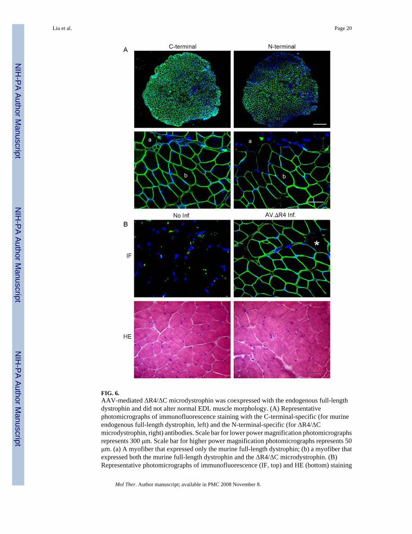
FIG. 6.AAV-mediated ΔR4/ΔC microdystrophin was coexpressed with the endogenous full-lengthdystrophin and did not alter normal EDL muscle morphology. (A) Representativephotomicrographs of immunofluorescence staining with the C-terminal-specific (for murineendogenous full-length dystrophin, left) and the N-terminal-specific (for ΔR4/ΔCmicrodystrophin, right) antibodies. Scale bar for lower power magnification photomicrographsrepresents 300 μm. Scale bar for higher power magnification photomicrographs represents 50μm. (a) A myofiber that expressed only the murine full-length dystrophin; (b) a myofiber thatexpressed both the murine full-length dystrophin and the ΔR4/ΔC microdystrophin. (B)Representative photomicrographs of immunofluorescence (IF, top) and HE (bottom) staining
Liu et al. Page 20
Mol Ther. Author manuscript; available in PMC 2008 November 8.
NIH
-PA Author Manuscript
NIH
-PA Author Manuscript
NIH
-PA Author Manuscript

of uninfected (left) and AV.ΔR4/ΔC-infected (right) BL10 EDL muscles. Immunofluorescencestaining was performed with anti-dystrophin N-terminal antibody (specific for ΔR4/ΔCmicrodystrophin, top). *A myofiber that was not transduced by AV.ΔR4/ΔC. Scale bar, 50μm.
Liu et al. Page 21
Mol Ther. Author manuscript; available in PMC 2008 November 8.
NIH
-PA Author Manuscript
NIH
-PA Author Manuscript
NIH
-PA Author Manuscript

FIG. 7.AAV-mediated ΔR4/ΔC microdystrophin expression did not compromise contractileproperties of the normal EDL muscle. The left EDL muscles of 4.5-month-old mdx mice wereinfected with AV.ΔR4/ΔC. The contralateral right EDL muscles served as sham-infectedcontrols. Muscle contractile assays were performed when mice were 9 months of age. (A)Force–frequency relationship between sham-infected (open bar) and AV.ΔR4/ΔC-infected(filled bar) EDL muscles. No statistically significant difference was seen between the twogroups ( P > 0.05). (B) Relative tetanic force drop during 10 cycles of eccentric contraction.Open circles, sham-infected EDL muscles (mean − SEM); closed circles, EDL muscles infectedwith AV.ΔR4/ΔC (mean + SEM). N = 3 pairs. There was no statistical difference betweenAV.ΔR4/ΔC-infected and sham-infected groups ( P > 0.05).
Liu et al. Page 22
Mol Ther. Author manuscript; available in PMC 2008 November 8.
NIH
-PA Author Manuscript
NIH
-PA Author Manuscript
NIH
-PA Author Manuscript

NIH
-PA Author Manuscript
NIH
-PA Author Manuscript
NIH
-PA Author Manuscript
Liu et al. Page 23TA
BLE
1C
hara
cter
istic
s of m
ice
and
EDL
mus
cles
infe
cted
by
AV
.RSV
.AP
ED
L c
hara
cter
istic
s at h
arve
stSt
rain
AV
.RSV
.AP
Na
Age
at i
nfec
tion
Age
at h
arve
stW
eigh
t (m
g)C
SA (m
m2 )b
Tra
nsdu
ctio
n (%
)
BL1
0N
o4
N/A
c77
day
s 8
.00
± 0.
291.
51 ±
0.0
5N
/Ac
BL1
0Y
es4
43 d
ays
77 d
ays
8.3
0 ±
0.16
1.56
± 0
.04
56.7
5 ±
5.88
dm
dxN
o5
N/A
c74
day
s11
.34
± 0.
332.
05 ±
0.0
6N
/Ac
mdx
Yes
542
day
s74
day
s11
.36
± 0.
162.
02 ±
0.0
753
.60
± 3.
88d
a Stud
y w
as p
erfo
rmed
in p
aire
d le
gs. T
he le
ft ED
L m
uscl
e w
as in
fect
ed w
ith A
V.R
SV.A
P, th
e rig
ht E
DL
mus
cle
of th
e sa
me
mou
se w
as n
ot in
fect
ed w
ith A
AV
.
b Cro
ss-s
ectio
nal a
rea
(cal
cula
ted
acco
rdin
g to
fibe
r len
gth)
.
c Not
app
licab
le.
d Ther
e w
as n
o st
atis
tical
diff
eren
ce in
tran
sduc
tion
effic
ienc
y be
twee
n B
L10
and
mdx
.
Mol Ther. Author manuscript; available in PMC 2008 November 8.

NIH
-PA Author Manuscript
NIH
-PA Author Manuscript
NIH
-PA Author Manuscript
Liu et al. Page 24TA
BLE
2C
hara
cter
istic
s of m
dx m
ice
and
mdx
ED
L m
uscl
es in
fect
ed b
y A
V.Δ
R4
ED
L c
hara
cter
istic
s at h
arve
stA
AV
Na
Age
at i
nfec
tion
Age
at h
arve
stW
eigh
t (m
g)C
SA (m
m2 )b
Tra
nsdu
ctio
n (%
)
AV
.AP
547
day
s (7
wk)
158
days
(5 m
)14
.32
± 0.
822.
81 ±
0.1
655
.88
± 2.
51d
AV
.ΔR
45
47 d
ays (
7 w
k)15
8 da
ys (5
m)
13.9
5 ±
0.79
2.74
± 0
.15
57.6
8 ±
4.29
dN
o in
fect
ion
4N
/Ac
374
days
(12
m)
14.0
5 ±
0.72
2.86
± 0
.12
N/A
cA
V.Δ
R4
427
6 da
ys (9
m)
374
days
(12
m)
14.7
3 ±
0.81
3.08
± 0
.15
31.7
7 ±
2.20
e
a Stud
y w
as p
erfo
rmed
in p
aire
d le
gs. T
he le
ft ED
L m
uscl
e w
as in
fect
ed w
ith A
V.Δ
R4,
the
right
ED
L m
uscl
e of
the
sam
e m
ouse
was
eith
er in
fect
ed w
ith A
V.A
P or
not
infe
cted
with
AA
V.
b Cro
ss-s
ectio
nal a
rea
(cal
cula
ted
acco
rdin
g to
fibe
r len
gth)
.
c Not
app
licab
le.
d Ther
e w
as n
o st
atis
tical
diff
eren
ce in
tran
sduc
tion
effic
ienc
y be
twee
n A
V.A
P an
d A
V.Δ
R4.
e AV
. ΔR
4 tra
nsdu
ctio
n ef
ficie
ncy
in 9
-mon
th-o
ld m
dx E
DL
was
sign
ifica
ntly
low
er th
an th
at in
7-w
eek-
old
mdx
ED
L.
Mol Ther. Author manuscript; available in PMC 2008 November 8.

NIH
-PA Author Manuscript
NIH
-PA Author Manuscript
NIH
-PA Author Manuscript
Liu et al. Page 25TA
BLE
3C
hara
cter
istic
s of B
L10
mic
e an
d B
L10
EDL
mus
cles
infe
cted
by
AV
.ΔR
4
ED
L c
hara
cter
istic
s at h
arve
stA
AV
Na
Age
at h
arve
stA
ge a
t inf
ectio
nW
eigh
t (m
g)C
SA (m
m2 )b
Tra
nsdu
ctio
n (%
)
No
infe
ctio
n3
N/A
c27
2 da
ys (9
m)
9.29
± 0
.02
1.95
± 0
.05
N/A
cA
V.Δ
R4
313
5 da
ys (4
.5 m
)27
2 da
ys (9
m)
9.31
± 0
.02
1.98
± 0
.03
59.6
7 ±
3.48
a Stud
y w
as p
erfo
rmed
in p
aire
d le
gs. T
he le
ft ED
L m
uscl
e w
as in
fect
ed w
ith A
V.Δ
R4,
the
right
ED
L m
uscl
e of
the
sam
e m
ouse
was
not
infe
cted
with
AA
V.
b Cro
ss-s
ectio
nal a
rea
(cal
cula
ted
acco
rdin
g to
fibe
r len
gth)
.
c Not
app
licab
le.
Mol Ther. Author manuscript; available in PMC 2008 November 8.
Related Documents