ADDIS ABABA UNIVERSITY, COLLEGE OF HEALTH SCIENCES, SCHOOL OF MEDICINE, DEPARTMENT OF, ORAL AND MAXILLOFACIAL SURGERY INCIDENCE AND PATTERNS OF MANDIBULAR FRACTURES IN ADDIS ABABA UNIVERSITY AFFLIATED HOSPITALS (YEKATIT 12 HOSPITAL MEDICAL COLLEGE AND ST.PETER SPECIALIZED HOSPITAL), FROM JANUARY, 2017 TO DECEMBER, 2019, ADDIS ABABA, ETHIOPIA BY: - ASSEFA ABERA BALCHA (ORAL AND MAXILLOFACIAL SURGERY RESIDENT) A RESEARCH PAPER TO BE SUBMITTED TO DEPARTMENT OF ORAL AND MAXILLOFACIAL SURGERY, COLLEGE OF HEALTH SCIENCES, ADDIS ABABA UNIVERSITY; IN PARTIAL FULLFILMENT FOR THE REQUIREMENT OF THE SPECIALTY CERTIFICATE IN ORAL AND MAXILLOFACIAL SURGERY AUGUST, 2020 ADDIS ABABA, ETHIOPIA

Welcome message from author
This document is posted to help you gain knowledge. Please leave a comment to let me know what you think about it! Share it to your friends and learn new things together.
Transcript
ADDIS ABABA UNIVERSITY, COLLEGE OF HEALTH SCIENCES, SCHOOL OF
MEDICINE, DEPARTMENT OF, ORAL AND MAXILLOFACIAL SURGERY
INCIDENCE AND PATTERNS OF MANDIBULAR FRACTURES IN ADDIS ABABA
UNIVERSITY AFFLIATED HOSPITALS (YEKATIT 12 HOSPITAL MEDICAL COLLEGE
AND ST.PETER SPECIALIZED HOSPITAL), FROM JANUARY, 2017 TO DECEMBER,
2019, ADDIS ABABA, ETHIOPIA
BY: - ASSEFA ABERA BALCHA (ORAL AND MAXILLOFACIAL SURGERY RESIDENT)
A RESEARCH PAPER TO BE SUBMITTED TO DEPARTMENT OF ORAL AND
MAXILLOFACIAL SURGERY, COLLEGE OF HEALTH SCIENCES, ADDIS ABABA
UNIVERSITY; IN PARTIAL FULLFILMENT FOR THE REQUIREMENT OF THE
SPECIALTY CERTIFICATE IN ORAL AND MAXILLOFACIAL SURGERY
AUGUST, 2020
ADDIS ABABA, ETHIOPIA
ADDIS ABABA UNIVERSITY, COLLEGE OF HEALTH SCIENCES
DEPARTMENT OF ORAL AND MAXILLOFACIAL SURGERY
INCIDENCE AND PATTERN OF MANDIBULAR FRACTURES IN ADDIS ABABA UNIVERSITY
AFFILIATED HOSPITALS (YEKATIT 12 HOSPITAL MEDICAL COLLEGE AND ST.PETER
SPECIALIZED HOSPITAL), ADDIS ABABA, ETHIOPIA
By: -Assefa Abera Balcha (Oral and Maxillofacial Surgery Resident)
Mobile: +251910912843
Email: [email protected]
Advisor:-Dr. Demerew Dejene Dana (Assistant Professor of Oral and Maxillofacial Surgery)
Addis Ababa University, College of Health Sciences, Department of Oral and Maxillofacial Surgery
Mobile: +251912224087
E-mail: [email protected]
Co-advisor:-Dr. Girma Uma H/Mariam (Assistant Professor of Oral and Maxillofacial Surgery)
Addis Ababa University, College of Health Sciences, Department of Oral and Maxillofacial Surgery
Mobile: +251933542664
Email: [email protected]
August, 2020
Addis Ababa, Ethiopia
II
ADDIS ABABA UNIVERSITY
SCHOOL OF POSTGRADUATE
This is to certify that, the thesis prepared by Assefa Abera entitled ‘Incidence and pattern of
mandible fractures in AAU affiliated hospitals (Yekatit 12 HMC and St.Peter’s specialized
Hospital) in Addis Ababa, Ethiopia over a retrospective period of 3 years from January
2017 to December 2019’ and submitted in partial fulfillment of the requirements for the
specialty certificate in Oral and Maxillofacial Surgery complies with the regulations of the
university and meets the accepted standards with respect to originality and quality.
Signed by the examining committee
Examiners: Dr. _________________ Signature _______________ Date_______________
Dr. _________________Signature _______________ Date_______________
Advisors: - Dr. Demerew Dejene Signature _______________ Date _______________
Dr. Girma Uma Signature _______________ Date_______________
I
Abstract
Objectives: The objective of this study was to assess the incidence and pattern of mandible
fractures in AAU affiliated hospitals in Addis Ababa, Ethiopia over a retrospective period of 3
years from January 2017 to December 2019 G C.
Methodology: Retrospective review of patient’s records was conducted among those patients
who visited AAU affiliated hospitals having mandibular fractures. The study was conducted
from November, 2019 to August, 2020. The patients chart with incomplete information and those
which are absent from shelf were excluded from the study. The data was entered, cleaned and
analyzed using Epi info data version 7.0 Software. Descriptive analysis was computed as
frequency of fractures, distribution of age, gender, etiology, diagnosis, and anatomical sites of
mandibular fractures.
Results: A total of 247 patients who were retrospectively evaluated at Yekatit 12 HMC and
St.Peter’s specialized hospital between January 2017 and December 2019 sustained 343
mandibular fractures (mean of 1.4, range 1-3). The incidence of mandibular fracture was higher
in male patients (83%) than in females (17%) (Male: Female ratio 5:1), and the peak incidence
was during the third decade for both genders. The most common site of fracture was the body
(26.53%), followed by the angle (23.9%), and parasymphysis (19.82%). Overall, interpersonal
violence (46.15%) was the most common cause followed by RTA (27.53%). In male patients, the
most common cause was interpersonal violence (50%); in females it was a RTA (34.14%). The
anatomical sites of fracture reflected their cause. A total of (n=76, 30.76%) patients sustained
other non-maxillofacial injuries of which head injury (n=58, 23.48%) is the most common.
Conclusions: The most common cause of mandibular fracture was interpersonal violence.
Mandibular fractures were more common in males than females with most patients aged 21-30
years. The most common fracture site was body of the mandible. The predominant treatment
modality was open reduction and internal fixation (ORIF).
Key words: Mandibular fractures, Body, Etiology, Interpersonal violence (IPV), ORIF.
II
Acknowledgement
First and foremost, I would like to praise the Almighty God for his mercy and help in the way of
my entire academic journey. Next, I would like to express my heartfelt gratitude to my advisor
Dr. Demerew Dejene and co-advisor Dr. Girma Uma who guided me in doing this research
paper reality. My sincere thanks also go to Yekatit 12 hospital medical college and St.Peter’s
specialized hospital staffs for their kind unbroken support throughout the data collection period.
Last but not least, I would like to extend my thanks to Addis Ababa University, College of
Health Sciences who gave me the valuable opportunity to conduct the study.
III
Acronyms
AAU - Addis Ababa University
CA - Car Accident
CHS - College of Health sciences
HMC - Hospital Medical College
IMF - Intermaxillary fixation
IPV- Interpersonal Violence
MMF-Maxillomandibular fixation
MVA - Motor Vehicle Accident
NOE - Naso-Orbitoethmoid Complex
OMF - Oral and maxillofacial
ORIF - Open reduction and internal fixation
RTA - Road Traffic Accident
SPSS - Statistical package for social sciences
WHO - World Health Organization
Yrs. - Years
ZMC # - Zygomaticomaxillary complex fracture
IV
Tables of contents
Abstract ...................................................................................................................................... I
Acknowledgement ..................................................................................................................... II
Acronyms ................................................................................................................................. III
Tables of contents .................................................................................................................... IV
List of tables and figures .......................................................................................................... VI
Chapter -1 ...................................................................................................................................1
1. Introduction .........................................................................................................................1
1.1 Background Information ...............................................................................................1
1.2 Statements of the problem .............................................................................................2
1.3 Significance of the study ...............................................................................................4
Chapter-2 ....................................................................................................................................5
2.1 Literature review ..................................................................................................................5
Chapter-3 .................................................................................................................................. 14
3. Objectives .......................................................................................................................... 14
3.1 General Objective ....................................................................................................... 14
3.2 Specific objectives ...................................................................................................... 14
Chapter-4 .................................................................................................................................. 15
4. Methods and Materials ....................................................................................................... 15
4.1 Study area and period .................................................................................................. 15
4.2 Study design ............................................................................................................... 15
4.3 Population ................................................................................................................... 15
4.3.1 Source population ................................................................................................ 15
4.3.2 Study population .................................................................................................. 15
4.4 Sample size and sampling technique ........................................................................... 16
V
4.4.1 Sample size Determination ................................................................................... 16
4.4.2 Sampling technique .............................................................................................. 16
4.4.3 Inclusion criteria………………………………………………………………. 16
4.4.3 Exclusion criteria…………………………………………………………….....16
4.5 Variables ..................................................................................................................... 16
4.5.1 Independent variables ……………………………………………………………..…16
4.5.2 Dependent variables ………………..…………………………………………………16
4.6 Data collection procedures .......................................................................................... 17
4.6.1 Disease Identification ........................................................................................... 17
4.6.2 Disease Classification and Categorization ............................................................ 17
4.7 Operational definitions and terms ................................................................................ 17
4.8 Data quality assurance................................................................................................. 17
4.8.1 Pre-test................................................................................................................. 17
4.8.2 Data collectors training and supervision ............................................................... 18
4.9 Data analysis ............................................................................................................... 18
4.10 Ethical Consideration .................................................................................................. 18
4.11 Dissemination plan ...................................................................................................... 18
Chapter-5 .................................................................................................................................. 19
Results ................................................................................................................................... 19
Chapter-6 .................................................................................................................................. 26
Discussions ......................................................................................................................... 26
Chapter -7 ................................................................................................................................. 28
Conclusions and Recommendations .................................................................................. 28
Annex I References ................................................................................................................... 29
Annex II Data collection format ................................................................................................ 32
VI
List of tables and figures
Table 1 Age and gender distribution of 247 patients having mandibular fracture at AAU, CHS
affiliated hospitals (Yekatit 12 HMC and St. Peter Specialized Hospital) from January, 2017 to
December, 2019.
Table 2 Distributions of 247 patients with mandibular fracture according to aetiology and gender
at AAU, CHS affiliated hospitals (Yekatit 12 HMC and St. Peter Specialized Hospital) from
January, 2017 to December, 2019.
Table 3 Distributions of 247 patients according to age groups and aetiology of fractures at AAU,
CHS affiliated hospitals (Yekatit 12 HMC and St. Peter Specialized Hospital) from January,
2017 to December, 2019.
Table 4 Distributions of mandibular fractures according to anatomical sites and gender at AAU,
CHS affiliated hospitals (Yekatit 12 HMC and St. Peter Specialized Hospital) from January,
2017 to December, 2019.
Table 5 Distributions of mandibular fractures according to anatomical sites and aetiology at
AAU, CHS affiliated hospitals (Yekatit 12 HMC and St. Peter Specialized Hospital) from
January, 2017 to December, 2019.
Table 6 Distributions of 247 patients according to anatomical sites and treatment modalities at
AAU, CHS affiliated hospitals (Yekatit 12 HMC and St. Peter Specialized Hospital) from
January, 2017 to December, 2019.
Figure 1 Distribution of 247 patients according to their geographic locations at AAU, CHS,
affiliated hospitals (Yekatit 12 HMC and St. Peter Specialized Hospital) from January, 2017 to
December, 2019.
Figure 2 Distributions of mandibular fractures according to treatment modalities at AAU, CHS,
affiliated hospitals (Yekatit 12 HMC and St. Peter Specialized Hospital) from January, 2017 to
December, 2019.
Figure 3 Distribution of 247 patients according to treatment modalities and gender at AAU,
CHS, affiliated hospitals (Yekatit 12 HMC and St. Peter Specialized Hospital) from January,
2017 to December, 2019.
1
CHAPTER-1
1. INTRODUCTION
1.1 Background Information
The maxillofacial region is one of the most fractured sites of the body. The management of these
fractures is a challenge requiring skill and experience. In repair of maxillofacial trauma,
functional and aesthetic reconstruction is a prime concern. Oral and maxillofacial injuries can
vary in severity ranging from minor soft tissue injuries to major fractures of the entire facial
skeleton (1).
The mandible is more prone for maxillofacial trauma and fractures due to its unique mobility,
shape, and chin prominence in the facial skeleton. It is the second most frequent of the facial
bones affected by traumatic injuries of all facial fractures. The mandible can be fractured alone
or in combination with a fracture of other bones in the maxillofacial region (2). A fractured
lower jaw is accompanied by pain, malocclusion and loss of masticatory function, speech
impairment, and esthetic disfigurement with psychological effects. It can result in severe pain
which prevents patient from feeding and airway obstruction (2)
Etiology of fracture is multifactorial and based variably on socioeconomic status, culture,
technology, demography, and economic factors. The etiology of mandibular fractures could be
caused by road traffic accidents (RTAs), accidental falls, assaults, industrial mishaps, sports
injuries and firearm injuries (3). RTA is the leading cause of mandibular fracture in developing
countries owing to poor enforcement of law and ensuring the abidance by the existing traffic and
speed limit regulations, while interpersonal violence is leading cause in developed countries (3).
Regarding with the management of mandibular fractures, the most widely applied treatment
modality is closed reduction with maxillomandibular fixation especially in developing countries
where advanced services are not widely available. In developed countries having advanced
setups, open reduction with internal fixation using plates and lag screws is the most practiced
modality of treatment (3).The most common complications related with closed reduction is TMJ
ankylosis, weight loss, poor oral hygiene related infections whereas ORIF has risk of nerve
injury, surgical scars, foreign body (plates and wires) related infections (3).
2
1.2 Statements of the problem
Mandibular fractures are the second, most-frequent facial injuries treated at a trauma center in
Switzerland. According to several studies, they account for 15.5% to 59% of all facial fractures.
The epidemiological data for facial and mandibular fractures varies among countries and changes
over time. The etiology of maxillofacial injuries is multi-factorial, mostly depending on socio-
economic, demographic, cultural, technological and environmental factors. Interpersonal
violence is the most common cause for mandibular fractures in North-American countries, North
European countries, Australia and New Zealand. In newly industrializing and less developed
countries such as Jordan or Nigeria, motor vehicle accidents are the most common cause for
mandibular fractures (4).
Maxillofacial bone fractures particularly mandibular fracture is one of the common problems
affecting the population worldwide. Globally the incidence of maxillofacial bone fractures as a
whole as well as mandibular bone fracture specifically is increasing with different figures
represented at different geographical location that mainly corresponding to cultures and
socioeconomics of the societies (5).
The majority of affected patients in Uttar prudish state, India were in the 2nd and 3rd decades. A
definitive relationship existed between RTA and the incidence of mandibular fractures. The
frequency further increased with consumption of social intoxicants. The most commonly
fractured site was Para symphysis either isolated or associated with other fractures in the
mandible (5).
The etiology and pattern of mandibular fracture vary considerably among different study
populations. Despite many reports about the incidence, diagnosis and treatment of mandibular
fracture there is limited knowledge about the specific pattern of mandibular fractures in South
Asian countries (6).
The sheer pace of modern life with high-speed travel as well as an increasingly violent and
intolerant society has made facial trauma a form of social disease from which no one is immune.
There are changes in patterns of facial injuries, extent, clinical features, and so forth resulting in
mild-to-massive disfigurement of maxillofacial skeleton along with functional loss.
3
Beside RTA and violence, direct/indirect trauma may also occur due to sport activities, falls, and
firearms. Occasionally, it may also be secondary to certain disease entities like cystic lesion,
neoplasms, and metabolic diseases (7).
Mandible is the only mobile bone of facial skeleton and there has been a significant increase in
number of cases in recent years. It is embryologically a membrane bone and is more commonly
fractured than the other bones of face. Mandibular fractures occur twice as often as midfacial
fractures. The energy required to fracture it being of the order of 44.6–74.4 kg/m, which is about
the same as the zygoma and about half that for the frontal bone. It is four times as much force is
required to fracture maxilla (7).
The mandible is fractured more frequently than any other facial bone, likely because it is
exposed and protruding. The etiology of these fractures is multifactorial, with the type and
frequency of fracture dependent on socioeconomic status, culture, technology, demography, and
economic factors. Despite these all major concerns, there is no recent comprehensive study has
assessed patterns of mandibular fracture in African countries (8).
4
1.3 Significance of the study
The facial area is one of the most common sites of injury. The mandible is fractured more
frequently than any other facial bone, because it is exposed and protruding. In addition to
functional loss, a mandibular fracture can result in mild to moderate impairment or defect.
Maxillofacial bone fractures in general and mandibular fractures in particular not only cause
morbidity and mortality but also has significant impact on facial cosmetics and psychosocial
concerns demanding considerable attention. So it can cause great economic and psychosocial on
the individuals, families and community as whole.
In worldwide, there is significant increase in number of mandibular fractures most commonly
affecting young male in productive age groups with the highest peak age ranging in 21–30 years
that has gross socioeconomic effect. The sheer pace of modern life with high speed travel has
increased its effects. Despite all these problems, there are few studies done in Africa and other
developing world regarding with incidence and patterns of mandibular fractures.
There are no previous study findings concerning incidence and patterns of mandibular fractures
in our country Ethiopia. Hence, this study assess incidence and patterns of mandibular fractures
based on etiology, gender, anatomic locations and treatment modalities used as this plays an
important role in the quality assurance of the health care process and the quality of life for
trauma patients.
Furthermore, in developing countries like Ethiopia where the number of road traffic accidents is
dramatically increasing with time, determining the frequency of mandibular fractures and their
patterns of occurrence can enhance early diagnosis and treatment, thereby reducing morbidity
and mortality associated with these fractures.
In addition, it is believed that, this study contributes much as base line data for the descriptive
epidemiologic study of maxillofacial fractures in general and mandibular fractures in particular,
intended treatment guidelines and policy making for future in the country.
5
CHAPTER-2
2.1. LITERATURE REVIEW
A total of 13,142 patients with mandibular fractures from participating trauma centers in USA
from 2001 to 2005 were included in the study of which 80% of patients were male. Fracture
occurring most frequently at 18 to 54 years of age. Mechanism of injury differed by gender, with
men most often sustaining mandibular fracture from assault (49.1%), followed by motor vehicle
accidents (MVAs; 25.4%) and falls (12.8%). Women most commonly sustained mandibular
fracture from MVAs (53.7%), followed by assault (14.5%) and falls (23.7%). Falls were a
significantly more common mechanism in patients who were at least 65 years old (9).
A retrospective study of 246 patients treated for mandibular fracture at the Toronto General
Hospital over a 5.5 years period (from 1995 to 2000) shows men those who aged 21 to 30 years
sustained the most mandibular fractures. The ratio of males to females was 5:1. Most fractures
were caused by violent assault (53.5%), followed by falls (21.5%) and sports activities (12.2%).
Alcohol was a contributing factor at the time of injury in 20.6% of fractures for which this
information was available. Nearly half of all cases were treated by open reduction (49.1%).
Complications occurred in 5.3% of patients (10).
A retrospective medical records review of a total of 181 patients with maxillofacial injury was
done in Montreal General Hospital, Canada between 1998 and 2003 and 307 mandibular
fractures were identified. About 52% of the fractures occurred in individuals 21 to 40 years of
age, 78% of patients were male, and there was wide ethnic diversity. About 60% of patients had
multiple mandibular fractures; 29% were symphysis fractures, 25% were condylar fractures and
23% were angle fractures. Assault was the most common mechanism of injury, with 29% of
fractures involving alcohol or illegal drug use. About 30% of patients had an associated facial
fracture, and more than one-third had another major injury (11).
The study done in Canada, Finland and Kuwait during the 1990–2000 at Institute of Dentistry,
University of Oulu and Oulu University Hospital, Oulu, Finland on a total of 596 patients having
818 mandibular fractures in Kuwait, 317 fractures on 228 patients in Canada and 417 fractures
on 268 patients in Finland. The mean age of the patients in Kuwait, Canada and Finland were
6
26.2, 31.7, and 30.7 years, respectively. Condylar fractures were more common in Finnish
patients (41%) than Canadian (35%) or Kuwaiti patients (21%). Condylar fractures caused by
falls were about 3.4 times more common in Kuwait and Finland compared to Canada. In Finland
the risk of road traffic accidents caused by condylar fracture was about 4 times higher than those
caused by other causes. In Canada male was about 2 times higher for the condylar fracture than
females. Female patients often had more multiple injuries than men in all three countries and
multiple fractures were observed especially in traumas caused by falling (12).
A retrospective review done at London teaching hospital between June 2005 and May 2010 on a
total of 1261 patients who sustained 1994 mandibular fractures shows the incidence of
mandibular fracture was higher in male patients (87%) than in females (13%) (Male: female ratio
6.6:1). The peak incidence was during the third decade for both genders. The most common site
of fracture was the angle (30%), followed by the parasymphysis (27%), and condyle (27%).
Overall, interpersonal violence was the most common causes (72%) followed by falls (18%). In
male patients, the most common cause was interpersonal violence (77%); in females it was a fall
(46%). Interpersonal violence typically resulted in fractures of the angle (36%) while road traffic
accidents and falls resulted in condylar fractures (28% and 53%, respectively). About 49.1%
were treated by open reduction and internal fixation (ORIF) while 29.4% were treated by IMF
and 21.5% were treated conservatively (8).
According to the retrospective study conducted in Portugal in the year period 1993 to 2002, on
521 patients with 681 mandibular fractures showed, motor-vehicle accident (MVA) was the most
common (53.9% patients) cause of fracture. Almost half of the patients (48.8%) were in the
oldest age group (16 to 18 years old). The condyle of the mandible was involved in 31.0% of the
fractures. Maxillomandibular (MMF) fixation was used in 534 (78.4%) fractures. Overall
mortality in this series was 0.6% (3 patients); mortality was caused by multiple traumas, mainly
head trauma (13).
The retrospective study conducted between January, 2000 and December, 2007 at the University
hospital of Bern, Switzerland’s largest Craniomaxillofacial surgery center in central Switzerland
on 420 patients with 707 mandibular fractures showed the two most common causes of injury
were road traffic accidents (28%) and various types of sports injuries (21%).
7
A total of 13% of the patients were under the influence of alcohol or drugs at admission.
Fractures were predominantly situated in the condyle/sub condyle (43%) and in the
symphysis/parasymphysis region (35%). Occurrences of fractures in the angle and in the body
were low, at 12% and 7% respectively (4).
In the city of Sao Paulo, Brazilian level I complex public trauma hospital ;one study done
between January and December, 2001 on a total of 91 patients having mandibular fractures
reveals motorcycle accident (22%) was the major cause of mandibular fractures followed by
physical aggression (15%) and height fall(11%). The mandibular anatomical sites of higher
fracture incidence were: symphysis (36.3%), body (34%) and condyle (6.6%). The most
commonly performed treatment modes were conservative approach or open reduction and
intraosseous fixation. The most predominantly affected age groups were 21-30 year-old males
(29.7%). Out of 91 patients, 18 (19.7%) had complication; 12 had infection (66%), malocclusion
was (2 cases; 10%). Trismus, lack of union, mobility and salivary fistula had 1 case each (14).
In Medina region King Fahad Hospital, Saudi Arabia: Oral Maxillofacial Surgery Department, a
retrospective study was done on a total of 197 patients with fracture of the mandible who were
admitted in the period of 3 years from 2013 to 2016 of which 165 were male and 32 female
patients. The ages ranged from 3 to 86 years with a mean of 24 yrs. A total of 260 fractures of
Mandible were documented. The largest number (113) of patients was found in the age group
between 16 and 30 years. Trauma caused by RTAs was the main etiology of the fractures
followed by falls and assault. The majority of the patients were in the role of vehicle drivers. The
condylar fracture was most frequently affected constituting the largest number (103) of fractures
followed by the angle (51), Parasymphysis (45), and then by the body (23) of the mandible.
Dentoalveolar fractures were present in 22 cases. Very less number of coronoid fractures (7),
followed by those of the ramus (5), and least number at the symphysis (4) were found (15).
The 5 years retrospective study conducted in Taleghani Hospital of Iran, from the beginning of
2013 until the end of 2017 on the total of 708 patients with maxillofacial fractures; most cases
were men (85.2%) and in the second and third decades of life (53.8%). The majority of the
fractures were in the mandible with the incidence rate of 64.7% (16).
8
According to a retrospective study done on an incidence and pattern of mandibular fracture in
Rohilkhand region, Uttar Pradesh state of India; the patient records and radiographs for 144
patients treated for mandibular fractures were reviewed between January 2012 to December 2013
and maximum incidence of fractures was observed among the individuals in 3rd decade (35.4%)
followed by 2nd and 4th decades, which exhibited 32 and 30 cases (22.2% and 20.8%),
respectively. Male to female ratio was (4:1) portraying a male predominance. Road traffic
accidents (RTAs) were observed to be the predominant etiological factor accounting for 79.2%
of the total injuries followed by assaults (11.8%) and falls (9%). Parasymphysis exhibited the
highest incidence (32.63%) amongst the anatomic sites, followed by body (18.75%), angle
(16.66%), condyle (15.27%), symphysis (12.50%), ramus (2.77%) and coronoid (1.38%) (5).
A 3 years (October 2010 to October 2013) retrospective analysis done in central India on a total
of 464 patients out of which (343) 79% were male and (91) 21% were female having mandibular
fractures with age ranging from 7 to 89 years showed the highest incidence (37.5%) of
mandibular fractures was in the age group of 21–30 years. The main cause was road traffic
accidents (RTAs, 68.8%) followed by falls (16.8%), assaults (11%) and other reasons (3.8%).
Parasymphyseal fractures were the most frequent 331 (41.1%), followed by condyle (135) and
angle (124) fractures in occurrence. Mandibular angle fractures were found mostly to be
associated with assault victims (6).
According to the Study conducted between 2012 t0 2015 on Pattern and Incidence of Mandibular
Fractures in Department of Oral and Maxillofacial Surgery, Career Post Graduate Institute of
Dental Sciences, Lucknow, India on of 66 patients of which 37 had a unilateral mandibular
fracture while 29 had bilateral fractures with maximum number of subjects were in the age group
21–30 years (28.8%) followed by 11–20 (25.8%), 31–40 (21.2%), <10 (13.6%), 41–50 (6.1%),
and 60 years and above (4.5%). Around three-fourth (75.76%) of patients were in the age range
11 to 40 years. More than four-fifth (81.8%) of patients were males. Only 12 (18.2%) patients
were female. The male to female ratio of the patients was 4.5: 1. Road traffic accident (68.2%)
was the cause of mandibular fractures in majority of subjects, followed by fall from height
(30.3%) and hit against object (1.5%). In 37.9% of cases, the mandible fracture was associated
with other injuries while in majority (62.1%) no such associated injury was observed.
9
Fracture of parasymphysis (31.4%), body (24.5%), sub condyle (20.6%), and angle (13.7%) were
the most common sites while fracture condyle (1%), coronoid (1.0%), dentoalveolar (1.0%), and
ramus (1.0%) were the least common fracture sites (7).
According to one study done in Department of Oral Medicine and Radiology, Faculty of Dental
Sciences, King George's Medical University, Lucknow, Uttar Pradesh, North India on
Prevalence of mandibular fracture in patients visiting a tertiary dental care hospital; the study
population consists of 1015 individuals aged between 7 and 68 years with the mean age of 33.49
± 11.79 years and majority of the patients were between 18 and 35 years of age. Males (78.6%)
dominated the study population than females (21.4%). The most common anatomic site for
mandibular fracture was parasymphysis region (40.3%) followed by angle (28.8%), condyle
(27.6%), and symphysis (12.5%) of mandible. The coronoid process of mandible (44, 4.3%) was
least involved in mandibular fracture. Males (42.4%) suffered more parasymphyseal injury than
females (32.7%). The mandibular angle fracture is more common in females (47.9%) than males
(23.6%) (17).
A 3 years retrospective study of maxillofacial injury cases was carried out at the newly created
B.P.S Government Medical College for Women, Khanpur Kalan, Sonepat, in India from
September 2011 to February 2013 on a total number of 474 patients with 86 mandibular fractures
was registered, males outnumbering female patients by a ratio of 2.9:1. Age of patients ranged
between 9 months and 72 years, maximum incidence occurring in the 18-34 year group of age.
Most injuries were caused by road traffic accidents (48.83%), followed by assaults (26.74%) and
sporting activities (13.95%). The most prominent site of mandibular fracture was parasymphysis
(27.90%), followed by angle (24.41%) and body (18.60%) regions. 30.23% of the patients with
mandible fractures were having multiple fracture sites. Also, 10% of the patients with mandible
fracture had associated mid-facial fractures. Closed reduction was done in 13.6% of patients,
open reduction and internal fixation was performed in 46.4% of cases, while 18.1% of them were
treated conservatively. The mean duration of hospitalization was 10.12 ± 6.24 days (18).
According to a retrospective descriptive study done on a total of 35 patients with mandibular
fractures in Department of Oral and Maxillofacial Surgery, Sinhgad Dental College and Hospital,
Pune, Maharashtra, India on the prevalence of mandibular fractures during the period of
February 2008 to September 2009, Out of 35 patients, thirty one were males (88.57%) and four
10
were females (11.43%) with a male: female ratio of 8:1. The peak occurrence is in young adults,
aged 21-30 years (n = 15, 42.86%). In case of etiologic of fracture, road traffic accidents (RTAs)
was the most common (n = 25, 71.43%) and condyle was most frequently involved site (n = 19,
38.78%). In most (n = 16, 45.71%) of the patients, an open reduction and rigid internal fixation
using bone plate and screws was done (19).
A 5 years retrospective review of Incidence and pattern of mandibular fractures in rural
population in Loni, Maharashtra, India during the period from January 2003 to December 2007
conducted on 324 patients reveals 486 mandibular fractures were identified; males formed 80.9%
and females 19.1% of the studied population, with peak incidence occurring in the 21–30 years
age group. The most common fractures site was parasymphysis (39.3%). The cause of
mandibular fractures was RTAs (42.9%), followed by falls (25.9%), assaults and interpersonal
violence (20.7%), and animal injuries (10.5%). The results exhibit that road traffic accidents
remain the major cause of mandibular trauma and animal injuries being found exclusively in
rural population (20).
The study carried out on 138 patients diagnosed with mandibular fractures in 2015 included 108
men (78.3%) and 30 women (21.7%), at the departments of oral and maxillofacial surgery of
three hospitals in Peshawar, Pakistan: Sardar Begum Dental College and Hospital, Rehman
Medical Institute, and Northwest General Hospital shows Most patients (56%) were aged 15-25
years, followed by those aged 26-35 years (26%). The most frequent cause of fractures was
RTAs; 59.42%), followed by falls (18.8%). RTAs were predominant in men (89%); whereas,
falls were predominant in women (80%). In patients with unilateral fractures, the most common
fracture site was the parasymphysis (24.6%) followed by the symphysis (10.1%). In patients with
bilateral fractures, the most common fracture sites were the parasymphysis and condyle (11.6%),
followed by the parasymphysis and angle (8.0%) (21).
A retrospective analysis of patients suffering from facial fractures and treated in 8 hospitals in
Kuwait during the years 1992-1997, shows there were 586 patients with facial fractures with a
male: female ratio of 5.4: 1. The age of the patients ranged from 8 months to 87 years, with a
mean of 27 and a median of 25 years. The majority (55.5%) were due to road traffic accidents.
Only mandibular fractures were suffered by 49%, only midfacial fractures by 36.7% and
combined mandibular and midfacial fractures by 14.3% of the patients. Altogether there were
11
923 fractures. The most frequent fractures were of zygomatico-maxillary complex (15%],
followed by mandibular molar-premolar area (14.7%). Serious concomitant injuries were found
in 141 patients (24%), of which 22 (3.8%) died as a results. 278 patients (47.4%) were treated by
open reduction, 247 (42.1%) of them using titanium miniplates. Conservative treatment (closed
reduction and/or maxillo-mandibular fixation) was performed in 180 patients (30.7%), while 128
patients (21.9%) were only observed (22).
The descriptive cross sectional retrospective study was undertaken on 435 patients with
mandibular fracture during the year 2014-2015 to determine the epidemiological and clinical
profile of patients presented with fractures of mandible and their different methods of treatment
modalities in Bangladesh College of Physicians and Surgeons reveals higher prevalence in male
(3.9:1), with occurrence peak between 21-30 years. The principal causes of fracture in this study
were RTA representing 54.02% followed by physical assault 17.24%, fall, Sports injury, Blow
by heavy objects, Tube well injury & others which includes Tire blast injury, Gunshot injury,
Iatrogenic cause, Pathological fracture, Boat handle injury, Penetrating injury by metal etc. The
most injured sites were in parasymphysis (26.31%) followed by angle of mandible 17.89% then
symphysis, condyle, body of mandible, dentoalveolar, ramus, coronoid process of mandible.
Most patients (70.11%) of mandible fractures were treated by closed reduction (arch bar, arch
bars with intermaxillary fixation, eyelet wiring & lateral compression plate) and 21.83% of
patients were treated with open reduction (miniplates fixation. 3D plate fixation). Only 8.05%
patients were managed by conservative approach (23).
According to the a 10-year retrospective analysis of 685 mandibular fracture cases treated in
Charles-Nicolle Hospital, Tunis, Tunisia during the time period of 1995 and 2004, the prevalence
of mandibular fractures was higher in male patients (sex ratio 6:1). Road traffic accidents were
the main cause of this trauma (45%), followed by assault (22%). Angle fractures were the most
common (24.8%) followed by parasymphyseal fractures (22.2%). The most frequent treatment
was closed reduction with MMF in 388 patients (56.6%). Trans osseous wiring was the most
commonly used method in open reductions. Tooth loss and neurological sensitive deficiency
were the most common sequels (24).
12
A review on incidence and patterns of maxillofacial fractures at Muhimbili National hospital,
Dar es Salaam, Tanzania from January 2003 to June 2009 on 118 patients having maxillofacial
fractures shows the males are more affected with (M: F ratio = 3.7:1). Peak incidence was in the
21-30 years age group that accounted for 53 (46.3 %) of the cases, followed by the 11 to 20 years
age group which comprised of 25 (accounting for 21.3%) of the cases. Majority, 110 (85.9%)
fractures, were occurred in the mandible, while 16 (13.6%) occurred in the maxilla and 2 (1.6%)
in the zygoma. The most frequent cause was violence (social altercation, domestic violence and
assaults), which accounted for 64 (54.2%) of all fractures causes, followed by motor traffic
accidents with 41 (34.7%) (25).
Another study done at Muhimbili National Hospital in Dar es Salaam, Tanzania, 1998–2003 on a
total of 314 patient records of 261 (83.1%) males and 53 (16.9%) females were retrieved (ratio
m:f = 5:1), age range 2–70 years, with most (41.4%) in the 20–29 year age group. Most of the
fractures occurred in the mandible 222 (70.7%). Most fractures were caused by assault 181
(57.6%) followed by falls 62 (19.7%), motor traffic accidents 43 (13.7%) and sports 25 (8%).
Most of the mandibular (98.2%) and zygomatic arch fractures (62.5%) were managed by closed
reduction, compared with alveolar bone fractures that were predominantly managed by open
reduction. Complications occurred in 17 (5.4%) patients and were mostly infections (26).
A 1 year retrospective study on incidence of facial fractures made on a total of 390 patients with
facial fractures at Khartoum teaching dental hospital; Sudan, during the period of January, 2010
to January 2011 indicates 14.1 % (55) of them were children below 16 years of age. Most facial
fractures 47 % were in the age group 12–16 years, followed by 29.1 % in the age group 7–11
years, while the least number of fractures were seen in 23.6 % in the age group below 6 years.
Most maxillofacial were caused by RTA 56.4 %, followed by daily activities 21.8 %, assault
16.4 %, and others 5.5 %. Most of the facial fractures occurred in the mandible 76.8 %, followed
by mid-face fractures; zygomatic complex fractures 13.5 %, Le Forte 6.7 % and NOE 1.3 %.
Intervention was in the form of conservative management in 49.1 % of the patients, 34.5 %
closed reduction (e.g. simple wire, lingual splints) and 16.4 % open reduction (open reduction
and internal fixation) (27).
13
A retrospective study made on Occurrence and pattern of mandibular fractures at Kisii District
Hospital, Kenya: hospital records revealed that 39 cases of mandibular fractures presented at
Kisii District hospital during a two-year period. 27 cases were due to interpersonal violence
while road traffic accidents and accidental falls accounted for 9 and 3 of the cases respectively.
The male ratio was 2.9:1. Majority (26 cases) of the patients were aged between 20 and 39 years.
The commonly involved fracture site was the left body of the mandible accounting for 20 of the
fractures (28).
A research done during 2-year period, September 2013–August 2015, in university of Gondar,
northern Ethiopia shows, 326 patients of maxillofacial trauma were treated of which 80% were
males with male to female ratio of 4.02:1. The mean age was 29.12 (± 8.62) with age range of
11–75 years. Majority of the study participants (47.2%) were within the age group of 21–
30 years. Interpersonal violence (75.8%) and Road traffic accident (21.5%) were the leading
causes. There was an associated injury in 79 (24.2%) patients in head and neck area, thoracic,
abdominal and extremities. Half of the patients were managed conservatively (49.7%) with
debridement and suture, while 45.7% of the patients were closed reduction and 4.6% were
surgical open reduction. There were 25 post procedure complications especially in mandibular
fractures (29).
14
CHAPTER-3
3. OBJECTIVES
3.1 General Objective
To assess the incidence and pattern of mandible fractures in AAU affiliated hospitals in
Addis Ababa, Ethiopia over a retrospective period of 3 years; January 2017 to December
2019.
3.2 Specific objectives
To determine sociodemographic distribution of mandibular fractures in AAU affiliated
hospitals (Yekatit 12 HMC and St. Peter Specialized Hospital).
To identify etiologies and frequency of mandibular fractures in the study population.
To assess patterns of mandibular fracture in AAU affiliated hospitals in Addis Ababa.
To determine incidence of mandibular fracture and the treatment modality given for
diagnosed cases in the study population.
15
CHAPTER-4
4. METHODS AND MATERIALS
4.1 Study area and period
The study was carried out among those patients with mandibular fractures who visited Addis
Ababa University affiliated hospitals (Yekatit 12HMC and St. Peter Specialized hospital) during
the study period. AAU is one of the renowned higher learning institutions in Ethiopia and it is
located in the capital city of the country, Addis Ababa. It provides health care service with 600
full time faculty staff members and 700 beds serving almost all the entire population as the only
tertiary level health care center. It contains of many departments such as Surgery, Dentistry,
Gynecology, Internal medicine, Ophthalmology, Pediatrics, Pharmacy, Pathology, laboratory and
other public health departments. It is the only tertiary level teaching center in the country until
recently. The College currently offers eight undergraduate and over 70 postgraduate programs.
Tikur Anbessa specialized hospital (TASH) is the teaching hospital of the College. In line with
the mission and vision of AAU, the CHS exercises unique roles in training highly skilled health
professionals at MSc, PhD, specialty and subspecialty levels. This allows it to contribute to the
expansion of quality health care, education and research in the country. The study was conducted
from November, 2019 to August, 2020 G C.
4.2 Study design
A retrospective secondary data like patient’s chart, admission and operation room log books
review were used.
4.3 Population
4.3.1 Source population
All patients who were treated at AAU affiliated hospitals (Yekatit 12 HMC and St. peter
specialized Hospital), Oral and maxillofacial department from January 2017 to December 2019.
4.3.2 Study population
All patients who had mandibular fractures and treated at AAU affiliated hospitals; maxillofacial
surgery department from January 2017 to December 2019 were taken as study population.
16
4.4 Sample size and sampling technique
4.4.1 Sample size Determination
All patients who came to AAU affiliated hospitals, oral and maxillofacial surgery department
and those diagnosed with mandibular fractures from January 2017 to December 2019 G.C were
taken as the study samples from the mentioned charts. All the patients with fully recorded charts
in the given time with given diagnosis were taken as valid study sample size.
4.4.2 Sampling technique
From log/registration books the list of all patients seen during January 2017 to December 2019
and diagnosed with mandibular fractures together with their chart number were gathered. Using
chart numbers, their charts were regained from card room for data collection.
4.4.3 Inclusion criteria
All patients who were treated for confirmed mandibular fracture at oral and maxillofacial surgery
department in the study period with completed record were included in the study.
4.4.4 Exclusion criteria
Those patient charts absent from shelf and incomplete charts were excluded from study sample
when it was found not quite enough to fill this specific data collection instruments.
4.5 Variables
4.5.1 Independent variables
-Age
-Sex
-Address
-Etiologies
4.5.2 Dependent Variables
-Pattern of mandibular fractures
-Treatment modalities
17
4.6 Data collection procedures
Data collection was undertaken in June, 2020 in Yekatit 12 HMC and St. Peter Specialized
hospital. Data was collected through medical record reviews of patients using a prepared data
extraction format to collect information on, socio-demographic, patient details, investigations,
comorbidities and diagnosis.
4.6.1 Disease Identification
Identification of disease was done by using typical clinical or radiographic features documented
in patient chart and the treatment delivered.
4.6.2 Disease Classification and Categorization
Disease classification and categorization was according to the standard text book of (Peterson’s
principles of oral and maxillofacial surgery) based on anatomical sites.
4.7 Operational definitions and Terms
Mandibular fracture:–is a break through the mandibular bone usually because of trauma and
some time as a result of pathologic conditions.
Polytrauma:–is the condition of a person who has been subjected to multiple traumatic injuries
to various part of the body.
Comorbidities:-any significant disease which coexists with mandibular fractures in trauma
patients.
Incidence:–the occurrence, rate or frequency of a disease or other undesirable things.
Patterns:–is the type of fracture weather complete or incomplete, displaced or non-displaced,
linear or comminuted depending on the severity of injuries.
4.8 Data quality assurance
4.8.1 Pre-test
For consistency purposes, prior to data collection the data collection format was pretested on
selected patient’s chart those who were not included in the final study, so that the data collecting
instruments were tested and based on the finding appropriate correction was taken including
estimation of the time needed for data collection, data collector ability to understand it.
18
4.8.2 Data collectors training and supervision
The data collectors were trained on how to collect the data in an orientation session on study
requirements including objectives of the study, definitions and the documentation processes,
prior to data collection. The data collection process was rigorous patient chart review. The
patient’s card number was used, to check validity and completeness of the information. The data
collectors were strictly supervised daily and the principal investigator has reviewed all filled
format so that any suggestion and corrections were given.
4.9 Data analysis
Completeness of the data was checked every day and was entered and cleaned using Epi-info
data version 7.0 and analyzed by the principal investigator. Descriptive analysis was computed
as frequency, percentages, cross tabulation of different variables were determined. Finally the
out puts of processed data were presented using tables and figures accordingly.
4.10 Ethical Consideration
Ethical clearance letter was obtained from Addis Ababa city government public health research
and emergency management directorate and St. Peter`s specialized hospital ethical review
committee office (ERCO).
4.11 Result Dissemination
Results of the study is disseminated to Addis Ababa University College Health Sciences ,Addis
Ababa city government public health research and emergency management directorate and St.
Peter`s specialized hospital ethical review committee office (ERCO).
19
CHAPTER-5
RESULTS
In between January, 2017 and December, 2019, the chart review of 247 patients identified 343
mandibular fractures (mean 1.4, range 1-3). A slightly increasing number of patients were
reported in each year of the study; 71 patients in 2017, 84 and 92 patients in 2018 and 2019
respectively.
Patient demographic details
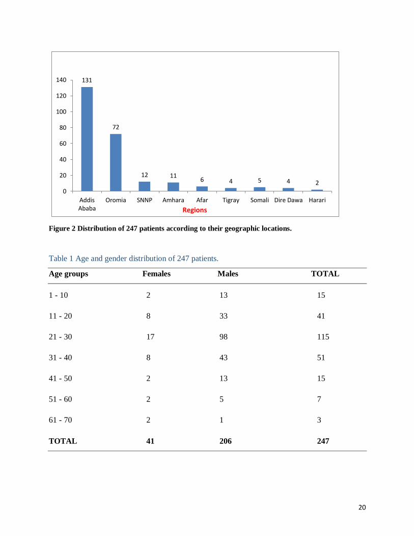
The majority of the patients were males (n=206, 83%), while the rest (n=41, 17%) were females
with the male to female ratio of 5:1. The age of patients ranged between 3 and 67 years, with the
mean of 27.70 years and median of 26.0 years. The maximum incidence occurred in 21-30 years age
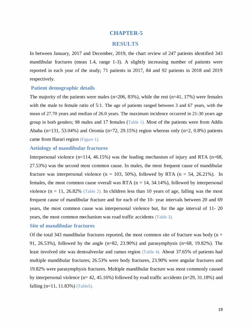
group in both genders; 98 males and 17 females (Table 1). Most of the patients were from Addis
Ababa (n=131, 53.04%) and Oromia (n=72, 29.15%) region whereas only (n=2, 0.8%) patients
came from Harari region (Figure 1).
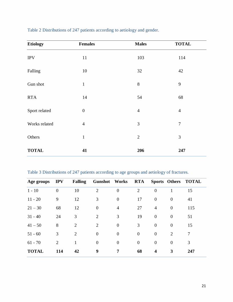
Aetiology of mandibular fractures
Interpersonal violence (n=114, 46.15%) was the leading mechanism of injury and RTA (n=68,
27.53%) was the second most common cause. In males, the most frequent cause of mandibular
fracture was interpersonal violence (n = 103, 50%), followed by RTA (n = 54, 26.21%). In
females, the most common cause overall was RTA (n = 14, 34.14%), followed by interpersonal
violence (n = 11, 26.82% (Table 2). In children less than 10 years of age, falling was the most
frequent cause of mandibular fracture and for each of the 10- year intervals between 20 and 69
years, the most common cause was interpersonal violence but, for the age interval of 11- 20
years, the most common mechanism was road traffic accidents (Table 3).
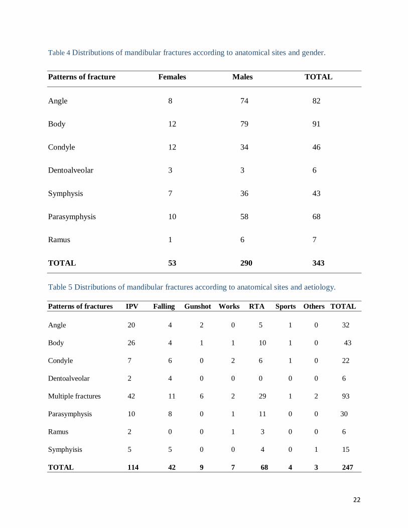
Site of mandibular fractures
Of the total 343 mandibular fractures reported, the most common site of fracture was body (n =
91, 26.53%), followed by the angle (n=82, 23.90%) and parasymphysis (n=68, 19.82%). The
least involved site was dentoalveolar and ramus region (Table 4). About 37.65% of patients had
multiple mandibular fractures; 26.53% were body fractures, 23.90% were angular fractures and
19.82% were parasymphysis fractures. Multiple mandibular fracture was most commonly caused
by interpersonal violence (n= 42, 45.16%) followed by road traffic accidents (n=29, 31.18%) and
falling (n=11, 11.83%) (Table5).
20
131
72
12 11 6 4 5 4 2
0
20
40
60
80
100
120
140
AddisAbaba
Oromia SNNP Amhara Afar Tigray Somali Dire Dawa Harari
Regions
Figure 2 Distribution of 247 patients according to their geographic locations.
Table 1 Age and gender distribution of 247 patients.
Age groups Females Males TOTAL
1 - 10 2 13 15
11 - 20 8 33 41
21 - 30 17 98 115
31 - 40 8 43 51
41 - 50 2 13 15
51 - 60 2 5 7
61 - 70 2 1 3
TOTAL 41 206 247
21
Table 2 Distributions of 247 patients according to aetiology and gender.
Etiology Females Males TOTAL
IPV 11 103 114
Falling 10 32 42
Gun shot 1 8 9
RTA 14 54 68
Sport related 0 4 4
Works related 4 3 7
Others 1 2 3
TOTAL 41 206 247
Table 3 Distributions of 247 patients according to age groups and aetiology of fractures.
Age groups IPV Falling Gunshot Works RTA Sports Others TOTAL
1 - 10 0 10 2 0 2 0 1 15
11 - 20 9 12 3 0 17 0 0 41
21 – 30 68 12 0 4 27 4 0 115
31 - 40 24 3 2 3 19 0 0 51
41 – 50 8 2 2 0 3 0 0 15
51 - 60 3 2 0 0 0 0 2 7
61 - 70 2 1 0 0 0 0 0 3
TOTAL 114 42 9 7 68 4 3 247
22
Table 4 Distributions of mandibular fractures according to anatomical sites and gender.
Patterns of fracture Females Males TOTAL
Angle 8 74 82
Body 12 79 91
Condyle 12 34 46
Dentoalveolar 3 3 6
Symphysis 7 36 43
Parasymphysis 10 58 68
Ramus 1 6 7
TOTAL 53 290 343
Table 5 Distributions of mandibular fractures according to anatomical sites and aetiology.
Patterns of fractures IPV Falling Gunshot Works RTA Sports Others TOTAL
Angle 20 4 2 0 5 1 0 32
Body 26 4 1 1 10 1 0 43
Condyle 7 6 0 2 6 1 0 22
Dentoalveolar 2 4 0 0 0 0 0 6
Multiple fractures 42 11 6 2 29 1 2 93
Parasymphysis 10 8 0 1 11 0 0 30
Ramus 2 0 0 1 3 0 0 6
Symphyisis 5 5 0 0 4 0 1 15
TOTAL 114 42 9 7 68 4 3 247
23
Role of patients and associated injuries
Among patients injured by RTA, the largest number were pedestrians (n=37, 43.02%) followed
by motorcycle drivers (n=19, 22.09%). The majority of males were involved in motorcycle +
vehicle driving (n=30) while more females were pedestrians (n=14). About 76 patients had other
associated injuries, of which head injury contributed larger number (n=58) followed by cervical
spine injury (n=10) and few patients had chest and abdominal injuries (n=8).
Combinations of mandibular fracture
The most common combinations were body + angle (n=23, 24.73%) and Parasymphysis + angle
(n = 14, 15.05%) fractures while the least was symphysis + ramus (n=1, 1.07%). Of the total 93
combined fractures almost all were treated by ORIF (n=89, 95.69%). Most of the fractures were
combinations of two while only three patients had combination of three fractures.
Treatment modalities
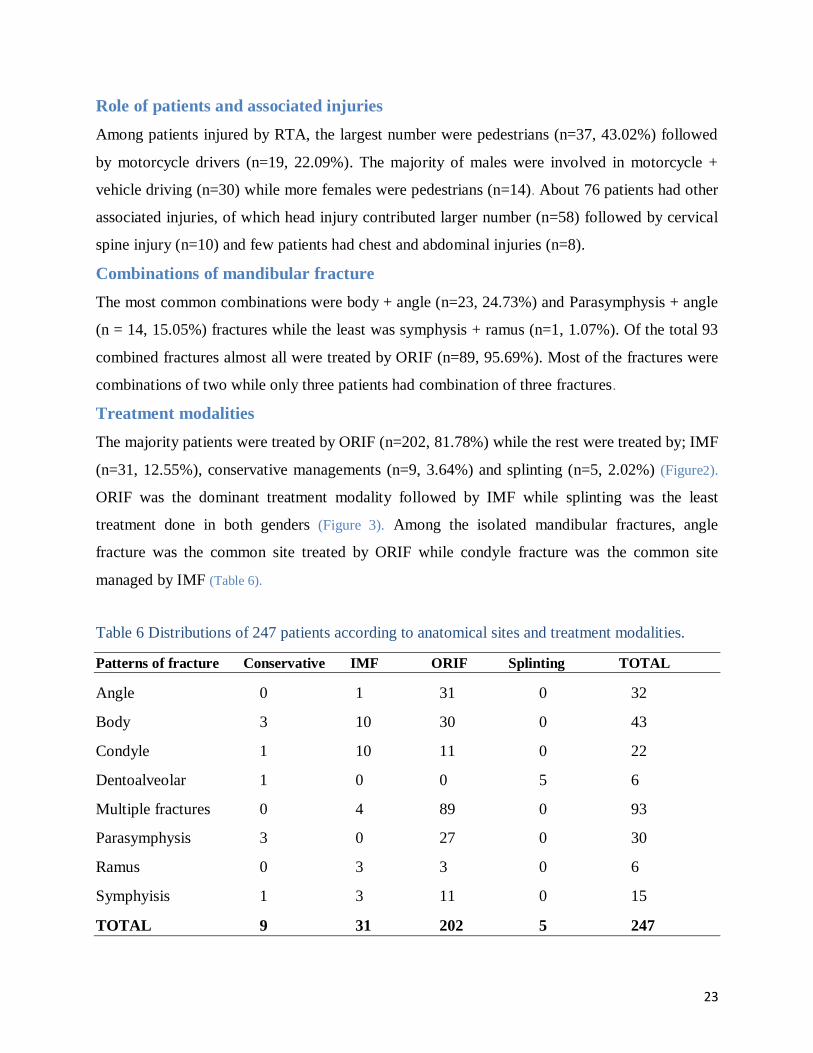
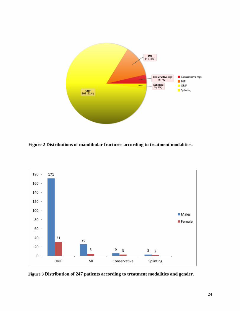
The majority patients were treated by ORIF (n=202, 81.78%) while the rest were treated by; IMF
(n=31, 12.55%), conservative managements (n=9, 3.64%) and splinting (n=5, 2.02%) (Figure2).
ORIF was the dominant treatment modality followed by IMF while splinting was the least
treatment done in both genders (Figure 3). Among the isolated mandibular fractures, angle
fracture was the common site treated by ORIF while condyle fracture was the common site
managed by IMF (Table 6).
Table 6 Distributions of 247 patients according to anatomical sites and treatment modalities.
Patterns of fracture Conservative IMF ORIF Splinting TOTAL
Angle 0 1 31 0 32
Body 3 10 30 0 43
Condyle 1 10 11 0 22
Dentoalveolar 1 0 0 5 6
Multiple fractures 0 4 89 0 93
Parasymphysis 3 0 27 0 30
Ramus 0 3 3 0 6
Symphyisis 1 3 11 0 15
TOTAL 9 31 202 5 247
24
Figure 2 Distributions of mandibular fractures according to treatment modalities.
171
26
6 3
31
5 3 2 0
20
40
60
80
100
120
140
160
180
ORIF IMF Conservative Splinting
Males
Female
Figure 3 Distribution of 247 patients according to treatment modalities and gender.
25
CHAPTER-6
DISCUSSIONS
Sociodemographic and Incidence
Socioeconomic, geographical, and cultural factors have been shown to influence the patterns of
mandibular fracture in a given population (5, 8, 10, 29). Most of the patients were from Addis
Ababa (53.04%) and Oromia region (29.15%) whereas only (0.8%) of patients came from Harari
region. This predominance could be explained by the fact that, Oromia region and Addis Ababa
city government are geographically close to the study area while other regions are on far
distance; they might also have maxillofacial surgery services in their respective more closer
areas. In line with other study, a comparable proportion of patients sustained mandibular
fractures at one site (44.9%) and 2 sites (52.4%) with mean of 1.4 fractures/mandible (8). The
overall 3 years incidence was almost similar which showed a little increments that showed
similarities with some other studies (11, 12).
The overall peak age group for mandibular fractures in this study for both gender was 21–30
years, which is similar to most studies in the literatures (6, 8, 10, 14, 20, 29). Mandibular fractures
have been reported in all age groups ranging from 3-67 years, which is consistent to most of the
studies (7, 17, 28, 29). The outcome of the study sample was predominantly male (83.4%) ;with
male to female ratio of 5:1 which is almost similar to the study conducted in Canada ,Kuwait and
Muhimbili National Hospital, Tanzania (10, 22, 26). These findings of the current study might be
attributed to the fact that, people are more active during second and third decades of life than
other decades, making them more vulnerable to trauma. Moreover, men participate in more
outdoor activities than women.
Etiology
The most common cause of mandibular fracture in the study group was interpersonal violence
(46.15%) followed by road traffic accidents (27.53%) and falling (17%) that coincided with the
studies made in Muhimbili national hospital in Tanzania, Kisi district hospital in Kenya and
Gondar university teaching hospital in Ethiopia (25, 28, 29). However, most of the literatures done
in developing countries showed RTA as the leading cause of mandibular fractures (23, 24, 27).
26
The principal cause of mandibular fracture varies with gender in the study group; in males
interpersonal violence (n=103, 50%) was most common cause followed by RTA (n= 54,
26.21%) whereas in females, the most common cause overall was RTA (n=14, 34.14%),
followed by interpersonal violence (n= 11, 26.82%) which is supported with one study done in
participating trauma centres of USA (9).
The majority of the patients injured by road traffic accidents (RTA) were motorcyclist + vehicle
drivers (n=30) in males whereas pedestrians (n=14) in females which might be explained by the
engagement of males in outdoor jobs like driving than females in Ethiopian culture. Over all, the
high prevalence of RTA was observed in pedestrians in the present study, which may reflect the
low level of community awareness on road traffic safety and road use. In addition, the absence of
pedestrian’s walkways in most of the roads in Addis Ababa, Ethiopia, may have contributed to
the higher vulnerability of pedestrians to motorized vehicles.
In children less than 10 years of age, falling was the most frequent cause of mandibular fracture;
for the age interval of 11- 20 years, the most common mechanism was road traffic accidents and
for the rest interval of 10 year age groups the leading cause of mandibular fracture was
interpersonal violence which is in line with one study conducted in London teaching hospital (8).
This might be due to low standard of socioeconomic life of the societies, low tolerance habit in
the community and frequent intertribal quarrelling in the country.
Pattern of fractures
The most common site of mandibular fracture was body (26.53%) followed by angle (23.9%)
and the least site fractured was ascending ramus which is in line with some literatures (1, 28),
however, some other studies showed condylar fracture predominance(13, 15, 19). The largest
number of patients had multiple mandibular fractures (37.65%) out of which body + angle
(24.73%) combined fracture comprised highest number while symphysis + ramus (1.04%)
fracture was least number recorded which is supported by one study result conducted in Montreal
General Hospital, Canada between 1998 and 2003 and 307 mandibular fractures (11). Out of the
total number of study sample, about 30.76% patients had non-facial associated injuries. Head
injury (23.48%) was the most common followed by cervical spine injury (4.05%) and these value
coincided with few literatures (1, 11).
27
Treatment modalities
The dominant treatment modality was ORIF (81.78%) followed by IMF (12.55%), while the rest
few patients were treated by splinting and conservative managements. Even though, open
reduction and internal fixation was the most common treatment used in many literatures, the
findings in this current study group showed higher value than those literatures (8, 10, 14, 19). This
finding could be explained by the reason that, most of the fracture patterns identified in the study
sample were multiple in natures and displaced. On the other hand, few literatures demonstrated
higher values of intermaxillary fixation as compared to other treatment options (13, 23). Among
the isolated single site mandibular fractures, angle fracture was the common site treated by ORIF
while condyle fracture was the common site managed by IMF. This variation might be related to
difficulty to achieve appropriate anatomic reduction while trying to treat angle fracture by
intermaxillary fixation due to increased muscle pull effects than other sites.
Limitations of the study
- Some of the patient’s charts were lost from the shelf while some others were incomplete
charts that made them difficult to include in the study sample.
- The study was unable draw valid conclusion regarding the impacts of patients social
history like their jobs and use of illicit substances on patterns of mandibular fracture as
these were not well documented in the patient’s charts.
28
CHAPTER-7
CONCLUSIONS AND RECOMMENDATIONS
CONCLUSIONS
The majority of the patients were from Addis Ababa and Oromia region. Most of the patients
who experienced mandibular fractures were young males. The major etiological factor was
interpersonal violence, followed by road traffic accidents (RTA) in all study age groups whereas
falling was the most common cause of fracture in children less than 10 years old and older
people above 60 years old. The most frequently fractured site was body of the mandible followed
by the angle. The most common combined fracture was body + angle. The dominant treatment
modality was open reduction and internal fixation (ORIF) especially for multiple site mandibular
fractures.
RECOMMENDATIONS
- The high incidence of interpersonal violence in the study group suggests a need for
government to prepare programs creating greater awareness on cultural, economic and
social problems and improve tolerance among communities to minimize the role of
violence in trauma.
- The significant role of RTA in causing mandibular fracture warns the government’s
body, transport office personnel, communities and other stake holders to work on road
safety rules so as to minimize the injuries.
- The high prevalence of falling among children and old age groups indicates, taking
special care in prevention of falling in these age groups is strongly recommended.
- Since the number/sites of fracture are influenced by the cause of trauma, the detailed
history on mechanism of injury and physical examination should be taken.
29
Annex I: References
1. Peterson LJ. Peterson's principles of oral and maxillofacial surgery: PMPH-USA; 2012.
2. Malik S, Singh G. Incidence, aetiology and pattern of mandible fractures in Sonepat, Haryana
(India). Int J Med Dent. 2014;4:51-9.
3. Fonseca RJ. Oral and Maxillofacial Surgery-E-Book: 3-Volume Set: Elsevier Health Sciences;
2017.
4. Zix J, Schaller B, Lieger O, Saulacic N, Thoren HA, Iizuka T. Incidence, aetiology and pattern
of mandibular fractures in central Switzerland. Swiss medical weekly. 2011;141(w13207):w13207.
5. Giri KY, Singh AP, Dandriyal R, Indra N, Rastogi S, Mall SK, et al. Incidence and pattern of
mandibular fractures in Rohilkhand region, Uttar Pradesh state, India: A retrospective study.
Journal of oral biology and craniofacial research. 2015;5(3):140-5.
6. Barde D, Mudhol A, Madan R. Prevalence and pattern of mandibular fracture in Central India.
National journal of maxillofacial surgery. 2014;5(2):153.
7. Natu SS, Pradhan H, Gupta H, Alam S, Gupta S, Pradhan R, et al. An epidemiological study
on pattern and incidence of mandibular fractures. Plastic surgery international. 2012;2012.
8. Rashid A, Eyeson J, Haider D, van Gijn D, Fan K. Incidence and patterns of mandibular
fractures during a 5-year period in a London teaching hospital. British journal of oral and
maxillofacial surgery. 2013;51(8):794-8.
9. Afrooz PN, Bykowski MR, James IB, Daniali LN, Clavijo-Alvarez JA. The epidemiology of
mandibular fractures in the United States, part 1: a review of 13,142 cases from the US National
Trauma Data Bank. Journal of oral and maxillofacial surgery. 2015;73(12):2361-6.
10. Sojat AJ, Meisami T, Sàndor GK, Clokie CM. Epidemiology of mandibular fractures treated
at the toronto general hospital: a review of 246 cases. Journal-Canadian Dental Association.
2001;67(11):640-5.
11. Czerwinski M, Parker W, Chehade A, Williams H. Identification of mandibular fracture
epidemiology in Canada: enhancing injury prevention and patient evaluation. Canadian Journal
of Plastic Surgery. 2008;16(1):36-40.
12. Oikarinen K, Thalib L, Sàndor GK, Schutz P, Clokie CM, Safar S, et al. Differences in the
location and multiplicity of mandibular fractures in Kuwait, Canada and Finland during the
1990s. Medical Principles and Practice. 2005;14(1):10-5.
30
13. Ferreira PC, Amarante JM, Silva AC, Pereira JM, Cardoso MA, Rodrigues JM. Etiology and
patterns of pediatric mandibular fractures in Portugal: a retrospective study of 10 years. Journal
of Craniofacial Surgery. 2004;15(3):384-91.
14. Martini MZ, Takahashi A, Oliveira Neto HGd, Carvalho Júnior JPd, Curcio R, Shinohara
EH. Epidemiology of mandibular fractures treated in a Brazilian level I trauma public hospital in
the city of São Paulo, Brazil. Brazilian dental journal. 2006;17(3):243-8.
15. Samman M, Ahmed SW, Beshir H, Almohammadi T, Patil SR. Incidence and pattern of
mandible fractures in the Madinah Region: A retrospective study. Journal of natural science,
biology, and medicine. 2018;9(1):59.
16. Akhlaghi F, Mafi N, Bastami F. Prevalence of Maxillofacial Fractures and Related Factors:
A Five-Year Retrospective Study. Trauma Monthly. 2019;24(4):e83974-e.
17. Chaurasia A, Katheriya G. Prevalence of mandibular fracture in patients visiting a tertiary
dental care hospital in North India. National journal of maxillofacial surgery. 2018;9(2):123.
18. Malik S, Singh G. Incidence, aetiology and pattern of mandible fractures in Sonepat,
Haryana (India). International Journal of Medical Dentistry. 2014;4(1):51.
19. Ghodke MH, Bhoyar SC, Shah SV. Prevalence of mandibular fractures reported at CSMSS
Dental College, aurangabad from february 2008 to september 2009. Journal of International
Society of Preventive & Community Dentistry. 2013;3(2):51.
20. Bither S, Mahindra U, Halli R, Kini Y. Incidence and pattern of mandibular fractures in rural
population: a review of 324 patients at a tertiary hospital in Loni, Maharashtra, India. Dental
traumatology. 2008;24(4):468-70.
21. Rashid S, Kundi JA, Sarfaraz A, Qureshi AU, Khan A. Patterns of Mandibular Fractures and
Associated Comorbidities in Peshawar, Khyber Pakhtunkhwa. Cureus. 2019;11(9).
22. Schutz P, Safar S, Al-Yassin S, Belal M, Korinek P. Maxillofacial fractures in Kuwait
between 1992–1997. Asian J Oral Maxillofac Surg. 2001;13:195-201.
23. Sultana F, Karim MR, Haider IA. Epidemiological & Clinical Profile of Patients Presented
with Mandible Fracture in a Tertiary Care Hospital. Journal of Bangladesh College of Physicians
and Surgeons. 2018;36(3):107-11.
24. Bouguila J, Zairi I, Khonsari R, Lankriet C, Mokhtar M, Adouani A. Mandibular fracture: a
10-year review of 685 cases treated in Charles-Nicolle Hospital (Tunis-Tunisia). Revue de
stomatologie et de chirurgie maxillo-faciale. 2009;110(2):81-5.
31
25. Kalyanyama B, Shubi F, Simon E. Maxillofacial fractures among patients attended at
Muhimbili National hospital, Dar es Salaam. Tanzania Medical Journal. 2009;24(1).
26. Deogratius BK, Isaac MM, Farrid S. Epidemiology and management of maxillofacial
fractures treated at Muhimbili National Hospital in Dar es Salaam, Tanzania, 1998–2003.
International dental journal. 2006;56(3):131-4.
27. Almahdi HM, Higzi MA. Maxillofacial fractures among Sudanese children at Khartoum
Dental Teaching Hospital. BMC research notes. 2016;9(1):120.
28. Akama M, Chindia M, Ndungu F. Occurrence and pattern of mandibular fractures at Kisii
District Hospital, Kenya. East African medical journal. 1993;70(11):732-3.
29. Teshome A, Andualem G, Tsegie R, Seifu S. Two years retrospective study of maxillofacial
trauma at a tertiary center in North West Ethiopia. BMC research notes. 2017;10(1):373.
32
Annex II: Data collection Instruments
ADDIS ABABA UNIVERSITY, COLLEGE OF HEALTH SCIENCES, SCHOOL OF
MEDICINE, DEPARTMENT OF ORAL AND MAXILLOFACIAL SURGERY
This data collection format to be filled by data collector is designed for the purpose of data
gathering to assess Incidence and Pattern of Mandibular Fractures in Addis Ababa University
affiliated Hospitals (Yekatit 12 Hospital Medical College and St. Peter Specialized Hospital).
Instruction: - please read the information carefully and put ‘’√ ’’mark in front of the
information given or encircle the choices given if applicable. More than one choice is possible; if
the check list is open ended, write the information on the space provided.
Date: - _________________________
Code number:-___________________
DATA CALLECTION FORMAT
1. Demographic data:-
Sex: Male Female Age
2. Geographic location (Region): -
a. Tigray g. Benishangul Gumuz
b. Afar h. Gambella
c. Amhara i. Harari
d. Oromia j. Addis Ababa
e. SNNP k. Dire Dawa
f. Somali
33
3. Occupation or role of the patient:-
a. Vehicle driver
b. Motor cyclist
c. Pedestrian
d. Others (specify) ------------------------------------------------
4. Causative factors:-
a. RTA
b. Assault
c. Falling
d. Sport related
e. Work related
f. Others (Specify) --------------------------------------------------
5. Patterns of mandibular fracture:-
a. Symphysis e. Parasymphysis
b. Body f. Angle
c. Ramus g. Condyle
d. Coronoid h. Dentoalveolar
6. Associated injuries
a. Head injury
b. Cervical spine injury
c. Chest and abdomen
d. Others (specify)_____________________________
7. Treatment modality
a. ORIF (open reduction and internal fixation)
b. IMF (Intermaxillary fixation)
c. Conservative management
Related Documents