Adaptive HIV-Specific B Cell-Derived Humoral Immune Defenses of the Intestinal Mucosa in Children Exposed to HIV via Breast-Feeding Sandrine Moussa 1 *, Mohammad-Ali Jenabian 2¤a , Jean Chrysostome Gody 3,4 , Josiane Le ´al 1 , Ge ´ rard Gre ´ senguet 4 , Alain Le Faou 1¤b , Laurent Be ´ lec 2,5 1 Institut Pasteur de Bangui, Laboratoire des Re ´ trovirus-VIH, Bangui, Central African Republic, 2 Assistance Publique - Ho ˆ pitaux de Paris, Ho ˆ pital Europe ´en Georges Pompidou, Laboratoire de Virologie, Paris, France, 3 Complexe Pe ´diatrique, Bangui, Central African Republic, 4 Unite ´ de Recherches et d’Intervention sur les Maladies Sexuellement Transmissibles et le SIDA, De ´partement de Sante ´ Publique, Faculte ´ des Sciences de la Sante ´ de Bangui, Bangui, Central African Republic, 5 Faculte ´ de Me ´decine Paris Descartes, Sorbonne Paris Cite ´, Paris, France Abstract Background: We evaluated whether B cell-derived immune defenses of the gastro-intestinal tract are activated to produce HIV-specific antibodies in children continuously exposed to HIV via breast-feeding. Methods: Couples of HIV-1-infected mothers (n = 14) and their breastfed non HIV-infected (n = 8) and HIV-infected (n = 6) babies, and healthy HIV-negative mothers and breastfed babies (n = 10) as controls, were prospectively included at the Complexe Pe ´diatrique of Bangui, Central African Republic. Immunoglobulins (IgA, IgG and IgM) and anti-gp160 antibodies from mother’s milk and stools of breastfed children were quantified by ELISA. Immunoaffinity purified anti-gp160 antibodies were characterized functionally regarding their capacity to reduce attachment and/or infection of R5- and X4- tropic HIV-1 strains on human colorectal epithelial HT29 cells line or monocyte-derived-macrophages (MDM). Results: The levels of total IgA and IgG were increased in milk of HIV-infected mothers and stools of HIV-exposed children, indicating the activation of B cell-derived mucosal immunity. Breast milk samples as well as stool samples from HIV-negative and HIV-infected babies exposed to HIV by breast-feeding, contained high levels of HIV-specific antibodies, mainly IgG antibodies, less frequently IgA antibodies, and rarely IgM antibodies. Relative ratios of excretion by reference to lactoferrin calculated for HIV-specific IgA, IgG and IgM in stools of HIV-exposed children were largely superior to 1, indicating active production of HIV-specific antibodies by the intestinal mucosa. Antibodies to gp160 purified from pooled stools of HIV- exposed breastfed children inhibited the attachment of HIV-1NDK on HT29 cells by 63% and on MDM by 77%, and the attachment of HIV-1JRCSF on MDM by 40%; and the infection of MDM by HIV-1JRCSF by 93%. Conclusions: The intestinal mucosa of children exposed to HIV by breast-feeding produces HIV-specific antibodies harbouring in vitro major functional properties against HIV. These observations lay the conceptual basis for the design of a prophylactic vaccine against HIV in exposed children. Citation: Moussa S, Jenabian M-A, Gody JC, Le ´al J, Gre ´senguet G, et al. (2013) Adaptive HIV-Specific B Cell-Derived Humoral Immune Defenses of the Intestinal Mucosa in Children Exposed to HIV via Breast-Feeding. PLoS ONE 8(5): e63408. doi:10.1371/journal.pone.0063408 Editor: Stefan Po ¨ hlmann, German Primate Center, Germany Received February 21, 2013; Accepted April 1, 2013; Published May 21, 2013 Copyright: ß 2013 Moussa et al. This is an open-access article distributed under the terms of the Creative Commons Attribution License, which permits unrestricted use, distribution, and reproduction in any medium, provided the original author and source are credited. Funding: External funding sources were received for this study by a french research organism (INSERM). The funders had no role in study design, data collection and analysis, decision to publish, or preparation of the manuscript. Competing Interests: The authors have declared that no competing interests exist. * E-mail: [email protected] ¤a Current address: Chronic Viral Illness Service, Montreal Chest Institute Research Institute, McGill University Health Centre, Montreal, Quebec, Canada ¤b Current address: EA 3452, CITHEFOR, Faculte ´ de Pharmacie, Universite ´ de Lorraine, Nancy, France Introduction The UNAIDS estimated that more than 330,000 (280,000– 380,000) children were newly infected by human immunodefi- ciency virus type 1 (HIV-1) through mother-to-child transmission (MTCT) worldwide in 2011, with the majority (.90%) occurring in sub-Saharan Africa [1]. The majority of MTCT occurs during pregnancy and birth. In addition, postnatal transmission of HIV-1 from HIV-infected mother to her child through prolonged breast- feeding is well recognized, and may account for one-third to half of new infant HIV-1 infections worldwide [2–10]. While studies of maternal or infant antiretroviral therapy during the period of breast-feeding have shown substantial potential for reduction of infant HIV infections [11–14], postnatal virus transmissions may continue to occur even in the setting of optimal antiretroviral prophylaxis [15].Therefore, development of immunologic strate- gies to reduce HIV transmission via breast milk remains important for improving survival of babies born to HIV-infected mothers in the developing world. Despite the babies daily exposure via their oral and gastroin- testinal mucosae to high amounts of cell-associated and cell-free PLOS ONE | www.plosone.org 1 May 2013 | Volume 8 | Issue 5 | e63408

Welcome message from author
This document is posted to help you gain knowledge. Please leave a comment to let me know what you think about it! Share it to your friends and learn new things together.
Transcript
Adaptive HIV-Specific B Cell-Derived Humoral ImmuneDefenses of the Intestinal Mucosa in Children Exposed toHIV via Breast-FeedingSandrine Moussa1*, Mohammad-Ali Jenabian2¤a, Jean Chrysostome Gody3,4, Josiane Leal1,
Gerard Gresenguet4, Alain Le Faou1¤b, Laurent Belec2,5
1 Institut Pasteur de Bangui, Laboratoire des Retrovirus-VIH, Bangui, Central African Republic, 2Assistance Publique - Hopitaux de Paris, Hopital Europeen Georges
Pompidou, Laboratoire de Virologie, Paris, France, 3Complexe Pediatrique, Bangui, Central African Republic, 4Unite de Recherches et d’Intervention sur les Maladies
Sexuellement Transmissibles et le SIDA, Departement de Sante Publique, Faculte des Sciences de la Sante de Bangui, Bangui, Central African Republic, 5 Faculte de
Medecine Paris Descartes, Sorbonne Paris Cite, Paris, France
Abstract
Background: We evaluated whether B cell-derived immune defenses of the gastro-intestinal tract are activated to produceHIV-specific antibodies in children continuously exposed to HIV via breast-feeding.
Methods: Couples of HIV-1-infected mothers (n = 14) and their breastfed non HIV-infected (n = 8) and HIV-infected (n = 6)babies, and healthy HIV-negative mothers and breastfed babies (n = 10) as controls, were prospectively included at theComplexe Pediatrique of Bangui, Central African Republic. Immunoglobulins (IgA, IgG and IgM) and anti-gp160 antibodiesfrom mother’s milk and stools of breastfed children were quantified by ELISA. Immunoaffinity purified anti-gp160 antibodieswere characterized functionally regarding their capacity to reduce attachment and/or infection of R5- and X4- tropic HIV-1strains on human colorectal epithelial HT29 cells line or monocyte-derived-macrophages (MDM).
Results: The levels of total IgA and IgG were increased in milk of HIV-infected mothers and stools of HIV-exposed children,indicating the activation of B cell-derived mucosal immunity. Breast milk samples as well as stool samples from HIV-negativeand HIV-infected babies exposed to HIV by breast-feeding, contained high levels of HIV-specific antibodies, mainly IgGantibodies, less frequently IgA antibodies, and rarely IgM antibodies. Relative ratios of excretion by reference to lactoferrincalculated for HIV-specific IgA, IgG and IgM in stools of HIV-exposed children were largely superior to 1, indicating activeproduction of HIV-specific antibodies by the intestinal mucosa. Antibodies to gp160 purified from pooled stools of HIV-exposed breastfed children inhibited the attachment of HIV-1NDK on HT29 cells by 63% and on MDM by 77%, and theattachment of HIV-1JRCSF on MDM by 40%; and the infection of MDM by HIV-1JRCSF by 93%.
Conclusions: The intestinal mucosa of children exposed to HIV by breast-feeding produces HIV-specific antibodiesharbouring in vitro major functional properties against HIV. These observations lay the conceptual basis for the design of aprophylactic vaccine against HIV in exposed children.
Citation: Moussa S, Jenabian M-A, Gody JC, Leal J, Gresenguet G, et al. (2013) Adaptive HIV-Specific B Cell-Derived Humoral Immune Defenses of the IntestinalMucosa in Children Exposed to HIV via Breast-Feeding. PLoS ONE 8(5): e63408. doi:10.1371/journal.pone.0063408
Editor: Stefan Pohlmann, German Primate Center, Germany
Received February 21, 2013; Accepted April 1, 2013; Published May 21, 2013
Copyright: � 2013 Moussa et al. This is an open-access article distributed under the terms of the Creative Commons Attribution License, which permitsunrestricted use, distribution, and reproduction in any medium, provided the original author and source are credited.
Funding: External funding sources were received for this study by a french research organism (INSERM). The funders had no role in study design, data collectionand analysis, decision to publish, or preparation of the manuscript.
Competing Interests: The authors have declared that no competing interests exist.
* E-mail: [email protected]
¤a Current address: Chronic Viral Illness Service, Montreal Chest Institute Research Institute, McGill University Health Centre, Montreal, Quebec, Canada¤b Current address: EA 3452, CITHEFOR, Faculte de Pharmacie, Universite de Lorraine, Nancy, France
Introduction
The UNAIDS estimated that more than 330,000 (280,000–
380,000) children were newly infected by human immunodefi-
ciency virus type 1 (HIV-1) through mother-to-child transmission
(MTCT) worldwide in 2011, with the majority (.90%) occurring
in sub-Saharan Africa [1]. The majority of MTCT occurs during
pregnancy and birth. In addition, postnatal transmission of HIV-1
from HIV-infected mother to her child through prolonged breast-
feeding is well recognized, and may account for one-third to half of
new infant HIV-1 infections worldwide [2–10]. While studies of
maternal or infant antiretroviral therapy during the period of
breast-feeding have shown substantial potential for reduction of
infant HIV infections [11–14], postnatal virus transmissions may
continue to occur even in the setting of optimal antiretroviral
prophylaxis [15].Therefore, development of immunologic strate-
gies to reduce HIV transmission via breast milk remains important
for improving survival of babies born to HIV-infected mothers in
the developing world.
Despite the babies daily exposure via their oral and gastroin-
testinal mucosae to high amounts of cell-associated and cell-free
PLOS ONE | www.plosone.org 1 May 2013 | Volume 8 | Issue 5 | e63408

HIV-1, estimated to be more than 700,000 viral particles per day
[16], HIV acquisition in exposed breastfed children occurs
infrequently. The overall probability of transmission via breast-
feeding was estimated to range from to 0.050 [17] to 0.064 [18]
percent per liter of breast milk ingested. Consumption of 0.5–1.0
liter of breast milk daily provides continuous exposure to
potentially infectious virus through the oral cavity and the
gastrointestinal mucosa. On the other hand, less than 10% of
babies born to HIV-infected women and breastfed during the first
6 months of life become infected postnatal [19], indicating low
efficiency of breast milk transmission which is in contrast with the
daily exposure to high amount of infectious viral particles. The low
frequency of breast-feeding acquisition suggests that anti-infective
factors in breast-feeding HIV-infected mothers as well as in HIV
exposed breastfed children are involved [20]. The fact that the
majority of breastfed babies of HIV-infected mothers remain
uninfected even after several months of breast-feeding constitutes
one of the major paradoxes of HIV transmission via breast milk
[21].
The majority of exposures to HIV-1 in breastfed children is
across oral mucosa, tonsillar tissue and gastrointestinal mucosa,
which are immunocompetent tissues, belonging to the afferent
branch of the mucosa-associated lymphoid tissue (MALT) [22].
Induction of mucosal immunity against HIV following prolonged
child exposure to infected breast milk is an attractive hypothesis
[21,23,24]. Previous studies showed that HIV-1-uninfected babies
exposed to HIV via breast-feeding may develop HIV-1-specific
salivary IgA [25] as well as systemic HIV-specific CD8 cytotoxic
immune responses [26].Overall, these observations suggest that
the specific humoral and cellular immune defenses are activated in
breastfed children. The confirmation that the child exposed to
HIV through breast-feeding actually develops protective specific
immunity against the acquisition of the virus could have major
importance for the demonstration of immunological correlates of
protection, and for the design of a prophylactic vaccine.
The aim of the present study was to evaluate whether B cell-
derived immune defenses of gastro-intestinal tract are activated to
produce HIV-specific antibodies in breastfed children continuous-
ly exposed to HIV via breast-feeding. For that purpose, HIV-
specific antibodies were first detected in immunoglobulins purified
from stools of breastfed children and characterized immunochem-
ically. Furthermore, their functional properties were assessed by
their capability to hamper in vitro the attachment of the virus to
intestinal epithelial cells and monocyte-derived macrophages
(MDM), and further by their aptitude to modulate negatively
HIV production in cell culture.
Materials and Methods
Inclusion of Mothers and their Breastfed BabiesCouples of HIV-1-infected mothers and their breastfed babies
were consecutively recruited at the Complexe Pediatrique, the
principal health care clinic for HIV-infected children held in
Bangui, the capital city of the Central African Republic [27–
29].The study was formally approved by the Scientific Committee
of the Faculte des Sciences de la Sante (‘‘FACSS’’) of Bangui (so-
called ‘‘Comite Scientifique Charge de la Validation des
Protocoles d’Etudes et des Resultats’’/’’CSCVPER’’) (agreement
2UB/FACSS/CSCVPER/05), constituting the National Ethical
Committee. Informed written consent was obtained from mothers
for themselves and on behalf of their respective child participating
in the study. All HIV-infected mother and their babies received
care, and when indicated antiretroviral treatment, according to the
WHO recommendations for the management of HIV infection in
resource-limited settings [30,31]. All HIV-infected children
received co-trimoxazole as prophylaxis against opportunistic
infections [32].
Inclusion criteria for HIV-infected mother-child couples in the
study were as follows: i) HIV-infected mother; ii) baby born from
HIV-infected mother and exposed to HIV by exclusive breast-
feeding from birth; iii) early diagnosis of HIV infection or non
HIV infection at time of sampling in babies born from HIV-
infected mother by molecular virological diagnosis; iv) mother and
babies care according to the national guidelines. The exclusion
criteria were: i) HIV diagnosis not formally established; ii) lack of
informal consent; iii) recent (,1 month) past history of gastro-
enteritis in breastfed children. Note that at time of period
inclusion, interruption of antiretroviral drugs availability through-
out the country unfortunately has not allowed to treat any HIV-
infected mothers or children for a period of at least one month
before inclusion.
Ten healthy volunteer HIV-seronegative breast-feeding women
and their breastfed HIV-non infected babies from the same setting
were also included as negative controls.
Collection and Processing of Clinical SamplesK3-EDTA-blood samples were obtained from study mothers
and their babies by venipuncture in Vacutainer tubes (Becton
Dickinson, Franklin Lakes, NJ, USA). The plasma was separated
after centrifugation at 10006g for 10 minutes, and aliquots were
kept frozen at 280uC within 2 hours after sampling until
processing. Of note, maternal milk and infant stool samples were
collected at the same time during the same visit.
Milk samples (10 ml) were collected manually, and then
centrifuged at 9,3006g for 20 minutes at +4uC, allowing
separation of the cellular, supernatant and lipid fractions, as
previously described [33]. The pellet and fat layer were discarded,
and the supernatant was collected, and aliquots were stored at
280uC until processing.
Stool samples from babies were collected at room temperature,
and then mixed with ‘‘cold buffer’’ conserved at +4uC. This bufferis constituted by phosphate buffered saline (PBS, pH=7.3)
containing 1 mM of the serine proteases inhibitor phenyl methyl
sulphonyl fluoride (Sigma Aldrich, St-Louis, MO, USA) (10% wt/
vol). The mixture was vortexed for at least 1 minute, and then let
for 10 minutes at room temperature, and then centrifuged at
2,7006g for 15 minutes at +4uC. Aliquots of resulting supernatantswere stored at 280uC until use.
Diagnosis of HIV InfectionMolecular diagnosis of HIV in children born from HIV-infected
mother was carried out by assessing the circulating plasma HIV-1
RNA load, as previously shown [34]. HIV-1 RNA load in plasma
from babies was measured by the Generic HIV-1 RNA
quantification assay (Biocentric, Bandol, France) using the ABI
PRISM 7000 real-time PCR system (Applied Biosystems,
California, USA), as previously described [35].
Antibodies and ReagentsAnti-human Fc fragment of IgA (a-chain specific), anti-human
Fc fragment of IgG (c-chain specific), and peroxidase (PO)-labeled
anti-human IgG(c-chain specific) were obtained from Pierce
(Rockford, IL, USA). Anti-human Fc fragment of IgM (m-chainspecific), biotinylated anti-human IgM (m-chain specific), and anti-
human lactoferrin (Lf), were obtained from Sigma Aldrich.
Horseradish peroxidase (HRPO)-labeled streptavidin was ob-
tained from Immunotech (Marseille, France). The anti-human
IgA-PO-conjugated, the anti-human Lf-PO-conjugated and anti-
HIV Abs in Stool of Breastfed HIV-Exposed Babies
PLOS ONE | www.plosone.org 2 May 2013 | Volume 8 | Issue 5 | e63408

human F(ab’)2-PO-conjugated were from our laboratory. Anti-
HIV-1 gp120 monoclonal antibodies IgG2G12 and IgG1B12
were obtained from the AIDS Reagent Program, Division of
AIDS, NIAID, NIH [36].
The gp160 antigen consisted of a purified preparation of
baculovirus-expressed recombinant gp160 (rgp160) derived from
the envelope of the HIV-1MN/LAI strain (kindly provided by
Aventis-Pasteur, Paris, France). Recombinant human macrophage
colony-stimulating factor (rhM-CSF) was from R&D Systems
Europe (Abingdon, United Kingdom). RPMI 1640 (with L-
glutamine) was provided by Cambrex (Verviers, Belgium), and
penicillin and streptomycin were provided by Invitrogen (Paisley,
United Kingdom). Medium for separation of lymphocytes (MSL)
was obtained from PAA (Les Mureaux, France), and fetal calf
serum (FCS) was provided by Eurobio (Les Ulis, France).
SepharoseH 4B was obtained from Sigma Aldrich and anti-human
F(ab’)2 from Jackson Immunoresearch (West Grove, PA, USA).
The HIV-1 p24 antigen capture enzyme-linked immunosorbent
assay (ELISA) was obtained from Innogenetics(Gent, Belgium).
Polyclonal anti-gp160 IgG was purified from a pool of sera from
HIV-1-infected patients (laboratoire de virologie, Hopital Eur-
opeen Georges Pompidou, Paris, France), to be used as positive
control in immunochemical assays detecting HIV-specific anti-
bodies, as previously described [37,38].
A stock solution of IVIg (50 mg/ml; 0.3 mM) corresponding to
pooled normal IgG obtained from plasma of healthy donors was
prepared in PBS and dialysed twice against large volume of PBS at
4uC to remove the stabilizing agents, as previously described [39].
IVIg contained mostly monomeric IgG (.95%) and was used as
negative control in functional inhibitory assays.
CellsPeripheral blood mononuclear cells (PBMC) were isolated from
buffy coats of healthy adult donors by Ficoll density gradient
centrifugation on MSL, as previously described [40]. Blood
samples from HIV-negative healthy donors for functional assays
were collected at the French Blood Establishment, Paris, France.
Informed written consents from all subjects were obtained before
blood sampling. The percentage of monocytes was determined by
flow cytometry (FACS Calibur, Becton Dickinson, NJ, USA) using
forward scatter and side scatter properties (FSC/SSC). PBMC
were re-suspended in RPMI 1640 medium supplemented with
glutamine, penicillin (100 IU/ml) and streptomycin (100 mg/ml).
Cells were seeded into 24 well-plates (Costar, Cambridge, MA) at
the concentration 16106 adherent cells/ml and incubated at 37uCfor 45 minutes. Non adherent cells were removed by 4 washes.
Adherent monocytes were incubated in RPMI medium with 10%
FCS, glutamine, and antibiotics in the presence of 10 ng/ml rhM-
CSF (10 ng/ml) to differentiate to macrophages, as previously
described [41]. The relative concentration of rhM-CSF improve
cell viability and maintained a neutral environment with respect to
activation marker quantitative expression (HLA-DR, CD14,
CD16), which remained similar to that of MDM cultured in
medium alone [41]. Half of the medium, including all supple-
ments, was replaced every 3 days. After 7 days of culture, adherent
cells corresponding to the macrophages-enriched fraction were
harvested, washed, and used for subsequent experiments [41,42].
At the time of collection, MDM were more than 90% pure,
expressing by flow cytometry analysis (CellQuest software, Becton
Dickinson) CD4+, CXCR4low, CCR5high+, CD14 (73%) and
CD11b (70%) (data not shown).
The HT-29 human colorectal epithelial cells line was provided
by the AmericanType Culture Collection (ATCC HTB-38,
Manassas, VA, USA). Cells were grown in RPMI 1640 medium
complemented with 10% FCS, penicillin (100 IU/ml) and
streptomycin (100 mg/ml). The HT-29 cells were CD42, DC-
SIGN2, CXCR4high+, CCR5low+ and GalCerhigh+ (data not
shown).
HIV StrainsPrimary X4-tropicstrain HIV-1NDK and R5-tropic strain HIV-
1JR-CSF were a gift of Prof. F. Barre-Sinoussi (Institut Pasteur,Paris,
France). Stocks of the HIV-1NDK strains were produced on IL-2-
activated peripheral blood lymphocytes (PBL) of healthy donors.
The HIV-1JR-CSF strain was amplified in MDM cultures. The
virus produced was clarified by centrifugation, and the HIV p24
concentration was determined by capture ELISA, and stored at
280uC. Tissue culture infective dose 50% (TCID50) of each stock
was calculated according to the Karber formula [43], 1 ng of p24
antigen corresponding to 1000 TCID50, as previously shown
[38].Primary strains are thought to be representative of viral
strains not adapted to their microcellular environment. Further-
more, monocytotropic (R5+) [44,45] and to a lesser extent
lymphocytotropic (X4+) [45] HIV-1 strains are present in breast
milk, and may participate to HIV mucosal crossing in exposed
receptive baby.
Quantification of Total IgA, IgG and IgM Antibodies inBreast Milk and Stool SamplesTotal immunoglobulins (IgA, IgG and IgM) in the mother’s
milk and in non-purified children’s stools were quantified by
asymmetrical ELISA, as previously described [46]. Briefly, plastic
plates were coated with goat anti-human a chain, c chain or mchain (all at 3 mg/ml) in PBS overnight at +4u, prior to washing
with PBS/0.1% Tween, and saturated with PBS/1% skimmed
milk. Serial dilutions of breast milk and stools supernatants were
then added for 1 hour at +37uC. After further washes, goat anti-human F(ab’)2 (2 mg/ml) coupled with peroxidase was added for 1
hour at 37uC. After extensive washes, the peroxidase activity was
revealed with o-phenylenediamine (OPD) (Sigma Aldrich), and the
optical density (OD) was read at 492 nm. Quantification of each
immunoglobulin class was assessed by extrapolation from standard
curves obtained by serial dilutions of a pool of 20 samples of
normal human colostrum, as described elsewhere [37,46].
Detection of Anti-gp160 Antibodies in Breast Milk andStool Samples and Calculation of their Specific activitiesThe detection of anti-gp160 antibodies in breast milk and
children’s stools was assessed by indirect ELISA, in part as
previously described [33]. Briefly, plastic plates were coated
overnight at +4u with rgp160 (1 mg/ml) in PBS. The plates were
washed with PBS/0.1% Tween prior to saturation with PBS/1%
skimmed powder milk. Dilutions of breast milk and stools
supernatants were then added, and incubated for 1 hour at
+37uC. After washing, peroxidase-labelled goat antibodies (2 mg/ml) to human F(ab’)2, IgA or IgG, were added for 1 hour at +37uCprior to addition of peroxidase substrate (OPD); for IgM
antibodies, biotinylated goat antibodies to IgM (0.5 mg/ml) were
first added for 1 hour at +37uC, followed by streptavidin-HRPO
for 10 minutes at +37uC, prior to addition of peroxidase substrate
(OPD). The cut-offs of F(ab’)2, IgA or IgG positivity for milk or
stools samples were defined as the mean OD plus 2 standard
deviations (SD) of the values obtained with breast milk or
children’s stools samples from the HIV-negative controls. The
cut-off of IgM positivity for milk or stools samples was defined as
the mean OD plus 3 SD of the values obtained with breast milk or
children’s stools samples from the HIV-negative controls. Finally,
HIV Abs in Stool of Breastfed HIV-Exposed Babies
PLOS ONE | www.plosone.org 3 May 2013 | Volume 8 | Issue 5 | e63408

the levels of IgA, IgG and IgM to gp160 in milk and stools samples
positive for anti-gp160 antibodies were expressed in arbitrary OD
(at 492 nm) units (AU).
The specific activities (SA) of antibodies to gp160 of the IgA
isotype (SAIgA to gp160) in breast milk (M) and stools (S) were
expressed as the ratio of AU (OD reactivity) per mg of total IgA,
according to the following formulae:
SAIgAtogp160,M~IgAtogp160½ �OD,M
totalIgA½ �M � 100
SAIgAtogp160,S~IgAtogp160½ �OD,S
totalIgA½ �S � 100
Similarly were calculated the SA of IgG to gp160 in milk
(SAIgG to gp160,M) and stools (SAIgG to gp160,S), as well as the
SA of IgM to gp160 in milk (SAIgM to gp160,M) and stools
(SAIgM to gp160,S).
Levels of Lactoferrin in Breast Milk and Stool SamplesLf in mother’s milk and in children’s stools was measured by
symmetrical ELISA. In brief, plastic plates were coated with anti-
human Lf (1 mg/ml) in PBS overnight at +4uC. The plates were
washed with PBS/0.1% Tween and saturated with PBS/1%
gelatin. Serial dilutions of mother’s milk, children’s stools
supernatants and human Lf in PBS (standard) were then added
in the plates and incubated for 1 hour at +37uC. After further
washes, goat anti-human Lf antibody coupled with peroxidase was
added for 1 hour at +37uC before addition of substrate (OPD) and
quantification of peroxidase activity by OD at 492 nm. Quanti-
fication of milk or stool Lf was assessed by extrapolation from the
standard curve obtained by serial dilutions of known amount of
human Lf.
Evaluation of Intestinal Production of StoolImmunoglobulins and Anti-gp160 AntibodiesLf is an iron-binding glycoprotein secreted from many epithelial
cells into most exocrine fluids, particularly in breast milk [47]. Lf is
thought to be poorly or not secreted by the intestinal mucosa of the
new born, and fecal Lf is mainly originating from breast milk in
breastfed infant [48].Indeed, the mean levels of fecal Lf reported in
the literature in bottle fed infants around 0.5 mg per day [49], thus
nearly 90% less than those usually reported in breast milk [50],
and around 12.5 mg per day in breastfed children, thus 25-fold
more than in bottle fed infants [49]. The fecal levels of Lf increase
in case of gastro-intestinal inflammatory or infectious diseases,
such as inflammatory bowel diseases, Crohn’s disease, ulcerative
colitis and gastro-enteritis [47,51]. In breastfed children, fecal Lf is
likely originating from breast milk intake, corresponding to
undegraded Lf passively seeped into the intestinal chyle, and to
a lesser extent from intestinal production, normally negligible in
absence of intestinal inflammation [52]. Similarly, it is possible to
consider that fecal immunoglobulins in breastfed children corre-
spond to undegraded immunoglobulins coming from breast milk
intake as well as to intestinal immunoglobulin production.
These latter considerations prompt us evaluating intestinal
production of stool immunoglobulins in breastfed children, taken
into account a simplified relationship between breast milk
immunoglobulins ([Ig]M) and Lf ([Lf]M), and stool immunoglob-
ulins ([Ig]S) and Lf ([Lf]S).
Thus, stool immunoglobulins is the sum of undegraded
immunoglobulins from breast milk ([Ig]S,bm), and intestinal
immunoglobulins production
([Ig]S,i): Ig½ �S~ Ig½ �S,bmz Ig½ �S,I .[Ig]S,bm corresponds to a frac-
tion a of [Ig]M : Ig½ �S,bm~a Ig½ �M.Finally,
Ig½ �S~a Ig½ �Mz Ig½ �S,i.Similarly, stool Lf is the sum of undegraded Lf from breast milk
([Lf]S,bm), and intestinal Lf production
([Lf]S,i): Lf½ �S~ Lf½ �S,bmz Lf½ �S,i. [Lf]S,bm corresponds to a
fraction a’ of [Lf]M : Lf½ �S,bm~a’ Lf½ �M. If one hypothesizes that
a~a’, because immunoglobulins and Lf are glycoproteins similarly
degraded in the intestinal chyle, and that the intestinal production
of Lf is negligible in the absence of gastro-intestinal inflammation
or gastro-enteritis ( Lf½ �S,i:&0), Lf½ �S~a Lf½ �M, and a~ Lf½ �SLf½ �M.
Thus, Ig½ �S~a Ig½ �Mz Ig½ �s,i~ Lf½ �SLf½ �M Ig½ �Mz Ig½ �S,iand
Ig½ �S,i~ Ig½ �S{(Lf½ �SLf½ �M ) Ig½ �M ð1Þ
In case of intestinal/fecal production of im-
munoglobulins,(I)w0, i:e:
Ig½ �Sw(Lf½ �SLf½ �M ) Ig½ �M, and
(Ig½ �SIg½ �M )
(Lf½ �SLf½ �M )
w1, i:e:
(Ig½ �SIg½ �M )
(Lf½ �MLf½ �S )
w1, i:e:
(Ig½ �SLf½ �S ) � ( Lf½ �M
Ig½ �M )w1
In opposite, in case of intestinal/fecal immunoglobulins
exclusively provided from breast milk, (Ig½ �SLf½ �S ) � (
Lf½ �MIg½ �M )v1.
Taken together, the following relative ratio of excretion (RRE).
RRE~(Ig½ �SLf½ �S ) � ( Lf½ �M
Ig½ �M )
may be used to evaluate the relative fecal/intestinal production of
immunoglobulins in feces from breastfed children, by reference to
Lf as breast milk intake factor, with the hypotheses that
immunoglobulins and Lf are glycoproteins similarly degraded
within intestinal chyle, and that the intestinal production of Lf is
negligible in the absence of gastro-intestinal inflammation or
gastro-enteritis in breastfed children. When this formula is applied
to HIV-specific antibodies, a RRE is superior to 1 in baby exposed
to HIV means that HIV-specific antibodies evidenced in stools are
not only originating from a passive ingestion of breast milk
antibodies, but rather indicates the infant intestinal mucosa likely
secretes actively HIV-specific antibodies.
HIV Abs in Stool of Breastfed HIV-Exposed Babies
PLOS ONE | www.plosone.org 4 May 2013 | Volume 8 | Issue 5 | e63408

Finally, in order to assess whether HIV-exposed children secrete
total and/or HIV-specific antibodies during breast-feeding in their
intestinal mucosa, we calculated the RRE of stool IgA (RREI-
gA,S), IgG (RREIgG,S), and IgM (RREIgM,S) by reference to Lf,
according to the following formulae:
RREIgA,S~(IgA½ �SLf½ �S ) � ( IgA½ �M
Lf½ �M )
RREIgG,S~(IgG½ �SLf½ �S ) � ( IgG½ �M
Lf½ �M )
RREIgM,S~(IgM½ �SLf½ �S ) � ( IgM½ �M
Lf½ �M )
Similarly, the RRE of stool gp160-specific IgA (RREIgA to
gp160,S), IgG (RREIgG to gp160,S), and IgM (RREIgM to
gp160,S), were calculated by reference to Lf, according to the
following formulae:
RREIgAtogp160,S~(IgAtogp160,S½ �
Lf½ �S )
(IgAtogp160,M½ �
Lf½ �M )
RREIgGtogp160,S~(IgGtogp160,S½ �
Lf½ �S )
(IgGtogp160,M½ �
Lf½ �M )
RREIgMtogp160,S~(IgMtogp160,S½ �
Lf½ �S )
(IgMtogp160,M½ �
Lf½ �M )
Thus,
RREIgAtogp160,S~(IgAtogp160½ �OD,S
Lf½ �S )
(IgAtogp160½ �OD,M
Lf½ �M )
RREIgGtogp160,S~(IgGtogp160½ �OD,S
Lf½ �S )
(IgGtogp160½ �OD,M
Lf½ �M )
RREIgMtogp160,S~(IgMtogp160½ �OD,S
Lf½ �S )
(IgMtogp160½ �OD,M
Lf½ �M )
The RRE of stool specific antibodies to gp160 of a given isotype
could be calculated only when the denominator is not zero, thus
when HIV-specific antibodies of the same isotype is detected in
corresponding breast milk.
Immunoaffinity Purification of Total Antibodies fromPooled Stools of Breastfed ChildrenTotal immunoglobulins were purified by immunoaffinity, as
previously described [46,53]. In brief, the anti-human F(ab’)2
was first coupled to SepharoseH 4B. Pool of stool samples
(supernatants) from HIV exposed non HIV-infected (group I)
and HIV-infected (group II) children, as well as from HIV non
exposed control babies were afterwards incubated with the
matrix overnight at +4uC before extensive washing of the
column with PBS until the OD of the effluent reached a value
of 0.001 at 280 nm. The column was then eluted with 0.2 M
glycine-HCl, pH 2.5. The eluate was rapidly neutralized with 1
MTris-HCl, pH 8.3, and dialysed against PBS overnight.
The isotype (IgA, IgG and IgM) composition of affinity-
purified anti-human F(ab’)2 was measured by ELISA. Plates
were coated with goat anti-human a-chain, c-chain or m-chain(all at 3 mg/ml) in PBS overnight at +4uC, prior to washing
with PBS/0.1% Tween and then saturated with PBS/1%
skimmed milk. Serial dilutions of pooled stools immunopurified
anti-F(ab’)2 antibodies were then added for one hour at +37uC.After further washes, goat anti-human F(ab’)2 (2 mg/ml) coupled
with peroxidase was added for 1 hour at +37uC. After extensivewashes, substrate (OPD) was added and peroxidase activity was
determined by OD at 492 nm. A pool of normal human sera
with known levels of IgA, IgG and IgM was used to obtain
standard curves.
Detection HIV-specific F(ab’)2 in Total ImmunoaffinityPurified Stool ImmunoglobulinsPlastic plates were coated overnight at +4uC with rgp160
(1 mg/ml) in PBS. The plates were washed with PBS/0.1%
Tween prior to saturation with PBS/1% skimmed milk. Serial
dilutions of antibodies purified from pooled stools were then
added and incubated for 1 hour at +37uC. After washing,
peroxidase-labelled goat anti-human F(ab’)2 (2 mg/ml) were
added for 1 hour at +37uC prior to addition of peroxidase
substrate (OPD). The levels of F(ab’)2 to gp160 in stools were
expressed in arbitrary OD (at 492 nm) units. The cut-off of
positivity was defined as the mean OD plus 2 SD of the values
obtained with breast milk samples from the HIV-negative
controls. The SA of purified F(ab’)2 to gp160 in pooled stools
from groups I and II were calculated as above as the ratio of
AU (OD reactivity at 492 nm) per mg of total IgAzIgGzIgMin each pool.
Inhibition of HIV-1 Attachment to HT29 Cells andMonocyte-derived Macrophages by Purified StoolsImmunoglobulinsHT29 cells or MDM (250,000 cells/well) were pre-incubated
with pooled stools immunoglobulins (30 or 50 mg/ml) purified
from HIV exposed group I and group II children for 1 hour at
+37uC before addition of 10 ng/ml HIV-1JRCSF p24 antigen and
HIV Abs in Stool of Breastfed HIV-Exposed Babies
PLOS ONE | www.plosone.org 5 May 2013 | Volume 8 | Issue 5 | e63408

HIV-1NDKp24 antigen for 1 hour at +37uC. Cells were then
extensively washed and lysed with 0.5% Triton, and HIV-1 p24
antigen levels were measured by ELISA. As attachment inhibition
positive control, 50 mg of Lf was added to cells prior to the
incubation with HIV, as previously described [54]. As attachment
inhibition negative controls, IVIg and total immunoglobulins
purified by immunoaffinity from pooled stools of HIV non
exposed control babies (50 mg/ml) were added to HT29 cells prior
to the incubation with HIV. Each experiment was carried out in
triplicate.
HIV-1 Infection Inhibition of Monocyte-derivedMacrophages by Purified Stools ImmunoglobulinsMDM were washed 2 times after 6 days of differentiation, and
seeded into 96-well culture plates (250,000 cells/well). At day 7,
pooled stools antibodies purified from HIV exposed group I and
group II children at 50 mg/ml were added to cells for 1 hour at
+37uC before addition of HIV-1JRCSF and HIV-1NDK (10 ng/
ml of p24 antigen). The cells were further incubated for 3 hours at
+37uC in a 5% CO2 atmosphere. After 4 washes to remove
exceeding virus, cells were cultured for 3 and 6 days. As inhibitory
positive control, monoclonal antibody IgG2G12 (20 mg/ml) was
added to MDM for 1 hour at +37uC before addition of HIV. As
negative controls, IVIg and total immunoglobulins purified from
pooled stools of HIV non exposed control babies (50 mg/ml) were
added to MDM prior to the incubation with HIV. The levels of
virus replication were estimated by HIV-1 p24 antigen ELISA on
the supernatant of cells culture. Each experiment was carried out
in triplicate.
Statistical AnalysisLevels of immunoglobulins, Lf, HIV-specific antibody SA, and
RRE were expressed as mean6standard error. Functional tests
were expressed as percentage 6 standard error. The non-
parametric Mann-Whitney U test was used for statistical analyses,
using GraphPad Prism 5.0 (San Diego, California, US) statistical
software. A P-value ,0.05 was considered as significant.
Results
Molecular Diagnosis of HIV Infection in Breastfed InfantsThe direct detection of circulating HIV RNA allows early
diagnosing of HIV infection in children aged less than 12 months
born from HIV-infected mother [30,55,56]. Among 36 couples of
HIV-1-infected mothers and their breastfed babies consecutively
recruited, 25 (69%) babies were negative for plasma HIV-1 RNA,
and thus HIV non infected, whereas 11 (31%) were positive, and
thus HIV-infected. We further selected mother-child couples
whose biological samples were in sufficient quantity for study
experiments, including 8 couples of HIV-1-infected mothers and
their breast milk exposed non infected babies (group I), and 6
couples of HIV-1-infected mothers and their breastfed infected
babies (group II). The main characteristics of the 14 study mother-
child couples are depicted in the Table 1. The mean duration of
exclusive breast-feeding in study couples was 4.5 months, without
difference in group I (mean duration: 4.6 months) and group II
(mean duration: 3.5 months). All but one (children #I) was
asymptomatic for HIV infection. At time of sampling, none of the
mothers had symptoms of mammary inflammation, and none of
the children showed gastro-intestinal symptoms or infectious
diarrhoea.
Quantification of Total IgA, IgG and IgM in Breast Milkand Children’s StoolsThe results of IgA, IgG and IgM levels in mothers’ milk and
children’s stools from couples of groups I and II, and from HIV-
negative control couples, are depicted in the Figure 1.
The mean concentrations of milk IgA in HIV-infected mothers
were higher than those of IgG, which were also higher than those
of IgM (milk IgA: 8086241 mg/ml in group I and 10256212 mg/ml in group II; milk IgG: 433641 mg/ml in group I and
447666 mg/ml in group II; milk IgM: 28611 mg/ml in group I
and 40620 mg/ml in group II; P,0.01 for all comparisons). In
HIV-negative control couples, the mean concentration of IgA in
milk (9696240 mg/ml) was higher than those of milk IgG
(78610 mg/ml) and IgM (1568 mg/ml), which were of similar
levels. Interestingly, the levels of milk IgG was 5.5 higher in HIV-
infected mothers than in HIV-negative control mothers (P,0.01).
The mean concentrations of stool IgA in babies born from HIV-
infected mothers were higher than those of IgG, which were
higher than those of IgM (stool IgA: 19096179 mg/ml in group I
and 17676287 mg/ml in group II; stool IgG: 104615 mg/ml in
group I and 108616 mg/ml in group II; stool IgM: 48616 mg/ml
in group I and 108658 mg/ml in group II; P,0.01 for all
comparisons excepting the comparison between stool IgG and
IgM in group II). In HIV-negative control couples, the mean
concentration of stool IgA (494648 mg/ml) was higher than those
of IgM (58627 mg/ml) (P,0.01), which were moderately higher
than those of IgG in stool (1763 mg/ml) (P,0.05). The levels of
stool IgA and IgG in babies born from HIV-infected mothers
(groups I and II) were higher than those of HIV-negative control
babies (P,0.01). The levels of stool IgM were higher than those of
HIV-negative babies only in group II (P,0.01).
HIV-specific F(ab’)2, IgA, IgG and IgM in Breast Milk andChildren’s StoolsThe detection of antibodies directed to gp160 in breast milk and
children’s stool samples and their corresponding calculated SA are
presented in the Table 2.
F(ab’)2, IgA and IgG to gp160 were present in all breast milk
samples of non-transmitting (group I) and transmitting (group II)
mothers, with specific anti-gp160 activity of 1.860.2 AU/mg for
IgA and 2.660.4 AU/mg for IgG. The mean SA of IgG antibodies
to gp160 was slightly higher than that of IgA to gp160 in milk
samples from group I (P,0.05) as from group II (P,0.02). HIV-
specific IgM were detected in only 2 to 14 (14%) breast milk
samples, including one-third of breast milk samples from women
of group II. Thus, IgA and IgG represented the major isotypes of
HIV-specific antibodies in breast milk samples from HIV-1-
infected mothers.
F(ab’)2, IgA, IgG or IgM to gp160 were present in nearly all
stool samples of children’s of the non-transmitting (group I) and
transmitting (group II) mothers, with specific anti-gp160 activity of
1.360.3 AU/mg for IgA, 12.161.6 AU/mg for IgG, and
0.460.3 AU/mg for IgM. The mean SA of IgG antibodies to
gp160 was 12 and 7 times higher than that of IgA to gp160 in stool
samples from group I and group II, respectively (P,0.01); and 24
and 40 times higher than that of IgM to gp160 in stool samples
from group I and group II, respectively (P,0.001). The mean SA
of IgA antibodies to gp160 was 2 and 5 times higher than that of
IgM to gp160 in stool samples from group I and group II,
respectively (P,0.01). Thus, HIV-specific antibodies of the IgA
and IgG isotypes could be generally detected in stool samples from
breastfed children whose mothers are HIV-1-infected, the IgG
isotype being the most important.
HIV Abs in Stool of Breastfed HIV-Exposed Babies
PLOS ONE | www.plosone.org 6 May 2013 | Volume 8 | Issue 5 | e63408

Table
1.Maincharacteristicsofstudymother-child
couples,including8couplesofHIV-1-infectedmothersan
dtheirbreastfednonHIV-infectedbab
ies(groupI),and6couplesof
HIV-1-infectedmothers
andtheirbreastfedHIV-1-infectedbab
ies(groupII).
Moth
ers
Breastfedinfants
Stu
dyGro
ups
Couple
Exclusivebreast-
feedingduration
(month
)Age
(year)
WHO
clinical
stage£
Mastitis
HIV-1
RNA
load
(log/m
l)*
WHO
clinical
stage$
Co-trimoxazole
pro
phylaxis
Gastro
-intestinal
sympto
ms
Gro
upI
(n=8)
#A
3.5
34
1No
Undetectab
leNA
NA
No
#B
425
1No
Undetectab
leNA
NA
No
#C
634
2No
Undetectab
leNA
NA
No
#D
623
1No
Undetectab
leNA
NA
No
#E
720
1No
Undetectab
leNA
NA
No
#F
324
1No
Undetectab
leNA
NA
No
#G
235
1No
Undetectab
leNA
NA
No
#H
2.5
25
1No
Undetectab
leNA
NA
No
Gro
upII
(n=6)
#I
432
1No
6.7
2Yes
No
#II
327
1No
5.1
1Yes
No
#III
426
1No
1.8
1Yes
No
#IV
224
1No
2.2
1Yes
No
#V
425
1No
4.6
1Yes
No
#VI
1.5
30
2No
6.3
1Yes
No
£WorldHealth
Organ
ization(W
HO)clinical
stag
ingofHIV/AIDSforad
ultsan
dad
olescentwithestab
lishedHIV
infectionaccordingto
the2010-revisedWHOrecommendationsforHIV
care
inin
bab
iesan
dchild
renforresource-
limitedsettings[31];
$WHOclinicalstag
ingofHIV/AIDSforchild
renwithestab
lishedHIV
infectionaccordingto
the2010-revisedWHOrecommendationsforHIV
care
inbab
iesan
dchild
renforresource-lim
itedsettings[30];CD4Tcellcountin
infants
were
notavailable
because
thenational
AIDSprogrammehas
focusedonuniversal
treatmentforinfants
less
than
12months,independentlyoftheirclinical
orim
munological
status;
*Plasm
aHIV-1
RNAload
was
measuredbytheGenericHIV-1
RNAquan
tificationassay(Biocentric,Ban
dol,Fran
ce),whichthreshold
ofdetectab
ility
is300copies/ml[35].ThemoleculardiagnosisofHIV
infectionwas
carriedoutat
timeofsamplin
g;thetimingofHIV
infectionin
studychild
rencould
notbeassessed.
NA:Notap
plicab
le.
doi:10.1371/journal.pone.0063408.t001
HIV Abs in Stool of Breastfed HIV-Exposed Babies
PLOS ONE | www.plosone.org 7 May 2013 | Volume 8 | Issue 5 | e63408

Figure 1. Levels of total IgA, IgG and IgM in breast milk (white bars in left) and children’s stools (black bars in right) samples from 8HIV-1-infected mothers and their breast milk exposed non HIV-infected babies (group I), 6 couples of HIV-1-infected mothers andtheir breastfed HIV-infected babies (group II), and 10 healthy HIV-negative breast-feeding women and their breastfed non HIV-infected babies as negative controls. The mean concentrations of milk IgA were higher than those of IgG or IgM in HIV-infected mothers as inHIV-negative mothers. The levels of milk IgG was 5.5 higher in HIV-infected mothers than in HIV-negative mothers. The levels of stool IgA and IgG inbabies born from HIV-infected mothers were higher than those of HIV-negative control babies. Immunoglobulins levels are expressed in mg/ml 6standard error. The stars indicate the significant differences by comparison to HIV-negative control samples (* P,0.01).doi:10.1371/journal.pone.0063408.g001
HIV Abs in Stool of Breastfed HIV-Exposed Babies
PLOS ONE | www.plosone.org 8 May 2013 | Volume 8 | Issue 5 | e63408

No antibodies to gp160 could be detected in the milk’s mothers
and stools ‘children samples from HIV-negative control mothers
and babies.
Relative Ratios of Excretion by Reference to Lactoferrin ofStool Immunoglobulins and Anti-gp160 AntibodiesThe calculations of RRE by reference to Lf of stool
immunoglobulins and HIV-specific antibodies in breastfed chil-
dren from groups I and II are depicted in the Figure 2.
For stool immunoglobulins, the RREIgA,S (group I: 7.262.3;
group II: 7.562.6; P.0.05) and RREIgM,S (group I: 11.765.7;
group II: 18.4612.6; P.0.05) were largely above 1, indicating
intestinal synthesis of IgA and IgM. In contrast, the mean
RREIgG,S (group I: 0.660.2; group II: 0.960.2; P.0.05) were
inferior to 1, indicating that the intestinal production of IgG is
likely less than the breast milk origin for this class. Similar
observations were found for stools total Ig from HIV-negative
control babies (data not shown).
For stool HIV-specific antibodies, the RRE were calculated
when HIV-specific antibodies of the same isotype was detected in
corresponding breast milk sample from the HIV-infected mother
(denominator different of zero).Thus, the RREIgA to gp160,S
(group I: 26.0614.7; group II: 25.668.0; P.0.05), RREIgG to
gp160,S (group I: 16.769.2; group II: 17.265.9; P.0.05) and
RREIgM to gp160,S (group I: not applicable; group II: 2.760.2)
were largely above 1, indicating intestinal synthesis of HIV-specific
IgA, IgG and sometimes IgM. The local synthesis of HIV-specific
IgA and IgG were 13- and 8- fold, respectively, higher than that of
HIV-specific IgM (P,0.01). The levels of intestinal production of
HIV-specific antibodies were similar in groups I and II whatever
the class of immunoglobulins (P.0.05 for all comparisons). Taken
together, the RRE calculations suggest that all babies exposed to
HIV via breast feeding, infected or not, synthesize intestinal HIV-
specific antibodies, mainly of the IgA and IgG isotypes.
Functional Activities Against HIV-1 of ImmunoglobulinsPurified from Pooled Stools Samples of ChildrenBreastfed by HIV-1-infected MilkTwo pools of stools samples from children of group I and from
group II were constituted, in order to be subjected to
immunoaffinity purification of total immunoglobulins. Resulting
purified pooled stools immunoglobulins contained F(ab’)2 to
gp160 (data not shown), showed similar SA [F(ab’)2 to gp160
purified from pooled stools of group I, 2.660.6 AU/mg, and of
group II, 3.261.0 AU/mg; P.0.05)], and were used for further
functional experiments.
The capability of purified pooled stools immunoglobulins to
inhibit the attachment of HIV-1 on HT29 cells and on MDM was
first evaluated. As shown in Figure 3A, pooled stools immunoglo-
bulins(30 mg/ml) purified from group I and group II inhibited the
attachment of HIV-1NDKon HT29 cells by 58.060.6% and
65.061.1%, respectively. Lf (50 mg) and the monoclonal antibody
IgG1B12 used as positive control inhibited the attachment of
HIV-1NDKon HT29 cells by 69.062.3%, and 43.564.5%,
respectively. As shown in Figure 3B, pooled stools immunoglobu-
lins(50 mg/ml) purified from group I and group II inhibited the
attachment of HIV-1NDKon MDM by 65.760.9% and
88.162.3%, respectively, and the attachment of HIV-1JRCSF
on MDM by 45.061.7% and 35.761.7%, respectively. Lf (50 mg)used as positive control inhibited the attachment of HIV-1NDKon
MDM by 71.360.9%, and the attachment of HIV-1JRCSFon
MDM by 72.762.0%. IVIg and total immunoglobulins purified
from pooled stools of HIV non exposed babies, used as negative
controls, inhibited the attachment of HIV-1NDKon MDM by
5.061.1% and 10.062.0%, respectively, and the attachment of
HIV-1JRCSFon MDM by 4.661.2% and 9.762.3%, respective-
ly.
The capability of pooled stools purified immunoglobulins to
inhibit the infection of MDM by HIV-1JRCSF was further
evaluated. As shown in Figure 3C, pooled stools immunoglobu-
lins(50 mg/ml) purified from group I and group II inhibited the
infection of MDM by 91.162.3% and 94.562.0%, respectively.
The monoclonal antibody IgG2G12 used as positive control
inhibited the infection of MDM by HIV-1JRCSF by 94.063.5%.
IVIg and total immunoglobulins purified from pooled stools of
HIV non exposed babies, used as negative controls, inhibited the
infection of MDM by HIV-1JRCSF by 1.761.2% and 7.062.5%,
respectively.
Discussion
Identifying factors involved in decreasing HIV transmission via
breast-feeding would provide important insights into the type of
immune responses required to protect against infant HIV
Table 2. Detection of F(ab’)2, IgA, IgG and IgM to gp160 andspecific activities (SA) of IgA, IgG and IgM to gp160, in milkand children’s stools from 8 couples of HIV-1-infected mothersand their breast milk exposed non HIV-infected babies (groupI), and 6 couples of HIV-1-infected mothers and their breastfedHIV-1-infected babies (group II).
Breast milk Children’s stools
Group I(n=8)
Group II(n = 6)
Group I(n =8)
Group II(n =6)
F(ab’)2 togp160a
Detection (n;%)*
8 (100%) 6 (100%) 8 (100%) 6 (100%)
IgA to gp160a Detection (n;%)*
8 (100%) 6 (100%) 7 (87%) 6 (100%)
SAIgA togp160**
2.160.2 1.660.2 0.960.2 1.760.6
IgG to gp160a Detection (n;%)*
8 (100%) 6 (100%) 8 (100%) 6 (100%)
SAIgG togp160**
2.760.1 2.660.4 12.061.5 12.261.8
IgM to gp160b Detection (n;%)*
0 (0%) 2 (33%) 3 (37%) 2 (33%)
SAIgM togp160**
NA 0.860.5 0.560.3 0.360.2
*n =number of positive samples, e.g. whose optical density (OD) by ELISA wasabove the calculated cut-offs of positivity for breast milk or stool samples;**The specific activities of antibodies to gp160 were expressed as arbitrary unitsof OD reactivity at 492 nm per mg of total immunoglobulin of a given isotype(mean6standard error);aF(ab’)2, IgA and IgG to gp160 were detected by indirect ELISA usingrecombinant gp160 as antigen, and peroxidase-labelled goat antibodies tohuman F(ab’)2, total IgA or IgG, as conjugates. The cut-offs of F(ab’)2, IgA or IgGpositivity for milk or stool samples were defined as the mean OD at 492 nmplus 2 standard deviations of the values obtained with breast milk or children’sstool samples from the HIV-negative controls;bIgM to gp160 were detected by biotine-streptavidine amplified indirect ELISAusing recombinant gp160 as antigen, and biotinylated goat antibodies to IgMas conjugate, followed by horseradish peroxidase-labeled streptavidinrevelation. To avoid the risk of false positivity, the cut-off of IgM positivity formilk or stool samples was defined as the mean OD plus 3 standard deviations ofthe values obtained with breast milk or children’s stool samples from the HIV-negative controls.NA: Not applicable.doi:10.1371/journal.pone.0063408.t002
HIV Abs in Stool of Breastfed HIV-Exposed Babies
PLOS ONE | www.plosone.org 9 May 2013 | Volume 8 | Issue 5 | e63408

acquisition. In the present study, the B cell immune intestinal
response to HIV was investigated using stool samples from
breastfed infants born from HIV-1-infected women. The stools
from non-infected as well as HIV-infected children exposed to
HIV-1 via breast-feeding contained HIV-specific antibodies,
mainly of the IgG isotype and to a lesser extent of the IgA
isotype. The RRE calculations by reference to Lf suggested that all
babies exposed to HIV via breast-feeding, infected or not,
synthesized actively intestinal HIV-specific antibodies, mainly of
the IgA and IgG isotypes. Furthermore, purified pooled stools
immunoglobulins containing anti-gp160 antibodies demonstrated
in vitro functional properties against HIV, by blocking the
attachment of HIV-1 on epithelial (HT29) and monocyte-derived
cells, and by inhibiting the viral infection of MDM. These findings
demonstrate that an intestinal humoral immune response to HIV
actively develops in infants born from HIV-1-infected mother and
breastfed with HIV-infected milk, likely in addition with breast
milk-derived passive seepage of ingested HIV-specific antibodies
from the HIV-infected mother. The intestinal production of HIV-
specific antibodies in HIV-exposed children via breast-feeding
indicates that inductive sites of the afferent branch of the MALT in
contact with ingested HIV particles coming from breast milk are
Figure 2. Relative ratios of excretion (RRE) by reference to lactoferrin of total IgA, IgG and IgM (A) and anti-gp160 antibodies of theIgA, IgG and IgM classes (B) in stool samples from8 HIV-1-infected mothers and their breast milk exposed non HIV-infected babies(group I) (grey bars) and from 6 couples of HIV-1-infected mothers and their breastfed HIV-1-infected babies (group II) (hatchedgrey bars). RRE is expressed as mean6standard error. The stars indicate the significant differences between the mean RRE of IgA, IgG and IgM, andthose of HIV-specific IgA, IgG and IgM in the whole 14 study babies breastfed by their HIV-infected mothers (groups I and II) (* P,0.01).doi:10.1371/journal.pone.0063408.g002
HIV Abs in Stool of Breastfed HIV-Exposed Babies
PLOS ONE | www.plosone.org 10 May 2013 | Volume 8 | Issue 5 | e63408

immunized against HIV antigens, and that the intestinal sites of
the efferent branch of the MALT actively produce HIV-specific
antibodies released within the intestinal lumen. The in vitro
blocking properties of HIV-specific antibodies purified from stools
of children exposed to HIV via breast-feeding suggest that
intestinal humoral immunity to HIV could be functional in vivo
against the virus, resulting in hampering the possibility of infants’
infection.
Figure 3. Functional activities against HIV-1 of immunoglobulins purified from pooled stool samples of children breastfed by HIV-1-infected milk. Immunoglobulins (Ig) purified by immunoaffinity from pooled stool samples from 8 HIV-1-infected mothers and their breast milkexposed non HIV-infected babies (group I) and 6 couples of HIV-1-infected mothers and their breastfed HIV-1-infected babies (group II), andcontaining F(ab’)2 to gp160 with similar specific activities, were constituted. A and B. Inhibition of the attachment of HIV-1NDKon HT29 cells and ofHIV-1NDKand HIV-1JRCSF on monocyte-derived macrophages (MDM) by Ig purified from pooled stools of children breastfed by HIV-1-infectedmilk.HT29 intestinal cell lines and MDM were incubated with HIV-1NDKor HIV-1JRCSFin the presence of 30 (A) or 50 (B) mg/ml of pooled stoolspurified Igfor 1 hour at 37uC. Cells were then washed, lysed, and quantities of attached virus were evaluated by HIV p24 antigen measurement in theculture lysate using capture ELISA. Lactoferrin and the monoclonal antibody IgG1B12 were used as positive controls for inhibition; IVIg and total Igpurified from pooled stools of HIV non exposed, HIV-seronegative babies were used as negative controls. The experiments were carried out intriplicate with cells from three different donors. The HIV-1NDK or HIV-1JRCSF attachment inhibitions are expressed as percentage 6 standard error ofthree independent experiments; C. Inhibition of the HIV-1JRCSF infection of MDM by Ig (50 mg/ml) purified from pooled stools of children breastfedby HIV-1-infected milk.The monoclonal antibody IgG2G12 was used as positive control for inhibition; IVIg and total Ig purified from pooled stools ofHIV non exposed, HIV-seronegative babies were used as negative controls. The levels of viral production at day 6 postinfection were assessed by HIVp24 antigen measurement in the culture supernatants using capture ELISA. The experiments were carried out in triplicate with cells from threedifferent donors. The HIV-1JRCSF infection inhibition is expressed as percentage 6 standard error of three independent experiments. Nota bene. HT-29 epithelial cells were stained with mouse phycoerythrin (PE)-conjugated monoclonal antibodies anti-CD4 (Leu3a) (Becton Dickinson Biosciences,Mountain View, CA) and CXCR4 (12G5) (BD PharMingen, Le Pont de Claix, France), and with fluorescein isothiocyanate (FITC)-conjugated anti-humanmonoclonal antibodies DC-SIGN (DCN46) and CCR5 (2D7) from BD Biosciences (San Diego, CA) and GalCer (MAB342) (Chemicon International, Paris,France). Analysis was assessed using a FACSCalibur flow cytometer and CellQuest software (BD Biosciences). Results are presented as the percentageof receptor-positive cells. Forty-six percent of HT-29 cells expressed GalCer, 29% CXCR4 and 10% CCR5, whereas very low (,0.1%) expression of CD4and DC-SIGN was detected.doi:10.1371/journal.pone.0063408.g003
HIV Abs in Stool of Breastfed HIV-Exposed Babies
PLOS ONE | www.plosone.org 11 May 2013 | Volume 8 | Issue 5 | e63408
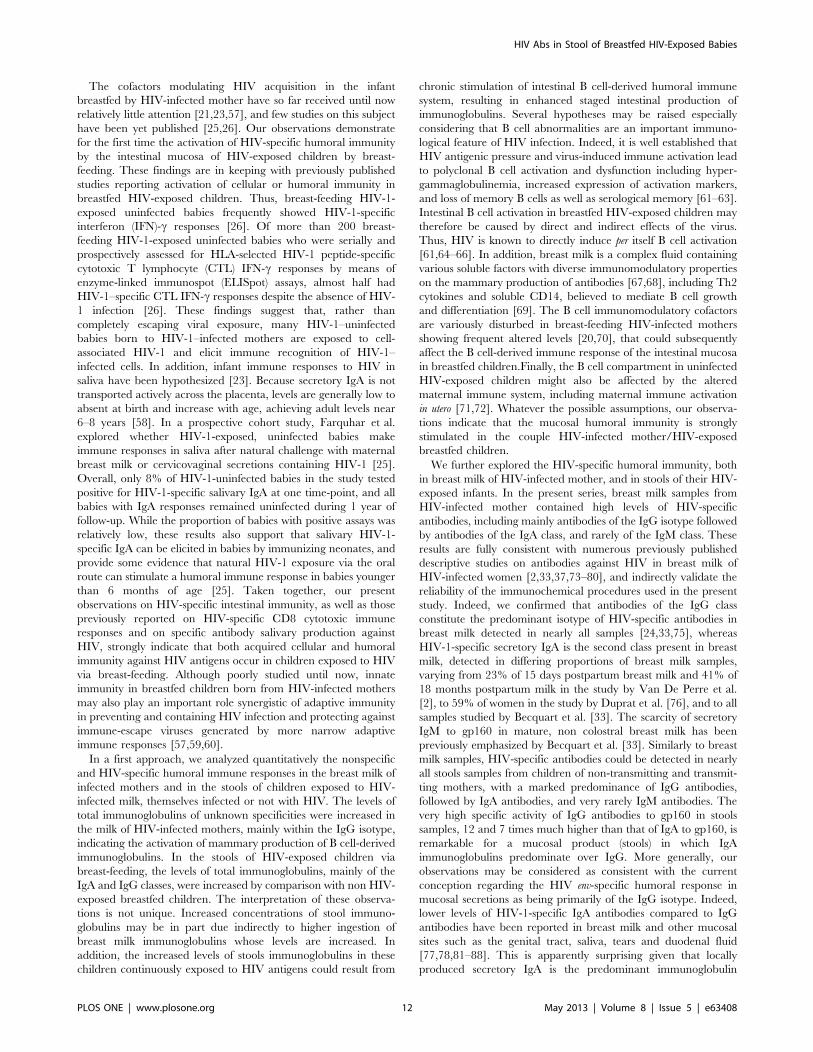
The cofactors modulating HIV acquisition in the infant
breastfed by HIV-infected mother have so far received until now
relatively little attention [21,23,57], and few studies on this subject
have been yet published [25,26]. Our observations demonstrate
for the first time the activation of HIV-specific humoral immunity
by the intestinal mucosa of HIV-exposed children by breast-
feeding. These findings are in keeping with previously published
studies reporting activation of cellular or humoral immunity in
breastfed HIV-exposed children. Thus, breast-feeding HIV-1-
exposed uninfected babies frequently showed HIV-1-specific
interferon (IFN)-c responses [26]. Of more than 200 breast-
feeding HIV-1-exposed uninfected babies who were serially and
prospectively assessed for HLA-selected HIV-1 peptide-specific
cytotoxic T lymphocyte (CTL) IFN-c responses by means of
enzyme-linked immunospot (ELISpot) assays, almost half had
HIV-1–specific CTL IFN-c responses despite the absence of HIV-
1 infection [26]. These findings suggest that, rather than
completely escaping viral exposure, many HIV-1–uninfected
babies born to HIV-1–infected mothers are exposed to cell-
associated HIV-1 and elicit immune recognition of HIV-1–
infected cells. In addition, infant immune responses to HIV in
saliva have been hypothesized [23]. Because secretory IgA is not
transported actively across the placenta, levels are generally low to
absent at birth and increase with age, achieving adult levels near
6–8 years [58]. In a prospective cohort study, Farquhar et al.
explored whether HIV-1-exposed, uninfected babies make
immune responses in saliva after natural challenge with maternal
breast milk or cervicovaginal secretions containing HIV-1 [25].
Overall, only 8% of HIV-1-uninfected babies in the study tested
positive for HIV-1-specific salivary IgA at one time-point, and all
babies with IgA responses remained uninfected during 1 year of
follow-up. While the proportion of babies with positive assays was
relatively low, these results also support that salivary HIV-1-
specific IgA can be elicited in babies by immunizing neonates, and
provide some evidence that natural HIV-1 exposure via the oral
route can stimulate a humoral immune response in babies younger
than 6 months of age [25]. Taken together, our present
observations on HIV-specific intestinal immunity, as well as those
previously reported on HIV-specific CD8 cytotoxic immune
responses and on specific antibody salivary production against
HIV, strongly indicate that both acquired cellular and humoral
immunity against HIV antigens occur in children exposed to HIV
via breast-feeding. Although poorly studied until now, innate
immunity in breastfed children born from HIV-infected mothers
may also play an important role synergistic of adaptive immunity
in preventing and containing HIV infection and protecting against
immune-escape viruses generated by more narrow adaptive
immune responses [57,59,60].
In a first approach, we analyzed quantitatively the nonspecific
and HIV-specific humoral immune responses in the breast milk of
infected mothers and in the stools of children exposed to HIV-
infected milk, themselves infected or not with HIV. The levels of
total immunoglobulins of unknown specificities were increased in
the milk of HIV-infected mothers, mainly within the IgG isotype,
indicating the activation of mammary production of B cell-derived
immunoglobulins. In the stools of HIV-exposed children via
breast-feeding, the levels of total immunoglobulins, mainly of the
IgA and IgG classes, were increased by comparison with non HIV-
exposed breastfed children. The interpretation of these observa-
tions is not unique. Increased concentrations of stool immuno-
globulins may be in part due indirectly to higher ingestion of
breast milk immunoglobulins whose levels are increased. In
addition, the increased levels of stools immunoglobulins in these
children continuously exposed to HIV antigens could result from
chronic stimulation of intestinal B cell-derived humoral immune
system, resulting in enhanced staged intestinal production of
immunoglobulins. Several hypotheses may be raised especially
considering that B cell abnormalities are an important immuno-
logical feature of HIV infection. Indeed, it is well established that
HIV antigenic pressure and virus-induced immune activation lead
to polyclonal B cell activation and dysfunction including hyper-
gammaglobulinemia, increased expression of activation markers,
and loss of memory B cells as well as serological memory [61–63].
Intestinal B cell activation in breastfed HIV-exposed children may
therefore be caused by direct and indirect effects of the virus.
Thus, HIV is known to directly induce per itself B cell activation
[61,64–66]. In addition, breast milk is a complex fluid containing
various soluble factors with diverse immunomodulatory properties
on the mammary production of antibodies [67,68], including Th2
cytokines and soluble CD14, believed to mediate B cell growth
and differentiation [69]. The B cell immunomodulatory cofactors
are variously disturbed in breast-feeding HIV-infected mothers
showing frequent altered levels [20,70], that could subsequently
affect the B cell-derived immune response of the intestinal mucosa
in breastfed children.Finally, the B cell compartment in uninfected
HIV-exposed children might also be affected by the altered
maternal immune system, including maternal immune activation
in utero [71,72]. Whatever the possible assumptions, our observa-
tions indicate that the mucosal humoral immunity is strongly
stimulated in the couple HIV-infected mother/HIV-exposed
breastfed children.
We further explored the HIV-specific humoral immunity, both
in breast milk of HIV-infected mother, and in stools of their HIV-
exposed infants. In the present series, breast milk samples from
HIV-infected mother contained high levels of HIV-specific
antibodies, including mainly antibodies of the IgG isotype followed
by antibodies of the IgA class, and rarely of the IgM class. These
results are fully consistent with numerous previously published
descriptive studies on antibodies against HIV in breast milk of
HIV-infected women [2,33,37,73–80], and indirectly validate the
reliability of the immunochemical procedures used in the present
study. Indeed, we confirmed that antibodies of the IgG class
constitute the predominant isotype of HIV-specific antibodies in
breast milk detected in nearly all samples [24,33,75], whereas
HIV-1-specific secretory IgA is the second class present in breast
milk, detected in differing proportions of breast milk samples,
varying from 23% of 15 days postpartum breast milk and 41% of
18 months postpartum milk in the study by Van De Perre et al.
[2], to 59% of women in the study by Duprat et al. [76], and to all
samples studied by Becquart et al. [33]. The scarcity of secretory
IgM to gp160 in mature, non colostral breast milk has been
previously emphasized by Becquart et al. [33]. Similarly to breast
milk samples, HIV-specific antibodies could be detected in nearly
all stools samples from children of non-transmitting and transmit-
ting mothers, with a marked predominance of IgG antibodies,
followed by IgA antibodies, and very rarely IgM antibodies. The
very high specific activity of IgG antibodies to gp160 in stools
samples, 12 and 7 times much higher than that of IgA to gp160, is
remarkable for a mucosal product (stools) in which IgA
immunoglobulins predominate over IgG. More generally, our
observations may be considered as consistent with the current
conception regarding the HIV env-specific humoral response in
mucosal secretions as being primarily of the IgG isotype. Indeed,
lower levels of HIV-1-specific IgA antibodies compared to IgG
antibodies have been reported in breast milk and other mucosal
sites such as the genital tract, saliva, tears and duodenal fluid
[77,78,81–88]. This is apparently surprising given that locally
produced secretory IgA is the predominant immunoglobulin
HIV Abs in Stool of Breastfed HIV-Exposed Babies
PLOS ONE | www.plosone.org 12 May 2013 | Volume 8 | Issue 5 | e63408

isotype in most mucosal secretions [89]. In animal models, low or
absent IgA responses have also been described in HIV-1-infected
chimpanzee [90] and simian immunodeficiency virus (SIV)-
infected macaques [91,92]. Thus, in striking contrast to other
mucosae-encountered microbial infections, HIV-1 and SIV do not
induce vigorous specific IgA responses in any body fluids
examined. The mechanism involved in this selective suppression
of HIV-1-specific responses in the IgA isotype in mucosal
secretions remains yet unresolved, and may be fundamental to
understand how HIV is capable to be shed in corporeal fluid, i.e. to
be produced by mucosal reservoirs despite the existence of
mucosal immunity against the virus [21].
The origin of humoral mucosal immunity to HIV in stools from
breastfed children born from HIV-infected mother remains
unknown. Antibodies in stools may a priori come from ingested
breast milk which contains HIV-specific antibodies, and from the
infants themselves. Thus, a certain proportion of stool antibodies
or F(ab’)2 moieties should correspond to undegraded or partially
degraded HIV-specific antibodies coming from breast milk intake.
In addition, HIV-specific antibodies in stools may be either
transferred from infant’s plasma by transudation or locally
produced by plasma cells belonging to the MALT [22], that
migrate to the efferent branch of intestinal mucosa from other
inductive mucosal sites, in particular, the oral- and gut- associated
lymphoid tissues [22,93]. In the present series, all infants were less
than 1 year old, thus were seropositive for routine HIV serology,
whatever their infectious status regarding HIV. Thus, plasma-
derived IgG may have passively transudated into intestinal
secretions, and account for a certain proportion of HIV-specific
IgG in stools. Furthermore, in order to evaluate the possibility of
intestinal production of HIV-specific antibodies, the relative fecal/
intestinal production of immunoglobulins and HIV-specific
antibodies in feces from breastfed children was evaluated by
calculating their RRE by reference to Lf as breast milk intake
factor. The working hypotheses were that immunoglobulins and Lf
are glycoproteins similarly degraded within intestinal chyle, and
that the intestinal production of Lf is negligible in the absence of
gastro-intestinal inflammation or gastro-enteritis in breastfed
children [49], as in the infants carefully selected for the study.
Assuming that the diffusion of milk immunoglobulins is similar to
that of Lf, the RRE calculation likely enables to evaluate the
partition between passive intake of immunoglobulins and intestinal
local production. The RRE by reference to Lf is quite similar to
the relative coefficient of excretion (RCE) previously proposed to
evaluate the mucosal excretion of immunoglublins by reference to
albumin [86,94,95]. For stool total immunoglobulins of unknown
specificities, the RRE calculations indicated intestinal synthesis of
total immunoglobulins, IgA and IgM, harboring RRE largely
above 1, whereas the stools IgG appeared mainly passively
deposited instead of locally produced. These latter observations
are reminiscent of the previous demonstration using the RCE
calculations by reference to albumin of active mucosal production
of polymeric IgA and IgM within the jejunal secretions whereas
IgG and monomeric IgA in the jejunal fluid are mainly plasma-
derived, and thus of transudative origin [94]. Applied to HIV-
specific antibodies, the calculations of RRE by reference to Lf for
HIV-specific IgA, IgG and IgM were largely superior to 1, likely
indicating that HIV-specific antibodies detected in stools are not
only originating from a passive ingestion of breast milk antibodies,
but rather from active intestinal secretions. Taken together, these
observations indicate that the infant intestinal mucosa of HIV-
exposed breastfed children, infected or not by HIV, likely secretes
actively HIV-specific antibodies, and thus that the HIV-specific
antibodies in their stools are locally-produced in addition with
possible passive transudation from plasma for the IgG class. The
inductive sites of HIV-specific antibodies intestinal production are
unknown, but free HIV particles and cell-associated virus present
in breast milk reach continuously the oral mucosa, the tonsillar
tissue and the upper gastrointestinal mucosa of exposed breastfed
babies, which are immunocompetent tissues including the
Waldeyer lymphoid tissue and the Peyer’s patches belonging to
the afferent branch of the MALT [22]. The effector sites of HIV-
specific antibodies intestinal production remain similarly un-
known, but may include the lymphoid tissue of the jejunal and
colonic mucosae, which belong to the efferent branch of the
MALT [22,96]. Acquired intestinal immunity against viral
antigens in children has been extensively described with other
infections than HIV, for example after natural intestinal infections
by rotavirus [97] or norovirus [98], or immunization with oral live
attenuated or inactivated poliovirus vaccine [99,100].
The functionality of purified pooled stools antibodies to HIV
was finally investigated using in vitro assays. The inhibitory
properties towards viral attachment to intestinal epithelial cells
and MDM of antibodies purified from pooled stools were first
assessed. In the present study, HT29 epithelial colon carcinoma
cell line was used to mimic the initial contact of HIV with
intestinal epithelial cells [101], as it is thought to occur during HIV
exposition of breastfed infants. Because a high proportion of HT29
epithelial cells expressed CXCR4 [102], X4-tropic HIV-1NDK
strain was used in attachment inhibition assay. In addition, MDM
were chosen because intestinal macrophages are considered as
prominent HIV-1 reservoir in chronically established HIV
infection [103]. MDM were used in attachment and infectivity
inhibition assays by X4-tropic HIV-1NDK or R5-tropic HIV-
1JRCSF strains, since both viral phenotypes may be present in
mucosal secretions [104]. Furthermore, R5-HIV-1-infected mac-
rophages in breast milk may be the most likely cells to transmigrate
across fetal oral and intestinal mucosal epithelia [105]. Pooled
immunoglobulins purified from stools of infants exposed to HIV
by breast-feeding, whatever the infant HIV status, inhibited the
attachment of HIV-1NDK on HT29 cells by 63% and on MDM
by 40%, and the attachment of HIV-1JRCSFon MDM by 77%.
In addition, purified pooled stools immunoglobulins inhibited the
HIV-1JRCSF infection of MDM by 93%. Otherwise, since the
stools of children breastfed by their HIV-infected mothers likely
contain a small proportion of HIV-specific antibodies coming from
breast milk, it is also likely that stools HIV-specific antibodies may
also harbor other functional inhibitory properties against HIV,
such as those previously demonstrated for breast milk specific
antibodies, like HIV-1 transcytosis inhibition [37,106], virus
neutralization and antibody-dependent cell cytotoxicity [79].
Taken together, these observations demonstrate that HIV-specific
antibodies purified from stools of HIV-exposed breastfed children
harbor in vitro major functional properties against HIV.
The correlates of protection against mucosal acquisition and
control of HIV-1 infection have not yet been clearly defined.
Humoral factors, innate immunity, and specific antibodies present
in external secretions, as well as cytotoxic lymphocytes distributed
in mucosal tissues, have been considered as involved in the
prevention and the local limitation of HIV at mucosal sites of viral
entry, especially in exposed non infected individuals [107,108].
The active intestinal synthesis of HIV-specific antibodies in HIV-
exposed, breastfed non infected infants is reminiscent of the genital
and oral mucosal immune response against HIV-1 in exposed
uninfected individuals [108–111]. The genital secretions collected
from HIV-1-exposed but persistently seronegative female sex
workers do not however contain uniformly and easily detectable
HIV-1-specific antibodies or neutralizing activities, and the
HIV Abs in Stool of Breastfed HIV-Exposed Babies
PLOS ONE | www.plosone.org 13 May 2013 | Volume 8 | Issue 5 | e63408

existence of adaptive HIV-specific humoral immunity in genital
mucosae exposed to HIV via sexual intercourse remains contro-
versial [112]. The possibility of intestinal production of HIV-
specific antibodies in HIV-exposed, breastfed non infected infants
may therefore constitute a particularly relevant model of natural
protection against HIV acquisition via mucosal routes. Indeed,
despite the infants’ daily exposure at their oral and gastrointestinal
mucosae to high amounts of cell-free and cell-associated HIV, the
virus acquisition in breastfed children occurs infrequently. The
puzzling fact that the majority of breastfed infants born from HIV-
infected mothers remain uninfected, even after several months of
breast-feeding, constitutes one of the major paradoxes of HIV
transmission via breast milk. The infrequent breast milk HIV
transmission despite prolonged exposure suggests that anti-
infective properties of breast milk may play a relevant protective
role against infection of exposed children, and that natural and/or
resistance to HIV in breastfed children could also be involved [21].
Finally, our observations raised acutely the issue of the putative
roles of functionally active HIV-specific, intestinal antibodies in
HIV transmission through breast-feeding. In the present series, the
HIV-specific intestinal immunity appeared qualitatively, quanti-
tatively and functionally similar in HIV-infected as in non HIV-
infected breastfed children. These features do not exclude a
protective role in vivo of the adaptive HIV-specific intestinal
immunity. Indeed, intestinal immunity to HIV developing after
exposition to HIV-infected milk should be envisioned in the
context of the multiple factors involved during HIV transmission
via breast-feeding, which is in essence a multifactorial process [21].
During the duration of exposure, HIV transmission by breast-
feeding may be associated with an imbalance between virus
ingestion and mucosal immune response directed against the virus,
and finally between infective and anti-infective properties of breast
milk [20,57] and exposed infant’s resistance [25,26]. For example,
in case of contamination during acute mastitis in the mother, the
potentially protective intestinal immunity directed to HIV would
have been insufficient to protect against exposure to large
quantities of virus occurring therefore transiently [113].
Finally, our observations constitutes the basis to further
investigate possible immunological correlates of protection in
HIV transmission via breast-feeding that could be noteworthy for
the design of a prophylactic vaccine. In developing countries
notably in sub-Saharan Africa, the postnatal mother to child
transmission of HIV-1 may continue even in the setting of optimal
antiretroviral therapy [15]. For this reason, the development of
new immunological strategies to reduce residual postnatal
transmission remains important in order to improve survival of
babies born from HIV-infected mothers in the developing world.
From the above results, we conclude that the intestinal mucosa of
children exposed to HIV by breast-feeding actively secretes HIV-
specific antibodies harboring in vitro functional properties against
HIV, and thus possibly protective in vivo. In vaccine perspective,
the demonstration of adaptive intestinal humoral immunity in
infants orally exposed to viral antigens provides the rational basis
for attempting to develop a mucosal prophylactic vaccination by
intestinal immunization against HIV in children orally exposed to
the virus.
Acknowledgments
We are grateful to all the women and their children who consented
participating in the study. We also thank the staff of the maternity clinics of
Castors, Boy-Rabe, Begoua and Hopital Communautaire in Bangui, where
the study was conducted and the staff of the Complexe Pediatrique of
Bangui. We are also very thankful to Dr. Antoinette Gody, Ministry of
Public Health, Bangui, as well as Pulcherie Pelembi and Julienne Ipero
from the Laboratoire des Retrovirus, Institut Pasteur de Bangui, for their
precious contributions. Finally, we are particularly grateful to the
association ARI, Paris, France, Dr Hakim Hocini, Vaccine Research
Institute, Creteil, France, and Mary Requena, INSERM, UMR1043,
Toulouse, France, for their contributions.
Author Contributions
Conceived and designed the experiments: LB SM. Performed the
experiments: SM JL. Analyzed the data: MAJ SM LB. Contributed
reagents/materials/analysis tools: JCG GG ALF. Wrote the paper: MAJ
SM LB.
References
1. UNAIDS (2011) Global HIV/AIDS response. Epidemic update and health
sector progress towards Universal Acces.
2. Van de Perre P, Simonon A, Hitimana DG, Dabis F, Msellati P, et al. (1993)
Infective and anti-infective properties of breastmilk from HIV-1-infected
women. Lancet 341: 914–918.
3. Datta P, Embree JE, Kreiss JK, Ndinya-Achola JO, Braddick M, et al. (1994)
Mother-to-child transmission of human immunodeficiency virus type 1: report
from the Nairobi Study. J Infect Dis 170: 1134–1140.
4. Bulterys M, Chao A, Dushimimana A, Saah A (1995) HIV-1 seroconversion
after 20 months of age in a cohort of breastfed children born to HIV-1-infected
women in Rwanda. AIDS 9: 93–94.
5. Bertolli J, St Louis ME, Simonds RJ, Nieburg P, Kamenga M, et al. (1996)
Estimating the timing of mother-to-child transmission of human immunode-
ficiency virus in a breast-feeding population in Kinshasa, Zaire. J Infect Dis
174: 722–726.
6. Bobat R, Moodley D, Coutsoudis A, Coovadia H (1997) Breastfeeding by HIV-
1-infected women and outcome in their infants: a cohort study from Durban,
South Africa. AIDS 11: 1627–1633.
7. Ekpini ER, Wiktor SZ, Satten GA, AdjorloloJohnson GT, Sibailly TS, et al.
(1997) Late postnatal mother-to-child transmission of HIV-1 in Abidjan, Cote
d’Ivoire. Lancet 349: 1054–1059.
8. Miotti PG, Taha TET, Kumwenda NI, Broadhead R, Mtimavalye LAR, et al.
(1999) HIV transmission through breastfeeding - A study in Malawi. Jama-
Journal of the American Medical Association 282: 744–749.
9. Fowler MG, Newell ML (2002) Breast-feeding and HIV-1 transmission in
resource-limited settings. Journal of Acquired Immune Deficiency Syndromes
30: 230–239.
10. John-Stewart G, Mbori-Ngacha D, Ekpini R, Janoff EN, Nkengasong J, et al.
(2004) Breast-feeding and Transmission of HIV-1. J Acquir Immune Defic
Syndr 35: 196–202.
11. Thior I, Lockman S, Smeaton LM, Shapiro RL, Wester C, et al. (2006)
Breastfeeding plus infant zidovudine prophylaxis for 6 months vs formula
feeding plus infant zidovudine for 1 month to reduce mother-to-child HIV
transmission in Botswana: a randomized trial: the Mashi Study. JAMA 296:
794–805.
12. Kumwenda NI, Hoover DR, Mofenson LM, Thigpen MC, Kafulafula G, et al.
(2008) Extended antiretroviral prophylaxis to reduce breast-milk HIV-1
transmission. N Engl J Med 359: 119–129.
13. Chasela CS, Hudgens MG, Jamieson DJ, Kayira D, Hosseinipour MC, et al.
(2010) Maternal or infant antiretroviral drugs to reduce HIV-1 transmission.
N Engl J Med 362: 2271–2281.
14. Shapiro RL, Hughes MD, Ogwu A, Kitch D, Lockman S, et al. (2010)
Antiretroviral regimens in pregnancy and breast-feeding in Botswana.
N Engl J Med 362: 2282–2294.
15. Wilson CB (2005) Developmental immunology and role of host defenses in
neonatal susceptibility. In: Remington J KJ, editor. Infectious diseases of the
fetus and newborn infant. Philadelphia, PA: WB Saunders Company. 37–49.
16. Lewis P, Nduati R, Kreiss JK, John GC, Richardson BA, et al. (1998) Cell-free
human immunodeficiency virus type 1 in breast milk. J Infect Dis 177: 34–39.
17. Neveu D, Viljoen J, Bland RM, Nagot N, Danaviah S, et al. (2011) Cumulative
exposure to cell-free HIV in breast milk, rather than feeding pattern per se,
identifies postnatally infected infants. Clinical Infectious Diseases 52: 819–825.
18. Richardson BA, John-Stewart GC, Hughes JP, Nduati R, Mbori-Ngacha D, et
al. (2003) Breast-milk infectivity in human immunodeficiency virus type 1-
infected mothers. J Infect Dis 187: 736–740.
19. Coovadia HM, Rollins NC, Bland RM, Little K, Coutsoudis A, et al. (2007)
Mother-to-child transmission of HIV-1 infection during exclusive breastfeeding
in the first 6 months of life: an intervention cohort study. Lancet 369: 1107–
1116.
20. Kourtis AP, Butera S, Ibegbu C, Belec L, Duerr A (2003) Breast milk and HIV-
1: vector of transmission or vehicle of protection? Lancet Infect Dis 3: 786–793.
HIV Abs in Stool of Breastfed HIV-Exposed Babies
PLOS ONE | www.plosone.org 14 May 2013 | Volume 8 | Issue 5 | e63408

21. Belec L, Kourtis AP (2012) B lymphocyte-derived humoral immune defenses in
breast milk transmission of the HIV-1. Adv Exp Med Biol 743: 139–160.
22. Brandtzaeg P (2003) Mucosal immunity: integration between mother and the
breast-fed infant. Vaccine 21: 3382–3388.
23. Farquhar C, John-Stewart G (2003) The role of infant immune responses and
genetic factors in preventing HIV-1 acquisition and disease progression. Clin
Exp Immunol 134: 367–377.
24. Van de Perre P (1999) Mother-to-child transmission of HIV-1: the ‘all mucosal’
hypothesis as a predominant mechanism of transmission. AIDS 13: 1133–1138.
25. Farquhar C, VanCott T, Bosire R, Bermudez C, Mbori-Ngacha D, et al.
(2008) Salivary human immunodeficiency virus (HIV)-1-specific immunoglob-
ulin A in HIV-1-exposed infants in Kenya. Clin Exp Immunol 153: 37–43.
26. John-Stewart GC, Mbori-Ngacha D, Payne BL, Farquhar C, Richardson BA,
et al. (2009) HV-1-specific cytotoxic T lymphocytes and breast milk HIV-1
transmission. J Infect Dis 199: 889–898.
27. Gody JC, Charpentier C, Mbitikon O, Si-Mohamed A, LeGoff J, et al. (2008)
High prevalence of antiretroviral drug resistance mutations in HIV-1 non-B
subtype strains from African children receiving antiretroviral therapy regimen
according to the 2006 revised WHO recommendations. J Acquir Immune
Defic Syndr 49: 566–569.
28. Moussa S, Pinson P, Pelembi P, Gody JC, Mbitikon O, et al. (2010) First data
on HIV-1 resistance mutations to antiretroviral drugs in Central African
Republic. AIDS Res Hum Retroviruses 26: 1247–1248.
29. Charpentier C, Gody JC, Mbitikon O, Moussa S, Matta M, et al. (2012)
Virological response and resistance profiles after 18 to 30 months of first- or
second2/third-line antiretroviral treatment: a cross-sectional evaluation in
HIV type 1-infected children living in the Central African Republic. AIDS Res
Hum Retroviruses 28: 87–94.
30. (WHO) WHO (2010) Antiretroviral therapy for HIV infection in babies and
children: Towards universal access. Recommendations for a public health
approach.
31. (WHO) WHO (2010) recommendations Antiretroviral therapy for HIV
infection in adults and adolescents. Antiretroviral therapy for a public health
approach.
32. Chintu C, Bhat GJ, Walker AS, Mulenga V, Sinyinza F, et al. (2004) Co-
trimoxazole as prophylaxis against opportunistic infections in HIV-infected
Zambian children (CHAP): a double-blind randomised placebo-controlled trial.
Lancet 364: 1865–1871.
33. Becquart P, Hocini H, Garin B, Sepou A, Kazatchkine MD, et al. (1999)
Compartmentalization of the IgG immune response to HIV-1 in breast milk.
AIDS 13: 1323–1331.
34. Rouet F, Sakarovitch C, Msellati P, Elenga N, Montcho C, et al. (2003)
Pediatric viral human immunodeficiency virus type 1 RNA levels, timing of
infection, and disease progression in African HIV-1-infected children.
Pediatrics 112: e289.
35. Rouet F, Chaix ML, Nerrienet E, Ngo-Giang-Huong N, Plantier JC, et al.
(2007) Impact of HIV-1 genetic diversity on plasma HIV-1 RNA Quantifi-
cation: usefulness of the Agence Nationale de Recherches sur le SIDA second-
generation long terminal repeat-based real-time reverse transcriptase polymer-
ase chain reaction test. J Acquir Immune Defic Syndr 45: 380–388.
36. Buchacher A, Predl R, Strutzenberger K, Steinfellner W, Trkola A, et al.
(1994) Generation of human monoclonal antibodies against HIV-1 proteins;
electrofusion and Epstein-Barr virus transformation for peripheral blood
lymphocyte immortalization. AIDS Res Hum Retroviruses 10: 359–369.
37. Becquart P, Hocini H, Levy M, Sepou A, Kazatchkine MD, et al. (2000)
Secretory anti-human immunodeficiency virus (HIV) antibodies in colostrum
and breast milk are not a major determinant of the protection of early postnatal
transmission of HIV. J Infect Dis 181: 532–539.
38. Chomont N, Hocini H, Gody JC, Bouhlal H, Becquart P, et al. (2008)
Neutralizing monoclonal antibodies to human immunodeficiency virus type 1
do not inhibit viral transcytosis through mucosal epithelial cells. Virology 370:
246–254.
39. Saidi H, Eslahpazir J, Carbonneil C, Carthagena L, Requena M, et al. (2006)
Differential modulation of human lactoferrin activity against both R5 and X4-
HIV-1 adsorption on epithelial cells and dendritic cells by natural antibodies.
J Immunol 177: 5540–5549.
40. Saidi H, Magri G, Carbonneil C, Nasreddine N, Requena M, et al. (2007) IFN-
gamma-activated monocytes weakly produce HIV-1 but induce the recruit-
ment of HIV-sensitive T cells and enhance the viral production by these
recruited T cells. J Leukoc Biol 81: 642–653.
41. Saidi H, Jenabian MA, Gombert B, Charpentier C, Mannarini A, et al. (2008)
Pre-clinical development as microbicide of zinc tetra-ascorbo-camphorate, a
novel terpenoid derivative: potent in vitro inhibitory activity against both R5-
and X4-tropic HIV-1 strains without significant in vivo mucosal toxicity. AIDS
Res Ther 5: 10.
42. Saidi H, Nasreddine N, Jenabian MA, Lecerf M, Schols D, et al. (2007)
Differential in vitro inhibitory activity against HIV-1 of alpha-(1–3)- and alpha-
(1–6)-D-mannose specific plant lectins: implication for microbicide develop-
ment. J Transl Med 5: 28.
43. Kimpton J, Emerman M (1992) Detection of replication-competent and
pseudotyped human immunodeficiency virus with a sensitive cell line on the
basis of activation of an integrated beta-galactosidase gene. J Virol 66: 2232–
2239.
44. Becquart P, Courgnaud V, Willumsen J, Van de Perre P (2007) Diversity of
HIV-1 RNA and DNA in breast milk from HIV-1-infected mothers. Virology363: 256–260.
45. Salazar-Gonzalez JF, Salazar MG, Learn GH, Fouda GG, Kang HH, et al.
(2011) Origin and evolution of HIV-1 in breast milk determined by single-genome amplification and sequencing. J Virol 85: 2751–2763.
46. Hocini H, Barra A, Belec L, Iscaki S, Preud’homme JL, et al. (1995) Systemic
and secretory humoral immunity in the normal human vaginal tract.
Scand J Immunol 42: 269–274.
47. Pfefferkorn MD, Boone JH, Nguyen JT, Juliar BE, Davis MA, et al. (2010)Utility of fecal lactoferrin in identifying Crohn disease activity in children.
J Pediatr Gastroenterol Nutr 51: 425–428.
48. Brock JH (1980) Lactoferrin in human milk: its role in iron absorption andprotection against enteric infection in the newborn infant. Arch Dis Child 55:
417–421.
49. Spik G, Brunet B, Mazurier-Dehaine C, Fontaine G, Montreuil J (1982)
Characterization and properties of the human and bovine lactotransferrinsextracted from the faeces of newborn infants. Acta Paediatr Scand 71: 979–
985.
50. Levay PF, Viljoen M (1995) Lactoferrin: a general review. Haematologica 80:252–267.
51. Langhorst J, Boone J (2012) Fecal lactoferrin as a noninvasive biomarker in
inflammatory bowel diseases. Drugs Today (Barc) 48: 149–161.
52. Goldman AS, Garza C, Schanler RJ, Goldblum RM (1990) Molecular forms of
lactoferrin in stool and urine from infants fed human milk. Pediatr Res 27:252–255.
53. Eslahpazir J, Jenabian MA, Bouhlal H, Hocini H, Carbonneil C, et al. (2008)
Infection of macrophages and dendritic cells with primary R5-tropic humanimmunodeficiency virus type 1 inhibited by natural polyreactive anti-CCR5
antibodies purified from cervicovaginal secretions. Clin Vaccine Immunol 15:872–884.
54. Carthagena L, Becquart P, Hocini H, Kazatchkine MD, Bouhlal H, et al.(2011) Modulation of HIV Binding to Epithelial Cells and HIV Transfer from
Immature Dendritic Cells to CD4 T Lymphocytes by Human Lactoferrin andits Major Exposed LF-33 Peptide. Open Virol J 5: 27–34.
55. Creek TL, Sherman GG, Nkengasong J, Lu L, Finkbeiner T, et al. (2007)
Infant human immunodeficiency virus diagnosis in resource-limited settings:issues, technologies, and country experiences. Am J Obstet Gynecol 197: S64–
71.
56. Sherman GG, Cooper PA, Coovadia AH, Puren AJ, Jones SA, et al. (2005)
Polymerase chain reaction for diagnosis of human immunodeficiency virusinfection in infancy in low resource settings. Pediatr Infect Dis J 24: 993–997.
57. Lehman DA, Farquhar C (2007) Biological mechanisms of vertical human
immunodeficiency virus (HIV-1) transmission. Rev Med Virol 17: 381–403.
58. C W (2005) Developmental immunology and role of host defenses in neonatalsusceptibility. In: Remington J, Klein J, ed. Infectious diseases of the fetus and
newborn infant, 6th edition. Philadelphia, PA: WB Saunders Company: 37–49.
59. Ballan WM, Vu BA, Long BR, Loo CP, Michaelsson J, et al. (2007) Natural
killer cells in perinatally HIV-1-infected children exhibit less degranulationcompared to HIV-1-exposed uninfected children and their expression of
KIR2DL3, NKG2C, and NKp46 correlates with disease severity. J Immunol179: 3362–3370.
60. Tiemessen CT, Shalekoff S, Meddows-Taylor S, Schramm DB, Papathana-
sopoulos MA, et al. (2009) Cutting Edge: Unusual NK cell responses to HIV-1
peptides are associated with protection against maternal-infant transmission ofHIV-1. J Immunol 182: 5914–5918.
61. De Milito A, Nilsson A, Titanji K, Thorstensson R, Reizenstein E, et al. (2004)
Mechanisms of hypergammaglobulinemia and impaired antigen-specifichumoral immunity in HIV-1 infection. Blood 103: 2180–2186.
62. Titanji K, Chiodi F, Bellocco R, Schepis D, Osorio L, et al. (2005) Primary
HIV-1 infection sets the stage for important B lymphocyte dysfunctions. AIDS
19: 1947–1955.
63. D’Orsogna LJ, Krueger RG, McKinnon EJ, French MA (2007) Circulatingmemory B-cell subpopulations are affected differently by HIV infection and
antiretroviral therapy. AIDS 21: 1747–1752.
64. Chong Y, Ikematsu H, Kikuchi K, Yamamoto M, Murata M, et al. (2004)Selective CD27+ (memory) B cell reduction and characteristic B cell alteration
in drug-naive and HAART-treated HIV type 1-infected patients. AIDS Res
Hum Retroviruses 20: 219–226.
65. Chong Y, Ikematsu H, Yamamoto M, Murata M, Yamaji K, et al. (2004)Increased frequency of CD27- (naive) B cells and their phenotypic alteration in
HIV type 1-infected patients. AIDS Res Hum Retroviruses 20: 621–629.
66. Lane HC, Masur H, Edgar LC, Whalen G, Rook AH, et al. (1983)Abnormalities of B-cell activation and immunoregulation in patients with the
acquired immunodeficiency syndrome. N Engl J Med 309: 453–458.
67. Agarwal S, Karmaus W, Davis S, Gangur V (2011) Immune markers in breast
milk and fetal and maternal body fluids: a systematic review of perinatalconcentrations. J Hum Lact 27: 171–186.
68. Cummins AG, Thompson FM (1997) Postnatal changes in mucosal immune
response: a physiological perspective of breast feeding and weaning. ImmunolCell Biol 75: 419–429.
69. Filipp D, Alizadeh-Khiavi K, Richardson C, Palma A, Paredes N, et al. (2001)
Soluble CD14 enriched in colostrum and milk induces B cell growth and
differentiation. Proc Natl Acad Sci U S A 98: 603–608.
HIV Abs in Stool of Breastfed HIV-Exposed Babies
PLOS ONE | www.plosone.org 15 May 2013 | Volume 8 | Issue 5 | e63408

70. Lepage P, Van de Perre P (2012) The immune system of breast milk:
antimicrobial and anti-inflammatory properties. Adv Exp Med Biol 743: 121–137.
71. Bunders M, Pembrey L, Kuijpers T, Newell ML (2010) Evidence of impact ofmaternal HIV infection on immunoglobulin levels in HIV-exposed uninfected
children. AIDS Res Hum Retroviruses 26: 967–975.
72. Filteau S (2009) The HIV-exposed, uninfected African child. Trop Med Int
Health 14: 276–287.
73. Van de Perre P (2000) Breast milk transmission of HIV-1. Laboratory and
clinical studies. Ann N Y Acad Sci 918: 122–127.
74. Van de Perre P, Hitimana DG, Lepage P (1988) Human immunodeficiencyvirus antibodies of IgG, IgA, and IgM subclasses in milk of seropositive
mothers. J Pediatr 113: 1039–1041.
75. Belec L, Bouquety JC, Georges AJ, Siopathis MR, Martin PM (1990)
Antibodies to human immunodeficiency virus in the breast milk of healthy,seropositive women. Pediatrics 85: 1022–1026.
76. Duprat C, Mohammed Z, Datta P, Stackiw W, Ndinya-Achola JO, et al. (1994)Human immunodeficiency virus type 1 IgA antibody in breast milk and serum.
Pediatr Infect Dis J 13: 603–608.
77. Lu FX (2000) Predominate HIV1-specific IgG activity in various mucosal
compartments of HIV1-infected individuals. Clin Immunol 97: 59–68.
78. Mestecky J, Jackson S, Moldoveanu Z, Nesbit LR, Kulhavy R, et al. (2004)
Paucity of antigen-specific IgA responses in sera and external secretions ofHIV-type 1-infected individuals. AIDS Res Hum Retroviruses 20: 972–988.
79. Fouda GG, Yates NL, Pollara J, Shen X, Overman GR, et al. (2011) HIV-
specific functional antibody responses in breast milk mirror those in plasma and
are primarily mediated by IgG antibodies. J Virol 85: 9555–9567.
80. Kuhn L, Trabattoni D, Kankasa C, Sinkala M, Lissoni F, et al. (2006) Hiv-
specific secretory IgA in breast milk of HIV-positive mothers is not associatedwith protection against HIV transmission among breast-fed infants. J Pediatr
149: 611–616.
81. Belec L, Georges AJ, Steenman G, Martin PM (1989) Antibodies to human
immunodeficiency virus in vaginal secretions of heterosexual women. J InfectDis 160: 385–391.
82. Belec L, Dupre T, Prazuck T, Tevi-Benissan C, Kanga JM, et al. (1995)
Cervicovaginal overproduction of specific IgG to human immunodeficiency
virus (HIV) contrasts with normal or impaired IgA local response in HIVinfection. J Infect Dis 172: 691–697.
83. Wolff H, Mayer K, Seage G, Politch J, Horsburgh CR, et al. (1992) A
comparison of HIV-1 antibody classes, titers, and specificities in paired semen
and blood samples from HIV-1 seropositive men. J Acquir Immune DeficSyndr 5: 65–69.
84. Artenstein AW, VanCott TC, Sitz KV, Robb ML, Wagner KF, et al. (1997)
Mucosal immune responses in four distinct compartments of women infected
with human immunodeficiency virus type 1: a comparison by site andcorrelation with clinical information. J Infect Dis 175: 265–271.
85. Haimovici F, Mayer KH, Anderson DJ (1997) Quantitation of HIV-1-specificIgG, IgA, and IgM antibodies in human genital tract secretions. J Acquir
Immune Defic Syndr Hum Retrovirol 15: 185–191.
86. Raux M, Finkielsztejn L, Salmon-Ceron D, Bouchez H, Excler JL, et al. (1999)
Comparison of the distribution of IgG and IgA antibodies in serum and variousmucosal fluids of HIV type 1-infected subjects. AIDS Res Hum Retroviruses
15: 1365–1376.
87. Wright PF, Kozlowski PA, Rybczyk GK, Goepfert P, Staats HF, et al. (2002)
Detection of mucosal antibodies in HIV type 1-infected individuals. AIDS ResHum Retroviruses 18: 1291–1300.
88. Alexander R, Mestecky J (2007) Neutralizing antibodies in mucosal secretions:
IgG or IgA? Curr HIV Res 5: 588–593.
89. Goldman AS (1993) The immune system of human milk: antimicrobial,
antiinflammatory and immunomodulating properties. Pediatr Infect Dis J 12:664–671.
90. Israel ZR, Marx PA (1995) Nonclassical mucosal antibodies predominate ingenital secretions of HIV-1 infected chimpanzees. J Med Primatol 24: 53–60.
91. Permar SR, Wilks AB, Ehlinger EP, Kang HH, Mahlokozera T, et al. (2010)Limited contribution of mucosal IgA to Simian immunodeficiency virus (SIV)-
specific neutralizing antibody response and virus envelope evolution in breast
milk of SIV-infected, lactating rhesus monkeys. J Virol 84: 8209–8218.92. Schafer F, Kewenig S, Stolte N, Stahl-Hennig C, Stallmach A, et al. (2002)
Lack of simian immunodeficiency virus (SIV) specific IgA response in the
intestine of SIV infected rhesus macaques. Gut 50: 608–614.93. Hanson LA, Ahlstedt S, Andersson B, Cruz JR, Dahlgren U, et al. (1984) The
immune response of the mammary gland and its significance for the neonate.Ann Allergy 53: 576–582.
94. Jonard PP, Rambaud JC, Dive C, Vaerman JP, Galian A, et al. (1984)
Secretion of immunoglobulins and plasma proteins from the jejunal mucosa.Transport rate and origin of polymeric immunoglobulin A. J Clin Invest 74:
525–535.95. Laibe S, Bard E, Biichle S, Vielle J, Millon L, et al. (2003) New sensitive
method for the measurement of lysozyme and lactoferrin to explore mucosalinnate immunity. Part II: time-resolved immunofluorometric assay used in HIV
patients with oral candidiasis. Clin Chem Lab Med 41: 134–138.
96. Brandtzaeg P FI (1994) The human mucosal B-cell system. In MucosalImmunology. In: Ogra PL MJ, Lamm ME, Strober W, Bienenstock J, McGhee
editor. Second Edition ed: Academic Press. 439–468.97. Ruggeri FM, Johansen K, Basile G, Kraehenbuhl JP, Svensson L (1998)
Antirotavirus immunoglobulin A neutralizes virus in vitro after transcytosis
through epithelial cells and protects infant mice from diarrhea. J Virol 72:2708–2714.
98. Long KZ, Garcia C, Ko G, Santos JI, Al Mamun A, et al. (2011) Vitamin Amodifies the intestinal chemokine and cytokine responses to norovirus infection
in Mexican children. J Nutr 141: 957–963.99. Ogra PL (1995) Comparative evaluation of immunization with live attenuated
and inactivated poliovirus vaccines. Ann N Y Acad Sci 754: 97–107.
100. Hird TR, Grassly NC (2012) Systematic review of mucosal immunity inducedby oral and inactivated poliovirus vaccines against virus shedding following oral
poliovirus challenge. PLoS Pathog 8: e1002599.101. Fantini J, Yahi N, Baghdiguian S, Chermann JC (1992) Human colon
epithelial cells productively infected with human immunodeficiency virus show
impaired differentiation and altered secretion. J Virol 66: 580–585.102. Bouhlal H, Chomont N, Haeffner-Cavaillon N, Kazatchkine MD, Belec L, et
al. (2002) Opsonization of HIV-1 by semen complement enhances infection ofhuman epithelial cells. J Immunol 169: 3301–3306.
103. Smith PD, Meng G, Salazar-Gonzalez JF, Shaw GM (2003) Macrophage HIV-1 infection and the gastrointestinal tract reservoir. J Leukoc Biol 74: 642–649.
104. Margolis L, Shattock R (2006) Selective transmission of CCR5-utilizing HIV-1:
the ‘gatekeeper’ problem resolved? Nat Rev Microbiol 4: 312–317.105. Tugizov SM, Herrera R, Veluppillai P, Greenspan D, Soros V, et al. (2012)
Differential transmission of HIV traversing fetal oral/intestinal epithelia andadult oral epithelia. J Virol 86: 2556–2570.
106. Hocini H, Becquart P, Bouhlal H, Chomont N, Ancuta P, et al. (2001) Active
and selective transcytosis of cell-free human immunodeficiency virus through atight polarized monolayer of human endometrial cells. J Virol 75: 5370–5374.
107. Moog C (2008) Immune responses that correlate with HIV-1 protection? AIDS22: 1461–1462.
108. Shearer G, Clerici M (2010) Historical perspective on HIV-exposedseronegative individuals: has nature done the experiment for us? J Infect Dis
202 Suppl 3: S329–332.
109. Belec L, Ghys PD, Hocini H, Nkengasong JN, Tranchot-Diallo J, et al. (2001)Cervicovaginal secretory antibodies to human immunodeficiency virus type 1
(HIV-1) that block viral transcytosis through tight epithelial barriers in highlyexposed HIV-1-seronegative African women. J Infect Dis 184: 1412–1422.
110. Hirbod T, Broliden K (2007) Mucosal immune responses in the genital tract of
HIV-1-exposed uninfected women. J Intern Med 262: 44–58.111. Hasselrot K (2009) Genital and oral mucosal immune response against HIV-1
in exposed uninfected individuals. Crit Rev Immunol 29: 369–377.112. Mestecky J, Wright PF, Lopalco L, Staats HF, Kozlowski PA, et al. (2011)
Scarcity or absence of humoral immune responses in the plasma and
cervicovaginal lavage fluids of heavily HIV-1-exposed but persistentlyseronegative women. AIDS Res Hum Retroviruses 27: 469–486.
113. Semba RD, Neville MC (1999) Breast-feeding, mastitis, and HIV transmission:nutritional implications. Nutr Rev 57: 146–153.
HIV Abs in Stool of Breastfed HIV-Exposed Babies
PLOS ONE | www.plosone.org 16 May 2013 | Volume 8 | Issue 5 | e63408
Related Documents