ACUTE STRESS-INDUCED CORTISOL ELEVATION ENHANCES MEMORY CONSOLIDATION OF SIMILAR ITEMS by Alice Jiang A thesis submitted to Johns Hopkins University in conformity with the requirements for the degree of Master of Science Baltimore, Maryland April 2016 © 2016 Alice Jiang All Rights Reserved

Welcome message from author
This document is posted to help you gain knowledge. Please leave a comment to let me know what you think about it! Share it to your friends and learn new things together.
Transcript
ACUTE STRESS-INDUCED CORTISOL ELEVATION ENHANCES MEMORY
CONSOLIDATION OF SIMILAR ITEMS
by Alice Jiang
A thesis submitted to Johns Hopkins University in conformity with the requirements for
the degree of Master of Science
Baltimore, Maryland April 2016
© 2016 Alice Jiang All Rights Reserved

ii
Abstract
Though considerable evidence has indicated that stress-induced changes in
cortisol crucially affect memory function, the precise relationship between cortisol and
long-term memory remains poorly understood. Cortisol has been shown to enhance
memory consolidation, the process of converting short-term memories into long-term
memories, and impair memory retrieval, the process of accessing previously stored
memories. However, very little is known about the way stress affects the hippocampus
and its subregions, which have recently been shown to contribute critical functions to
learning and memory. In the present study, we examined the effect of acute stress,
induced by the Trier Social Stress Test, on a memory task designed to differentially tax
the functioning of dentate gyrus and CA3 subregions of the hippocampus. The study
aimed to determine whether stress affects hippocampal-dependent memory consolidation
and retrieval processes in otherwise healthy young adults. It was observed that a stress-
induced elevation of glucocorticoids during memory consolidation enhanced the ability to
discriminate between highly similar stimuli while elevated glucocorticoids during
memory retrieval had no significant effect on memory performance. Additional findings
reported a dose-dependent effect of cortisol on memory function and negative
correlations with self-report ratings of depression, anxiety, and perceived stress. Taken
together, this study provides evidence that acute stress differentially affects the
consolidation and retrieval stages of memory, and particularly enhances the encoding of
highly similar items, a function thought to rely strongly on the dentate gyrus and CA3
subregions of the hippocampus.
Mentor/Principal Investigator: Arnold Bakker, Ph.D.
Advisor: Robert D. Horner, Ph.D.
iii
Acknowledgements
I would like to express the deepest gratitude for my mentor, Dr. Arnold Bakker,
for his continuous support and guidance on my research. He has been incredibly
instructive, patient, and encouraging, and has helped me grow and think critically as a
scientist. I could not have asked for a better mentor for my research experience.
I would also like to thank other members in the lab, especially Tammy Tran and
Carrie Speck, who have helped me at all levels of the research project. Tammy has been
an enormous source of advice and knowledge throughout this project in helping me with
the experimental design and data analysis. Carrie played an essential role in helping me
apply for IRB approval and work out project logistics.
Thanks to the Johns Hopkins University Department of Biology for their support
and assistance over the course of this program. I would like to especially thank my
seminar professor, Dr. Kathryn Tifft, for her invaluable mentorship, insightful
discussions, and constructive suggestions. I appreciate everything she has done for this
program and for me.
I am extremely grateful Dr. Farrah Madison for work in analyzing the saliva
samples in my project. I would also like to thank my undergraduate research assistants,
Ivar Ogden, Ashish Phal, Natalia Sonsin-Diaz, Elsa Olson, Anna Koerner, and Daniel
Alden, for their assistance in administering the Trier Social Stress Test. Without their
contribution, my project would not have been possible.
Finally, I would like to thank my family and friends for always supporting me and
being there for me through my research journey. I would not be where I am today without
their love and encouragement, and I am grateful to them everyday.

iv
Table of Contents
1. Introduction ..................................................................................................................... 1
1.1. Neuroendocrinology of stress ............................................................................ 2
1.2. Animal studies: stress response and memory function ...................................... 3
1.3. Human studies: stress response and memory function ....................................... 6
1.4. Pattern separation and pattern completion in long-term memory function ........ 11
1.5. Overview of the present study ............................................................................ 12
2. Methods ............................................................................................................................ 14
2.1. Participants ......................................................................................................... 14
2.2. Study design and procedure ............................................................................... 15
2.3. Saliva sampling and biochemical analysis ......................................................... 16
2.4. Behavioral pattern separation task ..................................................................... 16
2.5. Trier Social Stress Test ...................................................................................... 20
2.6. Self-report questionnaires .................................................................................. 21
3. Results .............................................................................................................................. 22
3.1. Parameters for analysis ....................................................................................... 22
3.2. Effectiveness of stress induction ........................................................................ 23
3.3. Memory performance ......................................................................................... 25
3.4. Cortisol response and memory ........................................................................... 27
3.5. Correlational analysis of self-report questionnaires ........................................... 29
4. Discussion ......................................................................................................................... 30
4.1. Effects of stress on memory performance .......................................................... 30
4.2. Correlates of pattern separation performance .................................................... 34
v
4.3. Possible mechanisms by which stress impacts memory .................................... 37
4.4. The role of adult neurogenesis in pattern separation .......................................... 38
4.5. Limitations ......................................................................................................... 40
4.6. Conclusion .......................................................................................................... 42
5. References ........................................................................................................................ 43
6. Curriculum Vitae ............................................................................................................ 57

vi
List of Tables
Table 1: Clinical data and analysis of control group participants ......................................... 23

vii
List of Figures
Figure 1 ................................................................................................................................. 19
1A: Outline of Study Design ..................................................................................... 19
1B: Behavioral pattern separation task ...................................................................... 19
1C: Sample stimuli and their lures ............................................................................ 19
Figure 2 ................................................................................................................................ 24
2A: STAI scores ........................................................................................................ 24
2B: Salivary cortisol concentration ........................................................................... 24
Figure 3 ................................................................................................................................. 25
Figure 4 ................................................................................................................................. 26
4A: Consolidation-stress discrimination of lures ...................................................... 26
4B: Retrieval-stress discrimination of lures .............................................................. 26
Figure 5 ................................................................................................................................. 27
Figure 6 ................................................................................................................................. 28
6A: Control cortisol response and memory ............................................................... 28
6B: Consolidation-stress cortisol response and memory .......................................... 28
6C: Retrieval-stress cortisol response and memory .................................................. 28
Figure 7 ................................................................................................................................. 29

1
1. Introduction
Exposure to stressful situations rapidly stimulates complex changes throughout
the body and brain. Although stress is generally considered detrimental to the organism’s
well being, it induces changes that are evolutionary relevant for survival. In stressful
situations, adrenal hormones are released to prepare the body to respond quickly to a
perceived threat; for instance, blood flow is directed towards the muscles to prepare for
action while accelerated breathing increases the availability of oxygen (for review, see
Lupien et al., 2007). Stress also has prominent effects on cognition. During the initial
stress response, rapidly unfolding neurochemical events exert immediate effects on
attention, sensory, and mnemonic processes (de Kloet et al., 2005). When elevated during
consolidation, the process of converting new memory traces into long-term storage, stress
hormones have memory-enhancing effects; however when elevated during retrieval, the
process of accessing previously stored memories, stress hormones may yield memory-
impairing effects (for review, see Wolf et al., 2003). Stress-enhanced memory
consolidation may have been evolutionarily adaptive to help adjust to a quickly changing
environment while stress-impaired memory retrieval may be functionally relevant to
prevent the interference of noise in the face of an immediate threat (de Kloet, 1998).
Prolonged exposure to stress, known as chronic stress, is detrimental to health as it
impairs growth and repair, suppresses immune function, and produces memory deficits
by causing atrophy of the hippocampus (Herbert et al., 2006, McEwen 2000).
Though many studies have investigated the effects of acute and chronic stress on
memory function broadly, there is limited research on the effect of stress on the different
aspects of memory function and the contributions of the memory processes instantiated

2
by the different subregions of the hippocampus. Although it is well established that the
hippocampus and the surrounding cortices in the medial temporal lobe are critically
important for episodic memory function, recent studies have highlighted the unique
contributions of the individual hippocampal subregions on the ability to learn and
remember information. In particular, the dentate gyrus (DG) and CA3 subregions of the
hippocampus contribute critically to every day memory function and appear to be
sensitive to stressors and aging (for review, see Yassa & Stark, 2011; Fa et al., 2014).
This study aims to investigate the effects of stress on hippocampal-dependent memory
and hippocampal subregion-specific functions. Given the prevalence of high stress in
many individuals today and the potentially lasting consequences of stress on the structure
and function of the brain, it is important to further elucidate the relationship between
stress and long-term memory function.
1.1. Neuroendocrinology of stress
Exposure to a stressful, e.g. threatening or demanding, situation leads to the
release of adrenal hormones that cause physiological and psychological changes to help
cope with the stressor. The initial rapid response occurs through the sympathetic nervous
system and results in the release of catecholamines. These hormones induce physical
changes characteristic of the “fight or flight” response, which include increased heart rate
and sweat production. They can also indirectly stimulate various regions of the brain,
most notably the amygdala (Roozendaal, Okuda, de Quervain, & McGaugh, 2006).
Catecholamines act through G-protein coupled receptors, producing fast-acting, transient
signaling cascades in the cell.

3
The secondary response to stress is activation of the hypothalamic-pituitary-
adrenal axis, named for the neuroendocrine organs that control this process. Through this
pathway, the hypothalamus secretes corticotrophin-releasing hormone, which stimulates
the pituitary gland to secrete adrenocorticotrophin, which in turn stimulates the adrenal
cortex to secrete glucocorticoids (cortisol in humans, corticosterone in rodents).
Glucocorticoids are steroid hormones that can pass through the blood brain barrier and
bind to two different intracellular receptors: mineralocorticoid receptors and
glucocorticoid receptors. These steroid receptors act as transcriptional regulators that
change gene expression. Typically, mineralocorticoid receptors have a higher affinity for
glucocorticoids, however, under times of stress, glucocorticoid receptors are
overwhelmingly saturated instead (for review, see Harris et al., 2013). Thus,
glucocorticoid receptors are believed to play a critical role in the stress response.
Particularly high levels of glucocorticoid receptors are found in the hippocampus, which
is known to be important for learning and memory. Glucocorticoids influence a range of
cellular functions including dendritic remodeling, neurogenesis, and intracellular
signaling that can be best described in terms of receptor activation (Herbert et al., 2006;
Bisaz, Conboy & Sandi, 2009).
1.2. Animal studies: stress response and memory function
Studies investigating the effects of stress on memory function in animals have
used a variety of tasks to measure different aspects of memory. In rodents, the effects of
glucocorticoids on hippocampal dependent spatial memory has been examined through
water maze spatial and cued training, while general memory retention has been studied
4
with object recognition tasks and conditioned taste aversion tasks (de Quervain et al.,
2009; Roozendaal et al., 2006). These studies show that memory processes depend on
different brain systems and require specific learning tasks.
Studies have differentiated between acute and chronic, or prolonged, exposure to
stressful conditions as they likely have differential effects on physiological and cognitive
functioning. An acute stress response is important for a quick reaction to threatening
situations and has been reported to have both positive and negative effects on cognition.
Acute systemic administration of glucocorticoids during or immediately after a training
experience enhanced memory consolidation, the process during which short-term
memories are converted into long-term memories (McGaugh & Roozendaal, 2002).
When conditioning paradigms were made more stressful for rats (e.g. by decreasing the
temperature in a water maze to uncomfortable levels), there was an increased secretion of
corticosterone and enhanced performance on memory tasks (Sandi, Loscertales & Guaza,
1997; Cordero, Merino & Sandi, 1998). The improvement presumably occurred as
glucocorticoid levels, initially elevated from the training condition, were sustained and
acted on the consolidation process, which takes places for an extended period of time
after training. The enhancement in memory performance has been observed for simple
conditioning paradigms, avoidance learning, and spatial tasks associated with
hippocampal function (de Kloet, Vreugdenhil, Oitzl, & Joels, 1998; de Kloet, Oitzl, &
Joels, 1999). McGaugh found that glucocorticoid effects were greatest when administered
immediately after training and were generally ineffective when administered several
hours after (1966, 1989). These results are consistent with the standard model of synaptic
consolidation, which suggests there is a critical time frame following learning during

5
which the molecular mechanisms supporting memory function are susceptible to
disruptions.
In addition to its enhancing effects on memory consolidation, elevated
glucocorticoids have also been implicated for its impairing effects in the memory
retrieval process. Animal studies in which testing occurred shortly after the
administration of glucocorticoids pre- or post-training, i.e. when glucocorticoid levels
were still elevated during memory retrieval, observed impaired performance on memory
tasks. To distinguish between glucocorticoid effects on consolidation versus retrieval, it
is necessary to maintain a long interval (i.e. 24 hours) between initial learning and
retention testing. Rats that were exposed to foot shock stress 30 minutes before a water-
maze task exhibited impaired retrieval of spatial memory from 24 hours prior (de
Quervain, Roozendaal & McGaugh, 1998). Retention performance was not impaired in
rats shocked two minutes or four hours before the water-maze task. These time-
dependent effects correspond to the plasma corticosterone levels at the time of testing,
suggesting a direct link between retrieval impairment and adrenocortical function.
Despite the varied effects of acute stress, chronic stress generally has detrimental
effects on cognition and mood. Chronic stress occurs from the prolonged and repeated
activation of the hypothalamic-pituitary-adrenal axis, causing a slow wear and tear on the
mind and body. In addition to obesity, reduced immune system function, and synaptic
deficits in the hippocampus (Kvarta et al., 2015), chronic exposure of rats to
glucocorticoids disrupt concentration and decision making (Dias-Ferreira et al., 2009),
induces depression-like and anxiety-like behaviors (Zhu et al., 2014), and impairs
working memory and spatial memory (Mizoguchi et al., 2000). Notably, mild stress
6
induction without glucocorticoid elevation (inhibited by metyrapone) did not show
depression-like behaviors in these animals, thus suggesting that the production and
circulation of glucocorticoids play a key role in the pathology of depression (Zhu et al.,
2014).
1.3. Human studies: stress response and memory function
While memory-enhancing effects of glucocorticoids on consolidation have also
been observed in humans (Buchanan & Lovallo, 2001; McCullough & Yonelinas, 2013),
other studies have failed to find the same beneficial effects (Rimmele, Domes, Mathiak,
Hautzinger, 2003). Furthermore, negative effects of cortisol on retrieval have been
observed in humans on the delayed recall of words (de Quervain, Roozendaal & Nitsch,
2000; Smeets, Otgaar, Candel & Wolf, 2008) and pictures (McCullough, Ritchey,
Ranganath & Yonelinas, 2015), but not in all studies (Boehringer et al., 2010; Rimmele et
al., 2015). Though most studies induce a single surge in glucocorticoids via drug
administration or through a stressful task, one study indicates that prolonged
glucocorticoid treatment (e.g. at least several days) is needed before impairing effects on
learning occur (Newcomer et al., 1999).
One explanation for the range of results observed might be due to the variety of
memory tasks used in these types of studies. A meta-analysis by Het et al. found that
eight out of 16 studies investigating acute effects of cortisol administration measured
declarative (explicit) long-term memory performance by using simple word lists and two
studies used word pairs (2005). Declarative memory, or memory for facts and events, can
be separated into recognition memory, a sense of familiarity for previously encountered
7
stimuli, and recollection, the ability to clearly recall past events. Recognition memory
was investigated in three studies while both recognition and recall performance was
measured in six studies. Seven studies tested immediate recall while eleven tested
delayed recall, and four studied both. Neuroimaging studies have observed differential
neuronal activation in the hippocampus and perirhinal cortex during recall and familiarity
of word and scene stimuli (Ryals et al., 2013) as well as variability in performance
between word and picture versions of the Free and Cued Selective Reminding Test
(Zimmerman et al., 2015). Moreover, there were limited correlations between three
episodic memory tests despite their purpose to assess the same psychological process
(Cheke & Clayton, 2013). Finally, some studies have used images (McCullough et al.,
2015), false recall of word pairs (Smeets et al., 2008), and perceptual priming (Holz et
al., 2014) while measuring memory performance. Overall, there are a plethora of tasks
used to test declarative memory in humans that will likely yield varied results depending
on the precise processes being investigated.
The heterogeneity in the findings of the various studies may also be attributed to
design factors, including time of day (morning vs. afternoon) and timing of
glucocorticoid treatment (before learning, after learning, or before retrieval) (for review,
see Het et al., 2005). In humans, cortisol is secreted on a diurnal rhythm, with
concentrations highest in the morning upon awakening and decreasing throughout the
day; cortisol concentrations then rise again during sleep. There is a period of relative
quiescence in the afternoon (around 2pm to 6pm) during which cortisol-related research
commonly occurs, though other studies have been conducted in the morning instead
(Kirschbaum et al., 1993, Hsu et al., 2003). A meta-analysis by Het et al. found

8
significant differences between studies that administered cortisol in the morning
compared to the afternoon, potentially due to rhythmic changes in cortisol concentrations
(2005). Furthermore, the secretion of glucocorticoids occurs with the secondary stress
response that takes longer to act throughout the body. Upon stress induction, peaks in
cortisol levels occur 10 min after cessation of stress exposure (Het et al., 2009; Petrowski
et al., 2010; Smeets et al., 2008), although peaks have also been observed 20-25 minutes
after the beginning of stress induction (Rohleder et al., 2001). The delayed peak response
could have caused elevated glucocorticoids to act on multiple memory processes. For
example, studies in which cortisol was administered before learning with recall being
tested immediately afterwards would be difficult to interpret since it is unclear which
memory phase(s) were affected (e.g. initial learning, consolidation, or retrieval).
Beyond the timing of the treatment, the levels of circulating glucocorticoids may
also have an impact on memory performance. Glucocorticoids have been predicted to act
on memory in an inverted U-shape dose response curve, where very high or very low
glucocorticoid levels are associated with weaker memory performance compared to
moderate glucocorticoid levels. This relationship has been reported in
neuropsychopharmacology experiments in which memory impairment was observed for
very high and very low stress responders, while memory enhancement was observed for
moderate stress responders (Kovacs et al., 1976; Flood et al, 1978). In a more recent
study, participants exposed to the cold-pressor stress test (a stress-inducing task that
involves placing the participant’s hand in cold water for up to three minutes) had overall
better performance on the free recall test compared to the control group (Andreano &
Cahill, 2006). Participants with a moderate glucocorticoid response from the cold-pressor
9
stress test had better recall performance compared to those with small or large stress
responses (Andreano & Cahill, 2006).
The effects of glucocorticoids on different types of memories are also still not
fully understood. Emotionally-charged memories have been reported to be better
remembered than neutral memories (LaBar & Cabeza, 2006). Some studies suggest that
emotional valence, the subjective evaluation of an experienced state, is less significant
than emotional arousal, the physiological and psychological reaction to a stimulus
through the sympathetic nervous system. Emotional valence is thought to be processed in
the prefrontal cortex while emotional arousal is processed by the amygdala (Kensinger
2004). The amygdala is especially important for processing emotional memories and
modulating the memory of arousing events. Lesions of the basolateral complex of the
amygdala (BLA) blocked the memory-enhancing effects of post-training injections of
glucocorticoids (McGaugh, 2004), but lesions of the adjacent central nucleus did not.
This highlights the critical role of the BLA in facilitating the consolidation of emotional
memories. Moreover, inhibition of beta-adrenergic receptors (thus preventing
catecholamines from binding) in the BLA blocked the memory-enhancing effects of
glucocorticoids (Roozendaal et al., 2006). These findings strongly suggest that
modulatory effects of glucocorticoids are mediated only in part by direct binding of
glucocorticoid receptors to the BLA and may require noradrenergic activation for
emotional memory processing. While most studies indicate that memory was enhanced
for emotional, but not neutral materials (Cahill et al., 2003; Smeets et al., 2008; Holz et
al., 2014), others have found enhanced recall of both neutral and emotional memories
(Nielson & Lorber, 2009) or even enhanced recall of neutral memories but not emotional
10
memories (Preuss & Wolf, 2009). Further analysis suggests that stress-related memory
enhancements occurred in recognition but not recollection, though it is remains unclear
which memory processes (e.g. recollection or recognition) were influenced by stress
(McCullough et al., 2013).
While acute stress can have enhancing as well as impairing effects, chronic stress
has predominantly negative impacts on the human body and brain. The experimental
induction of chronic stress in humans is obviously not feasible for ethical reasons,
however prolonged activation of the hypothalamic-pituitary-adrenal axis has been linked
to a range of health-damaging effects, including a weakened immune system (Sapolsky et
a., 2000), obesity (de Vriendt, Moreno, and Henauw, 2009), and poorer prognosis for
cancer and heart disease (Maddock and Pariante, 2001). Chronic stress is associated with
reduced hippocampal volume, a decrease in neurogenesis, and abnormal diurnal rhythms,
which can all lead to long-term detrimental effects on memory (McEwen, 2001; Schulz,
Kirschbaum, Pruner, & Hellhammer, 1998). In addition to inhibiting proliferation, excess
glucocorticoids reduce the survival of immature neurons in the dentate gyrus subregion of
the hippocampus (Wong & Herbert, 2015). Studies show that chronic stress results in
dendritic atrophy in the CA3 region of the hippocampus, leading to spatial memory and
declarative memory dysfunction (Herbert et al., 2006). Finally prolonged hypothalamic-
pituitary-adrenal axis alterations have been implicated in many other neurological and
psychiatric disorders including post-traumatic stress disorder (PTSD) and schizophrenia
(for review, see Wolf, 2007; Holz et al., 2014).

11
1.4. Pattern separation and pattern completion in long-term memory function
It is well established that the structures of the medial temporal lobe, particularly
the hippocampus and surrounding cortices, play a critical role in declarative memory, a
type of long-term memory that allows the storage and retrieval of facts and events
(Scoville & Milner, 1957). Computational studies of hippocampal memory function have
suggested that declarative memory is supported by two distinct and complementary
processes called pattern separation and pattern completion. Pattern separation, the ability
to differentiate highly similar experiences into distinct non-overlapping representations,
relies specifically on dentate gyrus (DG) and is crucial to reducing interference in the
formation of new episodic memories. Balanced against pattern separation is the process
of pattern competition, which allows for the re-instantiation of a memory based on partial
or noisy degraded input. The significance of pattern separation and the DG has been
previously examined in rodents exposed to environments of varying similarity (Leutgeb
et al., 2004; Wilson et al., 2005). Computational models of the hippocampus suggest that
the DG granule cells are especially well suited to facilitate pattern separation due to its
anatomic and network properties, such as the high density of granule cells, sparse pattern
of activation, and powerful projecting signals to CA3 pyramidal cells (for review, see
Yassa et al., 2011).
Recently, behavioral tasks to tax principles of pattern separation in humans have
been developed to parallel rodent studies in this area (Kirwan & Stark, 2007; Bakker et
al., 2008; Stark et al., 2013). While only an inferential measure, these behavioral tasks
can be used to assess neurobiological changes and indirectly measure DG and CA3-
mediated memory function. High-resolution imaging studies in humans have shown that

12
brain activity specifically in the DG/CA3 subregion1 of the hippocampus is consistent
with pattern separation, whereas brain activity in other regions, such as the CA1, the
subiculum, and the entorhinal and parahippocampal cortices, is consistent with pattern
completion (Bakker et al., 2008). Pattern separation is a particularly sensitive measure to
stressors and aging (for review, see Yassa & Stark, 2011; Fa et al., 2014). Impaired
pattern separation is also observed in multiple neurological and psychiatric conditions. In
patients with very early stages of Alzheimer’s disease, impairments in pattern separation
performance can be detected even before other symptoms of cognitive decline are
manifested (Stark et al., 2013). Depression and anxiety disorders such as PTSD have
been associated with pattern separation deficits that are attributable to dentate gyrus
dysfunction (for review, see Kheirbek et al., 2012; Shelton & Kirwan, 2013). These
findings emphasize the utility of using a pattern separation task as an indirect measures of
DG/CA3 functioning, i.e. in situations where imaging may not be possible or practical.
1.5. Overview of the present study
The present study aimed to investigate the effects of acute stress on DG/CA3-
dependent memory function, in particular pattern separation, to reflect an individual’s
ability to encode and retrieve novel information. Although many studies have focused on
the effects of stress on memory processes and some research has been done on other
factors influencing episodic memory, there are, to our knowledge, no reports that have
investigated the role of stress on pattern separation and completion. In this study, we used
1 In human neuroimaging studies, the spatial resolution of fMRI data remains insufficient to reliable separate the dentate gyrus and CA3 subregions of the hippocampus. In human fMRI studies, the DG and CA3 subregions are thus considered as a single locus of activation.

13
the Trier Social Stress Test (TSST), a laboratory protocol that combines public speaking,
mental arithmetic, threat anticipation, and social evaluation, to produce a robust and
reliable response in the hypothalamic-pituitary-adrenal axis in humans (Kirschbaum et
al., 1993). We also used an incidental encoding task designed to tax pattern separation
processes of memory (Kirwan & Stark, 2007; Bakker et al., 2008; Stark et al., 2013).
Together, these procedures assessed whether acute stress affects the discrimination
accuracy of highly overlapping stimuli. We hypothesized that acute stress would enhance
memory consolidation and impair retrieval due to mechanistic differences in each of the
processes, consistent with previous studies. Our results allowed us to infer behavioral
changes in pattern separation performance to the role of stress on specific hippocampal
subfields supporting pattern separation and pattern completion. Understanding acute
stress on critical memory processes in otherwise healthy young adults may shed light on
the how stress differently affects various memory processes and how dysfunctional
regulation of the system may cause memory deficits seen in neurological diseases.
14
2. Methods
2.1. Participants
Eighty healthy young adult males participated in the study (mean age, 19.72
years; standard deviation, 1.47 years; range, 18-24 years). Females were excluded to
avoid potential confounds due to menstrual cycle phases and use of hormonal
contraceptives (Kirschbaum et al., 1999; Bouma et al., 2009). Inclusion criteria required
all participants to be young adult males between the ages of 18 and 30 years old, native
English speakers, and able to provide written informed consent. Exclusion criteria
prevented individuals who met any of the following characteristics from participating in
the study: major psychiatric or behavioral disorders (including current major depression,
psychosis, bipolar disorder, post traumatic stress disorder/attention deficit disorder,
attention-deficit/hyperactivity disorder, autism, schizophrenia), major neurological
conditions (including multiple-sclerosis, intra-cerebral hemorrhage, embolic occlusion of
major cortical vessels, seizure disorder, progressive tumors), or presence of clinically
significant disease (cardiac disease, respiratory disease, primary or metastatic intracranial
neoplasm, severe head trauma). These criteria were evaluated through interviews and
self-report.
All participants provided written informed consent and were compensated with
class credit. The study was conducted in accordance with guidelines of the Johns Hopkins
Medical Institutions Institutional Review Board.
15
2.2. Study design and procedures
Participants were randomly assigned to one of four groups: consolidation-stress,
consolidation-control, retrieval-stress, and retrieval-control (the timeline of the
experimental groups is illustrated in Figure 1A). In the consolidation-stress condition,
exposure to stress was aimed to particularly influence the conversion of short-term
memories into long-term memories. Participants in the consolidation-stress group first
completed the encoding phase of the memory task, which presented images of objects for
the participant to study. Immediately afterwards, they were exposed to a stressful
situation through the Trier Social Stress Test (TSST). In the second visit that occurred 24
hours later, participants completed the recognition phase of the memory task, which
tested the participant’s memory for the images seen the day before. In the retrieval-stress
group, exposure to stress was aimed to influence the process of retrieving long-term
memories into working memory. Participants in the retrieval-stress group completed the
encoding phase of the memory task during their first visit, and then were exposed to a
stressful situation with the TSST and recognition phase of the memory task in the second
visit. All control participants in the study completed a non-stressful, control version of
the TSST (control-TSST). Of the control participants, half were exposed to the control-
TSST after the encoding phase of the memory task (consolidation-control group) while
the other half were exposed prior to the recognition phase of the memory task (retrieval-
control group). All participants were asked to provide saliva samples immediately prior
to and ten minutes following the TSST or control-TSST test. After all measures were
completed, participants were debriefed and compensated for their time.
16
2.3. Saliva sampling and biochemical analysis
In order to minimize differences in baseline cortisol levels and enable adequate
saliva sampling for cortisol assessment, participants were asked to refrain from the
consumption of alcohol and any recreational drugs 12 hours before each session; heavy
physical activity and caffeinated drinks 3 hours before each session; and food, non-
caffeinated drinks, tooth brushing/flossing, and smoking 1 hour before each session. To
reduce the impact of diurnal variation in cortisol levels, all testing was performed in the
afternoon between 3:00 P.M. and 6:00 P.M., when hormone levels are relatively stable.
Upon arrival, participants were asked to rinse their mouths with water to prevent
contamination of the saliva sample.
Saliva samples were collected to obtain free cortisol levels, a marker of
hypothalamic-pituitary-adrenal axis activity. Saliva samples were collected by the passive
drool method into 2mL cryovials. Samples were kept at -20°C until they were analyzed.
Saliva was assayed for salivary cortisol concentrations using a commercially available
immunoassay kit (Salimetrics). The minimal concentration of cortisol that could be
distinguished from 0 was 0.007 µg/dL. The intra-assay coefficient of variation (n=38)
was 3.7%, and the inter-assay coefficient of variation (n=5) was 10.2%.
2.4. Behavioral pattern separation task
The Behavioral Pattern Separation Task-Object Version (BPS-O) is a computer
based pattern separation task developed by Stark and colleagues that has been validated
and extensively studied (Stark et al., 2013, Stark et al., 2015).
17
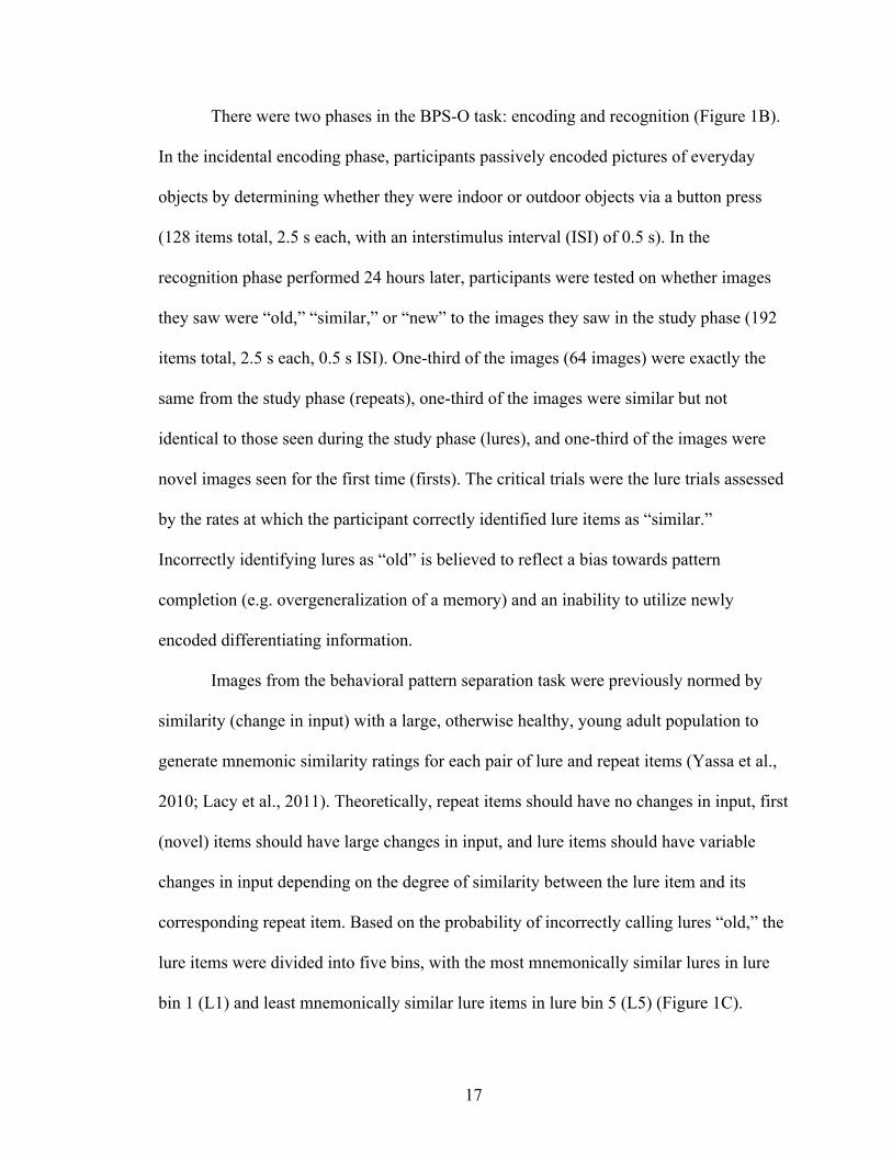
There were two phases in the BPS-O task: encoding and recognition (Figure 1B).
In the incidental encoding phase, participants passively encoded pictures of everyday
objects by determining whether they were indoor or outdoor objects via a button press
(128 items total, 2.5 s each, with an interstimulus interval (ISI) of 0.5 s). In the
recognition phase performed 24 hours later, participants were tested on whether images
they saw were “old,” “similar,” or “new” to the images they saw in the study phase (192
items total, 2.5 s each, 0.5 s ISI). One-third of the images (64 images) were exactly the
same from the study phase (repeats), one-third of the images were similar but not
identical to those seen during the study phase (lures), and one-third of the images were
novel images seen for the first time (firsts). The critical trials were the lure trials assessed
by the rates at which the participant correctly identified lure items as “similar.”
Incorrectly identifying lures as “old” is believed to reflect a bias towards pattern
completion (e.g. overgeneralization of a memory) and an inability to utilize newly
encoded differentiating information.
Images from the behavioral pattern separation task were previously normed by
similarity (change in input) with a large, otherwise healthy, young adult population to
generate mnemonic similarity ratings for each pair of lure and repeat items (Yassa et al.,
2010; Lacy et al., 2011). Theoretically, repeat items should have no changes in input, first
(novel) items should have large changes in input, and lure items should have variable
changes in input depending on the degree of similarity between the lure item and its
corresponding repeat item. Based on the probability of incorrectly calling lures “old,” the
lure items were divided into five bins, with the most mnemonically similar lures in lure
bin 1 (L1) and least mnemonically similar lure items in lure bin 5 (L5) (Figure 1C).
18
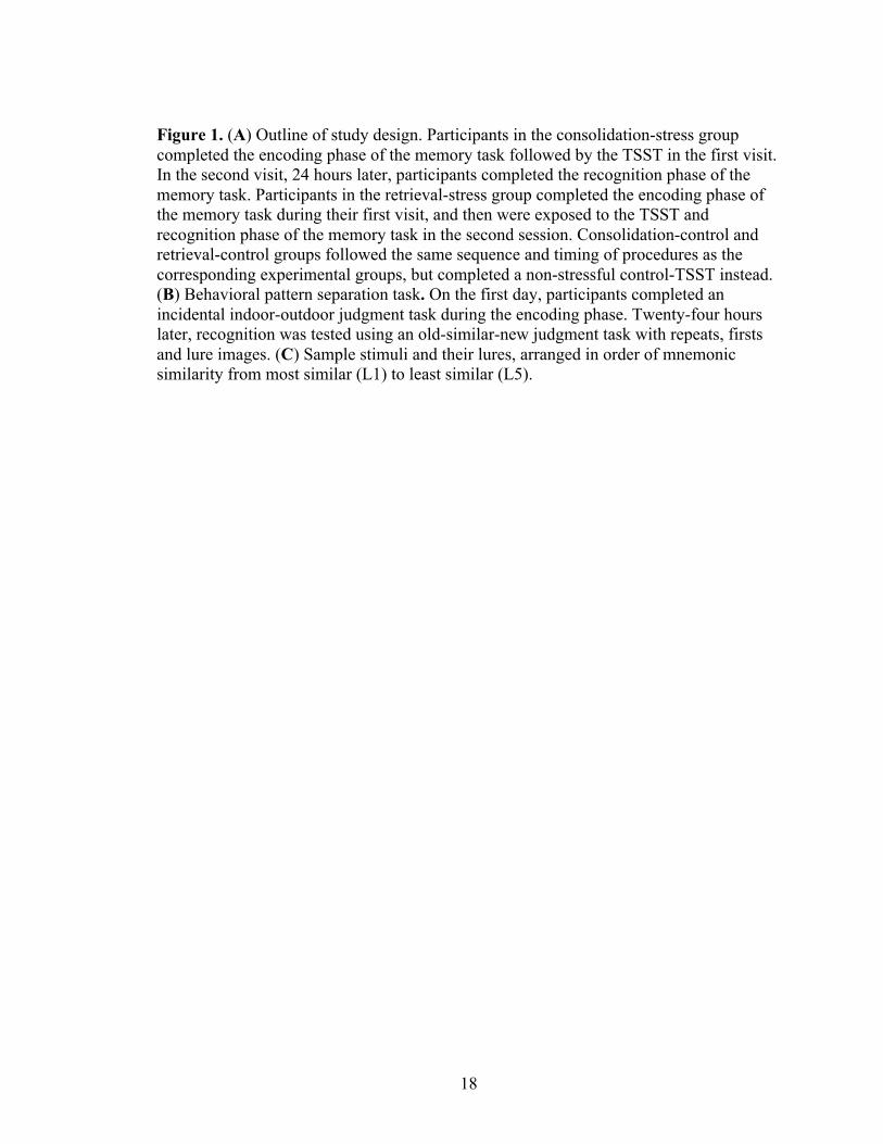
Figure 1. (A) Outline of study design. Participants in the consolidation-stress group completed the encoding phase of the memory task followed by the TSST in the first visit. In the second visit, 24 hours later, participants completed the recognition phase of the memory task. Participants in the retrieval-stress group completed the encoding phase of the memory task during their first visit, and then were exposed to the TSST and recognition phase of the memory task in the second session. Consolidation-control and retrieval-control groups followed the same sequence and timing of procedures as the corresponding experimental groups, but completed a non-stressful control-TSST instead. (B) Behavioral pattern separation task. On the first day, participants completed an incidental indoor-outdoor judgment task during the encoding phase. Twenty-four hours later, recognition was tested using an old-similar-new judgment task with repeats, firsts and lure images. (C) Sample stimuli and their lures, arranged in order of mnemonic similarity from most similar (L1) to least similar (L5).

19
B
A
C

20
2.5. Trier Social Stress Test
The standardized stressful component of the TSST was divided into three 5-
minute parts. In the first part, participants were asked to prepare a speech explaining why
they were the best candidates for an ideal job. Participants were allowed to use pen and
paper during the preparation, but were not allowed to use their notes during the
presentation. In the second part, the participants presented their speech to a panel of two
researchers, one male and one female, posing as expert judges. The judges maintained
neutral expressions and observed the participant without comment. If the participants did
not use the full 5 minutes allocated for the part, the judges asked the participants to
continue until time was up. In the final part of the TSST, the participants performed a
mental arithmetic task by serially subtracting the number 13 from 3087. If a mistake was
made, the judges asked the participants to start again from the beginning.
Prior to and immediately following the stressful portion of the TSST, participants
were given a 10-minute rest period and 10-minute recovery period. The rest period gave
participants a chance to reach baseline hormone levels before beginning the stressful task.
The recovery period served to coincide the post-TSST saliva sample with the delayed
cortisol peak response.
The standardized control version of the TSST was designed to be as similar as
possible to the TSST, but without the stressful components, e.g. specifically lacking the
social evaluative threat (Het et al., 2009). The control-TSST was performed in the same
room as the TSST. Instead of preparing a speech to give in front of a panel of judges, the
control-TSST gave participants 5 minutes to prepare for a 5-minute talk about a movie,
novel, or recent holiday trip. Participants were told that they would speak alone in an
21
empty room, and were given paper and pen to prepare and use throughout the task. The
researcher only entered the room between each part to give instructions to the
participants. After preparing and talking on the topic of their choice, participants were
asked to begin serially adding by increments of 15, starting at 0. The researcher, once
again, left the participants alone to complete the task, and controlled for the participants’
compliance by asking them for the final number reached in the serial addition.
2.6. Self-report questionnaires
During the 10-minute recovery period following the TSST, participants completed
self-report questionnaires, including the Beck Anxiety Inventory, designed to assess
severity of anxiety over the past week; the Beck Depression Inventory designed to assess
severity of depression over the past week; the Pittsburgh Sleep Quality Inventory aimed
to assess sleep quality over the past month; the Cohen Perceived Stress Scale to examine
perceived stress over the past month, and an exercise questionnaire to assess total hours
exercised over the past week. The State Trait Anxiety Inventory was also given to
participants before and after the TSST as a subjective measure of pre and post-
psychological stress.
22
3. Results
3.1. Parameters for analysis
Accuracy for correctly identifying novel and repeated images was used to ensure
that participants understood and completed the task as instructed. Eleven participants (2
from the consolidation-stress group, 2 from consolidation-control, 4 from retrieval-stress,
and 3 from retrieval-control) were excluded from data analysis due to poor memory task
performance, which was defined as scoring below a 33% (chance) correct response rate
for first or repeat images. Seven participants (3 from the consolidation-stress group, 2
from consolidation-control, 1 from retrieval-stress, and 1 from retrieval-control) were
further excluded from cortisol data analysis due to undetectable salivary cortisol levels in
the samples collected either pre- or post-TSST administration. After removing these
participants, the consolidation-stress group included 21 participants, the consolidation-
control group included 10 participants, retrieval-stress group included 20 participants,
and retrieval-control group included 11 participants.
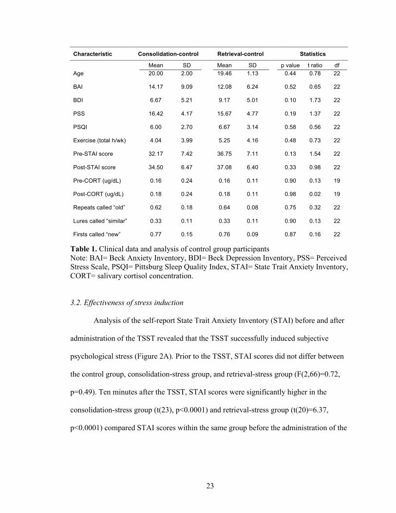
We examined differences between the two control subgroups (consolidation-
control and retrieval-control) and found no significant differences between the groups for
all values measured (Table 1). Therefore we collapsed the data from these groups to form
a single combined control group (n=24 for behavioral data, n=21 for cortisol analysis) for
the rest of our analysis.
23
Characteristic Consolidation-control Retrieval-control Statistics
Mean SD Mean SD p value t ratio df Age 20.00 2.00
19.46 1.13
0.44 0.78 22
BAI 14.17 9.09
12.08 6.24
0.52 0.65 22
BDI 6.67 5.21
9.17 5.01
0.10 1.73 22
PSS 16.42 4.17
15.67 4.77
0.19 1.37 22
PSQI 6.00 2.70
6.67 3.14
0.58 0.56 22
Exercise (total h/wk) 4.04 3.99
5.25 4.16
0.48 0.73 22
Pre-STAI score 32.17 7.42 36.75 7.11 0.13 1.54 22
Post-STAI score 34.50 6.47 37.08 6.40 0.33 0.98 22
Pre-CORT (ug/dL) 0.16 0.24 0.16 0.11 0.90 0.13 19
Post-CORT (ug/dL) 0.18 0.24 0.18 0.11 0.98 0.02 19
Repeats called “old” 0.62 0.18 0.64 0.08 0.75 0.32 22
Lures called “similar” 0.33 0.11 0.33 0.11 0.90 0.13 22
Firsts called “new” 0.77 0.15 0.76 0.09 0.87 0.16 22
Table 1. Clinical data and analysis of control group participants Note: BAI= Beck Anxiety Inventory, BDI= Beck Depression Inventory, PSS= Perceived Stress Scale, PSQI= Pittsburg Sleep Quality Index, STAI= State Trait Anxiety Inventory, CORT= salivary cortisol concentration.
3.2. Effectiveness of stress induction
Analysis of the self-report State Trait Anxiety Inventory (STAI) before and after
administration of the TSST revealed that the TSST successfully induced subjective
psychological stress (Figure 2A). Prior to the TSST, STAI scores did not differ between
the control group, consolidation-stress group, and retrieval-stress group (F(2,66)=0.72,
p=0.49). Ten minutes after the TSST, STAI scores were significantly higher in the
consolidation-stress group (t(23), p<0.0001) and retrieval-stress group (t(20)=6.37,
p<0.0001) compared STAI scores within the same group before the administration of the

24
TSST. Participants in the control conditions did not exhibit an increase in subjective
stress following the control-TSST (t(23)=1.18, p=0.25).
To verify that the TSST also induced a physiological stress response, we
compared the salivary cortisol levels between stress and control groups (Figure 2B). Prior
to the TSST, concentrations of salivary cortisol did not differ between the control group,
consolidation-stress group, or retrieval-stress group (F(2,59)=1.218, p=0.30). Ten
minutes after the TSST, concentrations of salivary cortisol were significantly higher in
the consolidation stress group (t(20)=4.90, p<0.0001) and retrieval stress group
(t(19)=5.26, p<0.0001) compared to the salivary concentrations within the same group
before the administration of the TSST. Participants in the control conditions did not
exhibit an increase in salivary cortisol following the control-TSST (t(20)=0.47, p=0.65).
Figure 2. The TSST induces a psychological and physiological response to stress. Mean (A) STAI scores and (B) salivary cortisol concentrations are higher post-TSST for stress groups but not for control groups. Error bars represent standard error of the mean. ***p<0.001
0.0
0.2
0.4
0.6
CO
RT
(ug/
dL)
Consolidation-stress
Retrieval-stress
ControlsPre Post Pre Post Pre Post
*** ***
0
20
40
60
80
STAI
sco
re
Consolidation-stress
Retrieval-stress
ControlsPre Post Pre Post Pre Post
*** ***
A B

25
3.3. Memory performance
Participants identified repeats as “old” and firsts as “new” at similar rates across
groups (Figure 3). This performance was consistent with a previous study using a similar
population (Borota et al., 2013).
To determine whether the ability to differentiate the critical lure items was
affected by stress, we examined the proportion of lure stimuli correctly identified as
“similar.” Multiple paired t-tests were used to compare pattern separation performance
between the consolidation-stress and control groups as well as the retrieval-stress and
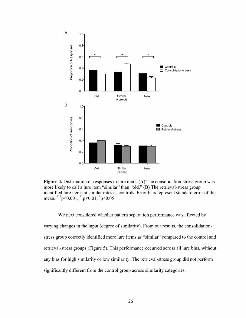
control groups. Pattern separation performance is defined as the proportion of correct
responses to lure items. Participants in the consolidation-stress condition were
significantly more likely to correctly call lure items “similar” than the participants in the
control condition, who were more likely to call lure items “old” (F(2, 138)=15.90,
p<0.0001) (Figure 4A). There was no significant difference in lure response between
retrieval-stress and control groups (F(2,129)=1.055, p=0.35) (Figure 4B).
Figure 3. Stress during consolidation, but not retrieval, enhances the identification of lure items. Error bars represent standard error of the mean. ***p<0.001
0.0
0.2
0.4
0.6
0.8
1.0
Pro
porti
on o
f Cor
rect
Res
pons
es
Consolidation-stressRetrieval-stress
Controls
Repeats Lures Firsts
***
26
Figure 4. Distribution of responses to lure items (A) The consolidation-stress group was more likely to call a lure item “similar” than “old.” (B) The retrieval-stress group identified lure items at similar rates as controls. Error bars represent standard error of the mean. ***p<0.001, **p<0.01, *p<0.05
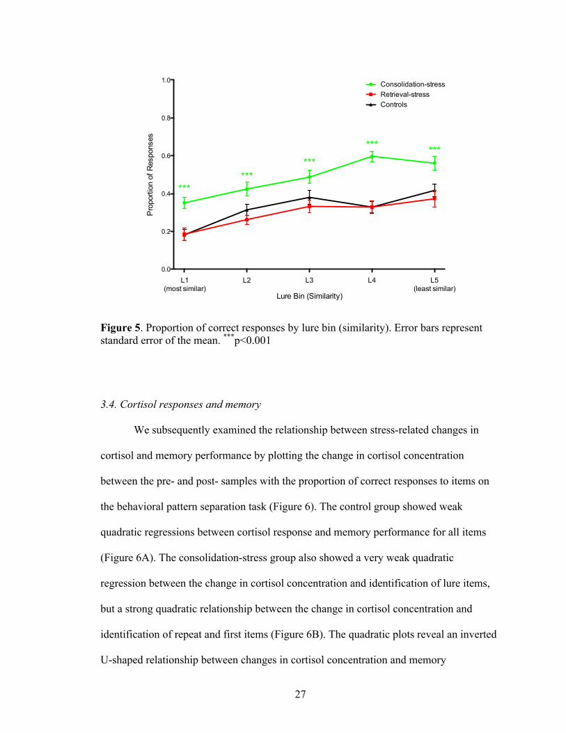
We next considered whether pattern separation performance was affected by
varying changes in the input (degree of similarity). From our results, the consolidation-
stress group correctly identified more lure items as “similar” compared to the control and
retrieval-stress groups (Figure 5). This performance occurred across all lure bins, without
any bias for high similarity or low similarity. The retrieval-stress group did not perform
significantly different from the control group across similarity categories.
Old Similar New
0.0
0.2
0.4
0.6
0.8
1.0
Pro
porti
on o
f Res
pons
esConsolidation-stress Controls
*** ***
Old Similar New
0.0
0.2
0.4
0.6
0.8
1.0
Pro
porti
on o
f Res
pons
es
ControlsRetrieval-stress
B
A
(correct)
(correct)
27
Figure 5. Proportion of correct responses by lure bin (similarity). Error bars represent standard error of the mean. ***p<0.001
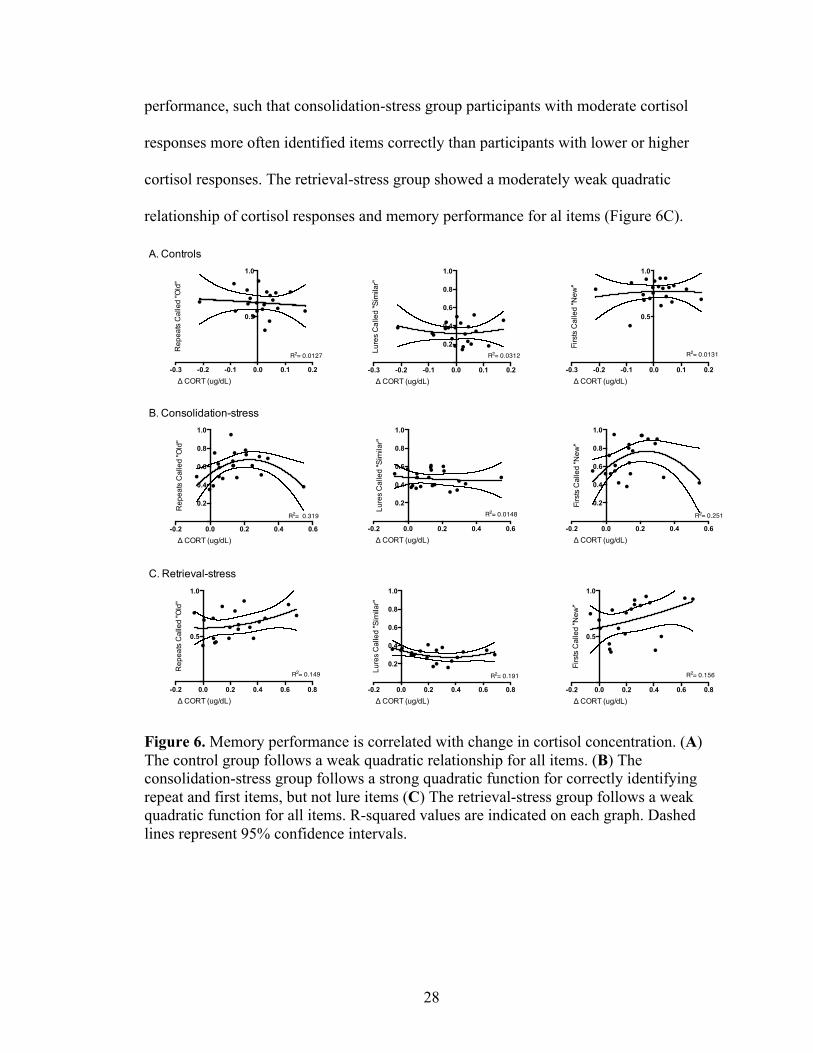
3.4. Cortisol responses and memory
We subsequently examined the relationship between stress-related changes in
cortisol and memory performance by plotting the change in cortisol concentration
between the pre- and post- samples with the proportion of correct responses to items on
the behavioral pattern separation task (Figure 6). The control group showed weak
quadratic regressions between cortisol response and memory performance for all items
(Figure 6A). The consolidation-stress group also showed a very weak quadratic
regression between the change in cortisol concentration and identification of lure items,
but a strong quadratic relationship between the change in cortisol concentration and
identification of repeat and first items (Figure 6B). The quadratic plots reveal an inverted
U-shaped relationship between changes in cortisol concentration and memory
0.0
0.2
0.4
0.6
0.8
1.0
Lure Bin (Similarity)
Pro
porti
on o
f Res
pons
es
Consolidation-stressRetrieval-stressControls
L1(most similar)
L2 L3 L4 L5(least similar)
******
***
*** ***
28
performance, such that consolidation-stress group participants with moderate cortisol
responses more often identified items correctly than participants with lower or higher
cortisol responses. The retrieval-stress group showed a moderately weak quadratic
relationship of cortisol responses and memory performance for al items (Figure 6C).
Figure 6. Memory performance is correlated with change in cortisol concentration. (A) The control group follows a weak quadratic relationship for all items. (B) The consolidation-stress group follows a strong quadratic function for correctly identifying repeat and first items, but not lure items (C) The retrieval-stress group follows a weak quadratic function for all items. R-squared values are indicated on each graph. Dashed lines represent 95% confidence intervals.
-0.3 -0.2 -0.1 0.0 0.1 0.2
0.5
1.0
Δ CORT (ug/dL)
Rep
eats
Cal
led
"Old
"
R2= 0.0127
-0.2 0.0 0.2 0.4 0.6
0.2
0.4
0.6
0.8
1.0
Δ CORT (ug/dL)
Rep
eats
Cal
led
"Old
"
R2= 0.319
-0.2 0.0 0.2 0.4 0.6 0.8
0.5
1.0
Δ CORT (ug/dL)
Rep
eats
Cal
led
"Old
"
R2= 0.149
-0.3 -0.2 -0.1 0.0 0.1 0.2
0.2
0.4
0.6
0.8
1.0
Δ CORT (ug/dL)Lu
res
Cal
led
"Sim
ilar"
R2= 0.0312
-0.2 0.0 0.2 0.4 0.6
0.2
0.4
0.6
0.8
1.0
Δ CORT (ug/dL)
Lure
s C
alle
d "S
imila
r"
R2= 0.0148
-0.2 0.0 0.2 0.4 0.6 0.8
0.2
0.4
0.6
0.8
1.0
Δ CORT (ug/dL)
Lure
s C
alle
d "S
imila
r"
R2= 0.191
-0.3 -0.2 -0.1 0.0 0.1 0.2
0.5
1.0
Δ CORT (ug/dL)
Firs
ts C
alle
d "N
ew"
R2= 0.0131
-0.2 0.0 0.2 0.4 0.6
0.2
0.4
0.6
0.8
1.0
Δ CORT (ug/dL)
Firs
ts C
alle
d "N
ew"
R2= 0.251
-0.2 0.0 0.2 0.4 0.6 0.8
0.5
1.0
Δ CORT (ug/dL)
Firs
ts C
alle
d "N
ew"
R2= 0.156
A. Controls
B. Consolidation-stress
C. Retrieval-stress
29
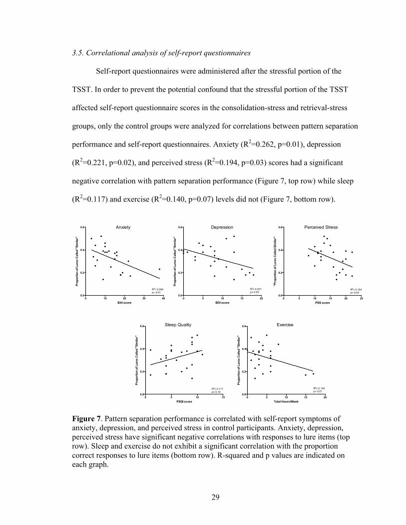
3.5. Correlational analysis of self-report questionnaires
Self-report questionnaires were administered after the stressful portion of the
TSST. In order to prevent the potential confound that the stressful portion of the TSST
affected self-report questionnaire scores in the consolidation-stress and retrieval-stress
groups, only the control groups were analyzed for correlations between pattern separation
performance and self-report questionnaires. Anxiety (R2=0.262, p=0.01), depression
(R2=0.221, p=0.02), and perceived stress (R2=0.194, p=0.03) scores had a significant
negative correlation with pattern separation performance (Figure 7, top row) while sleep
(R2=0.117) and exercise (R2=0.140, p=0.07) levels did not (Figure 7, bottom row).
Figure 7. Pattern separation performance is correlated with self-report symptoms of anxiety, depression, and perceived stress in control participants. Anxiety, depression, perceived stress have significant negative correlations with responses to lure items (top row). Sleep and exercise do not exhibit a significant correlation with the proportion correct responses to lure items (bottom row). R-squared and p values are indicated on each graph.
0 10 20 30 400.0
0.2
0.4
0.6
BAI score
Prop
ortio
n of
Lur
es C
alle
d "S
imila
r"
R2= 0.262p= 0.01
0 5 10 15 20 250.0
0.2
0.4
0.6
PSS score
"Pro
port
ion
of L
ures
Cal
led
Sim
ilar"
R2= 0.194p= 0.03
0 5 10 15 200.0
0.2
0.4
0.6
Total Hours/Week
Prop
ortio
n of
Lur
es C
alle
d "S
imila
r"
R2= 0.140p= 0.07
0 5 10 15 200.0
0.2
0.4
0.6
BDI score
Prop
ortio
n of
Lur
es C
alle
d "S
imila
r"
R2= 0.221p= 0.02
0 5 10 150.0
0.2
0.4
0.6
PSQI score
Prop
ortio
n of
Lur
es C
alle
d "S
imila
r"
R2= 0.117p= 0.10
Anxiety Depression Perceived Stress
Sleep Quality Exercise

30
4. Discussion
The present study examined the relationship of acute stress on memory
consolidation and retrieval in healthy young adults. We used a behavioral pattern
separation task to assess memory processes thought to be supported by the DG/CA3
hippocampal subregions during stress. Our results show that acute stress induced during
consolidation, the process of converting short-term memories into long-term memories,
enhanced pattern separation performance, reflecting an increased ability to differentiate
and recall highly similar items. Participants in the consolidation-stress group exhibited
higher response accuracy in identifying lure items across all degrees of similarity. Stress-
related changes in cortisol exhibited an inverted-U shaped relationship in identifying
repeat and first items in the consolidation-stress group, suggesting that moderate amounts
of stress improves the consolidation of this type of information while low and high
amounts of stress had detrimental effects on consolidation. Our results additionally show
that stress induced during retrieval does not significantly impact memory compared to
controls. Finally, results from this study showed that pattern separation performance was
negatively correlated with self-reported anxiety, depression, and perceived stress, such
that the most severe symptoms were related to an impaired ability to correctly identify
lure items.
4.1. Effects of stress on memory performance
The findings reported here are consistent with previous studies that observed
enhancing effects of cortisol on memory performance (de Kloet et al., 1999; McGaugh &
Roozendaal, 2002). Studies of the stress response in animals have consistently

31
demonstrated the beneficial effects of glucocorticoids on memory consolidation. As
described previously, human studies have reported much more varied effects of stress on
memory consolidation. For instance, Cahill, Gorski, & Le (2003) found that the cold-
pressor stress test enhanced memory of emotionally arousing images but not neutral ones.
Yet in another study using the cold-pressor stress test, memory enhancement was
observed for both neutral and negative images (McCullough & Yonelinas, 2013). Many
other studies in humans suggest that cortisol only has an effect for emotionally arousing
experiences and does not modulate neutral information (Kuhlmann & Wolf, 2006; Holz
et al., 2014). Despite using emotionally neutral images in our memory task, our results
show an enhancing effect of cortisol on memory consolidation, potentially supporting the
notion that pattern separation is indeed a more sensitive measure to stress than other
memory tasks. The behavioral pattern separation task has previously been used in
research on age-related changes in cognitive function. In a study by Stark et al.,
performance on this task showed a linearly decreasing memory function with age when
no changes in traditional measures of recognition memory were observed (2013). These
findings support the sensitivity of pattern separation in detecting changes in memory
function to various stimuli.
Our results show that acute stress during consolidation significantly enhances the
ability to discriminate between similar stimuli. Participants in the consolidation-stress
group were more likely to correctly respond “similar” to lure items, which is indicative of
improved pattern separation abilities. This improved performance was observed for all
lure items, regardless of mnemonic similarity, in a linear relationship such that there was
higher response accuracy in identifying lure items of least similarity compared to lure

32
items of most similarity. For the lure items, the proportion of “new” responses shifted
less than the proportion of “old” responses shifted towards “similar” responses,
reflecting a change in memory accuracy rather than a complete failure of recall or
recognition (misses). The observed lower frequency of “old” responses to lure items in
the consolidation-stress group is also indicative of decreased pattern completion and
further supports the effect of stress during consolidation on memory function.
Our finding that stress induced immediately before retrieval did not have
significant effects on memory is inconsistent with some previous reports. In both animals
and humans, most studies observed impairing effects of glucocorticoids on memory
retrieval (for reviews, see Wolf, 2003; Het et al., 2004; de Quervain, 2009). However,
some studies have found contradictory results. In one study, administration of a cortisol-
suppressing drug led to significantly impaired free recall of emotional texts (Rimmele et
al., 2015). In another study, highly stressed participants with large increases on measures
of arousal were able to compensate for the impairing effects on retrieval and perform
comparably to controls (Boehringer et al., 2010). It is possible that the participants in the
present study also experienced both high stress and high arousal, and thus did not exhibit
impaired memory retrieval. Yet, this is unlikely to be the sole reason given that the
consolidation-stress group underwent the same stress induction as the retrieval-stress
group. Recently there has been evidence, primarily from animals but also from human
studies, that the balance between mineralocorticoid and glucocorticoid receptor activation
of different brain regions determines glucocorticoid effects on memory retrieval
(Rimmele et al., 2015; Harris et al., 2013). Though glucocorticoid receptors are believed
to be the major effectors of the stress response pathway, mineralocorticoid receptor
33
activity and location may also mediate this response. Future experiments should consider
the context of mineralocorticoids and glucocorticoid receptor activation when
investigating the role of glucocorticoids in memory processes.
The observed quadratic relationship between stress-induced cortisol with memory
performance is consistent with previous studies (Andreano & Cahill, 2006; McCullough
et al., 2015). The consolidation-stress group exhibited the strongest inverted U-shaped
relationship between cortisol concentrations and the proportion of correct responses to
repeat and novel items on the behavioral pattern separation task. Notably, the responses
to lure items did not exhibit a strong quadratic relationship for the consolidation-stress
group. The observed dose-response curve may be a result of stress impacting various
memory trade-offs, such as that between “gist,” the general theme, and "visual details,
specific information, of an experience. In previous studies, arousal has enhanced “gist”
memory but not memory for the visual details of studied images (Adolphs et al., 2001,
2001; Denburg et al., 2003). As the discrimination of lure stimuli requires memory for
visual details of the studied images, a limited relationship between changes in cortisol
concentration and response accuracy for lure items in the consolidation-stress group is
consistent with reports of a trade-off between the strengthening of the gist but not an
increase for the amount of detail for an event or stimulus. In the responses to repeats and
firsts for the consolidation-stress group, stress led to an increase in memory performance
up to a certain point, beyond which additional stress became detrimental.
The retrieval-stress group showed weakly positive (repeats and firsts) and weakly
negative (lures) relationships between amounts of stress and memory performance, again
supporting the idea that stress during retrieval did not have a significant effect. The weak
34
quadratic regression observed in controls between stress-induced cortisol changes and
memory responses were expected. The control group did not show significant changes in
cortisol concentrations before and after the control-TSST, and thus were not predicted to
exhibit an inverted U-response curve or other type of change relationship.
4.2. Correlates of pattern separation performance
The negative relationship observed between depression and pattern separation
performance is consistent with previous research. Studies have reliably demonstrated a
reduction in hippocampal volume and impairment in memory performance in patients
with depression (for review, see Campbell et al., 2004). These changes have been
attributed to neuronal death and decreased adult neurogenesis necessary to support
pattern separation processes. Depressed individuals treated with antidepressants showed
larger dentate gyrus volume and more neural progenitor cells, suggesting that
antidepressants may remediate memory deficits in depression through increased
neurogenesis (Boldrini et al., 2009). Studies suggest that individuals with depression have
a tendency to overgeneralize stimuli in the environment, a pathology that has been
associated with functioning of the hippocampus and its subregions (Becker & Wojtowicz,
2007; Sahay & Hen, 2007). Accordingly, impairments in pattern separation may be an
indicator for both the behavior and underlying pathology of depression and other mood
disorders. Only a few other studies have correlated self-report depression symptoms with
pattern separation performance (Dery et al., 2013; Shelton et al., 2013). Our results were
highly consistent with these studies, suggesting that depression symptoms are closely
35
correlated with pattern separation performance, which may reflect a reduction in
neurogenesis.
Anxiety-like behaviors have previously been tested in rats through observations
on the open field test (measuring willingness to explore in an exposed space) and
novelty-suppressed feeding test (measuring the latency of an animal to eat familiar food
in an novel environment) (Sahay et al., 2011). Mice that were genetically manipulated for
increased neurogenesis, the process thought to facilitate pattern separation, showed a
decrease in anxiety-like behaviors (Sahay et al., 2011). In other tasks, mice have been
tested on their ability to distinguish between an aversive context, in which they would
receive a shock, and a similar but safe (no shock) context (McHugh et al., 2007;
Kheirbek, Tannenholz, & Hen, 2012). High discrimination between contexts was
observed by exploratory behavior, while low discrimination between contexts led to
freezing behavior. In humans, however, no studies thus far have investigated the
relationship between self-report anxiety and pattern separation performance. Our results
are consistent with previous animal studies that suggest increased anxiety decreases
discriminatory ability, and show this correlation in humans for the first time.
Sleep quality has also been not been well investigated in relation to pattern
separation. We are only aware of one study, to date, that directly investigated the effect of
sleep on the behavioral responses of pattern separation and pattern completion in humans.
In this study, there was better memory discrimination in participants who slept (when
testing occurred 12 hours later, on the following day) compared to those who did not
(when testing occurred 12 hours later, on during the same day), consistent with a bias
36
toward pattern separation following sleep (James, 2014). However, our results did not
find a significant correlation between sleep quality and pattern separation performance.
Although we did not find a significant correlation between total hours exercised
per week and pattern separation performance, increasing evidence suggests that physical
exercise promotes synaptic plasticity and hippocampal neurogenesis. Studies in mice
found that running enhanced spatial pattern separation and increased the proliferation of
young neurons (Creer et al., 2010; Snyder et al., 2009). The decline in neurogenesis in
aged mice was reversed to 50% of the levels of young controls after a voluntary running
wheel was introduced to sedentary mice (van Praag et al., 2005). A previous study
reported that there was no effect of exercise on learning in rats, however in this study,
forced treadmill training was used rather than voluntary wheel running, thus suggesting a
difference in voluntary and forced exercise (Barnes et al., 1991). In humans, individuals
who experienced the greatest improvement in aerobic fitness showed significant post-
exercise enhancement in the ability to distinguish similar lures (Dery et al., 2013). A
possible reason we did not observed this relationship was because the exercise
questionnaire employed in this study did not ask about certain fitness criteria (e.g.
changes in fitness level) and rather only asked about baseline exercise habits. A more
extensive exercise questionnaire (e.g. including questions on exercise intensity, aerobic
vs. nonaerobic exercise) may yield significant correlations to pattern separation
performance.
37
4.3. Possible mechanisms by which stress impacts memory
Several influential models have attempted to explain the contradictory findings of
facilitating and detrimental acute glucocorticoid effects on memory (de Quervain et al.,
2009; Joels, 2006). Recently, a model has related the timing and dosage of
glucocorticoids to the characteristics of neural activation (Sandi, 2011; McCullough &
Yonelinas, 2013). In this model, moderate levels of post-encoding stress may be
associated with better memory performance compared to that of lower or higher levels of
stress. Our analysis of the salivary cortisol levels support the inverted U-dose response
curve hypothesis in which moderate levels of glucocorticoids observes optimal memory
performance.
Glucocorticoid-induced changes to synaptic strength, membrane excitability, or
remodeling of neuronal structure may have mediated pattern separation performance
(Herbert et al., 2006). Rapid processes can occur when glucocorticoids interact with
membrane-bound receptors while delayed processes (protein-synthesis dependent) occur
when glucocorticoids bind to intracellular receptors to alter gene expression in the
nucleus. The glucocorticoid effects in the present study likely occurred through non-
genomic mechanisms mediated by membrane-bound receptors, as behavioral measures
were taken shortly following stress induction. Activation of the noradrenergic system
through the TSST may also have induced a mechanism in which norepinephrine and
glucocorticoids interacted to produce a G-protein mediated signaling cascade
(Roozendaal, Williams, & McGaugh, 1999). Norepinephrine binding to beta-
adrenoceptors, G-protein coupled receptors, in the BLA may enhance memory
consolidation through downstream activation of cAMP, a signing molecule (Ferry,

38
Roozendaal, & McGaugh, 1999). Overall, short-term and long-term activation of
glucocorticoids may act through different mechanisms, and future studies should aim to
uncover these processes by which the DG/CA3 is influenced by stress.
4.4. The role of adult neurogenesis in pattern separation
New adult-born neurons are believed to play a role in pattern separation (for
review, see Aimone et al., 2011). Produced by adult neurogenesis, young neurons exhibit
distinct physiological properties compared to mature neurons. For example, four-to-eight
week-old granular cells show greater synaptic plasticity and increased excitability
compared to the older granular cell population (Ming & Song, 2011). Studies that ablate
neurogenesis in adult mice found impaired performance on tasks shown to engage pattern
separation, such as contextual fear discrimination learning and radial maze delayed non-
match to place (Clelland et al., 2009; McHugh et al., 2007). Furthermore, genetically
improving the survival and proliferation of adult-born neurons enhanced rodents’ abilities
to distinguish between similar contexts (Sahay et al., 2011). The enhancement in pattern
separation ability can attributed to increased plasticity and hyperexcitability of young
neurons relative to mature neurons. Young neurons have a lower threshold of activation,
thus requiring weaker inputs to fire, which make the more readily available to respond to
subtle changes in the environment (Markwardt, Wadiche, & Overstreet-Wadiche, 2009).
Granule cells also have multiple place fields that remap quickly with small changes in
environmental context (Leutgeb et al., 2007). Taken together, loss-of-function and gain-
of-function studies support the role of these cells in discriminating between fine details
and overlapping contextual representations.
39
Levels of adult neurogenesis may change as an adaptive response to different
environments (for review, see Sahay et al., 2011). From the proliferation of neural
precursors to the survival of newborn neurons, each phase of adult neurogenesis can be
influenced by numerous physiological and environmental factors. Overall, neurogenesis-
based circuit alterations are more relevant for reflecting longer-term changes in the
environment. For instance, living in an enriching environment that promotes exploration
and learning increases the survival of young adult-born neurons as they create a need for
increased neurogenesis to maintain nonoverlapping representations (Kempermann et al.,
1997). Learning is a major regulator of adult neurogenesis only if the tasks depend on the
hippocampus, and specific learning paradigms can regulate the survival of new neurons
in animals (for review, see Zhao et al., 2008). On the other hand, stressful environments
may lead to decreased neurogenesis to enable organisms to generalize across similar
situations and avoid potential danger in the future.
Disruptions in the normal regulation of neurogenesis may result in excessive or
impaired pattern separation, either of which can be maladaptive for the individual.
Excessive pattern separation may prevent the normal processing of information due to a
fixation on fine details or preoccupation with minutiae; these behaviors can be seen in
autism spectrum disorders and obsessive-compulsive personality disorder (Soulieres et
al., 2009). In contrast, impaired pattern separation may lead to overgeneralization and a
decreased ability to discriminate between multiple contexts. For individuals with panic
disorder or PTSD, the reduced ability to distinguish between experiences may cause a
neutral stimulus to trigger a traumatic memory. Impaired pattern separation and the
tendency to overgeneralize are also characteristic of depression and aging (Yassa et al.,
40
2010; Shelton & Kirwan, 2013). While our results are cannot conclude a causal
relationship between neurogenesis and pattern separation, they are consistent with the
hypothesis that a reduction in neurogenesis is both related to impaired pattern separation
performance and symptoms in anxiety and depression.
Overall, neurogenesis is implicated in a range of neurological disorders, and may
be assessed behaviorally with pattern separation tasks. The identification of specific
cognitive markers such as pattern separation deficits can allow for the development of
approaches targeting underlying neural circuits. Recent improvements in behavioral
assessments of human pattern separation and imaging methods of the hippocampal
subregions has led to significant progress in understanding the relationship underlying
pattern separation, neurogenesis, and neurological disorders.
4.5. Limitations
There are several limitations to the present study. First, with all behavioral tasks,
there is a concern of whether the observed behavior is a true reflection the underlying
neurobiological process. Here, pattern separation performance is used to assess function
of the DG/CA3 subregion of the hippocampus. While the behavioral pattern separation
task specifically taxes the DG/CA3 subregions, future studies will need to determine if
the current findings can be replicated using more direct measures of hippocampal
subregion function, such as functional magnetic resonance imaging studies. Nevertheless,
behavioral assays have been critical to establishing much of the information known in the
field, and pattern separation remains a useful indicator or for brain-specific measures
(Deng et al., 2010).

41
Second, acute stress triggers multiple responses in the body beyond
hypothalamic-pituitary-adrenal axis activation, and causes other hormones besides
cortisol to be released. For example, norepinephrine, vasopressin, and pro-inflammatory
cytokines, are also all secreted in response to stress. Since such changes occur in response
to the same stimuli, there is a strong association between stress response systems. For
example, peaks in salivary alpha-amylase, an indicator of the sympathetic-adrenal-
medullary axis, and cortisol, an indicator of the hypothalamic-pituitary-adrenal axis, were
closely correlated following a cold-pressor stress test (Smeets et al., 2008). Extensive
evidence supports that salivary cortisol is a reliable and valid biomarker in stress
research, and continues to be used as the biomarker of choice (Hellhammer, Wust, &
Kudielka, 2009).
Third, the present study recruited an all-male sample, and thus the results may not
be readily generalized to females. Females were excluded because they tend to exhibit
smaller stress responses and more variable changes in salivary cortisol levels (Kajantie
and Phillips, 2006). Stress effects are also less consistently observed in females
depending on menstrual cycle phases and the use of oral contraceptives (Kirschbaum et
al., 1999; Bouma et al., 2009). In this study, we chose the population with the most robust
and stable stress response, as cortisol levels served as an independent variable for
memory performance. Many studies measuring or manipulating the stress response limit
their population to males (e.g. Andreano & Cahill, 2006; Henckens et al., 2009;
McCulllogh et al., 2015). However, there is evidence suggesting that gender differences
may moderate the link between stress and memory performance. Future research should
consider a sample with both genders in order to better make comparisons.

42
Finally, only emotionally neutral images were shown in the present study. It
would be important to assess the pattern separation performance of both emotionally
neutral and emotionally charged images, and future studies should consider the effect of
emotional valence and arousal on pattern separation behavior. Many neurological
conditions that show impaired pattern separation performance are closely related to
emotional responses. For example, in anxiety disorders, the inability to distinguish
between neutral and emotional or fearful events may lead to intrusive memories and
clinical symptoms.
4.6. Conclusion
The present study demonstrates the important role of acute stress in modulating
hippocampal-dependent memory function. Specifically, an enhancement in the pattern
separation of neutral objects was observed when stress was induced during memory
consolidation. The improved ability to discriminate between stimuli occurred without
bias across varying levels of similarity in lure items. No significant effect on pattern
separation performance was observed when stress was induced on memory retrieval. This
study is the first to focus on the effects of acute stress on pattern separation, a
computational process of episodic memory that is specifically supported by the DG/CA3
subregions of the hippocampus. Our findings provide evidence that the memory
consolidation process is associated with the DG/CA3 region and facilitated by acute
stress hormones.

43
5. References
Adolphs, R., Denburg, N. L., & Tranel, D. (2001). The amygdala’s role in long-term
declarative memory for gist and detail. Behavioral Neuroscience, 115, 983–992.
Aimone, J. B., Deng, W., & Gage, F. H. (2010). Adult neurogenesis: integrating theories
and separating functions. Trends in Cognitive Sciences, 14(7), 325-337.
Andreano, J. M., & Cahill, L. (2006). Glucocorticoid release and memory consolidation
in men and women. Psychological Science, 17(6), 466-470.
Barnes, C. A., Forster, M. J., Fleshner, M., Ahanotu, E. N., Laudenslager, M. L., Mazzeo,
R. S., ... & Lal, H. (1991). Exercise does not modify spatial memory, brain
autoimmunity, or antibody response in aged F-344 rats. Neurobiology of
aging, 12(1), 47-53.
Bakker, A., Kirwan, C. B., Miller, M., & Stark, C. E. (2008). Pattern separation in the
human hippocampal CA3 and dentate gyrus. Science, 319(5870), 1640-1642.
Becker, S., & Wojtowicz, J. M. (2007). A model of hippocampal neurogenesis in
memory and mood disorders. Trends in Cognitive Sciences, 11(2), 70-76.
Bisaz, R., Conboy, L., & Sandi, C. (2009). Learning under stress: a role for the neural
cell adhesion molecule NCAM. Neurobiology of Learning and Memory, 91(4),
333-342.
Boehringer, A., Schwabe, L., & Schachinger, H. (2010). A combination of high stress-
induced tense and energetic arousal compensates for impairing effects of stress on
memory retrieval in men. Stress, 13(5), 444-453.

44
Boldrini, M., Underwood, M.D., Hen, R., Rosoklija, G. B., Dwork, A.J., John Mann J., et
al. (2009). Antidepressants increase neural progenitor cells in the human
hippocampus. Neurpsychopharmacology, 2376-89.
Borota, D., Murray, E., Keceli, G., Chang, A., Watabe, J. M., Ly, M., & Yassa, M. A.
(2014). Post-study caffeine administration enhances memory consolidation in
humans. Nature Neuroscience, 17(2), 201-203.
Bouma, E. M., Riese, H., Ormel, J., Verhulst, F. C., & Oldehinkel, A. J. (2009).
Adolescents’ cortisol responses to awakening and social stress; effects of gender,
menstrual phase and oral contraceptives. The TRAILS study.
Psychoneuroendocrinology, 34(6), 884-893.
Buchanan, T. W., & Lovallo, W. R. (2001). Enhanced memory for emotional material
following stress-level cortisol treatment in humans.
Psychoneuroendocrinology, 26(3), 307-317.
Cahill, L., Gorski, L., & Le, K. (2003). Enhanced human memory consolidation with
post-learning stress: Interaction with the degree of arousal at encoding. Learning
& Memory, 10(4), 270-274.
Campbell, S., Marriott, M., Nahmias, C., MacQueen, G.M. (2004). Lower hippocampal
volume in patients suffering from depression: a meta-analysis. The American
Journal of Psychiatry, 161, 598-607.
Cheke, L. G., & Clayton, N. S. (2013). Do different tests of episodic memory produce
consistent results in human adults? Learning & Memory, 20(9), 491-498.
45
Clelland, C. D., Choi, M., Romberg, C., Clemenson, G. D., Fragniere, A., Tyers, P. &
Bussey, T. J. (2009). A functional role for adult hippocampal neurogenesis in
spatial pattern separation. Science, 325(5937), 210-213.
Cordero, M. I., Merino, J. J., & Sandi, C. (1998). Correlational relationship between
shock intensity and corticosterone secretion on the establishment and subsequent
expression of contextual fear conditioning. Behavioral Neuroscience, 112(4),
885-891.
Creer, D. J., Romberg, C., Saksida, L. M., van Praag, H., & Bussey, T. J. (2010).
Running enhances spatial pattern separation in mice. Proceedings of the National
Academy of Sciences, 107(5), 2367-2372.
De Kloet, E. R., Joels, M., Holsboer, F. (2005). Stress and the brain: from adaptation to
disease. Nature Neuroscience, 2, 753-758.
De Kloet, E. R., Oitzl, M. S., & Joëls, M. (1999). Stress and cognition: are corticosteroids
good or bad guys? Trends in Neurosciences, 22(10), 422-426.
De Kloet, E. R., Vreugdenhil, E., Oitzl, M. S., & Joels, M. (1998). Brain corticosteroid
receptor balance in health and disease 1. Endocrine Reviews, 19(3), 269-301.
De Quervain, D.J., Aerni, A., Schelling, G., & Roozendaal, B. (2009). Glucocorticoids
and the regulation of memory in health and disease. Frontiers in
Neuroendocrinology, 30(3), 358-370.
De Quervain, D.J., Roozendaal, B., & McGaugh, J. L. (1998). Stress and glucocorticoids
impair retrieval of long-term spatial memory. Nature,394(6695), 787-790.

46
De Quervain, D.J., Roozendaal, B., Nitsch, R. M., McGaugh, J. L., & Hock, C. (2000).
Acute cortisone administration impairs retrieval of long-term declarative memory
in humans. Nature Neuroscience, 3(4), 313-314.
De Vriendt, T., Moreno, L. A., & De Henauw, S. (2009). Chronic stress and obesity in
adolescents: scientific evidence and methodological issues for epidemiological
research. Nutrition, Metabolism and Cardiovascular Diseases, 19(7), 511-519.
Denburg, N. L., Buchanan, T. W., Tranel, D., & Adolphs, R. (2003). Evidence for
preserved emotional memory in normal older persons. Emotion, 3, 239–253.
Deng W., Aimone, J. B., Gage, F. H. (2010). New neurons and new memories: how does
adult hippocampal neurogenesis affect learning and memory? Nat Rev Neurosci.,
11, 339–350.
Dery, N., Pilgrim, M., Gibala, M., Gillen, J., Wojtowicz, J. M., MacQueen, G., & Becker,
S. (2013). Adult hippocampal neurogenesis reduces memory interference in
humans: opposing effects of aerobic exercise and depression. Frontiers in
Neuroscience, 7, 66.
Dias-Ferreira, E., Sousa, J.C., Melo, I., Morgado, P., Mesquita, A.R., Cerqueira, J.J.,
Costa, R.M., Sousa, N. (2009). Chronic Stress Causes Frontostriatal
Reorganization and Affects Decision-Making. Science, 325(5940), 621–625
Fa, M., Xia, L., Anunu, R., Kehat, O., Kriebel, M., Volkmer, H., & Richter-Levin, G.
(2014). Stress modulation of hippocampal activity– Spotlight on the dentate
gyrus. Neurobiology of Learning and Memory, 112, 53-60.
47
Ferry, B., Roozendaal, B., & McGaugh, J. L. (1999). Basolateral amygdala noradrenergic
influences on memory storage are mediated by an interaction between β-and α1-
adrenoceptors. The Journal of Neuroscience, 19(12), 5119-5123.
Flood, J. F., Vidal, D., Bennett, E. L., Orme, A. E., Vasquez, S., & Jarvik, M. E. (1978).
Memory facilitating and anti-amnesic effects of corticosteroids. Pharmacology
Biochemistry and Behavior, 8(1), 81-87.
Harris, A. P., Holmes, M. C., de Kloet, E. R., Chapman, K. E., & Seckl, J. R. (2013).
Mineralocorticoid and glucocorticoid receptor balance in control of HPA axis and
behaviour. Psychoneuroendocrinology, 38, 648–658.
Hellhammer, D. H., Wüst, S., & Kudielka, B. M. (2009). Salivary cortisol as a biomarker
in stress research. Psychoneuroendocrinology, 34(2), 163-171.
Herbert, J., Goodyer, I. M., Grossman, A. B., Hastings, M. H., De Kloet, E. R.,
Lightman, S. L., & Seckl, J. R. (2006). Do corticosteroids damage the
brain? Journal of Neuroendocrinology, 18(6), 393-411.
Het, S., Ramlow, G., Wolf, O.T. (2005). A meta-analytic review of the effect of acute
cortisol administration on human memory. Psychoneuroendocrinology 30, 771-
884.
Het, S., Rohleder, N., Schoofs, D., Kirschbaum, C., Wolf, O., (2009). Neuroendocrine
and psychometric evaluation of a placebo version of the ‘Trier Social Stress Test’.
Psychoneuroendocrinology, 34, 1075–1086.
Holz, E., Lass-Hennemann, J., Streb, M., Pfaltz, M., & Michael, T. (2014). Effects of
acute cortisol administration on perceptual priming of trauma-related
material. PloS one, 9(9), e104864.

48
Hsu, F. C., Garside, M. J., Massey, A. E., & McAllister-Williams, R. H. (2003). Effects
of a single dose of cortisol on the neural correlates of episodic memory and error
processing in healthy volunteers. Psychopharmacology, 167(4), 431-442.
James, J. R. (2014). The Effect of Sleep-Dependent Consolidation on Pattern Separation
and Pattern Completion in Delayed Retrieval (Doctoral dissertation). Retrieved
from BYU ScholarsArchive.
Joels, M. (2006). Corticosteroid effects in the brain: U-shape it. Trends in
Pharmacological Sciences, 27(5), 244-250.
Kajantie, E., & Phillips, D. I. (2006). The effects of sex and hormonal status on the
physiological response to acute psychosocial
stress.Psychoneuroendocrinology, 31(2), 151-178.
Kempermann, G., Kuhn, H. G., & Gage, F. H. (1997). More hippocampal neurons in
adult mice living in an enriched environment. Nature,386 (6624), 493-495.
Kensinger, E. A. (2004). Remembering emotional experiences: The contribution of
valence and arousal. Reviews in the Neurosciences, 15, 241–251.
Kheirbek, M.A., Klemenhagen, K.C., Sahay, A., & Hen. (2012). Neurogenesis and
generalization: a new approach to stratify and treat anxiety disorders. Nature
Neuroscience, 15(12), 1613-1620.
Kheirbek, M.A., Tannenholz, L. & Hen, R. (2012). NR2B-dependent plasticity of adult-
born granule cells is necessary for context discrimination. The Journal of
Neuroscience, 32, 8696–8702.
Kirschbaum, C., Kudielka, B. M., Gaab, J., Schommer, N. C., & Hellhammer, D. H.
(1999). Impact of gender, menstrual cycle phase, and oral contraceptives on the

49
activity of the hypothalamus-pituitary-adrenal axis. Psychosomatic Medicine,
61(2), 154-162.
Kirschbaum, C., Pirke, K. M., & Hellhammer, D. H. (1993). The ‘Trier Social Stress
Test’–a tool for investigating psychobiological stress responses in a laboratory
setting. Neuropsychobiology, 28, 76-81.
Kirwan, C. B., & Stark, C. E. (2007). Overcoming interference: An fMRI investigation of
pattern separation in the medial temporal lobe. Learning & Memory, 14(9), 625-
633.
Kovacs, G. L., Telegdy, G., & Lissák, K. (1976). 5-Hydroxytryptamine and the mediation
of pituitary-adrenocortical hormones in the extinction of active avoidance
behaviour. Psychoneuroendocrinology,1(3), 219-230.
Kuhlmann, S., & Wolf, O. T. (2006). Arousal and cortisol interact in modulating memory
consolidation in healthy young men. Behavioral neuroscience, 120(1), 217.
Kvarta, M. D., Bradbrook, K. E., Dantrassy, H. M., Bailey, A. M., & Thompson, and S.
M. (2015). Corticosterone mediates the synaptic and behavioral effects of chronic
stress at rat hippocampal temporoammonic synapses. Journal of Neurophysiology,
114(3), 1713–1724.
LaBar, K. S., & Cabeza, R. (2006). Cognitive neuroscience of emotional memory. Nature
Reviews Neuroscience, 7(1), 54-64.
Lacy, J. W., Yassa, M. A., Stark, S. M., Muftuler, L. T., & Stark, C. E. (2011). Distinct
pattern separation related transfer functions in human CA3/dentate and CA1
revealed using high-resolution fMRI and variable mnemonic similarity. Learning
& Memory, 18(1), 15-18.

50
Leutgeb, J. K., Leutgeb, S., Moser, M. B. & Moser, E. I. (2007). Pattern separation in the
dentate gyrus and CA3 of the hippocampus. Science, 315, 961–966.
Leutgeb, S., Leutgeb, J. K., Treves, A., Moser, M. B., & Moser, E. I. (2004). Distinct
ensemble codes in hippocampal areas CA3 and CA1. Science, 305(5688), 1295-
1298.
Lupien, S. J., Maheu, F., Tu, M., Fiocco, A., & Schramek, T. E. (2007). The effects of
stress and stress hormones on human cognition: Implications for the field of brain
and cognition. Brain & Cognition, 65(3), 209-237.
Maddock, C., & Pariante, C. M. (2001). How does stress affect you? An overview of
stress, immunity, depression and disease. Epidemiol. Psychiatr. Sci., 10(03), 153-
162.
Markwardt, S. J., Wadiche, J. I., and Overstreet-Wadiche, L. S. (2009). Input-specific
GABAergic signaling to newborn neurons in adult dentate gyrus. J. Neurosci. 29,
15063–15072.
McCullough, A. M., & Yonelinas, A. P. (2013). Cold-pressor stress after learning
enhances familiarity-based recognition memory in men. Neurobiology of
Learning and Memory, 106, 11-17.
McCullough, A. M., Ritchey, M., Ranganath, C., Yonelinas, A. (2015). Differential
effects of stress-induced cortisol response on recollection and familiarity-based
recognition memory. Neurobiology of Learning and Memory 123, 1-10.
McEwen, B. S. (2001). Plasticity of the hippocampus: adaptation to chronic stress and
allostatic load. Annals of the New York Academy of Sciences, 933(1), 265-277.
51
McEwen, B. S. (2000). Allostasis and allostatic load: Implications for
neuropsychopharmacology. Neuropsychopharmacology, 22(2), 108–124.
McGaugh, J. L. (1966). Time-dependent processes in memory storage. Science,
153(3742), 1351-1358.
McGaugh, J. L. (1989). Involvement of hormonal and neuromodulatory systems in the
regulation of memory storage. Annual Review of Neuroscience,12(1), 255-287.
McGaugh, J. L. (2004). The amygdala modulates the consolidation of memories of
emotionally arousing experiences. Annu. Rev. Neurosci., 27, 1-28.
McGaugh, J. L., & Roozendaal, B. (2002). Role of adrenal stress hormones in forming
lasting memories in the brain. Current Opinion in Neurobiology, 12(2), 205-210.
McHugh, T. J., Jones, M. W., Quinn, J. J., Balthasar, N., Coppari, R., Elmquist, J. K., ...
& Tonegawa, S. (2007). Dentate gyrus NMDA receptors mediate rapid pattern
separation in the hippocampal network. Science, 317(5834), 94-99.
McHugh, T.J. et al. (2007). Dentate gyrus NMDA receptors mediate rapid pattern
separation in the hippocampal network. Science, 317, 94–99.
Ming, G. L., & Song, H. (2011). Adult neurogenesis in the mammalian brain: significant
answers and significant questions. Neuron, 70(4), 687-702.
Mizoguchi, K., Yuzurihara, M., Ishige, A., Sasaki, H., Chui, D.H., Tabira, T. (2000).
Chronic stress induces impairment of spatial working memory because of
prefrontal dopaminergic dysfunction. J Neurosci, 20(4), 1568–74
Newcomer, J. W., Selke, G., Melson, A. K., Hershey, T., Craft, S., Richards, K., &
Alderson, A. L. (1999). Decreased memory performance in healthy humans

52
induced by stress-level cortisol treatment. Archives of General Psychiatry, 56(6),
527-533.
Nielson, K. A., & Lorber, W. (2009). Enhanced post-learning memory consolidation is
influenced by arousal predisposition and emotion regulation but not by stimulus
valence or arousal. Neurobiology of Learning and Memory, 92(1), 70-79.
Oliveira, T. G., Chan, R. B., Bravo, F. V., Miranda, A., Silva, R. R., Zhou, B., ... &
Sousa, N. (2016). The impact of chronic stress on the rat brain lipidome.
Molecular Psychiatry, 21(1), 80-88.
Orchinik, M., Murray, T. F., & Moore, F. L. (1991). A corticosteroid receptor in neuronal
membranes. Science, 252(5014), 1848-1851.
Petrowski, K., Herold, U., Joraschky, P., Wittchen, H.U., Kirschbaum, C. (2010). A
striking pattern of cortisol non-responsiveness to psychosocial stress in patients
with panic disorder with concurrent normal cortisol awakening responses.
Psychoneuroendocrinology 35, 414–421.
Preuss, D., & Wolf, O. T. (2009). Post-learning psychosocial stress enhances
consolidation of neutral stimuli. Neurobiology of Learning and Memory, 92(3),
318-326.
Rimmele, U., Besedovsky, L., Lange, T., Born, J. (2015). Emotional memory can be
persistently weakened by suppressing cortisol during retrieval. Neurobiology of
Learning and Memory 199, 102-107.
Rimmele, U., Domes, G., Mathiak, K., & Hautzinger, M. (2003). Cortisol has different
effects on human memory for emotional and neutral stimuli. Neuroreport, 14(18),
2485-2488.
53
Rohleder, N., Schommer, N.C., Hellhammer, D.H., Engel, R., Kirschbaum, C. (2001).
Sex differences in glucocorticoid sensitivity of proinflammatory cytokine
production after psychosocial stress. Psychosom. Med. 63, 966–972.
Roozendaal, B., Okuda, S., De Quervain, D. F., & McGaugh, J. L. (2006).
Glucocorticoids interact with emotion-induced noradrenergic activation in
influencing different memory functions. Neuroscience, 138(3), 901-910.
Roozendaal, B., Okuda, S., va der Zee, E.A., McGaugh, J.L. (2006). Glucocorticoid
enhancement of memory requires arousal-induced noradrenergic activation in the
basolateral amygdala. Proc. Natl. Acad. Sci. USA 103, 6471-6746.
Roozendaal, B., Williams, C. L., & McGaugh, J. L. (1999). Glucocorticoid receptor
activation in the rat nucleus of the solitary tract facilitates memory consolidation:
involvement of the basolateral amygdala. European Journal of Neuroscience,
11(4), 1317-1323.
Ryals, A. J., Cleary, A. M., & Seger, C. A. (2013). Recall versus familiarity when recall
fails for words and scenes: The differential roles of the hippocampus, perirhinal
cortex, and category-specific cortical regions. Brain research, 1492, 72-91.
Sahay, A., & Hen, R. (2007). Adult hippocampal neurogenesis in depression. Nature
Neuroscience, 10(9), 1110-1115.
Sahay, A., Scobie, K. N., Hill, A. S., O'Carroll, C. M., Kheirbek, M. A., Burghardt, N. S.,
& Hen, R. (2011). Increasing adult hippocampal neurogenesis is sufficient to
improve pattern separation. Nature, 472(7344), 466-470.
Sandi, C. (2011). Glucocorticoids act on glutamatergic pathways to affect memory
processes. Trends in Neurosciences, 34(4), 165-176.

54
Sandi, C., Loscertales, M., & Guaza, C. (1997). Experience‐dependent facilitating effect
of corticosterone on spatial memory formation in the water maze. European
Journal of Neuroscience, 9(4), 637-642.
Sapolsky, R. M., Romero, L. M., & Munck, A. U. (2000). How do glucocorticoids
influence stress responses? Integrating permissive, suppressive, stimulatory, and
preparative actions 1. Endocrine reviews, 21(1), 55-89.
Schulz, P., Kirschbaum, C., Prü.ner, J., & Hellhammer, D. (1998). Increased free cortisol
secretion after awakening in chronically stressed individuals due to work
overload. Stress and Health, 14(2), 91–97.
Scoville, W. B. & Milner, B. (1957). Loss of recent memory after bilateral hippocampal
lesions. J Neurol. Neurosurg Psychiatr, 20, 11-21.
Shelton, D. J. & Kirwan, C. B. (2013). A possible negative influence of depression on the
ability to overcome memory interference. Behavioural brain research, 256, 20-
26.
Smeets, T., Otgaar, H., Candel, I., & Wolf, O. T. (2008). True or false? Memory is
differentially affected by stress-induced cortisol elevations and sympathetic
activity at consolidation and retrieval. Psychoneuroendocrinology, 33(10), 1378-
1386.
Snyder, J. S., Glover, L. R., Sanzone, K. M., Kamhi, J. F., & Cameron, H. A. (2009). The
effects of exercise and stress on the survival and maturation of adult‐generated
granule cells. Hippocampus, 19(10), 898-906.
55
Soulieres, I., Dawson, M., Samson, F., Barbeau, E. B., Sahyoun, C. P., Strangman, G. E.
& Mottron, L. (2009). Enhanced visual processing contributes to matrix reasoning
in autism. Human brain mapping, 30(12), 4082-4107.
Stark, S. M., Stevenson, R., Wu, C., Rutledge, S., & Stark, C. E. (2015). Stability of age-
related deficits in the mnemonic similarity task across task variations. Behavioral
Neuroscience, 129(3), 257.
Stark, S. M., Yassa, M. A., Lacy, J. W., & Stark, C. E. (2013). A task to assess behavioral
pattern separation (BPS) in humans: Data from healthy aging and mild cognitive
impairment. Neuropsychologia, 51(12), 2442-2449.
Van Praag, H., Kempermann, G., & Gage, F. H. (1999). Running increases cell
proliferation and neurogenesis in the adult mouse dentate gyrus. Nature
Neuroscience, 2(3), 266-270.
Van Praag, H., Shubert, T., Zhao, C., & Gage, F. H. (2005). Exercise enhances learning
and hippocampal neurogenesis in aged mice. The Journal of
Neuroscience, 25(38), 8680-8685.
Wilson, I. A., Ikonen, S., Gallagher, M., Eichenbaum, H., & Tanila, H. (2005). Age-
associated alterations of hippocampal place cells are subregion specific. The
Journal of Neuroscience, 25(29), 6877-6886.
Wolf, O. T. (2003). HPA axis and memory. Best Practice & Research Clinical
Endocrinology and Metabolism, 17(2), 287-299.
Wolf, O. T. (2007). The influence of stress hormones on emotional memory: Relevance
for psychopathy. Acta Psychologia, 127, 513-531.
56
Wong, E. Y. & Herbert, J. (2015). The corticoid environment: a determining factor for
neural progenitors’ survival in the adult hippocampus. Eur J Neurosci 20, 2491–
2498.
Yassa, M. A. & Stark, C. E. (2011). Pattern separation in the hippocampus. Trends in
Neurosciences, 34(10), 515-525.
Yassa, M. A., Lacy, J. W., Stark, S. M., Albert, M. S., Gallagher, M., & Stark, C. E.
(2011). Pattern separation deficits associated with increased hippocampal CA3
and dentate gyrus activity in nondemented older adults. Hippocampus, 21(9), 968-
979.
Zhao, C., Deng, W., & Gage, F. H. (2008). Mechanisms and functional implications of
adult neurogenesis. Cell, 132(4), 645-660.
Zhu, L. J., Liu, M. Y., Li, H., Liu, X., Chen, C., Han, Z., ... & Zhu, D. Y. (2014). The
different roles of glucocorticoids in the hippocampus and hypothalamus in
chronic stress-induced HPA axis hyperactivity. PloS one, 9(5), e97689.
Zimmerman, M. E., Katz, M. J., Wang, C., Burns, L. C., Berman, R. M., Derby, C. A., &
Lipton, R. B. (2015). Comparison of “Word” vs.“Picture” version of the Free and
Cued Selective Reminding Test (FCSRT) in older adults. Alzheimer's &
dementia: diagnosis, assessment & disease monitoring, 1(1), 94-100.

57
6. Curriculum Vitae
ALICE JIANG 47213 Scarlet Dr. S, Novi, MI 48374, USA
(248) 787-9510; [email protected] EDUCATION Johns Hopkins University, Krieger School of Arts and Sciences Baltimore, MD Master of Science in Molecular & Cellular Biology, candidate May 2016 Bachelor of Science in Molecular & Cellular Biology Aug 2012- May 2015 Minor in Psychology
Dean’s List: Fall 2012, Fall 2013, Spring 2014, Fall 2014, Spring 2015
RESEARCH EXPERIENCE Johns Hopkins Medical Institute, Dept of Psychiatric Neuroimaging Baltimore, MD Research Assistant; Bakker Lab Sept 2013-present
• Conducted independent research investigating the role of psychological and physiological factors on long-term memory function in young adults
• Performed detailed manual segmentations of the medial temporal lobe in fMRI scans with AFNI
• Aided in patient visits in a study on ApoE-4 allelic variations and memory impairment in the elderly
Jiangsu Academy of Agricultural Services Nanjing, China Research Assistant; Yu Lab Jan 2014, Nov 2014- Jan 2015
• Assisted in sample preparation, data collection, and manuscript writing • Publication: Wang, T. T., Li, Y. S., Jiang, A. C., Lu, M. X., Liu, X. J., & Yu, X.
Y. (2015). Suppression of Chlorantraniliprole Sorption on Biochar in Soil–Biochar Systems. Bulletin of Environ Contamin and Toxicology, 95(3), 401-406.
TEACHING EXPERIENCE Johns Hopkins University, Krieger School of Arts and Sciences Baltimore, MD Teaching Assistant, General Biology I and II Lecture and Lab Aug 2015- present
• Led a section of 20-24 students for three-hour laboratory periods every week • Graded lab assignments and kept updated records of student grades • Proctored and graded exams and quizzes for the lecture course
WORK EXPERIENCE Desmond Tutu HIV Foundation Youth Centre Cape Town, South Africa Intern June 2015-July 2015
• Conducted interviews to identify barriers in female adolescent transition from youth to adult clinics
• Developed and proposed a program to be implemented at the Youth Center to facilitate clinic transition
58
Johns Hopkins University Office of Academic Support Baltimore, MD Study Consultant Aug 2014-present
• Mentored undergraduate students on strengthening academic skills (e.g. time management, note taking)
• Tracked progress throughout the semester & tailored strategies to the student
VOLUNTEER EXPERIENCE Hopkins Emergency Response Unit (HERU) Baltimore, MD Crew Member; Driver; Selections, Public Relations Committee Sept 2012-May 2015
• Served as the first response for medical emergencies on the JHU Homewood campus (8 hours/week)
• Worked with JHU Security, Baltimore City Fire Department, and the Student Health and Wellness Center
• Engaged in the selections process by reading applications, leading group process, conducting interviews
Advocates for Baltimore Community Health Baltimore, MD President (2014-2015); Secretary (2013-2014) Sept 2013-present
• Established new clinic partnership after the previously-affiliated clinic closed • Communicated between executive board, group members, and clinic coordinators • Oversaw responsibilities of all board members; facilitates executive board and
general body meetings, including initiating the meeting and preparing an agenda in advance
Musicare Baltimore, MD Publicity and Programming Officer Jan 2014-present
• Created all programs and flyers, scheduled performances at hospitals, played music for patients
Shepherd’s Clinic Baltimore, MD Joy Wellness Center Front Desk Volunteer Sept 2014-Dec 2014
• Scheduled appointments, completed insurance forms, prepared patient charts
Providence Park Hospital Novi, MI Medical Surgery/Oncology; Infusion Center Volunteer May 2014-Aug 2014
• Worked with nurses and technicians to ensure patient comfort; transported patients, set up rooms, and delivered water, snacks, blankets
People’s Community Health Centers Baltimore, MD Student Medical Assistant Sept 2013-May 2014
• Assisted patients in obtaining health insurance; triaged patients; completed medical history and risk assessment paperwork with patients, photocopied medical records
Related Documents