ACUTE SINUSITI S -Anira Iqbal Batch 2011

Welcome message from author
This document is posted to help you gain knowledge. Please leave a comment to let me know what you think about it! Share it to your friends and learn new things together.
Transcript

ACUTESINUSITI
S
-Anira Iqbal
Batch 2011

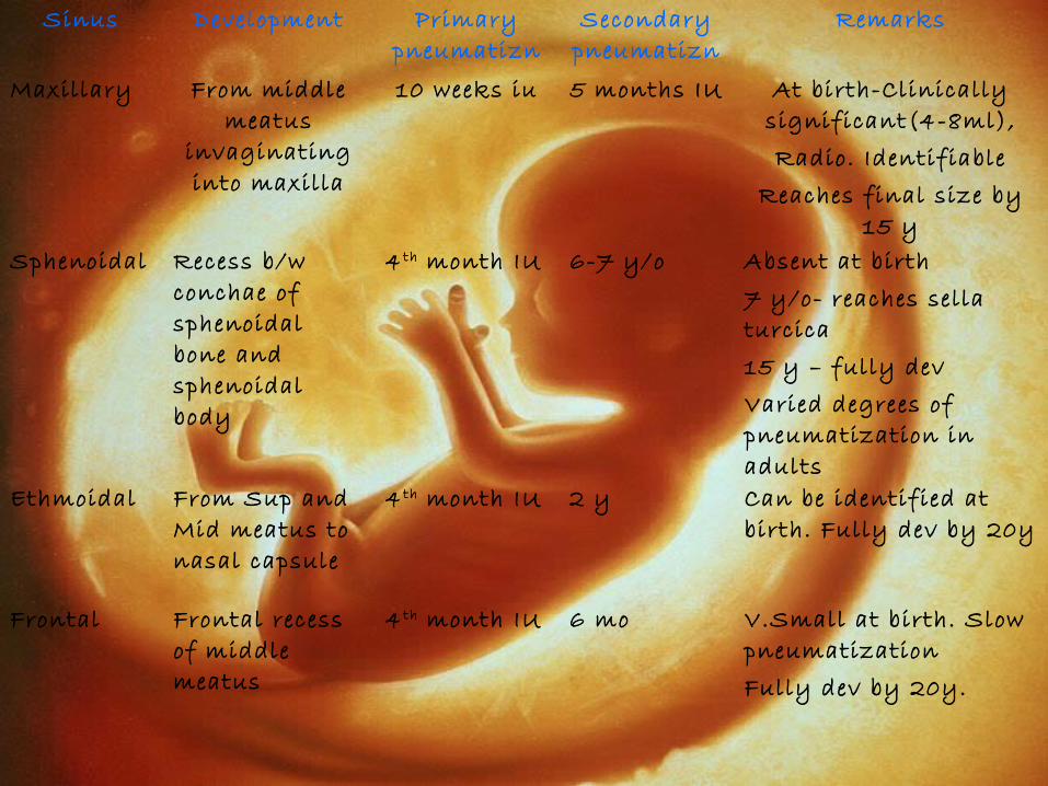
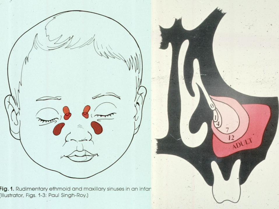
EMBRYOLOGY• Begin to develop in 3 r d fetal month• As outpouchings of mucous
membranes of Superior and Middle Meatus
• 2 processes – Primary pneumatization Secondary
pneumatization• Primary – Differential growth
Diverticular pouches/recesses expansion of wall itself to elaborate air space
• Secondary – Expansion outside wall occupies space within craniofacial bones
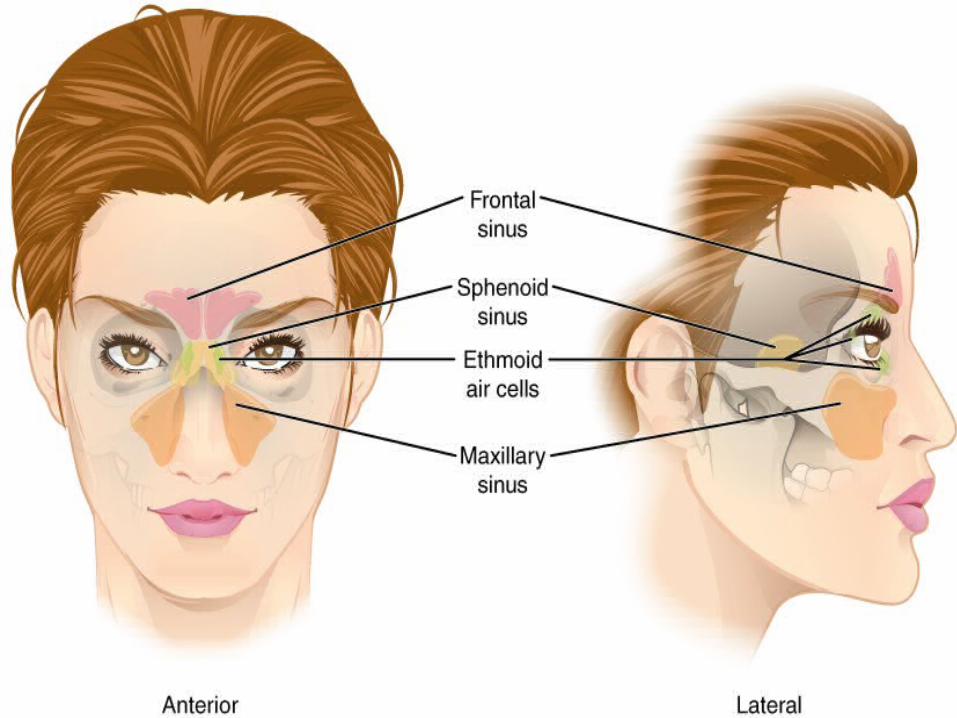
Sinus Development Primary pneumatizn
Secondary pneumatizn
Remarks
Maxillary From middle meatus
invaginating into maxilla
10 weeks iu 5 months IU At birth-Clinically significant(4-8ml),Radio. Identifiable
Reaches final size by 15 y
Sphenoidal Recess b/w conchae of sphenoidal bone and sphenoidal body
4 th month IU 6-7 y/o Absent at birth7 y/o- reaches sella turcica15 y – fully devVaried degrees of pneumatization in adults
Ethmoidal From Sup and Mid meatus to nasal capsule
4 th month IU 2 y Can be identified at birth. Fully dev by 20y
Frontal Frontal recess of middle meatus
4 th month IU 6 mo V.Small at birth. Slow pneumatizationFully dev by 20y.
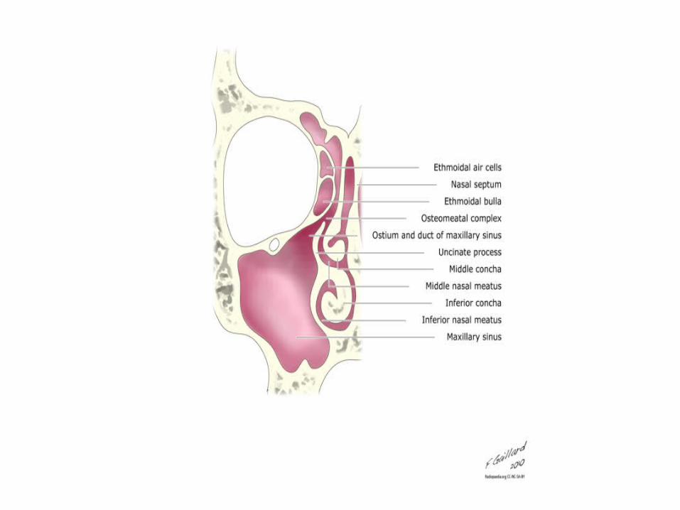
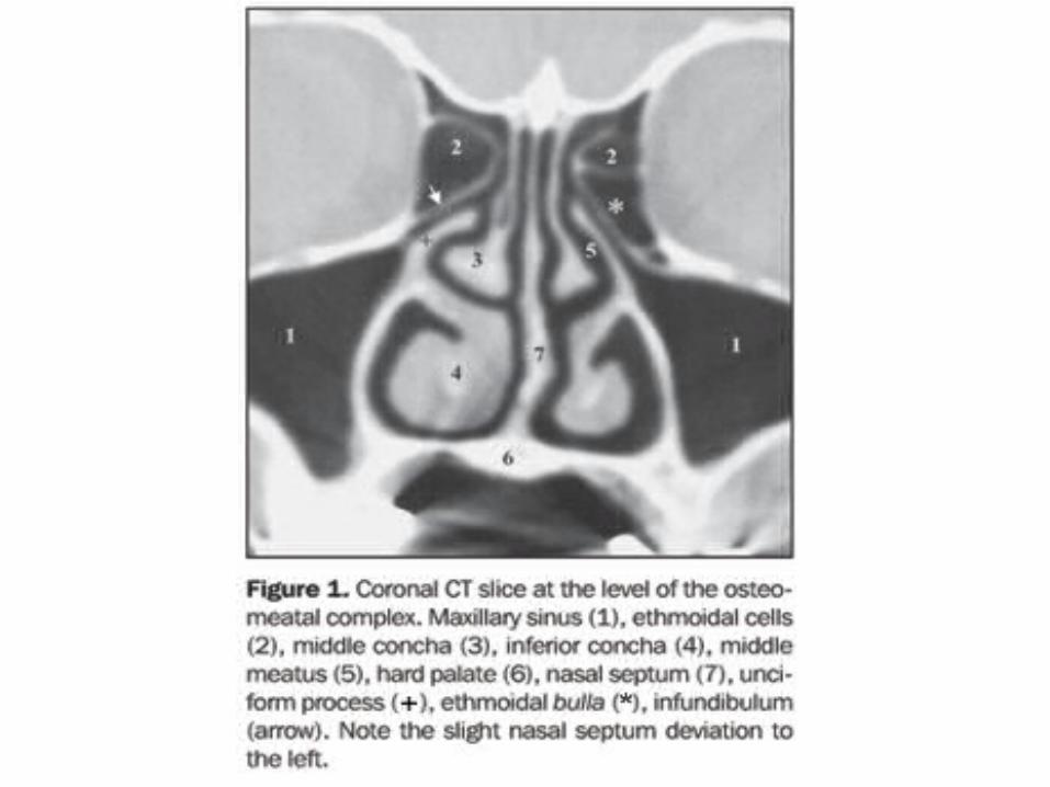
OSTEOMEATAL COMPLEX
• Common channel that links frontal sinus, ant. and middle ethmoid sinus, and max sinus to middle meatus allows air flow and mucociliary drainage needs to be patent for drainage of secretions in sinusitis
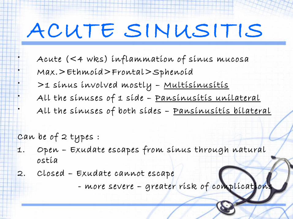
ACUTE SINUSITIS• Acute (<4 wks) inflammation of sinus mucosa • Max.>Ethmoid>Frontal>Sphenoid• >1 sinus involved mostly – Multisinusitis• All the sinuses of 1 side – Pansinusitis unilateral• All the sinuses of both sides – Pansinusitis bilateral
Can be of 2 types :1. Open – Exudate escapes from sinus through natural
ostia2. Closed – Exudate cannot escape - more severe – greater risk of complications

ETIOLOGYEXCITING CAUSES1. Nasal infections - Nasal mucosa Sinus mucosa MCC – Viral >
Bacterial>>Fungal2. Swimming/Diving – Infected water Ostia of sinuses - Chlorine Chemical
inflammation3. Trauma – Compound # or penetrating injuries
infection4. Dental infection – Molar/Premolar infection/extraction
Max. sinus

PREDISPOSING CAUSES Local GeneralLOCAL1. Obstruction to sinus ventilation and drainage• Nasal packing• DNS• Hypertrophic turbinates• Allergy – oedema of sinus ostia• Nasal polypi• Benign/Malignant neoplasm2. Stasis of secretions in nasal cavity• Cystic fibrosis – high viscosity of secretions• Enlarged adenoids - obstruction• Choanal atresia - obstruction
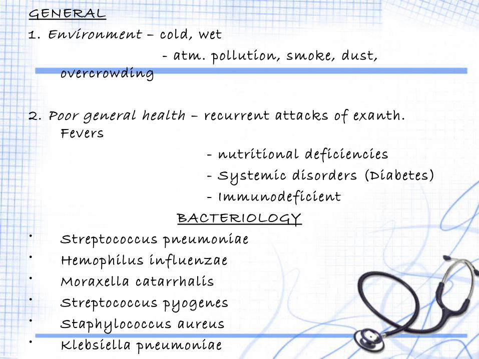
GENERAL1. Environment – cold, wet - atm. pollution, smoke, dust,
overcrowding
2. Poor general health – recurrent attacks of exanth. Fevers
- nutritional deficiencies - Systemic disorders (Diabetes) - Immunodeficient
BACTERIOLOGY• Streptococcus pneumoniae• Hemophilus influenzae• Moraxella catarrhalis• Streptococcus pyogenes• Staphylococcus aureus• Klebsiella pneumoniae• Anaerobic org. – Dental infections
PATHOLOGY Infection Acute Inflammation of sinus mucosa
Hyperemia Exudation (serous mucopurulent/purulent) Outpouring of PMNs Increased activity of serous and mucus glands severe infection destruction of mucosal lining
If failure of ostium to drain EmpyemaIf destruction of bony walls complications
• Mild/Non suppurative – less virulent, good immunity, drainage
• Severe/Suppurative
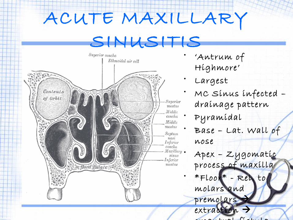
ACUTE MAXILLARY SINUSITIS
• ‘Antrum of Highmore’
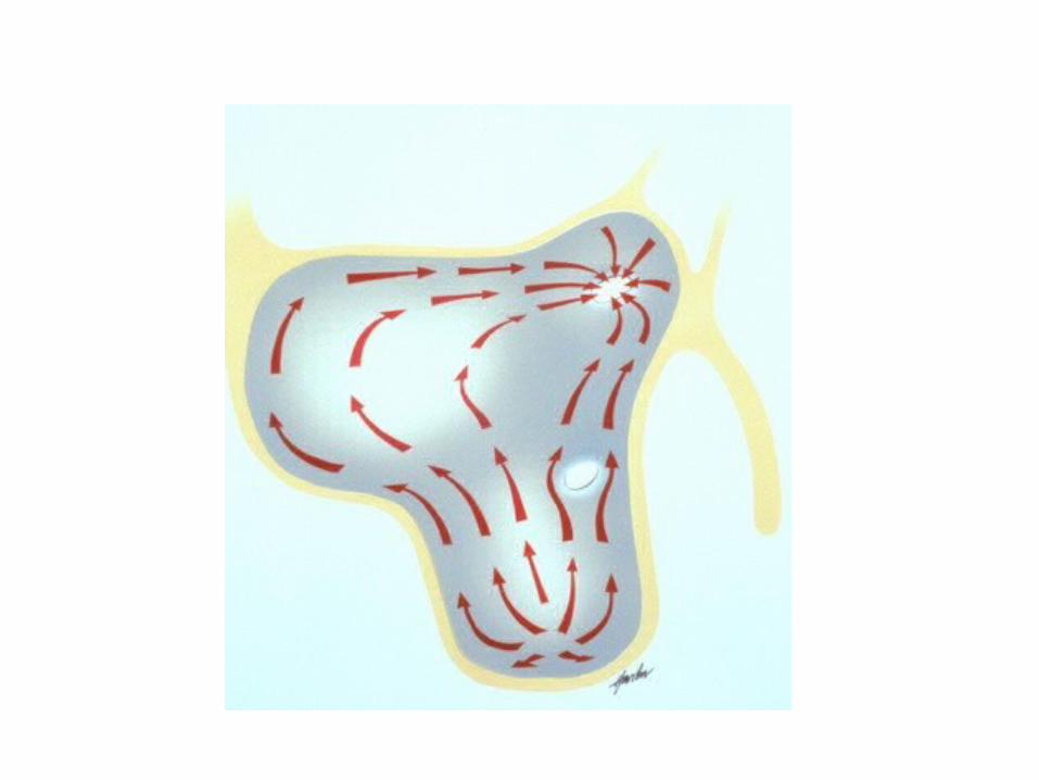
• Largest• MC Sinus infected –
drainage pattern• Pyramidal• Base – Lat. Wall of
nose• Apex – Zygomatic
process of maxilla• *Floor* - Rel. to
molars and premolars extraction oroantral fistula
• Capacity – 15 ml

ETIOLOGY1. MCC – Viral rhinitis2. 2 n d MCC – Bacterial invasion3. Diving/swimming4. *Dental infections* - Periapical dental abscess Tooth extraction5. Trauma – Compound # Penetrating injuries Gunshot wounds
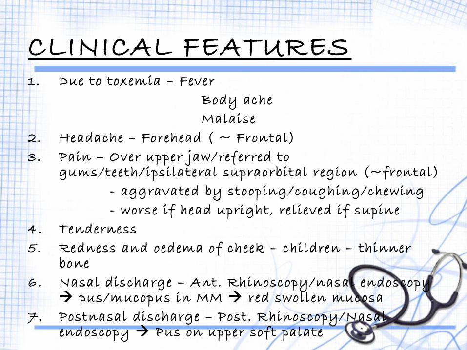
CLINICAL FEATURES1. Due to toxemia – Fever Body ache Malaise2. Headache – Forehead ( ~ Frontal)3. Pain – Over upper jaw/referred to
gums/teeth/ipsilateral supraorbital region (~frontal) - aggravated by stooping/coughing/chewing - worse if head upright, relieved if supine4. Tenderness5. Redness and oedema of cheek – children – thinner
bone6. Nasal discharge – Ant. Rhinoscopy/nasal endoscopy
pus/mucopus in MM red swollen mucosa7. Postnasal discharge – Post. Rhinoscopy/Nasal
endoscopy Pus on upper soft palate
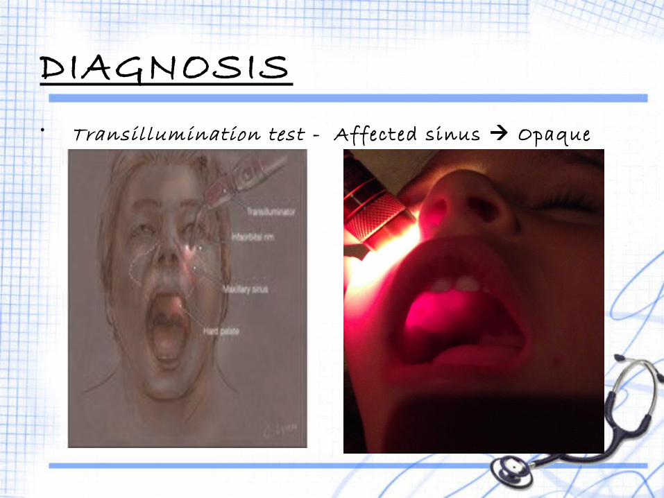
DIAGNOSIS• Transillumination test - Affected sinus Opaque
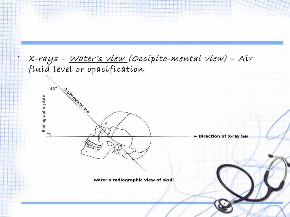
• X-rays – Water’s view (Occipito-mental view) – Air fluid level or opacification


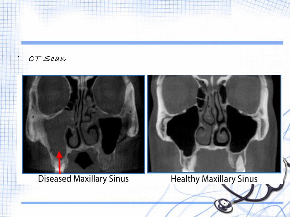
• CT Scan
COMPLICATIONS1. Subacute/Chronic sinusitis2. Frontal sinusitis – Oedema obstruction of OMC
obstruction of frontal sinus drainage pathway3. Osteitis/Osteomyelitis of maxilla4. Orbital cellulitis/abscess – Spread of infection a. direct – roof of
maxillary sinus b. indirect –
ethmoid sinus
ACUTE FRONTAL SINUSITIS
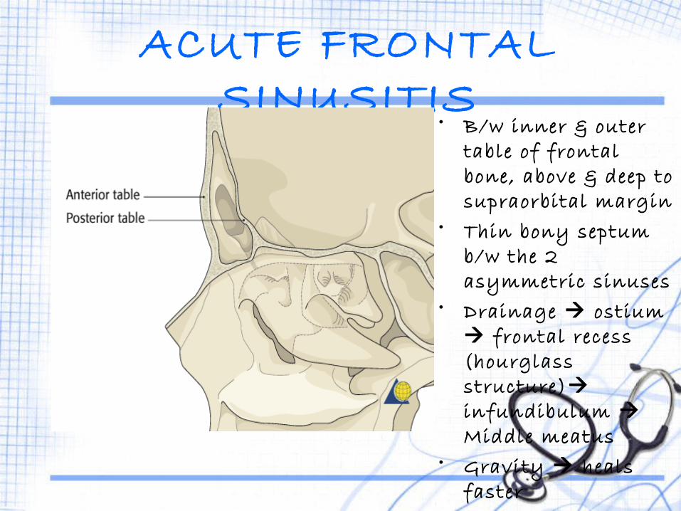
• B/w inner & outer table of frontal bone, above & deep to supraorbital margin
• Thin bony septum b/w the 2 asymmetric sinuses
• Drainage ostium frontal recess (hourglass structure) infundibulum Middle meatus
• Gravity heals faster


ETIOLOGY1. Viral rhinitis2. Bacterial invasions3. Diving/Swimming4. Trauma5. Ipsilateral Maxillary/Ethmoid sinusitis
CLINICAL FEATURES1. Frontal headache – Medial brow area ‘Office Headache’ – Comes up
on waking Gradually increases Peak at mid-day Subsides
2. Tenderness – Tapping - Pressure upwards on floor of frontal
sinus3. Oedema of upper eyelid4. Nasal discharge – Nasal endoscopy – vertical streak
of mucopus high up in anterior part of middle meatus

• X- ray – Water’s view
DIAGNOSIS

• CT Scan

COMPLICATIONS1. Orbital cellulitis2. Osteomyelitis of frontal bone and fistula formation3. Meningitis4. Extradural abscess5. Frontal lobe abscess6. Chronic frontal sinusitis
ACUTE ETHMOID SINUSITIS
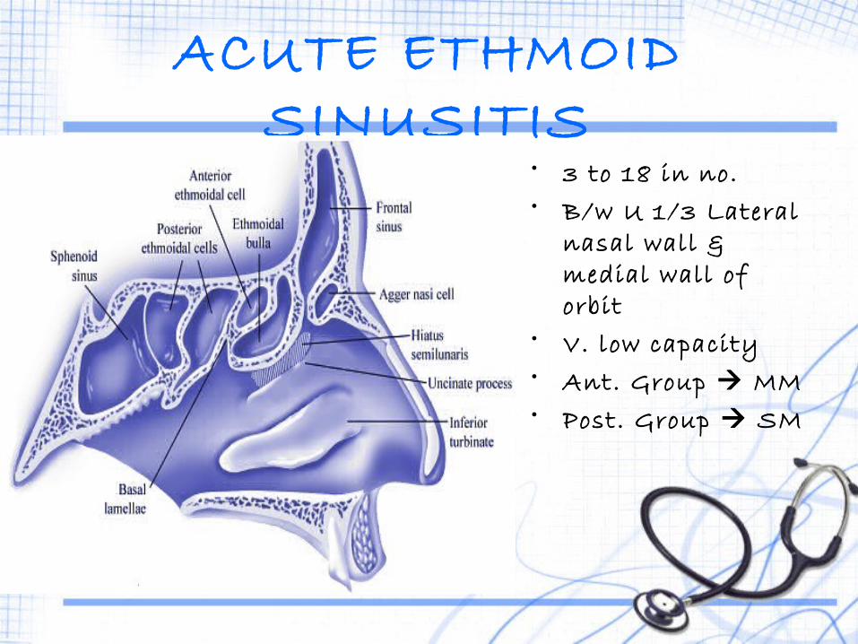
• 3 to 18 in no.• B/w U 1/3 Lateral
nasal wall & medial wall of orbit
• V. low capacity• Ant. Group MM• Post. Group SM
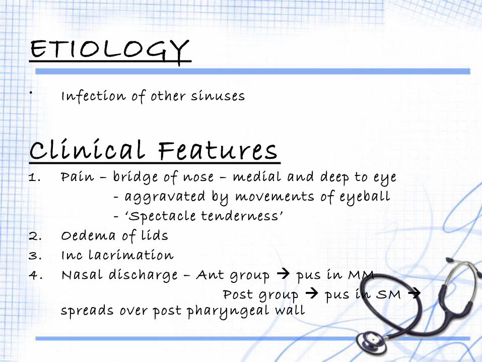
ETIOLOGY• Infection of other sinuses
Clinical Features1. Pain – bridge of nose – medial and deep to eye - aggravated by movements of eyeball - ‘Spectacle tenderness’2. Oedema of lids3. Inc lacrimation4. Nasal discharge – Ant group pus in MM Post group pus in SM
spreads over post pharyngeal wall

COMPLICATIONS1. Orbital cellulitis and abscess2. Optic Nerve Visual deterioration blindness3. Cavernous sinus thrombosis4. Extradural abscess5. Meningitis6. Brain abscess
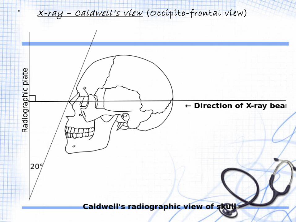
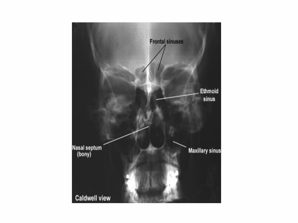
• X-ray – Caldwell ’s view (Occipito-frontal view)
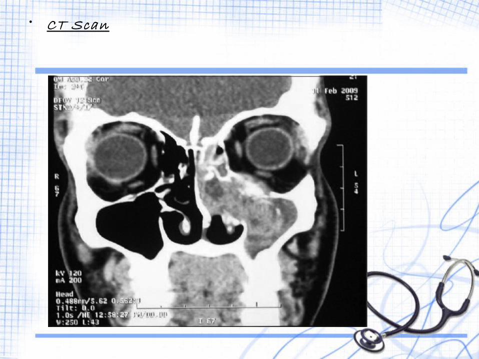
• CT Scan
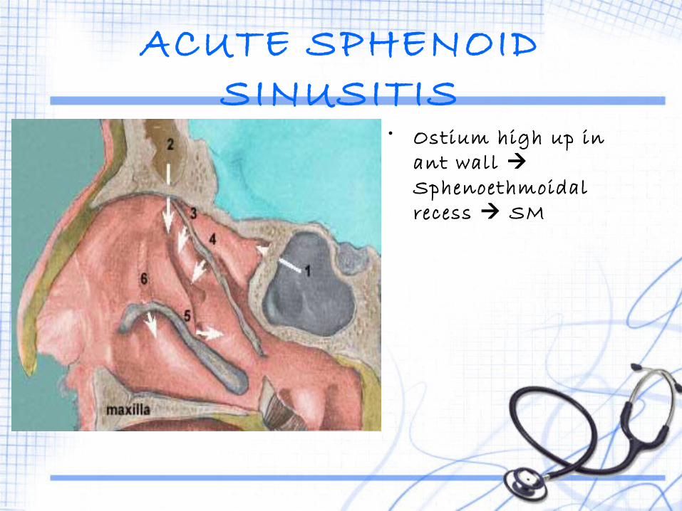
ACUTE SPHENOID SINUSITIS
• Ostium high up in ant wall Sphenoethmoidal recess SM
ETIOLOGY• Isolated involvement – rare• + Pansinusitis/Post. Ethmoidal sinusitis
Clinical Features1. Headache occiput/vertex - maybe referred to mastoid2. Postnasal discharge – Posterior rhinoscopy pus
on roof and post. Wall of NP

• X-ray – Lateral view• CT Scan

THANK YOU!
Related Documents