BRIEF COMMUNICATION IMMUNE NETWORK Vol. 13, No. 6: 289-294, December, 2013 http://dx.doi.org/10.4110/in.2013.13.6.289 pISSN 1598-2629 eISSN 2092-6685 289 Received on November 20, 2013. Revised on December 7, 2013. Accepted on December 9, 2013. CC This is an open access article distributed under the terms of the Creative Commons Attribution Non-Commercial License (http://creativecommons.org/licenses/by-nc/3.0) which permits unrestricted non-commercial use, distribu- tion, and reproduction in any medium, provided the original work is properly cited. *Corresponding Author. Kyoungho Suk, Department of Pharmacology, Kyungpook National University School of Medicine, 680, Gukchaebosang Street, Joong-gu, Daegu, Korea. Tel: 82-53-420-4835; Fax: 82-53-256-1566; E-mail: ksuk@knu. ac.kr # Present Address. Department of Neurosurgery, Biological Chemistry, and Neuroscience, Neurosurgery Pain Research Institute, Johns Hopkins School of Medicine, Baltimore, MD 21205, USA Keywords: Lipocalin-2, Acute nociception, Pathological pain, Neuroinflammation, Microglia, Spinal cord Abbreviations: KO, knockout; LCN2, lipocalin-2; SNI, spared nerve injury; PBS, phosphate-buffered saline; PWT, paw withdrawal threshold; WT, wild-type Acute Phase Protein Lipocalin-2 Is Associated with Formalin-induced Nociception and Pathological Pain Mithilesh Kumar Jha 1 , Sangmin Jeon 1# , Myungwon Jin 1 , Won-Ha Lee 2 and Kyoungho Suk 1 * 1 Department of Pharmacology, Brain Science & Engineering Institute, Kyungpook National University School of Medicine, Daegu 700-422, 2 School of Life Sciences and Biotechnology, Kyungpook National University, Daegu 700-422, Korea Lipocalin-2 (LCN2) is an acute-phase protein induced by in- jury, infection, or other inflammatory stimuli. LCN2 binds small hydrophobic ligands and interacts with cell surface re- ceptor to regulate diverse cellular processes. The role of LCN2 as a chemokine inducer in the central nervous system (CNS) has been previously reported. Based on the previous participation of LCN2 in neuroinflammation, we investigated the role of LCN2 in formalin-induced nociception and patho- logical pain. Formalin-induced nociceptive behaviors (licking/ biting) and spinal microglial activation were significantly re- duced in the second or late phase of the formalin test in Lcn2 knockout mice. Likewise, antibody-mediated neutralization of spinal LCN2 attenuated the mechanical hypersensitivity induced by peripheral nerve injury in mice. Taken together, our results suggest that LCN2 can be therapeutically tar- geted, presumably for both prevention and reversal of acute inflammatory pain as well as pathological pain. [Immune Network 2013;13(6):289-294] INTRODUCTION Inflammation, defined as the immune system response to in- jury or infection, is an intrinsically beneficial event that leads to the removal of initiating noxious stimuli or offending fac- tors and the restoration of tissue structures and physiological functions. Usually an acute inflammatory response is resolved successfully and the damaged tissue is repaired, rather than becoming persistent and dysfunctional. It may be anticipated, therefore, that the failure of resolving acute inflammation may predispose to autoimmunity, chronic dysplastic inflammation, and excessive tissue damage (1). Acute inflammatory pain hy- persensitivity is an outcome of failure in resolving acute in- flammation (2,3). Correspondingly, pathological pain occurs in response to extensive, intense, or prolonged tissue or nerve damages and results in extended discomfort and abnor- mal sensitivity. The neuropathic pain caused by central or pe- ripheral nerve injuries results in the perception of pain even in the presence of normally innocuous stimuli. Pathological pain, or chronic pain, is caused by nerve damages such as nerve compression, nerve trauma, diabetes, infection with herpes zoster virus, autoimmune disease, or cancer (4). Activation of glial cells, especially microglia and astrocytes, in the dorsal horn of the spinal cord and their release of proinflammatory mediators like cytokines, chemokines, pros- taglandins, and nitric oxide (NO) have been well documented as the key players in the pathogenesis of pathological pain (5-12). A cascade of events regulated by these mediators re- sults in central sensitization and pain enhancement (13). Lipocalin-2 (LCN2), which is also known in humans as neu-

Welcome message from author
This document is posted to help you gain knowledge. Please leave a comment to let me know what you think about it! Share it to your friends and learn new things together.
Transcript
BRIEF COMMUNICATION
IMMUNE NETWORK Vol. 13, No. 6: 289-294, December, 2013
http://dx.doi.org/10.4110/in.2013.13.6.289
pISSN 1598-2629 eISSN 2092-6685
289
Received on November 20, 2013. Revised on December 7, 2013. Accepted on December 9, 2013.CC This is an open access article distributed under the terms of the Creative Commons Attribution Non-Commercial
License (http://creativecommons.org/licenses/by-nc/3.0) which permits unrestricted non-commercial use, distribu-tion, and reproduction in any medium, provided the original work is properly cited.
*Corresponding Author. Kyoungho Suk, Department of Pharmacology, Kyungpook National University School of Medicine, 680, Gukchaebosang Street, Joong-gu, Daegu, Korea. Tel: 82-53-420-4835; Fax: 82-53-256-1566; E-mail: ksuk@knu. ac.kr
#Present Address. Department of Neurosurgery, Biological Chemistry, and Neuroscience, Neurosurgery Pain Research Institute, Johns Hopkins School of Medicine, Baltimore, MD 21205, USA
Keywords: Lipocalin-2, Acute nociception, Pathological pain, Neuroinflammation, Microglia, Spinal cord
Abbreviations: KO, knockout; LCN2, lipocalin-2; SNI, spared nerve injury; PBS, phosphate-buffered saline; PWT, paw withdrawal threshold; WT, wild-type
Acute Phase Protein Lipocalin-2 Is Associated with Formalin-induced Nociception and Pathological PainMithilesh Kumar Jha1, Sangmin Jeon1#, Myungwon Jin1, Won-Ha Lee2 and Kyoungho Suk1*1Department of Pharmacology, Brain Science & Engineering Institute, Kyungpook National University School of Medicine, Daegu 700-422, 2School of Life Sciences and Biotechnology, Kyungpook National University, Daegu 700-422, Korea
Lipocalin-2 (LCN2) is an acute-phase protein induced by in-jury, infection, or other inflammatory stimuli. LCN2 binds small hydrophobic ligands and interacts with cell surface re-ceptor to regulate diverse cellular processes. The role of LCN2 as a chemokine inducer in the central nervous system (CNS) has been previously reported. Based on the previous participation of LCN2 in neuroinflammation, we investigated the role of LCN2 in formalin-induced nociception and patho-logical pain. Formalin-induced nociceptive behaviors (licking/ biting) and spinal microglial activation were significantly re-duced in the second or late phase of the formalin test in Lcn2 knockout mice. Likewise, antibody-mediated neutralization of spinal LCN2 attenuated the mechanical hypersensitivity induced by peripheral nerve injury in mice. Taken together, our results suggest that LCN2 can be therapeutically tar-geted, presumably for both prevention and reversal of acute inflammatory pain as well as pathological pain.[Immune Network 2013;13(6):289-294]
INTRODUCTION
Inflammation, defined as the immune system response to in-
jury or infection, is an intrinsically beneficial event that leads
to the removal of initiating noxious stimuli or offending fac-
tors and the restoration of tissue structures and physiological
functions. Usually an acute inflammatory response is resolved
successfully and the damaged tissue is repaired, rather than
becoming persistent and dysfunctional. It may be anticipated,
therefore, that the failure of resolving acute inflammation may
predispose to autoimmunity, chronic dysplastic inflammation,
and excessive tissue damage (1). Acute inflammatory pain hy-
persensitivity is an outcome of failure in resolving acute in-
flammation (2,3). Correspondingly, pathological pain occurs
in response to extensive, intense, or prolonged tissue or
nerve damages and results in extended discomfort and abnor-
mal sensitivity. The neuropathic pain caused by central or pe-
ripheral nerve injuries results in the perception of pain even
in the presence of normally innocuous stimuli. Pathological
pain, or chronic pain, is caused by nerve damages such as
nerve compression, nerve trauma, diabetes, infection with
herpes zoster virus, autoimmune disease, or cancer (4).
Activation of glial cells, especially microglia and astrocytes,
in the dorsal horn of the spinal cord and their release of
proinflammatory mediators like cytokines, chemokines, pros-
taglandins, and nitric oxide (NO) have been well documented
as the key players in the pathogenesis of pathological pain
(5-12). A cascade of events regulated by these mediators re-
sults in central sensitization and pain enhancement (13).
Lipocalin-2 (LCN2), which is also known in humans as neu-

Role of LCN2 in Acute Nociception and Pathological PainMithilesh Kumar Jha, et al.
290 IMMUNE NETWORK Vol. 13, No. 6: 289-294, December, 2013
trophil gelatinase-associated lipocalin (NGAL) (14) and in
mice as 24p3 (15), is a 25-kDa secretory protein that belongs
to the lipocalin family (16). It binds small hydrophobic li-
gands and interacts with cell surface receptors (24p3R and
megalin) to regulate diverse cellular processes. LCN2 is also
an acute-phase protein induced by an injury, infection, or
other inflammatory stimuli (17). LCN2 has been found to be
increased in the prefrontal cortex during inflammatory pain
(18) and in the lumbar segment of the spinal cord during pe-
ripheral nerve injury-induced neuropathic pain (7). Similarly,
LCN2 has been documented to regulate stress-induced changes
in spine morphology, neuronal excitability, and anxiety (19).
LCN2 is secreted by glial cells (20-22) and acts as a chemokine
inducer under neuroinflammatory conditions (23). Recently,
LCN2 has been reported to play the critical role in the devel-
opment of pain hypersensitivity following peripheral nerve in-
jury, suggesting that it mediates neuropathic pain by inducing
chemokine expression and subsequent microglial activation
(7). Based on these earlier findings, we have speculated that
the involvement of LCN2 in the pathogenesis of acute in-
flammatory pain and the neutralization of LCN2 is a potential
pharmacological approach for the prevention and treatment of
pathological pain. We have tested these hypotheses using for-
malin-induced nociceptive behavioral tests and a spared nerve
injury (SNI) model. Our findings, based on Lcn2-deficient mice
and antibody-mediated neutralization of spinal LCN2, suggest
that LCN2 contributes to the pathogenesis of acute in-
flammatory pain as well as pathological pain.
MATERIALS AND METHODS
Mouse breeding and maintenance All experiments were conducted in accordance with the ani-
mal care guidelines of the National Institutes of Health. All
efforts were made to minimize the number of animals used
and animal suffering. Male Lcn2 wild-type (WT, Lcn2+/+) and
Lcn2 knockout (KO, Lcn2−/−) mice aged 8∼10 weeks were
used. LCN2−/−
mice were a gift from Dr. Shizuo Akira
(Osaka University, Japan). Lcn2+/+ and Lcn2−/−
mice were
back-crossed for eight to ten generations onto the C57BL/6
background to generate homozygous and heterozygous ani-
mals free of background effects on phenotypes, as described
previously (23,24). Genotypes were confirmed by polymerase
chain reaction (PCR) of genomic DNA (25). The C57BL/6
mice used for breeding were purchased from Samtako (Osan,
Korea). Animals were housed under a 12 h light/dark cycle
(lights on 07:00∼19:00) at a constant ambient temperature
of 23±2oC with food and water provided ad libitum. Animals
participated in only one experiment.
Formalin testFormalin solution in saline (5%; 10μl) was injected into the
plantar surface of the left hindpaw. Mice were then observed
for 40 min, and times spent licking or biting injected hindpaws
were recorded. The first 10 min post-formalin injection was
considered the early/first phase, and the period between 15 min
and 40 min the late/second phase (2). Total times spent licking
or biting injected paws were measured with a stopwatch by an
experimenter blinded to animal genotype or treatment.
Activities were recorded over consecutive 5-min periods.
Intrathecal injection of LCN2 antibodyA single intrathecal injection was performed by direct lumbar
puncture between the L5 and L6 vertebrae using a 25μl
Hamilton syringe with a 30-gauge needle, as described pre-
viously (26). LCN2 antibody was purchased from R&D Systems
(Minneapolis, MN) and diluted in phosphate-buffered saline
(PBS). Mice were injected intrathecally with 1μg of LCN2 an-
tibody in a volume of 10μl. Precise intrathecal localization
was confirmed by a tail flick upon penetration. PBS was used
as the vehicle control.
Peripheral nerve injuryThe SNI model that mimics human neuropathic pain re-
lated to peripheral nerve injury was used as described pre-
viously (7,27,28). Briefly, surgery was performed on the left
side, defined as ipsilateral side, of mice under 2% isoflurane
anesthesia. The contralateral sides were left intact. For the SNI,
the three peripheral branches (the sural, common peroneal,
and tibial nerves) of the sciatic nerve were exposed, and the
tibial and common peroneal branches were ligated with 6-0
silk thread. A ∼2 mm segment distal to the ligation site of
the two nerves was removed, while the sural nerve was left
intact. All wounds were irrigated with sterile saline and
closed in layers post-surgery. SNI surgery was performed 30
min after the intrathecal injection of LCN2 antibody or
vehicle. The day of surgery was set as day 0.
Assessments of peripheral nerve injury-induced pain behaviorsMice were allowed to acclimate after arrival in the animal care
unit for 1 week, and placed in the test room used for 1 h
Role of LCN2 in Acute Nociception and Pathological PainMithilesh Kumar Jha, et al.
291IMMUNE NETWORK Vol. 13, No. 6: 289-294, December, 2013
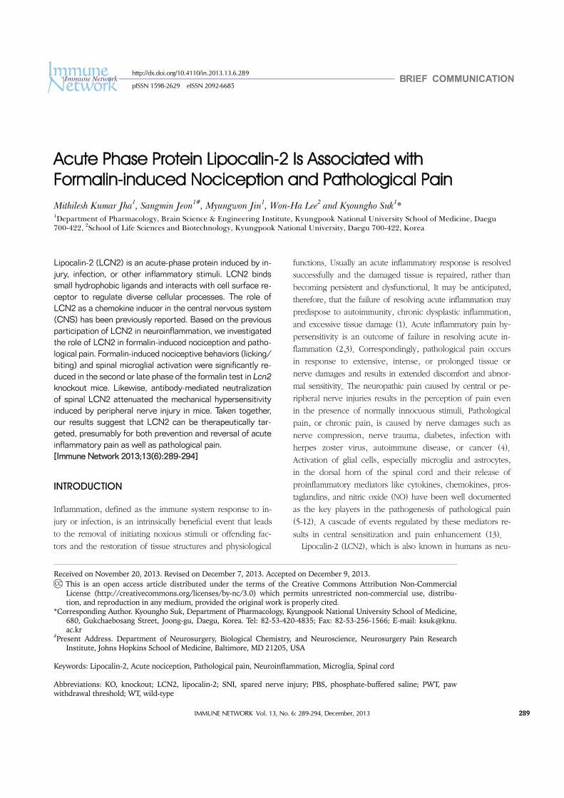
Figure 1. Attenuation of acute inflammatory pain in Lcn2 KO mice. (A) Formalin (5%, 10μl) is administered intraplantarly to the left hindpaw of mice, then nociceptive behavior is measured as shown in the experimental timeline. (B) The behaviors of wild-type (WT) and Lcn2 KO mice are compared for 40 min after the injection. Times spent licking or biting injected hindpaws are recorded. (C) The first 10 min post-injection is defined as the early phase, and the period between 15 and 40 min post-injection as the late phase. The results are means±SEMs. *p<0.05, n.s.=not significant (n=7∼9).
the day before experiments. Animals were habituated to the
test room for at least 1 h before behavioral testing, which
was performed by an experimenter unaware of the treatment
condition. Mechanical sensitivity was tested using a loga-
rithmic series of calibrated Semmes-Weinstein monofilaments
(von Frey hairs; Stoelting, Wood Dale, IL), as previously de-
scribed (29). Briefly, mice were acclimated for 20 min in in-
verted individual acrylic boxes with wire mesh floors to pro-
vide access to the ventral side of the hindpaws. Von Frey
hairs were presented perpendicularly to the lateral region of
left and right hindpaws, and held in position for approx-
imately 5 s with enough force to cause a slight bend. Two
trials per paw were conducted with an interval of at least 3
min. A positive response was defined as abrupt paw
withdrawal. Paw withdrawal thresholds (PWTs) were de-
termined by increasing and decreasing forces and estimated
using Dixon's up-and-down method (30).
Immunofluorescence analysisDeeply anesthetized mice were perfused through the aorta
with 0.1 M PBS followed by 4% paraformaldehyde (PFA)
fixative. Lumbar spinal cord segments (L4-L6) were dissected
out, post-fixed in the same PFA fixative overnight, and cry-
oprotected in 30% sucrose in 0.1 M PBS overnight at 4oC.
A cryostat was used to prepare 30μm sections of the spinal
cord kept in 0.1 M PBS. Sections were then blocked with
4% normal serum in 0.3% Triton X-100 for 90 min at room
temperature, and then incubated with primary antibodies
against Iba-1 (mouse, 1:1,000; Wako, Osaka, Japan) over-
night at 4oC. Sections were then incubated with Cy3-conjugat-
ed secondary antibodies (1:200; Jackson ImmunoResearch,
West Grove, PA). Slides were washed, coverslipped with
Vectashield mounting medium (Vector Laboratories, Burlin-
game, CA), and visualized under a fluorescence microscope.
Quantification and statistical analysis All results are presented as mean±standard error of mean
(SEM). The effects of different treatments were compared us-
ing Student’s t-test, with p-values of <0.05 considered stat-
istically significant.
RESULTS AND DISCUSSION
Attenuation of formalin-induced nociceptive behavior and spinal microglial activation in Lcn2 KO miceThe involvement of LCN2 in acute inflammatory pain was as-
sessed using the formalin test as explained in the ex-
perimental timeline (Fig. 1A). An intraplantar injection of for-
malin resulted in a typical biphasic pain response (2). The
first or early phase occurred immediately post-injection due
to the direct stimulation of nociceptors, whereas the second
or late phase occurred after a short period of quiescence
(typically 10∼15 min post-injection), during which inflam-
matory responses took place. This phase reflects central sen-
sitization and continued afferent input (Fig. 1B). No sig-
nificant difference was observed between Lcn2 KO and WT
mice during the early phase, but behavioral responses during
the late phase were significantly diminished in Lcn2 KO mice
(Fig. 1C). Acute nociceptive pain is caused by the stimulation
of peripheral nerve fibers that normally respond only to stim-
uli approaching or exceeding a harmful intensity. In the for-
malin-induced acute inflammatory pain model, Lcn2 defi-
ciency did not influence the early phase of formalin-induced
behavior [direct chemical effect on peripheral nociceptors
(31)]. In contrast to the early phase, late phase-responses
[central sensitization in the spinal cord (31)] were significantly
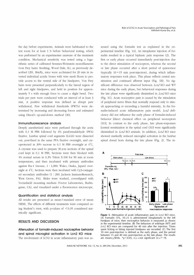
diminished in Lcn2 KO animals. In addition, Lcn2 KO mice
showed markedly reduced microglial activation in the lumbar
spinal dorsal horn during the late phase (Fig. 2). The in-
Role of LCN2 in Acute Nociception and Pathological PainMithilesh Kumar Jha, et al.
292 IMMUNE NETWORK Vol. 13, No. 6: 289-294, December, 2013
Figure 2. Reduction of microglial activation in Lcn2 KO mice. (A) The lumbar segment (L4-6) of the spinal cord is sampled 30 min after the intraplantar injection of formalin (5%, 10μl) as shown in the experimental timeline. (B) Photomicrographs showing Iba-1 immuno-reactivity in the dorsal horns of the ipsilateral lumbar spinal cords of WT and Lcn2 KO mice. The results are representative of at least three independent experiments. Scale bars=200μm.
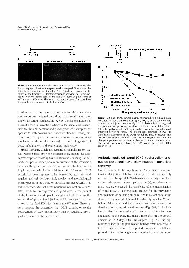
Figure 3. Spinal LCN2 neutralization attenuated SNI-induced pain behavior. (A) LCN2 antibody (0.1μg/μl, 10μl), or the same volume of vehicle, is injected intrathecally 30 min before SNI surgery, and the pain test was performed as shown in the experimental timeline. (B) In the ipsilateral side, SNI significantly reduces the paw withdrawal threshold (PWT) to force. The SNI-induced decrease in PWT is significantly attenuated in the LCN2-neutralized mice compared with control animals at 1 day and 2 days after SNI surgery. No significant change in pain-related behavior is observed in the contralateral side. The results are means±SEMs. *p<0.05 versus the vehicle (PBS) group (n=3).
duction and maintenance of pain hypersensitivity is consid-
ered to be due to spinal cord dorsal horn sensitization, also
known as central sensitization (32,33). Central sensitization is
a specific form of synaptic plasticity in the spinal cord respon-
sible for the enhancement and prolongation of nociceptive re-
sponses to both noxious and innocuous stimuli. Growing evi-
dence supports glia as an important source of inflammatory
mediators fundamentally involved in the pathogenesis of
acute inflammatory and pathological pain (34,35).
Spinal microglia, which also respond to proinflammatory sig-
nals released from other non-neuronal cells, amplify the noci-
ceptive response following tissue inflammation or injury (36,37).
Acute peripheral nociception is an outcome of the interaction
between the peripheral and the central sensitization, which
implicates the activation of glial cells (38). Moreover, LCN2
protein has been reported to be secreted by glial cells, and
regulate glial cell death/survival, motility, and morphological
phenotypes in an autocrine or paracrine manner (20,21). This
led us to speculate that acute peripheral nociception is trans-
lated into LCN2 overexpression in spinal cord. In the present
study, formalin caused spinal microglial activation during the
second (late) phase after injection, which was significantly re-
duced in the Lcn2 KO mice than in the WT mice. These re-
sults support the contention that LCN2 contributes to the
pathogenesis of acute inflammatory pain by regulating micro-
glial activation in the spinal cord.
Antibody-mediated spinal LCN2 neutralization atte-nuated peripheral nerve injury-induced mechanical sensitivityOn the basis of the findings from the Lcn2-deficient mice and
intrathecal injection of LCN2 protein, Jeon et al. have recently
reported that the spinal LCN2-chemokine axis may contribute
to the pathogenesis of neuropathic pain (7). In reference to
these results, we tested the possibility of the neutralization
of spinal LCN2 as a therapeutic strategy for the prevention
and treatment of pathological pain. Anti-LCN2 antibody at the
dose of 1μg was administered intrathecally to mice 30 min
before SNI surgery, and the pain response was measured as
described in the experimental timeline (Fig. 3A). In the ipsi-
lateral sides, SNI reduced PWT to force, and this effect was
attenuated in the LCN2-neutralized mice than in the control
animals at 1∼2 days after SNI surgery (Fig. 3B). No sig-
nificant change in the pain-related behavior was observed in
the contralateral sides. As reported previously, LCN2 ex-
pressed in the lumbar segment of dorsal spinal cord following
Role of LCN2 in Acute Nociception and Pathological PainMithilesh Kumar Jha, et al.
293IMMUNE NETWORK Vol. 13, No. 6: 289-294, December, 2013
SNI surgery might have played an important role in the onset
of neuropathic pain (7). Consistent with this notion, the intra-
thecally administered LCN2 antibody neutralized the ex-
pressed spinal LCN2 responsible for the development of the
neuropathic pain.
In conclusion, our results suggest that LCN2 participates in
behavioral responses to acute inflammatory stimuli and sub-
stantiates the critical involvement of LCN2 in the genesis of
central sensitization and pathological pain hypersensitivity.
These findings indicate that LCN2 can possibly be therapeuti-
cally targeted for both prevention and reversal of acute in-
flammatory and pathological pain.
ACKNOWLEDGEMENTS
This work was supported by a grant from the Korea Health-
care Technology R&D Project, Ministry of Health & Welfare,
Republic of Korea (A111345), and by the National Research
Foundation (NRF) grants funded by the Ministry of Education,
Science and Technology (MEST) of the Korean government
(2008-0062282).
CONFLICTS OF INTEREST
The authors declare that they have no conflicts of interest.
REFERENCES
1. Nathan, C. 2002. Points of control in inflammation. Nature 420: 846-852.
2. Jha, M. K., S. Jeon, and K. Suk. 2012. Glia as a Link between Neuroinflammation and Neuropathic Pain. Immune Netw. 12: 41-47.
3. Lyman, M., D. G. Lloyd, X. Ji, M. P. Vizcaychipi, and D. Ma. 2013. Neuroinflammation: The role and consequences. Neurosci. Res. In press: http://doi.org/10.1016/j.neures. 2013.10.004.
4. Campbell, J. N. and R. A. Meyer. 2006. Mechanisms of neu-ropathic pain. Neuron 52: 77-92.
5. Scholz, J. and C. J. Woolf. 2007. The neuropathic pain triad: neurons, immune cells and glia. Nat. Neurosci. 10: 1361- 1368.
6. Watkins, L. R., E. D. Milligan, and S. F. Maier. 2001. Glial activation: a driving force for pathological pain. Trends Neurosci. 24: 450-455.
7. Jeon, S., M. K. Jha, J. Ock, J. Seo, M. Jin, H. Cho, W. H. Lee, and K. Suk. 2013. Role of lipocalin-2-chemokine axis in the development of neuropathic pain following peripheral nerve injury. J. Biol. Chem. 288: 24116-24127.
8. Ji, R. R., T. Berta, and M. Nedergaard. 2013. Glia and pain:
Is chronic pain a gliopathy? Pain In press: http://dx.doi. org/10.1016/j.pain.2013.06.022.
9. Milligan, E. D. and L. R. Watkins. 2009. Pathological and protective roles of glia in chronic pain. Nat. Rev. Neurosci. 10: 23-36.
10. Inoue, K. and M. Tsuda. 2009. Microglia and neuropathic pain. Glia 57: 1469-1479.
11. Gao, Y. J. and R. R. Ji. 2010. Chemokines, neuronal-glial in-teractions, and central processing of neuropathic pain. Pharmacol. Ther. 126: 56-68.
12. Raghavendra, V., F. Tanga, M. D. Rutkowski, and J. A. DeLeo. 2003. Anti-hyperalgesic and morphine-sparing actions of propentofylline following peripheral nerve injury in rats: mechanistic implications of spinal glia and proinflammatory cytokines. Pain 104: 655-664.
13. Woolf, C. J. 2011. Central sensitization: implications for the diagnosis and treatment of pain. Pain 152: S2-15.
14. Cowland, J. B. and N. Borregaard. 1997. Molecular character-ization and pattern of tissue expression of the gene for neu-trophil gelatinase-associated lipocalin from humans. Genomics 45: 17-23.
15. Hraba-Renevey, S., H. Turler, M. Kress, C. Salomon, and R. Weil. 1989. SV40-induced expression of mouse gene 24p3 in-volves a post-transcriptional mechanism. Oncogene 4: 601- 608.
16. Flower, D. R. 1996. The lipocalin protein family: structure and function. Biochem. J. 318(Pt 1): 1-14.
17. Nilsen-Hamilton, M., Q. Liu, J. Ryon, L. Bendickson, P. Lepont, and Q. Chang. 2003. Tissue involution and the acute phase response. Ann. N. Y. Acad. Sci. 995: 94-108.
18. Poh, K. W., J. F. Yeo, C. S. Stohler, and W. Y. Ong. 2012. Comprehensive gene expression profiling in the prefrontal cortex links immune activation and neutrophil infiltration to antinociception. J. Neurosci. 32: 35-45.
19. Mucha, M., A. E. Skrzypiec, E. Schiavon, B. K. Attwood, E. Kucerova, and R. Pawlak. 2011. Lipocalin-2 controls neuronal excitability and anxiety by regulating dendritic spine for-mation and maturation. Proc. Natl. Acad. Sci. U. S. A. 108: 18436-18441.
20. Lee, S., J. Lee, S. Kim, J. Y. Park, W. H. Lee, K. Mori, S. H. Kim, I. K. Kim, and K. Suk. 2007. A dual role of lipocalin 2 in the apoptosis and deramification of activated microglia. J. Immunol. 179: 3231-3241.
21. Lee, S., J. Y. Park, W. H. Lee, H. Kim, H. C. Park, K. Mori, and K. Suk. 2009. Lipocalin-2 is an autocrine mediator of re-active astrocytosis. J. Neurosci. 29: 234-249.
22. Lee, S., W. H. Lee, M. S. Lee, K. Mori, and K. Suk. 2012. Regulation by lipocalin-2 of neuronal cell death, migration, and morphology. J. Neurosci. Res. 90: 540-550.
23. Lee, S., J. H. Kim, J. W. Seo, H. S. Han, W. H. Lee, K. Mori, K. Nakao, J. Barasch, and K. Suk. 2011. Lipocalin-2 Is a chemokine inducer in the central nervous system: role of chemokine ligand 10 (CXCL10) in lipocalin-2-induced cell migration. J. Biol. Chem. 286: 43855-43870.
24. Flo, T. H., K. D. Smith, S. Sato, D. J. Rodriguez, M. A. Holmes, R. K. Strong, S. Akira, and A. Aderem. 2004. Lipocalin 2 mediates an innate immune response to bacterial infection by sequestrating iron. Nature 432: 917-921.

Role of LCN2 in Acute Nociception and Pathological PainMithilesh Kumar Jha, et al.
294 IMMUNE NETWORK Vol. 13, No. 6: 289-294, December, 2013
25. Nairz, M., I. Theurl, A. Schroll, M. Theurl, G. Fritsche, E. Lindner, M. Seifert, M. L. Crouch, K. Hantke, S. Akira, F. C. Fang, and G. Weiss. 2009. Absence of functional Hfe pro-tects mice from invasive Salmonella enterica serovar Typhimurium infection via induction of lipocalin-2. Blood 114: 3642-3651.
26. Hylden, J. L. and G. L. Wilcox. 1980. Intrathecal morphine in mice: a new technique. Eur. J. Pharmacol. 67: 313-316.
27. Decosterd, I. and C. J. Woolf. 2000. Spared nerve injury: an animal model of persistent peripheral neuropathic pain. Pain 87: 149-158.
28. Tegeder, I., M. Costigan, R. S. Griffin, A. Abele, I. Belfer, H. Schmidt, C. Ehnert, J. Nejim, C. Marian, J. Scholz, T. Wu, A. Allchorne, L. Diatchenko, A. M. Binshtok, D. Goldman, J. Adolph, S. Sama, S. J. Atlas, W. A. Carlezon, A. Parsegian, J. Lotsch, R. B. Fillingim, W. Maixner, G. Geisslinger, M. B. Max, and C. J. Woolf. 2006. GTP cyclohydrolase and tetrahy-drobiopterin regulate pain sensitivity and persistence. Nat. Med. 12: 1269-1277.
29. Chaplan, S. R., F. W. Bach, J. W. Pogrel, J. M. Chung, and T. L. Yaksh. 1994. Quantitative assessment of tactile allodynia in the rat paw. J. Neurosci. Methods 53: 55-63.
30. Dixon, W. J. 1980. Efficient analysis of experimental observations. Annu. Rev. Pharmacol. Toxicol. 20: 441-462.
31. Hunskaar, S. and K. Hole. 1987. The formalin test in mice:
dissociation between inflammatory and non-inflammatory pain. Pain 30: 103-114.
32. Latremoliere, A. and C. J. Woolf. 2009. Central sensitization: a generator of pain hypersensitivity by central neural plasticity. J. Pain 10: 895-926.
33. Woolf, C. J. and M. W. Salter. 2000. Neuronal plasticity: in-creasing the gain in pain. Science 288: 1765-1769.
34. Austin, P. J. and G. Moalem-Taylor. 2010. The neuro-immune balance in neuropathic pain: involvement of inflammatory im-mune cells, immune-like glial cells and cytokines. J. Neuroimmunol. 229: 26-50.
35. Hashioka, S., T. Miyaoka, R. Wake, M. Furuya, and J. Horiguchi. 2013. Glia: an important target for anti-in-flammatory and antidepressant activity. Curr. Drug Targets 14: 1322-1328.
36. Skaper, S. D., P. Giusti, and L. Facci. 2012. Microglia and mast cells: two tracks on the road to neuroinflammation. FASEB J. 26: 3103-3117.
37. Vega-Avelaira, D., J. J. Ballesteros, and J. A. Lopez-Garcia. 2013. Inflammation-induced hyperalgesia and spinal microglia reactivity in neonatal rats. Eur. J. Pain. 17: 1180-1188.
38. Nowak, L., D. Zurowski, J. Dobrogowski, J. Wordliczek, and P. J. Thor. 2012. Pentoxifylline modifies central and periph-eral vagal mechanism in acute and chronic pain models. Folia Med. Cracov. 52: 83-95.
Related Documents