ACUTE PANCREATITIS SAMIR EL ANSARY

Welcome message from author
This document is posted to help you gain knowledge. Please leave a comment to let me know what you think about it! Share it to your friends and learn new things together.
Transcript
ACUTE PANCREATITIS
SAMIR EL ANSARY

https://www.facebook.com/groups/1451610115129555/#!/groups/1451610115129555/
Wellcome in our new group ..... Dr.SAMIR EL ANSARY
Acute pancreatitis
AP is an inflammatory condition of the pancreas
that has a broad spectrum of severity ranging
from mild and self-limited to severe and
associated with multiorgan failure.
Clinically it is associated with the acute onset of
abdominal pain and elevation of serum
biochemical markers.
The underlying pathophysiology is early and
inappropriate activation of digestive enzymes
within acinar cells.

•Different degrees of AP, and how are they
defined?
Mild AP
Pancreatic inflammation (of any cause) without
persistent organ failure or local complications.
Severe acute pancreatitis (SAP)
Associated with systemic complications,
including multiple-organ failure, and local
complications, such as necrosis, abscess,
and/or pseudocyst. This degree of pancreatitis requires aggressive therapy in an
intensive care setting to prevent significant morbidity or mortality.

Causes of AP?
Gallstone-related disease and excessive
alcohol intake comprising 70% of all cases.
hypertriglyceridemia (serum triglyceride level
above 1500 mg/dL), trauma, and
hypercalcemia.
Smoking is a risk factor for AP in a time- and
dose-dependent manner.

Causes of AP?
A large number of medications have been
implicated in causing AP, including angiotensin-
converting enzyme inhibitors, furosemide,
tetracycline, aminosalicylic acid, corticosteroids,
procainamide, thiazides, metronidazole, and
ranitidine.
Because of the varied duration between exposure and
development of symptoms, and the usual lack of a clear
mechanism, it is often difficult to identify a drug as the
sole cause of AP.

Causes of AP?
True idiopathic cases of AP are diminishing as
more genetic causes of the disease are
discovered.
Endoscopic retrograde
cholangiopancreatography (ERCP) causes
pancreatitis in approximately 5% of individuals
who undergo this procedure.
The presenting signs and symptoms of
AP?
AP is characterized by the sudden onset of
abdominal pain, classically located in the
epigastrium and usually associated with nausea
and/or vomiting.
Radiation of pain from the epigastrium
through to the back that is alleviated with the
patient leaning forward is a typical but not a
necessary feature.
Tachycardia related to pain or volume depletion
and low-grade fever may be present.

The presenting signs and symptoms of
AP?Two additional findings associated with severe
pancreatitis may be present
the Grey Turner and Cullen signs.•The Grey Turner sign is an ecchymosis of the
flank due to retroperitoneal hemorrhage. When present, it usually occurs 3 to 7 days after the onset of pain
and is indicative of severe pancreatitis.
•The Cullen sign is periumbilical ecchymosis
associated with both severe necrotizing
pancreatitis and retroperitoneal hemorrhage of
various causes.

•Are amylase and/or lipase measurements
helpful in the diagnosis?
The most commonly used diagnostic markers are
serum amylase and lipase.
Although serum amylase levels have a high
sensitivity in the first 24 hours the specificity is
very low.
In addition to pancreatitis, elevated amylase
levels are seen with many other conditions
including bowel infarction, renal failure,
perforated peptic ulcer, trauma to the salivary
glands, and macroamylasemia.

•Are amylase and/or lipase measurements
helpful in the diagnosis?
In contrast, serum lipase levels are both more
specific and more sensitive than amylase
measurements.
Of note, no correlation exists between
the absolute levels of amylase and
lipase and the severity of pancreatitis.
Role of imaging in the diagnosis
of AP
Abdominal plain radiographs may be
nonspecific or reveal an ileus if the pancreatitis
is severe.
Ultrasound evaluation of the pancreas itself is
often limited by overlying bowel gas and/or
patient discomfort. This test may, however,
detect signs of biliary abnormalities in cases
where this cause is suspected.

Role of imaging in the diagnosis
of AP
Computed tomography (CT) with oral and
intravenous (IV) contrast
Can offer information regarding severity of
disease and development of complications.
Features that may be identified on CT include
evidence of inflammation (pancreatic
parenchymal edema or peripancreatic fat
stranding), peripancreatic or intrapancreatic
fluid collections, pancreas perfusion, and
presence and extent of pancreatic necrosis.

•Should all patients have imaging studies
done at the time of presentation?
The timing of performing CT in the evaluation of
AP has been frequently debated and studied.
Obtaining CT early in the course of illness has
not been shown to establish alternative
diagnoses or change clinical management.
The need for cross-sectional imaging should be
evaluated on a case-by-case basis and reserved
for those who do not improve clinically after
several days of supportive therapy and/or in
whom worrisome symptoms such as fever or
leukocytosis develop.

•Should all patients have imaging studies
done at the time of presentation?
Advanced imaging should be considered early in
a patient's course of illness if the diagnosis itself
is uncertain or if suspicion exists of a
complication from AP that, if identified, would
significantly alter management, or if alternative
diagnoses requiring surgical management are
considered.

•What if the patient cannot receive
contrast for imaging?
Magnetic resonance imaging (MRI) has a
growing role in the diagnosis and management
of AP and is a reasonable option in patients
who cannot receive iodinated contrast for CT.
Enhanced MRl requires the administration of
gadolinium, which has been implicated in
severe toxic side effects (nephrogenic
systemic fibrosis) in patients with
compromised renal function.

•What if the patient cannot receive
contrast for imaging?
Good correlation has been noted, however,
when comparing magnetic resonance
cholangiopancreatography (MRCP), with or
without gadolinium contrast, with CT in the
evaluation AP.
MRCP also has the added benefit of being able to
better define the pancreatic and biliary ductal system
in cases where this cause is suspected but the pretest
probability is too low to proceed directly to ERCP.

Determination of the severity and
prognosis of AP
Recognizing and differentiating mild AP from
Severe AP is important so that patients can be
triaged to the appropriate setting and
treatment plan.
Over decades, several clinical predictors have
emerged.
Although all are imperfect, they are
considered superior to clinical judgment alone.

Ranson criteriaWere one of the earliest and widely used
scoring systems.
Their major disadvantage was that they
required 48 hours to complete.
The Acute Physiology and Chronic Health
Evaluation (APACHE) II system, developed
to evaluate critically ill patients, has also been
used to differentiate mild AP from SAP {
Severe AP }.
The major disadvantage of this system is that many find it
cumbersome as it requires 12 physiologic measures to calculate.

A CT severity index
(Balthazar score )Has been developed and often used to predict
severity of pancreatitis on the basis of
radiographic features.
The bedside index of severity in AP
(BISAP) score integrates the systemic
inflammatory response syndrome (SIRS)
criteria and can be calculated relatively
quickly on admission.

RANSON PROGNOSTIC SIGNSETIOLOGY OF PANCREATITIS
{ GALL STONES AND NON GALL STONES }
At initial presentation :
•70
•White blood cell count (k/mm3)
•Lactate dehydrogenase (U/L)
During first 48 hours
•Decrease in hematocrit
•Elevation in blood urea nitrogen (mg/dl )
•Base deficit (mmol/L)
•Fluid sequestration (L)

The treatment for AP
The mainstay of treatment in AP is aggressive
supportive and symptomatic therapy that
includes volume repletion, pain control,
nutritional support, correction of electrolyte
abnormalities, treatment of infection (if
present), and treatment of associated or
causative conditions.
Adequate volume repletion and restoration
of perfusion to pancreatic microcirculation is
imperative to stave off progression of disease
and development of local complications.
Inadequate volume repletion is associated
with higher rates of pancreatic necrosis.
No randomized trials exist to guide rate or
volume of fluid administration. Most experts
recommend isotonic crystalloid infusion rates
of 250 to 300 Ml/hr or greater for the first 48
hours or enough to maintain urine output at 0.5
ml/kg/hr.

Narcotics are usually necessary to establish
pain control.
IV morphine or hydromorphone at 2- to 4-hour
intervals should be considered.
Occasionally, continuous infusion with
additional patient-administered boluses is
necessary.
Antisecretory agents have been considered
for use in pancreatitis.
The inhibitory effect of octreotide, a
pharmaceutical analog of somatostatin, on
pancreatic enzyme secretion has led to its
study in the treatment of AP.
The largest randomized trial comparing
placebo with octreotide in the treatment of
moderate or severe pancreatitis
found no significant difference
With regard to mortality, rate of new
complications, rate of surgical intervention,
duration of pain, or length of hospital stay.
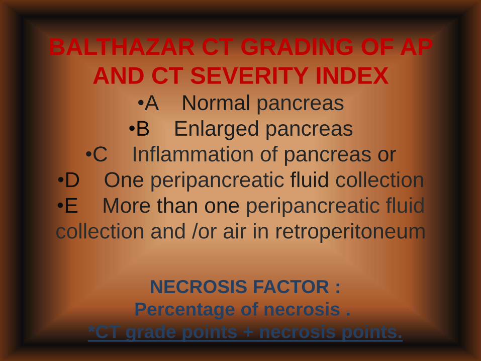
BALTHAZAR CT GRADING OF AP
AND CT SEVERITY INDEX•A Normal pancreas
•B Enlarged pancreas
•C Inflammation of pancreas or
•D One peripancreatic fluid collection
•E More than one peripancreatic fluid
collection and /or air in retroperitoneum
NECROSIS FACTOR :
Percentage of necrosis .
*CT grade points + necrosis points.
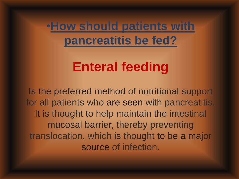
•How should patients with
pancreatitis be fed?
Enteral feeding
Is the preferred method of nutritional support
for all patients who are seen with pancreatitis.
It is thought to help maintain the intestinal
mucosal barrier, thereby preventing
translocation, which is thought to be a major
source of infection.

Enteral feeding
No strong evidence exists that nasojejunal
feeding is advantageous over nasogastric
feeding.
Still, many experts recommend fluoroscopic or
endoscopic placement of a jejunal, or
postpyloric, feeding tube if possible.

Parenteral nutritionshould be initiated in patients unable to
tolerate oral feeding because of pain,
ileus, and/or nausea.
Opinion with regard to the timing of
initiation of parenteral nutrition
ranges from 2 to 5 days after
presentation.

•Should all patients with AP
receive antibiotics?
No. The use of prophylactic antibiotics to
prevent infection of pancreatic necrosis has
been controversial but is not currently
recommended.
No significant benefit has been found with
regard to mortality, rates of infected
necrosis,need for operative treatment, or overall
infections when administering prophylactic
antibiotics.

Broad-spectrum antibiotics should be
administered if objective evidence exists of
infected necrosis on the basis of clinical status
(i.e., fever) or cultured aspirate.
Some experts believe antibiotics are warranted
when evidence is seen on CT of necrosis in >
30% of the pancreas.
In such cases, the use of antibiotics should be
limited to 7 to 14 days because of the risk of
fungal superinfection.

If the patient's condition continues to
deteriorate, he or she should be
evaluated for minimally invasive
and/or surgical debridement or
necrosectomy .

The bedside index of severity in AP
BlSAP SCORE :1 point for each of the following if present. 0 ooints if absenl
•BUN >25 mg/dL (8.9 mmol/L)
•Impaired mental state status
•SIRS (two or more of the following) :
•Pulse > 90 beats/min
•Respiratory rate >20/min or
•PaC02 < 32 mm Hg
•Temperature >38" C or <36" C
•WBC >12,000 or <4000 cells/mm3
or > 10% immarure neutrophils
•Age > 60 yr
•Pleural effusion

The most common bacteria responsible
for infected pancreatic necrosis?
Culprit infective agents are usually gut derived,
including
Escherichia coli, Bacteroides, and
Enterococcus.
Staphylococcus and Pseudomonas
are also potential pathogens and should be
considered.

Fungal infectionof necrosis
Is rare but more common when prophylactic
antibiotics are given.
It is unclear whether fungal superinfection has
any impact on mortality.

Role of surgery for AP
Surgery may be necessary for delayed
complications of AP (i.e., pancreatic
pseudocysts, persistent sterile necrosis) or for
cholecystectomy to prevent future episodes of
biliary pancreatitis.
Surgical necrosectomy is the gold standard
treatment for infected pancreatic necrosis
resulting from severe AP.

Retrospective reviews addressing timing of
intervention found that postponing surgery
until 4 or 6 weeks after admission correlated
with decreased mortality compared with
earlier intervention.
Expert panels recommend delaying
necrosectomy at least 3 to 4 weeks after
hospitalization if possible, while
administering antibiotics and allowing the
necrosis to organize.

Principles of surgical management of
infected pancreatic necrosis include
debridement of all infected necrotic
material and drainage of the remaining
pancreatic bed.
Debridement is done bluntly and
gently, with hydrosonic irrigation
frequently used, to avoid vascular
injury.

Adequate debridement may require
multiple trips to the operating room.
The current favored drainage option
includes closure of the abdomen over
multiple large closed sump drains
with or without irrigation.
These patients are usually critically ill
and require vigorous supportive care.
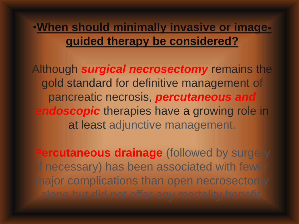
•When should minimally invasive or image-
guided therapy be considered?
Although surgical necrosectomy remains the
gold standard for definitive management of
pancreatic necrosis, percutaneous and
endoscopic therapies have a growing role in
at least adjunctive management.
Percutaneous drainage (followed by surgery
if necessary) has been associated with fewer
major complications than open necrosectomy
alone but did not offer any mortality benefit.

•One third to one half of patients who undergo
percutaneous drainage have no need for surgical
necrosectomy. CT-guided percutaneous drainage of
necrotizing pancreatic collections may be considered
as a bridge to necrosectomy in patients with sepsis
who are too ill
to proceed directly to surgery.
Drainage of a walled-off necrotic cavity by
endoscopic ultrasonography (EUS) via transgastric
or transduodenal approach has been shown to be
effective; however, this modality should be considered
only in a carefully selected patient population and is
dependent on local expertise.

Additional treatment options for acute
biliary pancreatitis
Early ERCP has previously been the standard of
care for patients with AP suspected to be due to
gallstones.
All patients who have signs or symptoms of
cholangitis should have early ERCP to relieve
biliary obstruction.

Other patients with AP in which cholangitis
is not present should be evaluated on a
case-by-case basis with the understanding
that investigations have shown that early
ERCP in patients with predicted mild or
severe pancreatitis does not significantly
reduce the risk for overall complications or
mortality.
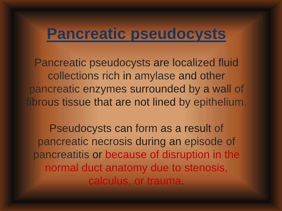
Pancreatic pseudocysts
Pancreatic pseudocysts are localized fluid
collections rich in amylase and other
pancreatic enzymes surrounded by a wall of
fibrous tissue that are not lined by epithelium.
Pseudocysts can form as a result of
pancreatic necrosis during an episode of
pancreatitis or because of disruption in the
normal duct anatomy due to stenosis,
calculus, or trauma.

Pancreatic pseudocysts may be
asymptomatic, present with pain alone, or
present with a variety of other clinical
complications including
bleeding, infection, or rupture.
Rare complications include gastric outlet
and/or biliary obstruction and thrombosis of
splenic or portal veins with development
of gastric varices.
Diagnosis is usually made on the basis of
clinical and radiographic evidence.

The best approach to the
management of pseudocysts
Pancreatic pseudocysts require treatment if
they become symptomatic or develop a
complication .
Surgical, percutaneous, and endoscopic
approaches have all been used to manage
these collections.
No randomized trials have been performed to
compare these modalities.

Endoscopic drainage has advantages of
being less invasive, more cost-effective, and
associated with lower lengths of stay than
surgery, but its use may be limited on the
basis of anatomy.
Modality of treatment for pancreatic
pseudocysts should be based on a
combination of factors including patient
comorbidities and clinical status, site and
characteristics of the lesion, and available
local expertise.

COMMON CAUSES OF ACUTE
PANCREATITIS :•Biliary-Gallstones, parasites, or
malignancy
•Alcohol
•Drugs
•Trauma, toxins
•Idiopathic, ischemic, infectious, inherited
•Metabolic-hyperlipidemia, hypercalcemia
•ERCP
•Smoking

GOOD LUCK
SAMIR EL ANSARY
ICU PROFESSOR
AIN SHAMS
CAIRO
https://www.facebook.com/groups/1451610115129555/#!/groups/1451610115129555/
Wellcome in our new group ..... Dr.SAMIR EL ANSARY
Related Documents