Go to: Go to: Go to: Go to: Brain . 2013 Aug; 136(8): 2539–2549. Published online 2013 Jul 3. doi: 10.1093/brain/awt177 PMCID: PMC3722353 Acute lesions that impair affective empathy Richard Leigh , Kenichi Oishi , John Hsu , Martin Lindquist , Rebecca F. Gottesman , Samson Jarso , Ciprian Crainiceanu , Susumu Mori , and Argye E. Hillis 1 Department of Neurology, Johns Hopkins University School of Medicine, Baltimore, Maryland, USA 2 Department of Radiology, Johns Hopkins University School of Medicine, Baltimore, Maryland, USA 3 Department of Biostatistics, Bloomberg School of Public Health, Johns Hopkins University, Baltimore, Maryland, USA 4 Department of Physical Medicine and Rehabilitation, Johns Hopkins University School of Medicine, Baltimore, Maryland, USA 5 Department of Cognitive Science, Krieger School of Arts and Sciences, Johns Hopkins University, Baltimore, Maryland, USA Corresponding author. Correspondence to: Argye E. Hillis, MD Department of Neurology Meyer 6-113, Johns Hopkins Hospital, 600 North Wolfe Street, Baltimore, MD 21287, USA E-mail: [email protected] *These authors contributed equally to this work. Received 2013 Feb 19; Revised 2013 Apr 9; Accepted 2013 May 12. Copyright © The Author (2013). Published by Oxford University Press on behalf of the Guarantors of Brain. All rights reserved. For Permissions, please email: [email protected] This article has been cited by other articles in PMC. Abstract Functional imaging studies of healthy participants and previous lesion studies have provided evidence that empathy involves dissociable cognitive functions that rely on at least partially distinct neural networks that can be individually impaired by brain damage. These studies converge in support of the proposal that affective empathy—making inferences about how another person feels—engages at least the following areas: prefrontal cortex, orbitofrontal gyrus, anterior insula, anterior cingulate cortex, temporal pole, amygdala and temporoparietal junction. We hypothesized that right-sided lesions to any one of these structures, except temporoparietal junction, would cause impaired affective empathy (whereas bilateral damage to temporoparietal junction would be required to disrupt empathy). We studied 27 patients with acute right hemisphere ischaemic stroke and 24 neurologically intact inpatients on a test of affective empathy. Acute impairment of affective empathy was associated with infarcts in the hypothesized network, particularly temporal pole and anterior insula. All patients with impaired affective empathy were also impaired in comprehension of affective prosody, but many patients with impairments in prosodic comprehension had spared affective empathy. Patients with impaired affective empathy were older, but showed no difference in performance on tests of hemispatial neglect, volume of infarct or sex distribution compared with patients with intact affective empathy. Keywords: empathy, stroke, emotion perception, magnetic resonance imaging, prosody Introduction Nearly all human interaction requires that we make inferences about what other people think and feel. 1,2,* 2,* 2 3 1,3 2 3 2 1,4,5

Welcome message from author
This document is posted to help you gain knowledge. Please leave a comment to let me know what you think about it! Share it to your friends and learn new things together.
Transcript

Go to:Go to:
Go to:Go to:
Brain. 2013 Aug; 136(8): 2539–2549.Published online 2013 Jul 3. doi: 10.1093/brain/awt177
PMCID: PMC3722353
Acute lesions that impair affective empathyRichard Leigh, Kenichi Oishi, John Hsu, Martin Lindquist, Rebecca F. Gottesman, Samson Jarso,Ciprian Crainiceanu, Susumu Mori, and Argye E. Hillis
1 Department of Neurology, Johns Hopkins University School of Medicine, Baltimore, Maryland, USA2 Department of Radiology, Johns Hopkins University School of Medicine, Baltimore, Maryland, USA3 Department of Biostatistics, Bloomberg School of Public Health, Johns Hopkins University, Baltimore, Maryland, USA4 Department of Physical Medicine and Rehabilitation, Johns Hopkins University School of Medicine, Baltimore, Maryland, USA5 Department of Cognitive Science, Krieger School of Arts and Sciences, Johns Hopkins University, Baltimore, Maryland, USA
Corresponding author.Correspondence to: Argye E. Hillis, MD Department of Neurology Meyer 6-113, Johns Hopkins Hospital, 600 North Wolfe Street,Baltimore, MD 21287, USA E-mail: [email protected]*These authors contributed equally to this work.
Received 2013 Feb 19; Revised 2013 Apr 9; Accepted 2013 May 12.
Copyright © The Author (2013). Published by Oxford University Press on behalf of the Guarantors of Brain. All rights reserved. ForPermissions, please email: [email protected]
This article has been cited by other articles in PMC.
Abstract
Functional imaging studies of healthy participants and previous lesion studies have provided evidencethat empathy involves dissociable cognitive functions that rely on at least partially distinct neuralnetworks that can be individually impaired by brain damage. These studies converge in support of theproposal that affective empathy—making inferences about how another person feels—engages at leastthe following areas: prefrontal cortex, orbitofrontal gyrus, anterior insula, anterior cingulate cortex,temporal pole, amygdala and temporoparietal junction. We hypothesized that right-sided lesions to anyone of these structures, except temporoparietal junction, would cause impaired affective empathy(whereas bilateral damage to temporoparietal junction would be required to disrupt empathy). Westudied 27 patients with acute right hemisphere ischaemic stroke and 24 neurologically intact inpatientson a test of affective empathy. Acute impairment of affective empathy was associated with infarcts inthe hypothesized network, particularly temporal pole and anterior insula. All patients with impairedaffective empathy were also impaired in comprehension of affective prosody, but many patients withimpairments in prosodic comprehension had spared affective empathy. Patients with impaired affectiveempathy were older, but showed no difference in performance on tests of hemispatial neglect, volumeof infarct or sex distribution compared with patients with intact affective empathy.
Keywords: empathy, stroke, emotion perception, magnetic resonance imaging, prosody
Introduction
Nearly all human interaction requires that we make inferences about what other people think and feel.
1,2,* 2,* 2 3 1,3 23 2 1,4,5

This ability to take the perspective of another person provides an important foundation for ourrelationships, communication, negotiations and other social activities. Perspective-taking is animportant component of empathy (de Waal, 2012), along with emotional contagion (Stotland andDunn, 1963; de Waal, 2012), or sharing the other person’s perceived emotional state. Several importantneurological and neuropsychiatric diseases are known to disrupt various aspects of empathy, includingautism (Dziobek et al., 2008), frontotemporal dementia (Eslinger et al., 2005; Viskontas et al., 2007),traumatic head injury (Eslinger, 1998; McDonald and Flanagan, 2004; Neumann et al., 2012) andschizophrenia (Shamay-Tsoory et al., 2007; Lee et al., 2011). Numerous functional imaging studieshave investigated the brain regions engaged in empathy, and have found distinct regions involved inemotional contagion versus cognitive and emotional perspective-taking (the ability to infer whatanother person thinks or feels). These studies have led to the proposal that there are dissociable neuraland cognitive systems of empathy. One is a developmentally and phylogenetically ‘early’ system ofemotional contagion that may involve the mirror neuron system and the right inferior frontal gyrus(Bodini et al., 2004; Shamay-Tsoory, 2011), as well as the right temporal pole, superior temporal gyrus,fusiform, insula and amygdala (Carr et al., 2003). Emotional contagion may depend on areas of thebrain that are activated in association with recognizing emotions of others through prosody (tone ofvoice), facial expression and gestures, in right fusiform cortex as well as superior temporal sulcus andamygdala (Gorno-Tempini et al., 2001). The second ‘later’ system, which some think of as the secondstage of empathy (Decety and Jackson, 2004) is a higher level perspective-taking system that dependson the medial prefrontal cortex (Eslinger, 1998; Shamay-Tsoory et al., 2003). Perspective-taking can befurther subdivided into cognitive and emotional perspective-taking. Perspective-taking may depend ona number of cognitive functions such as cognitive flexibility (Decety and Jackson, 2004; Rankin et al.,2005), attention and working memory, abstract reasoning (Rankin et al., 2006), belief attribution andassignment of agency (Saxe and Kanwisher 2003; Samson et al., 2004; Saxe et al., 2004; Decety andLamm, 2007). Thus, affective empathy, the ability to recognize and make judgements about howanother person feels, includes both emotional contagion and emotional perspective-taking.
The neural networks supporting affective empathy have been studied primarily through task-relatedand task-free (‘resting-state’) functional MRI. A task-free functional MRI study of neurologicallynormal adults investigated intrinsic functional dynamics of affective compared with cognitive empathy(Cox et al., 2012). This study showed that a dominance of affective empathy compared to cognitiveempathy was associated with functional connectivity between ventral anterior insula, orbitofrontalcortex, amygdala, and anterior cingulate, whereas a dominance of cognitive empathy was associatedwith functional connectivity between parts of the brainstem, superior temporal sulcus and ventralanterior insula. Task-related functional imaging studies also confirm an important role of various partsof the frontal cortex, anterior insula, anterior cingulate cortex and right amygdala in empathy, asrevealed by large coordinate-based activation likelihood estimation meta-analyses. In one activationlikelihood estimation meta-analysis of 112 experiments on affective empathy, Bzdok et al. (2012)reported that the following areas showed significant activation in association with empathy tasks acrossstudies: bilateral dorsomedial prefrontal cortex, right greater than left inferior frontal cortex, bilateralanterior insula, anterior and posterior cingulate, bilateral temporoparietal junction, right amygdala,right middle temporal gyrus, right superior temporal sulcus, bilateral thalamus, right hippocampus,midbrain and right pallidum. A few meta-analyses have focused on a particular type of affectiveempathy: recognizing another person’s negative emotion caused by pain. Gu et al. (2012) reported thatanterior insula and anterior cingulate cortex were the areas most commonly activated in associationwith recognition of another person’s pain, on the basis of an activation likelihood estimation meta-
analysis of 28 functional MRI studies on empathetic pain (using keywords ‘empathy’, ‘empathetic’,‘sympathy’, ‘emotional contagion’, ‘altruism’ or ‘compassion’ combined with ‘pain’ and ‘functionalMRI’). Similarly, an image-based meta-analysis of nine independent functional MRI studies ofempathy for pain and an activation likelihood estimation meta-analysis of 32 studies of empathy forpain identified activation in bilateral anterior insula, anterior cingulate, precuneus and thalamus,associated with empathy for other people’s pain (Lamm et al., 2011).
Two recent studies have investigated the influence of neuropeptides that affect amygdala function onempathy. The first reported that intranasal oxytocin increased affective, but not cognitive empathy, asmeasured by a multifaceted empathy task; and improved learning performance on an association taskwhen social, but not non-social, reinforcers were used (Hurlemann et al., 2010). These authors alsoreported that two females with selective damage to bilateral amygdala due to Urbach-Wiethe diseasewere impaired in affective, but not cognitive, empathy on their multifaceted empathy task, and showednormal learning on an association task when non-social reinforcers, but not social, reinforcers, wereused. Stone et al., (2003) also reported cases of impaired empathy performance associated withbilateral amygdala lesions in two patients. More recently, investigators studied the effects of intranasalvasopressin, which is considered to have opposing effects of oxytocin, on activation in amygdala andother brain regions during empathy in a functional MRI study of affective empathy (Brunnlieb et al.,2013). This study revealed that vasopressin modulated activation of right amygdala associated withaffective empathy and increased connectivity between right amygdala and medial prefrontal cortex andinferior parietal cortex in association with the empathy task.
Together, these studies indicate an important role of at least the following areas in recognizing andmaking inferences about the emotions of another person (affective empathy): prefrontal cortex,orbitofrontal gyrus, anterior insula, anterior cingulate cortex, temporal pole and amygdala. Thetemporoparietal junction is another area that is nearly always activated in tasks that require perspective-taking (Corbetta et al., 2008), but seems to be important for more general aspects of mentalizing orbelief attribution than in affective empathy (Sebastian et al., 2012). For example, it is equally engagedin third person visuospatial judgements as third person judgements of another person’s feelings oremotions (Schnell et al., 2011). Moreover, because it is not specific to emotional processing, wehypothesize that a unilateral lesion would not disrupt its role in assignment of agency or attributing abelief or feeling to another person. Rather, we expect bilateral damage to this region would be requiredto disrupt its role in empathy.
Lesions localized to some of the regions that are activated in functional imaging studies are reported tocause deficits specific to cognitive versus affective empathy (Hynes et al., 2006; Shamay-Tsoory andAharon-Peretz, 2007; Gu et al., 2012). Such studies are important to the field, because it is critical toshow that regions activated during a task are critical to that task, and not just engaged by it (or activelyinhibited during it), as ‘activation’ (blood oxygen level-dependent signal in a region that is correlatedwith the task) can reflect any of these possibilities (Thompson-Schill et al., 1998; Fellows et al., 2005).However, most of the studies of impaired empathy have been conducted in patients with poorlylocalized lesions, such as autism, dementia, and traumatic head injury. The few studies of affectiveempathy in patients with focal lesions, such as stroke, have included just one to two patients with focallesions (Stuss and Anderson 2004; Samson et al., 2005, 2007; Roldan et al., 2011; Couto et al., 2012)or have involved a heterogeneous population, of which only a few had stroke (and others hadmeningioma, head injury and other lesions) (Shamay-Tsoory et al., 2003, 2005, 2009; Shamay-Tsooryand Aharon-Peretz, 2007). A voxel-based morphometry study of 123 patients with Alzheimer’s
Go to:Go to:
disease, progressive supranuclear palsy, corticobasal degeneration and frontotemporal dementia usingcaregivers’ ratings on the Interpersonal Reactivity Index (Davis, 1983) to evaluate empathy revealedthat impairments in empathy measured by the sum of ‘empathetic concern’ (reflecting emotionalcontagion) and ‘perspective-taking’ significantly correlated with the volume of grey matter in righttemporal pole, fusiform gyrus and medial inferior frontal region (Rankin et al., 2006). Both empatheticconcern and perspective-taking subscale scores alone correlated with volume of grey matter volume inright temporal pole. Other studies of behavioural variant frontotemporal dementia, a relatively focalneurodegenerative disease characterized by impaired social conduct and emotional function, includingempathy, have also reported a role of right temporal pole and/or orbitofrontal atrophy in empathydeficits (Rankin et al., 2005; Kipps and Hodges, 2006; Viskontas et al., 2007). For example, a detailedstudy of a patient with hereditary multiple exostoses and frontotemporal dementia revealed severelyimpaired affective empathy associated with atrophy in right anterior temporal lobe and orbitofrontalgyrus (Narvid et al., 2009). Right insula also showed atrophy. Therefore, we sought to test thehypothesis, derived from functional imaging studies in healthy participants and previous lesion studies,that lesions to components of a network of brain regions involving right prefrontal cortex, orbitofrontalgyrus, temporal pole, anterior insula, anterior cingulate cortex and amygdala cause deficits in affectiveempathy. We focused on patients with right hemisphere lesions because studies of neurodegenerativedisease have consistently shown greater correlation between impairments in empathy and atrophy inthe right hemisphere compared to the left hemisphere (Rankin et al., 2005, 2006; Eslinger et al., 2011).Older lesions studies, which frequently compared small numbers of right hemisphere stroke to lefthemisphere stroke, without further lesion localization, also found a greater impact of right hemisphereon empathy (Brune and Brune-Cohrs, 2006). Finally, functional imaging studies have also indicated aspecialized role of the right hemisphere in emotional aspects of empathy and sympathy (Decety andChaminade, 2003). We also determined whether deficits in this type of empathy were related to lesionsite alone, or also influenced by lesion volume, age, impairments of attention (neglect), or impairmentsof prosody comprehension. We tested these hypotheses in patients with acute stroke (within 48 h ofonset), before the opportunity for reorganization of structure-function relationships, rehabilitation, orrecovery. This approach allowed us to determine if a lesion in a particular area had the predicted effecton the task.
Materials and methods
Participants
We enrolled a consecutive series of 27 patients with acute ischaemic right hemisphere stroke whoprovided informed consent to participate in the study and had none of the following exclusion criteria:(i) neurological disease other than stroke; (ii) reduced level of consciousness or on-going sedation; and(iii) inability to have MRI due to claustrophobia, implanted ferrous metal, or weight >300 lb. We alsoenrolled 24 patients with transient ischaemic attack who had normal MRI and normal neurologicalexamination at the time of testing to serve as normal controls as they had the same demographiccharacteristics and the same stressors of hospitalization as the stroke patients. They also had to havenone of the exclusion criteria and no stroke.
Imaging
Patients underwent a stroke protocol MRI, including diffusion-weighted imaging, FLAIR and 3D time-of-flight angiography of the intracranial vessels. Diffusion-weighted imaging was acquired using

single-shot spin-echo echo-planar imaging, in the transverse plane covering the entire brain with a b-value of 1000 (s/mm ) and with a least diffusion weighting (b0).
Stroke volume measurement
To define boundaries of acute stroke lesion(s) (hereafter termed stroke map) to measure the strokevolume of each participant, a threshold of >30% intensity increase from the unaffected area in thediffusion-weighted image was applied, and then a neurologist (K.O.), who was blinded to the clinicalinformation, manually modified the boundary to avoid false-positive and false-negative areas (Oishi etal., 2009). This procedure was performed on RoiEditor (www.MRIstudio.org).
Image processing
The least diffusion weighted image (b0) with T -weighted contrast was transformed to the JHU-MNI-b0 atlas using affine transformation followed by the large deformation diffeomorphic metric mapping(Oishi et al., 2009). The resultant matrices were applied to the stroke map for the normalization.Customized version of the JHU-MNI Brain Parcellation Map (cmrm.med.jhmi.edu) was overlaid onthe normalized stroke map to investigate % volume of the selected anatomical structures (prefrontalcortex, orbitofrontal gyrus, anterior cingulate cortex, anterior insula, temporal pole and amygdala)affected by acute stroke (Fig. 1). This procedure was performed on DiffeoMap (www.MRIstudio.org).
Figure 1Procedures for the image processing. (A) Images in original space.(B) The diffusion weighted image (DWI) was used to 3D definethe area with acute infarction (stroke map) shown as a red contourin the least diffusion weighted image (b0) and diffusion ...
Behavioural testing
Patients underwent cognitive testing within 24 h of admission to the hospital. Testing of affectiveempathy included eight questions requiring inference about emotions of individuals in short videotapedscenarios and two questions requiring inference about emotions of individuals in stories that were readto the patients (Box 1). To control for deficits in general attention and recent memory, patients werealso asked factual questions about the stories. To evaluate emotional contagion, the facial expressions,comments, and tone of voice of patients were evaluated when the patients watched the videos andlistened to the stories.
Box 1 Example of a story from the Affective Empathy Task
Participants listen to the following story, and then select a printed word that most accurately depictsthe emotion in response to the question.
John was waiting for the bus that would bring his girlfriend, Cathy. He was planning to ask her tomarry him. When the bus arrived, Cathy got off the bus, talking to a very handsome man. Theywere smiling at each other.
How do you thing John feels?
Happy Jealous Scared Bored Excited Relieved
2
2

Go to:Go to:
The story continues. Cathy saw John. She smiled and waved. She said, ‘John, I would like you tomeet my brother.’
How do you thing John feels now?
Angry Jealous Scared Bored Excited Relieved
Patients were also administered the Interpersonal Reactivity Index. This test is a self-administered testthat includes four types of questions designed to evaluate cognitive empathy (perspective-taking andfantasy scales) and affective empathy (empathetic concern and personal distress).
Patients were also administered the prosody comprehension subtest of the Aprosodia Battery (Ross andMonnot, 2008) to evaluate for deficits in comprehension in affective prosody. In this test, participantslisten to emotionally-neutral sentences, monosyllables (ba ba ba), or asyllabic tones (ah) produced indifferent tones of voice, and select (from written choices) the corresponding emotion (e.g. happy, sad,angry, surprised).
Patients were administered a general test of hemispatial neglect, including a test of detecting left versusright-sided gaps in circles scattered across a page (Ota et al., 2001), and copying a scene of a house,two trees and a fence, and bisection of a 10-inch line.
Statistical analysis
We first identified a cut-off score for normal performance on the affective empathy test, byadministering the test to 24 neurologically normal control subjects (transient ischaemic attack patientswith normal MRI and normal neurological examination at the time of testing). We determined therange and distribution of scores for control subjects with comparable age, education and socioeconomicbackgrounds of our stroke patients, which allowed us to identify a cut-off that would be outside of thenormal range of scores for this population.
We then identified whether or not a lesion in the proposed network of areas critical for affectiveempathy (right prefrontal, orbitofrontal gyrus, temporal pole, anterior cingulate, anterior insula, oramygdala) in the right hemisphere is associated with impaired affective empathy, using Fisher’s exacttests, for the entire network as a whole, including only the 27 stroke patients in the analysis. For thetwo areas most strongly associated with impaired affective empathy, we evaluated the Pearsoncorrelation between severity of impairment (error rate on the affective empathy task) and percentage ofdamage in each of the two areas.
We then evaluated differences between stroke patients with lesions in this network and patients withoutlesions in this network (in a case control study), with regard to score on our affective empathy test,tests of affective prosody, tests of hemispatial neglect, as well as age and volume of infarct.
Finally, we evaluated whether or not patients with impaired affective empathy were different frompatients with spared affective empathy in terms of scores on prosody comprehension, volume ofinfarct, age, and scores on neglect.
Results
Affective Empathy Task: range of normal performance
The 24 control participants ranged in age from 35–79 years, mean ± standard deviation (SD) = 52.5 ±15.5. The stroke patients were similar, ranging in age from 26–75 years, mean ± SD = 54.5 ± 13.6. Inboth the control group and the stroke group, 33% of patients were female.
The range of scores on the Affective Empathy Task for control patients was 0 to 20% errors. The meanscore ± SD was 5.8 ± 7.8. The distribution of scores for controls was as follows: 58.3% made 0%errors; 25% made 10% errors; and 16.7% made 20% errors. Therefore, we used >20% errors as the cut-off for normal performance; ≥30% errors was considered impaired. This score was more than 2 SDabove the mean for normal controls.
The effect of acute ischaemic lesions in right hemisphere affective empathy networkon affective empathy task performance
We examined the regions of interest in the right hemisphere that most commonly show activationduring affective empathy tasks, to determine if a lesion in one or more of the components of theproposed network caused impaired affective empathy (≥30% error). The regions of interest included:right prefrontal cortex, orbitofrontal gyrus, anterior cingulate cortex, anterior insula, temporal pole andamygdala. We considered a region of interest as affected only if at least 1% of the area was infarcted ondiffusion weighted imaging trace image. We identified 10 patients with lesions in one or more of theregions of interest. Nine (90%) of the 10 patients had impaired affective empathy. One patient with alesion involving the anterior insula did not have impaired affective empathy using the 30% error cut-off, although the patient made 20% errors on the task.
Table 1 shows the percentage of infarct in each of the regions of interest for each of the 10 patientswith damage to the network. All of these patients who had lesions in the temporal pole also had lesionsin the anterior insula. The one patient who had insular damage but no damage to the temporal pole isthe patient who made only 20% errors on the affective empathy task (in the normal range).
Table 1Error rate and percentage of damage to each region of interest inthe ‘empathy network’ for the 10 patients with network lesions
Of the 14 patients with impaired affective empathy, nine (64.3%) had lesions to one or more of theregions of interest we identified on the basis of the functional neuroimaging literature as engaged inaffective empathy. The remaining five patients had lesions in the right thalamus (three patients) or rightposterior superior frontal gyrus and middle frontal gyrus, posterior segment (two patients). One patientwith a posterior superior and middle frontal gyrus lesion also had a lesion in the right precuneus. Theseare all areas that have shown activation in one or more of the functional imaging studies of affectiveempathy reviewed above.
There was a very strong association between the presence of a lesion in one or more region of interestin the network hypothesized to underlie affective empathy and impairments on our tasks of affectiveempathy (Fisher’s exact = 0.004). Figure 2 shows examples of patients with impaired performance onthe affective empathy task and lesions in each of the regions of interest. The strongest association (andonly significant) associations were between impaired affective empathy and infarct in the temporal pole(Fisher’s exact = 0.04) or in the anterior insula (Fisher’s exact = 0.03). When we further divided thetemporal pole into the pole of superior and middle temporal gyri, the strongest association was betweenimpaired affective empathy and infarct of the superior temporal gyrus pole (Fisher’s exact = 0.003).

There were seven patients with both superior temporal gyrus pole and anterior insular lesions who hadimpaired affective empathy. For all 27 stroke patients, there was also a significant correlation betweenerror rate on the affective empathy task and the volume of infarct in (i) temporal pole (r = 0.48; P =0.014); (ii) superior temporal gyrus pole alone (r = 0.49; P = 0.013); and (iii) anterior insula (r = 0.40;P = 0.047). There were only a few patients with>1% damage to right prefrontal cortex (n = 3), anteriorcingulate (n = 2), and amygdala (n = 4). Importantly, however, there were no patients with lesions to>1% of any of these three regions of interest or the temporal pole who had spared affective empathy onour test. Some lesions involved more than one of the regions of interest (Table 1).
Figure 2Representative individuals with acute infarction in the prefrontalcortex (A, cyan contour), anterior cingulate cortex (B, pinkcontour), anterior insular cortex (C, yellow contour), orbitofrontalcortex (D, blue contour), amygdala (E, green contour) ...
As expected, there was no association between impaired affective empathy and a unilateral lesioninvolving the temporal parietal junction. There were three patients with impaired affective empathywhose lesions included the temporal parietal junction, but also three patients with spared affectiveempathy whose lesions included that area.
Performance on affective empathy and other cognitive measures in patients withlesions in the hypothesized network supporting affective empathy versus lesionsoutside the network
Our second goal was to evaluate differences between stroke patients with lesions in the proposed‘affective empathy network’ and patients with lesions outside this network, with regard to score on ouraffective empathy test, tests of affective prosody, tests of hemispatial neglect, as well as age andvolume of infarct. To accomplish this goal, we carried out a very small case-control study includingnine patients with lesions in the regions of interest hypothesized to underlie affective empathy,compared with nine patients with stroke, but not involving any of these six regions of interest, matchedrelatively closely for lesion volume, age and sex. To identify these patients, we excluded all patientswith lesions >100 cm and <1.45 cm . This exclusion yielded patients with the characteristics in Table 2. We compared the two groups using Mann-Whitney tests (because these data were notnormally distributed).
Table 2Characteristics of patients with and without lesions in the‘Affective Empathy Network’ (right prefrontal cortex,orbitofrontal gyrus, temporal pole, anterior insula, anteriorcingulate, amygdala), excluding those with lesions >100 ...
Patients who had lesions in the proposed ‘affective empathy network’ (prefrontal cortex, orbitofrontalgyrus, anterior cingulate cortex, anterior insula, temporal pole, and/or amygdala) made significantlymore errors on the affective empathy task than patients without lesions to the network (median = 30%versus 0%; Z = −2.5; P = 0.01). There were no significant differences between patients with andwithout lesions in the ‘affective empathy network’ with regard to lesion volume, age, neglect orprosody scores.
3 3
Go to:Go to:
Although patients were administered the Interpersonal Reactivity Index, which has been shown to be areliably measure of cognitive and affective empathy in neurologically normal individuals and somepatient populations (Rankin et al., 2005), we could not use it to detect deficits in empathy in ourpopulation. Only one participant had a score that was >1.5 SD below normal on the perspective-takingsubscore; and this participant was a transient ischaemic attack (neurologically normal) control subject.Likewise, only one participant (an acute stroke patient) showed performance on the empathetic concernsubscore that was >1.5 SD below the mean for normal controls.
We also did not use the scores for emotional contagion based on observations of facial expressionsduring the videotapes, because nearly all patients were scored as ‘happy’ (given choices of happy,jealous, scared, bored, excited or relieved). The patient testers found it difficult to assess theparticipants’ emotions while watching the videos. Most patients did not show outward signs ofemotion, such as change in facial expression or make spontaneous comments about their feelings. Theywere scored as happy if they were pleasant and judged to be happy to continue the experiment.
Differences between patients with impaired affective empathy versus patients withspared affective empathy
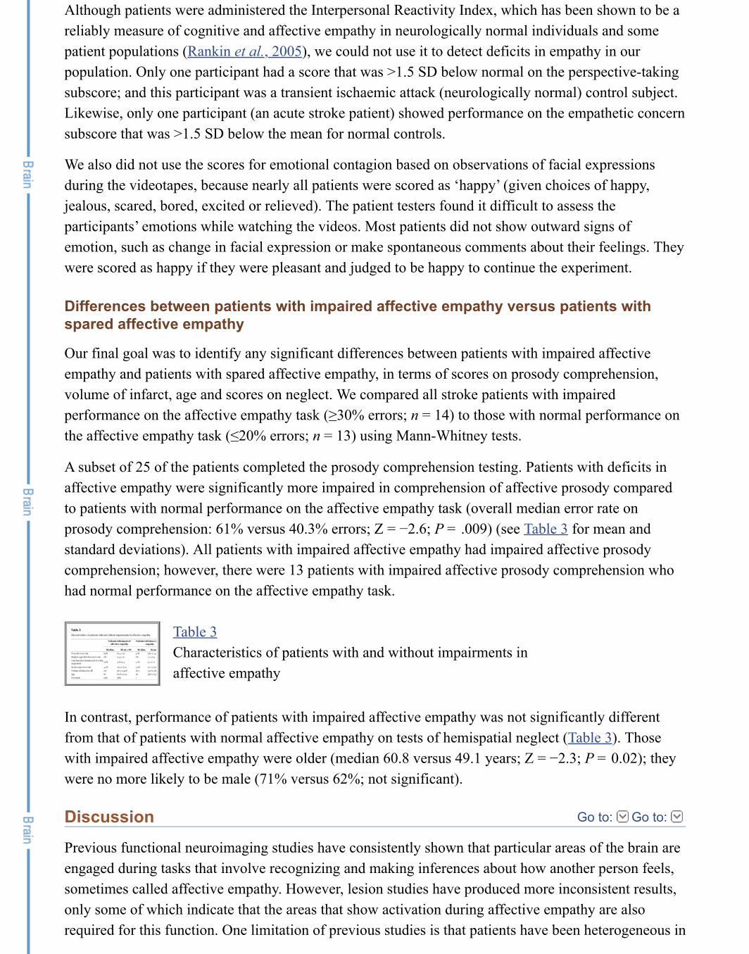
Our final goal was to identify any significant differences between patients with impaired affectiveempathy and patients with spared affective empathy, in terms of scores on prosody comprehension,volume of infarct, age and scores on neglect. We compared all stroke patients with impairedperformance on the affective empathy task (≥30% errors; n = 14) to those with normal performance onthe affective empathy task (≤20% errors; n = 13) using Mann-Whitney tests.
A subset of 25 of the patients completed the prosody comprehension testing. Patients with deficits inaffective empathy were significantly more impaired in comprehension of affective prosody comparedto patients with normal performance on the affective empathy task (overall median error rate onprosody comprehension: 61% versus 40.3% errors; Z = −2.6; P = .009) (see Table 3 for mean andstandard deviations). All patients with impaired affective empathy had impaired affective prosodycomprehension; however, there were 13 patients with impaired affective prosody comprehension whohad normal performance on the affective empathy task.
Table 3Characteristics of patients with and without impairments inaffective empathy
In contrast, performance of patients with impaired affective empathy was not significantly differentfrom that of patients with normal affective empathy on tests of hemispatial neglect (Table 3). Thosewith impaired affective empathy were older (median 60.8 versus 49.1 years; Z = −2.3; P = 0.02); theywere no more likely to be male (71% versus 62%; not significant).
Discussion
Previous functional neuroimaging studies have consistently shown that particular areas of the brain areengaged during tasks that involve recognizing and making inferences about how another person feels,sometimes called affective empathy. However, lesion studies have produced more inconsistent results,only some of which indicate that the areas that show activation during affective empathy are alsorequired for this function. One limitation of previous studies is that patients have been heterogeneous in

aetiology and time post-onset of lesion. Some lesions, such as meningiomas and other slow growingtumours, often fail to produce deficits, even when they arise in areas of the brain where a stroke orother sudden onset lesion would typically cause a specific deficit. Furthermore, when patients arestudied a long time after stroke or other focal lesion, they may have originally had the deficit associatedwith the lesioned area, but may have recovered due to rehabilitation or spontaneous recovery, as otherareas of the brain can assume the functions of the damaged ones. The few previous lesion studies ofempathy have included patients with slow growing tumours or patients who are a long time post onsetof stroke or other focal injury. We tried to correct for these particular limitations of previous studies bystudying patients immediately after onset of acute stroke, before the opportunity for reorganization,recovery or rehabilitation.
We were able to confirm that lesions within the hypothesized network were associated with an errorrate on an affective empathy task that is outside the error rate made by neurological normal controls.We also showed, in a small case-control study that patients with lesions involving the network hadhigher error rates on the affective empathy task than a fairly well-matched group of patients withsimilar sized lesions that do not involve the network.
The most commonly affected areas in the patients with impaired affective empathy in this study werethe anterior insula and the temporal pole (specifically the pole of the superior temporal gyrus). Theanterior insula is an area that is commonly affected in acute middle cerebral artery stroke (Finley et al.,2003). Nevertheless, we found that those who had impaired affective empathy were more likely to havelesions in the anterior insula than those who did not have impaired affective empathy. The temporalpole is not an area commonly involved in stroke (Caviness et al., 2002). Yet, 50% of the patients withimpaired affective empathy had lesions in temporal pole, and no patients with lesions in temporal polehad normal performance on the affective empathy task. In both areas, percentage of damage correlatedwith error rate on the affective empathy task. All patients with temporal pole lesions had anteriorinsular lesions, raising the possibility that damage to only one or the other may be associated withaffective empathy. However, there are converging data from other sources for a role of each of theseareas in affective empathy. As reviewed above, the anterior insula nearly always shows activation inassociation with affective empathy tasks in functional imaging studies (Bzdok et al., 2012). Thetemporal pole is an area where it has been more difficult to reveal activation in functional imagingstudies, but recent studies have shown activation in association with affective empathy (Jimura et al.,2010). Furthermore, both right anterior insula and right temporal pole atrophy have been associatedwith impaired affective empathy in neurodegenerative disease (Rankin et al., 2005, 2006; Kipps andHodges, 2006; Narvid et al., 2009; Lee et al., 2012). These areas were not the only critical componentsof the network; other areas included right prefrontal and fronto-orbito cortex, anterior cingulate andamygdala.
All but five patients with significant impairments in affective empathy had lesions in one or more ofthe regions of interest in the proposed network. Two of these patients had small lesions in part of theright posterior superior frontal gyrus and middle frontal gyrus, close to ventromedial prefrontal cortex(ventral superior frontal gyrus). It is possible that a larger part of the frontal cortex, rather than justventromedial prefrontal cortex, as proposed by some authors, is critical for affective empathy.
The three remaining patients with impaired affective empathy had right thalamic infarcts. The thalamushas an important role in relaying sensory and multimodal information to the prefrontal cortex forfurther processing. It is reasonable to assume that it may play an important role in perception ofauditory and visual cues about another person’s emotions, and relaying this information to the cortex to

enable inferences to be drawn. Four out of nine patients with right thalamic infarcts had impairments inaffective empathy. The thalamus consists of several nuclei with quite distinct functions, so whether ornot affective empathy was disrupted may have depended on the nuclei involved. However, theresolution of our imaging was not adequate to determine which nuclei were included in the infarct. Ofnote, consistent with our hypothesis that the thalamus may be important in affective empathy, theactivation likelihood estimation meta-analysis of 112 functional MRI studies of affective empathyrevealed the right posterior thalamus and left anterior thalamus among the areas commonly activated inassociation with empathy (Bzdok et al., 2012).
The temporoparietal junction is an area that consistently is engaged in ‘mentalizing’ or cognitiveperspective-taking, although it does not appear to be specific to empathy (Decety and Lamm, 2007;Schnell et al., 2011; Sebastian et al., 2012). Although one study has indicated that a right parietallesion might cause impairment in empathy (Shamay-Tsoory et al., 2003), most studies have raised thepossibility that either temporoparietal junction alone might be adequate to handle the role inperspective-taking, which may include attributing a belief to another person (Shamay-Tsoory et al.,2003). In this case, a unilateral temporoparietal junction would not be expected to substantially impairempathy. Consistent with this view, in our study, lesions in this area alone were not associated withimpaired affective empathy.
Not surprisingly, patients with impaired affective empathy also had significantly higher error rates inthe closely related task of recognizing the emotion in tone of voice. However, we do not believe thatimpaired recognition of prosody was responsible for the deficit in affective empathy, becauserecognition of emotion from tone of voice was not required to answer any of the questions correctly.Furthermore, a patient with severely impaired recognition and production of prosody performed thistask accurately (and scored normally on the Interpersonal Reactivity Index) (Dara et al., 2012).Furthermore, in this study, 13 patients had impaired prosody comprehension, but normal performanceon the empathy task. Rather, we believe that some of the areas of the brain necessary for affectiveempathy may also be necessary for comprehension of affective prosody.
Finally, we found that impairments in affective empathy appear to be independent of impairments inspatial attention (neglect) after right hemisphere stroke, indicating that it is not likely to simply be amarker of ‘severe’ right cortical stroke.
One limitation of the study is that we were not able to demonstrate a dissociation between perspective-taking and emotional contagion; that is, we could not distinguish between neurologically normalparticipants and stroke patients in emotional contagion, either using the self-administered InterpersonalReactivity Index or technicians’ observations of facial expressions, gestures, and tone of voice duringvideos and stories. Acute right hemisphere stroke patients are known to have other deficits thatinterfere with expression of emotional concern (e.g. impaired expression of emotion through prosody;Ross et al., 1997) and self-assessment of deficits (anosognosia, perhaps including changes in emotionalcontagion compared to baseline). Therefore, if they have impaired emotional contagion, we wereunable to detect it using the Interpersonal Reactivity Index or observation of facial expression/gestures.Our task of affective empathy may have conflated emotional contagion and affective perspective-taking, because the questions about videos may have been answered in part by recognizing and sharingin the emotions of another person. Future studies may use the caregiver assessments of emotionalcontagion as well as perspective-taking, as has been done for patients with frontotemporal dementia,who also have limited insight (Rankin et al., 2006; Eslinger et al., 2011). Like our stroke patients,patients with frontotemporal dementia showed no difference from controls on their self-assessment of

Go to:Go to:
Go to:Go to:
Go to:Go to:
empathy, although caregivers rated them as having impaired perspective-taking and empathetic concernat the same time (Eslinger et al., 2011). Alternatively, emotional contagion can be assessed withautonomic responses, such as skin conductance response and heart rate changes, when presented withemotional scenes or stories (Balconi and Bortolotti, 2012).
Another limitation is that we did not assess patients with left hemisphere stroke in this study. Previousstudies of dementia (Rankin et al., 2006; Eslinger et al., 2011) and focal lesions (Shamay-Tsoory et al.,2003, 2005) have indicated that right hemisphere lesions are more likely than left hemisphere lesions tocause deficits in empathy. However, one study of penetrating traumatic brain injuries found that lesionsinvolving left but not right ventromedial frontal cortex affected performance on affective theory ofmind tasks (Leopold et al., 2012). One might argue that penetrating traumatic brain injuries, althoughthey have a focal component, also have a more diffuse and bilateral impact that may have affectedperformance. Nevertheless, the role of the left medial prefrontal cortex deserves further investigation.
Despite its limitations, our study is a relatively large study of the effect of acute focal ischaemic lesionsin carefully selected regions of interest on affective empathy. This study provides converging evidencethat areas identified as engaged in the task are indeed necessary for the task. Furthermore, the studyindicates that one or more nucleus of the right thalamus also likely plays an important role in thisnetwork.
Acknowledgements
We are grateful to the patients who participated in this study.
Funding
This work was supported by: National Institute of Neurological Disorders and Stroke, RO1NS47691(to A.E.H.), and The Yousem Family Research Fund and NICHD R01 HD065955 (to K.O.).
References
1. Balconi M, Bortolotti A. Empathy in cooperative versus non-cooperative situations: thecontribution of self-report measures and autonomic responses. Appl Psychophysiol Biofeedback.2012;37:161–9. [PubMed]
2. Bodini B, Iacoboni M, Lenzi GL. Acute stroke effects on emotions: an interpretation through themirror system. Curr Opin Neurol. 2004;17:55–60. [PubMed]
3. Brune M, Brune-Cohrs U. Theory of mind—evolution, ontogeny, brain mechanisms andpsychopathology. Neurosci Biobehav Rev. 2006;30:437–55. [PubMed]
4. Brunnlieb C, Munte TF, Tempelmann C, Heldmann M. Vasopressin modulates neural responsesrelated to emotional stimuli in the right amygdala. Brain Res. 2013;1499:29–42. [PubMed]
5. Bzdok D, Schilbach L, Vogeley K, Schneider K, Laird AR, Langner R, et al. Parsing the neuralcorrelates of moral cognition: ALE meta-analysis on morality, theory of mind, and empathy.Brain Struct Funct. 2012;217:783–96. [PMC free article] [PubMed]
6. Carr L, Iacoboni M, Dubeau MC, Mazziotta JC, Lenzi GL. Neural mechanisms of empathy inhumans: a relay from neural systems for imitation to limbic areas. Proc Natl Acad Sci USA.2003;100:5497–502. [PMC free article] [PubMed]
7. Caviness V, Makris N, Montinaro E, Sahin N, Bates J, Schwamm L, et al. Anatomy of stroke,Part I: An MRI-based topographic and volumetric system of analysis. Stroke. 2002;33:2549–56.[PubMed]
8. Corbetta M, Patel G, Shulman GL. The reorienting system of the human brain: fromenvironment to theory of mind. Neuron. 2008;58:306–24. [PMC free article] [PubMed]
9. Couto B, Sedeno L, Sposato LA, Sigman M, Riccio PM, Salles A, et al. Insular networks foremotional processing and social cognition: comparison of two case reports with either cortical orsubcortical involvement. Cortex. 2013;49:1420–34. [PubMed]
10. Cox CL, Uddin LQ, Di MA, Castellanos FX, Milham MP, Kelly C. The balance between feelingand knowing: affective and cognitive empathy are reflected in the brain's intrinsic functionaldynamics. Soc Cogn Affect Neurosci. 2012;7:727–37. [PMC free article] [PubMed]
11. Dara C, Kirsch-Darrow L, Ochfeld E, Slenz J, Agranovich A, Vasconcellos-Faria A, et al.Impaired emotion processing from vocal and facial cues in frontotemporal dementia compared toright hemisphere stroke. Neurocase. 2012 Advance Access published on July 25, 2012.[PMC free article] [PubMed]
12. Davis MH. Measuring individual differences in empathy: evidence for a multidimensionalapproach. J Pers Soc Psychol. 1983;44:113–26.
13. de Waal FB. The antiquity of empathy. Science. 2012;336:874–6. [PubMed]14. Decety J, Chaminade T. When the self represents the other: a new cognitive neuroscience view
on psychological identification. Conscious Cogn. 2003;12:577–96. [PubMed]15. Decety J, Jackson PL. The functional architecture of human empathy. Behav Cogn Neurosci Rev.
2004;3:71–100. [PubMed]16. Decety J, Lamm C. The role of the right temporoparietal junction in social interaction: how low-
level computational processes contribute to meta-cognition. Neuroscientist. 2007;13:580–93.[PubMed]
17. Dziobek I, Rogers K, Fleck S, Bahnemann M, Heekeren HR, Wolf OT, et al. Dissociation ofcognitive and emotional empathy in adults with Asperger syndrome using the multifacetedempathy test (MET) J Autism Dev Disord. 2008;38:464–73. [PubMed]
18. Eslinger PJ. Neurological and neuropsychological bases of empathy. Eur Neurol. 1998;39:193–9.[PubMed]
19. Eslinger PJ, Dennis K, Moore P, Antani S, Hauck R, Grossman M. Metacognitive deficits infrontotemporal dementia. J Neurol Neurosurg Psychiatry. 2005;76:1630–5. [PMC free article][PubMed]
20. Eslinger PJ, Moore P, Anderson C, Grossman M. Social cognition, executive functioning, andneuroimaging correlates of empathic deficits in frontotemporal dementia. J Neuropsych ClinNeurosci. 2011;23:74–82. [PMC free article] [PubMed]
21. Fellows LK, Heberlein AS, Morales DA, Shivde G, Waller S, Wu DH. Method matters: anempirical study of impact in cognitive neuroscience. J Cogn Neurosci. 2005;17:850–8. [PubMed]
22. Finley A, Saver J, Alger J, Pregenzer M, Leary M, Ovbiagele B. Diffusion weighted imagingassessment of insular vulnerability in acute middle cerebral artery infarctions [abstract] Stroke.2003;34:259. discussion.
23. Gorno-Tempini ML, Pradelli S, Serafini M, Pagnoni G, Baraldi P, Porro C, et al. Explicit andincidental facial expression processing: an fMRI study. Neuroimage. 2001;14:465–73. [PubMed]
24. Gu X, Gao Z, Wang X, Liu X, Knight RT, Hof PR, et al. Anterior insular cortex is necessary forempathetic pain perception. Brain. 2012;135:2726–35. [PMC free article] [PubMed]
25. Hurlemann R, Patin A, Onur OA, Cohen MX, Baumgartner T, Metzler S, et al. Oxytocinenhances amygdala-dependent, socially reinforced learning and emotional empathy in humans. JNeurosci. 2010;30:4999–5007. [PubMed]
26. Hynes CA, Baird AA, Grafton ST. Differential role of the orbital frontal lobe in emotional versus

cognitive perspective-taking. Neuropsychologia. 2006;44:374–83. [PubMed]27. Jimura K, Konishi S, Asari T, Miyashita Y. Temporal pole activity during understanding other
persons' mental states correlates with neuroticism trait. Brain Res. 2010;1328:104–12. [PubMed]28. Kipps CM, Hodges JR. Theory of mind in frontotemporal dementia. Soc Neurosci. 2006;1:235–
44. [PubMed]29. Lamm C, Decety J, Singer T. Meta-analytic evidence for common and distinct neural networks
associated with directly experienced pain and empathy for pain. Neuroimage. 2011;54:2492–502.[PubMed]
30. Lee SE, Seeley WW, Poorzand P, Rademakers R, Karydas A, Stanley CM, et al. Clinicalcharacterization of bvFTD due to FUS neuropathology. Neurocase. 2012;18:305–17.[PMC free article] [PubMed]
31. Lee J, Zaki J, Harvey PO, Ochsner K, Green MF. Schizophrenia patients are impaired inempathic accuracy. Psychol Med. 2011;41:2297–304. [PMC free article] [PubMed]
32. Leopold A, Krueger F, Dal Monte O, Pardini M, Pulaski SJ, Solomon J, et al. Damage to the leftventromedial prefrontal cortex impacts affective theory of mind. Soc Cogn Affect Neurosci.2012;7:871–80. [PMC free article] [PubMed]
33. McDonald S, Flanagan S. Social perception deficits after traumatic brain injury: interactionbetween emotion recognition, mentalizing ability and social communication. Neuropsychology.2004;18:572–9. [PubMed]
34. Narvid J, Gorno-Tempini ML, Slavotinek A, Dearmond SJ, Cha YH, Miller BL. Of brain andbone: the unusual case of Dr A. Neurocase. 2009;15:190–205. [PMC free article] [PubMed]
35. Neumann D, Zupan B, Babbage DR, Radnovich AJ, Tomita M, Hammond F, et al. Affectrecognition, empathy and dysosmia after traumatic brain injury. Arch Phys Med Rehabil.2012;93:1414–20. [PubMed]
36. Oishi K, Faria A, Jiang H, Li X, Akhter K, Zhang J, et al. Atlas-based whole brain white matteranalysis using large deformation diffeomorphic metric mapping: application to normal elderlyand Alzheimer's disease participants. Neuroimage. 2009;46:486–99. [PMC free article] [PubMed]
37. Ota H, Fujii T, Suzuki K, Fukatsu R, Yamadori A. Dissociation of body-centered and stimulus-centered representations in unilateral neglect. Neurology. 2001;57:2064–9. [PubMed]
38. Rankin KP, Gorno-Tempini ML, Allison SC, Stanley CM, Glenn S, Weiner MW, et al. Structuralanatomy of empathy in neurodegenerative disease. Brain. 2006;129(Pt 11):2945–56.[PMC free article] [PubMed]
39. Rankin KP, Kramer JH, Miller BL. Patterns of cognitive and emotional empathy infrontotemporal lobar degeneration. Cogn Behav Neurol. 2005;18:28–36. [PubMed]
40. Roldan GE, Cerquetti D, Tenca E, Leiguarda R. The impact of bilateral cerebellar damage ontheory of mind, empathy and decision making. Neurocase. 2011;17:270–5. [PubMed]
41. Ross ED, Monnot M. Neurology of affective prosody and its functional-anatomic organization inright hemisphere. Brain Lang. 2008;104:51–74. [PubMed]
42. Ross ED, Thompson RD, Yenkosky J. Lateralization of affective prosody in brain and thecallosal integration of hemispheric language functions. Brain Lang. 1997;56:27–54. [PubMed]
43. Samson D, Apperly IA, Chiavarino C, Humphreys GW. Left temporoparietal junction isnecessary for representing someone else's belief. Nat Neurosci. 2004;7:499–500. [PubMed]
44. Samson D, Apperly IA, Humphreys GW. Error analyses reveal contrasting deficits in ‘theory ofmind’: neuropsychological evidence from a 3-option false belief task. Neuropsychologia.2007;45:2561–9. [PubMed]
45. Samson D, Apperly IA, Kathirgamanathan U, Humphreys GW. Seeing it my way: a case of a

selective deficit in inhibiting self-perspective. Brain. 2005;128:1102–11. [PubMed]46. Saxe R, Carey S, Kanwisher N. Understanding other minds: linking developmental psychology
and functional neuroimaging. Annu Rev Psychol. 2004;55:87–124. [PubMed]47. Saxe R, Kanwisher N. People thinking about thinking people. The role of the temporo-parietal
junction in ‘theory of mind’ Neuroimage. 2003;19:1835–42. [PubMed]48. Schnell K, Bluschke S, Konradt B, Walter H. Functional relations of empathy and mentalizing:
an fMRI study on the neural basis of cognitive empathy. Neuroimage. 2011;54:1743–54.[PubMed]
49. Sebastian CL, Fontaine NM, Bird G, Blakemore SJ, Brito SA, McCrory EJ, et al. Neuralprocessing associated with cognitive and affective theory of mind in adolescents and adults. SocCogn Affect Neurosci. 2012;7:53–63. [PMC free article] [PubMed]
50. Shamay-Tsoory SG. The neural bases for empathy. Neuroscientist. 2011;17:18–24. [PubMed]51. Shamay-Tsoory SG, Aharon-Peretz J. Dissociable prefrontal networks for cognitive and affective
theory of mind: a lesion study. Neuropsychologia. 2007;45:3054–67. [PubMed]52. Shamay-Tsoory SG, Aharon-Peretz J, Levkovitz Y. The neuroanatomical basis of affective
mentalizing in schizophrenia: comparison of patients with schizophrenia and patients withlocalized prefrontal lesions. Schizophr Res. 2007;90:274–83. [PubMed]
53. Shamay-Tsoory SG, Aharon-Peretz J, Perry D. Two systems for empathy: a double dissociationbetween emotional and cognitive empathy in inferior frontal gyrus versus ventromedialprefrontal lesions. Brain. 2009;132:617–27. [PubMed]
54. Shamay-Tsoory SG, Tomer R, Berger BD, Aharon-Peretz J. Characterization of empathy deficitsfollowing prefrontal brain damage: the role of the right ventromedial prefrontal cortex. J CognNeurosci. 2003;15:324–37. [PubMed]
55. Shamay-Tsoory SG, Tomer R, Berger BD, Goldsher D, Aharon-Peretz J. Impaired ‘affectivetheory of mind’ is associated with right ventromedial prefrontal damage. Cogn Behav Neurol.2005;18:55–67. [PubMed]
56. Stone VE, Baron-Cohen S, Calder A, Keane J, Young A. Acquired theory of mind impairmentsin individuals with bilateral amygdala lesions. Neuropsychologia. 2003;41:209–20. [PubMed]
57. Stotland E, Dunn RE. Empathy, self-esteem and birth order. J Abnorm Soc Psychol.1963;66:532–40. [PubMed]
58. Stuss DT, Anderson V. The frontal lobes and theory of mind: developmental concepts from adultfocal lesion research. Brain Cogn. 2004;55:69–83. [PubMed]
59. Thompson-Schill SL, Swick D, Farah MJ, D'Esposito M, Kan IP, Knight RT. Verb generation inpatients with focal frontal lesions: a neuropsychological test of neuroimaging findings. Proc NatlAcad Sci USA. 1998;95:15855–60. [PMC free article] [PubMed]
60. Viskontas IV, Possin KL, Miller BL. Symptoms of frontotemporal dementia provide insights intoorbitofrontal cortex function and social behavior. Ann N Y Acad Sci. 2007;1121:528–45.[PubMed]
Articles from Brain are provided here courtesy of Oxford University Press
Related Documents