Acute Kidney Injury(AKI) And Deep Venous Thrombosis(DVT) DR. Akbar

Welcome message from author
This document is posted to help you gain knowledge. Please leave a comment to let me know what you think about it! Share it to your friends and learn new things together.
Transcript
Acute Kidney Injury(AKI)And
Deep Venous Thrombosis(DVT)
DR. Akbar
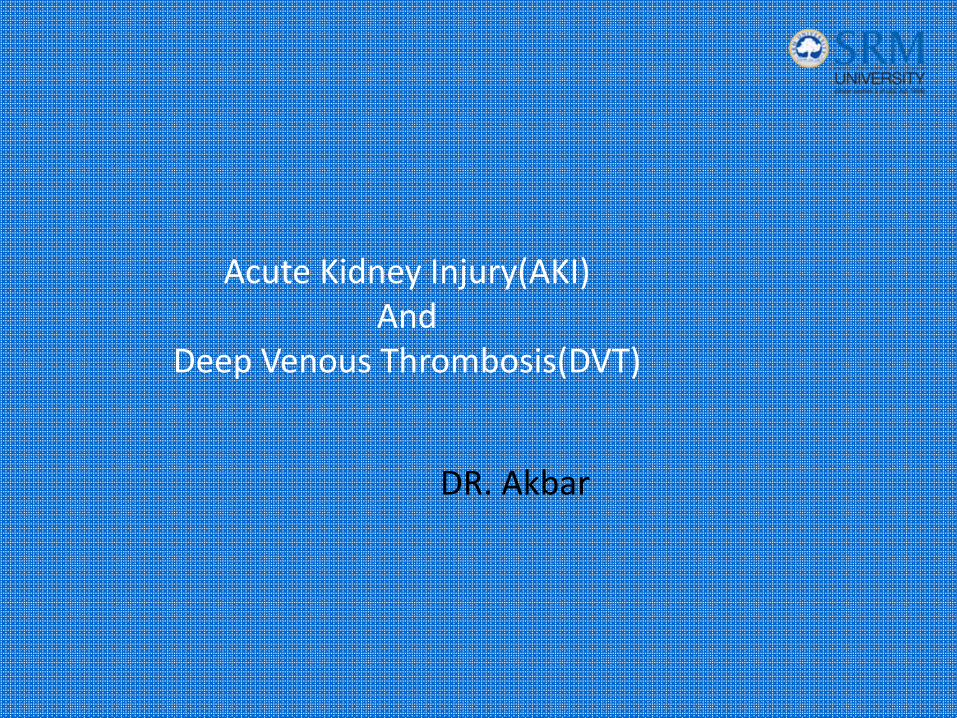
Definition of Acute Kidney Injury (AKI) based on “Acute Kidney Injury
Network”
Stage Increase in Serum Creatinine
Urine Output
1 1.5‐2 times baselineOR0.3 mg/dl increase from baseline
<0.5 ml/kg/h for >6 h
2 2‐3 times baseline <0.5 ml/kg/h for >12 h
3 3 times baseline OR0.5 mg/dl increase if baseline>4mg/dlORAny RRT given
<0.3 ml/kg/h for >24 hORAnuria for >12 h
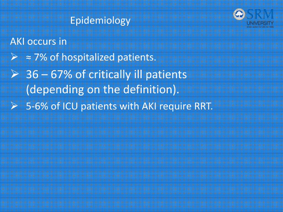
Epidemiology
AKI occurs in
≈ 7% of hospitalized patients.
36 – 67% of critically ill patients (depending on the definition).5‐6% of ICU patients with AKI require RRT.
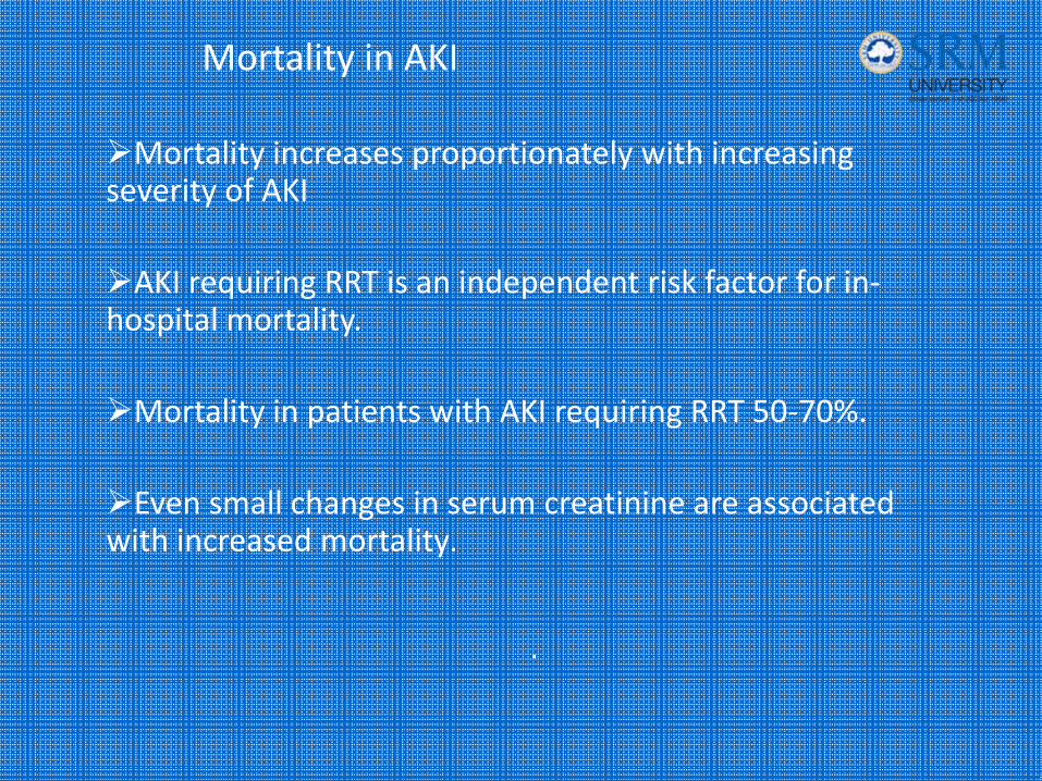
Mortality in AKI
Mortality increases proportionately with increasing severity of AKI
AKI requiring RRT is an independent risk factor for in‐hospital mortality.
Mortality in patients with AKI requiring RRT 50‐70%.
Even small changes in serum creatinine are associated with increased mortality.
.
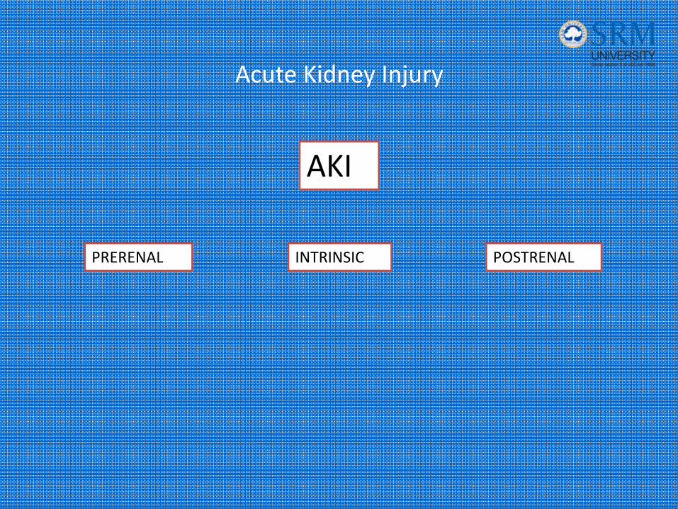
Acute Kidney Injury
PRERENAL
AKI
INTRINSIC POSTRENAL
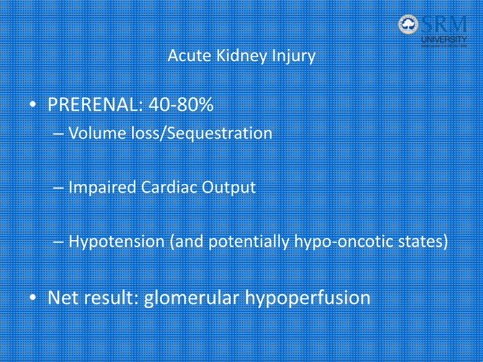
Acute Kidney Injury
• PRERENAL: 40‐80%– Volume loss/Sequestration
– Impaired Cardiac Output
– Hypotension (and potentially hypo‐oncotic states)
• Net result: glomerular hypoperfusion
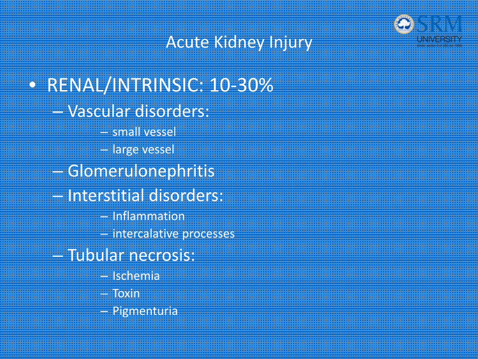
Acute Kidney Injury
• RENAL/INTRINSIC: 10‐30%– Vascular disorders:
– small vessel– large vessel
– Glomerulonephritis– Interstitial disorders:
– Inflammation– intercalative processes
– Tubular necrosis:– Ischemia– Toxin– Pigmenturia
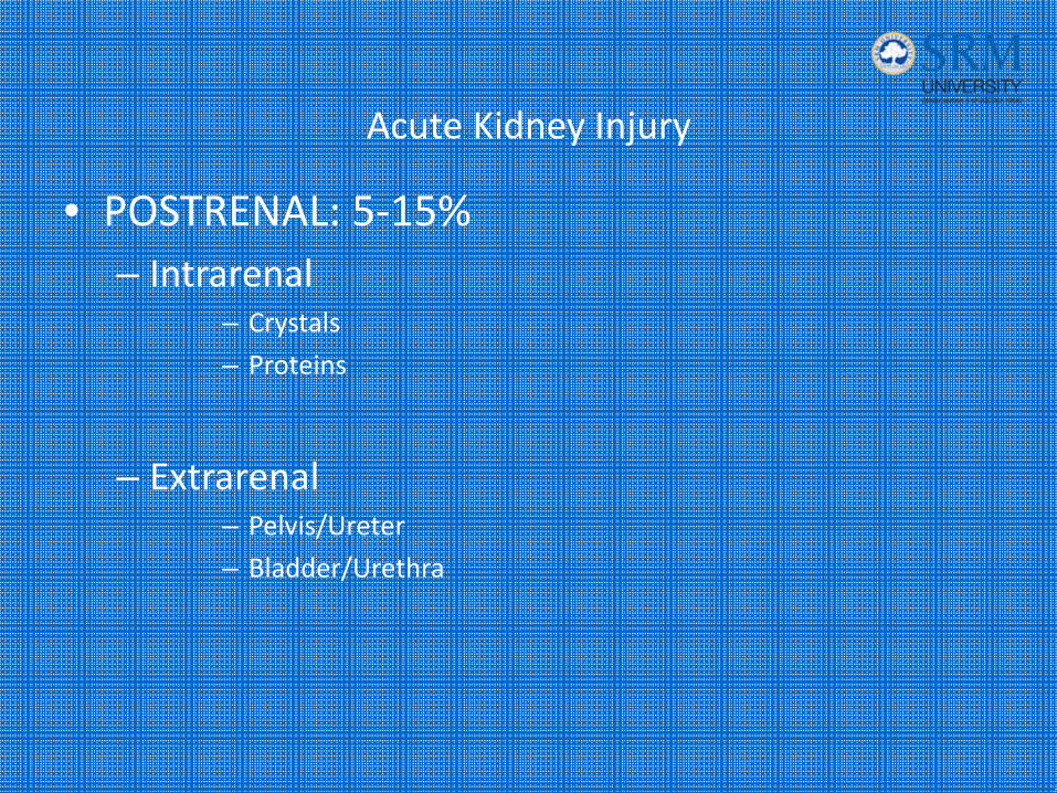
Acute Kidney Injury
• POSTRENAL: 5‐15%– Intrarenal
– Crystals
– Proteins
– Extrarenal– Pelvis/Ureter
– Bladder/Urethra
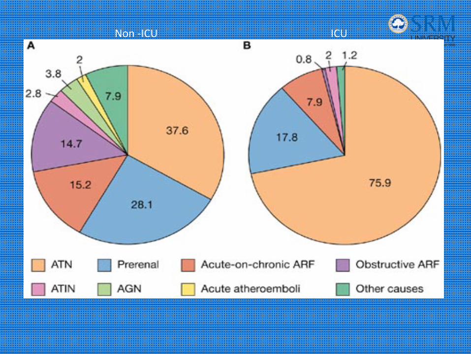
Non ‐ICU ICU
Acute Kidney Injury• Prerenal and ATN encountered most often in the hospital setting: 70‐75% in many studies
• Most common diagnostic consideration is therefore between these two conditions
• Prerenal:1. Intravascular volume depletion2. Hypotension3. Edematous states4. Localized renal ischemia
• ATN:1. All causes for prerenal, leading to post‐ischemic ATN2. Toxins
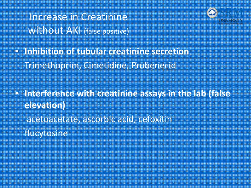
Increase in Creatininewithout AKI (false positive)
• Inhibition of tubular creatinine secretion
Trimethoprim, Cimetidine, Probenecid
• Interference with creatinine assays in the lab (false elevation)
acetoacetate, ascorbic acid, cefoxitin
flucytosine
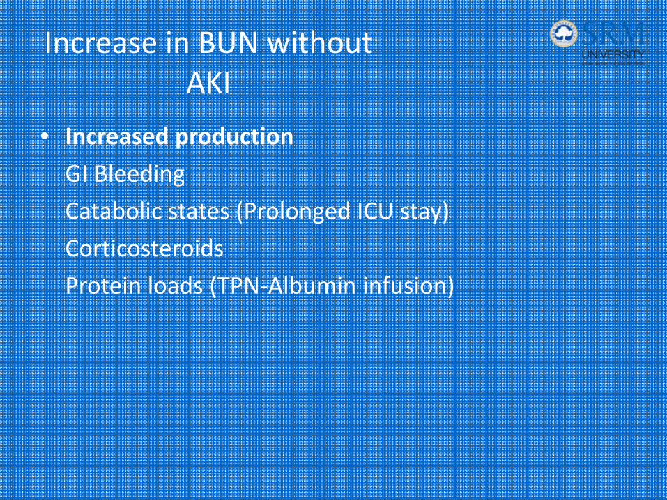
Increase in BUN without AKI
• Increased production
GI Bleeding
Catabolic states (Prolonged ICU stay)
Corticosteroids
Protein loads (TPN‐Albumin infusion)
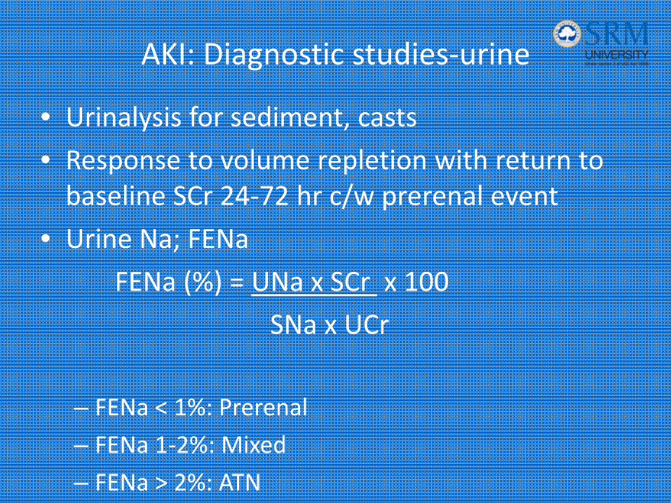
AKI: Diagnostic studies‐urine
• Urinalysis for sediment, casts
• Response to volume repletion with return to baseline SCr 24‐72 hr c/w prerenal event
• Urine Na; FENa
FENa (%) = UNa x SCr x 100
SNa x UCr
– FENa < 1%: Prerenal
– FENa 1‐2%: Mixed
– FENa > 2%: ATN
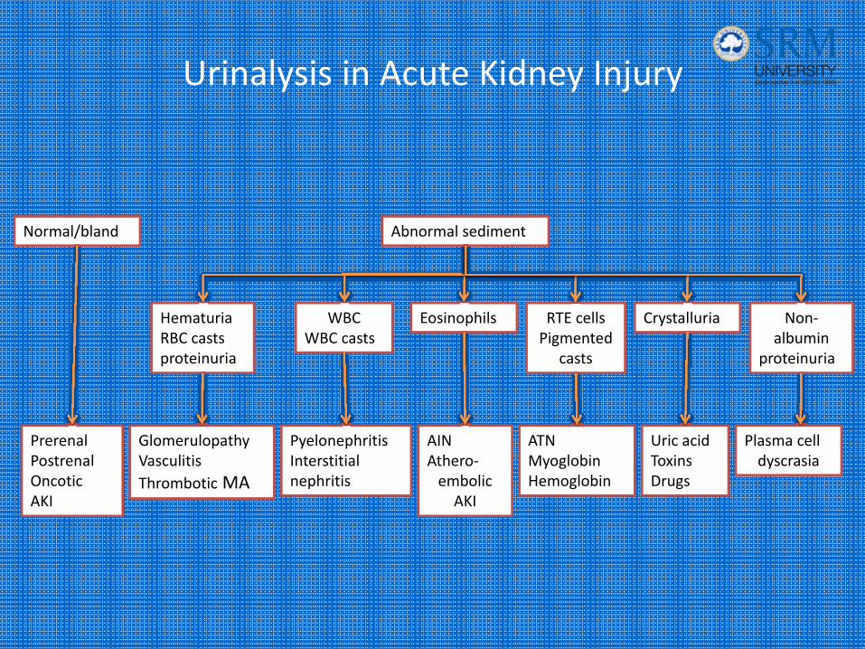
Urinalysis in Acute Kidney Injury
PrerenalPostrenalOncoticAKI
GlomerulopathyVasculitisThrombotic MA
PyelonephritisInterstitial nephritis
AINAthero‐embolic AKI
ATNMyoglobinHemoglobin
Uric acidToxinsDrugs
Plasma cell dyscrasia
HematuriaRBC castsproteinuria
WBCWBC casts
Eosinophils RTE cellsPigmented
casts
Crystalluria Non‐albumin
proteinuria
Abnormal sedimentNormal/bland
Acute Kidney Injury
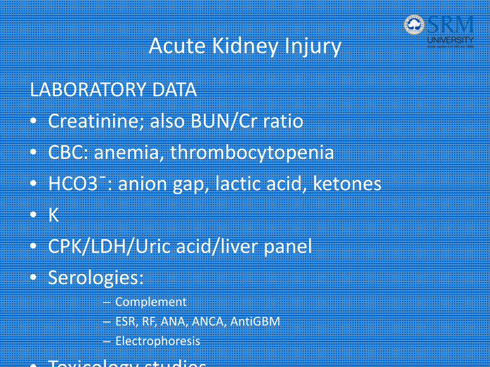
LABORATORY DATA
• Creatinine; also BUN/Cr ratio
• CBC: anemia, thrombocytopenia
• HCO3ˉ: anion gap, lactic acid, ketones
• K
• CPK/LDH/Uric acid/liver panel
• Serologies:– Complement
– ESR, RF, ANA, ANCA, AntiGBM
– Electrophoresis
• Toxicology studies
Acute Kidney Injury
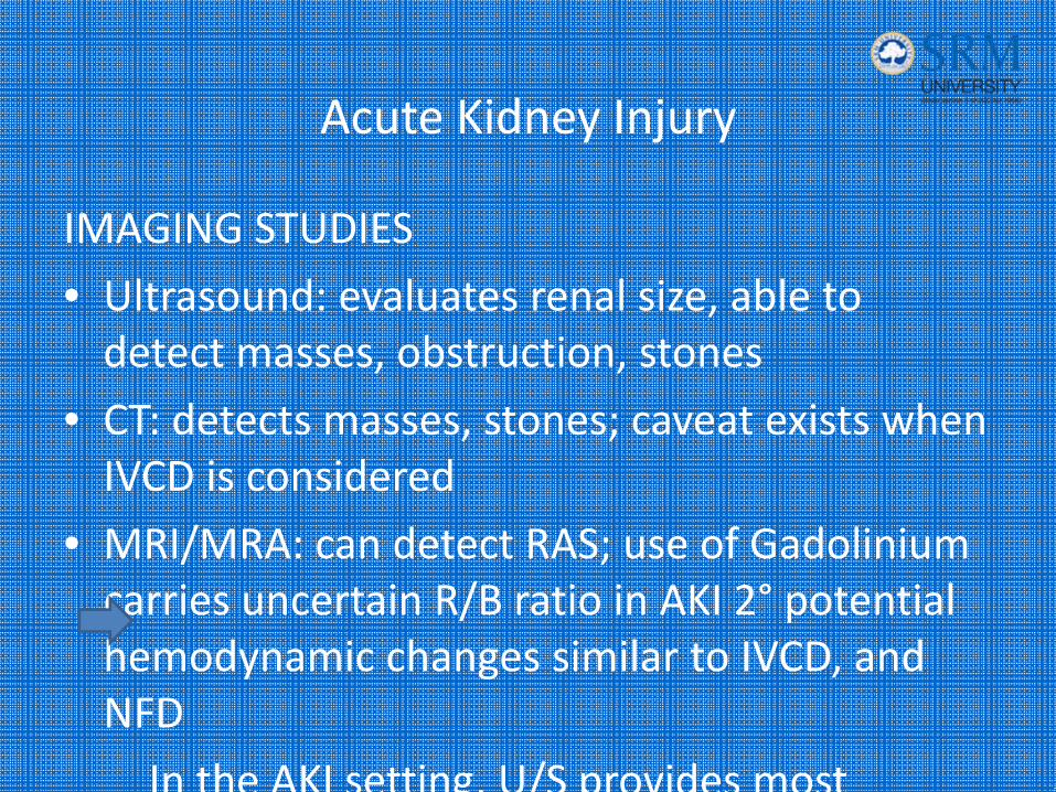
IMAGING STUDIES
• Ultrasound: evaluates renal size, able to detect masses, obstruction, stones
• CT: detects masses, stones; caveat exists when IVCD is considered
• MRI/MRA: can detect RAS; use of Gadolinium carries uncertain R/B ratio in AKI 2° potential hemodynamic changes similar to IVCD, and NFD
In the AKI setting, U/S provides most
Prevention of AKI in ICU
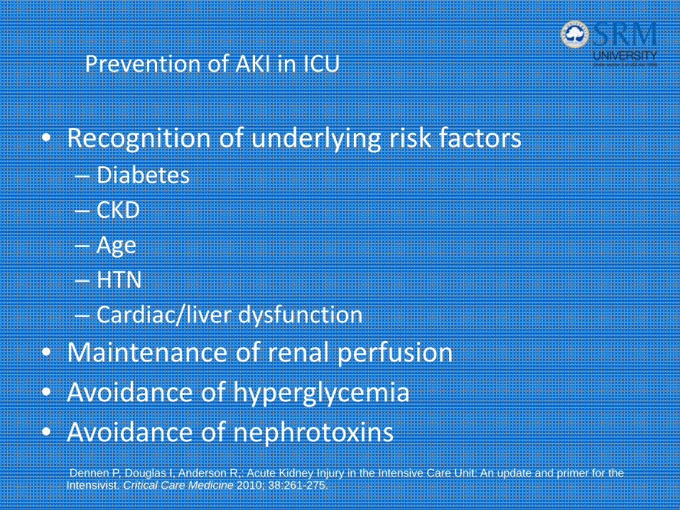
• Recognition of underlying risk factors– Diabetes– CKD– Age – HTN– Cardiac/liver dysfunction
• Maintenance of renal perfusion• Avoidance of hyperglycemia• Avoidance of nephrotoxins
Dennen P, Douglas I, Anderson R,: Acute Kidney Injury in the Intensive Care Unit: An update and primer for the Intensivist. Critical Care Medicine 2010; 38:261-275.
Management of AKI in ICU
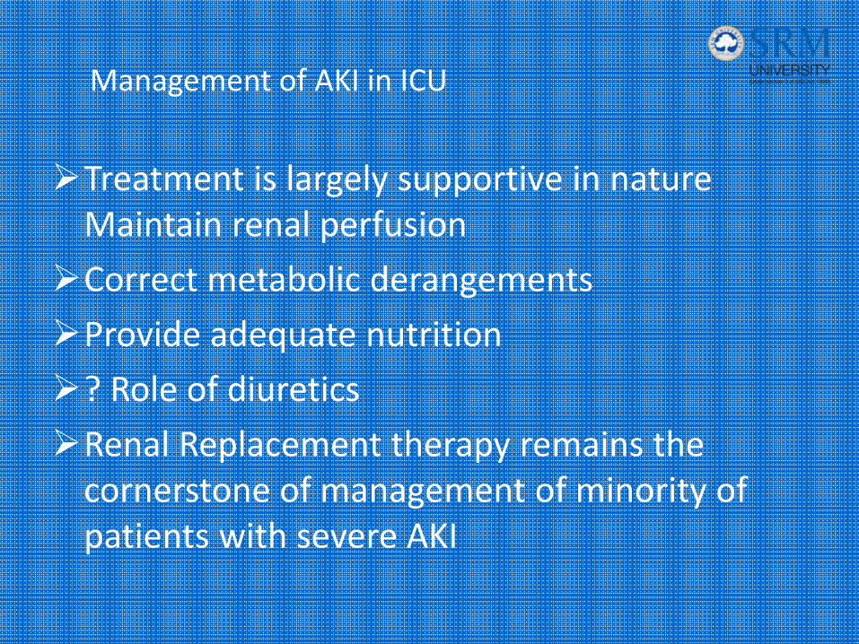
Treatment is largely supportive in nature Maintain renal perfusion
Correct metabolic derangements
Provide adequate nutrition
? Role of diuretics
Renal Replacement therapy remains the cornerstone of management of minority of patients with severe AKI
Maintaining renal perfusion
• Human kidney has a compromised ability to autoregulate in AKI.
• Maintaining haemodynamic stability and avoiding volume depletion are a priority in AKI.
• The individual BP target depends on age, co‐morbidities (HTN) and the current acute illness.
• A generally accepted target remains MAP ≥
Volume resuscitation – which fluid?
• no statistical difference between volume resuscitation with saline or albumin in survival rates or need for RRT.
Renal vasodilators?
renal dose dopamine (<5 μg/kg of body weight/min)increases RBF and, to a lesser extent, GFR. Dopamine isunable to prevent or alter the course of ischaemic ornephrotoxic AKI]. Furthermore, dopamine, even at lowdoses, can induce tachy‐arrhythmia’s, myocardialischaemia, and extravasation out of the vein can causesevere necrosis .Thus, the routine administration ofdopamine to patients for the prevention of AKI orincipient AKI is no longer justified.
Is there a role for Fenoldopam in prevention or treatment of AKI in ICU setting?
Dopamine‐1 receptor agonist, lack of Dopamine‐2, and alpha‐1 receptor effect, make it a potentially safer drug than Dopamine!
Reduces in hospital mortality and the need for RRT in AKI
Reverses renal hypoperfusion more effectively than renal dose Dopamine
So far so good specially in cardiothoracic ICU patients, awaiting more powered trials in other groups!
Is there a role for diuretics in the treatment of AKI in ICU setting?
• Loop diuretics may convert an oliguric into a non‐oliguric form of AKI that may allow easier fluid and/or nutritional support of the patient. Volume overload in AKI patients is common and diuretics may provide symptomatic benefit in that situation. However, loop diuretics are neither associated with improved survival, nor with better recovery of renal function in AKI.
EPO
• Erythropoietin (EPO) has tissue‐protective effects and prevents tissue damage during ischaemia and inflammation, and currently trials are performed with EPO in the prevention of AKI post‐cardiac surgery, CIN and post‐kidney transplantation.
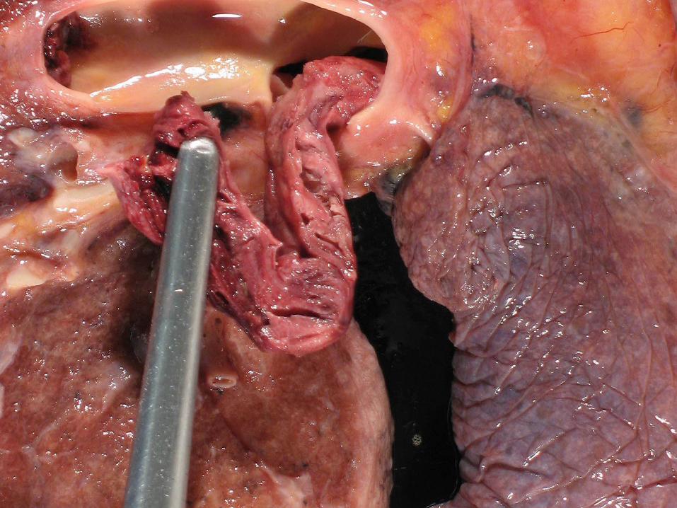
Deep vein thrombosis
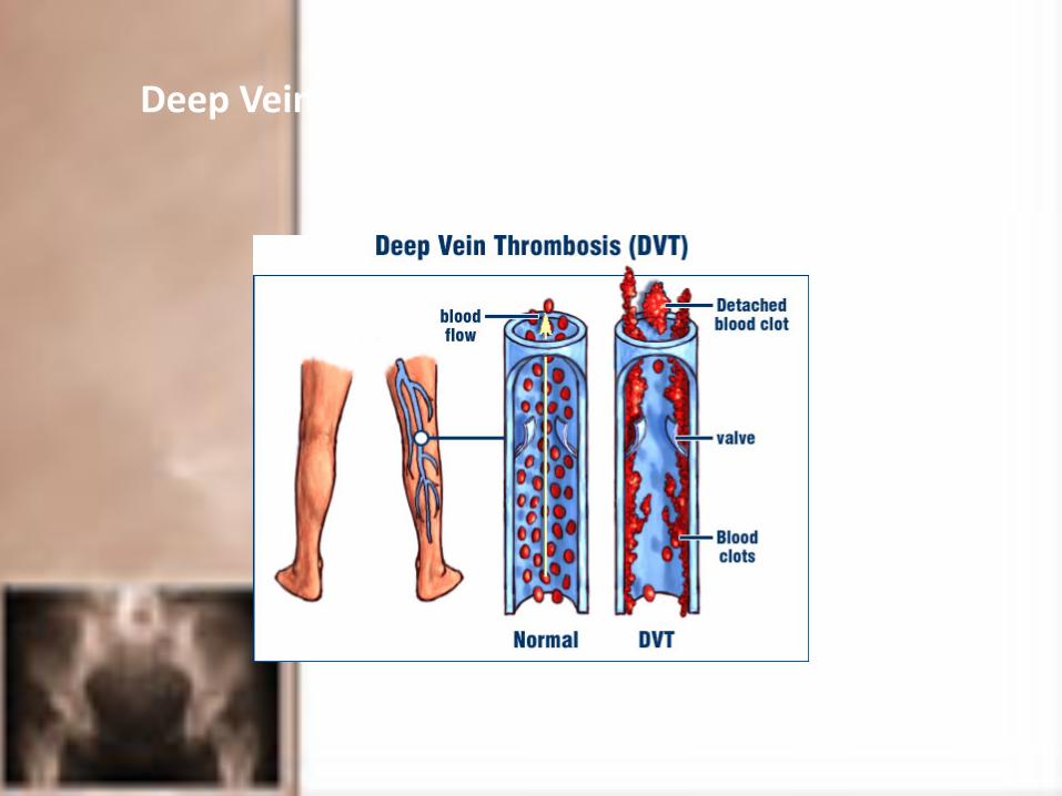
Deep Vein Thrombosis

Deep Vein Thrombosis
• 80 % proximal thrombosis
• 20% calf vein thrombosis
• 50% progress to PE
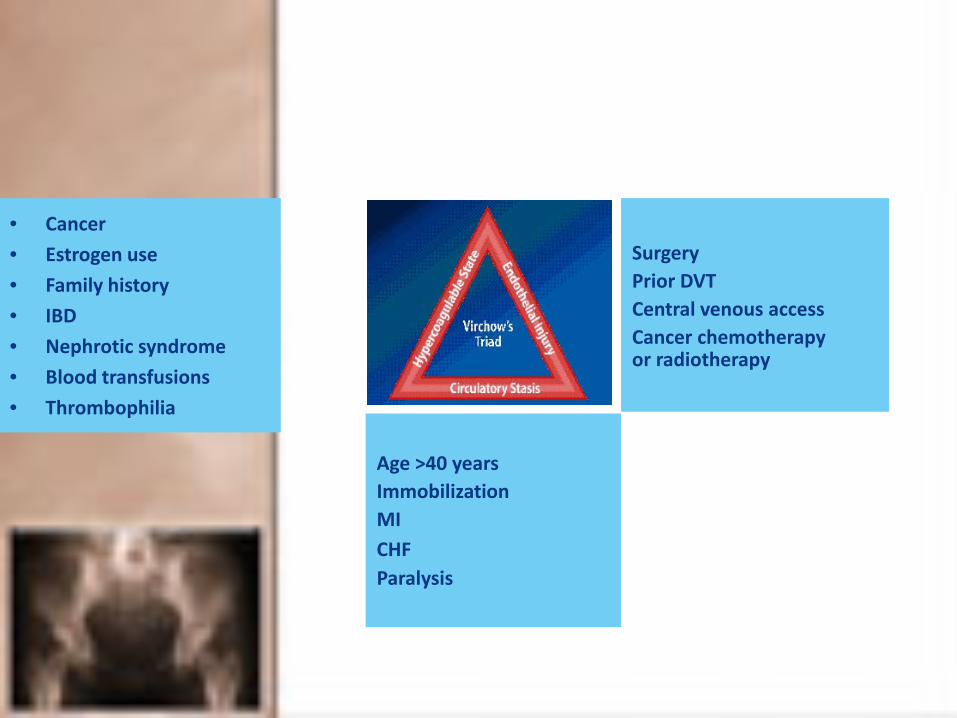
• Cancer
• Estrogen use
• Family history
• IBD
• Nephrotic syndrome
• Blood transfusions
• Thrombophilia
SurgeryPrior DVTCentral venous accessCancer chemotherapyor radiotherapy
Age >40 yearsImmobilizationMICHFParalysis
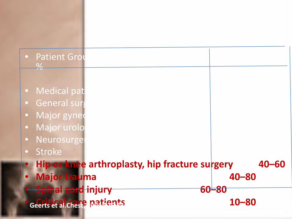
• Patient Group DVT Prevalence%
• Medical patients 10–20• General surgery 15–40• Major gynecologic surgery 15–40• Major urologic surgery 15–40• Neurosurgery 15–40• Stroke 20–50• Hip or knee arthroplasty, hip fracture surgery 40–60• Major trauma 40–80• Spinal cord injury 60–80• Critical care patients 10–80Geerts et al.Chest, 2004;126:338S

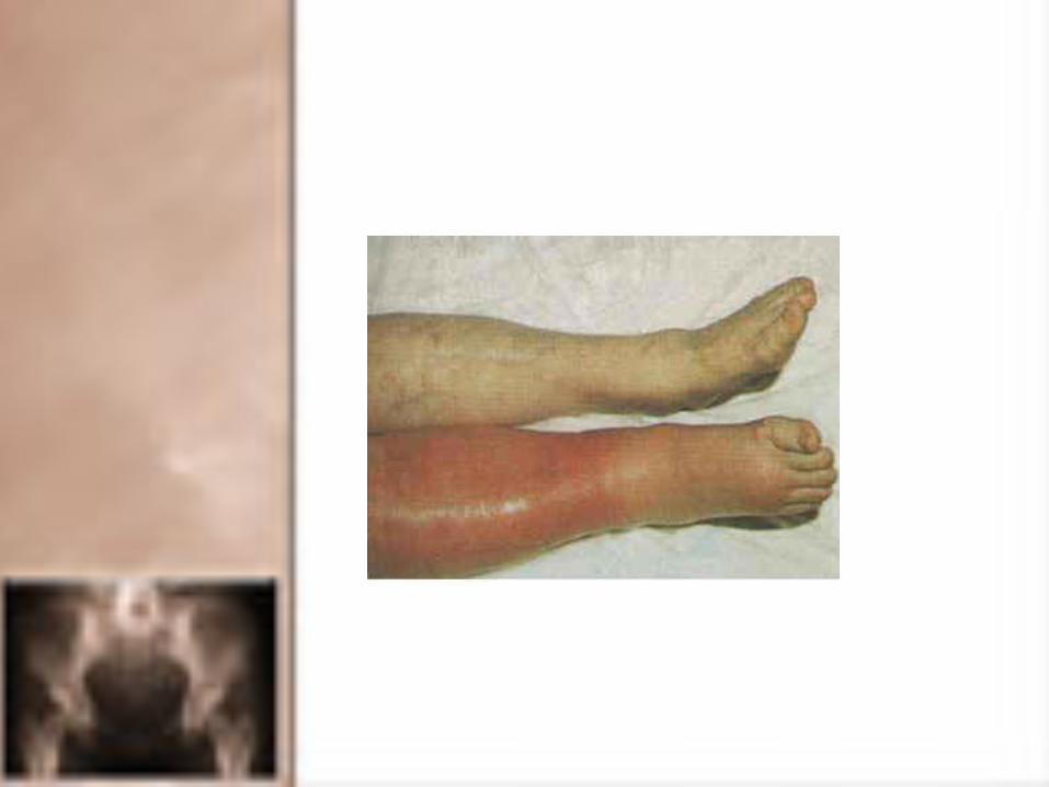
Signs and symptoms
• Discoloration of the legs
• Calf or leg pain or tenderness
• Swelling of the leg or lower limb
• Warm skin
• Surface veins become more visible
• Leg fatigue

Importance of DVT diagnosis
• If left untreated, can suffer fatal PE
• Treatment of proximal DVT reduces the risk of fatal PE to <1% and the risk of developing recurrent DVTs to 5%
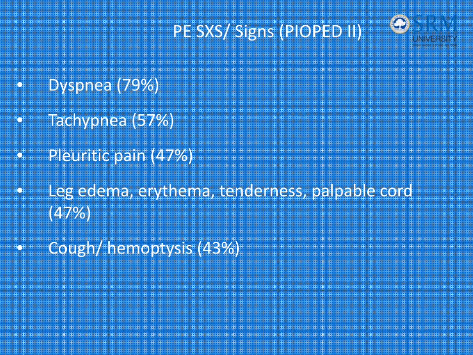
PE SXS/ Signs (PIOPED II)
• Dyspnea (79%)
• Tachypnea (57%)
• Pleuritic pain (47%)
• Leg edema, erythema, tenderness, palpable cord (47%)
• Cough/ hemoptysis (43%)
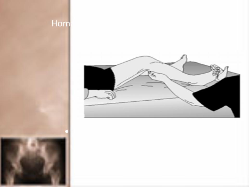
Homan’s Sign
• Forcible ankle dorsiflexion with the knee flexed causing deep calf pain

Wells criteria
• Malignancy (Rxn < 6 mos or palliative)+1
• Lower limb paralysis/ recent plaster cast +1
• Bedridden for > 3 days/ surgery (< 4 weeks) +1
• Localized tenderness along deep veins +1
• Entire leg swollen +1
• Calf swelling > 3 cm asymptotic side

Wells criteria
• > 3: High ‐75%
• 1‐2 :Moderate ‐17%
• < 0 :Low ‐3%
Screening test
D‐dimer test:• Degradation product of a cross‐linked fibrin blood clot
Increased in:• Arterial and venous thrombosis• Major surgery• Hemorrhage• Trauma• Pregnancy• cancer
Definitive investigation
• Venography : Gold standard
• Ultrasonography:• Compression ultrasound• Duplex ultrasonography• Colour coded Doppler ultrasonography
• Computed tomography
• Magnetic resonance imaging
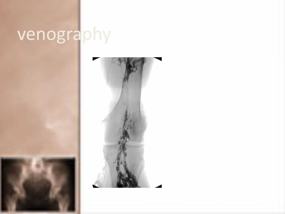
venography

Management
• Medical management
• Surgical management

Medical management
• Compressional stockings
• Heparin
• Warfarin
• Vitamin k antagonist

Mechanisms of Elastic Compression Stockings
• ↓ Venous dilatation
• ↑ Flow velocity
• Improve valve function
• ↑ Tissue Factor Pathway Inhibitor
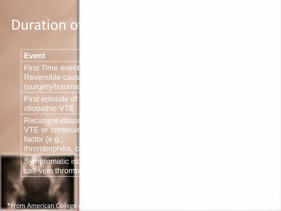
Duration of Anticoagulation for DVT
Event DurationFirst Time event of Reversible cause (surgery/trauma)
At least 3 mos
First episode of idiopathic VTE
At least 6 mos
Recurrent idiopathic VTE or continuing risk factor (e.g., thrombophilia, cancer)
At least 12 mos
Symptomatic isolated calf-vein thrombosis
6 to 12 weeks
*From American College of Chest Physicians

Prophylaxsis
Risk group Recommended prophylaxis
• Hip replacement Warfarin, LMWH, fondaparinux
• Knee replacement Warfarin, LMWH, IPC, fondaparinux (Arixtra)
• Hip Fracture Warfarin, LMWH, fondaparinux
• Major trauma LMWH, IPC• Abdominal surgery UFH, LMWH, IPC, warfarin, ES

Effective prophylaxis
• Subcutaneous heparin‐5000IU Q8H for 5 days
• Aspirin 150mg started preoperatively and continued till 45 days
• Graduated elastic compression stockings
• Intermittent pneumatic compression
• Mechanical foot pumps

Surgical management
• IVC filter
• Embolectomy
• Thrombolysis
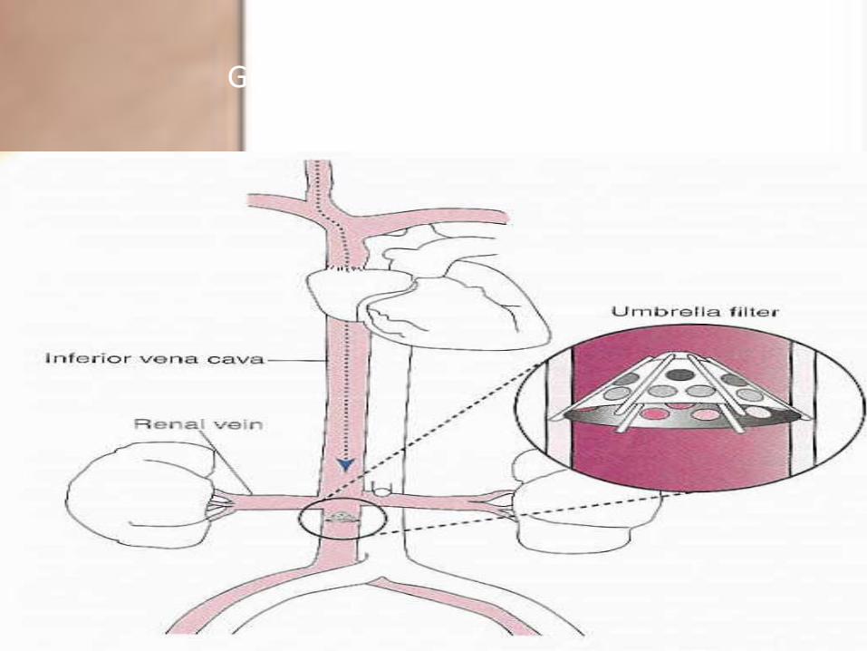
Greenfield stainless steel filter

Indication
• Pulmonary embolism with contraindication to anticoagulation
• Recurrent pulmonary embolism despite adequate anticoagulation.

Embolectomy
• Indication:
– In those where thrombolysis is contraindicated or fails

Thrombolysis
• Considered once P.E. diagnosed• If chosen, hold anticoagulation during thrombolysis infusion, then resumed
• Associated with higher incidence of major hemorrhage
Thank you
Related Documents