Acute Exacerbations of Idiopathic Pulmonary Fibrosis Harold R. Collard 1 , Bethany B. Moore 2 , Kevin R. Flaherty 2 , Kevin K. Brown 3 , Robert J. Kaner 4 , Talmadge E. King Jr. 1 , Joseph A. Lasky 5 , James E. Loyd 6 , Imre Noth 7 , Mitchell A. Olman 8 , Ganesh Raghu 9 , Jesse Roman 10 , Jay H. Ryu 11 , David A. Zisman 12 , Gary W. Hunninghake 13 , Thomas V. Colby 14 , Jim J. Egan 15 , David M. Hansell 16 , Takeshi Johkoh 17 , Naftali Kaminski 18 , Dong Soon Kim 19 , Yasuhiro Kondoh 20 , David A. Lynch 21 , Joachim Müller-Quernheim 22 , Jeffrey L. Myers 23 , Andrew G. Nicholson 24 , Moisés Selman 25 , Galen B. Toews 2 , Athol U. Wells 26 , Fernando J. Martinez 2 , and Idiopathic Pulmonary Fibrosis Clinical Research Network Investigators Correspondence and requests for reprints should be addressed to Fernando J. Martinez, M.D., University of Michigan Health System, 1500 East Medical Center Drive, 3916 Taubman Center, Ann Arbor, MI 48109-0360. E-mail: [email protected]. Conflict of Interest Statement: H.R.C. received $5,000 from InterMune for advisory board activities in 2006. B.B.M. does not have a financial relationship with a commercial entity that interest in the subject of this manuscript. K.R.F. received $4,100 in 2005 and $3,000 in 2007 from Boehringer Ingelheim; $2,500 in 2006 from InterMune; and $1,200 in 2005 from Fibrogen for serving on advisory boards related to pulmonary fibrosis. K.K.B. has served as a consultant or spoken to the following companies interested in IPF: Actelion, Amgen, Genzyme, Wyeth, Biogen, Boehringer Ingelheim, Novartis, and Lung Rx. He or his institution have received grants to support the performance of treatment trials in IPF from Actelion, InterMune, Biogen, and Genzyme. R.J.K. does not have a financial relationship with a commercial entity that interest in the subject of this manuscript. T.E.K. has served on advisory boards for InterMune and for GlaxoSmithKline (GSK); and served as a consultant for Nektar, Alexza, Astra-Zeneca, Biogen, Centocor, Fibrogen, Genzyme, Human Genome Sciences, Merck, and CoTherix. J.A.L. has been supported by Novartis for an investigator-initiated study, for service on an advisory board by InterMune, and by Actelion for service of advisory and data safety monitoring boards. J.E.L. does not have a financial relationship with a commercial entity that interest in the subject of this manuscript. I.N. has participated as a speaker for various pharmaceutical companies (GSK, Pfizer, Boehringer Ingelheim, in 2006 for various amounts). He has also received honoraria for participating in advisory board meetings for Novartis in 2005 and GSK in 2006. I.N.'s institution has received grants and contracts from Actelion, CoTherix, Centocor, Genzyme, InterMune, Novartis, and Roche, all ongoing in 2007. M.A.O. does not have a financial relationship with a commercial entity that interest in the subject of this manuscript. G.R. served as a steering committee member for BUILD-1 and serves as a steering committee member for BUILD-3 studies for IPF sponsored by Actelion. J.R. in 2006 received $10,000 for research conducted as part of a multicenter trial sponsored by InterMune. J.H.R. does not have a financial relationship with a commercial entity that interest in the subject of this manuscript. D.A.Z. received speaker honoraria and served on advisory board for InterMune ($7,000 in 2006, $6,000 in 2005; $6,000 in 2004). D.A.Z. has received research grants from InterMune, Actelion, and CoTherix for participating in multicenter clinical trials. G.W.H. does not have a financial relationship with a commercial entity that interest in the subject of this manuscript. T.V.C. does not have a financial relationship with a commercial entity that interest in the subject of this manuscript. J.J.E. does not have a financial relationship with a commercial entity that interest in the subject of this manuscript. D.M.H. received $6,000 for giving advice and grading HRCTs for clinical trial run by InterMune in 2005/2006. T.J. does not have a financial relationship with a commercial entity that interest in the subject of this manuscript. N.K. received $5,000 for serving on a Biogen Idec advisory board in November 2005. N.K. is also a recipient of an investigator-initiated grant from Biogen Idec in August 2006 ($674,800 for 2 yr). D.S.K. does not have a financial relationship with a commercial entity that interest in the subject of this manuscript. Y.K. does not have a financial relationship with a commercial entity that interest in the subject of this manuscript. D.A.L. has received less than $5,000 in 2004, 2005, and 2006 from InterMune, for interpretation of CT scans. He has received more than $5,000 from Encysive for consultation on clinical trials. He received $6,000 in 2006 for service on an advisory board for Actelion. J.M.-Q. received a research grant of $180,000 from Boehringer Ingelheim, Germany, in 2003 through 2006 for an in vitro study on fibrogenic mechanisms. J.M.-Q received a research grant of $80,000 from Altana, Germany, in 2003 through 2006 for an in vitro study on alveolar epithelia. J.L.M. does not have a financial relationship with a commercial entity that interest in the subject of this manuscript. A.G.N. received $4,000 for reviewing slides for InterMune Ltd in 2004–2006 and £8,600 for reviewing slides for Actelion Ltd in 2006 in relation to entry into drug trials for patients with IPF. M.S. does not have a financial relationship with a commercial entity that interest in the subject of this manuscript. G.B.T. does not have a financial relationship with a commercial entity that interest in the subject of this manuscript. A.U.W. does not have a financial relationship with a commercial entity that interest in the subject of this manuscript. F.J.M. is a consultant for Altana Pharma and has received compensation greater than $10K. F.J.M has been a member of several advisory boards, CME committees, and the speakers' bureau for Boehringer Ingelheim, Pfizer, and GSK. His total compensation per company was greater than $10K. In addition, F.J.M. is on an advisory board for Novartis and a speaker's bureau for Sepracor and Astra, receiving less than $10K per company. F.J.M. has been an investigator for industry-sponsored studies for GSK, Boehringer Ingelheim, and Actelion. The Idiopathic Pulmonary Fibrosis Clinical Research Network (IPFnet) consists of a central data coordinating center (Duke Clinical Research Institute) and 11 clinical sites, as follows: Emory University, Gulf South Research Consortium (Tulane University and University of Alabama, Birmingham); Mayo Clinic, Rochester; National Jewish Medical and Research Center; University of California, Los Angeles; University of California, San Francisco; University of Chicago; University of Michigan Health System; University of Washington; Vanderbilt University; and Weill Cornell Medical College. NIH Public Access Author Manuscript Am J Respir Crit Care Med. Author manuscript; available in PMC 2007 November 26. Published in final edited form as: Am J Respir Crit Care Med. 2007 October 1; 176(7): 636–643. NIH-PA Author Manuscript NIH-PA Author Manuscript NIH-PA Author Manuscript

Welcome message from author
This document is posted to help you gain knowledge. Please leave a comment to let me know what you think about it! Share it to your friends and learn new things together.
Transcript
Acute Exacerbations of Idiopathic Pulmonary Fibrosis
Harold R. Collard1, Bethany B. Moore2, Kevin R. Flaherty2, Kevin K. Brown3, Robert J.Kaner4, Talmadge E. King Jr.1, Joseph A. Lasky5, James E. Loyd6, Imre Noth7, Mitchell A.Olman8, Ganesh Raghu9, Jesse Roman10, Jay H. Ryu11, David A. Zisman12, Gary W.Hunninghake13, Thomas V. Colby14, Jim J. Egan15, David M. Hansell16, TakeshiJohkoh17, Naftali Kaminski18, Dong Soon Kim19, Yasuhiro Kondoh20, David A. Lynch21,Joachim Müller-Quernheim22, Jeffrey L. Myers23, Andrew G. Nicholson24, MoisésSelman25, Galen B. Toews2, Athol U. Wells26, Fernando J. Martinez2, and IdiopathicPulmonary Fibrosis Clinical Research Network Investigators
Correspondence and requests for reprints should be addressed to Fernando J. Martinez, M.D., University of Michigan Health System,1500 East Medical Center Drive, 3916 Taubman Center, Ann Arbor, MI 48109-0360. E-mail: [email protected] of Interest Statement: H.R.C. received $5,000 from InterMune for advisory board activities in 2006. B.B.M. does not have afinancial relationship with a commercial entity that interest in the subject of this manuscript. K.R.F. received $4,100 in 2005 and $3,000in 2007 from Boehringer Ingelheim; $2,500 in 2006 from InterMune; and $1,200 in 2005 from Fibrogen for serving on advisory boardsrelated to pulmonary fibrosis. K.K.B. has served as a consultant or spoken to the following companies interested in IPF: Actelion, Amgen,Genzyme, Wyeth, Biogen, Boehringer Ingelheim, Novartis, and Lung Rx. He or his institution have received grants to support theperformance of treatment trials in IPF from Actelion, InterMune, Biogen, and Genzyme. R.J.K. does not have a financial relationshipwith a commercial entity that interest in the subject of this manuscript. T.E.K. has served on advisory boards for InterMune and forGlaxoSmithKline (GSK); and served as a consultant for Nektar, Alexza, Astra-Zeneca, Biogen, Centocor, Fibrogen, Genzyme, HumanGenome Sciences, Merck, and CoTherix. J.A.L. has been supported by Novartis for an investigator-initiated study, for service on anadvisory board by InterMune, and by Actelion for service of advisory and data safety monitoring boards. J.E.L. does not have a financialrelationship with a commercial entity that interest in the subject of this manuscript. I.N. has participated as a speaker for variouspharmaceutical companies (GSK, Pfizer, Boehringer Ingelheim, in 2006 for various amounts). He has also received honoraria forparticipating in advisory board meetings for Novartis in 2005 and GSK in 2006. I.N.'s institution has received grants and contracts fromActelion, CoTherix, Centocor, Genzyme, InterMune, Novartis, and Roche, all ongoing in 2007. M.A.O. does not have a financialrelationship with a commercial entity that interest in the subject of this manuscript. G.R. served as a steering committee member forBUILD-1 and serves as a steering committee member for BUILD-3 studies for IPF sponsored by Actelion. J.R. in 2006 received $10,000for research conducted as part of a multicenter trial sponsored by InterMune. J.H.R. does not have a financial relationship with acommercial entity that interest in the subject of this manuscript. D.A.Z. received speaker honoraria and served on advisory board forInterMune ($7,000 in 2006, $6,000 in 2005; $6,000 in 2004). D.A.Z. has received research grants from InterMune, Actelion, and CoTherixfor participating in multicenter clinical trials. G.W.H. does not have a financial relationship with a commercial entity that interest in thesubject of this manuscript. T.V.C. does not have a financial relationship with a commercial entity that interest in the subject of thismanuscript. J.J.E. does not have a financial relationship with a commercial entity that interest in the subject of this manuscript. D.M.H.received $6,000 for giving advice and grading HRCTs for clinical trial run by InterMune in 2005/2006. T.J. does not have a financialrelationship with a commercial entity that interest in the subject of this manuscript. N.K. received $5,000 for serving on a Biogen Idecadvisory board in November 2005. N.K. is also a recipient of an investigator-initiated grant from Biogen Idec in August 2006 ($674,800for 2 yr). D.S.K. does not have a financial relationship with a commercial entity that interest in the subject of this manuscript. Y.K. doesnot have a financial relationship with a commercial entity that interest in the subject of this manuscript. D.A.L. has received less than$5,000 in 2004, 2005, and 2006 from InterMune, for interpretation of CT scans. He has received more than $5,000 from Encysive forconsultation on clinical trials. He received $6,000 in 2006 for service on an advisory board for Actelion. J.M.-Q. received a researchgrant of $180,000 from Boehringer Ingelheim, Germany, in 2003 through 2006 for an in vitro study on fibrogenic mechanisms. J.M.-Qreceived a research grant of $80,000 from Altana, Germany, in 2003 through 2006 for an in vitro study on alveolar epithelia. J.L.M. doesnot have a financial relationship with a commercial entity that interest in the subject of this manuscript. A.G.N. received $4,000 forreviewing slides for InterMune Ltd in 2004–2006 and £8,600 for reviewing slides for Actelion Ltd in 2006 in relation to entry into drugtrials for patients with IPF. M.S. does not have a financial relationship with a commercial entity that interest in the subject of thismanuscript. G.B.T. does not have a financial relationship with a commercial entity that interest in the subject of this manuscript. A.U.W.does not have a financial relationship with a commercial entity that interest in the subject of this manuscript. F.J.M. is a consultant forAltana Pharma and has received compensation greater than $10K. F.J.M has been a member of several advisory boards, CME committees,and the speakers' bureau for Boehringer Ingelheim, Pfizer, and GSK. His total compensation per company was greater than $10K. Inaddition, F.J.M. is on an advisory board for Novartis and a speaker's bureau for Sepracor and Astra, receiving less than $10K per company.F.J.M. has been an investigator for industry-sponsored studies for GSK, Boehringer Ingelheim, and Actelion.The Idiopathic Pulmonary Fibrosis Clinical Research Network (IPFnet) consists of a central data coordinating center (Duke ClinicalResearch Institute) and 11 clinical sites, as follows: Emory University, Gulf South Research Consortium (Tulane University andUniversity of Alabama, Birmingham); Mayo Clinic, Rochester; National Jewish Medical and Research Center; University of California,Los Angeles; University of California, San Francisco; University of Chicago; University of Michigan Health System; University ofWashington; Vanderbilt University; and Weill Cornell Medical College.
NIH Public AccessAuthor ManuscriptAm J Respir Crit Care Med. Author manuscript; available in PMC 2007 November 26.
Published in final edited form as:Am J Respir Crit Care Med. 2007 October 1; 176(7): 636–643.
NIH
-PA Author Manuscript
NIH
-PA Author Manuscript
NIH
-PA Author Manuscript

1Department of Medicine, San Francisco General Hospital, University of California, San Francisco, SanFrancisco, California 2Department of Medicine, University of Michigan, Ann Arbor, Michigan 3Departmentof Medicine, National Jewish Medical and Research Center, Denver, Colorado 4Department of Medicine andGenetic Medicine, Weill Cornell Medical College, New York, New York 5Department of Medicine, TulaneUniversity, New Orleans, Louisiana 6Department of Medicine, Vanderbilt University, Nashville, Tennessee7Department of Medicine, University of Chicago, Chicago, Illinois 8Department of Medicine and Pathology,University of Alabama at Birmingham, Birmingham, Alabama 9Department of Medicine, University ofWashington, Seattle, Washington 10Department of Medicine, Emory University, Atlanta, Georgia11Department of Medicine, Mayo Clinic, Rochester, Minnesota 12Department of Medicine, University ofCalifornia Los Angeles, Los Angeles, California 13Department of Medicine, University of Iowa, Iowa City,Iowa 14Department of Laboratory Medicine and Pathology, Mayo Clinic Scottsdale, Scottsdale, Arizona15Mater Misericordiae University Hospital, University College, Dublin, Ireland 16Department of Radiology,Royal Brompton Hospital, Imperial College London, London, United Kingdom 17Department of Radiology,Osaka University, Osaka, Japan 18Department of Medicine, University of Pittsburgh, Pittsburgh,Pennsylvania 19Division of Pulmonary and Critical Care Medicine, Asan Medical Center, University ofUlsan, Seoul, South Korea 20Department of Respiratory Medicine and Allergy, Tosei General Hospital, Aichi,Japan 21Department of Radiology, National Jewish Medical and Research Center, Denver, Colorado22Department of Pneumology, University Medical Center Freiburg, Freiburg, Germany 23Department ofPathology, University of Michigan, Ann Arbor, Michigan 24Department of Histopathology, Royal BromptonHospital, Imperial College London, London, United Kingdom 25Instituto Nacional de EnfermedadesRespiratorias, Mexico City, Mexico 26Department of Respiratory Medicine, Royal Brompton Hospital,Imperial College London, London, United Kingdom
AbstractThe natural history of idiopathic pulmonary fibrosis (IPF) has been characterized as a steady,predictable decline in lung function over time. Recent evidence suggests that some patients mayexperience a more precipitous course, with periods of relative stability followed by acutedeteriorations in respiratory status. Many of these acute deteriorations are of unknown etiology andhave been termed acute exacerbations of IPF. This perspective is the result of an international effortto summarize the current state of knowledge regarding acute exacerbations of IPF. Acuteexacerbations of IPF are defined as acute, clinically significant deteriorations of unidentifiable causein patients with underlying IPF. Proposed diagnostic criteria include subjective worsening over 30days or less, new bilateral radiographic opacities, and the absence of infection or another identifiableetiology. The potential pathobiological roles of infection, disordered cell biology, coagulation, andgenetics are discussed, and future research directions are proposed.
Keywordsacute exacerbation; pulmonary fibrosis; diagnosis; definition
Idiopathic pulmonary fibrosis (IPF) is the most common form of idiopathic interstitialpneumonia (1,2). No therapy has been proven effective, and median survival from the time ofdiagnosis is approximately 3 years (3–5). Historically, IPF has been described as a graduallyprogressive disease, characterized by steady worsening of symptoms, lung function, and gasexchange (6,7). More recently, it has been recognized that some patients with IPF experienceacute respiratory deterioration, suggesting that the clinical course may be less predictable (8).Many of these acute declines are idiopathic—that is, without clinically apparent infection, leftheart failure, pulmonary embolism, or other identifiable cause. These episodes of idiopathicacute deterioration have been termed acute exacerbations of IPF (9,10).
Collard et al. Page 2
Am J Respir Crit Care Med. Author manuscript; available in PMC 2007 November 26.
NIH
-PA Author Manuscript
NIH
-PA Author Manuscript
NIH
-PA Author Manuscript

This perspective summarizes the current state of knowledge regarding acute exacerbations ofIPF, proposes a definition and diagnostic criteria, and suggests areas for future investigation.It is an international perspective, combining the efforts of the Idiopathic Pulmonary FibrosisClinical Research Network (IPFnet), an NHLBI-sponsored collaborative formed to advancethe clinical care of patients with IPF, with those of experienced individual investigators. It isthe authors' strong belief that a consensus approach to defining acute exacerbations of IPF isessential to furthering our knowledge of this condition.
Significance of Acute ExacerbationA number of studies have reported on the clinical significance of acute exacerbations of IPF(Table 1) (9–27). These studies are primarily retrospective case series and provide discordantestimates of incidence and mortality rates. This is likely due to differences in study design,particularly substantial variation in the case definition of acute exacerbation.
Three recent studies provide information regarding the incidence and mortality of acuteexacerbations of IPF. A randomized, controlled trial of anticoagulant therapy in IPF includedrehospitalization (which included acute exacerbation) as a secondary endpoint (16). Of 56patients followed for approximately 3 years, 32 (57%) were rehospitalized for acuteexacerbation and 53% of those patients died. Importantly, subjects in this study were initiallyenrolled during an admission to the hospital, suggesting the population recruited was enrichedfor previous acute exacerbation or progressive disease. A retrospective review of anobservational cohort of 147 patients with IPF identified 11 who met criteria for acuteexacerbation (14). The 2-year incidence of acute exacerbation was reported at 9.6%, andmortality at 78%. The time to development of acute exacerbation from the subjects' initial visitswas between 3 and 60 months. Identifying acute exacerbations in this study was hindered bythe retrospective analysis, as illustrated by 14 additional cases of acute worsening that couldnot be included due to lack of data. Finally, acute exacerbation was included as a secondaryendpoint in a randomized, double-blinded, placebo-controlled trial of pirfenidone for thetreatment of IPF (13). Of 107 patients followed for 6 months, 5 developed acute exacerbation,1 of whom died. All cases were in the placebo group, suggesting that the incidence in untreatedIPF might be higher (14%).
Current Diagnosis and Management of Acute ExacerbationThere is no established consensus approach to the diagnosis of acute exacerbation of IPF.Although specific criteria vary considerably among the published case series, most studiesrequire a combination of the following: worsening of dyspnea within days to weeks (generally< 30 d); evidence of abnormal gas exchange as defined by a low partial pressure of arterialoxygen (PaO2)/percentage of inspired oxygen (FIO2) ratio or a decrease in PaO2; newradiographic opacities; and an absence of an alternative explanation, such as infection, leftheart failure, or pulmonary embolism (9,10–20). There is variation in the rigor with which thislast criterion, particularly with regard to the evaluation for possible infection, has beenapproached.
Treatment of acute exacerbation of IPF has generally consisted of high-dose corticosteroids,but there are no data from controlled trials to prove their efficacy. Cyclosporin A has beenstudied, but no convincing evidence of benefit has been demonstrated (17,20). Data fromrandomized clinical trials involving patients with IPF suggest possible roles for anticoagulantand antifibrotic therapies. Fifty-six subjects randomized to anticoagulant therapy over 3 yearshad a lower mortality from acute exacerbation than subjects who were untreated (18 vs. 71%)(16). This study was limited by several factors that prevent definitive conclusions (28). Mostimportant, the study was not blinded and had a large and differential dropout of 25% in thetreatment group that eliminated the benefits of randomization. A recent placebo-controlled trial
Collard et al. Page 3
Am J Respir Crit Care Med. Author manuscript; available in PMC 2007 November 26.
NIH
-PA Author Manuscript
NIH
-PA Author Manuscript
NIH
-PA Author Manuscript

of pirfenidone showed no difference in the primary endpoint of change in lowest oxygensaturation with exercise, but it was stopped early because of a statistically significant decreasein the risk of acute exacerbation in the pirfenidone-treated arm, suggesting a potentialpreventive role for this agent (13). Although this difference was statistically significant, therewere only five acute exacerbation events on which this analysis was based, and confirmationof this potential preventive effect in a larger, longer term study is necessary.
Recognized Components of Acute ExacerbationRisk Factors
Acute exacerbations of IPF appear to occur at any time during the course of disease, and forsome patients, may be the presenting manifestation of their disease. Importantly, the risk of anexacerbation does not appear to be linked to the level of pulmonary function derangement(14), although in one prospective series, patients with lower forced vital capacity had moretotal and respiratory hospitalizations during subsequent follow-up (29). There is no clearassociation with age or smoking history, but acute exacerbations seem to be more common inmen. Several reports suggest that surgical lung biopsy may be a risk factor, althoughdistinguishing these cases from post-surgical acute respiratory distress syndrome is difficult(30–34).
Clinical FeaturesAcute to subacute worsening of dyspnea is invariable in patients with acute exacerbation ofIPF. Development of new or worsening dyspnea generally occurs within 30 days, althoughcertain series have reported longer time courses (11,24). Cough, fever, and flulike symptomsare additional findings (12,14,18). Many patients present with severe hypoxemia, andrespiratory failure requiring mechanical ventilation is common.
Laboratory FindingsSignificant gas exchange abnormalities have been almost universally described (10,12–20).The most common criteria for abnormal gas exchange have been a PaO2/FIO2 ratio of less than225 and a decrease over time in PaO2 of 10 mm Hg or greater. Systematic studies of serum andbronchoalveolar lavage fluid (BALF) from patients with acute exacerbations of IPF are notavailable. Exaggerated ST2 protein, IL-8, and α-defensin levels have been reported in somepatients with acute exacerbations, suggesting the importance of activated T cells andneutrophils (35–38). These levels correlate negatively with lung function parameters. Anincrease in BALF neutrophils has also been reported (12,14,18). Serum KL-6, neutrophilelastase, and lactate hydrogenase (LDH) levels have been suggested as markers of acuteexacerbation of IPF (39).
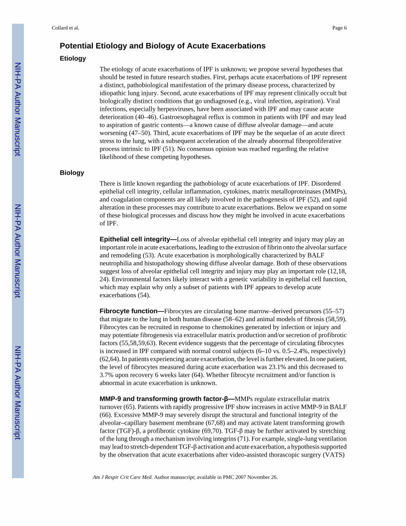
RadiologyEarly reports of acute exacerbation of IPF describe diffuse ground-glass opacities on plainchest radiograph (9,10). High-resolution computed tomography (HRCT) generallydemonstrates bilateral ground-glass abnormality with or without areas of consolidation,superimposed on the bibasilar subpleural reticular abnormality, traction bronchiectasis, andhoneycomb change typical of usual interstitial pneumonia (UIP) pattern (Figure 1) (11,12,14).
Three HRCT patterns of abnormality have been suggested: peripheral, multifocal, and diffuseground glass (11). The multifocal and diffuse patterns have been associated with diffusealveolar damage superimposed on UIP on surgical lung biopsy. Survival may be related to thedegree of CT involvement (11,12,14).
Collard et al. Page 4
Am J Respir Crit Care Med. Author manuscript; available in PMC 2007 November 26.
NIH
-PA Author Manuscript
NIH
-PA Author Manuscript
NIH
-PA Author Manuscript

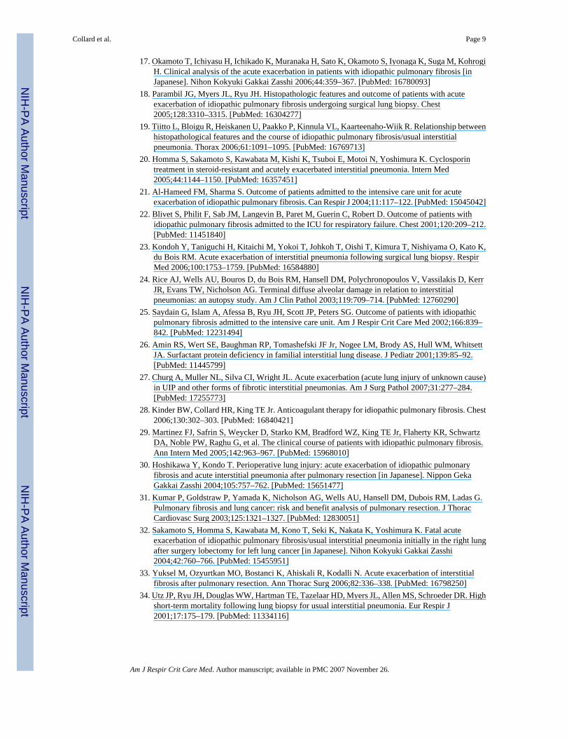
HistopathologyDiffuse alveolar damage superimposed on underlying UIP is the most commonly describedfinding when surgical lung biopsy is performed (Figure 2) (11,10,12,14,18,24,27). Diffusealveolar damage has been described in nearly three-quarters of the lung specimens reported inthe literature, and, in all but one case, the findings were associated with histologic evidence ofunderlying UIP. Organizing pneumonia without other evidence of organizing diffuse alveolardamage and extensive fibroblastic foci have also been described in a few cases (27).
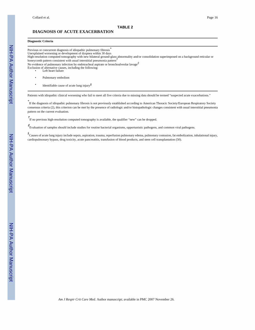
Proposed Definition and Diagnostic CriteriaWe propose the following definition for acute exacerbation of IPF: an acute, clinicallysignificant deterioration of unidentifiable cause in a patient with underlying IPF. Proposeddiagnostic criteria for acute exacerbation of IPF are listed in Table 2. Clinical deteriorationsof IPF that are of unknown cause but do not fulfill the criteria listed due to missing data shouldbe termed “suspected acute exacerbations of IPF.” This could include the following cases:those that are of more than 30 days' duration, those that have unilateral ground-glassabnormalities, or those that have not undergone endotracheal aspirate or BAL evaluation. Indeveloping these criteria, we have tried to reconcile the desire for diagnostic specificity withour lack of understanding regarding the etiology and pathobiology of acute exacerbations. Itis hoped that the diagnostic criteria presented here will serve to standardize inclusion criteriain future research studies, and that these criteria will be revised and modified as we gainadditional insight and information.
Several items regarding the proposed diagnostic criteria deserve further emphasis andexplanation. First, the choice of 30 days or less as the time course for acute exacerbation, whilebased on published experience, is recognized as arbitrary. Certainly, cases of idiopathic clinicaldeterioration over longer periods of time (e.g., 2 mo) may share a similar pathobiology.However, it was believed that a relatively short time period was important to separate acuteworsening from the more gradual progression seen in many patients with IPF. Second, therequirement for documentation of abnormal gas exchange present in previously publisheddiagnostic criteria has been removed. Any diagnostic specificity gained from includingdocumentation of abnormal gas exchange as a requirement was believed to be outweighed bythe decreased sensitivity (cases with clear exacerbation but without previous data points wouldbe excluded, and there would be bias toward more severe disease; i.e., patients with comparisondata would more likely be sicker and/or have more advanced disease) its inclusion would cause.Third, endotracheal aspirate or BAL is recommended to investigate a potentially infectiousetiology. Evaluation of these samples should include routine studies for bacterial organisms,opportunistic pathogens (e.g., Pneumocystis jiroveci), and common respiratory viruses.Although the epidemiology of viral respiratory infections varies globally, relevant viruses inNorth America would include influenza A and B, parainfluenza 1–4, respiratory syncytial virusA and B, human metapneumovirus, adenoviruses, rhinovirus, and coronaviruses. In casescomplicated by suspected nosocomial infection, the etiologic role of organisms identified onculture should be decided based on clinical judgment. Fourth, acute exacerbations of otherforms of interstitial lung disease (e.g., nonspecific interstitial pneumonia) have beendeliberately excluded, although they may turn out to have a similar etiology and/orpathobiology. As more is learned about acute exacerbations in IPF, it may be appropriate tobroaden the definition to include acute exacerbations of other conditions. Fifth, the exclusionof alternative etiologies, such as heart failure and pulmonary embolism, is left intentionallyvague, because the diagnostic approach to these conditions is complex and beyond the scopeof this article to adequately describe.
Collard et al. Page 5
Am J Respir Crit Care Med. Author manuscript; available in PMC 2007 November 26.
NIH
-PA Author Manuscript
NIH
-PA Author Manuscript
NIH
-PA Author Manuscript
Potential Etiology and Biology of Acute ExacerbationsEtiology
The etiology of acute exacerbations of IPF is unknown; we propose several hypotheses thatshould be tested in future research studies. First, perhaps acute exacerbations of IPF representa distinct, pathobiological manifestation of the primary disease process, characterized byidiopathic lung injury. Second, acute exacerbations of IPF may represent clinically occult butbiologically distinct conditions that go undiagnosed (e.g., viral infection, aspiration). Viralinfections, especially herpesviruses, have been associated with IPF and may cause acutedeterioration (40–46). Gastroesophageal reflux is common in patients with IPF and may leadto aspiration of gastric contents—a known cause of diffuse alveolar damage—and acuteworsening (47–50). Third, acute exacerbations of IPF may be the sequelae of an acute directstress to the lung, with a subsequent acceleration of the already abnormal fibroproliferativeprocess intrinsic to IPF (51). No consensus opinion was reached regarding the relativelikelihood of these competing hypotheses.
BiologyThere is little known regarding the pathobiology of acute exacerbations of IPF. Disorderedepithelial cell integrity, cellular inflammation, cytokines, matrix metalloproteinases (MMPs),and coagulation components are all likely involved in the pathogenesis of IPF (52), and rapidalteration in these processes may contribute to acute exacerbations. Below we expand on someof these biological processes and discuss how they might be involved in acute exacerbationsof IPF.
Epithelial cell integrity—Loss of alveolar epithelial cell integrity and injury may play animportant role in acute exacerbations, leading to the extrusion of fibrin onto the alveolar surfaceand remodeling (53). Acute exacerbation is morphologically characterized by BALFneutrophilia and histopathology showing diffuse alveolar damage. Both of these observationssuggest loss of alveolar epithelial cell integrity and injury may play an important role (12,18,24). Environmental factors likely interact with a genetic variability in epithelial cell function,which may explain why only a subset of patients with IPF appears to develop acuteexacerbations (54).
Fibrocyte function—Fibrocytes are circulating bone marrow–derived precursors (55–57)that migrate to the lung in both human disease (58–62) and animal models of fibrosis (58,59).Fibrocytes can be recruited in response to chemokines generated by infection or injury andmay potentiate fibrogenesis via extracellular matrix production and/or secretion of profibroticfactors (55,58,59,63). Recent evidence suggests that the percentage of circulating fibrocytesis increased in IPF compared with normal control subjects (6–10 vs. 0.5–2.4%, respectively)(62,64). In patients experiencing acute exacerbation, the level is further elevated. In one patient,the level of fibrocytes measured during acute exacerbation was 23.1% and this decreased to3.7% upon recovery 6 weeks later (64). Whether fibrocyte recruitment and/or function isabnormal in acute exacerbation is unknown.
MMP-9 and transforming growth factor-β—MMPs regulate extracellular matrixturnover (65). Patients with rapidly progressive IPF show increases in active MMP-9 in BALF(66). Excessive MMP-9 may severely disrupt the structural and functional integrity of thealveolar–capillary basement membrane (67,68) and may activate latent transforming growthfactor (TGF)-β, a profibrotic cytokine (69,70). TGF-β may be further activated by stretchingof the lung through a mechanism involving integrins (71). For example, single-lung ventilationmay lead to stretch-dependent TGF-β activation and acute exacerbation, a hypothesis supportedby the observation that acute exacerbations after video-assisted thorascopic surgery (VATS)
Collard et al. Page 6
Am J Respir Crit Care Med. Author manuscript; available in PMC 2007 November 26.
NIH
-PA Author Manuscript
NIH
-PA Author Manuscript
NIH
-PA Author Manuscript

were more prominent in the intraoperatively ventilated lung (24). Thus, increases in MMP-9and TGF-β activation may promote the development of acute exacerbation in IPF.
Disordered coagulation and fibrinolysis—Disordered coagulation and fibrinolysis maybe important components of acute exacerbations of IPF (16). Studies of patients with stableIPF have demonstrated a procoagulant and antifibrinolytic alveolar environment (29,72–74),and a similar environment has been described in the acute respiratory distress syndrome(ARDS) (74–76). The presence of disordered coagulation and fibrinolysis in stable IPF andthe clinical and pathologic similarities of acute exacerbations with ARDS support a role forthese mechanisms.
Genetic predisposition—A polymorphism in erythrocyte complement receptor 1 andmutations in surfactant protein genes are present in selected patients with IPF (77,78). Recently,the presence of heterozygous mutations in the telomerase reverse transcriptase (hTERT) and/or RNA component (hTR) genes encoding telomerase components have been reported inseveral families with IPF and in one patient with sporadic IPF (79,80). These mutations leadto shortened telomeres that may limit the regenerative capacity of alveolar epithelial cells andcontribute to the pathobiology of acute exacerbation.
Future DirectionsOur understanding of acute exacerbations of IPF is limited and key questions remain. Thereare several important areas for future research. Investigators should focus on characterizingthe epidemiology of acute exacerbations. What is the true incidence of acute exacerbations?Are they a common complication of IPF? What is their prognostic significance? What are therisk factors (clinical, biological, and genetic) for their development? Are acute exacerbationsof IPF different from acute exacerbations of other forms of interstitial lung disease? Large,prospective cohorts of well-defined patients are needed to answer these questions. The role ofcorticosteroids and other therapies, such as anticoagulation, for the treatment of acuteexacerbation remains unclear. A better understanding of the epidemiology of this condition isessential to the design of future treatment trials aimed at clarifying treatment issues.
Studies of the underlying pathobiology of acute exacerbations of IPF are also criticallyimportant. Do acute exacerbations of IPF represent a distinct pathobiological manifestation ofthe primary disease process or are they caused by occult complications such as infection andaspiration? What are the roles of inflammation, coagulation, epithelial cell biology, andgenetics? Future studies should prospectively collect biological samples from subjectsexperiencing acute exacerbations of IPF. A systematic search for evidence of infection, perhapsusing pan-viral and bacterial microarrays to look for novel agents, is important. Additionalstudies could include gene expression arrays of lung tissues or of BALF cells, and proteomicanalysis of plasma and BAL. Given the clinical, radiographic, and histopathologic similaritiesbetween acute exacerbation of IPF and ARDS, the role of factors associated with thepathobiology of ARDS should also be explored. It is possible that similar biologicalmechanisms are involved in these two conditions. If so, treatment approaches targeting patientswith ARDS could be beneficial to patients with acute exacerbation of IPF.
To answer these and other questions, a systematic and concerted effort to identify patientsundergoing acute exacerbations of IPF is an essential first step. We propose that the definitionand diagnostic criteria presented above be used for these efforts. The development ofmulticenter, national, or international registries to prospectively collect clinical data andbiological samples from patients with acute exacerbations of IPF would allow investigatorssufficient numbers of subjects to successfully study this condition. It is only through thestandardization of terminology and collaboration of effort that important progress will be made.
Collard et al. Page 7
Am J Respir Crit Care Med. Author manuscript; available in PMC 2007 November 26.
NIH
-PA Author Manuscript
NIH
-PA Author Manuscript
NIH
-PA Author Manuscript

Acknowledgements
Supported by National Institutes of Health (NIH) HL86516 (H.R.C.), NHLBI SCOR HL6767 (K.K.B.), NIHHL080383 (R.J.K.), NIH HL083901 (J.A.L.), and NIH HL58655 and NIH 080685 (M.A.O.).
References1. American Thoracic Society/European Respiratory Society. American Thoracic Society/European
Respiratory Society international multidisciplinary consensus classification of the idiopathicinterstitial pneumonias. Am J Respir Crit Care Med 2002;165:277–304. [PubMed: 11790668]
2. American Thoracic Society/European Respiratory Society. Idiopathic pulmonary fibrosis: diagnosisand treatment. International consensus statement. Am J Respir Crit Care Med 2000;161:646–664.[PubMed: 10673212]
3. Bjoraker JA, Ryu JH, Edwin MK, Myers JL, Tazelaar HD, Schroeder DR, Offord KP. Prognosticsignificance of histopathologic subsets in idiopathic pulmonary fibrosis. Am J Respir Crit Care Med1998;157:199–203. [PubMed: 9445300]
4. Flaherty KR, Travis WD, Colby TV, Toews GB, Kazerooni EA, Gross BH, Jain A, Strawderman RL,Flint A, Lynch JP, et al. Histopathologic variability in usual and nonspecific interstitial pneumonias.Am J Respir Crit Care Med 2001;164:1722–1727. [PubMed: 11719316]
5. Nicholson AG, Colby TV, Dubois RM, Hansell DM, Wells AU. The prognostic significance of thehistologic pattern of interstitial pneumonia in patients presenting with the clinical entity of cryptogenicfibrosing alveolitis. Am J Respir Crit Care Med 2000;162:2213–2217. [PubMed: 11112140]
6. Carrington CB, Gaensler EA, Coutu RE, Fitzgerald MX, Gupta RG. Natural history and treated courseof usual and desquamative interstitial pneumonia. N Engl J Med 1978;298:801–809. [PubMed:634315]
7. Stack BHR, Choo-Kang YEJ, Heard BE. The prognosis of cryptogenic fibrosing alveolitis. Thorax1972;27:535–542. [PubMed: 4538877]
8. Kim DS, Collard HR, King TE Jr. Classification and natural history of the idiopathic interstitialpneumonias. Proc Am Thorac Soc 2006;3:285–292. [PubMed: 16738191]
9. Kondo, A.; Saiki, S. Acute exacerbation in idiopathic interstitial pneumonia (IIP). In: Harasawa, M.;Fukuchi, Y.; Morinari, H., editors. Interstitial pneumonia of unknown etiology. Tokyo, Japan:University of Tokyo Press; 1989. p. 33-42.Intractable Diseases Research Foundation Publication No.27
10. Kondoh Y, Taniguchi H, Kawabata Y, Yokoi T, Suzuki K, Takagi K. Acute exacerbation in idiopathicpulmonary fibrosis: analysis of clinical and pathologic findings in three cases. Chest 1993;103:1808–1812. [PubMed: 8404104]
11. Akira M, Hamada H, Sakatani M, Kobayashi C, Nishioka M, Yamamoto S. CT findings during phaseof accelerated deterioration in patients with idiopathic pulmonary fibrosis. AJR Am J Roentgenol1997;168:79–83. [PubMed: 8976924]
12. Ambrosini V, Cancellieri A, Chilosi M, Zompatori M, Trisolini R, Saragoni L, Poletti V. Acuteexacerbation of idiopathic pulmonary fibrosis: report of a series. Eur Respir J 2003;22:821–826.[PubMed: 14621091]
13. Azuma A, Nukiwa T, Tsuboi E, Suga M, Abe S, Nakata K, Taguchi Y, Nagai S, Itoh H, Ohi M, etal. Placebo-controlled trial of pirfenidone in patients with idiopathic pulmonary fibrosis. Am J RespirCrit Care Med 2005;171:1040–1047. [PubMed: 15665326]
14. Kim DS, Park JH, Park BK, Lee JS, Nicholson AG, Colby T. Acute exacerbation of idiopathicpulmonary fibrosis: frequency and clinical features. Eur Respir J 2006;27:143–150. [PubMed:16387947]
15. Kondoh Y, Taniguchi H, Yokoi T, Nishiyama O, Ohishi T, Kato T, Suzuki K, Suzuki R.Cyclophosphamide and low-dose prednisolone in idiopathic pulmonary fibrosis and fibrosingnonspecific interstitial pneumonia. Eur Respir J 2005;25:528–533. [PubMed: 15738299]
16. Kubo H, Nakayama K, Yanai M, Suzuki T, Yamaya M, Watanabe M, Sasaki H. Anticoagulant therapyfor idiopathic pulmonary fibrosis. Chest 2005;128:1475–1482. [PubMed: 16162746]
Collard et al. Page 8
Am J Respir Crit Care Med. Author manuscript; available in PMC 2007 November 26.
NIH
-PA Author Manuscript
NIH
-PA Author Manuscript
NIH
-PA Author Manuscript
17. Okamoto T, Ichiyasu H, Ichikado K, Muranaka H, Sato K, Okamoto S, Iyonaga K, Suga M, KohrogiH. Clinical analysis of the acute exacerbation in patients with idiopathic pulmonary fibrosis [inJapanese]. Nihon Kokyuki Gakkai Zasshi 2006;44:359–367. [PubMed: 16780093]
18. Parambil JG, Myers JL, Ryu JH. Histopathologic features and outcome of patients with acuteexacerbation of idiopathic pulmonary fibrosis undergoing surgical lung biopsy. Chest2005;128:3310–3315. [PubMed: 16304277]
19. Tiitto L, Bloigu R, Heiskanen U, Paakko P, Kinnula VL, Kaarteenaho-Wiik R. Relationship betweenhistopathological features and the course of idiopathic pulmonary fibrosis/usual interstitialpneumonia. Thorax 2006;61:1091–1095. [PubMed: 16769713]
20. Homma S, Sakamoto S, Kawabata M, Kishi K, Tsuboi E, Motoi N, Yoshimura K. Cyclosporintreatment in steroid-resistant and acutely exacerbated interstitial pneumonia. Intern Med2005;44:1144–1150. [PubMed: 16357451]
21. Al-Hameed FM, Sharma S. Outcome of patients admitted to the intensive care unit for acuteexacerbation of idiopathic pulmonary fibrosis. Can Respir J 2004;11:117–122. [PubMed: 15045042]
22. Blivet S, Philit F, Sab JM, Langevin B, Paret M, Guerin C, Robert D. Outcome of patients withidiopathic pulmonary fibrosis admitted to the ICU for respiratory failure. Chest 2001;120:209–212.[PubMed: 11451840]
23. Kondoh Y, Taniguchi H, Kitaichi M, Yokoi T, Johkoh T, Oishi T, Kimura T, Nishiyama O, Kato K,du Bois RM. Acute exacerbation of interstitial pneumonia following surgical lung biopsy. RespirMed 2006;100:1753–1759. [PubMed: 16584880]
24. Rice AJ, Wells AU, Bouros D, du Bois RM, Hansell DM, Polychronopoulos V, Vassilakis D, KerrJR, Evans TW, Nicholson AG. Terminal diffuse alveolar damage in relation to interstitialpneumonias: an autopsy study. Am J Clin Pathol 2003;119:709–714. [PubMed: 12760290]
25. Saydain G, Islam A, Afessa B, Ryu JH, Scott JP, Peters SG. Outcome of patients with idiopathicpulmonary fibrosis admitted to the intensive care unit. Am J Respir Crit Care Med 2002;166:839–842. [PubMed: 12231494]
26. Amin RS, Wert SE, Baughman RP, Tomashefski JF Jr, Nogee LM, Brody AS, Hull WM, WhitsettJA. Surfactant protein deficiency in familial interstitial lung disease. J Pediatr 2001;139:85–92.[PubMed: 11445799]
27. Churg A, Muller NL, Silva CI, Wright JL. Acute exacerbation (acute lung injury of unknown cause)in UIP and other forms of fibrotic interstitial pneumonias. Am J Surg Pathol 2007;31:277–284.[PubMed: 17255773]
28. Kinder BW, Collard HR, King TE Jr. Anticoagulant therapy for idiopathic pulmonary fibrosis. Chest2006;130:302–303. [PubMed: 16840421]
29. Martinez FJ, Safrin S, Weycker D, Starko KM, Bradford WZ, King TE Jr, Flaherty KR, SchwartzDA, Noble PW, Raghu G, et al. The clinical course of patients with idiopathic pulmonary fibrosis.Ann Intern Med 2005;142:963–967. [PubMed: 15968010]
30. Hoshikawa Y, Kondo T. Perioperative lung injury: acute exacerbation of idiopathic pulmonaryfibrosis and acute interstitial pneumonia after pulmonary resection [in Japanese]. Nippon GekaGakkai Zasshi 2004;105:757–762. [PubMed: 15651477]
31. Kumar P, Goldstraw P, Yamada K, Nicholson AG, Wells AU, Hansell DM, Dubois RM, Ladas G.Pulmonary fibrosis and lung cancer: risk and benefit analysis of pulmonary resection. J ThoracCardiovasc Surg 2003;125:1321–1327. [PubMed: 12830051]
32. Sakamoto S, Homma S, Kawabata M, Kono T, Seki K, Nakata K, Yoshimura K. Fatal acuteexacerbation of idiopathic pulmonary fibrosis/usual interstitial pneumonia initially in the right lungafter surgery lobectomy for left lung cancer [in Japanese]. Nihon Kokyuki Gakkai Zasshi2004;42:760–766. [PubMed: 15455951]
33. Yuksel M, Ozyurtkan MO, Bostanci K, Ahiskali R, Kodalli N. Acute exacerbation of interstitialfibrosis after pulmonary resection. Ann Thorac Surg 2006;82:336–338. [PubMed: 16798250]
34. Utz JP, Ryu JH, Douglas WW, Hartman TE, Tazelaar HD, Myers JL, Allen MS, Schroeder DR. Highshort-term mortality following lung biopsy for usual interstitial pneumonia. Eur Respir J2001;17:175–179. [PubMed: 11334116]
Collard et al. Page 9
Am J Respir Crit Care Med. Author manuscript; available in PMC 2007 November 26.
NIH
-PA Author Manuscript
NIH
-PA Author Manuscript
NIH
-PA Author Manuscript

35. Mukae H, Iiboshi H, Nakazato M, Hiratsuka T, Tokojima M, Abe K, Ashitani J, Kadota J, MatsukuraS, Kohno S. Raised plasma concentrations of alpha-defensins in patients with idiopathic pulmonaryfibrosis. Thorax 2002;57:623–628. [PubMed: 12096207]
36. Tajima S, Oshikawa K, Tominaga S, Sugiyama Y. The increase in serum soluble ST2 protein uponacute exacerbation of idiopathic pulmonary fibrosis. Chest 2003;124:1206–1214. [PubMed:14555548]
37. Ziegenhagen MW, Zabel P, Zissel G, Schlaak M, Muller-Quernheim J. Serum level of interleukin 8is elevated in idiopathic pulmonary fibrosis and indicates disease activity. Am J Respir Crit CareMed 1998;157:762–768. [PubMed: 9517588]
38. Akira M, Sakatani M, Hara H. Thin-section CT findings in rheumatoid arthritis-associated lungdisease: CT patterns and their courses. J Comput Assist Tomogr 1999;23:941–948. [PubMed:10589572]
39. Yokoyama A, Kohno N, Hamada H, Sakatani M, Ueda E, Kondo K, Hirasawa Y, Hiwada K.Circulating KL-6 predicts the outcome of rapidly progressive idiopathic pulmonary fibrosis. Am JRespir Crit Care Med 1998;158:1680–1684. [PubMed: 9817725]
40. Egan JJ, Stewart JP, Hasleton PS, Arrand JR, Carroll KB, Woodcock AA. Epstein-Barr virusreplication within pulmonary epithelial cells in cryptogenic fibrosing alveolitis. Thorax1995;50:1234–1239. [PubMed: 8553293]
41. Hogg JC. Epstein-Barr virus and cryptogenic fibrosing alveolitis. Thorax 1995;50:1232. [PubMed:8553292]
42. Tsukamoto K, Hayakawa H, Sato A, Chida K, Nakamura H, Miura K. Involvement of Epstein-Barrvirus latent membrane protein 1 in disease progression in patients with idiopathic pulmonary fibrosis.Thorax 2000;55:958–961. [PubMed: 11050267]
43. Yonemaru M, Kasuga I, Kusumoto H, Kunisawa A, Kiyokawa H, Kuwabara S, Ichinose Y, ToyamaK. Elevation of antibodies to cytomegalovirus and other herpes viruses in pulmonary fibrosis. EurRespir J 1997;10:2040–2045. [PubMed: 9311499]
44. Semenzato G, de Rossi A, Agostini C. Human retroviruses and their aetiological link to pulmonarydiseases. Eur Respir J 1993;6:925–929. [PubMed: 8396535]
45. Stewart JP, Egan JJ, Ross AJ, Kelly BG, Lok SS, Hasleton PS, Woodcock AA. The detection ofEpstein-Barr virus DNA in lung tissue from patients with idiopathic pulmonary fibrosis. Am J RespirCrit Care Med 1999;159:1336–1341. [PubMed: 10194186]
46. Tang YW, Johnson JE, Browning PJ, Cruz-Gervis RA, Davis A, Graham BS, Brigham KL, Oates JAJr, Loyd JE, Stecenko AA. Herpesvirus DNA is consistently detected in lungs of patients withidiopathic pulmonary fibrosis. J Clin Microbiol 2003;41:2633–2640. [PubMed: 12791891]
47. Raghu G, Freudenberger TD, Yang S, Curtis JR, Spada C, Hayes J, Sillery JK, Pope CE II, PellegriniCA. High prevalence of abnormal acid gastro-oesophageal reflux in idiopathic pulmonary fibrosis.Eur Respir J 2006;27:136–142. [PubMed: 16387946]
48. Raghu G, Yang ST, Spada C, Hayes J, Pellegrini CA. Sole treatment of acid gastroesophageal refluxin idiopathic pulmonary fibrosis: a case series. Chest 2006;129:794–800. [PubMed: 16537884]
49. Sweet MP, Patti MG, Leard LE, Golden JA, Hays SR, Hoopes C, Theodore PR. Gastroesophagealreflux in patients with idiopathic pulmonary fibrosis referred for lung transplantation. J ThoracCardiovasc Surg 2007;133:1078–1084. [PubMed: 17382656]
50. Ware LB, Matthay MA. The acute respiratory distress syndrome. N Engl J Med 2000;342:1334–1349. [PubMed: 10793167]
51. Selman M, King TE Jr, Pardo A. Idiopathic pulmonary fibrosis: prevailing and evolving hypothesesabout its pathogenesis and implications for therapy. Ann Intern Med 2001;134:136–151. [PubMed:11177318]
52. Selman M, Thannickal VJ, Pardo A, Zisman DA, Martinez FJ, Lynch JP. Idiopathic pulmonaryfibrosis: pathogenesis and therapeutic approaches. Drugs 2004;64:405–430. [PubMed: 14969575]
53. Kuhn C III, Boldt J, King TE Jr, Crouch E, Vartio T, McDonald JA. An immunohistochemical studyof architectural remodeling and connective tissue synthesis in pulmonary fibrosis. Am Rev RespirDis 1989;140:1693–1703. [PubMed: 2604297]
Collard et al. Page 10
Am J Respir Crit Care Med. Author manuscript; available in PMC 2007 November 26.
NIH
-PA Author Manuscript
NIH
-PA Author Manuscript
NIH
-PA Author Manuscript

54. Schwartz DA. The importance of gene-environment interactions and exposure assessment inunderstanding human diseases. J Expo Sci Environ Epidemiol 2006;16:474–476. [PubMed:17109021]
55. Bucala R, Spiegel LA, Chesney J, Hogan M, Cerami A. Circulating fibrocytes define a new leukocytesubpopulation that mediates tissue repair. Molecular Med 1994;1:71–81.
56. Chesney J, Bacher M, Bender A, Bucala R. The peripheral blood fibrocyte is a potent antigen-presenting cell capable of priming naive T cells in situ. Proc Natl Acad Sci USA 1997;94:6307–6312.[PubMed: 9177213]
57. Chesney J, Metz C, Stavitsky AB, Bacher M, Bucala R. Regulated production of type I collagen andinflammatory cytokines by peripheral blood fibrocytes. J Immunol 1998;160:419–425. [PubMed:9551999]
58. Moore BB, Kolodsick JE, Thannickal VJ, Cooke K, Moore TA, Hogaboam C, Wilke CA, Toews GB.CCR2-mediated recruitment of fibrocytes to the alveolar space after fibrotic injury. Am J Pathol2005;166:675–684. [PubMed: 15743780]
59. Phillips RJ, Burdick MD, Hong K, Lutz MA, Murray LA, Xue YY, Belperio JA, Keane MP, StrieterRM. Circulating fibrocytes traffic to the lungs in response to CXCL12 and mediate fibrosis. J ClinInvest 2004;114:438–446. [PubMed: 15286810]
60. Schmidt M, Sun G, Stacey M, Mori L, Mattoli S. Identification of circulating fibrocytes as precursorsof bronchial myofibroblasts in asthma. J Immunol 2003;170:380–389. [PubMed: 12817021]
61. Nihlberg K, Larsen K, Hultgardh-Nilsson A, Malmstrom A, Bjermer L, Westergren-Thorsson G.Tissue fibrocytes in patients with mild asthma: a possible link to thickness of reticular basementmembrane? Respir Res 2006;7:50. [PubMed: 16571120]
62. Mehrad B, Burdick MD, Zisman DA, Keane MP, Belperio JA, Strieter RM. Circulating peripheralblood fibrocytes in human fibrotic interstitial lung disease. Biochem Biophys Res Commun2007;353:104–108. [PubMed: 17174272]
63. Moore BB, Murray L, Das A, Wilke CA, Herrygers AB, Toews GB. The role of CCL12 in therecruitment of fibrocytes and lung fibrosis. Am J Respir Cell Mol Biol 2006;35:175–181. [PubMed:16543609]
64. Gilpin, S.; Moeller, A.; Baatjes, A.; Martin, G.; Ask, K.; O'Bryne, P.; Cox, G.; Gauldie, J.; Kolb, M.Circulating human matrix-producing progenitor cells in pulmonary fibroproliferative disorders;Presented at the International Colloquium on Lung Fibrosis; 2006 September 7–10; Eltville,Germany.
65. Nagase H, Visse R, Murphy G. Structure and function of matrix metalloproteinases and TIMPs.Cardiovasc Res 2006;69:562–573. [PubMed: 16405877]
66. Suga M, Iyonaga K, Okamoto T, Gushima Y, Miyakawa H, Akaike T, Ando M. Characteristicelevation of matrix metalloproteinase activity in idiopathic interstitial pneumonias. Am J Respir CritCare Med 2000;162:1949–1956. [PubMed: 11069839]
67. Atkinson JJ, Senior RM. Matrix metalloproteinase-9 in lung remodeling. Am J Respir Cell Mol Biol2003;28:12–24. [PubMed: 12495928]
68. Pardo A, Ruiz V, Arreola JL, Ramirez R, Cisneros-Lira J, Gaxiola M, Barrios R, Kala SV, LiebermanMW, Selman M. Bleomycin-induced pulmonary fibrosis is attenuated in gamma-glutamyltranspeptidase-deficient mice. Am J Respir Crit Care Med 2003;167:925–932. [PubMed: 12468440]
69. Wang H, Kochevar IE. Involvement of UVB-induced reactive oxygen species in TGF-betabiosynthesis and activation in keratinocytes. Free Radic Biol Med 2005;38:890–897. [PubMed:15749385]
70. Yu Q, Stamenkovic I. Cell surface-localized matrix metalloproteinase-9 proteolytically activatesTGF-beta and promotes tumor invasion and angiogenesis. Genes Dev 2000;14:163–176. [PubMed:10652271]
71. Munger JS, Huang X, Kawakatsu H, Griffiths MJ, Dalton SL, Wu J, Pittet JF, Kaminski N, Garat C,Matthay MA, et al. The integrin alpha v beta 6 binds and activates latent TGF beta 1: a mechanismfor regulating pulmonary inflammation and fibrosis. Cell 1999;96:319–328. [PubMed: 10025398]
72. Gunther A, Mosavi P, Ruppert C, Heinemann S, Temmesfeld B, Velcovsky HG, Morr H, GrimmingerF, Walmrath D, Seeger W. Enhanced tissue factor pathway activity and fibrin turnover in the alveolar
Collard et al. Page 11
Am J Respir Crit Care Med. Author manuscript; available in PMC 2007 November 26.
NIH
-PA Author Manuscript
NIH
-PA Author Manuscript
NIH
-PA Author Manuscript

compartment of patients with interstitial lung disease. Thromb Haemost 2000;83:853–860. [PubMed:10896238]
73. Perez RL, Duncan A, Hunter RL, Staton GW Jr. Elevated D dimer in the lungs and blood of patientswith sarcoidosis. Chest 1993;103:1100–1106. [PubMed: 8131447]
74. Olman, MA. Mechanisms of fibroproliferation in acute lung injury. In: Matthay, MA.; Lenfant, C.,editors. Acute respiratory distress syndrome. New York: Marcel Dekker; 2003. p. 313-354.
75. Gaggar A, Olman MA. Biologic markers of mortality in acute lung injury. Clin Chim Acta2006;372:24–32. [PubMed: 16797514]
76. Prabhakaran P, Ware LB, White KE, Cross MT, Matthay MA, Olman MA. Elevated levels ofplasminogen activator inhibitor-1 in pulmonary edema fluid are associated with mortality in acutelung injury. Am J Physiol Lung Cell Mol Physiol 2003;285:L20–L28. [PubMed: 12730079]
77. Zorzetto M, Ferrarotti I, Trisolini R, Agli LL, Scabini R, Novo M, De Silvestri A, Patelli M, MartinettiM, Cuccia M, et al. Complement receptor 1 gene polymorphisms are associated with idiopathicpulmonary fibrosis. Am J Respir Crit Care Med 2003;168:330–334. [PubMed: 12773320]
78. Lawson WE, Loyd JE. The genetic approach in pulmonary fibrosis: can it provide clues to thiscomplex disease? Proc Am Thorac Soc 2006;3:345–349. [PubMed: 16738199]
79. Armanios MY, Chen JJ, Cogan JD, Alder JK, Ingersoll RG, Markin C, Lawson WE, Xie M, VultoI, Phillips JA III, et al. Telomerase mutations in families with idiopathic pulmonary fibrosis. N EnglJ Med 2007;356:1317–1326. [PubMed: 17392301]
80. Tsakiri K, Cronkhite J, Kuan P, Xing C, Raghu G, Weissler J, Rosenblatt R, Shay J, Garcia C. Adult-onset pulmonary fibrosis caused by mutations in telemorase. Proc Natl Acad Sci USA2007;104:7552–7557. [PubMed: 17460043]
81. Stern JB, Mal H, Groussard O, Brugiere O, Marceau A, Jebrak G, Fournier M. Prognosis of patientswith advanced idiopathic pulmonary fibrosis requiring mechanical ventilation for acute respiratoryfailure. Chest 2001;120:213–219. [PubMed: 11451841]
Collard et al. Page 12
Am J Respir Crit Care Med. Author manuscript; available in PMC 2007 November 26.
NIH
-PA Author Manuscript
NIH
-PA Author Manuscript
NIH
-PA Author Manuscript

Figure 1.High-resolution computed tomography (HRCT) of acute exacerbation of idiopathic pulmonaryfibrosis (IPF). (A, B) HRCT images through right upper and lower lung of a patient with stableIPF show typical features of usual interstitial pneumonia pattern: peripheral and basalpredominant reticular abnormality with traction bronchiectasis and subpleural honeycombing.A small amount of ground-glass abnormality is present. (C, D) HRCT images through the rightupper and lower lung of the same patient during an acute exacerbation of IPF show new,extensive ground-glass abnormality superimposed on the background of lung fibrosis.
Collard et al. Page 13
Am J Respir Crit Care Med. Author manuscript; available in PMC 2007 November 26.
NIH
-PA Author Manuscript
NIH
-PA Author Manuscript
NIH
-PA Author Manuscript

Figure 2.Surgical lung biopsy of acute exacerbation of idiopathic pulmonary fibrosis. (A) Lowmagnification photomicrograph shows chronic interstitial pneumonia with heterogeneousdistribution and honeycomb change typical of usual interstitial pneumonia (originalmagnification, ×20; hematoxylin-and-eosin stain). (B) Higher magnification photomicrographshows area of acute diffuse alveolar damage in same biopsy specimen characterized by well-developed eosinophilic hyaline membranes (arrows) (original magnification, ×100;hematoxylin-and-eosin stain).
Collard et al. Page 14
Am J Respir Crit Care Med. Author manuscript; available in PMC 2007 November 26.
NIH
-PA Author Manuscript
NIH
-PA Author Manuscript
NIH
-PA Author Manuscript

NIH
-PA Author Manuscript
NIH
-PA Author Manuscript
NIH
-PA Author Manuscript
Collard et al. Page 15TA
BLE
1PU
BL
ISH
ED
ST
UD
IES
OF
AC
UT
E E
XA
CE
RB
AT
ION
Publ
icat
ion
Stud
y D
esig
nN
o.*
Age
(yr)
M/F
Inci
denc
e (%
)M
orta
lity
Rat
e (%
)
Chu
rg a
nd c
olle
ague
s, 20
07 (2
7)R
etro
spec
tive
revi
ew o
f lun
g bi
opsy
cas
es9
623/
6N
/A22
Tiitt
o an
d co
lleag
ues,
2006
(19)
Aut
opsy
revi
ew9
N/A
N/A
21N
/AO
kam
oto
and
colle
ague
s, 20
06(1
7)R
etro
spec
tive
revi
ew o
f hos
pita
l adm
issi
ons
2868
20/8
12.5
(2-y
r inc
iden
ce)
86
Kim
and
col
leag
ues,
2006
(13)
Ret
rosp
ectiv
e lo
ngitu
dina
l coh
ort
1163
6/2
9.6
(2-y
r inc
iden
ce)
78K
ondo
h an
d co
lleag
ues,
2006
(23)
Ret
rosp
ectiv
e re
view
of p
ost-l
ung
biop
syca
ses
363
3/0
N/A
67
Para
mbi
l and
col
leag
ues,
2005
(18)
Cas
e se
ries
770
5/2
N/A
86
Azu
ma
and
colle
ague
s, 20
05 (1
3)R
ando
miz
ed c
ontro
lled
trial
5N
/AN
/A5
(9-m
o in
cide
nce)
20K
ubo
and
colle
ague
s, 20
05 (1
6)R
ando
miz
ed c
ontro
lled
trial
32N
/AN
/A57
(3-y
r inc
iden
ce)
53K
ondo
h an
d co
lleag
ues,
2005
(15)
Pros
pect
ive
trial
661
3/0
22 (5
-yr i
ncid
ence
)N
/AH
omm
a an
d co
lleag
ues,
2005
(20)
Cas
e se
ries
1070
10/0
N/A
100
Al-H
amee
d an
d Sh
arm
a, 2
004
(21)
Ret
rosp
ectiv
e re
view
of I
CU
adm
issi
ons
2569
23/2
N/A
96
Ric
e an
d co
lleag
ues,
2003
(24)
Aut
opsy
revi
ew12
6610
/2N
/A10
0A
mbr
osin
i and
col
leag
ues,
2003
(12)
Cas
e se
ries
570
4/1
N/A
80
Sayd
ain
and
colle
ague
s, 20
02 (2
5)R
etro
spec
tive
revi
ew o
f IC
U a
dmis
sion
s15
N/A
N/A
47N
/ASt
ern
and
colle
ague
s, 20
01 (8
1)R
etro
spec
tive
revi
ew o
f IC
U a
dmis
sion
s14
N/A
N/A
61>9
0B
livet
and
col
leag
ues,
2001
(22)
Ret
rosp
ectiv
e re
view
of I
CU
adm
issi
ons
6N
/AN
/A40
100
Aki
ra a
nd c
olle
ague
s, 19
97 (1
1)C
ase
serie
s17
6314
/3N
/A53
Kon
doh
and
colle
ague
s, 19
93 (1
0)C
ase
serie
s3
613/
0N
/A0
Kon
do a
nd S
aiki
, 198
9 (9
)Su
rvey
51N
/AN
/A33
96K
ondo
and
Sai
ki, 1
989
(9)
Ret
rosp
ectiv
e co
hort
4N
/AN
/A18
N/A
Def
initi
on o
f abb
revi
atio
ns: I
CU
= in
tens
ive
care
uni
t; M
/F =
mal
e/fe
mal
e; N
/A =
not
ava
ilabl
e.
* Num
ber o
f acu
te e
xace
rbat
ions
repo
rted.
Am J Respir Crit Care Med. Author manuscript; available in PMC 2007 November 26.
NIH
-PA Author Manuscript
NIH
-PA Author Manuscript
NIH
-PA Author Manuscript
Collard et al. Page 16
TABLE 2DIAGNOSIS OF ACUTE EXACERBATION
Diagnostic Criteria
Previous or concurrent diagnosis of idiopathic pulmonary fibrosis*Unexplained worsening or development of dyspnea within 30 daysHigh-resolution computed tomography with new bilateral ground-glass abnormality and/or consolidation superimposed on a background reticular orhoneycomb pattern consistent with usual interstitial pneumonia pattern†
No evidence of pulmonary infection by endotracheal aspirate or bronchoalveolar lavage‡Exclusion of alternative causes, including the following:
• Left heart failure
• Pulmonary embolism
• Identifiable cause of acute lung injury§
Patients with idiopathic clinical worsening who fail to meet all five criteria due to missing data should be termed “suspected acute exacerbations.”
*If the diagnosis of idiopathic pulmonary fibrosis is not previously established according to American Thoracic Society/European Respiratory Society
consensus criteria (2), this criterion can be met by the presence of radiologic and/or histopathologic changes consistent with usual interstitial pneumoniapattern on the current evaluation.
†If no previous high-resolution computed tomography is available, the qualifier “new” can be dropped.
‡Evaluation of samples should include studies for routine bacterial organisms, opportunistic pathogens, and common viral pathogens.
§Causes of acute lung injury include sepsis, aspiration, trauma, reperfusion pulmonary edema, pulmonary contusion, fat embolization, inhalational injury,
cardiopulmonary bypass, drug toxicity, acute pancreatitis, transfusion of blood products, and stem cell transplantation (50).
Am J Respir Crit Care Med. Author manuscript; available in PMC 2007 November 26.
Related Documents