“Acute Coronary Syndrome” July 24, 2013

“Acute Coronary Syndrome”
Jan 15, 2016
“Acute Coronary Syndrome”. July 24, 2013. Item 72. - PowerPoint PPT Presentation
Welcome message from author
This document is posted to help you gain knowledge. Please leave a comment to let me know what you think about it! Share it to your friends and learn new things together.
Transcript

“Acute Coronary Syndrome”
July 24, 2013
Item 72• A 78 year old man is evaluated in the ED with chest
pain. The patient reports that the pain, which is present in the left substernal area, began at rest, and has been present for 12 hours. He reports no similar episodes of chest pain. Medical history is significant for hypertension and a 30-pack year history of ongoing tobacco use. His only medication is nifedipine.
• On PE, temperature is 37.90C, BP 130/80 mm Hg, pulse rate is 72/minute and respiration rate is 12/min. BMI is 28. A normal carotid upstroke without carotid bruits is noted, jugular venous pulsations are normal and S1 and S2 are heard without murmurs. Lung fields are clear, distal pulses are normal and no peripheral edema is present.

Item 72 (Con’t)• Serum creatinine kinase level is 500
units/L and troponin I level is 26 ng/mL. Lab findings are otherwise normal.
• EKG shows sinus rhythm at 70/min; 2 mm ST-segment elevation in leads II, III and aVF; and 1 mm ST segment depression in leads V2 and V3. He is taken to the cardiac cath lab and found to have single vessel coronary disease with severe stenosis of the proximal left anterior descending coronary artery.
Item 72 (Con’t)
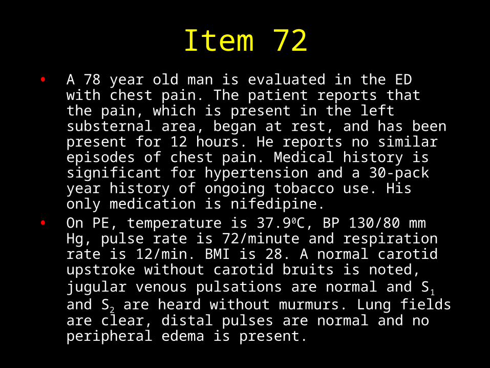
• Which of the following is the most appropriate treatment?
A. Coronary artery bypass surgery
B. Intracoronary thrombolytic therapy
C. Medical therapy
D. Primary percutaneous coronary intervention
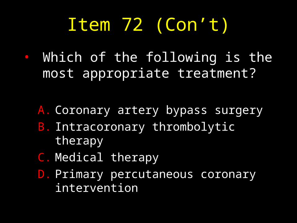
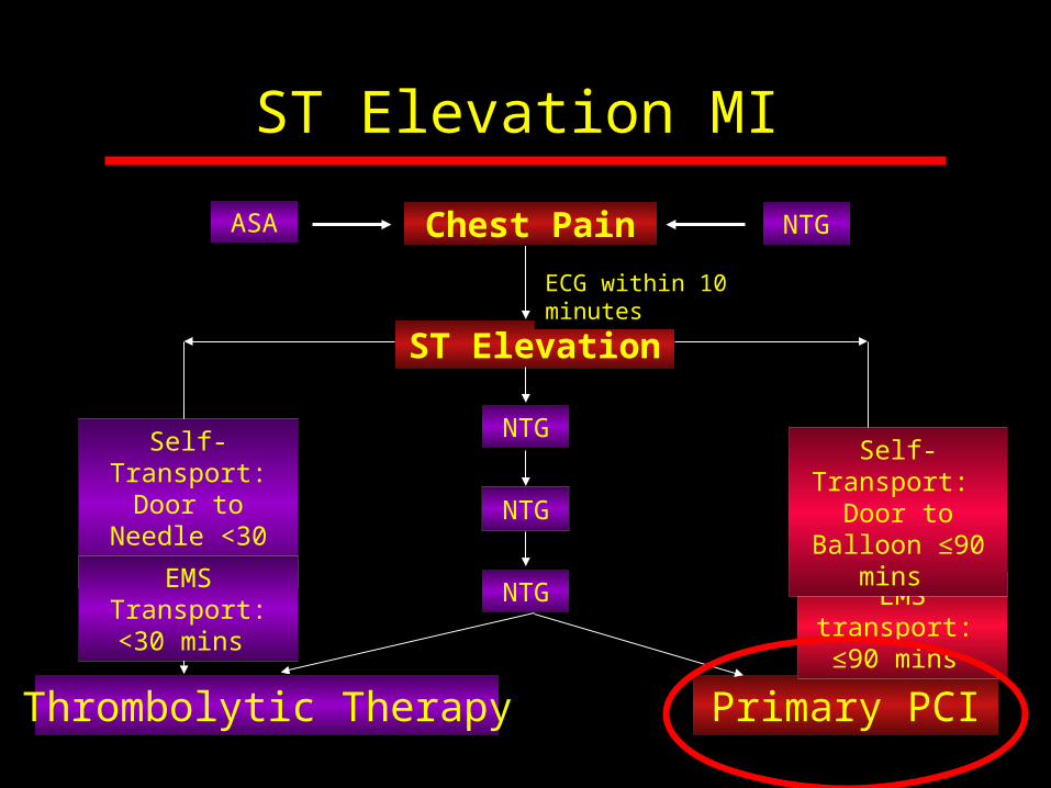
STEMI Care and Time to Treatment Goals
• Primary PCI is the recommended method of reperfusion when it can be performed in a timely fashion by experienced operators.
I
AIIa IIb III
2013 ACC/AHA Guideline
JACC 2013;61:e1-63
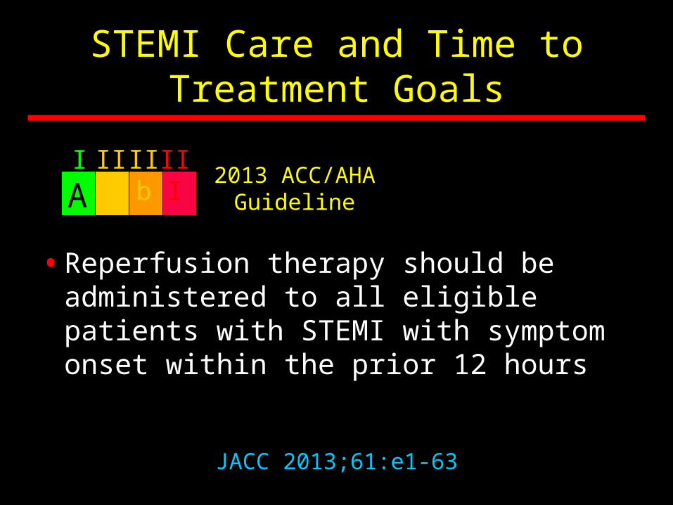
STEMI Care and Time to Treatment Goals
• Reperfusion therapy should be administered to all eligible patients with STEMI with symptom onset within the prior 12 hours
I
AIIa IIb III
2013 ACC/AHA Guideline
JACC 2013;61:e1-63

STEMI Care and Time to Treatment Goals
• Reperfusion therapy is reasonable for patients with STEMI within the prior 12 to 24 hours who have clinical and/or ECG evidence of ongoing ischemia. Primary PCI is the preferred strategy in this population
I
BIIa IIb III
2013 ACC/AHA Guideline
JACC 2013;61:e1-63
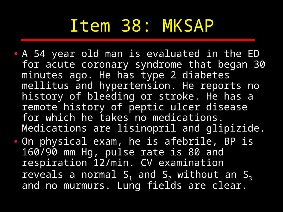
Item 38: MKSAP
• A 54 year old man is evaluated in the ED for acute coronary syndrome that began 30 minutes ago. He has type 2 diabetes mellitus and hypertension. He reports no history of bleeding or stroke. He has a remote history of peptic ulcer disease for which he takes no medications. Medications are lisinopril and glipizide.
• On physical exam, he is afebrile, BP is 160/90 mm Hg, pulse rate is 80 and respiration 12/min. CV examination reveals a normal S1 and S2 without an S3 and no murmurs. Lung fields are clear.
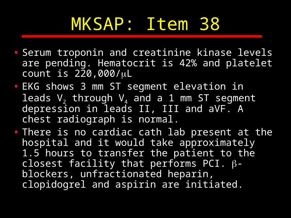
MKSAP: Item 38
• Serum troponin and creatinine kinase levels are pending. Hematocrit is 42% and platelet count is 220,000/L
• EKG shows 3 mm ST segment elevation in leads V2 through V4 and a 1 mm ST segment depression in leads II, III and aVF. A chest radiograph is normal.
• There is no cardiac cath lab present at the hospital and it would take approximately 1.5 hours to transfer the patient to the closest facility that performs PCI. -blockers, unfractionated heparin, clopidogrel and aspirin are initiated.

MKSAP: Item 38
• Which of the following is the most appropriate management?
A. Abciximab and thrombolytic therapy
B. Await the results of troponin and CK
C. Thrombolytic therapy
D. Transfer for primary PCI

STEMI Care and Time to Treatment Goals
• If the symptom duration is within 3 hours and the expected door to balloon time minus the expected door to needle time is:–Within 1 hour, primary PCI is preferred
–Greater than 1 hour, fibrinolytic therapy is generally preferred.
I
BIIa IIb III
2004 ACC/AHA Guideline
Circulation 2004;110:588-636

Door to Balloon Time for Transfer and Direct Arrival Patients, National CV Data Registry (NCDR)
Tim
e (
Min
utes
)
0
60
90
120
180
Year
30
2005 QI
Am Heart J 2011;161:76-83
150
210
2005 Q3
2006 Q1
2006 Q3
2007 Q1
2007 Q3
Transfer PCI
Direct PCITarget Door to Balloon
Time

Transfer and Direct PCI Door to Balloon Time P
erce
ntag
e of
Pat
ient
s
0
20
30
40
50
Door to Balloon Time (hours)
10
1 2 3 4 5 6
90 minutes
Direct PCI = 79 min
Transfer PCI = 149 min
9.7%
63.4% (n=86,382)
(n=29,248)
Am Heart J 2011;161:76-83

STEMI Care and Time to Treatment Goals
• Immediate transfer to a PCI-capable hospital for primary PCI is recommended strategy for STEMI patients who initially arrive at or are transported to a non-PCI-capable hospital with a FMC-to-device time goal of 120 minutes or less.
I
BIIa IIb III
2013 ACCF/AHA Guideline

STEMI Patient, First Medical Contact
PCI Capable Hospital
FMC* to Device Time
≤90 mins
Non-PCI Capable Hospital
*FMC: First Medical Contact
Anticipated FMC* to Device Time
≥120 min
Thrombolytic Therapy within
30 mins
FMC* to Device Time ≤120 min
Cath Lab for PCI
Door In Door Out (DIDO) ≤30
mins
Transfer for Primary PCI
JACC 2013;61:e1-63

Acute Coronary Syndrome
Definition
A constellation of clinical symptoms due to acute
myocardial ischemiaCirculation 2011,123:e426-e579
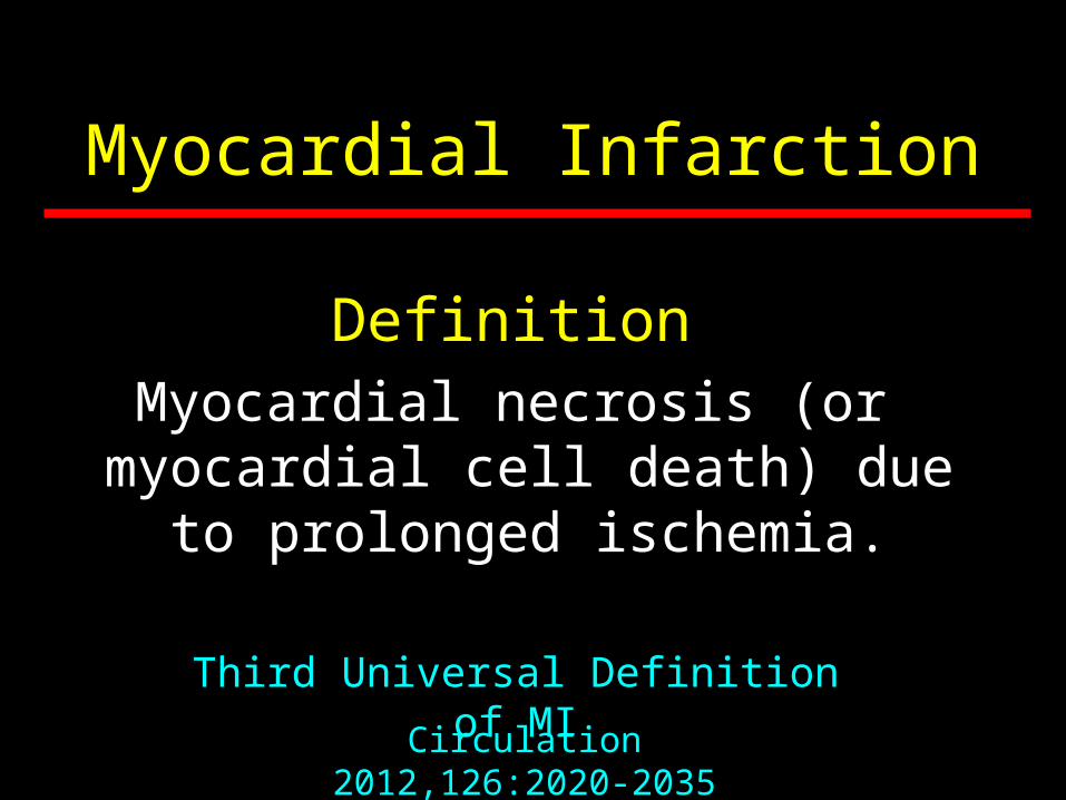
Myocardial Infarction
DefinitionMyocardial necrosis (or
myocardial cell death) due to prolonged ischemia.
Circulation 2012,126:2020-2035
Third Universal Definition of MI
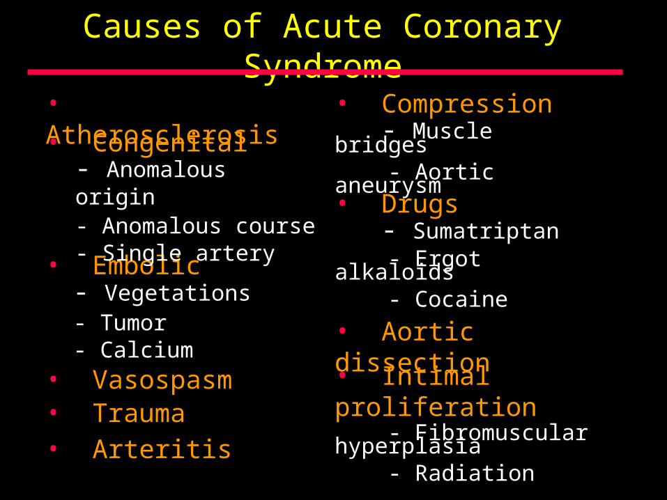
Causes of Acute Coronary Syndrome
• Congenital
• Embolic
• Vasospasm• Trauma
• Compression - Muscle bridges - Aortic aneurysm
• Drugs - Sumatriptan - Ergot alkaloids - Cocaine
• Arteritis
• Aortic dissection
• Intimal proliferation - Fibromuscular hyperplasia - Radiation
• Atherosclerosis
- Anomalous origin- Anomalous course- Single artery
- Vegetations- Tumor- Calcium
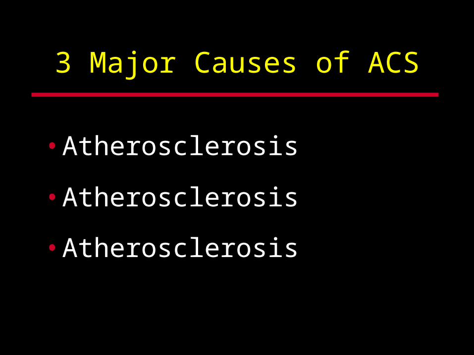
3 Major Causes of ACS
• Atherosclerosis
• Atherosclerosis
• Atherosclerosis
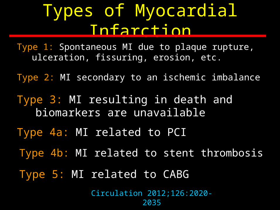
Types of Myocardial Infarction
Type 1: Spontaneous MI due to plaque rupture, ulceration, fissuring, erosion, etc.
Type 2: MI secondary to an ischemic imbalance
Type 3: MI resulting in death and biomarkers are unavailable
Type 4a: MI related to PCI
Type 4b: MI related to stent thrombosis
Type 5: MI related to CABG
Circulation 2012;126:2020-2035

MI Type 1

Plaque Rupture

Healed Plaque

Erosion

Atherosclerotic Vessel
Thrombotic Occlusion
Progressive Narrowing of the Arterial Lumen
Lipid CoreVessel Lumen
Progressive Narrowing (Time)
Clot

Atherosclerotic Vessel
Plaque Rupture
Platelet Adhesion
Activation and Aggregation
Thrombus Formation
Thrombotic Occlusion
MI
Stroke
Vascular Death
Plaque Rupture and Atherothrombosis
Lipid CoreVessel Lumen
Am J Med 1996;101:199-209
Thrombus
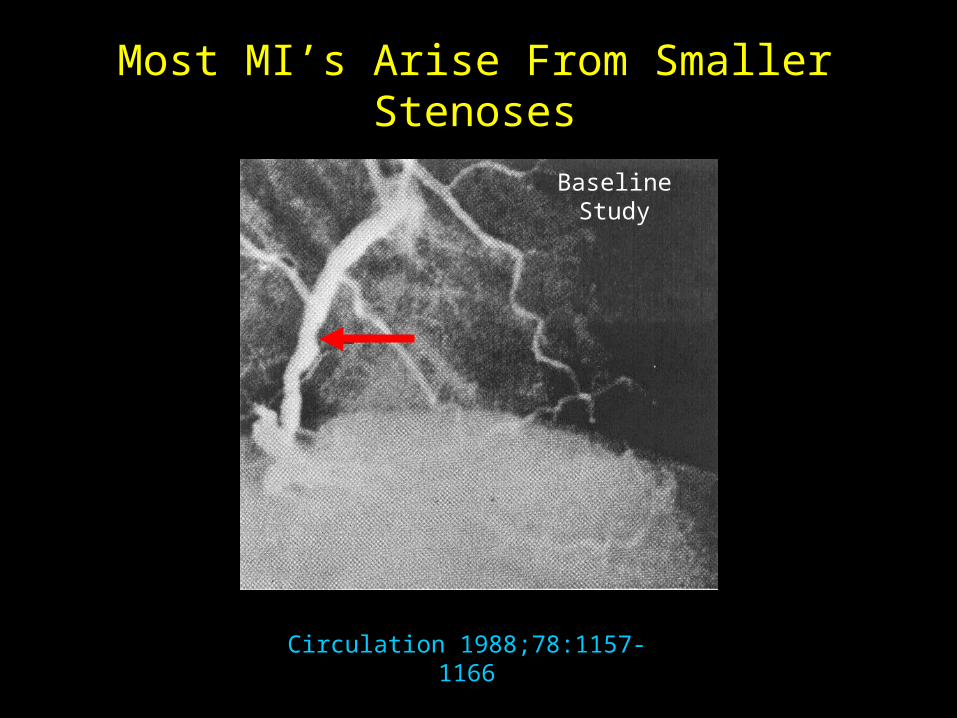
Circulation 1988;78:1157-1166
Baseline Study
Most MI’s Arise From Smaller Stenoses
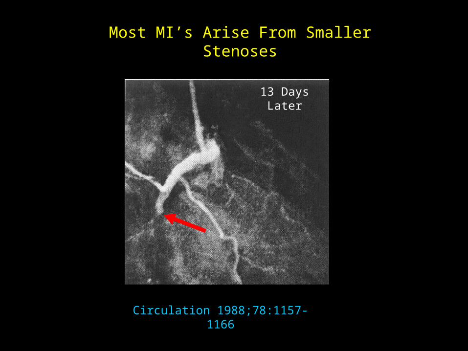
Circulation 1988;78:1157-1166
13 Days Later
Most MI’s Arise From Smaller Stenoses
Circulation 1988;78:1157-1166
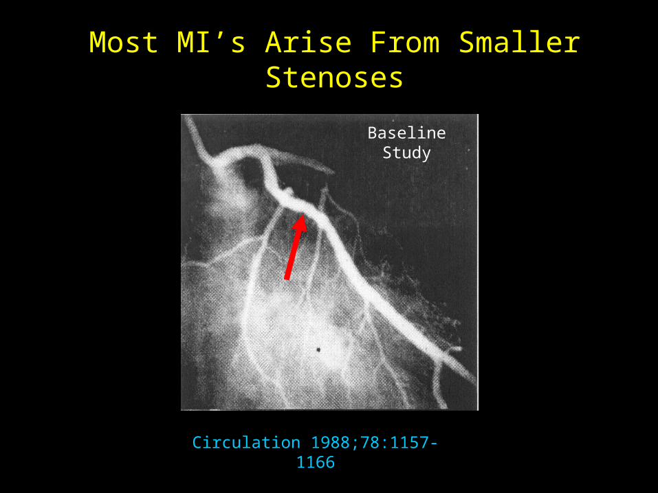
Baseline Study
Most MI’s Arise From Smaller Stenoses

Circulation 1988;78:1157-1166
2 months later
Most MI’s Arise From Smaller Stenoses
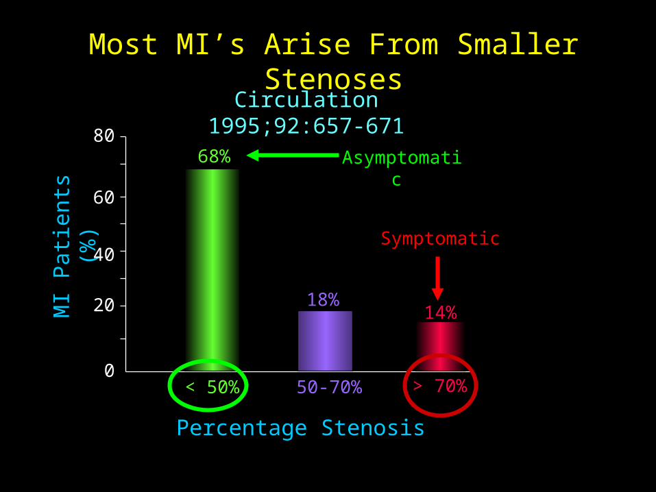
Most MI’s Arise From Smaller StenosesM
I P
atie
nts
(%)
0
20
40
60
80
< 50% 50-70% > 70%
Percentage Stenosis
68%
18%14%
Circulation 1995;92:657-671
Symptomatic
Asymptomatic
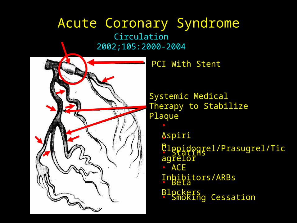
Acute Coronary SyndromeCirculation 2002;105:2000-2004
PCI With Stent
Systemic Medical Therapy to Stabilize Plaque
• Aspirin
• Clopidogrel/Prasugrel/Ticagrelor
• Statins
• ACE Inhibitors/ARBs
• Beta Blockers
• Smoking Cessation

Multiple Plaques in ACSM
I P
atie
nts
(%)
0
10
20
30
40
Culprit Lesion 2 3
Number of Ruptured Plaques in Addition to Culprit Lesion Detected by IVUS
21%25%
12.5%
Circulation 2002;106:804-808
1
29%
7.5%4.5%
4 5
79% of patients had >1 plaque ruptured

The Asymptomatic Progression of CAD
Initial Presentation
0 10 20 30 40 706050
Levy D, Textbook of CV Medicine 1998
WOMEN (70.4 years)
MEN (65.8 years)
ACS or Sudden Cardiac Death
46%
62%
AHA: Heart Disease and Stroke Statistics-2006 Update

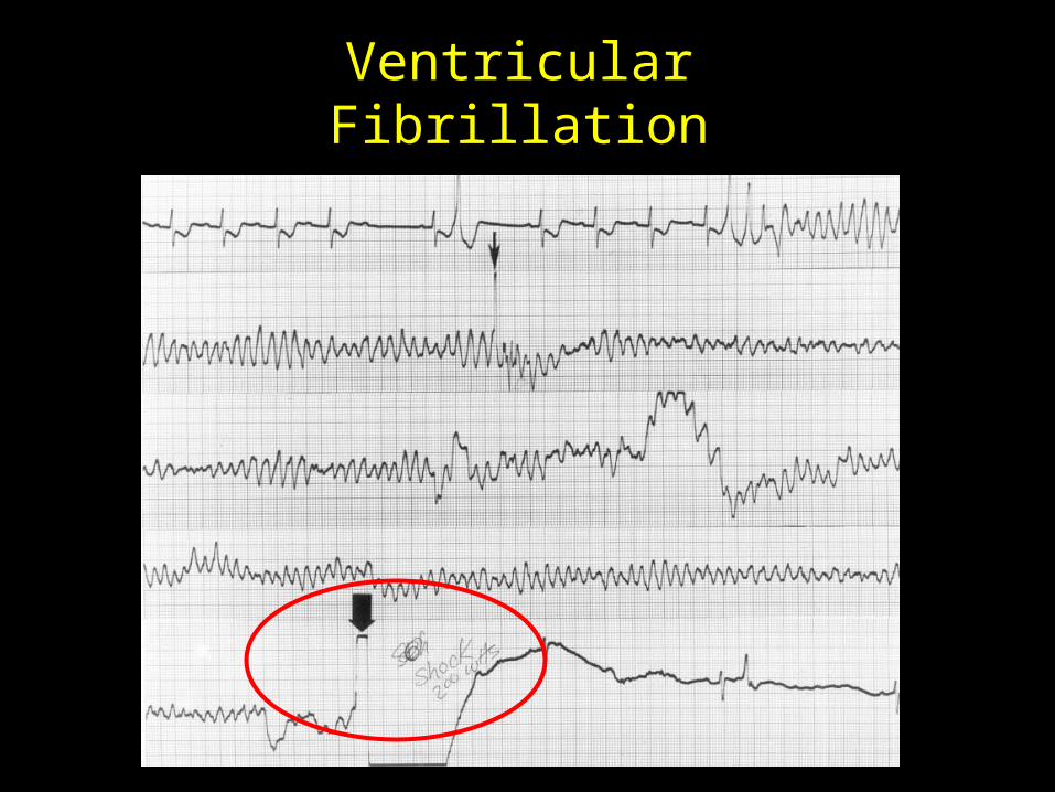
Ventricular Fibrillation
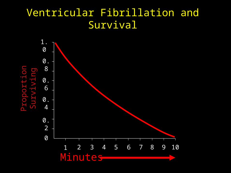
Ventricular Fibrillation and SurvivalP
ropo
rtio
n S
urvi
ving
0
0.4
0.6
0.8
1.0
Minutes
0.2
1 2 3 4 5 6 7 8 9 10
Ventricular Fibrillation

Deaths due to Acute MI
• In-hospital mortality had improved significantly– 1960’s – prior to introduction of CCUs, in-
hospital mortality averaged ~25-30%.– 1980’s – CCU, pre-reperfusion era ~16%– 1990 - 2000’s – era of fibrinolysis, coronary
interventions, those who participated in clinical trials, one month mortality is ~4-6%
Eur HJ 2208;29:2909-2945
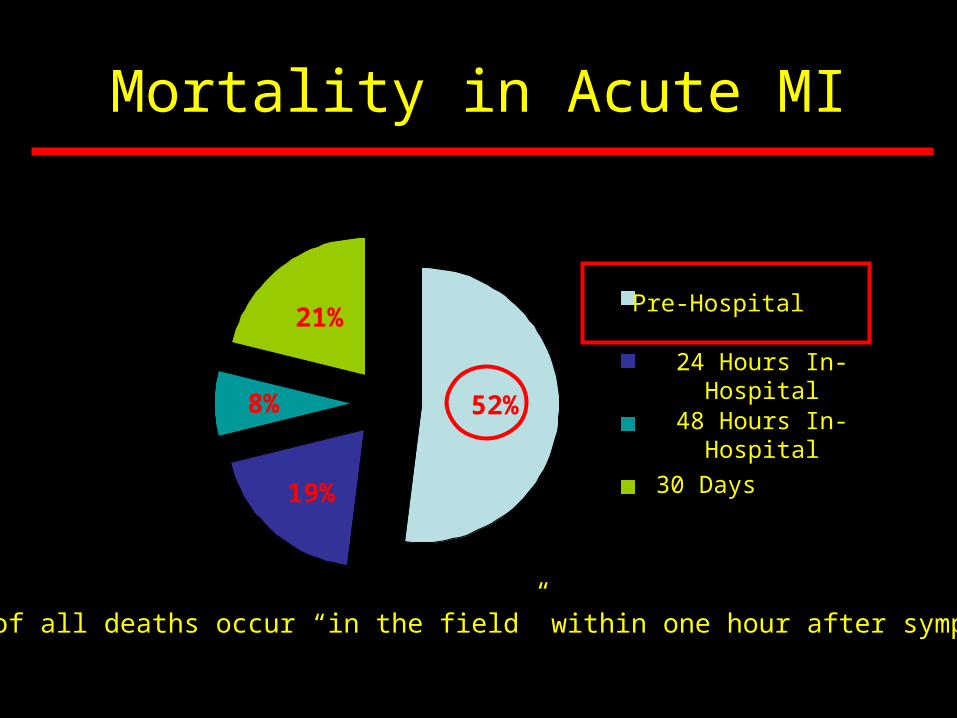
Mortality in Acute MI
Pre-Hospital
24 Hrs- InHospital
48 Hrs- InHospital
30 Days
Pre-Hospital
52%
24 Hours In-Hospital
48 Hours In-Hospital
30 Days
21%
19%
8%
One-half of all deaths occur “in the field” within one hour after symptom onset
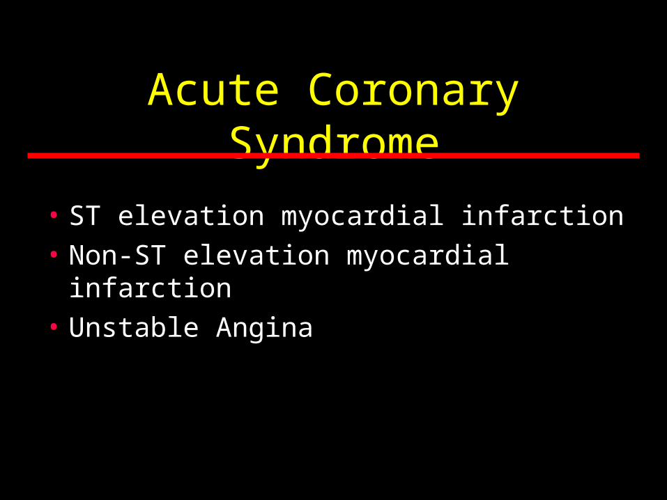
Acute Coronary Syndrome
• ST elevation myocardial infarction
• Non-ST elevation myocardial infarction
• Unstable Angina
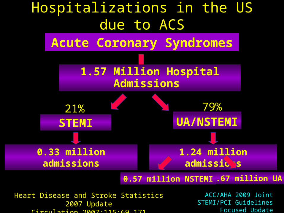
Hospitalizations in the US due to ACS
Acute Coronary Syndromes
1.57 Million Hospital Admissions
0.33 million admissions 1.24 million admissions
Heart Disease and Stroke Statistics 2007 UpdateCirculation 2007;115:69-171
0.67 million UA0.57 million NSTEMI
79%21%STEMI UA/NSTEMI
ACC/AHA 2009 Joint STEMI/PCI Guidelines Focused Update

Rates of Acute MI, 1999 - 2008In
cide
nce
Ra
te
(No
. of c
ase
s/10
0,0
00
pe
r p
ers
on-y
ear)
0
100
150
200
250
Year
50
1999 2000 2001 2002 2003 2004 2005 2006 2007 2008
JACC 2013;61e7300
STEMI
Non-STEMI
MI
Acute Coronary Syndrome
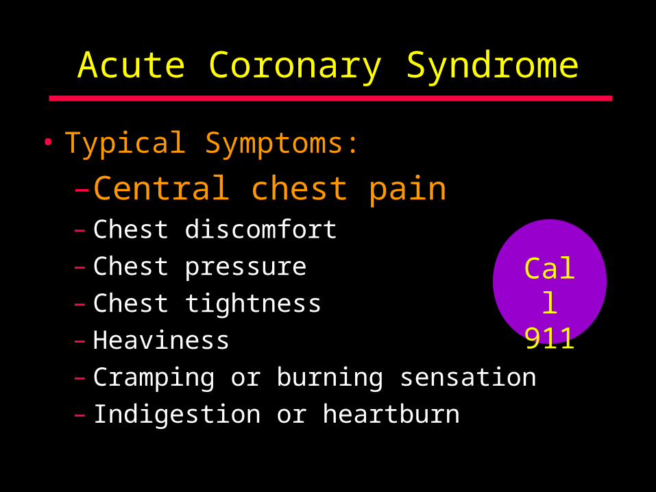
• Typical Symptoms:
–Central chest pain– Chest discomfort– Chest pressure– Chest tightness– Heaviness – Cramping or burning sensation– Indigestion or heartburn
Call 911

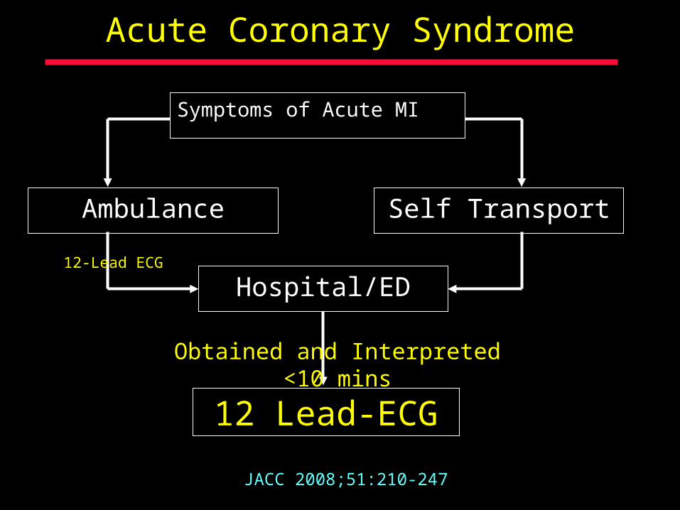
Acute Coronary Syndrome
Symptoms of Acute MI
Call 911
Ambulance Transport
Self Transport
Recommended Discouraged
Hospitalized
JACC 2008;51:210-247
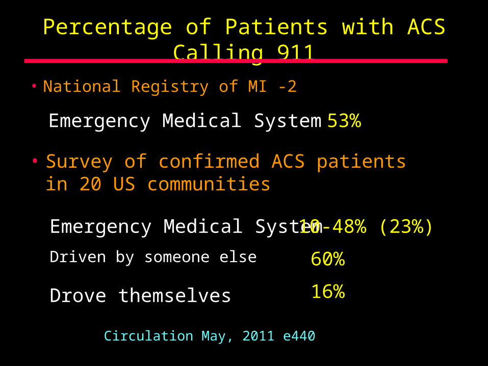
Percentage of Patients with ACS Calling 911
• National Registry of MI -2 53%
• Survey of confirmed ACS patients in 20 US communities
Emergency Medical System 10-48% (23%)
Emergency Medical System
Driven by someone else 60%
Drove themselves 16%
Circulation May, 2011 e440

Acute Coronary Syndrome
• Physical signs:– No physical signs diagnostic of Acute MI
– Activation of autonomic nervous system• Pallor• Sweating• Hypotension or narrow pulse pressure• Irregularities in heart rate, bradycardia,
tachycardia
– Basal rales
– Third heart sound
Acute Coronary Syndrome
Symptoms of Acute MI
Ambulance
JACC 2008;51:210-247
Self Transport
12 Lead-ECG
Hospital/ED
Obtained and Interpreted <10 mins
12-Lead ECG
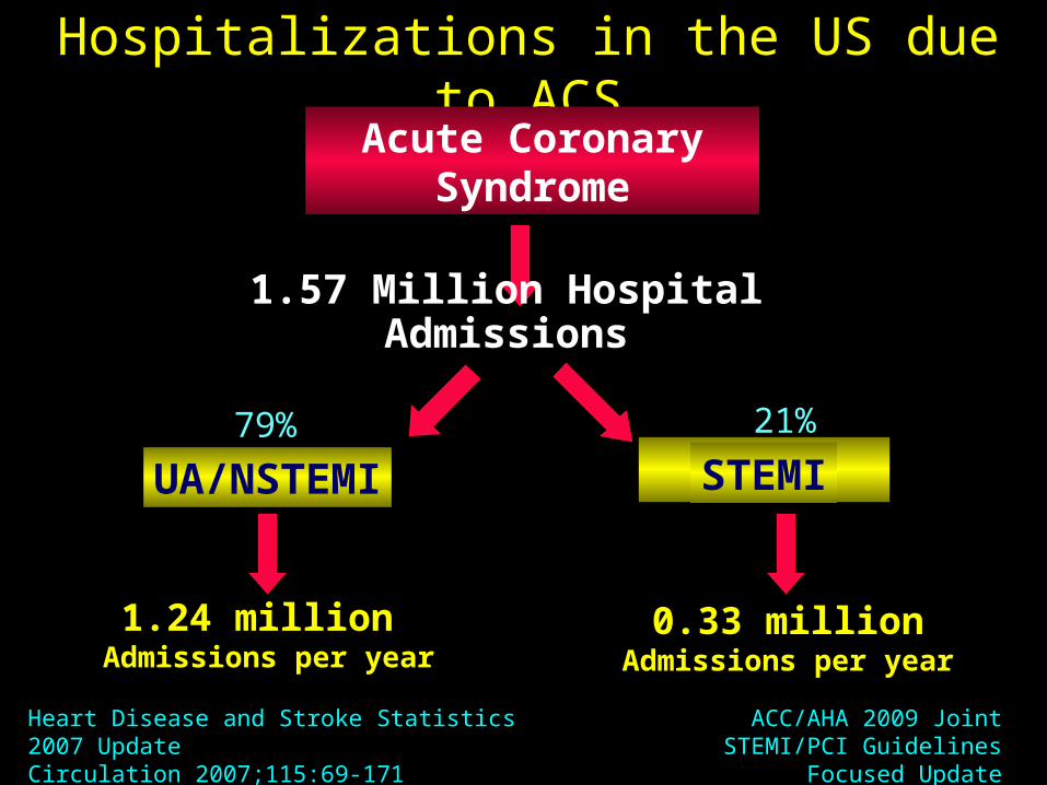
Hospitalizations in the US due to ACSAcute Coronary
Syndrome
1.57 Million Hospital Admissions
UA/NSTEMI STEMI
1.24 million Admissions per year
0.33 millionAdmissions per year
Heart Disease and Stroke Statistics 2007 UpdateCirculation 2007;115:69-171
79% 21%
ACC/AHA 2009 Joint STEMI/PCI Guidelines Focused Update

Pathophysiology
Fuster V et al. NEJM. 1992; 326: 310-318.Davies MJ et al. Circulation. 1990; 82 (Suppl II): II-38, II-46.
Lipid Lipid Pool
MacrophagesMacrophages
Stress, tensile,Stress, tensile,internalinternal
Shear forces,Shear forces,externalexternal
Atheroscleroticplaque
Fissure
Plaquerupture
LargeLargeFissureFissure
SmallSmallFissureFissure
Mural thrombusMural thrombus(unstable angina/(unstable angina/non-ST elevation MI)non-ST elevation MI)
Occlusive thrombusOcclusive thrombus(ST Elevation MI)(ST Elevation MI)
Thrombus
Acute Coronary Syndromes
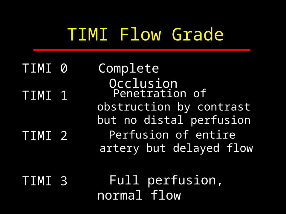
TIMI Flow Grade
TIMI 0 Complete Occlusion
TIMI 1
TIMI 2
TIMI 3
Penetration of obstruction by contrast but no distal perfusion
Perfusion of entire artery but delayed flow
Full perfusion, normal flow
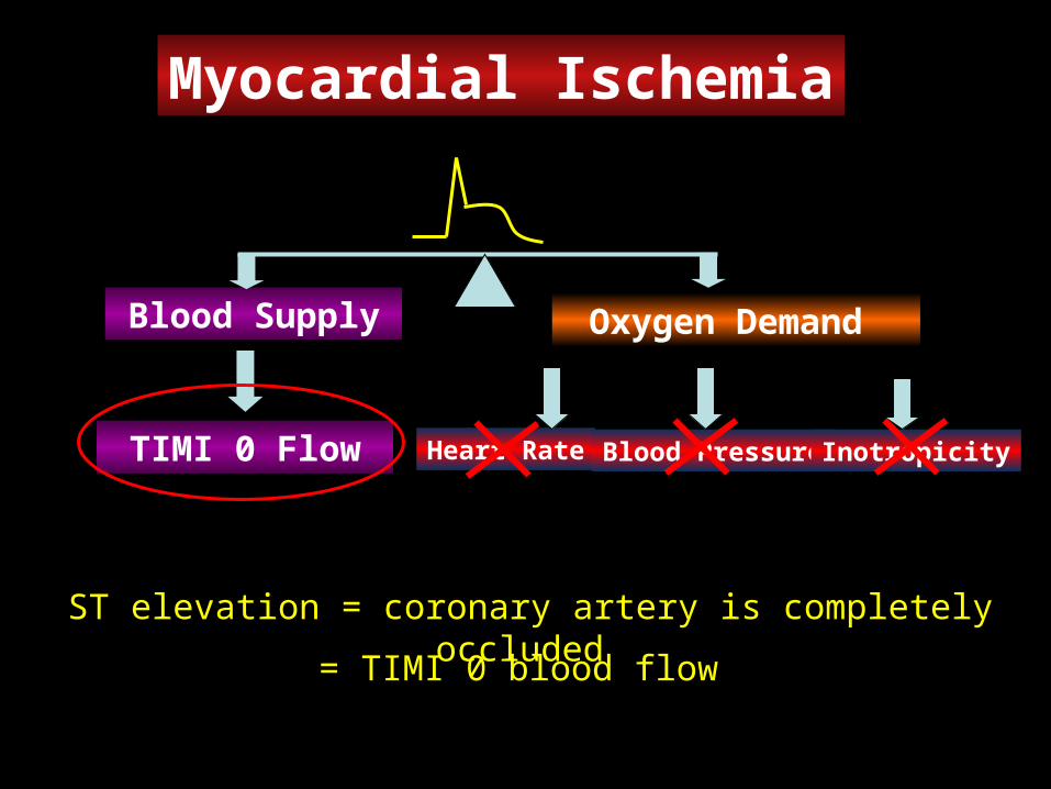
Myocardial Ischemia
Heart Rate
Oxygen Demand Blood Supply
Blood Pressure Inotropicity
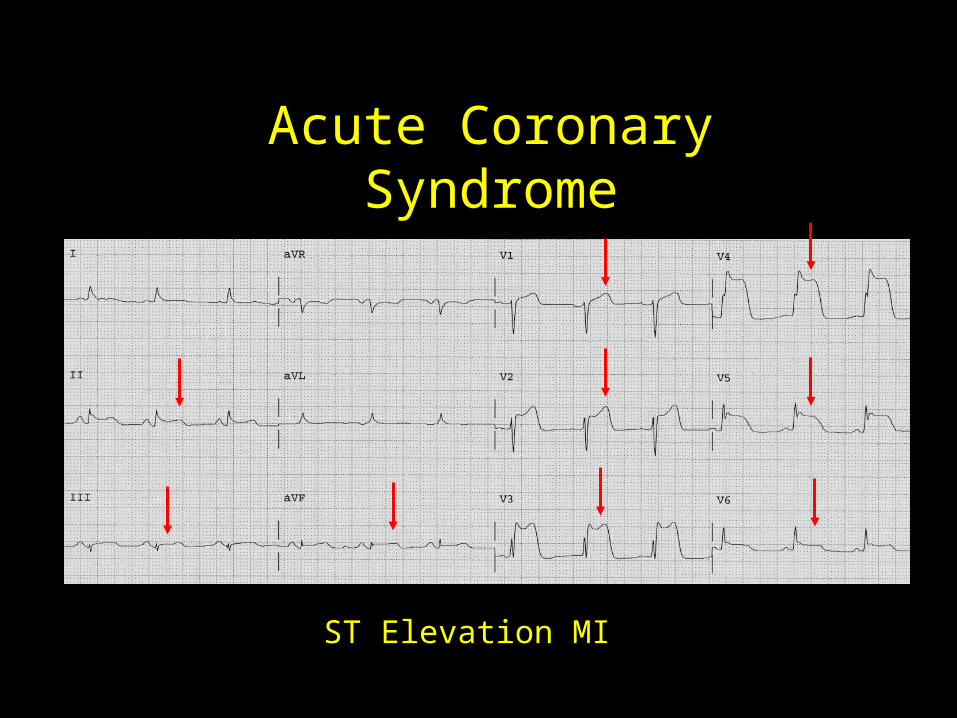
ST elevation = coronary artery is completely occluded
TIMI 0 Flow
= TIMI 0 blood flow
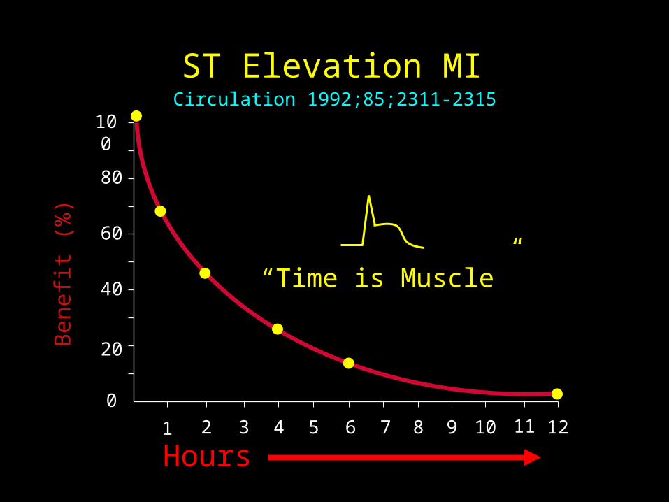
ST Elevation MIB
enef
it (%
)
0
40
60
80
100
Hours
20
1 2 3 4 5 6 7 8
“Time is Muscle”
9 10 11 12
Circulation 1992;85;2311-2315

The 12-Lead ECG
The 12-lead ECG is the only modality that can best identify the presence of a
completely occluded coronary artery

Diagnostic and Therapeutic Pathways in Patients With and Without ST-Segment Elevation
Hamm CW et al. Lancet. 2001;358:1533-1538.2002 ACC/AHA UA/NSTEMI Guideline Update. Available at: www.acc.org
Acute Coronary Syndrome
ECG
ST Elevation
Thrombolysis, PCIAspirin, clopidogrel,UFH or LMWH, 2B/3A
antagonists-blockers, nitrates
No ST Elevation

Normal ECG
Acute Coronary Syndrome
ST Elevation MI

Acute Coronary Syndrome
Chest PainASA
Aspirin
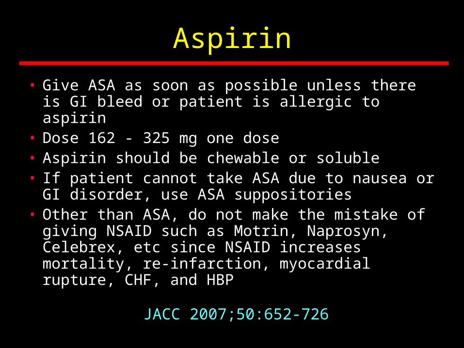
• Give ASA as soon as possible unless there is GI bleed or patient is allergic to aspirin
• Dose 162 - 325 mg one dose• Aspirin should be chewable or soluble• If patient cannot take ASA due to nausea or GI
disorder, use ASA suppositories• Other than ASA, do not make the mistake of
giving NSAID such as Motrin, Naprosyn, Celebrex, etc since NSAID increases mortality, re-infarction, myocardial rupture, CHF, and HBP
JACC 2007;50:652-726

Acute Coronary Syndrome
ST Elevation
NTG
NTG
NTG
Chest Pain
ECG within 10 minutes
ASA NTG
Arrival in ED

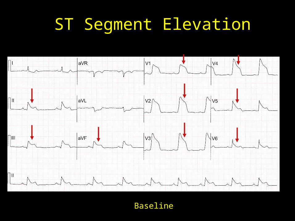
ST Segment Elevation
Baseline ECG

ST Segment Elevation
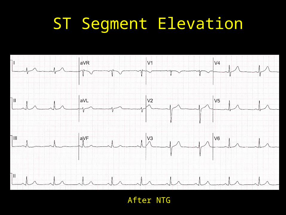
After NTG
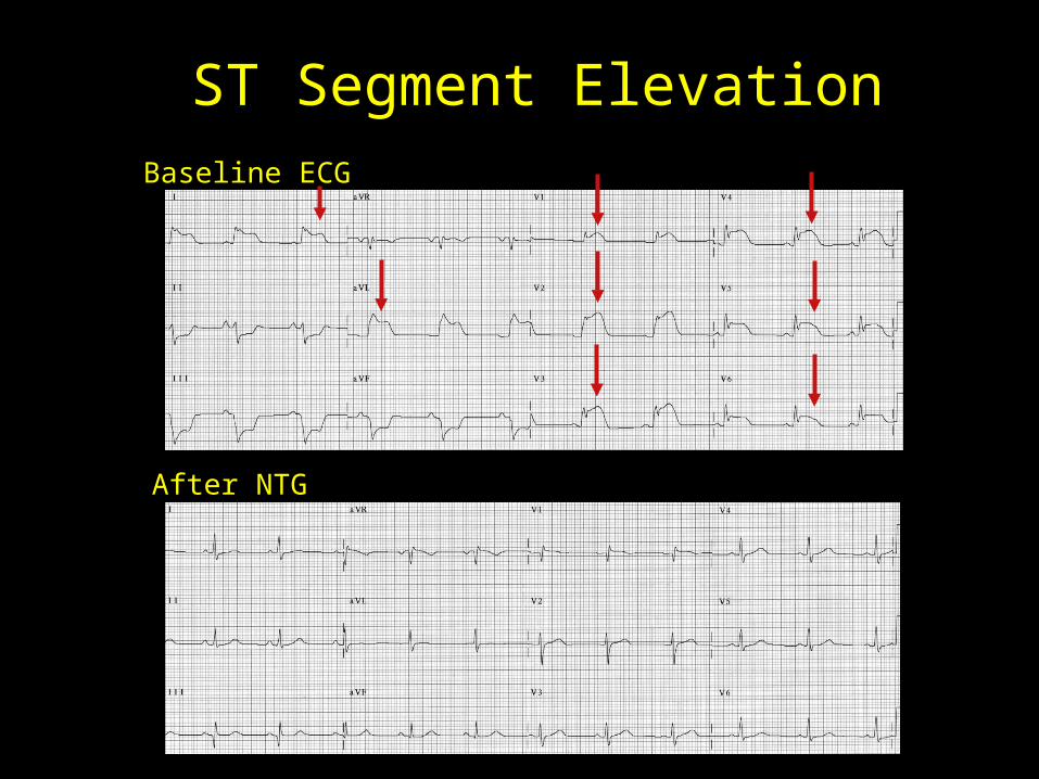
ST Segment ElevationBaseline ECG
After NTG
ST Segment Elevation
Baseline
ST Segment Elevation
After NTG

ST Segment Elevation
After NTG
Baseline
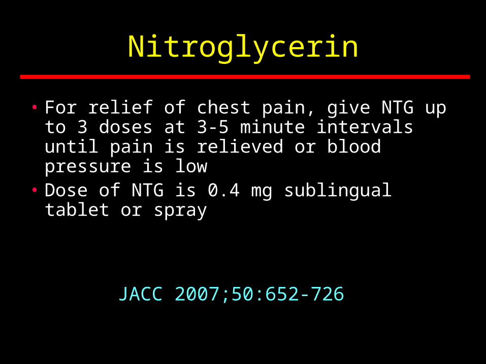
Nitroglycerin
• For relief of chest pain, give NTG up to 3 doses at 3-5 minute intervals until pain is relieved or blood pressure is low
• Dose of NTG is 0.4 mg sublingual tablet or spray
JACC 2007;50:652-726
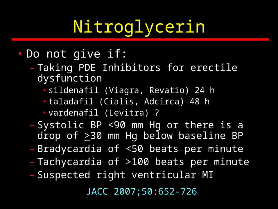
Nitroglycerin
• Do not give if: – Taking PDE Inhibitors for erectile dysfunction
• sildenafil (Viagra, Revatio) 24 h • taladafil (Cialis, Adcirca) 48 h • vardenafil (Levitra) ?
– Systolic BP <90 mm Hg or there is a drop of >30 mm Hg below baseline BP
– Bradycardia of <50 beats per minute– Tachycardia of >100 beats per minute– Suspected right ventricular MI
JACC 2007;50:652-726
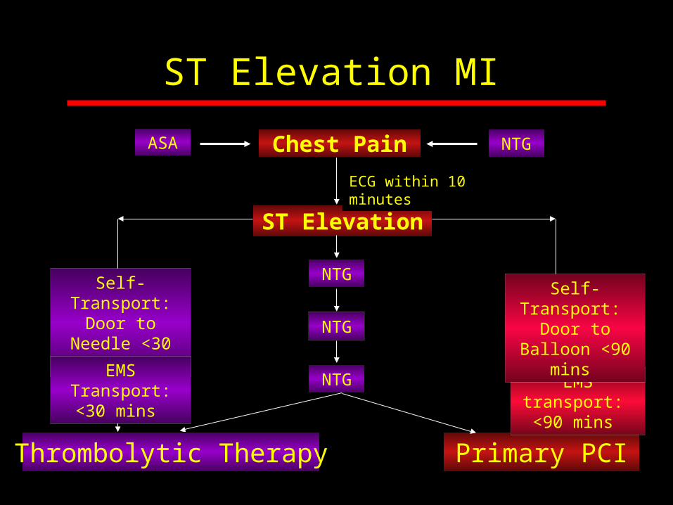
ST Elevation MI
ST Elevation
NTG
NTG
NTG
Primary PCI
EMS transport: <90 mins
Self-Transport: Door to Needle
<30 mins
EMS Transport: <30 mins
Self-Transport: Door to Balloon
<90 mins
Chest Pain
Thrombolytic Therapy
ECG within 10 minutes
ASA NTG

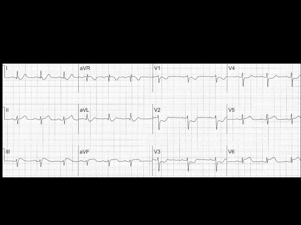
Acute Inferior MI
Thrombolytic Therapy

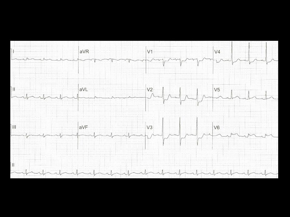
Post Thrombolytic Therapy
One Hour Later
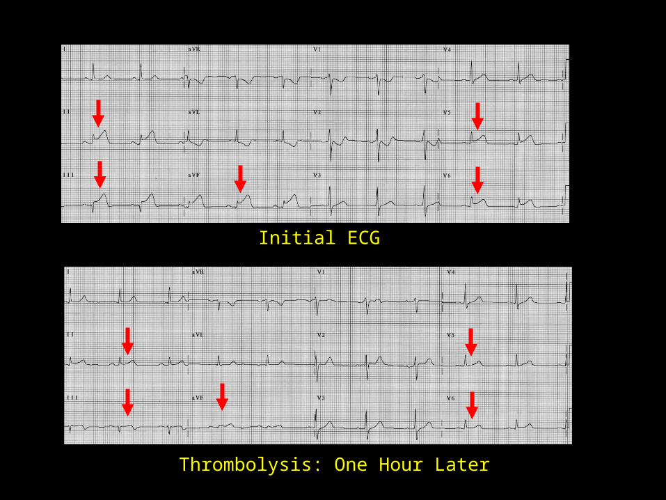
Initial ECG
Thrombolysis: One Hour Later

Thrombolysis
• No contraindication to thrombolysis• Best results within 2 hours after onset of
symptoms• Hemodynamically stable:
– Not in cardiogenic shock or CHF or with mechanical complications of AMI

Absolute Contraindications• Any prior ICH• Known structural cerebral
vascular lesion (AVM)• Known malignant intracranial
neoplasm (primary/metastatic)• Ischemic stroke within 3
months• Suspected aortic dissection• Active bleeding or bleeding
diathesis (excluding menses)• Closed head or facial trauma
within 3 months
Relative Contraindications• History of chronic severe, poorly
controlled hypertension• Severe uncontrolled hypertension
(SBP >180 mm Hg or DBP >110 mm Hg)
• History of prior ischemic stroke >3 mos, dementia or IC pathology
• Traumatic or prolonged (>10 mins) CPR or major surgery <3 weeks
• Recent (2-4 weeks) internal bleeding
• Pregnancy
• Active peptic ulcer
• Current use of anticoagulants
Contraindications to Thrombolysis
Thrombolytic Therapy and Mortality According to Admission ECG
Live
s S
aved
per
Tho
usan
d
0
20
40
60
BBB Anterior ST Elevation
Inferior ST Elevation
49%
37%
8%
Lancet 1994;343:311-322
10
30
50
-10
-14%
ST DepressionAdmission ECG
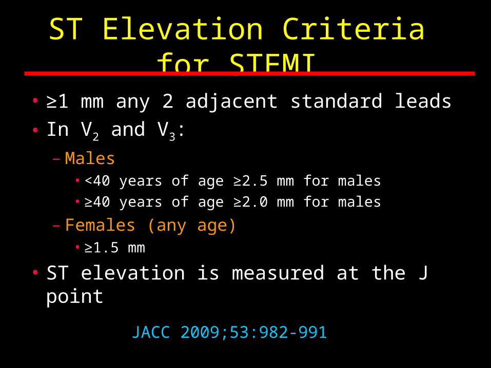
ST Elevation Criteria for STEMI
• ≥1 mm any 2 adjacent standard leads
• In V2 and V3:
– Males• <40 years of age ≥2.5 mm for males• ≥40 years of age ≥2.0 mm for males
– Females (any age)• ≥1.5 mm
• ST elevation is measured at the J point
JACC 2009;53:982-991

Fibrinolytic Agents
Fibrin-specific
JACC 2013;61e78-140
● Tenecteplase (TNK-tPA)
● Reteplase (rPA)
● Alteplase (tPA)
Non-fibrin-specific
● Streptokinase
Patency Rate90 min TIMI 2 or 3
85%
84%
73-84%
60-68%(No longer marketed in the US)
ST Elevation MI
ST Elevation
NTG
NTG
NTG
Primary PCI
EMS transport: ≤90 mins
Self-Transport: Door to Needle
<30 mins
EMS Transport: <30 mins
Self-Transport: Door to Balloon
≤90 mins
Chest Pain
Thrombolytic Therapy
ECG within 10 minutes
ASA NTG


AMI: Post PCI

ST Segment Elevation
Admission
Post PCI
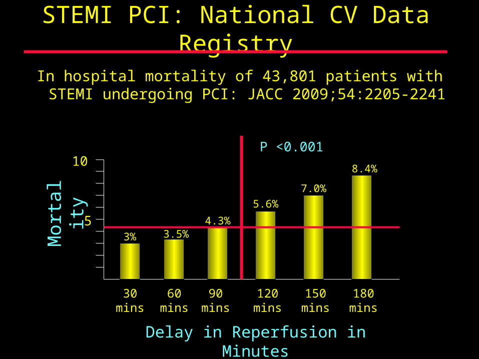
STEMI PCI: National CV Data Registry
In hospital mortality of 43,801 patients with STEMI undergoing PCI: JACC 2009;54:2205-2241
30 mins
60 mins
3% 3.5%4.3%
90 mins
120 mins
5.6%
5
10
150 mins
7.0%
180 mins
8.4%
Mor
talit
y
Delay in Reperfusion in Minutes
P <0.001

Primary PCI vs IV Thrombolytic Therapy for Acute MI: Review of 23 Randomized Trials
• 23 randomized clinical trials with 7739 patients with STEMI– Thrombolytic therapy = 3867– Primary PCI = 3872
• Results: Primary PCI was better than thrombolytic therapy at reducing short-term and long-term death, non-fatal reinfarction, stroke and combined endpoint of death, non-fatal reinfarction and stroke
• Conclusion: Primary PCI is more effective than thrombolytic therapy for the treatment of STEMI
Lancet 2003;361:13-20
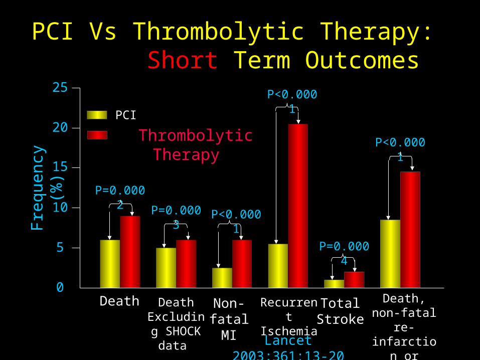
PCI Vs Thrombolytic Therapy: Short Term OutcomesF
requ
ency
(%
)
0
5
10
15
20
25
Death
P=0.0002
P=0.0003
Death Excluding SHOCK
data
P<0.0001
Non-fatal MI
P<0.0001
Recurrent Ischemia
Thrombolytic TherapyPCI
P=0.0004
Total Stroke
Death, non-fatal re-
infarction or stroke
P<0.0001
Lancet 2003;361:13-20

PCI Vs Thrombolytic Therapy: Long Term Outcomes
Fre
quen
cy (
%)
0
10
20
30
40
50
Death
P=0.0019
P=0.0053
Death Excluding SHOCK
data
P<0.0001
Non-fatal MI
P<0.0001
Recurrent Ischemia
Thrombolytic TherapyPTCA
Data Not Available
Total Stroke
Death, non-fatal re-
infarction or stroke
P<0.0001
Lancet 2003;361:13-20
* *

Options for Transport of Patients With STEMI and Initial Reperfusion Treatment
EMS Transport
Onset of symptoms of
STEMI
9-1-1EMS
Dispatch
EMS on-scene• Encourage 12-lead ECGs.
• Consider prehospital fibrinolytic if capable and EMS-to-needle within
30 min.
GOALS
PCIcapable
Not PCIcapable
Hospital fibrinolysis:
Door-to-Needle
within 30 min.
EMS Triage
Plan
Inter-HospitalTransfer
Golden Hour = first 60 min. Total ischemic time: within 120 min.
Patient EMS Prehospital fibrinolysisEMS-to-needlewithin 30 min.
EMS transportEMS-to-balloon within 90
min..Patient self-transport
Hospital door-to-balloon within 90 min.
Dispatch1 min.
5 min.
8 min.
Antman EM, et al. J Am Coll Cardiol 2008. Published ahead of print on December 10, 2007. Available at http://content.onlinejacc.org/cgi/content/full/j.jacc.2007.10.001.
PCIcapable
Fibrinolysis Door-to-Needle or
FMC to Needle
< 30 mins
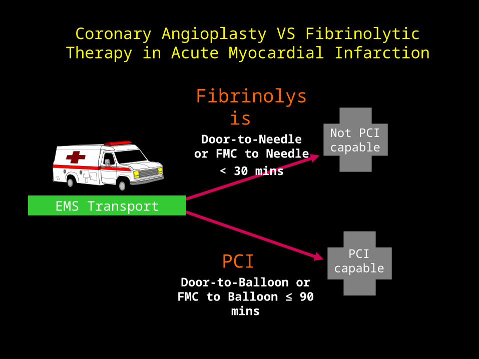
Coronary Angioplasty VS Fibrinolytic Therapy in Acute Myocardial Infarction
EMS Transport
PCI Door-to-Balloon or FMC
to Balloon ≤ 90 mins
Not PCIcapable
PCIcapable
Not PCIcapable
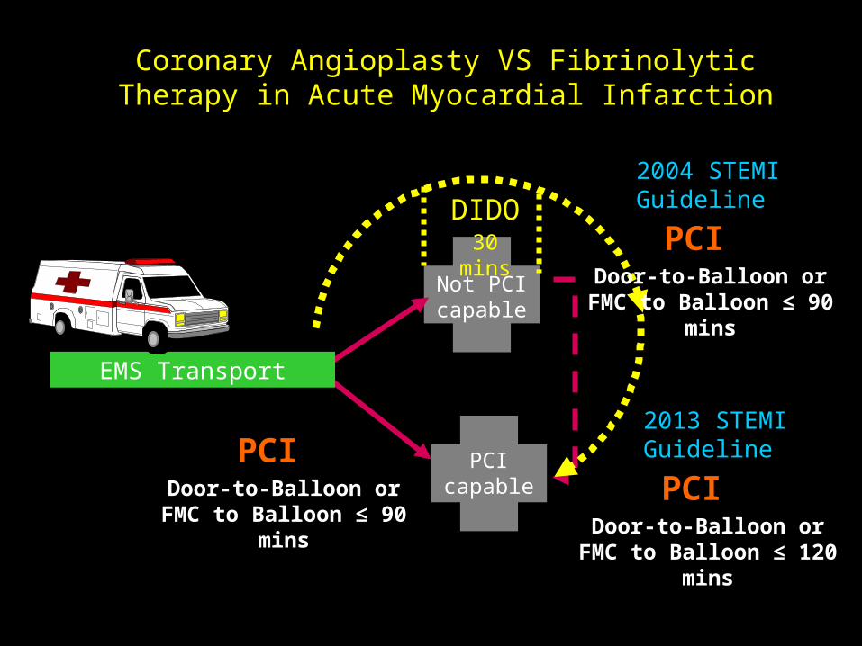
Coronary Angioplasty VS Fibrinolytic Therapy in Acute Myocardial Infarction
EMS Transport
PCI Door-to-Balloon or FMC
to Balloon ≤ 90 mins
2004 STEMI Guideline
PCI Door-to-Balloon or FMC
to Balloon ≤ 90 mins
2013 STEMI Guideline
PCI Door-to-Balloon or FMC to Balloon ≤ 120 mins
DIDO30 mins

Mortality and Ejection FractionO
ne Y
ear
Car
diac
Mor
talit
y (%
)
0
20
30
40
50
Radionuclide Ejection Fraction (%)
10
10 20 30 40 50 60 70 800
< 20%N = 799
Mean EF = 46%
> 60%40-59%
20-39%

STEMI: Standard Therapy
• Thrombolytic Agent or PCI• Aspirin
• Beta Blockers within 24 hours
• ACE Inhibitors or ARB’s within 24 hours
• Aldosterone antagonists for EF ≤40%
• Statins before hospital discharge
• Heparin
• Clopidogrel

Related Documents