See discussions, stats, and author profiles for this publication at: https://www.researchgate.net/publication/233745578 Activity-regulated RNA editing in select neuronal subfields in hippocampus Article in Nucleic Acids Research · November 2012 DOI: 10.1093/nar/gks1045 · Source: PubMed CITATIONS 23 READS 30 4 authors: Ales Balik Academy of Sciences of the Czech Republic 31 PUBLICATIONS 502 CITATIONS SEE PROFILE Andrew Charles Penn University of Sussex 19 PUBLICATIONS 575 CITATIONS SEE PROFILE Zsofia Nemoda Semmelweis University 64 PUBLICATIONS 1,508 CITATIONS SEE PROFILE Ingo Greger University of Cambridge 59 PUBLICATIONS 1,535 CITATIONS SEE PROFILE All content following this page was uploaded by Andrew Charles Penn on 01 December 2016. The user has requested enhancement of the downloaded file.

Welcome message from author
This document is posted to help you gain knowledge. Please leave a comment to let me know what you think about it! Share it to your friends and learn new things together.
Transcript

Seediscussions,stats,andauthorprofilesforthispublicationat:https://www.researchgate.net/publication/233745578
Activity-regulatedRNAeditinginselectneuronalsubfieldsinhippocampus
ArticleinNucleicAcidsResearch·November2012
DOI:10.1093/nar/gks1045·Source:PubMed
CITATIONS
23
READS
30
4authors:
AlesBalik
AcademyofSciencesoftheCzechRepublic
31PUBLICATIONS502CITATIONS
SEEPROFILE
AndrewCharlesPenn
UniversityofSussex
19PUBLICATIONS575CITATIONS
SEEPROFILE
ZsofiaNemoda
SemmelweisUniversity
64PUBLICATIONS1,508CITATIONS
SEEPROFILE
IngoGreger
UniversityofCambridge
59PUBLICATIONS1,535CITATIONS
SEEPROFILE
AllcontentfollowingthispagewasuploadedbyAndrewCharlesPennon01December2016.
Theuserhasrequestedenhancementofthedownloadedfile.
Activity-regulated RNA editing in select neuronalsubfields in hippocampusAles Balik1,2, Andrew C. Penn1, Zsofia Nemoda1 and Ingo H. Greger1,*
1Neurobiology Division, MRC Laboratory of Molecular Biology, Cambridge, CB2 0QH, UK and 2Institute ofPhysiology, ASCR v.v.i., 142 20 Prague, Czech Republic
Received August 11, 2012; Revised October 3, 2012; Accepted October 8, 2012
ABSTRACT
RNA editing by adensosine deaminases is a wide-spread mechanism to alter genetic information inmetazoa. In addition to modifications in non-codingregions, editing contributes to diversification ofprotein function, in analogy to alternative splicing.However, although splicing programs respond toexternal signals, facilitating fine tuning and homeo-stasis of cellular functions, a similar regulation hasnot been described for RNA editing. Here, we showthat the AMPA receptor R/G editing site is dynamic-ally regulated in the hippocampus in response toactivity. These changes are bi-directional, reversibleand correlate with levels of the editase Adar2. Thisregulation is observed in the CA1 hippocampalsubfield but not in CA3 and is thus subfield/celltype-specific. Moreover, alternative splicing ofthe flip/flop cassette downstream of the R/G site isclosely linked to the editing state, which is regulatedby Ca2+. Our data show that A-to-I RNA editing hasthe capacity to tune protein function in response toexternal stimuli.
INTRODUCTION
Adenosine-to-inosine (A-to-I) RNA editing is a uniquemechanism to expand and diversify functions of theprotein repertoire (1). Select adenosines in pre-mRNAare targeted by enzymatic deamination to inosine, whichis read as guanosine during translation. Editing by adeno-sine deaminases acting on RNA (Adars) requires complexRNA secondary structures, which in most cases areformed between the editing-site region and a complemen-tary inverted repeat sequence (1). In addition to alteringreading frames, A-to-I editing targets non-coding regions,which impacts on splicing and RNA metabolism (2–4).
A-to-I editing targets are abundant in nervous systemswhere Adar levels and in turn the concentration ofinosine-containing RNA are highest (5–7). Proteinsinvolved in synaptic transmission, including ion channelsand G-protein coupled receptors, are recoded at strategicpositions in both vertebrates and invertebrates (6,8,9).These changes result in profound alterations in neuronalsignaling and can give rise to severe neurological disorderswhen mis-regulated (10,11). A dramatic example isprovided by alpha-amino-3-hydroxy-5-methyl-4-isoxa-zolepropionic acid (AMPA)-type ionotropic glutamatereceptors, the main mediators of fast excitatory neuro-transmission (12). Here, editing targets the channel poreand the ligand-binding domain (LBD) of the receptor (13).Editing at the Q/R site in the pore affects ion selectivity(rendering the edited channel Ca2+ impermeable) andsubunit assembly (14,15). Q/R editing is essential forsurvival of the organism and is compromised in avariety of neurological disorders, including epilepsy(10,16). In contrast, editing at the R/G site alters thespeed of recovery from a non-conducting, desensitizedstate and thereby influences how the receptor decodestrains of incoming action potentials (17). Like the Q/Rsite, R/G editing also modulates receptor assembly bylimiting the capacity of GluA2 to form homomericchannels in the endoplasmic reticulum (ER) and therebypromotes the formation of (functionally diverse) AMPARheterotetramers (18). Together, editing of the GluA2subunit has a profound impact on AMPAR-mediatedneurotransmission at various levels.Alternative splicing can be subject to regulation by
external stimuli, including hormones and cell depolariza-tion, thus providing adaptive means to orchestrate proteindiversification (19). In many cases, this involves Ca2+ sig-naling. A well-described example is the splicing regulationof the STREX exon in BK potassium channel transcriptsvia activation of Ca2+/calmodulin kinase IV (20). In thecase of AMPA receptors, alternative splicing of the
*To whom correspondence should be addressed. Tel: +44 1223 402 173; Fax: +44 1223 402 310; Email: [email protected] addresses:Andrew C. Penn, Univ. de Bordeaux and CNRS, Interdisciplinary Institute for Neuroscience, UMR 5297, F-33000 Bordeaux, France.Zsofia Nemoda, Institute of Medical Chemistry, Molecular Biology and Pathobiochemistry, Semmelweis University, Budapest, Hungary.
Nucleic Acids Research, 2012, 1–11doi:10.1093/nar/gks1045
! The Author(s) 2012. Published by Oxford University Press.This is an Open Access article distributed under the terms of the Creative Commons Attribution License (http://creativecommons.org/licenses/by-nc/3.0/), whichpermits non-commercial reuse, distribution, and reproduction in any medium, provided the original work is properly cited. For commercial re-use, please [email protected].
Nucleic Acids Research Advance Access published November 20, 2012 at IN
IST-CNRS on January 5, 2013
http://nar.oxfordjournals.org/D
ownloaded from
mutually exclusive flip/flop (i/o) exons (encoding residueswithin the LBD dimer interface) responds to Ca2+throughL-type Ca2+ channels (21). The i/o cassette lies immedi-ately downstream of the R/G site and similarly impacts onAMPAR biogenesis and gating (22–25). Although Adar1can be induced under specific pathological conditions [e.g.response to viral infection (26)], whether editing by Adarscould also be regulated by physiological cues is currentlyunclear.Here, we report that RNA editing by Adar2 responds to
activity in an intact neuronal circuit. This regulation iscell-type-specific, bi-directional and involves Ca2+ influx.Moreover, not all editing sites respond to the same degree,which will be linked to features of the RNA substrate andAdar selectivity. The AMPAR GluA2 R/G site showsbi-directional regulation, which is reversible. R/G editingcorrelates with Adar2 mRNA levels, which are elevatedunder high-activity conditions but reduced when activity islowered. In addition, editing is closely correlated to alter-native splicing at the alternative i/o exons, positioned im-mediately downstream of the R/G site. Recoding thisediting site in response to external cues will shapeAMPAR biogenesis and kinetics and is thereby expectedto tune excitatory neurotransmission.
MATERIALS AND METHODS
Slice preparation and treatments
All procedures were carried out in accordance with UKHome Office regulations. Sprague–Dawley rathippocampi were dissected from pups (postnatal age 5days) in a sucrose-modified Gey’s balanced salt solution,which was (in mM): sucrose (175), NaCl (50), KCl (2.5)Na2HPO4 (0.85), KH2PO4 (0.66), MgSO4 (0.28), MgCl2(2) CaCl2 (0.5), glucose (25) and 10 mg/ml phenol red(!330 mOsm, pH 7.3). Transverse hippocampal slices(350-mm thick) were cultured using the roller-tubemethod on collagen-coated coverslips in an incubator at36"C without humidity or CO2 control (27) or using aninterface method. Culture medium contained (all fromGIBCO) 50% Basal Medium Eagles, 25% Hank’sbalanced salt solution, 25% heat-inactivated horseserum, 1mM L-Glutamine and 6.5 g/l dextrose (320mOsm). At 3–4 days in vitro, slices were fed with culturemedium supplemented with 1 mM each cytosine b-D-arabinofuranoside, 5-fluoro-20-deoxyuridine and uridine(Sigma). Slices were fed the following day and twiceweekly thereafter (without antimitotics).Slices were cultured for at least 3 weeks prior to treat-
ments. For treatments, slices were fed with culturemedium containing tetrodotoxin (TTX, 1 mM), bicuculline(BIC, 20 mM), Nifedipine (NIF, 100 mM), Carbachol (Carb20 mM), kainate (10 mM) or no drug (CTRL). Slices werereturned to the incubator for a duration of 48 h, afterwhich they were dissected for molecular biology.Acute rat brain tissue was dissected in a phosphate
buffer salt solution (PBS, pH 7.2). Transversehippocampal slices (400 mm) were cut using a tissuechopper (McIllwain). Slices were collected in ice-coldPBS and hippocampal subfields were dissected under a
dissection microscope (Zeiss) and RNA extracted asdescribed below.
Molecular biology
RNA was extracted from tissue with Trizol according tothe manufacturer’s instructions (Invitrogen). The nucleicacid pellet was re-suspended in RNase-free water, treatedwith DNase I and used as template for random primedcDNA synthesis catalyzed by Moloney Murine LeukemiaVirus (MMLV) reverse transcriptase (Promega).Polymerase chain reaction (PCR) was conducted withThermus aquaticus (Taq) polymerase (Invitrogen) usingstandard protocols. Amplicons were either sequenceddirectly (Geneservice, UK) or cloned into pGEM T-Easyvector (Promega) and transformed into Escherichia colifor individual clone sequencing (GATC, Germany). Peakheights in sequence chromatograms were quantified usingPeakPicker software (28). To quantify splicing, the meanof the first five different base positions for the alternativelyspliced exon were used. To calculate simultaneous 95%confidence intervals (CIs) for the multinomial subunitvariant proportions in Figure 2C, we used the method ofGoodman (29).
For analysis of Adar2 splicing (and thus self-editing),the PCR amplicon of the ROI was run on agarose gelsand post-stained with ethidium bromide. Quantificationswere made on gel images by measuring band peakintensities using ImageJ (NIH).
Quantitative real-time PCR
Alternations in ADARs expression levels were determinedby real-time PCR. Pre-designed TaqMan assays (Adar1Rn00508006_m1, Adar2 Rn00563671_ml, GAPD4352338E; b-2-microglobulin Rn00560865_m1; ABI)were used and validated according to manufacture proto-cols. Reactions were run on the Rotor-Gene 6000 andanalyzed with Rotor-Gene Software1.7 (Qiagen).
Bioinformatics
The R/G editing substrates of GluA2–4 published for rat(17) were used as a Basic Local Alignment Search Tool(BLAST) or BLAST-Like Alignment Tool (BLAT) searchqueries to indentify homologues from the online Ensembland Pre-Ensembl vertebrate assemblies (30–32). Specieswith diploid genomes were selected to represent majortaxonomic branches in vertebrate evolution. Sharksequence was retrieved from the whole genome shotgunsequence database on the Institute of Molecular and CellBiology server (33). The sequence coordinates andassociated information are documented inSupplementary Table S1. The subunit identity of theediting substrates (and the adjacent translated exon 13sequence) for the paralogs was confirmed from pairwisealignment output information using Clustalw2 (www.ebi.ac.uk/Tools/msa/clustalw2/, data not shown). Sequenceswere then aligned using Multiple Alignment based onFast Fourier Transform (MAFFT) by iterative refinementwith pairwise alignment information [G-INS-i; Version6.903; (34)]. Sequence similarity was calculated for eachparalog and data output from the PLOTCON application
2 Nucleic Acids Research, 2012
at INIST-CN
RS on January 5, 2013http://nar.oxfordjournals.org/
Dow
nloaded from
in European Molecular Biology Open Software Suite(EMBOSS) using the EDNA scoring matrix and awindow size of 1. The g-centroid consensus structurewas predicted from the alignment using CentroidAlifoldwith a default inference engine [Version 0.0.9; (35)]. Thecommon structure was superimposed onto the rat orconsensus pre-mRNA sequence and visualized withVisualization Applet for RNA (VARNA) [Version 3.8;(36)]. The posteriors were used to plot the probabilitiesof base pairs shown in the consensus structure. Topredict the strength of the exon 13 splice donor beforeand after editing, we used an algorithm developed by C.Burge, available at: http://genes.mit.edu/burgelab/maxent/Xmaxentscan_scoreseq.html.
Data analysis
Most statistical comparisons were performed usingnon-parametric methods in Prism 5.0b or Instat 3.1a(Graphpad Software Inc). Other statistical analyses wereperformed using R 2.81 (http://www.r-project.org/).Graphical presentation of data was carried out in Excel(Microsoft).
RESULTS
Activity-mediated RNA editing in CA1 neurons
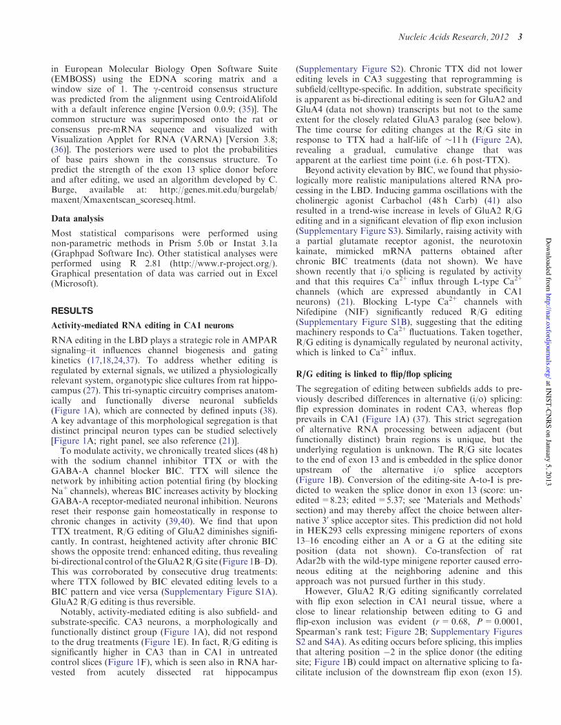
RNA editing in the LBD plays a strategic role in AMPARsignaling–it influences channel biogenesis and gatingkinetics (17,18,24,37). To address whether editing isregulated by external signals, we utilized a physiologicallyrelevant system, organotypic slice cultures from rat hippo-campus (27). This tri-synaptic circuitry comprises anatom-ically and functionally diverse neuronal subfields(Figure 1A), which are connected by defined inputs (38).A key advantage of this morphological segregation is thatdistinct principal neuron types can be studied selectively[Figure 1A; right panel, see also reference (21)].
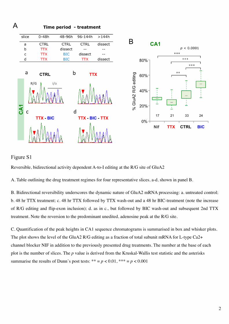
To modulate activity, we chronically treated slices (48 h)with the sodium channel inhibitor TTX or with theGABA-A channel blocker BIC. TTX will silence thenetwork by inhibiting action potential firing (by blockingNa+channels), whereas BIC increases activity by blockingGABA-A receptor-mediated neuronal inhibition. Neuronsreset their response gain homeostatically in response tochronic changes in activity (39,40). We find that uponTTX treatment, R/G editing of GluA2 diminishes signifi-cantly. In contrast, heightened activity after chronic BICshows the opposite trend: enhanced editing, thus revealingbi-directional control of theGluA2R/G site (Figure 1B–D).This was corroborated by consecutive drug treatments:where TTX followed by BIC elevated editing levels to aBIC pattern and vice versa (Supplementary Figure S1A).GluA2 R/G editing is thus reversible.
Notably, activity-mediated editing is also subfield- andsubstrate-specific. CA3 neurons, a morphologically andfunctionally distinct group (Figure 1A), did not respondto the drug treatments (Figure 1E). In fact, R/G editing issignificantly higher in CA3 than in CA1 in untreatedcontrol slices (Figure 1F), which is seen also in RNA har-vested from acutely dissected rat hippocampus
(Supplementary Figure S2). Chronic TTX did not lowerediting levels in CA3 suggesting that reprogramming issubfield/celltype-specific. In addition, substrate specificityis apparent as bi-directional editing is seen for GluA2 andGluA4 (data not shown) transcripts but not to the sameextent for the closely related GluA3 paralog (see below).The time course for editing changes at the R/G site inresponse to TTX had a half-life of !11 h (Figure 2A),revealing a gradual, cumulative change that wasapparent at the earliest time point (i.e. 6 h post-TTX).Beyond activity elevation by BIC, we found that physio-
logically more realistic manipulations altered RNA pro-cessing in the LBD. Inducing gamma oscillations with thecholinergic agonist Carbachol (48 h Carb) (41) alsoresulted in a trend-wise increase in levels of GluA2 R/Gediting and in a significant elevation of flip exon inclusion(Supplementary Figure S3). Similarly, raising activity witha partial glutamate receptor agonist, the neurotoxinkainate, mimicked mRNA patterns obtained afterchronic BIC treatments (data not shown). We haveshown recently that i/o splicing is regulated by activityand that this requires Ca2+ influx through L-type Ca2+
channels (which are expressed abundantly in CA1neurons) (21). Blocking L-type Ca2+ channels withNifedipine (NIF) significantly reduced R/G editing(Supplementary Figure S1B), suggesting that the editingmachinery responds to Ca2+fluctuations. Taken together,R/G editing is dynamically regulated by neuronal activity,which is linked to Ca2+ influx.
R/G editing is linked to flip/flop splicing
The segregation of editing between subfields adds to pre-viously described differences in alternative (i/o) splicing:flip expression dominates in rodent CA3, whereas flopprevails in CA1 (Figure 1A) (37). This strict segregationof alternative RNA processing between adjacent (butfunctionally distinct) brain regions is unique, but theunderlying regulation is unknown. The R/G site locatesto the end of exon 13 and is embedded in the splice donorupstream of the alternative i/o splice acceptors(Figure 1B). Conversion of the editing-site A-to-I is pre-dicted to weaken the splice donor in exon 13 (score: un-edited=8.23; edited=5.37; see ‘Materials and Methods’section) and may thereby affect the choice between alter-native 30 splice acceptor sites. This prediction did not holdin HEK293 cells expressing minigene reporters of exons13–16 encoding either an A or a G at the editing siteposition (data not shown). Co-transfection of ratAdar2b with the wild-type minigene reporter caused erro-neous editing at the neighboring adenine and thisapproach was not pursued further in this study.However, GluA2 R/G editing significantly correlated
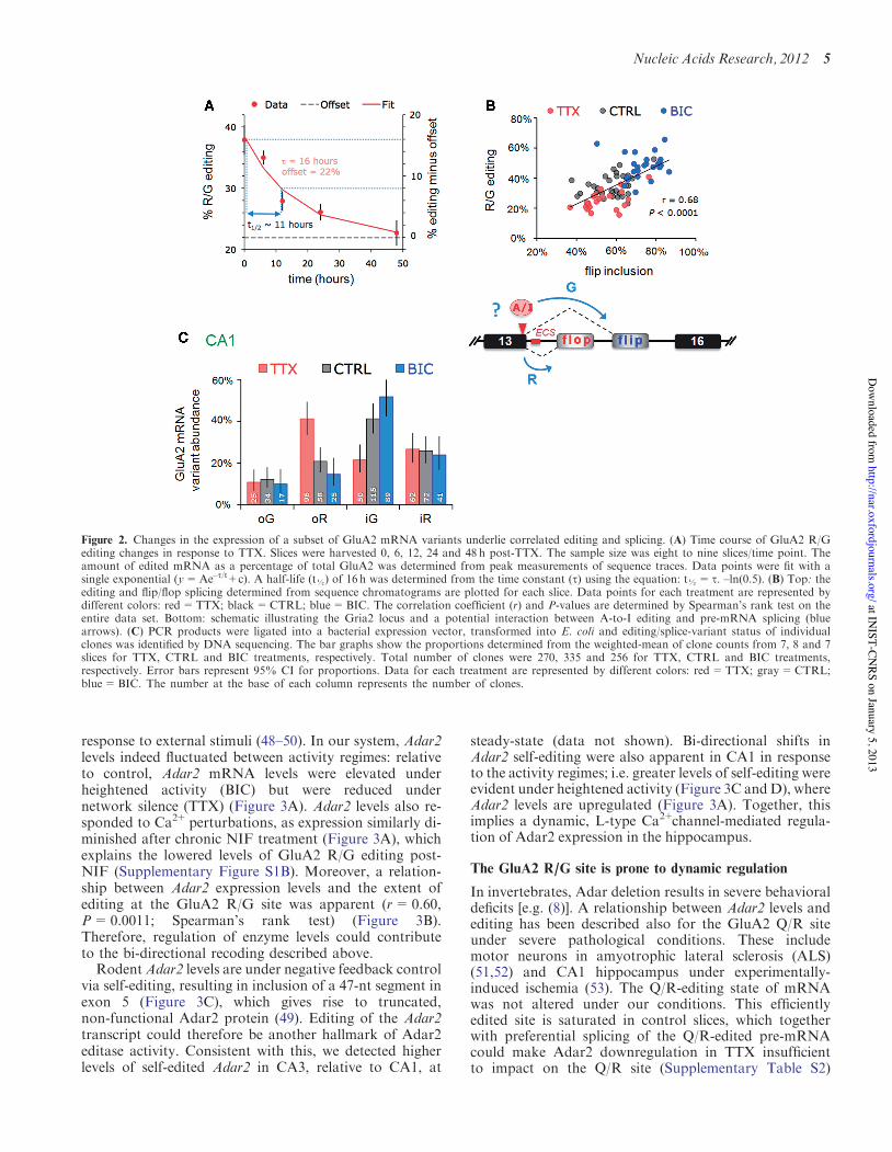
with flip exon selection in CA1 neural tissue, where aclose to linear relationship between editing to G andflip-exon inclusion was evident (r=0.68, P=0.0001,Spearman’s rank test; Figure 2B; Supplementary FiguresS2 and S4A). As editing occurs before splicing, this impliesthat altering position #2 in the splice donor (the editingsite; Figure 1B) could impact on alternative splicing to fa-cilitate inclusion of the downstream flip exon (exon 15).
Nucleic Acids Research, 2012 3
at INIST-CN
RS on January 5, 2013http://nar.oxfordjournals.org/
Dow
nloaded from
Furthermore, analysis of individual clones revealed thatthe distinction between drug treatments apparentlyresulted from a shift in the balance of edited-flip (i-G)and unedited-flop variants (o-R). Specifically, BICboosted edited-flip (i-G) representation, whereasunedited flop variants (o-R) prevailed after chronicTTX. This coupling between editing and splicing wasalso apparent in CA3 tissue and for the GluA3 R/G site,although neither responded to changes in activity (Figures1E and 4B; Supplementary Figure S4B). These findingssuggest a potential mechanistic link between R/G editingand downstream splice site choice (Figure 2B, lowerpanel), which extends earlier data where editing wasreported to repress splicing of the adjacent flop exon ina reporter system (42).
Adar levels fluctuate in response to neural activity
A-to-I editing is catalyzed by editase (Adar) dimers(43,44); individual substrates are recognized by the
catalytically active Adar1 or Adar2, or by a combinationof the two enzymes. Editing at the GluA2 R/G site ismediated mostly by Adar2, whereas both enzymes targetthe R/G site in GluA3 (45,46). Adar2 is expressed athigher levels in subfield CA3 than in CA1 (!3-fold inadult rat hippocampus). This was not the case forAdar1, which is expressed homogeneously acrosshippocampal subfields (Supplementary Figure S5A). Themore prominent level of Adar2 in CA3 is also evident within situ hybridization data of mouse hippocampus(Supplementary Figure S5B) (47). Related to this Adar2expression profile, GluA2 R/G editing is consistentlyhigher in CA3, in both acutely dissected and inorganotypic hippocampal slices (Figure 1F andSupplementary Figure S2).
This raises the question as to whether changes in Adar2expression could contribute to the changes in GluA2 R/Gediting in CA1. Adar2 expression is under negativefeedback control and has been shown to fluctuate in
Figure 1. Activity-dependent changes in GluA2 R/G editing localized to hippocampal CA1. (A) Nissl stain of an organotypic culture of a transversehippocampal slice cultured for 3 weeks. The major subfields of the trisynaptic circuit are annotated: CA1, CA3 and the dentate gyrus. Right top andbottom show close-up images of CA1 and CA3 cells respectively. (B) Schematic illustrating the Gria2 locus encompassing exons 13–16; the region ofinterest (ROI), encodes segment 2 of the ligand-binding domain, the alternatively spliced i/o exons and the A/I RNA editing site, where arginine (R)is recoded to a glycine (G). The editing complementary sequence (ECS) forms a highly conserved pre-mRNA secondary structure encompassing thesplice site (see Figure 4A). (C) Sequence traces show part of the ROI including the A/I editing site and the exon–exon junction between exon 13 andthe flip/flop exon. Changes in GluA2 editing and splicing from mRNA extracted from CA1 tissue after 48 h TTX or BIC treatments. CTRL treatmentrepresents mock feeding without drugs. (D) Quantification of the peak heights in CA1 sequence chromatograms is summarized in box and whiskerplots. The plot shows levels of GluA2 R/G editing as a fraction of total subunit mRNA for the different drug treatments. The median is representedby a gray line. The box and whiskers show the interquartile range (25–75%) and the total range (min–max) of the data, respectively. The number atthe base of each plot is the number of slices. The P-value is derived from the Kruskal–Wallis test statistic and the asterisks summarize the results ofDunn’s post-tests: **P< 0.01, ***P< 0.001. (E) As in Figure 1D but for mRNA extracted from micro-dissected CA3 tissue after 48 h treatments withTTX or BIC. Editing and splicing of GluA2 are invariant across drug treatments. (F) Box and whisker plots summarizing GluA2 R/G editing levelsbetween subfields (CA1 and CA3) determined from peak height ratios. The P-value is derived from the Mann–Whitney U-statistic, ***P< 0.0001.
4 Nucleic Acids Research, 2012
at INIST-CN
RS on January 5, 2013http://nar.oxfordjournals.org/
Dow
nloaded from
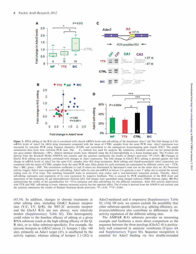
response to external stimuli (48–50). In our system, Adar2levels indeed fluctuated between activity regimes: relativeto control, Adar2 mRNA levels were elevated underheightened activity (BIC) but were reduced undernetwork silence (TTX) (Figure 3A). Adar2 levels also re-sponded to Ca2+perturbations, as expression similarly di-minished after chronic NIF treatment (Figure 3A), whichexplains the lowered levels of GluA2 R/G editing post-NIF (Supplementary Figure S1B). Moreover, a relation-ship between Adar2 expression levels and the extent ofediting at the GluA2 R/G site was apparent (r=0.60,P=0.0011; Spearman’s rank test) (Figure 3B).Therefore, regulation of enzyme levels could contributeto the bi-directional recoding described above.
RodentAdar2 levels are under negative feedback controlvia self-editing, resulting in inclusion of a 47-nt segment inexon 5 (Figure 3C), which gives rise to truncated,non-functional Adar2 protein (49). Editing of the Adar2transcript could therefore be another hallmark of Adar2editase activity. Consistent with this, we detected higherlevels of self-edited Adar2 in CA3, relative to CA1, at
steady-state (data not shown). Bi-directional shifts inAdar2 self-editing were also apparent in CA1 in responseto the activity regimes; i.e. greater levels of self-editing wereevident under heightened activity (Figure 3C andD), whereAdar2 levels are upregulated (Figure 3A). Together, thisimplies a dynamic, L-type Ca2+channel-mediated regula-tion of Adar2 expression in the hippocampus.
The GluA2 R/G site is prone to dynamic regulation
In invertebrates, Adar deletion results in severe behavioraldeficits [e.g. (8)]. A relationship between Adar2 levels andediting has been described also for the GluA2 Q/R siteunder severe pathological conditions. These includemotor neurons in amyotrophic lateral sclerosis (ALS)(51,52) and CA1 hippocampus under experimentally-induced ischemia (53). The Q/R-editing state of mRNAwas not altered under our conditions. This efficientlyedited site is saturated in control slices, which togetherwith preferential splicing of the Q/R-edited pre-mRNAcould make Adar2 downregulation in TTX insufficientto impact on the Q/R site (Supplementary Table S2)
Figure 2. Changes in the expression of a subset of GluA2 mRNA variants underlie correlated editing and splicing. (A) Time course of GluA2 R/Gediting changes in response to TTX. Slices were harvested 0, 6, 12, 24 and 48 h post-TTX. The sample size was eight to nine slices/time point. Theamount of edited mRNA as a percentage of total GluA2 was determined from peak measurements of sequence traces. Data points were fit with asingle exponential (y=Ae–t/t+c). A half-life (t!) of 16 h was determined from the time constant (t) using the equation: t!= t. –ln(0.5). (B) Top: theediting and flip/flop splicing determined from sequence chromatograms are plotted for each slice. Data points for each treatment are represented bydifferent colors: red=TTX; black=CTRL; blue=BIC. The correlation coefficient (r) and P-values are determined by Spearman’s rank test on theentire data set. Bottom: schematic illustrating the Gria2 locus and a potential interaction between A-to-I editing and pre-mRNA splicing (bluearrows). (C) PCR products were ligated into a bacterial expression vector, transformed into E. coli and editing/splice-variant status of individualclones was identified by DNA sequencing. The bar graphs show the proportions determined from the weighted-mean of clone counts from 7, 8 and 7slices for TTX, CTRL and BIC treatments, respectively. Total number of clones were 270, 335 and 256 for TTX, CTRL and BIC treatments,respectively. Error bars represent 95% CI for proportions. Data for each treatment are represented by different colors: red=TTX; gray=CTRL;blue=BIC. The number at the base of each column represents the number of clones.
Nucleic Acids Research, 2012 5
at INIST-CN
RS on January 5, 2013http://nar.oxfordjournals.org/
Dow
nloaded from
(45,54). In addition, changes to chronic treatments atother editing sites, including GluK2 Kainate receptorsites (Y/C, I/V, Q/R), the 5HT-2C serotonin receptorand the GluA3 R/G site (see above), were relativelymodest (Supplementary Table S2). This heterogeneitycould relate to the baseline efficacy of editing at a givenRNA substrate (such as the high editing efficacy of the Q/R site), or to Adar substrate-specificity. For example, theintronic hotspots in GRIA2 intron 11: hotspot 1 (the+60site), primarily an Adar1 target (45), is unaffected by theactivity regimes, whereas editing of hotspot 2 (+262) is
Adar2-mediated and is responsive [Supplementary TableS2; (54)]. Of note, we cannot exclude the possibility thatother substrate-specific factors (e.g. splicing efficiency, ac-tivators/inhibitors) also contribute to differences in theactivity regulation of the different editing sites.
The AMPAR R/G substrate provides an interestingexample and facilitates a more direct comparison as thesequence between the three paralogs (GluA2–4) is particu-larly well conserved in amniotic vertebrates (Figure 4Aand Supplementary Figure S6). Sequence recognition isessential for Adar2 binding via two double-stranded
Figure 3. RNA editing at the R/G site is correlated with altered mRNA levels and self-editing of the deaminase Adar2. (A) The fold change in CA1mRNA levels of Adar2 for (48 h) drug treatments compared with the mean of CTRL samples from the same PCR runs. Adar2 expression wasmeasured by real-time PCR using Taqman chemistry (FAM) and normalized to the endogenous house-keeping gene Gapdh (VIC). The graphsummarizes data from four real-time PCR runs. The !!CT method was used for analysis. By validation, standard curves run for primer-probesets gave primer efficiencies >90%. Almost identical results were obtained using the b-2-microglobulin as a house keeping gene. The P-values arederived from the Kruskall–Wallis ANOVA test statistic and the asterices summarize the results of Dunn’s post-tests: *P< 0.05. (B) Changes inGluA2 R/G editing are positively correlated with changes in Adar2 expression. The fold change in GluA2 R/G editing is plotted against the foldchange in mRNA levels of Adar2 for the same CA1 samples after 48 h drug treatments. Both editing and Gapdh-normalized Adar2 expression arecompared with the mean of CTRL samples from the same PCR runs. Data points for each treatment are represented by different colors: red=TTX;blue=BIC; green=NIF. The correlation coefficient (r) and P-values are determined by Spearman’s rank test on the entire data set. (C) Neuronalactivity triggers Adar2 auto-regulation by self-editing. Adar2 edits its own pre-mRNA in intron 2 to generate a new 30 splice site to extend the secondcoding exon by 47 nt (top). The resulting frameshift leads to premature stop codon and a non-functional truncated protein. Thereby, Adar2self-editing represents auto-regulation of its own expression by negative feedback. This is assayed by PCR amplification of the ROI (top) andseparation of the fragments by gel electrophoresis (bottom left). Gel images were quantified using ImageJ software (NIH) (bottom right). (D) Plotsummarizing the results of the quantification for +47 nt inclusion and thus self-editing for the different treatments. After 48 h activity deprivationwith TTX and NIF, self-editing is lower, whereas increased activity has the opposite effect. The P-value is derived from the ANOVA test statistic andthe asterices summarize the results of Student–Newman–Keuls post-tests: *P< 0.05, ***P< 0.001.
6 Nucleic Acids Research, 2012
at INIST-CN
RS on January 5, 2013http://nar.oxfordjournals.org/
Dow
nloaded from
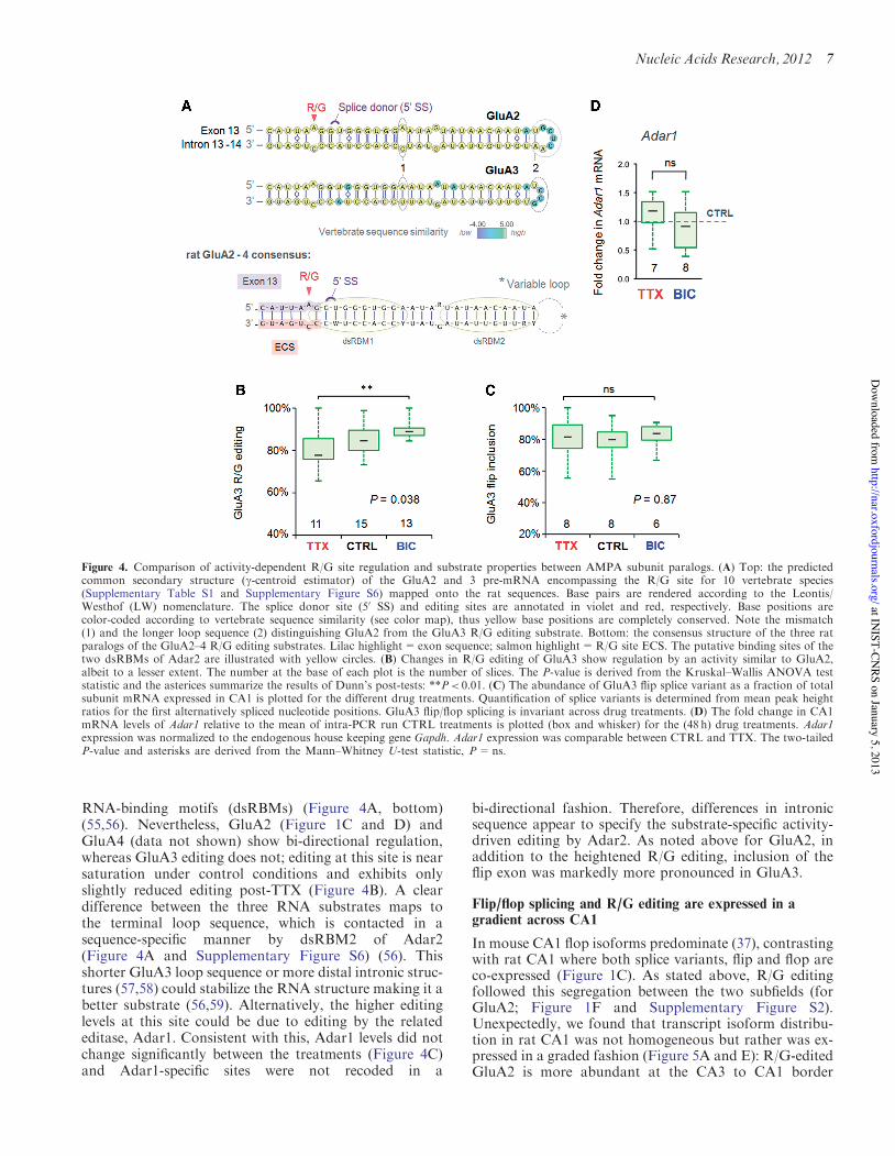
RNA-binding motifs (dsRBMs) (Figure 4A, bottom)(55,56). Nevertheless, GluA2 (Figure 1C and D) andGluA4 (data not shown) show bi-directional regulation,whereas GluA3 editing does not; editing at this site is nearsaturation under control conditions and exhibits onlyslightly reduced editing post-TTX (Figure 4B). A cleardifference between the three RNA substrates maps tothe terminal loop sequence, which is contacted in asequence-specific manner by dsRBM2 of Adar2(Figure 4A and Supplementary Figure S6) (56). Thisshorter GluA3 loop sequence or more distal intronic struc-tures (57,58) could stabilize the RNA structure making it abetter substrate (56,59). Alternatively, the higher editinglevels at this site could be due to editing by the relatededitase, Adar1. Consistent with this, Adar1 levels did notchange significantly between the treatments (Figure 4C)and Adar1-specific sites were not recoded in a
bi-directional fashion. Therefore, differences in intronicsequence appear to specify the substrate-specific activity-driven editing by Adar2. As noted above for GluA2, inaddition to the heightened R/G editing, inclusion of theflip exon was markedly more pronounced in GluA3.
Flip/flop splicing and R/G editing are expressed in agradient across CA1
In mouse CA1 flop isoforms predominate (37), contrastingwith rat CA1 where both splice variants, flip and flop areco-expressed (Figure 1C). As stated above, R/G editingfollowed this segregation between the two subfields (forGluA2; Figure 1F and Supplementary Figure S2).Unexpectedly, we found that transcript isoform distribu-tion in rat CA1 was not homogeneous but rather was ex-pressed in a graded fashion (Figure 5A and E): R/G-editedGluA2 is more abundant at the CA3 to CA1 border
Figure 4. Comparison of activity-dependent R/G site regulation and substrate properties between AMPA subunit paralogs. (A) Top: the predictedcommon secondary structure (g-centroid estimator) of the GluA2 and 3 pre-mRNA encompassing the R/G site for 10 vertebrate species(Supplementary Table S1 and Supplementary Figure S6) mapped onto the rat sequences. Base pairs are rendered according to the Leontis/Westhof (LW) nomenclature. The splice donor site (50 SS) and editing sites are annotated in violet and red, respectively. Base positions arecolor-coded according to vertebrate sequence similarity (see color map), thus yellow base positions are completely conserved. Note the mismatch(1) and the longer loop sequence (2) distinguishing GluA2 from the GluA3 R/G editing substrate. Bottom: the consensus structure of the three ratparalogs of the GluA2–4 R/G editing substrates. Lilac highlight=exon sequence; salmon highlight=R/G site ECS. The putative binding sites of thetwo dsRBMs of Adar2 are illustrated with yellow circles. (B) Changes in R/G editing of GluA3 show regulation by an activity similar to GluA2,albeit to a lesser extent. The number at the base of each plot is the number of slices. The P-value is derived from the Kruskal–Wallis ANOVA teststatistic and the asterices summarize the results of Dunn’s post-tests: **P< 0.01. (C) The abundance of GluA3 flip splice variant as a fraction of totalsubunit mRNA expressed in CA1 is plotted for the different drug treatments. Quantification of splice variants is determined from mean peak heightratios for the first alternatively spliced nucleotide positions. GluA3 flip/flop splicing is invariant across drug treatments. (D) The fold change in CA1mRNA levels of Adar1 relative to the mean of intra-PCR run CTRL treatments is plotted (box and whisker) for the (48 h) drug treatments. Adar1expression was normalized to the endogenous house keeping gene Gapdh. Adar1 expression was comparable between CTRL and TTX. The two-tailedP-value and asterisks are derived from the Mann–Whitney U-test statistic, P=ns.
Nucleic Acids Research, 2012 7
at INIST-CN
RS on January 5, 2013http://nar.oxfordjournals.org/
Dow
nloaded from
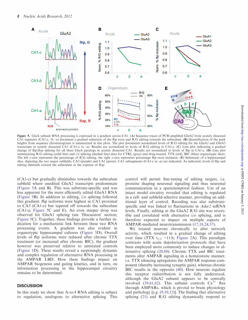
(CA1-c) but gradually diminishes towards the subiculumsubfield where unedited GluA2 transcripts predominate(Figure 5A and B). This was substrate-specific and wasless apparent for the more efficiently edited GluA3 RNA(Figure 5B). In addition to editing, i/o splicing followedthis gradient: flip isoforms were highest in CA1 proximalto CA3 (CA1-c) but tapered off towards the subiculum(CA1-a; Figure 5C and E). An even steeper drop wasobserved for GluA1 splicing (see ‘Discussion’ section;Figure 5C). Together, these findings provide a further in-dication for a mechanistic link between these two RNAprocessing events. A gradient was also evident inorganotypic hippocampal cultures (Figure 5D). Overalllevels of flip isoforms were reduced after chronic TTXtreatment (or increased after chronic BIC), the gradienthowever was preserved relative to untreated controls(Figure 1D). These results reveal a surprisingly dynamicand complex regulation of alternative RNA processing inthe AMPAR LBD. How these findings impact onAMPAR biogenesis and gating kinetics, and in turn, oninformation processing in the hippocampal circuitryremains to be determined.
DISCUSSION
In this study we show that A-to-I RNA editing is subjectto regulation, analogous to alternative splicing. This
control will permit fine-tuning of editing targets, i.e.proteins shaping neuronal signaling and thus neuronalcommunication in a spatiotemporal fashion. Use of anintact model circuitry revealed that editing is regulatedin a cell- and subfield-selective manner, providing an add-itional layer of control. Recoding was also substrate-specific and was linked to fluctuations in Adar2 mRNAlevels. Finally, editing at the GluA2 R/G site was revers-ible and correlated with alternative i/o splicing, and istherefore expected to impact on multiple aspects ofAMPAR-mediated neurotransmission (17,18,24,37).
We treated neurons chronically to alter networkactivity, which resulted in a gradual change of editingover time (TTX t1/2 !11 h; Figure 2A). This paradigmcontrasts with acute depolarization protocols that havebeen employed more commonly to induce changes in al-ternative splicing (20,60). Chronic TTX and BIC treat-ments alter AMPAR signaling in a homeostatic manner,i.e. TTX silencing upregulates the AMPAR response com-ponent (thereby increasing synaptic gain), whereas chronicBIC results in the opposite (40). How neurons regulatethis receptor redistribution is not fully understood,although the GluA2 subunit appears to be centrallyinvolved (39,61,62). This subunit controls Ca2+ fluxthrough AMPARs, which is pivotal to brain physiologyand pathology [e.g. (9,10,12)]. The finding that alternativesplicing (21) and R/G editing dynamically respond to
Figure 5. GluA subunit RNA processing is expressed in a gradient across CA1. (A) Sequence traces of PCR-amplified GluA2 from acutely dissectedCA1 segments (CA1-c, -b, -a) document a gradual reduction of the flip exon and R/G editing towards the subiculum. (B) Quantification of the peakheights from sequence chromatograms is summarized in line plots. The plot documents normalized levels of R/G editing for the GluA2 and GluA3transcripts in acutely dissected CA1 (CA1-c to -a). Results are normalized to levels of R/G editing in CA1-c. (C) Line plot indicating a gradualchange of flip/flop splicing for all three GluA paralogs in acutely dissected CA1. Results are normalized to levels of flip in CA1-c. (D) Line plotsummarizing R/G editing (solid line) and i/o splicing (dashed line) data for CTRL (gray) and drug-treated, TTX (red), BIC (blue) organotypic slices.The left y-axis represents the percentage of R/G editing, the right y-axis represents percentage flip exon inclusion. (E) Schematic of a hippocampalslice, depicting the two major subfields, CA3 (purple) and CA1 (green). CA1 subsegments (CA1-c to -a) are indicated. As indicated, levels of flip andediting diminish toward the subiculum at the expense of flop.
8 Nucleic Acids Research, 2012
at INIST-CN
RS on January 5, 2013http://nar.oxfordjournals.org/
Dow
nloaded from
fluctuations in activity will have consequences forAMPAR subunit assembly (23,63), which in addition toaltering channel properties at synapses could have second-ary effects on trafficking to synapses. Besides impactingER exit and endocytic traffic, AMPARs of differentstoichiometries have the capacity to be targeted to differ-ent dendritic regions within individual neurons (64).
An interplay between the R/G site and i/o splicing hasbeen addressed in earlier studies in vitro (65) and it wasshown that GluA2 R/G editing represses splicing of theproximal (flop) intron (42). Here, we extend this relation-ship to the endogenous transcript in neuronal tissue. Acorrelation between R/G editing and the downstreamflip exon was apparent in hippocampal subfields, whichfollowed activity manipulations in a bi-directional and re-versible manner. Editing is expected to weaken the splicedonor site. Because editing and splicing occur,co-transcriptional (66) RNA polymerase II processivitymight be a decisive factor in this regulation (67). Thisidea is supported by the fact that in the GluA1 transcript,which harbors a stronger splice donor site in exon 13(score: 11.08; see ‘Materials and Methods’ section) thanGluA2, splicing to the adjacent flop exon is more prom-inent. In fact, a steeper drop of GluA1 flip, relativeto GluA2 flip, is seen across the gradient in CA1(Figure 5C). Accordingly, Adar2, which is undercomplex cellular control (48,68) and responded to theactivity treatments in a bi-directional fashion(Figure 3A), might contribute to regulating i/o splicingby weakening the exon 13 splice donor.
While this manuscript was in preparation, a relatedstudy that used dissociated cortical neurons, describedactivity-regulated A-to-I RNA editing (69). The authorsutilized high-throughput Illumina sequencing platforms toassess genome-wide changes in editing in response to acuteand chronic changes in activity. This article provides acomplementary study to the work described here.Together, these data suggest that RNA editing byADARs provides a powerful and dynamic regulation ofneuronal communication.
SUPPLEMENTARY DATA
Supplementary Data are available at NAR Online:Supplementary Tables 1 and 2, Supplementary Figures1–6, Supplementary Methods and SupplementaryReference [70].
ACKNOWLEDGEMENTS
We thank the MRC LMB Biomedical Facility for helpwith animal work and the LMB workshop for help withvarious pieces of specific equipment. We thank Jill Hanbyand Jernej Ule for critically reading of the manuscript.
FUNDING
FEBS and EMBO short-term fellowships (to A.B. andZ.N.); Czech Academy of Sciences [M200110971,RVO:67985823 to A.B.]; Grant Agency of the Czech
Republic [P304/12/G069 to A.B.]; Royal Society (toI.H.G.); Medical Research Council (MRC) [U105174197to A.B., A.C.P., Z.N. and I.H.G.]. Funding for openaccess charge: MRC.
Conflict of interest statement. None declared.
REFERENCES
1. Bass,B.L. (2002) RNA editing by adenosine deaminases that acton RNA. Annu. Rev. Biochem., 71, 817–846.
2. Jepson,J.E. and Reenan,R.A. (2008) RNA editing in regulatinggene expression in the brain. Biochim. Biophys. Acta, 1779,459–470.
3. Hundley,H.A. and Bass,B.L. (2012) ADAR editing indouble-stranded UTRs and other noncoding RNA sequences.Trends Biochem. Sci., 35, 377–383.
4. Peng,Z., Cheng,Y., Tan,B.C., Kang,L., Tian,Z., Zhu,Y.,Zhang,W., Liang,Y., Hu,X., Tan,X. et al. (2012) Comprehensiveanalysis of RNA-Seq data reveals extensive RNA editing in ahuman transcriptome. Nat. Biotechnol., 30, 253–260.
5. Paul,M.S. and Bass,B.L. (1998) Inosine exists in mRNA attissue-specific levels and is most abundant in brain mRNA.EMBO J., 17, 1120–1127.
6. Hoopengardner,B., Bhalla,T., Staber,C. and Reenan,R. (2003)Nervous system targets of RNA editing identified by comparativegenomics. Science, 301, 832–836.
7. Tariq,A. and Jantsch,M.F. (2012) Transcript diversification in thenervous system: A to I RNA editing in CNS function and diseasedevelopment. Front. Neurosci., 6, 99.
8. Palladino,M.J., Keegan,L.P., O’Connell,M.A. and Reenan,R.A.(2000) A-to-I pre-mRNA editing in Drosophila is primarilyinvolved in adult nervous system function and integrity. Cell, 102,437–449.
9. Seeburg,P.H. and Hartner,J. (2003) Regulation of ion channel/neurotransmitter receptor function by RNA editing. Curr. Opin.Neurobiol., 13, 279–283.
10. Maas,S., Kawahara,Y., Tamburro,K.M. and Nishikura,K. (2006)A-to-I RNA editing and human disease. RNA Biol., 3, 1–9.
11. Rosenthal,J.J. and Seeburg,P.H. (2012) A-to-I RNA editing:effects on proteins key to neural excitability. Neuron, 74, 432–439.
12. Traynelis,S.F., Wollmuth,L.P., McBain,C.J., Menniti,F.S.,Vance,K.M., Ogden,K.K., Hansen,K.B., Yuan,H., Myers,S.J.,Dingledine,R. et al. (2010) Glutamate receptor ion channels:structure, regulation, and function. Pharmacol. Rev., 62, 405–496.
13. Seeburg,P.H. (1996) The role of RNA editing in controllingglutamate receptor channel properties. J. Neurochem., 66, 1–5.
14. Greger,I.H., Khatri,L., Kong,X. and Ziff,E.B. (2003) AMPAreceptor tetramerization is mediated by Q/R editing. Neuron, 40,763–774.
15. Sommer,B., Kohler,M., Sprengel,R. and Seeburg,P.H. (1991)RNA editing in brain controls a determinant of ion flow inglutamate-gated channels. Cell, 67, 11–19.
16. Feldmeyer,D., Kask,K., Brusa,R., Kornau,H.C., Kolhekar,R.,Rozov,A., Burnashev,N., Jensen,V., Hvalby,O., Sprengel,R. et al.(1999) Neurological dysfunctions in mice expressing differentlevels of the Q/R site-unedited AMPAR subunit GluR-B. Nat.Neurosci., 2, 57–64.
17. Lomeli,H., Mosbacher,J., Melcher,T., Hoger,T., Geiger,J.R.,Kuner,T., Monyer,H., Higuchi,M., Bach,A. and Seeburg,P.H.(1994) Control of kinetic properties of AMPA receptor channelsby nuclear RNA editing. Science, 266, 1709–1713.
18. Greger,I.H., Akamine,P., Khatri,L. and Ziff,E.B. (2006)Developmentally regulated, combinatorial RNA processingmodulates AMPA receptor biogenesis. Neuron, 51, 85–97.
19. Li,Q., Lee,J.A. and Black,D.L. (2007) Neuronal regulationof alternative pre-mRNA splicing. Nat. Rev. Neurosci., 8,819–831.
20. Xie,J. and Black,D.L. (2001) A CaMK IV responsive RNAelement mediates depolarization-induced alternative splicing of ionchannels. Nature, 410, 936–939.
Nucleic Acids Research, 2012 9
at INIST-CN
RS on January 5, 2013http://nar.oxfordjournals.org/
Dow
nloaded from
21. Penn,A.C., Balik,A., Wozny,C., Cais,O. and Greger,I.H. (2012)Activity-mediated AMPA receptor remodeling, driven byalternative splicing in the ligand-binding domain. Neuron, 76,503–510.
22. Coleman,S.K., Moykkynen,T., Cai,C., von Ossowski,L.,Kuismanen,E., Korpi,E.R. and Keinanen,K. (2006)Isoform-specific early trafficking of AMPA receptor flip and flopvariants. J. Neurosci., 26, 11220–11229.
23. Coleman,S.K., Moykkynen,T., Hinkkuri,S., Vaahtera,L.,Korpi,E.R., Pentikainen,O.T. and Keinanen,K. (2010) Ligand-binding domain determines endoplasmic reticulum exit of AMPAreceptors. J. Biol Chem., 285, 36032–36039.
24. Greger,I.H., Ziff,E.B. and Penn,A.C. (2007) Moleculardeterminants of AMPA receptor subunit assembly. TrendsNeurosci., 30, 407–416.
25. Penn,A.C., Williams,S.R. and Greger,I.H. (2008) Gating motionsunderlie AMPA receptor secretion from the endoplasmicreticulum. EMBO J., 27, 3056–3068.
26. Liu,Y. and Samuel,C.E. (1999) Editing of glutamate receptorsubunit B pre-mRNA by splice-site variants ofinterferon-inducible double-stranded RNA-specific adenosinedeaminase ADAR1. J. Biol. Chem., 274, 5070–5077.
27. Gahwiler,B.H. (1981) Organotypic monolayer cultures of nervoustissue. J. Neurosci. Methods, 4, 329–342.
28. Ge,B., Gurd,S., Gaudin,T., Dore,C., Lepage,P., Harmsen,E.,Hudson,T.J. and Pastinen,T. (2005) Survey of allelic expressionusing EST mining. Genome Res., 15, 1584–1591.
29. Goodman,L.A. (1965) On simultaneous confidence intervals formultinomial proportions. Technometrics, 7, 247–254.
30. Altschul,S.F., Madden,T.L., Schaffer,A.A., Zhang,J., Zhang,Z.,Miller,W. and Lipman,D.J. (1997) Gapped BLAST andPSI-BLAST: a new generation of protein database searchprograms. Nucleic Acids Res., 25, 3389–3402.
31. Birney,E., Andrews,T.D., Bevan,P., Caccamo,M., Chen,Y.,Clarke,L., Coates,G., Cuff,J., Curwen,V., Cutts,T. et al. (2004)An overview of Ensembl. Genome Res., 14, 925–928.
32. Kent,W.J. (2002) BLAT–the BLAST-like alignment tool. GenomeRes., 12, 656–664.
33. Venkatesh,B., Kirkness,E.F., Loh,Y.H., Halpern,A.L., Lee,A.P.,Johnson,J., Dandona,N., Viswanathan,L.D., Tay,A., Venter,J.C.et al. (2007) Survey sequencing and comparative analysisof the elephant shark (Callorhinchus milii) genome. PLoS Biol.,5, e101.
34. Katoh,K., Kuma,K., Miyata,T. and Toh,H. (2005) Improvementin the accuracy of multiple sequence alignment program MAFFT.Genome Inform., 16, 22–33.
35. Hamada,M., Sato,K. and Asai,K. (2011) Improving the accuracyof predicting secondary structure for aligned RNA sequences.Nucleic Acids Res., 39, 393–402.
36. Darty,K., Denise,A. and Ponty,Y. (2009) VARNA: Interactivedrawing and editing of the RNA secondary structure.Bioinformatics, 25, 1974–1975.
37. Sommer,B., Keinanen,K., Verdoorn,T.A., Wisden,W.,Burnashev,N., Herb,A., Kohler,M., Takagi,T., Sakmann,B. andSeeburg,P.H. (1990) Flip and flop: a cell-specific functional switchin glutamate-operated channels of the CNS. Science, 249,1580–1585.
38. Witter,M.P., Wouterlood,F.G., Naber,P.A. and Van Haeften,T.(2000) Anatomical organization of the parahippocampal-hippocampal network. Ann. NY Acad. Sci., 911, 1–24.
39. Turrigiano,G.G. (2008) The self-tuning neuron: synaptic scaling ofexcitatory synapses. Cell, 135, 422–435.
40. Turrigiano,G.G., Leslie,K.R., Desai,N.S., Rutherford,L.C. andNelson,S.B. (1998) Activity-dependent scaling of quantalamplitude in neocortical neurons. Nature, 391, 892–896.
41. Fisahn,A., Pike,F.G., Buhl,E.H. and Paulsen,O. (1998)Cholinergic induction of network oscillations at 40 Hz in thehippocampus in vitro. Nature, 394, 186–189.
42. Schoft,V.K., Schopoff,S. and Jantsch,M.F. (2007) Regulation ofglutamate receptor B pre-mRNA splicing by RNA editing.Nucleic Acids Res., 35, 3723–3732.
43. Gallo,A., Keegan,L.P., Ring,G.M. and O’Connell,M.A. (2003) AnADAR that edits transcripts encoding ion channel subunitsfunctions as a dimer. EMBO J., 22, 3421–3430.
44. Valente,L. and Nishikura,K. (2007) RNA binding-independentdimerization of adenosine deaminases acting on RNA anddominant negative effects of nonfunctional subunits on dimerfunctions. J. Biol. Chem., 282, 16054–16061.
45. Higuchi,M., Maas,S., Single,F.N., Hartner,J., Rozov,A.,Burnashev,N., Feldmeyer,D., Sprengel,R. and Seeburg,P.H. (2000)Point mutation in an AMPA receptor gene rescues lethality inmice deficient in the RNA-editing enzyme ADAR2. Nature, 406,78–81.
46. Wang,Q., Khillan,J., Gadue,P. and Nishikura,K. (2000)Requirement of the RNA editing deaminase ADAR1 gene forembryonic erythropoiesis. Science, 290, 1765–1768.
47. Lein,E.S., Hawrylycz,M.J., Ao,N., Ayres,M., Bensinger,A.,Bernard,A., Boe,A.F., Boguski,M.S., Brockway,K.S., Byrnes,E.J.et al. (2007) Genome-wide atlas of gene expression in the adultmouse brain. Nature, 445, 168–176.
48. Feng,Y., Sansam,C.L., Singh,M. and Emeson,R.B. (2006) AlteredRNA editing in mice lacking ADAR2 autoregulation. Mol. Cell.Biol., 26, 480–488.
49. Rueter,S.M., Dawson,T.R. and Emeson,R.B. (1999) Regulation ofalternative splicing by RNA editing. Nature, 399, 75–80.
50. Gurevich,I., Englander,M.T., Adlersberg,M., Siegal,N.B. andSchmauss,C. (2002) Modulation of serotonin 2C receptor editingby sustained changes in serotonergic neurotransmission. J.Neurosci., 22, 10529–10532.
51. Hideyama,T., Yamashita,T., Aizawa,H., Tsuji,S., Kakita,A.,Takahashi,H. and Kwak,S. (2012) Profound downregulation ofthe RNA editing enzyme ADAR2 in ALS spinal motor neurons.Neurobiol. Dis., 45, 1121–1128.
52. Hideyama,T., Yamashita,T., Suzuki,T., Tsuji,S., Higuchi,M.,Seeburg,P.H., Takahashi,R., Misawa,H. and Kwak,S. (2010)Induced loss of ADAR2 engenders slow death of motorneurons from Q/R site-unedited GluR2. J. Neurosci., 30,11917–11925.
53. Peng,P.L., Zhong,X., Tu,W., Soundarapandian,M.M., Molner,P.,Zhu,D., Lau,L., Liu,S., Liu,F. and Lu,Y. (2006)ADAR2-dependent RNA editing of AMPA receptor subunitGluR2 determines vulnerability of neurons in forebrain ischemia.Neuron, 49, 719–733.
54. Penn,A.C., Balik,A. and Greger,I.H. (2012) Steric antisenseinhibition of AMPA receptor Q/R editing reveals tight couplingto intronic editing sites and splicing. Nucleic Acids Res.,doi:10.1093/nar/gks1044.
55. Dawson,T.R., Sansam,C.L. and Emeson,R.B. (2004) Structureand sequence determinants required for the RNA editing ofADAR2 substrates. J. Biol. Chem., 279, 4941–4951.
56. Stefl,R., Oberstrass,F.C., Hood,J.L., Jourdan,M.,Zimmermann,M., Skrisovska,L., Maris,C., Peng,L., Hofr,C.,Emeson,R.B. et al. (2010) The solution structure of the ADAR2dsRBM-RNA complex reveals a sequence-specific readout of theminor groove. Cell, 143, 225–237.
57. Aruscavage,P.J. and Bass,B.L. (2000) A phylogenetic analysisreveals an unusual sequence conservation within introns involvedin RNA editing. RNA, 6, 257–269.
58. Daniel,C., Veno,M.T., Ekdahl,Y., Kjems,J. and Ohman,M. (2012)A distant cis acting intronic element induces site-selective RNAediting. Nucleic Acids Res., 40, 9876–9886.
59. Stefl,R. and Allain,F.H. (2005) A novel RNA pentaloop foldinvolved in targeting ADAR2. RNA, 11, 592–597.
60. Lee,J.A., Xing,Y., Nguyen,D., Xie,J., Lee,C.J. and Black,D.L.(2007) Depolarization and CaM kinase IV modulate NMDAreceptor splicing through two essential RNA elements.PLoS Biol., 5, e40.
61. Gainey,M.A., Hurvitz-Wolff,J.R., Lambo,M.E. andTurrigiano,G.G. (2009) Synaptic scaling requires the GluR2subunit of the AMPA receptor. J. Neurosci., 29, 6479–6489.
62. Pozo,K. and Goda,Y. (2010) Unraveling mechanisms ofhomeostatic synaptic plasticity. Neuron, 66, 337–351.
63. Brorson,J.R., Li,D. and Suzuki,T. (2004) Selective expressionof heteromeric AMPA receptors driven by flip-flop differences.J. Neurosci., 24, 3461–3470.
64. Toth,K. and McBain,C.J. (1998) Afferent-specific innervation oftwo distinct AMPA receptor subtypes on single hippocampalinterneurons. Nat. Neurosci., 1, 572–578.
10 Nucleic Acids Research, 2012
at INIST-CN
RS on January 5, 2013http://nar.oxfordjournals.org/
Dow
nloaded from

65. Bratt,E. and Ohman,M. (2003) Coordination ofediting and splicing of glutamate receptor pre-mRNA. RNA, 9,309–318.
66. Ryman,K., Fong,N., Bratt,E., Bentley,D.L. and Ohman,M. (2007)The C-terminal domain of RNA Pol II helps ensure thatediting precedes splicing of the GluR-B transcript. RNA, 13,1071–1078.
67. de la Mata,M., Munoz,M.J., Allo,M., Fededa,J.P., Schor,I.E. andKornblihtt,A.R. (2011) RNA polymerase II elongation at thecrossroads of transcription and alternative splicing. Genet. Res.Int., 2011, 309865.
68. Marcucci,R., Brindle,J., Paro,S., Casadio,A., Hempel,S.,Morrice,N., Bisso,A., Keegan,L.P., Del Sal,G. and O’Connell,M.A.(2011) Pin1 and WWP2 regulate GluR2 Q/R site RNA editing byADAR2 with opposing effects. EMBO J., 30, 4211–4222.
69. Sanjana,N.E., Levanon,E.Y., Hueske,E.A., Ambrose,J.M. andLi,J.B. (2012) Activity-dependent A-to-I RNA editing in ratcortical neurons. Genetics, 192, 281–287.
70. Higuchi,M., Single,F.N., Kohler,M., Sommer,B., Sprengel,R. andSeeburg,P.H. (1993) RNA editing of AMPA receptor subunitGluR-B: a base-paired intron-exon structure determines positionand efficiency. Cell, 75, 1361–1370.
Nucleic Acids Research, 2012 11
at INIST-CN
RS on January 5, 2013http://nar.oxfordjournals.org/
Dow
nloaded from
1
Supplemental Material !
!
Inventory of Supplemental Material
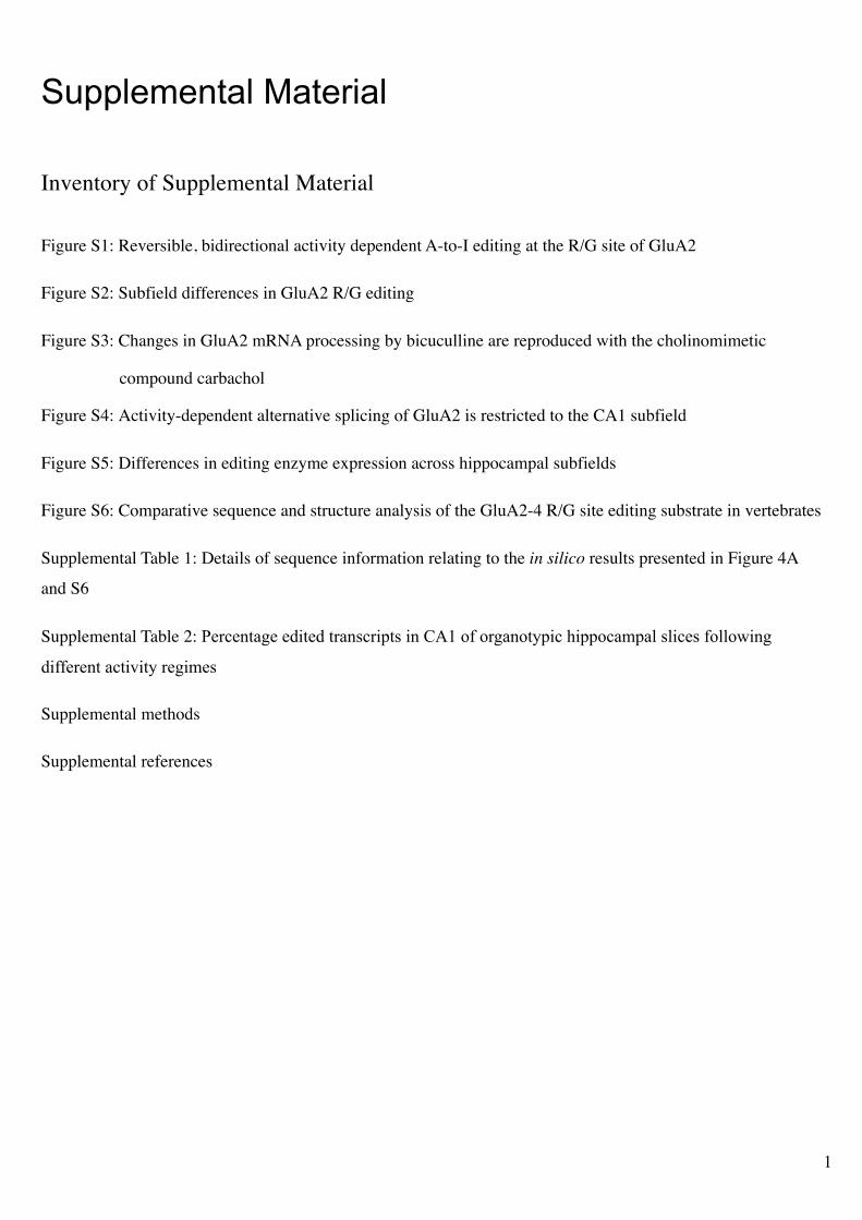
Figure S1: Reversible, bidirectional activity dependent A-to-I editing at the R/G site of GluA2
Figure S2: Subfield differences in GluA2 R/G editing
Figure S3: Changes in GluA2 mRNA processing by bicuculline are reproduced with the cholinomimetic
compound carbachol
Figure S4: Activity-dependent alternative splicing of GluA2 is restricted to the CA1 subfield
Figure S5: Differences in editing enzyme expression across hippocampal subfields
Figure S6: Comparative sequence and structure analysis of the GluA2-4 R/G site editing substrate in vertebrates
Supplemental Table 1: Details of sequence information relating to the in silico results presented in Figure 4A
and S6
Supplemental Table 2: Percentage edited transcripts in CA1 of organotypic hippocampal slices following
different activity regimes
Supplemental methods
Supplemental references !
2
!
Figure S1!
Reversible, bidirectional activity dependent A-to-I editing at the R/G site of GluA2
A. Table outlining the drug treatment regimes for four representative slices, a-d, shown in panel B.
B. Bidirectional reversibility underscores the dynamic nature of GluA2 mRNA processing: a. untreated control;
b. 48 hr TTX treatment; c. 48 hr TTX followed by TTX wash-out and a 48 hr BIC-treatment (note the increase
of R/G editing and flip-exon inclusion); d. as in c., but followed by BIC wash-out and subsequent 2nd TTX
treatment. Note the reversion to the predominant unedited, adenosine peak at the R/G site.
C. Quantification of the peak heights in CA1 sequence chromatograms is summarised in box and whisker plots.
The plot shows the level of the GluA2 R/G editing as a fraction of total subunit mRNA for L-type Ca2+
channel blocker NIF in addition to the previously presented drug treatments. The number at the base of each
plot is the number of slices. The p value is derived from the Kruskal-Wallis test statistic and the asterisks
summarise the results of Dunn’s post tests: ** = p < 0.01, *** = p < 0.001
3
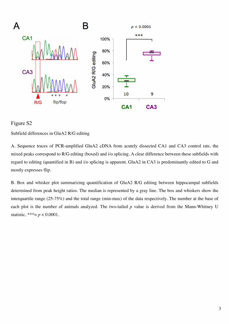
Figure S2!
Subfield differences in GluA2 R/G editing
A. Sequence traces of PCR-amplified GluA2 cDNA from acutely dissected CA1 and CA3 control rats, the
mixed peaks correspond to R/G editing (boxed) and i/o splicing. A clear difference between these subfields with
regard to editing (quantified in B) and i/o splicing is apparent. GluA2 in CA3 is predominantly edited to G and
mostly expresses flip.
B. Box and whisker plot summarizing quantification of GluA2 R/G editing between hippocampal subfields
determined from peak height ratios. The median is represented by a gray line. The box and whiskers show the
interquartile range (25-75%) and the total range (min-max) of the data respectively. The number at the base of
each plot is the number of animals analyzed. The two-tailed p value is derived from the Mann-Whitney U
statistic, ***= p < 0.0001.
4
!
!
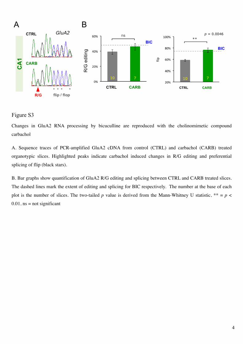
Figure S3!
Changes in GluA2 RNA processing by bicuculline are reproduced with the cholinomimetic compound
carbachol
A. Sequence traces of PCR-amplified GluA2 cDNA from control (CTRL) and carbachol (CARB) treated
organotypic slices. Highlighted peaks indicate carbachol induced changes in R/G editing and preferential
splicing of flip (black stars).
B. Bar graphs show quantification of GluA2 R/G editing and splicing between CTRL and CARB treated slices.
The dashed lines mark the extent of editing and splicing for BIC respectively. The number at the base of each
plot is the number of slices. The two-tailed p value is derived from the Mann-Whitney U statistic, ** = p <
0.01, ns = not significant
!
5
!
!
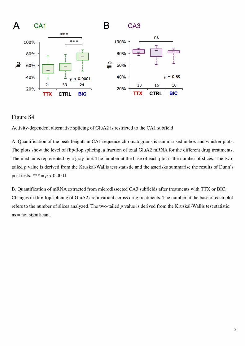
Figure S4!
Activity-dependent alternative splicing of GluA2 is restricted to the CA1 subfield
A. Quantification of the peak heights in CA1 sequence chromatograms is summarised in box and whisker plots.
The plots show the level of flip/flop splicing, a fraction of total GluA2 mRNA for the different drug treatments.
The median is represented by a gray line. The number at the base of each plot is the number of slices. The two-
tailed p value is derived from the Kruskal-Wallis test statistic and the asterisks summarise the results of Dunn’s
post tests: *** = p < 0.0001
B. Quantification of mRNA extracted from microdissected CA3 subfields after treatments with TTX or BIC.
Changes in flip/flop splicing of GluA2 are invariant across drug treatments. The number at the base of each plot
refers to the number of slices analyzed. The two-tailed p value is derived from the Kruskal-Wallis test statistic:
ns = not significant.
6
!
!
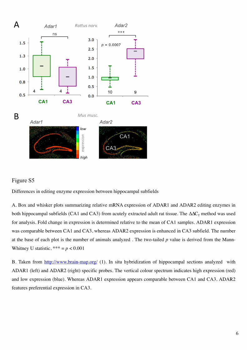
Figure S5!
Differences in editing enzyme expression between hippocampal subfields
A. Box and whisker plots summarizing relative mRNA expression of ADAR1 and ADAR2 editing enzymes in
both hippocampal subfields (CA1 and CA3) from acutely extracted adult rat tissue. The !!CT method was used
for analysis. Fold change in expression is determined relative to the mean of CA1 samples. ADAR1 expression
was comparable between CA1 and CA3, whereas ADAR2 expression is enhanced in CA3 subfield. The number
at the base of each plot is the number of animals analyzed . The two-tailed p value is derived from the Mann-
Whitney U statistic, *** = p < 0.001
B. Taken from http://www.brain-map.org/ (1). In situ hybridization of hippocampal sections analyzed with
ADAR1 (left) and ADAR2 (right) specific probes. The vertical colour spectrum indicates high expression (red)
and low expression (blue). Whereas ADAR1 expression appears comparable between CA1 and CA3, ADAR2
features preferential expression in CA3.
7
!
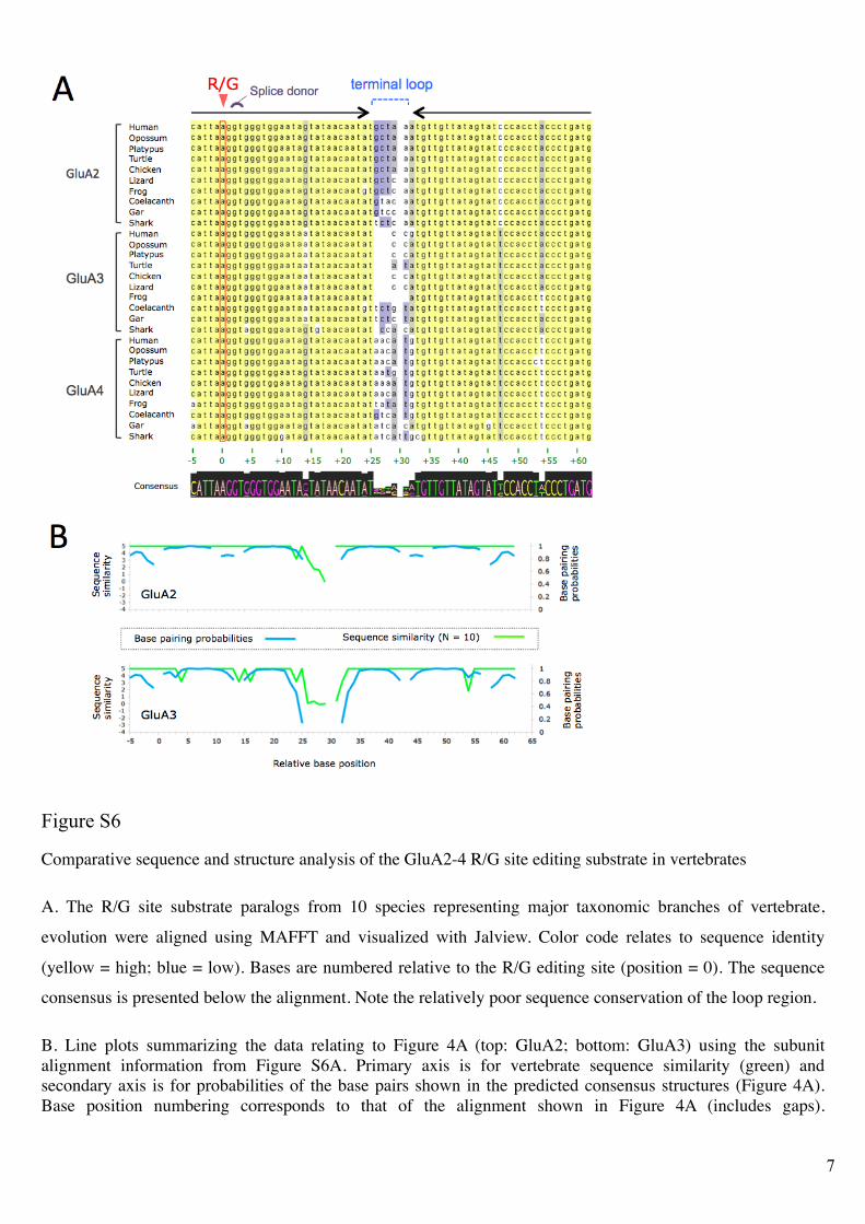
Figure S6!
Comparative sequence and structure analysis of the GluA2-4 R/G site editing substrate in vertebrates
A. The R/G site substrate paralogs from 10 species representing major taxonomic branches of vertebrate,
evolution were aligned using MAFFT and visualized with Jalview. Color code relates to sequence identity
(yellow = high; blue = low). Bases are numbered relative to the R/G editing site (position = 0). The sequence
consensus is presented below the alignment. Note the relatively poor sequence conservation of the loop region.
B. Line plots summarizing the data relating to Figure 4A (top: GluA2; bottom: GluA3) using the subunit
alignment information from Figure S6A. Primary axis is for vertebrate sequence similarity (green) and
secondary axis is for probabilities of the base pairs shown in the predicted consensus structures (Figure 4A).
Base position numbering corresponds to that of the alignment shown in Figure 4A (includes gaps).
8
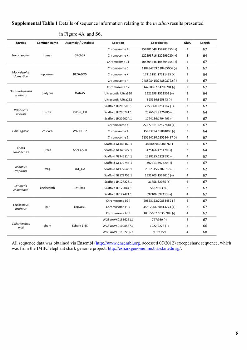
Supplemental Table 1 Details of sequence information relating to the in silico results presented
in Figure 4A and S6.
!"#$%#&' !"##"$%$&#'% ())'#*+,%-%.&/&*&)'% 0"1&/2"$% !""342$&/')% 5+6(% 0'$7/8%
!"#$%$&$%'()( *+,-,*.),/*+,-,*0++(123( -( 67
!"#$%$&$%'(4( *--+5,6*7/*--+55.-.(123( 0( 64 !"#"$%&'()*$ "8%9:( ;<!"06(
!"#$%$&$%'(**( *.+,.))),/*.+,.)6++(123( )( 67
!"#$%$&$%'(+( **,),)6+5/**,),+.77(1=3( -( 67
!"#$%$&$%'(4( *6-***,*/*6-**),+(123( 0( 64 +"*",)-'.(%$
,"#)%/(0&$$>$&&8%( ?<@AB@+(
!"#$%$&$%'()( -),,.,)*+/-),,.,6--(1=3( )( 67
!"#$%$&$%'(*-( *)-.,,56/*)-.5-.)(1=3( -( 67
CDE#9F$:EGH(CDE#905.( *+-*55,/*+--0.-(123( 0( 64 12*(/."2.3*0.4%$
&*&/(*4%$>D9EI>8&( @AJA+(
CDE#9F$:EGH(CDE#9*5-( ,7++07/,7+,)0(1=3( )( 67
KF9LL$DM(NO-.,+.+P*( --+0,7./--+)*76(123( -( 67
KF9LL$DM(NO-.76)*P*( -0677,*/-0675,+(1=3( 0( 64 5)-",(%04%$
%(*)*%(%$E8#ED'( Q'DKG:R*P.(
KF9LL$DM(NO-.5.-)P*( *65)*,7/*65))50(1=3( )( 67
!"#$%$&$%'()( --+66+**/--+66,*,(123( -( 67
!"#$%$&$%'()( *+,,065)/*+,,).5,(1=3( 0( 64 6&--4%$7&--4%$ F"GFS':( TAKOC!-(
!"#$%$&$%'(*( *,++0)*5./*,++0))56(1=3( )( 67
KF9LL$DM(;U0)0*75P*( 0,0,.75/0,0,067/=*( -( 67
KF9LL$DM(;U0)0+--P*( )6+*77/)6+)6.(123( 0( 64 8*"-(%$
0&2"-(*)*%(%$DGV9#M( A:$!9#-P.(
KF9LL$DM(;U0)0**)P*( *--,--+/*--,+0-(1=3( )( 67
KF9LL$DM(;U*6-6)7P*( 05--*0/05-+-.(123( -( 67
KF9LL$DM(;U*6-7)7P*( -0,-0*+/-0,-7*6(1=3( 0( 62 9)*"'4%$
/2"'(0&-(%$L#$H( N;WR)P-(
KF9LL$DM(;U*6-6++P*( *+0-6.0/*+00.*.(123( )( 67
KF9LL$DM(NO*-6--7P*( 0*6+,/0-.7+(123( -( 67
KF9LL$DM(NO*-,.))P*( +70-/+505(1=3( 0( 67 :&/(#)2(&$
0.&-4#*&)$F$'D9F9:E"( U9E!"9*(
KF9LL$DM(NO*-6)-*P*( 756*.7/756)*0(123( )( 67
!"#$%$&$%'(U;)( -.,+0*+-/-.,+0)+5(1=3( -( 67
!"#$%$&$%'(U;6( 0,,*-577/0,,*0-60(123( 0( 67 :)'(%"%/)4%$
"04-&/4%$H9#( U'>@F8*(
!"#$%$&$%'(U;0( *.0++7,-/*.0++5,5(1=3( )( 67
T;K(AAX4.*+07-7*P*( 6-6/5,5(1=3( -( 67
T;K(AAX4.*.-,+76P*( *5--/---,(123( 0( 66 ;&--"2.(*0.4%$
#(-(($&"9#S( Y&"9#S(*P)4(
T;K(AAX4.**5--77P*( 5+*/*-+5( )( 68
All sequence data was obtained via Ensembl (http://www.ensembl.org, accessed 07/2012) except shark sequence, which
was from the IMBC elephant shark genome project: http://esharkgenome.imcb.a-star.edu.sg/.
9
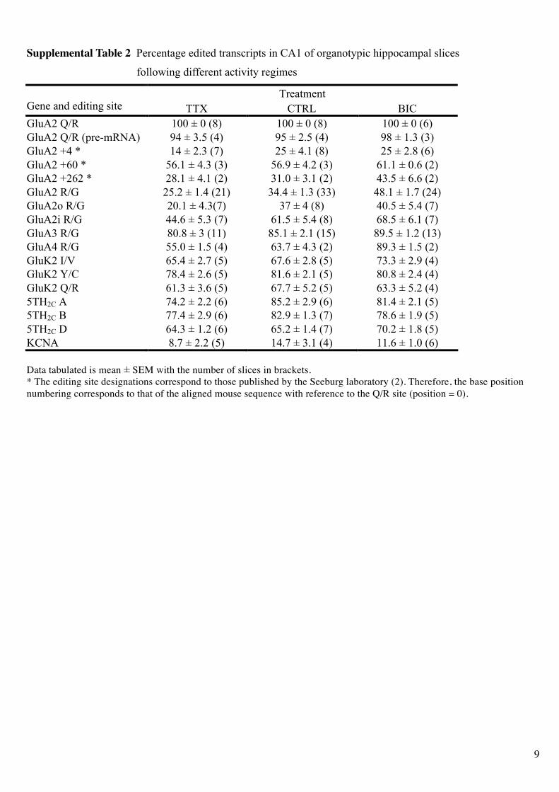
Supplemental Table 2 Percentage edited transcripts in CA1 of organotypic hippocampal slices
following different activity regimes
Treatment Gene and editing site TTX CTRL BIC
GluA2 Q/R 100 ± 0 (8) 100 ± 0 (8) 100 ± 0 (6)
GluA2 Q/R (pre-mRNA) 94 ± 3.5 (4) 95 ± 2.5 (4) 98 ± 1.3 (3)
GluA2 +4 * 14 ± 2.3 (7) 25 ± 4.1 (8) 25 ± 2.8 (6)
GluA2 +60 * 56.1 ± 4.3 (3) 56.9 ± 4.2 (3) 61.1 ± 0.6 (2)
GluA2 +262 * 28.1 ± 4.1 (2) 31.0 ± 3.1 (2) 43.5 ± 6.6 (2)
GluA2 R/G 25.2 ± 1.4 (21) 34.4 ± 1.3 (33) 48.1 ± 1.7 (24)
GluA2o R/G 20.1 ± 4.3(7) 37 ± 4 (8) 40.5 ± 5.4 (7)
GluA2i R/G 44.6 ± 5.3 (7) 61.5 ± 5.4 (8) 68.5 ± 6.1 (7)
GluA3 R/G 80.8 ± 3 (11) 85.1 ± 2.1 (15) 89.5 ± 1.2 (13)
GluA4 R/G 55.0 ± 1.5 (4) 63.7 ± 4.3 (2) 89.3 ± 1.5 (2)
GluK2 I/V 65.4 ± 2.7 (5) 67.6 ± 2.8 (5) 73.3 ± 2.9 (4)
GluK2 Y/C 78.4 ± 2.6 (5) 81.6 ± 2.1 (5) 80.8 ± 2.4 (4)
GluK2 Q/R 61.3 ± 3.6 (5) 67.7 ± 5.2 (5) 63.3 ± 5.2 (4)
5TH2C A 74.2 ± 2.2 (6) 85.2 ± 2.9 (6) 81.4 ± 2.1 (5)
5TH2C B 77.4 ± 2.9 (6) 82.9 ± 1.3 (7) 78.6 ± 1.9 (5)
5TH2C D 64.3 ± 1.2 (6) 65.2 ± 1.4 (7) 70.2 ± 1.8 (5)
KCNA 8.7 ± 2.2 (5) 14.7 ± 3.1 (4) 11.6 ± 1.0 (6)
Data tabulated is mean ± SEM with the number of slices in brackets.
* The editing site designations correspond to those published by the Seeburg laboratory (2). Therefore, the base position
numbering corresponds to that of the aligned mouse sequence with reference to the Q/R site (position = 0).
10
Supplementary Methods
Preparation of slice cultures
For roller-tube cultures, coverslips (12 x 24 mm, Kindler O’GmbH) were placed into alumina holders (Thomas
Scientific) and washed (with sonication) in Millipore water, 96 % ethanol and then allowed to dry on sterile
tissue paper prior to autoclaving. All the following procedures were carried out under sterile conditions,
dissection tools were baked and solutions were sterile filtered. Coverslips were returned to the autoclaved holders
and submerged in freshly prepared 25 µg/ml poly-D-lysine solution (30 kDa, P7280, Sigma) to coat for 5
minutes. Afterwards coverslips were washed three times with autoclaved Millipore water and allowed to dry on
sterile tissue paper. Collagen Type I from rat tail (Fluka or Sigma) was prepared in aqueous solution at 1 mg/ml
and 50–100 µL was spot into the centre of each coverslip and spread to a diameter of ~10 mm. Cross-linking to
form a collagen matrix was achieved by exposing coverslips to ammonia vapour from droplets of 5 N ammonium
hydroxide solution (318612, Sigma) for 5–10 minutes. Coverslips were then washed in autoclaved Millipore
water (containing 10 µg/ml phenol red) until further washing caused no change of indicator colour. Collagen-
coated coverslips were maintained in a sealed jar containing Hanks balanced salt solution (HBSS) for up to a
week prior to the continuing the remainder of the protocol.
Rat brain tissue was dissected in a sucrose modified Gey’s balanced salt solution, which was (in mM): sucrose
(175), NaCl (50), KCl (2.5) Na2HPO4 (0.85), KH2PO4 (0.66), MgSO4 (0.28), MgCl2 (2) CaCl2 (0.5), glucose (25)
and 10 mg/ml phenol red (~ 330 mOsm, pH 7.3). Transverse hippocampal slices were cut at 350 µm thickness
using a McIllwain tissue chopper and individually transferred in dissection medium using a wide-lumen pipette
and positioned with a fine paint brush on the surface of the collagen matrix in the centre of the coverslip.
Dissection medium was carefully aspirated and the slices were wet with a couple of drops of pre-equilibrated (37
°C / 5 % CO2) slice culture medium. Culture medium contained 50 % Basal Medium Eagles (BME), 25 %
HBSS, 25 % heat-inactivated horse serum, 1 mM L-Glutamine and 6.5 g/L D-glucose (320 mOsm). The medium
did not contain antibiotics. Coverslips were transferred into flat-bottomed polystyrene culture tubes (156758,
Nunc) containing 0.75 ml culture medium, the screw cap was sealed tight and the slices were maintained in the
culture incubator overnight. The following day culture tubes were transferred to a custom-made roller drum:
angled ~8 ° and rotating at ~10 rotations per hour (rph). Throughout the entire culture process, incubator was set
to 36 °C with no humidity or CO2 control.
For interface cultures, Millicell cell culture inserts 0.4µm pore size (PICM0RG50, Millipore) were placed in 6-
well plates containing 1ml of prewarmed culture medium (as above). Three slices were individulally transferred
and positioned on the membrane of each insert and gently washed with culture medium. Slices were maintained
in an incubator at 37°C and supplied 5% CO2.
11
Reverse Transcription (RT)-PCR and expression analysis
RT-PCR was conducted using standard instruments and protocols (see main text). The region of interest in
GluA1, 2, 3 and Adar2 were amplified from 2 µl of cDNA with primer pairs at 0.5 µM final concentration for 35
(GluAs) or 42 (Adar2) cycles. All primer sequences are available upon request. PCR amplicons were run on 1.5 -
2.5 % agarose gels and post-stained with ethidium bromide.
To determine the extent of editing and mutually exclusive splicing, 10 µl of each PCR product was cleaned up
using 2 µl of ExoSAP-IT (USB Corporation), sequenced (Geneservice, UK) and individual sequnce analyzed
using PeakPicker software (Version 0.5; (3).
Quantifications of Adar2 PCR amplicons were made on gel images by measuring band peak intensities using
ImageJ (NIH, http://rsb.info.nih.gov/ij/).
Quantitative PCR
The expression levels of Adar1 and 2, relatively to Gapdh or !-2-microglobulin, were measured by real-time
PCR using TaqMan®
pre-developed assays (Applied Biosystems) on cDNA samples. PCR reactions were
prepared in a final volume of 10 !l, containing 0.5 µl of 20x TaqMan probe mix for each gene of interest (FAM
labeled) and GAPDH or ! -2-microglobulin (VIC labeled), 5 µl diluted cDNA and 5µl of 2x TaqMan Universal
PCR Master Mix (Applied Biosystems). PCR reactions were run on a Rotor-Gene 6000 (Qiagen) or a
LightCycler480 (Roche) under the following thermal conditions: 95°C for 10 min (to activate the polymerase),
followed by 45 cycles of denaturation at 95°C for 15 s, and a single annealing and extension step at 60°C for 45s.
Post-reaction data were analyzed with respective software associated with quantitative PCR device. The ""CT
method was used for relative quantification analysis.
Additionally, the expression levels of Adar2, relative to !-2-microglobulin, were also measured by SYBR-green
based quantitative PCR. Reactions for individual gene were prepared in a final volume of 10 !l, containing 1 µl
of 5 µM primer pair, 5µl of Rotor-Gene SYBR Green PCR Kit (Qiagene) and 4 µl diluted cDNA. PCR reactions
were run on a Rotor-Gene 6000 or a LightCycler480 under the following thermal conditions: 95°C for 10 min,
followed by 40 cycles of denaturation at 95°C for 5 s, annealing at 60°C for 10s and extension step at 72°C for
10s. All primer sequences are available upon request.

12
Supplemental References
1. Lein, E.S., Hawrylycz, M.J., Ao, N., Ayres, M., Bensinger, A., Bernard, A., Boe, A.F., Boguski, M.S.,
Brockway, K.S., Byrnes, E.J. et al. (2007) Genome-wide atlas of gene expression in the adult mouse
brain. Nature, 445, 168-176.
2. Higuchi, M., Single, F.N., Kohler, M., Sommer, B., Sprengel, R. and Seeburg, P.H. (1993) RNA editing
of AMPA receptor subunit GluR-B: a base-paired intron-exon structure determines position and
efficiency. Cell, 75, 1361-1370.
3. Ge, B., Gurd, S., Gaudin, T., Dore, C., Lepage, P., Harmsen, E., Hudson, T.J. and Pastinen, T. (2005)
Survey of allelic expression using EST mining. Genome Res, 15, 1584-1591.
Related Documents


![VPS35 regulates developing mouse hippocampal neuronal ... · postnatal day 10 (P10)] (Fig. 1A). The expression appeared to be peaked at the neonatal stage (P10–P15) of the hippocampus](https://static.cupdf.com/doc/110x72/5d67223488c993a9318b4652/vps35-regulates-developing-mouse-hippocampal-neuronal-postnatal-day-10-p10.jpg)


