eScholarship provides open access, scholarly publishing services to the University of California and delivers a dynamic research platform to scholars worldwide. Lawrence Berkeley National Laboratory Lawrence Berkeley National Laboratory Peer Reviewed Title: Activity of the kinesin spindle protein inhibitor ispinesib (SB-715992) in models of breast cancer Author: Purcell, James W Publication Date: 03-11-2011 Publication Info: Lawrence Berkeley National Laboratory Permalink: http://escholarship.org/uc/item/4d53585s DOI: 10.1158/1078-0432.CCR-09-1498 Local Identifier: LBNL Paper LBNL-4328E Preferred Citation: Clinical Cancer Research, 16, 566-576, 1-15-2010

Welcome message from author
This document is posted to help you gain knowledge. Please leave a comment to let me know what you think about it! Share it to your friends and learn new things together.
Transcript
eScholarship provides open access, scholarly publishingservices to the University of California and delivers a dynamicresearch platform to scholars worldwide.
Lawrence Berkeley National LaboratoryLawrence Berkeley National Laboratory
Peer Reviewed
Title:Activity of the kinesin spindle protein inhibitor ispinesib (SB-715992) in models of breast cancer
Author:Purcell, James W
Publication Date:03-11-2011
Publication Info:Lawrence Berkeley National Laboratory
Permalink:http://escholarship.org/uc/item/4d53585s
DOI:10.1158/1078-0432.CCR-09-1498
Local Identifier:LBNL Paper LBNL-4328E
Preferred Citation:Clinical Cancer Research, 16, 566-576, 1-15-2010
1
Activity of the Kinesin Spindle Protein Inhibitor Ispinesib (SB-715992) in Models of
Breast Cancer
James W. Purcell1, Jefferson Davis1, Mamatha Reddy1, Shamra Martin1 , Kimberly
Samayoa1, Hung Vo1, Karen Thomsen1, Peter Bean1, Wen Lin Kuo2, Safiyyah Ziyad2,
Jessica Billig2, Heidi S. Feiler2, Joe W. Gray2, Kenneth W. Wood1, and Sylvaine Cases1
1Cytokinetics, Inc., 280 East Grand Ave, South San Francisco, California
2Lawrence Berkeley National Laboratory, Life Sciences Division, Berkeley, California
Running title: ispinesib activity in models of breast cancer
Keywords: breast cancer, KSP, ispinesib
Correspondence:
Kenneth W. Wood
Cytokinetics, Inc
280 East Grand Ave
South San Francisco, CA 94080
2
Translational Relevance
Microtubule targeted therapies form an integral part of treatment regimens for breast
cancers, most often in combination with other therapies. Of the clinically limiting
toxicities of microtubule-targeted therapies, neuropathy is uniquely unrelated to anti-
proliferative drug action. Inhibitors of Kinesin Spindle Protein (KSP) have emerged as
candidate non-neurotoxic anti-mitotic cancer therapies. One of these KSP inhibitors,
ispinesib, has been evaluated in a Phase II clinical trial in women with locally advanced
or metastatic breast cancer, producing several partial responses. In this report we evaluate
the activity of ispinesib as a single agent in models of several breast cancer subgroups
and examined tolerability and efficacy of ispinesib combined with various standards-of-
care. This is the first study to explore activity of a KSP inhibitor in models of breast
cancer, and to identify attractive KSP inhibitor drug combinations. Our results highlight
clinical settings in which KSP inhibitors may be of clinical utility.
3
Abstract
Purpose: Ispinesib (SB-715992) is a potent inhibitor of kinesin spindle protein (KSP), a
kinesin motor protein essential for the formation of a bipolar mitotic spindle and cell
cycle progression through mitosis. Clinical studies of ispinesib have demonstrated a 9%
response rate in patients with locally advanced or metastatic breast cancer, and a
favorable safety profile without significant neurotoxicities, gastrointestinal toxicities or
hair loss. To better understand the potential of ispinesib in the treatment of breast cancer
we explored the activity of ispinesib alone and in combination several therapies approved
for the treatment of breast cancer.
Experimental Design: We measured the ispinesib sensitivity and pharmacodynamic
response of breast cancer cell lines representative of various subtypes in vitro and as
xenografts in vivo, and tested the ability of ispinesib to enhance the anti-tumor activity of
approved therapies.
Results: In vitro, ispinesib displayed broad anti-proliferative activity against a panel of
53 breast cell-lines. In vivo, ispinesib produced regressions in each of five breast cancer
models, and tumor free survivors in three of these models. The effects of ispinesib
treatment on pharmacodynamic markers of mitosis and apoptosis were examined in vitro
and in vivo, revealing a greater increase in both mitotic and apoptotic markers in the
MDA-MB-468 model than in the less sensitive BT-474 model. In vivo, ispinesib
enhanced the anti-tumor activity of trastuzumab, lapatinib, doxorubicin, and
capecitabine, and exhibited activity comparable to paclitaxel and ixabepilone.

4
Conclusions: These findings support further clinical exploration of KSP inhibitors for
the treatment of breast cancer.
5
Introduction
Chemotherapy remains a cornerstone in the treatment of breast cancer. Microtubule-
targeted anti-mitotic agents feature prominently in therapeutic regimens. Among these,
the taxanes paclitaxel and docetaxel form an integral part of most adjuvant therapy
regimens, and play a major role in the treatment of metastatic disease. Additional
microtubule-targeted therapies include vinorelbine, vinblastine and the recently approved
ixabepilone. These therapies are usually administered as part of a combination regimen
with an anthracycline (doxorubicin), an antimetabolite (capecitabine), a platinum
(carboplatin), or with HER2-targeted therapy such as trastuzumab.
Therapeutic regimens containing microtubule-targeted agents often produce clinically
limiting toxicities including myelosuppression, neuropathy, alopecia and gastrointestinal
toxicities (1, 2). Neuropathy is the only toxicity unrelated to anti-proliferative activity,
and is likely due to the effects of these drugs on neuronal microtubules (3). One strategy
to identify novel anti-mitotic cancer therapies with improved tolerability profiles is to
target mitosis-specific enzymes, eliminating target-related neurotoxicities (4).
Kinesin Spindle Protein (KSP) is a motor protein with an exclusive and essential role in
mitosis (5-7). It is required early in mitosis to separate the centrosomes of the emerging
spindle poles, thus driving establishment of a bipolar mitotic spindle. Failure to establish
a bipolar spindle results in an extended cell cycle delay in mitosis, after which cells may
experience a variety of fates including abnormal exit from mitosis, resumption of the cell
cycle and apoptosis (8-12). The essential role of KSP in cell cycle progression through
6
mitosis in normal and tumor cells alike suggests that anti-tumor activity of KSP inhibitors
is most-likely attributable to post-mitotic response pathways that remain poorly
understood.
Ispinesib (SB-715992), an allosteric small molecule inhibitor of KSP kinesin motor
ATPase (13), was the first small molecule inhibitor of KSP that advanced to cancer
clinical trials. Results from multiple phase I and II clinical studies of ispinesib in patients
suffering from a variety of cancers confirm the absence of significant neurotoxicities,
alopecia or gastrointestinal toxicities (14-22). The most common toxicity associated with
ispinesib was neutropenia, with a reproducible nadir 6-8 days following administration
and full recovery by day 14.
In a preliminary Phase II trial in women with locally advanced or metastatic breast cancer
progressing despite an anthracycline and a taxane treatment, ispinesib was administered
once every 21 days and produced a response rate of 9%, with reductions in tumor size of
46% to 69% and response durations of 6.9 to 19.1 weeks (20). A Phase I/II is ongoing in
patients with locally advanced or metastatic breast cancer previously untreated with
chemotherapy for advanced disease, administering ispinesib on a 2 week schedule (23).
We have evaluated the activity of ispinesib as a single agent in models of specific breast
cancer subgroups (ER positive, Her2 positive, triple negative) and examined tolerability
and efficacy of ispinesib combined with various standards-of-care for breast cancer.

7
This study represents the first in depth biological examination of KSP inhibition in breast
cancer. Our findings support the ongoing clinical investigation of ispinesib as a treatment
option in breast cancer patients.
8
Materials and Methods
Cell culture
Cell lines were obtained from ATCC and from collections developed by Drs. Steve Ethier
and Adi Gazdar. KPL4 was kindly provided by Dr J. Kurebayashi (Kawasaki Medical
School, Kurashiki, Okayama, Japan). Cell culture reagents were from Cellgro-Mediatech
(Herndon, VA).
Cell growth inhibition and data analysis
Cells were plated in log phase of growth in 96-well plates and treated for 72h with a
range of ispinesib concentrations from 3.3x10⁻⁵ to 8.5x10⁻¹¹M. Cell growth was measured
using Cell Titer Glo assays (Promega, Madison, WI) and luminescence read-out was
recorded using BIO-TEK FLx800. Data were analyzed according to the method described
previously by the NCI/NIH DTP Human Tumor Cell Line Screen Process
(http://dtp.nci.nih.gov/branches/btb/ivclsp.html) (24). The % growth curve is calculated
as [(T-T0)/(C- T0)] x100, where T0 is the cell count on day 0, C is the vehicle control (e.g.
0.3% DMSO without drug) cell count on day 3, T is the cell count at the test
concentration. The GI50 value is the drug concentration that results in 50% growth after
72h of drug exposure.
Western blot analyses
Cells were treated with 150nM ispinesib (3 to 7-fold GI50 values) and lysed in RIPA
buffer, (50mM Tris pH7.5, 150mM NaCl, 1% NP40, 0.5% sodium deoxycholate, 0.1%
9
SDS, 1% complete protease inhibitor cocktail [Roche Biochemicals, Indianapolis, IN]).
5ug of protein were separated on 4-20% tris-glycine gels (Invitrogen, Carlsbad, CA) and
transferred onto PVDF membranes using Invitrogen’s iBlot system. Primary antibodies
for Bax, Bid, xIAP, Bcl2, p-Bcl2 (Ser70), and Bcl-XL (54H6) were from Cell Signaling
(Danvers, MA). Other primary antibodies used were: PARP (BD Pharminogen, San
Diego, CA), GAPDH-6C5 (Santa Cruz Biotechnology, Columbia, SC), cyclin-B and
cyclin-E-HE12 (Upstate-Millipore, Billerica, MA). Secondary antibodies were infra-red
680/800CW Licor (Lincoln, NE) and signal detection and analysis were performed on a
Licor-Odyssey imaging system.
DNA cell cycle analysis by flow cytometry
Cells were treated with 150nM ispinesib, fixed in 85% ice-cold ethanol, resuspended in
PBS containing 10ug/ml propidium iodide DNA stain (Sigma Aldrich, St. Louis, MO)
and 250ug/ml RNase A (Sigma Aldrich), and analyzed with a FACSCaliber flow
cytometer (Becton Dickinson, Franklin Lakes, NJ). Cell cycle analyses were performed
with FLOWJO (Treestar Inc, Ashland, OR).
Xenograft studies
Protocols for xenograft studies were approved by the Cytokinetics Institutional Animal
Care and Use Committee (IACUC). Female mice (7-8 weeks) obtained from Charles
River (Hollister, CA) were implanted on their flank with 107 cells in 100l 1:1
PBS/matrigel (BD Biosciences, San Jose, CA). Nu/Nu mice were used for all tumor
models, except BT474 and MDA-MB-468 which were established in Fox-Chase SCID
10
mice. BT474 tumors were generated by subcutaneously implanting 30mm3 tumor
fragments from previously established xenografts. For MCF7 xenograft formation, mice
were implanted subcutaneously at the base of the neck with 90 day release 0.36mg 17β-
estradiol pellets (Innovative Research of America, Sarasota, FL) 3 days prior to tumor
cells implantation. Tumor volume (length x width2)/2 and body weight were measured
twice weekly. For efficacy studies, drug treatment started when tumor volume was
~100mm3 and mice were sacrificed at 60 days post treatment or when tumor volume
reached 1500mm3. Drug treated mice were categorized as a partial regression (PR) if 3
consecutive tumor measurements were less than half the starting tumor volume on day 0
of treatment, a complete regression (CR) if tumor volume was less than 12.5mm3 for 3
consecutive measurements, and a tumor free survivor (TFS) if it had no measurable
tumor or remained a CR at the end of the study. Tumor growth inhibition (TGI) is
defined as the percentage difference in tumor volume between vehicle and drug treated
groups, determined on the final day when all tumor volumes in the vehicle group are
below 1000mm3.
Unpaired t-test statistical analyses were performed on all xenograft studies to determine
the significance of differences in tumor volume (p-value<0.05) between vehicle and drug
treated mice. Unpaired t-tests were also used to determine if a significant improvement in
efficacy occurred when ispinesib was combined with approved agents commonly used in
breast cancer, relative to the single agent alone
Drugs
11
All drugs were dosed at their maximum tolerated dose (MTD) unless otherwise stated,
and drug volumes were 200l/25g mouse. Ispinesib was formulated in 10% ethanol, 10%
cremophor, 80% D5W (dextrose 5%) and dosed intraperitoneally (i.p.) on a q4dx3
schedule (3 doses given every 4 days) at 10mg/kg in Nu/Nu mice or 8mg/kg in SCID
mice, unless otherwise stated. Trastuzumab (Genentech, South San Francisco, CA) was
dosed i.p. twice weekly for 4 weeks at 10mg/kg. Doxorubicin (LGM pharmaceuticals,
Boca Raton, FL) was formulated in 0.9% saline and dosed q4dx3 at 3mg/kg in Nu/Nu
mice or on days 1, 7, and 21 at 2.5mg/kg in Fox-Chase SCID mice. Lapatinib
(GlaxoSmithKline, London, U.K.) was formulated in 0.5%
hydroxypropylmethylcellulose and 0.1% Tween-80 in water and was dosed orally twice
daily for 3 weeks at 40mg/kg. Capecitabine (Roche, Basel, Switzerland) was formulated
in 40mM citrate buffer (pH 6) in 0.5% methylcellulose and was orally dosed daily at
450mg/kg for 14 days. Paclitaxel (Natural Pharmaceuticals, Arcadia, CA) and
ixabepilone (Bristol Myers Squibb, Princeton, NJ) were formulated in 10% ethanol, 10%
cremophor, 80% D5W and dosed intravenously (i.v.) q4dx3 at their respective MTDs of
30mg/kg and 5mg/kg. Vehicle treated control mice were injected i.p. q4dx3 with a
formulation of 10% ethanol, 10% cremophor, 80% D5W.
Immunohistochemistry
Mice with a tumor volume of ~250mm3 received a single dose of ispinesib (10mg/kg).
Tumors were dissected, fixed in 10% buffered formalin, embedded in paraffin, and 5m
tissue sections were mounted on slides. Antigen retrieval was performed by boiling in
50mM citrate buffer pH 5.5, sections were incubated in 3% hydrogen peroxide for 5
12
minutes at RT, washed in PBS-0.1% Tween and blocked in 10% goat serum (Jackson
Laboratories, Bar Harbor, ME). For phospho-histone-H3 (PH3) staining, we used a 1:250
dilution of primary antibody and a 1:500 dilution of AlexaFluor 488nm secondary
antibody (Molecular Probes-Invitrogen). Images were taken with a Nikon-Eclipse TE-
2000U microscope at 10x magnification and captured using Metamorph software to
quantify PH3 expression by computing the area-ratio of PH3 positive cells per total cells.
For Ki67/cleaved caspase-3 double staining, primary antibodies and a Biocare’s Mach2
secondary-HRP-ALP cocktail were used according to manufacturer’s guidelines (Biocare
Medical LLC, Concord, CA). Non-fluorescent images were taken on an Olympus-BX41
microscope at 20x magnification.
13
Results
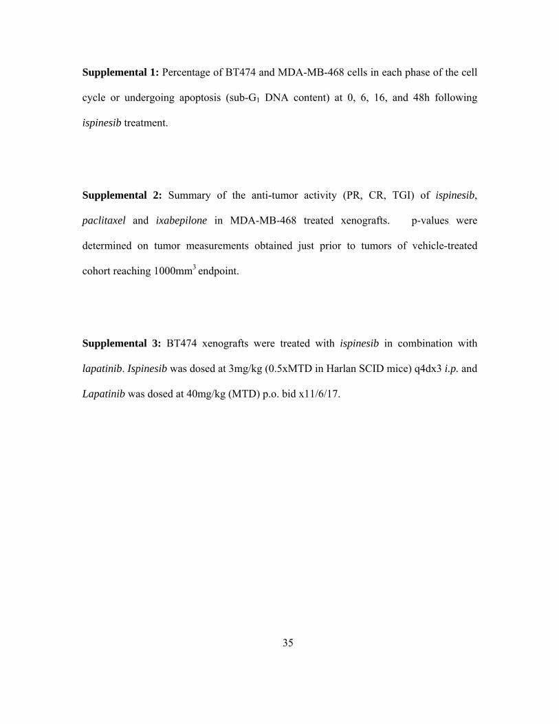
Sensitivity of human breast cancer cell lines to ispinesib in vitro. We investigated the
possibility that specific breast cancer subtypes might exhibit particular sensitivity to
ispinesib in a panel of 50 human breast tumor cell lines of representative of diverse
primary tumor histotypes and genetic backgrounds, and in three normal mammary
epithelial lines, MCF10A, MCF10F and MCF12A (Fig. 1A) (25). Cells were treated with
increasing concentrations of ispinesib and ranked according to the concentration of drug
required to reduce growth by 50% (GI50) (Fig.1A). All lines exhibited sensitivities
between 7.4nM and 600nM, with most falling within a ten-fold range, between 7.4nM
and 80nM. Three lines, all of luminal subtype, exhibited sensitivities between 100 and
600nM. Across this relatively narrow range of sensitivity we were unable to discern any
obvious correlation with subtype, receptor expression or mutational status.
We selected two cell lines, BT474, a HER2 positive luminal cell line (GI50 for ispinesib
of 45nM) and MDA-MB-468, a basal A triple negative cell line, (GI50 of 19nM) and
characterized the kinetics of cell cycle and apoptotic responses to ispinesib in vitro
following exposure to 150nM ispinesib, greater than 3-fold the GI50 value for both cell
lines (Fig.1B). In the absence of drug, the proportion of cells with in G2 or M phases of
the cell cycle in MDA-MB-468 was twice that of BT474. After exposure to 150nM
ispinesib, this proportion increased transiently in both lines, consistent with KSP-induced
mitotic arrest. Maximal accumulation of mitotic cells occurred after 16h of treatment in
MDA-MB-468 cells and 48h in BT474 cells. At 48h, MDA-MB-468 displayed a much
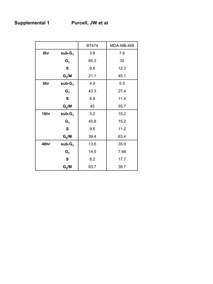
higher proportion of apoptotic cells (sub-G1 DNA content) (35%), than BT474 cells
14
(13.6%), (Supplemental 1). These findings are consistent with a more rapid and penetrant
onset of cell death following mitotic arrest in MDA-MB-468 than in BT474.
We also evaluated the effects of ispinesib on the abundance of cell cycle and apoptosis-
related proteins (Fig.1C). Expression of the pro-apoptotic proteins BAX and Bid was
higher in MDA-MB-468 than in BT-474, while the anti-apoptotic protein Bcl-XL was
lower. Bcl2 levels were not different between the two lines, though phosphorylation on
Serine 70 was greater in BT474. The significance of this modification is unclear but has
been previously associated with potentiating and abrogating Bcl2 anti-apoptotic activity
(26).
The onset of apoptosis was preceded by accumulation of cyclin B, a marker of mitosis
(27). In MDA-MB-468 cells, cyclin B expression was maximal at 16h and remained
elevated for at least 48h, consistent with an abundance of mitotic cells. In contrast, in BT-
474 cells cyclin B levels were generally lower, maximal accumulation was observed at 6h
and diminished thereafter. Cyclin E, which normally accumulates to maximal levels in
late G1 phase of the cell cycle (28), increased slightly in BT474 after ispinesib treatment,
but in MDA-MB-468 cells was almost undetectable. The abundance of cyclin A was
minimally affected by drug exposure and we observed no changes in the abundance of
cyclin D (data not shown).
Efficacy of ispinesib as a single agent in pre-clinical breast cancer models. To
determine the extent of ispinesib anti-tumor activity in breast cancer models in vivo, we
chose cell lines that exhibited different in vitro sensitivity to ispinesib and represent
15
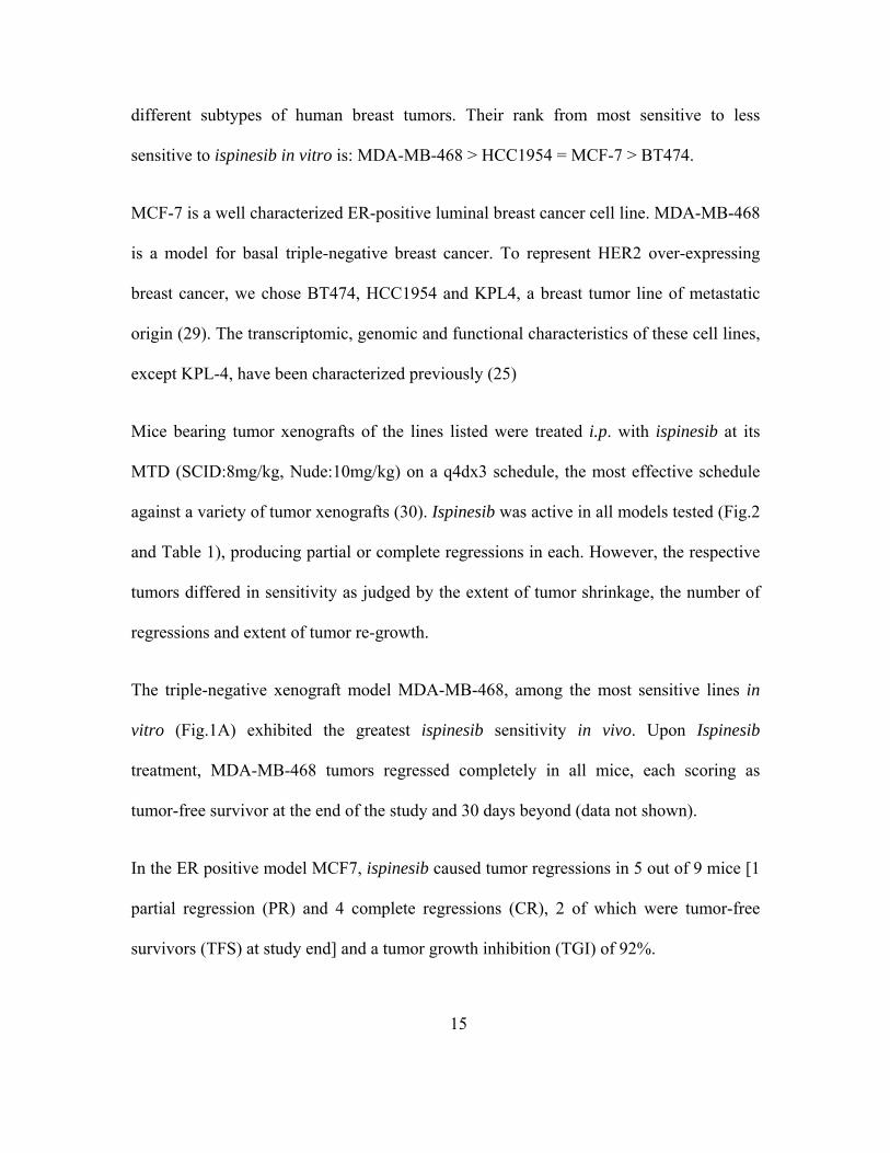
different subtypes of human breast tumors. Their rank from most sensitive to less
sensitive to ispinesib in vitro is: MDA-MB-468 > HCC1954 = MCF-7 > BT474.
MCF-7 is a well characterized ER-positive luminal breast cancer cell line. MDA-MB-468
is a model for basal triple-negative breast cancer. To represent HER2 over-expressing
breast cancer, we chose BT474, HCC1954 and KPL4, a breast tumor line of metastatic
origin (29). The transcriptomic, genomic and functional characteristics of these cell lines,
except KPL-4, have been characterized previously (25)
Mice bearing tumor xenografts of the lines listed were treated i.p. with ispinesib at its
MTD (SCID:8mg/kg, Nude:10mg/kg) on a q4dx3 schedule, the most effective schedule
against a variety of tumor xenografts (30). Ispinesib was active in all models tested (Fig.2
and Table 1), producing partial or complete regressions in each. However, the respective
tumors differed in sensitivity as judged by the extent of tumor shrinkage, the number of
regressions and extent of tumor re-growth.
The triple-negative xenograft model MDA-MB-468, among the most sensitive lines in
vitro (Fig.1A) exhibited the greatest ispinesib sensitivity in vivo. Upon Ispinesib
treatment, MDA-MB-468 tumors regressed completely in all mice, each scoring as
tumor-free survivor at the end of the study and 30 days beyond (data not shown).
In the ER positive model MCF7, ispinesib caused tumor regressions in 5 out of 9 mice [1
partial regression (PR) and 4 complete regressions (CR), 2 of which were tumor-free
survivors (TFS) at study end] and a tumor growth inhibition (TGI) of 92%.
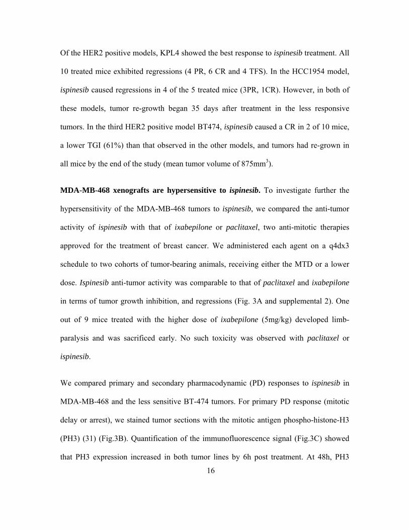
16
Of the HER2 positive models, KPL4 showed the best response to ispinesib treatment. All
10 treated mice exhibited regressions (4 PR, 6 CR and 4 TFS). In the HCC1954 model,
ispinesib caused regressions in 4 of the 5 treated mice (3PR, 1CR). However, in both of
these models, tumor re-growth began 35 days after treatment in the less responsive
tumors. In the third HER2 positive model BT474, ispinesib caused a CR in 2 of 10 mice,
a lower TGI (61%) than that observed in the other models, and tumors had re-grown in
all mice by the end of the study (mean tumor volume of 875mm3).
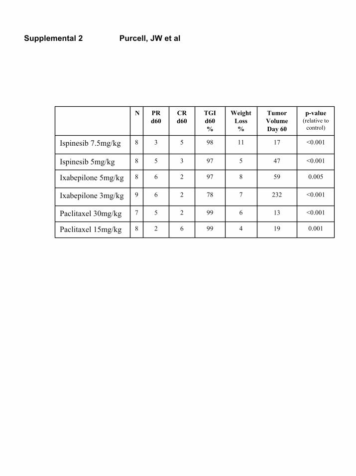
MDA-MB-468 xenografts are hypersensitive to ispinesib. To investigate further the
hypersensitivity of the MDA-MB-468 tumors to ispinesib, we compared the anti-tumor
activity of ispinesib with that of ixabepilone or paclitaxel, two anti-mitotic therapies
approved for the treatment of breast cancer. We administered each agent on a q4dx3
schedule to two cohorts of tumor-bearing animals, receiving either the MTD or a lower
dose. Ispinesib anti-tumor activity was comparable to that of paclitaxel and ixabepilone
in terms of tumor growth inhibition, and regressions (Fig. 3A and supplemental 2). One
out of 9 mice treated with the higher dose of ixabepilone (5mg/kg) developed limb-
paralysis and was sacrificed early. No such toxicity was observed with paclitaxel or
ispinesib.
We compared primary and secondary pharmacodynamic (PD) responses to ispinesib in
MDA-MB-468 and the less sensitive BT-474 tumors. For primary PD response (mitotic
delay or arrest), we stained tumor sections with the mitotic antigen phospho-histone-H3
(PH3) (31) (Fig.3B). Quantification of the immunofluorescence signal (Fig.3C) showed
that PH3 expression increased in both tumor lines by 6h post treatment. At 48h, PH3
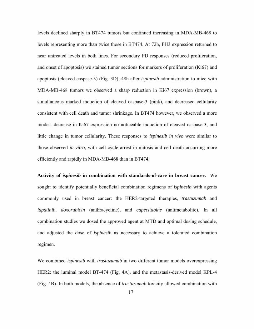
17
levels declined sharply in BT474 tumors but continued increasing in MDA-MB-468 to
levels representing more than twice those in BT474. At 72h, PH3 expression returned to
near untreated levels in both lines. For secondary PD responses (reduced proliferation,
and onset of apoptosis) we stained tumor sections for markers of proliferation (Ki67) and
apoptosis (cleaved caspase-3) (Fig. 3D). 48h after ispinesib administration to mice with
MDA-MB-468 tumors we observed a sharp reduction in Ki67 expression (brown), a
simultaneous marked induction of cleaved caspase-3 (pink), and decreased cellularity
consistent with cell death and tumor shrinkage. In BT474 however, we observed a more
modest decrease in Ki67 expression no noticeable induction of cleaved caspase-3, and
little change in tumor cellularity. These responses to ispinesib in vivo were similar to
those observed in vitro, with cell cycle arrest in mitosis and cell death occurring more
efficiently and rapidly in MDA-MB-468 than in BT474.
Activity of ispinesib in combination with standards-of-care in breast cancer. We
sought to identify potentially beneficial combination regimens of ispinesib with agents
commonly used in breast cancer: the HER2-targeted therapies, trastuzumab and
lapatinib, doxorubicin (anthracycline), and capecitabine (antimetabolite). In all
combination studies we dosed the approved agent at MTD and optimal dosing schedule,
and adjusted the dose of ispinesib as necessary to achieve a tolerated combination
regimen.
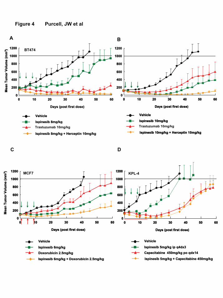
We combined ispinesib with trastuzumab in two different tumor models overexpressing
HER2: the luminal model BT-474 (Fig. 4A), and the metastasis-derived model KPL-4
(Fig. 4B). In both models, the absence of trastuzumab toxicity allowed combination with
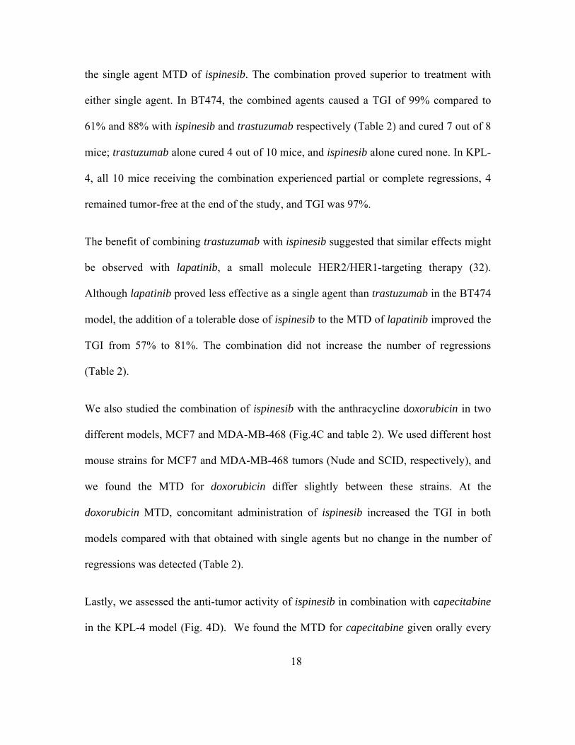
18
the single agent MTD of ispinesib. The combination proved superior to treatment with
either single agent. In BT474, the combined agents caused a TGI of 99% compared to
61% and 88% with ispinesib and trastuzumab respectively (Table 2) and cured 7 out of 8
mice; trastuzumab alone cured 4 out of 10 mice, and ispinesib alone cured none. In KPL-
4, all 10 mice receiving the combination experienced partial or complete regressions, 4
remained tumor-free at the end of the study, and TGI was 97%.
The benefit of combining trastuzumab with ispinesib suggested that similar effects might
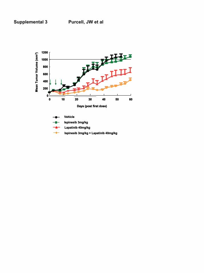
be observed with lapatinib, a small molecule HER2/HER1-targeting therapy (32).
Although lapatinib proved less effective as a single agent than trastuzumab in the BT474
model, the addition of a tolerable dose of ispinesib to the MTD of lapatinib improved the
TGI from 57% to 81%. The combination did not increase the number of regressions
(Table 2).
We also studied the combination of ispinesib with the anthracycline doxorubicin in two
different models, MCF7 and MDA-MB-468 (Fig.4C and table 2). We used different host
mouse strains for MCF7 and MDA-MB-468 tumors (Nude and SCID, respectively), and
we found the MTD for doxorubicin differ slightly between these strains. At the
doxorubicin MTD, concomitant administration of ispinesib increased the TGI in both
models compared with that obtained with single agents but no change in the number of
regressions was detected (Table 2).
Lastly, we assessed the anti-tumor activity of ispinesib in combination with capecitabine
in the KPL-4 model (Fig. 4D). We found the MTD for capecitabine given orally every
19
day for 2 weeks was 450mg/kg, comparable to previously reports (33). The maximum
dose of ispinesib that we could co-administer with capecitabine without treatment-related
toxicity was half its single agent MTD (5mg/kg). In these conditions, although the mean
tumor volume was similar to that in mice treated with capecitabine alone we observed a
clear increase in the number of tumor regressions, including one tumor-free survivor and
an increase in TGI (Table 2).
20
Discussion
In this study, ispinesib has demonstrated significant anti-tumor activity in diverse pre-
clinical models of breast cancer, supporting its potential for therapeutic intervention in
breast cancer. In vitro, ispinesib inhibited proliferation of all 53 breast cell lines tested,
including 3 non-malignant and 50 cancer cell lines derived from distinct breast tumor
types. GI50 values spanned a 100-fold range and fall between 10nM and 100nM for most
cell lines. Ispinesib exhibited no apparent specificity for histopathological sub-type
(luminal A, luminal B, basal) or receptor status (HER2, ER/PR). Interestingly, its profile
of activity differed from that of other anti-mitotic agents such as paclitaxel that inhibited
cell growth over a larger concentration range and were more potent against models of
basal breast cancer (34). Identification of genomic and transcriptomic differences
correlating with relative sensitivity to ispinesib may reveal the basis for differential
sensitivity of these cell lines in culture and as xenografts, providing biomarkers
predictive of disease response to ispinesib.
Ispinesib was also active in vivo in various breast cancer subtypes, inducing complete
regressions or cures in ER positive (MCF7), HER2 positive (BT474, HCC1954, KPL4)
and triple negative (MDA-MB-468) models, suggesting that it might be useful in the
treatment of a broad range of breast cancers.
Xenografts of the triple-negative MDA-MB-468 cell line were exquisitely sensitive to
ispinesib. In vitro this cell line scored among the most sensitive. In vivo, all mice were
cured and remained tumor-free for at least 30 additional days. The basis for this strikingly
21
efficacious response is unclear but our data suggest that cell cycle abnormalities might
present a favorable environment for ispinesib activity.
Prior to drug treatment in vitro and in vivo, MDA-MB-468 cells displayed a relatively
high proportion of cells in mitosis (with a 4N DNA content or positive for the mitotic
antigen, phospho-histone H3) compared to the less sensitive BT474 cells. BT474 cells
appear to transiently arrest in mitosis and then escape from M-phase, re-entering
interphase as suggested by accumulation of cyclin E. The loss of cyclin E expression and
the increased and longer duration of cyclin B expression in MDA-MB-468 cells are
consistent with ispinesib inducing a penetrant and sustained mitotic arrest in these cells.
This suggests a deregulation of the G1/S transition and interestingly, deregulation of
cyclin E expression is commonly observed in breast cancer (35). MDA-MB-468 cells
also harbor mutations in the regulators of the G1 checkpoint Rb and p53. Additional
experiments will be required to determine if these cell cycle alterations play a role in
relatively greater sensitivity of MDA-MB-468 to ispinesib.
The elevated expression of the pro-apoptotic proteins BAX and Bid, and the reduced
expression of anti-apoptotic proteins phospho-Bcl-2 and Bcl-XL are consistent with
increased induction of apoptosis following ispinesib treatment. Previous observations
have linked elevated BAX expression to the induction of apoptosis by KSP inhibition (8,
9) and differences in apoptotic responses have been proposed to be predictive of
sensitivity to anti-mitotic drugs such as KSP inhibitors (10). Importantly, our in vitro
observations were confirmed by pharmacodynamic studies in vivo. In both MDA-MB-
468 and BT474 tumors we observed ispinesib-induced increases in the mitotic antigen
22
phospho-histone H3. Ispinesib treatment was also associated with cleavage of caspase-3,
a marker of apoptosis, decreased staining for Ki67, a marker of active proliferation, and
decrease in tumor cellularity. These ispinesib-induced pharmacodynamic changes were
greater in MDA-MB-468 tumors compared with BT474 tumors, consistent with the
greater rate of regressions observed in MDA-MB-468 xenografts compared with BT474
xenografts.
Ispinesib compared favorably with approved anti-mitotic agents (paclitaxel and
ixabepilone) in the MDA-MB-468 model of the basal subtype of breast cancer with all
three agents producing complete and partial regressions. Consistent with neuropathy
being a common side-effect of ixabepilone therapy (3, 36, 37), we observed severe limb-
paralysis in some mice receiving ixabepilone. However, neurotoxicity is uncommon in
patients receiving ispinesib, and this side-effect was not recorded in preclinical studies,
most likely due to the absence of ispinesib’s target KSP from post-mitotic neurons (6).
Our findings also show that pre-clinically ispinesib is well-tolerated when combined with
doxorubicin, capecitabine, trastuzumab and lapatanib, therapies commonly used in
treatment of breast cancer. Administration of ispinesib at doses tolerated with the MTD
of these agents enhanced their anti-tumor activity as demonstrated by higher TGI values
and increased tumor regressions.
A particularly beneficial combination was that of ispinesib with trastuzumab. The HER-2
positive models we tested were somewhat less sensitive to single-agent ispinesib than
either the ER-positive or triple-negative models. However, in two models of HER2
23
positive breast cancer, ispinesib combined with trastuzumab enhanced the activity of
either single agent by increasing TGI or the number of regressions and cures. HER2
inhibition is known to potentiate the activity of anti-mitotic agents such as paclitaxel (38,
39) and this drug combination is currently the standard-of-care for patients with
advanced/metastatic HER2 positive breast cancer (40, 41). The improved efficacy of
combining KSP inhibition and HER2 inhibition in the preclinical setting, together with
the favorable clinical toxicity profile of ispinesib (20, 23) suggests that a combination
with trastuzumab may be of clinical benefit.
In patients with advanced or metastatic breast cancer, capecitabine is a standard-of-care
(42). A Phase I study demonstrated that the commonly used dose of capecitabine,
2000mg/m2, is well-tolerated with the full recommended Phase II dose of ispinesib
administered every 21 days (18mg/m2) (43). In preclinical studies, toxicity prevented us
from combining capecitabine and ispinesib at the respective single agent MTD.
However, we found that doses of ispinesib below MTD potentiated the anti-tumor
activity of capecitabine administered at its MTD. These findings suggest that ispinesib
and capecitabine represent a potentially beneficial combination for the treatment of
advanced breast cancer, a setting where ispinesib has already demonstrated activity.
We have shown that in vitro and in vivo, ispinesib displays a broad spectrum of activity
against breast cancer models representative of various human breast tumor types. In vivo,
the addition of ispinesib enhanced the anti-tumor activity of several therapies that are
current standards-of-care for the treatment of breast cancer. This robust preclinical anti-
tumor activity, coupled with evidence of clinical activity and favorable tolerability profile
24
in patients with breast cancer, support the continued investigation of ispinesib as a
promising therapeutic agent in breast cancer.
Acknowledgements
This work was supported by Cytokinetics, Inc, the Director, Office of Science, Office of
Biological & Environmental Research, of the U.S. Department of Energy under Contract
No. DE-AC02-05CH11231, by the National Institutes of Health, National Cancer
Institute grant P50 CA 58207, the P50 CA 83639, and by a Cytokinetics grant to JWG.
Thanks to Peter Lambert for advice on statistical analysis.
References
1. Bishop, J. F., Dewar, J., Toner, G. C., Smith, J., Tattersall, M. H., Olver, I. N.,
Ackland, S., Kennedy, I., Goldstein, D., Gurney, H., Walpole, E., Levi, J.,
Stephenson, J., and Canetta, R. Initial paclitaxel improves outcome compared
with CMFP combination chemotherapy as front-line therapy in untreated
metastatic breast cancer. J Clin Oncol, 17: 2355-2364, 1999.
2. Nabholtz, J. M. and Riva, A. Taxane/anthracycline combinations: setting a new
standard in breast cancer? Oncologist, 6 Suppl 3: 5-12, 2001.
25
3. Rowinsky, E. K., Eisenhauer, E. A., Chaudhry, V., Arbuck, S. G., and
Donehower, R. C. Clinical toxicities encountered with paclitaxel (Taxol). Semin.
Oncol., 20: 1-15, 1993.
4. Jackson, J. R., Patrick, D. R., Dar, M. M., and Huang, P. S. Targeted anti-mitotic
therapies: can we improve on tubulin agents? Nat Rev Cancer, 7: 107-117, 2007.
5. Sawin, K. E., LeGuellec, K., Philippe, M., and Mitchison, T. J. Mitotic spindle
organization by a plus-end-directed microtubule motor. Nature, 359: 540-543,
1992.
6. Sakowicz, R., Finer, J. T., Beraud, C., Crompton, A., Lewis, E., Fritsch, A., Lee,
Y., Mak, J., Moody, R., Turincio, R., Chabala, J. C., Gonzales, P., Roth, S.,
Weitman, S., and Wood, K. W. Anti-tumour activity of a kinesin inhibitor. Cancer
Res., 64: 3276-3280, 2004.
7. Blangy, A., Lane, H. A., d'Herin, P., Harper, M., Kress, M., and Nigg, E. A.
Phosphorylation by p34cdc2 regulates spindle association of human Eg5, a
kinesin-related motor essential for bipolar spindle formation in vivo. Cell, 83:
1159-1169, 1995.
8. Tao, W., South, V. J., Diehl, R. E., Davide, J. P., Sepp-Lorenzino, L., Fraley, M.
E., Arrington, K. L., and Lobell, R. B. An inhibitor of the kinesin spindle protein
activates the intrinsic apoptotic pathway independently of p53 and de novo
protein synthesis. Mol Cell Biol, 27: 689-698, 2007.
9. Tao, W., South, V. J., Zhang, Y., Davide, J. P., Farrell, L., Kohl, N. E., Sepp-
Lorenzino, L., and Lobell, R. B. Induction of apoptosis by an inhibitor of the
26
mitotic kinesin KSP requires both activation of the spindle assembly checkpoint
and mitotic slippage. Cancer Cell, 8: 49-59, 2005.
10. Shi, J., Orth, J. D., and Mitchison, T. Cell type variation in responses to
antimitotic drugs that target microtubules and kinesin-5. Cancer Res, 68: 3269-
3276, 2008.
11. Gascoigne, K. E. and Taylor, S. S. Cancer cells display profound intra- and
interline variation following prolonged exposure to antimitotic drugs. Cancer
Cell, 14: 111-122, 2008.
12. Gascoigne, K. E. and Taylor, S. S. How do anti-mitotic drugs kill cancer cells? J
Cell Sci, 122: 2579-2585, 2009.
13. Lad, L., Luo, L., Carson, J. D., Wood, K. W., Hartman, J. J., Copeland, R. A., and
Sakowicz, R. Mechanism of inhibition of human KSP by ispinesib. Biochemistry,
47: 3576-3585, 2008.
14. Burris, H. A., Lorusso, P., Jones, S., Guthrie, T. M., Orr, J. B., Williams, D. D.,
Hodge, J. P., Bush, M., and Sabry, J. Phase I trial of novel kinesin spindle protein
(KSP) inhibitor SB-715992 IV days 1, 8, 15 q 28 days. In: 2004 ASCO Annual
Meeting 2004.
15. Beekman, K. W., Dunn, R., Colevas, D., Davis, N., Clark, J., Agamah, E.,
Thomas, S., Nichols, K., Redman, B., and Stadler, W. University of Chicago
Consortium phase II study of ispinesib (SB-715992) in patients (pts) with
advanced renal cell carcinoma (RCC). In: ASCO Annual Meetting 2007, pp.
Abstract 15573.
27
16. Chu, Q., Holen, K. D., Rowinsky, E. K., Wilding, G., Volkman, J. L., Orr, J. B.,
Williams, D. D., Hodge, J. P., Kerfoot, C. A., and Sabry, J. A phase I study of
novel kinesin spindle protein (KSP) inhibtor, SB-715992 administered
intravenously once every 21 days. In: 40th ASCO Annual Meeting, New Orleans,
LA, 2004, pp. Abstract 2078.
17. Knox, J. J., Gill, S., Synold, T. W., Biagi, J. J., Major, P., Feld, R., Cripps, C.,
Wainman, N., Eisenhauer, E., and Seymour, L. A phase II and pharmacokinetic
study of SB-715992, in patients with metastatic hepatocellular carcinoma: a study
of the National Cancer Institute of Canada Clinical Trials Group (NCIC CTG
IND.168). Invest New Drugs, 26: 265-272, 2008.
18. Lee, C. W., Belanger, K., Rao, S. C., Petrella, T. M., Tozer, R. G., Wood, L.,
Savage, K. J., Eisenhauer, E. A., Synold, T. W., Wainman, N., and Seymour, L. A
phase II study of ispinesib (SB-715992) in patients with metastatic or recurrent
malignant melanoma: a National Cancer Institute of Canada Clinical Trials Group
trial. Invest New Drugs, 26: 249-255, 2008.
19. LoRusso, J. B., Jones, J. B., Gadgeel, S., Willcutt, N., Helmke, W., Orr, J.,
Hodge, J., Williams, D., Pandite, L., Sabry, J., Ho, P., and Burris, H. A Phase I
Study To Determine the Safety and Pharmacokinetics of Intravenous
Administration of SB-715992, a Novel Kinesin Spindle Protein (KSP) Inhibitor,
on a Once Weekly for Three Consecutive Weeks Schedule in Patients With
Refractory Solid Tumors. In: European Cancer Conference 2003.
28
20. Miller, K. N., C.; Ang, P.; Brufsky, A. M.; Lees, S. C.; Dees, E. C.; Piccart, M.;
Verrill, M.; Wardley, A.; Loftiss, J.; Yeoh, S.; Hodge, J.; Williams, D.; Dar, M.;
Ho, P. T. C. Phase II, open label study of SB-715992 (Ispinesib) in subjects with
advanced or metastatic breast cancer. Poster 1089. In: 28th Annual San Antonio
Breast Cancer Symposium, San Antonio, TX, 2005, pp. Abstract 1089.
21. Shahin, M. S., Braly, P., Rose, P., Malpass, T., Bailey, H., Alvarez, R. D., Hodge,
J., Bowen, C., and Buller, R. A phase II, open-label study of ispinesib (SB-
715992) in patients with platinum/taxane refractory or resistant relapsed ovarian
cancer. In: Annual ASCO Meeting 2007, pp. Abstract 5562.
22. Tang, P. A., Siu, L. L., Chen, E. X., Hotte, S. J., Chia, S., Schwarz, J. K., Pond,
G. R., Johnson, C., Colevas, A. D., Synold, T. W., Vasist, L. S., and Winquist, E.
Phase II study of ispinesib in recurrent or metastatic squamous cell carcinoma of
the head and neck. Invest New Drugs, 26: 257-264, 2008.
23. Gomez, H., Castaneda, C., Philco, M., Pimentel, P., Falcon, S., Escandon, R.,
Saikali, K., Conlan, M., Seroogy, J., and Wolff, A. A phase I-II trial of Ispinesib,
a kinesin spindle protein inhibitor, dosed every two weeks in patients with locally
advanced or metastatic breast cancer previously untreated with chemotherapy for
metastatic disease or recurrence. In: 31st Annual San Antonio Breast Cancer
Symposium, San Antonio, TX, 2008, pp. Abstract 2148.
24. Monks, A., Scudiero, D., Skehan, P., Shoemaker, R., Paull, K., Vistica, D., Hose,
C., Langley, J., Cronise, P., Vaigro-Wolff, A., and et al. Feasibility of a high-flux
29
anticancer drug screen using a diverse panel of cultured human tumor cell lines. J
Natl Cancer Inst, 83: 757-766, 1991.
25. Neve, R. M., Chin, K., Fridlyand, J., Yeh, J., Baehner, F. L., Fevr, T., Clark, L.,
Bayani, N., Coppe, J. P., Tong, F., Speed, T., Spellman, P. T., DeVries, S., Lapuk,
A., Wang, N. J., Kuo, W. L., Stilwell, J. L., Pinkel, D., Albertson, D. G.,
Waldman, F. M., McCormick, F., Dickson, R. B., Johnson, M. D., Lippman, M.,
Ethier, S., Gazdar, A., and Gray, J. W. A collection of breast cancer cell lines for
the study of functionally distinct cancer subtypes. Cancer Cell, 10: 515-527,
2006.
26. Haldar, S., Basu, A., and Croce, C. M. Serine-70 is one of the critical sites for
drug-induced Bcl2 phosphorylation in cancer cells. Cancer Res, 58: 1609-1615,
1998.
27. Dutta, A., Chandra, R., Leiter, L. M., and Lester, S. Cyclins as markers of tumor
proliferation: immunocytochemical studies in breast cancer. Proc Natl Acad Sci U
S A, 92: 5386-5390, 1995.
28. Lew, D. J., Dulic, V., and Reed, S. I. Isolation of three novel human cyclins by
rescue of G1 cyclin (Cln) function in yeast. Cell, 66: 1197-1206, 1991.
29. Kurebayashi, J., Otsuki, T., Tang, C. K., Kurosumi, M., Yamamoto, S., Tanaka,
K., Mochizuki, M., Nakamura, H., and Sonoo, H. Isolation and characterization of
a new human breast cancer cell line, KPL-4, expressing the Erb B family
receptors and interleukin-6. Br J Cancer, 79: 707-717, 1999.
30
30. Johnson, R. K., McCabe, F. L., Cauder, E., Innlow, L., Whitacre, M., Winkler, J.
D., Bergnes, G., Feng, B., Smith, W., Morgans, D., Wood, K., and Jackson, J. R.
SB-715992, a potent and selective inhibitor of the mitotic kinesin KSP,
demonstrates broad-spectrum activity in advanced murine tumors and human
tumor xenografts. In: Proc. Am. Assoc. Cancer Res., San Francisco, CA, 2002,
pp. Abstr. 1335.
31. Juan, G., Traganos, F., James, W. M., Ray, J. M., Roberge, M., Sauve, D. M.,
Anderson, H., and Darzynkiewicz, Z. Histone H3 phosphorylation and expression
of cyclins A and B1 measured in individual cells during their progression through
G2 and mitosis. Cytometry, 32: 71-77, 1998.
32. Moy, B. and Goss, P. E. Lapatinib: current status and future directions in breast
cancer. Oncologist, 11: 1047-1057, 2006.
33. Higgins, B., Kolinsky, K., Linn, M., Adames, V., Zhang, Y. E., Moisa, C., Dugan,
U., Heimbrook, D., and Packman, K. Antitumor activity of capecitabine and
bevacizumab combination in a human estrogen receptor-negative breast
adenocarcinoma xenograft model. Anticancer Res, 27: 2279-2287, 2007.
34. O'Brien, C., Cavet, G., Pandita, A., Hu, X., Haydu, L., Mohan, S., Toy, K.,
Rivers, C. S., Modrusan, Z., Amler, L. C., and Lackner, M. R. Functional
genomics identifies ABCC3 as a mediator of taxane resistance in HER2-amplified
breast cancer. Cancer Res, 68: 5380-5389, 2008.
35. Smith, A. P., Henze, M., Lee, J. A., Osborn, K. G., Keck, J. M., Tedesco, D.,
Bortner, D. M., Rosenberg, M. P., and Reed, S. I. Deregulated cyclin E promotes
31
p53 loss of heterozygosity and tumorigenesis in the mouse mammary gland.
Oncogene, 25: 7245-7259, 2006.
36. Denduluri, N., Low, J. A., Lee, J. J., Berman, A. W., Walshe, J. M., Vatas, U.,
Chow, C. K., Steinberg, S. M., Yang, S. X., and Swain, S. M. Phase II trial of
ixabepilone, an epothilone B analog, in patients with metastatic breast cancer
previously untreated with taxanes. J Clin Oncol, 25: 3421-3427, 2007.
37. Perez, E. A., Lerzo, G., Pivot, X., Thomas, E., Vahdat, L., Bosserman, L., Viens,
P., Cai, C., Mullaney, B., Peck, R., and Hortobagyi, G. N. Efficacy and safety of
ixabepilone (BMS-247550) in a phase II study of patients with advanced breast
cancer resistant to an anthracycline, a taxane, and capecitabine. J Clin Oncol, 25:
3407-3414, 2007.
38. Baselga, J., Norton, L., Albanell, J., Kim, Y. M., and Mendelsohn, J.
Recombinant humanized anti-HER2 antibody (Herceptin) enhances the antitumor
activity of paclitaxel and doxorubicin against HER2/neu overexpressing human
breast cancer xenografts. Cancer Res, 58: 2825-2831, 1998.
39. Lee, S., Yang, W., Lan, K. H., Sellappan, S., Klos, K., Hortobagyi, G., Hung, M.
C., and Yu, D. Enhanced sensitization to taxol-induced apoptosis by herceptin
pretreatment in ErbB2-overexpressing breast cancer cells. Cancer Res, 62: 5703-
5710, 2002.
40. Buzdar, A. U., Ibrahim, N. K., Francis, D., Booser, D. J., Thomas, E. S.,
Theriault, R. L., Pusztai, L., Green, M. C., Arun, B. K., Giordano, S. H.,
Cristofanilli, M., Frye, D. K., Smith, T. L., Hunt, K. K., Singletary, S. E., Sahin,
32
A. A., Ewer, M. S., Buchholz, T. A., Berry, D., and Hortobagyi, G. N.
Significantly higher pathologic complete remission rate after neoadjuvant therapy
with trastuzumab, paclitaxel, and epirubicin chemotherapy: results of a
randomized trial in human epidermal growth factor receptor 2-positive operable
breast cancer. J Clin Oncol, 23: 3676-3685, 2005.
41. Slamon, D. J., Leyland-Jones, B., Shak, S., Fuchs, H., Paton, V., Bajamonde, A.,
Fleming, T., Eiermann, W., Wolter, J., Pegram, M., Baselga, J., and Norton, L.
Use of chemotherapy plus a monoclonal antibody against HER2 for metastatic
breast cancer that overexpresses HER2. N Engl J Med, 344: 783-792, 2001.
42. Findlay, M., von Minckwitz, G., and Wardley, A. Effective oral chemotherapy for
breast cancer: pillars of strength. Ann Oncol, 19: 212-222, 2008.
43. Rodon, J., Till, E., Patnaik, A., Takimoto, C., Kathman, S., Williams, D., Vasist,
L., Bowen, C., Hodge, J., Dar, M., and Tolcher, A. Phase I Study of lspinesib in
Combination with Capecitabine in Patients with Advanced Solid Tumors. In: 18th
Annual EORTC-NCI-AACR Symposium on Molecular Targets and Cancer
Therapeutics, Prague, Czech Republic, 2006.
33
Figure Legends
Figure 1: Anti-tumor activity of ispinesib in vitro against models of breast cancer. A,
Growth inhibition at 50% (GI50) induced by ispinesib was determined for 53 breast cell-
lines of luminal, basal A, basal B and non-cancerous origin. B, Differences in cell cycle
profiles in MDA-MB-468, a cell line sensitive to ispinesib, compared to a less sensitive
cell line, BT474, after treatment with 150nM ispinesib (3- to 7-fold GI50). C, Expression
of cell cycle markers (cyclin A, cyclin B, and cyclin E), apoptotic proteins (Bax, Bid, p-
Bcl2, Bcl2, Bcl-XL) was analyzed by western blotting in MDA-MB-468 and BT474 cells
following treatment with 150nM ispinesib.
Figure 2: Anti-tumor activity of ispinesib in vivo in pre-clinical models of breast
cancer. Vehicle control (black) and ispinesib (green) at its MTD (10mg/kg in Nude mice,
8mg/kg in SCID mice) were dosed i.p. q4dx3 in models of ER positive (MCF7), Her2
positive (KPL4, HCC1954, BT474) and triple negative (MDA-MB-468) breast cancer.
All cell lines were grown in Nude mice except BT474 and MDA-MB-468 grown in SCID
mice. Arrows indicate the days on which ispinesib was administered.
Figure 3: Mitotic arrest and apoptosis induced in the MDA-MB-468 ispinesib-
hypersensitive xenograft model. A, Anti-tumor activity of ispinesib compared with
paclitaxel and ixabepilone in MDA-MB-468 xenografts in SCID mice. Arrows indicate
the days on which the respective drugs were administered. B, Mice with MDA-MB-468
34
and BT474 xenografts were treated with a single 10mg/kg i.p. dose of ispinesib, tumors
sections were stained for the mitotic antigen phospho-histone H3 (PH3, green) and
nuclear DAPI (blue). PH3 images were taken at 10x magnification. C, Quantification of
ispinesib-induced PH3 staining in MDA-MB-468 and BT474 xenografts (calculated as
the area of PH3 positive signal relative to the area of DNA positive signal). D, Ki67
(brown) was used as a marker of cellular proliferation and cleaved caspase-3 (pink) as a
marker of apoptosis; images were taken at 20x magnification.
Figure 4: Ispinesib enhances the anti-tumor activity of therapies approved for the
treatment of breast cancer. Combination of ispinesib with trastuzumab in A, BT474 and
B, KPL4 xenografts. Ispinesib was dosed i.p. q4dx3 at its MTD (10mg/kg) in Nu/Nu
mice (KPL4 xenografts) and 8mg/kg in Fox-Chase SCID mice (BT474 xenografts).
Trastuzumab was dosed i.p. twice weekly for 4 weeks at 10mg/kg. C, Combination of
ispinesib with the anthracycline doxorubicin . Nu/Nu mice with MCF7 xenografts were
treated i.p q4dx3 with ispinesib (6mg/kg) and i.v. q4dx3 with doxorubicin (2.5mg/kg). D,
Combination of ispinesib with capecitabine in KPL4 xenografts in Nu/Nu mice. Ispinesib
was dosed q4dx3 i.p. at 5mg/kg (0.5xMTD), capecitabine was dosed at 450mg/kg (MTD)
p.o. qdx14. Arrows indicate the days on which ispinesib was administered.
35
Supplemental 1: Percentage of BT474 and MDA-MB-468 cells in each phase of the cell
cycle or undergoing apoptosis (sub-G1 DNA content) at 0, 6, 16, and 48h following
ispinesib treatment.
Supplemental 2: Summary of the anti-tumor activity (PR, CR, TGI) of ispinesib,
paclitaxel and ixabepilone in MDA-MB-468 treated xenografts. p-values were
determined on tumor measurements obtained just prior to tumors of vehicle-treated
cohort reaching 1000mm3 endpoint.
Supplemental 3: BT474 xenografts were treated with ispinesib in combination with
lapatinib. Ispinesib was dosed at 3mg/kg (0.5xMTD in Harlan SCID mice) q4dx3 i.p. and
Lapatinib was dosed at 40mg/kg (MTD) p.o. bid x11/6/17.
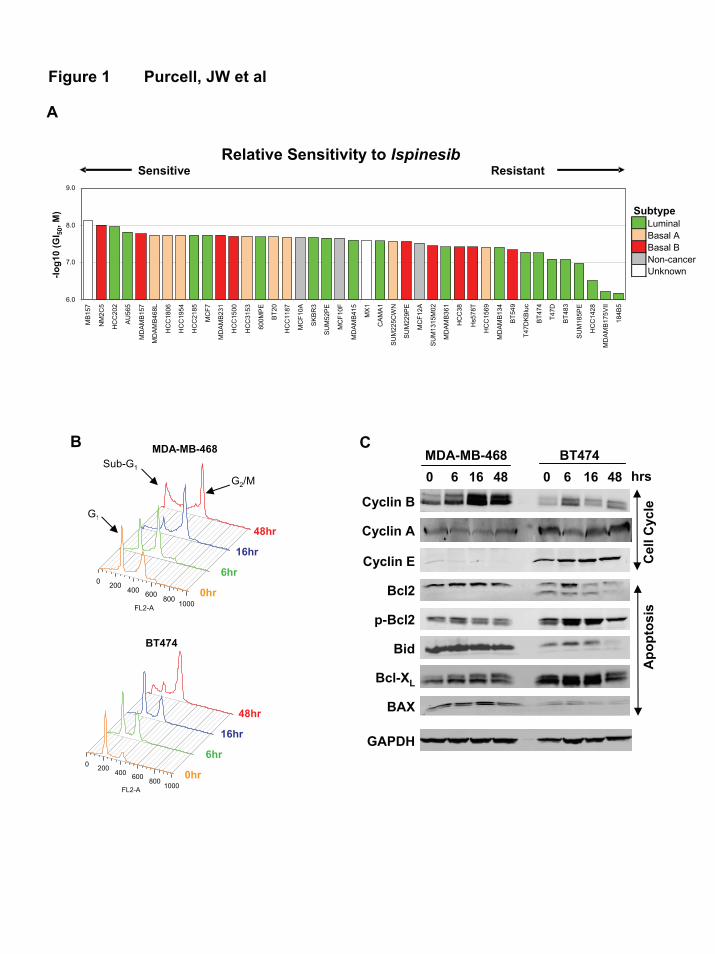
Figure 1 Purcell, JW et al
A
B
G1
BT474
MDA-MB-468
0 200400 600
8001000FL2-A
0 200 400 600 8001000FL2-A
48hr
16hr
6hr
0hr
Sub-G1
G2/M
48hr
16hr
6hr
0hr
0 6 16 48 0 6 16 48MDA-MB-468 BT474
hrs
Apo
ptos
isC
ell C
ycle
C
Cyclin A
Cyclin E
Cyclin B
GAPDH
BAX
Bid
p-Bcl2
Bcl2
Bcl-XL
Relative Sensitivity to IspinesibSensitive Resistant
6.0
7.0
8.0
9.0
MB1
57
NM
2C5
HC
C20
2
AU
565
MD
AMB1
57
MD
AMB4
68L
HC
C18
06
HC
C19
54
HC
C21
85
MC
F7
MD
AMB2
31
HC
C15
00
HC
C31
53
600M
PE
BT20
HC
C11
87
MC
F10A
SK
BR
3
SU
M52
PE
MC
F10F
MD
AMB4
15
MX
1
CAM
A1
SU
M22
5CW
N
SU
M22
9PE
MC
F12A
SU
M13
15M
02
MD
AMB3
61
HC
C38
Hs5
78T
HC
C15
69
MD
AMB1
34
BT54
9
T47D
KBl
uc
BT47
4
T47D
BT48
3
SU
M18
5PE
HC
C14
28
MD
AMB1
75VI
I
184B
5
-log1
0 (G
I 50, M
) SubtypeLuminalBasal ABasal BNon-cancerUnknown
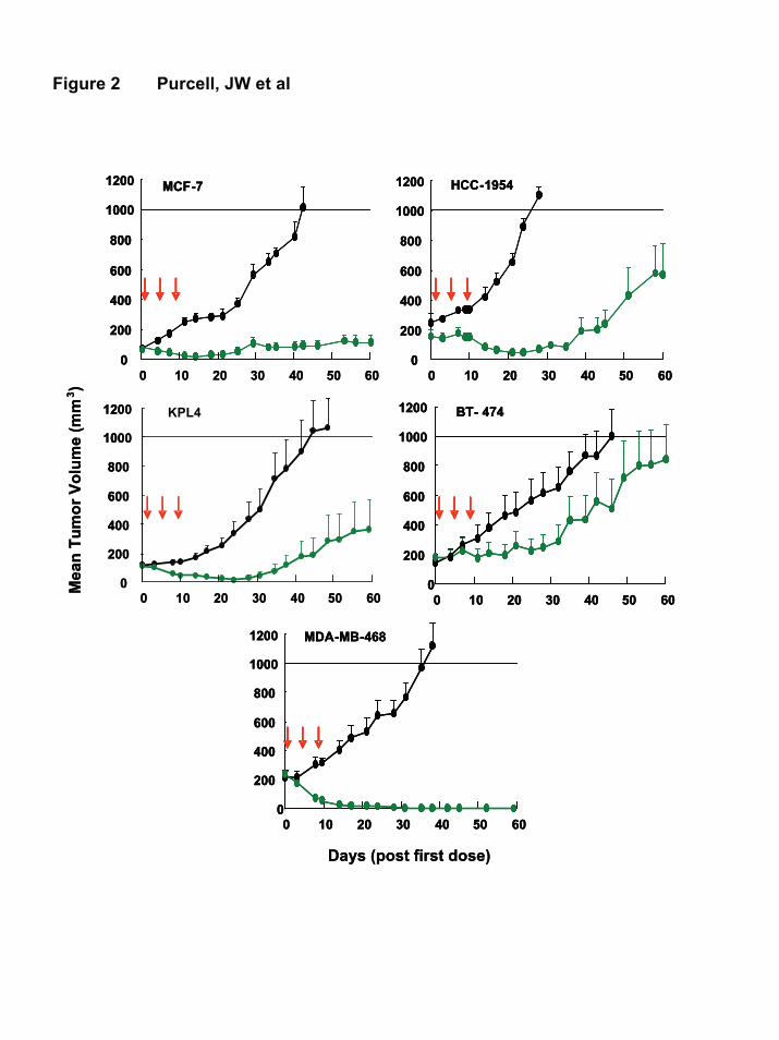
Figure 2 Purcell, JW et al
0
200
400
600
800
1000
1200
0 10 20 30 40 50 60
KPL4
Mea
n Tu
mor
Vol
ume
(mm
3 )
Days (post first dose)
0
200
400
600
800
1000
1200
0 10 20 30 40 50 60
BT- 474
0
200
400
600
800
1000
1200
0 10 20 30 40 50 60
HCC-1954
0
200
400
600
800
1000
1200
0 10 20 30 40 50 60
MCF-7
0
200
400
600
800
1000
1200
0 10 20 30 40 50 60
KPL4
0
200
400
600
800
1000
1200
0 10 20 30 40 50 60
KPL4
Mea
n Tu
mor
Vol
ume
(mm
3 )
Days (post first dose)
0
200
400
600
800
1000
1200
0 10 20 30 40 50 60
BT- 474
0
200
400
600
800
1000
1200
0 10 20 30 40 50 60
BT- 474
0
200
400
600
800
1000
1200
0 10 20 30 40 50 60
MDA-MB-468
0
200
400
600
800
1000
1200
0 10 20 30 40 50 60
MDA-MB-468
0
200
400
600
800
1000
1200
0 10 20 30 40 50 60
MDA-MB-468
0
200
400
600
800
1000
1200
0 10 20 30 40 50 60
HCC-1954
0
200
400
600
800
1000
1200
0 10 20 30 40 50 60
HCC-1954
0
200
400
600
800
1000
1200
0 10 20 30 40 50 60
MCF-7
0
200
400
600
800
1000
1200
0 10 20 30 40 50 60
MCF-7
Figure 3 Purcell, JW et al
Vehicle
Ispinesib 7.5mg/kg ip q4dx3
Ixabepilone 5mg/kg iv q4dx3
Paclitaxel 30mg/kg iv q4dx3
Ixabepilone 3mg/kg iv q4dx3
Ispinesib 5mg/kg ip q4dx3
Paclitaxel 15mg/kg iv q4dx3
Vehicle
Ispinesib 7.5mg/kg ip q4dx3
Ixabepilone 5mg/kg iv q4dx3
Paclitaxel 30mg/kg iv q4dx3
Ixabepilone 3mg/kg iv q4dx3
Ispinesib 5mg/kg ip q4dx3
Paclitaxel 15mg/kg iv q4dx3
Vehicle
Ispinesib 7.5mg/kg ip q4dx3
Ixabepilone 5mg/kg iv q4dx3
Paclitaxel 30mg/kg iv q4dx3
Ixabepilone 3mg/kg iv q4dx3
Ispinesib 5mg/kg ip q4dx3
Paclitaxel 15mg/kg iv q4dx310
100
1000
0 10 20 30 40 50 60
Mea
n Tu
mor
Vol
ume
(mm
3 )
Days (post first dose)
10
100
1000
0 10 20 30 40 50 60
Mea
n Tu
mor
Vol
ume
(mm
3 )
Days (post first dose)
10
100
1000
0 10 20 30 40 50 60
Mea
n Tu
mor
Vol
ume
(mm
3 )
Days (post first dose)
A
pH3 DNA
MDA-MB-468
0hr Untreated Ispinesib 24hr Ispinesib 48hr
BT474
B
C
1
2
3
4
untreated 6 24 48 72Time (hrs post treatment)
Abu
ndan
ce o
f PH
3-po
sitiv
e ce
lls re
lativ
e to
unt
erat
ed.
BT474MDA-MB-468
MDA-MB-468
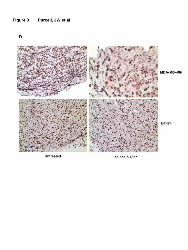
Figure 3 Purcell, JW et al
D
MDA-MB-468
BT474
Untreated Ispinesib 48hr
0
200
400
600
800
1000
1200
0 10 20 30 40 50 60
Days (post first dose)
0
200
400
600
800
1000
1200
0 10 20 30 40 50 60
Days (post first dose)
0
200
400
600
800
1000
1200
0 10 20 30 40 50 60
Days (post first dose)
0
200
400
600
800
1000
1200
0 10 20 30 40 50 60
Days (post first dose)
B
C D
0
200
400
600
800
1000
1200
0 10 20 30 40 50 60
Mea
n Tu
mor
Vol
ume
(mm
3 )
Days (post first dose)
Vehicle
Ispinesib 6mg/kgDoxorubicin 2.5mg/kg
Ispinesib 6mg/kg + Doxorubicin 2.5mg/kg
0
200
400
600
800
1000
1200
0 10 20 30 40 50 60
Mea
n Tu
mor
Vol
ume
(mm
3 )
Days (post first dose)
0
200
400
600
800
1000
1200
0 10 20 30 40 50 60
Mea
n Tu
mor
Vol
ume
(mm
3 )
Days (post first dose)
Vehicle
Ispinesib 6mg/kgDoxorubicin 2.5mg/kg
Ispinesib 6mg/kg + Doxorubicin 2.5mg/kg
Figure 4 Purcell, JW et al
KPL4
MCF7 KPL-4
Vehicle
Ispinesib 5mg/kg ip q4dx3Capecitabine 450mg/kg po qdx14
Ispinesib 5mg/kg + Capecitabine 450mg/kg
Vehicle
Ispinesib 5mg/kg ip q4dx3Capecitabine 450mg/kg po qdx14
Ispinesib 5mg/kg + Capecitabine 450mg/kg
A
Vehicle
Ispinesib 8mg/kgHerceptin 10mg/kg
Ispinesib 8mg/kg + Herceptin 10mg/kg
Vehicle
Ispinesib 8mg/kgHerceptin 10mg/kg
Ispinesib 8mg/kg + Herceptin 10mg/kg
0
200
400
600
800
1000
1200
0 10 20 30 40 50 60
Days (post first dose)
0
200
400
600
800
1000
1200
0 10 20 30 40 50 60
Days (post first dose)
BT474
Mea
n Tu
mor
Vol
ume
(mm
3 )M
ean
Tum
or V
olum
e (m
m3 )
Mea
n Tu
mor
Vol
ume
(mm
3 )
Trastuzumab 10mg/kg
0
200
400
600
800
1000
1200
0 10 20 30 40 50 60
Days (post first dose)
Vehicle
Ispinesib 10mg/kgHerceptin 10mg/kg
Ispinesib 10mg/kg + Herceptin 10mg/kg
0
200
400
600
800
1000
1200
0 10 20 30 40 50 60
Days (post first dose)
Vehicle
Ispinesib 10mg/kgHerceptin 10mg/kg
Ispinesib 10mg/kg + Herceptin 10mg/kg
0
200
400
600
800
1000
1200
0 10 20 30 40 50 60
Days (post first dose)
Vehicle
Ispinesib 10mg/kgHerceptin 10mg/kg
Ispinesib 10mg/kg + Herceptin 10mg/kg
Trastuzumab 10mg/kg
BT474 MDA-MB-468
0hr
6hr
16hr
48hr
G2/M 63.7 38.7
sub-G1 3.8 7.6
G1 65.3 35
S 9.8 12.3
G2/M 21.1 45.1
sub-G1 4.9 5.5
G1 43.3 27.4
S 6.8 11.4
G2/M 45 55.7
sub-G1 5.2 10.2
G1 45.8 15.2
S 9.6 11.2
G2/M 39.4 63.4
sub-G1 13.6 35.9
G1 14.5 7.68
S 8.2 17.7
Supplemental 1 Purcell, JW et al
Supplemental 2 Purcell, JW et al
N PR d60
CR d60
TGId60%
Weight Loss%
Tumor Volume Day 60
11 17
47
59
232
13
19
5
8
7
6
4
p-value (relative to
control)
Ispinesib 7.5mg/kg 8 3 5 98 <0.001
Ispinesib 5mg/kg 8 5 3 97 <0.001
Ixabepilone 5mg/kg 8 6 2 97 0.005
Ixabepilone 3mg/kg 9 6 2 78 <0.001
Paclitaxel 30mg/kg 7 5 2 99 <0.001
Paclitaxel 15mg/kg 8 2 6 99 0.001
Supplemental 3 Purcell, JW et al
0
200
400
600
800
1000
1200
0 10 20 30 40 50 60
Mea
n Tu
mor
Vol
ume
(mm
3 )
Days (post first dose)
Vehicle
Ispinesib 3mg/kgLapatinib 40mg/kg
Ispinesib 3mg/kg + Lapatinib 40mg/kg
0
200
400
600
800
1000
1200
0 10 20 30 40 50 60
Mea
n Tu
mor
Vol
ume
(mm
3 )
Days (post first dose)
0
200
400
600
800
1000
1200
0 10 20 30 40 50 60
Mea
n Tu
mor
Vol
ume
(mm
3 )
Days (post first dose)
Vehicle
Ispinesib 3mg/kgLapatinib 40mg/kg
Ispinesib 3mg/kg + Lapatinib 40mg/kg
Related Documents