ARTICLE 1 NATURE COMMUNICATIONS | 2:576 | DOI: 10.1038/ncomms1585 | www.nature.com/naturecommunications © 2011 Macmillan Publishers Limited. All rights reserved. Received 6 Jun 2011 | Accepted 3 Nov 2011 | Published 6 Dec 2011 DOI: 10.1038/ncomms1585 Activin is an important orchestrator of wound repair, but its potential role in skin carcinogenesis has not been addressed. Here we show using different types of genetically modified mice that enhanced levels of activin in the skin promote skin tumour formation and their malignant progression through induction of a pro-tumourigenic microenvironment. This includes accumulation of tumour-promoting Langerhans cells and regulatory T cells in the epidermis. Furthermore, activin inhibits proliferation of tumour-suppressive epidermal γδ T cells, resulting in their progressive loss during tumour promotion. An increase in activin expression was also found in human cutaneous basal and squamous cell carcinomas when compared with control tissue. These findings highlight the parallels between wound healing and cancer, and suggest inhibition of activin action as a promising strategy for the treatment of cancers overexpressing this factor. 1 Department of Biology, Institute of Cell Biology, ETH Zurich, 8093 Zurich, Switzerland. 2 Department of Dermatology, University of Lausanne, 1011 Lausanne, Switzerland. 3 Department of Immunology and Microbial Science, The Scripps Research Institute, La Jolla, California 92037, USA. 4 Department of Dermatology, University Hospital, 8091 Zurich, Switzerland. *These authors contributed equally to this work. Correspondence and requests for materials should be addressed to S.W. (email: [email protected]). Activin enhances skin tumourigenesis and malignant progression by inducing a pro-tumourigenic immune cell response Maria Antsiferova 1 , Marcel Huber 2, *, Michael Meyer 1, *, Aleksandra Piwko-Czuchra 1, *, Tamara Ramadan 1 , Amanda S. MacLeod 3 , Wendy L. Havran 3 , Reinhard Dummer 4 , Daniel Hohl 2 & Sabine Werner 1

Welcome message from author
This document is posted to help you gain knowledge. Please leave a comment to let me know what you think about it! Share it to your friends and learn new things together.
Transcript
ARTICLE
1NATURE COMMUNICATIONS | 2:576 | DOI: 10.1038/ncomms1585 | www.nature.com/naturecommunications
© 2011 Macmillan Publishers Limited. All rights reserved.
Received 6 Jun 2011 | Accepted 3 Nov 2011 | Published 6 Dec 2011 DOI: 10.1038/ncomms1585
Activin is an important orchestrator of wound repair, but its potential role in skin carcinogenesis
has not been addressed. Here we show using different types of genetically modifi ed mice that
enhanced levels of activin in the skin promote skin tumour formation and their malignant
progression through induction of a pro-tumourigenic microenvironment. This includes
accumulation of tumour-promoting Langerhans cells and regulatory T cells in the epidermis.
Furthermore, activin inhibits proliferation of tumour-suppressive epidermal γ δ T cells, resulting
in their progressive loss during tumour promotion. An increase in activin expression was also
found in human cutaneous basal and squamous cell carcinomas when compared with control
tissue. These fi ndings highlight the parallels between wound healing and cancer, and suggest
inhibition of activin action as a promising strategy for the treatment of cancers overexpressing
this factor.
1 Department of Biology, Institute of Cell Biology , ETH Zurich , 8093 Zurich, Switzerland . 2 Department of Dermatology, University of Lausanne , 1011
Lausanne , Switzerland . 3 Department of Immunology and Microbial Science, The Scripps Research Institute , La Jolla , California 92037, USA . 4 Department
of Dermatology, University Hospital , 8091 Zurich , Switzerland . * These authors contributed equally to this work. Correspondence and requests for materials
should be addressed to S.W. (email: [email protected] ) .
Activin enhances skin tumourigenesis and malignant progression by inducing a pro-tumourigenic immune cell response Maria Antsiferova 1 , Marcel Huber 2 , * , Michael Meyer 1 , * , Aleksandra Piwko-Czuchra 1 , * , Tamara Ramadan 1 ,
Amanda S. MacLeod 3 , Wendy L. Havran 3 , Reinhard Dummer 4 , Daniel Hohl 2 & Sabine Werner 1
ARTICLE
2
NATURE COMMUNICATIONS | DOI: 10.1038/ncomms1585
NATURE COMMUNICATIONS | 2:576 | DOI: 10.1038/ncomms1585 | www.nature.com/naturecommunications
© 2011 Macmillan Publishers Limited. All rights reserved.
There are remarkable similarities between wound healing and cancer 1 , and the presence of a ‘ wound healing gene expres-sion signature ’ is a hallmark of highly malignant carcinomas 2 .
Th erefore, it is of particular interest to identify wound-regulated genes and to characterize their functions in cancer development and progression. Major orchestrators of wound repair are activins, which belong to the transforming growth factor (TGF)- β superfamily of growth and diff erentiation factors. Th e most abundant activin variant is activin A, the homodimer formed by two β A subunits. Activins exert their biological eff ects through activation of transmembrane serine / threonine kinase receptors. Binding to a type II activin recep-tor (ActRII or ActRIIB) leads to the phosphorylation and activation of a type I activin receptor (ActRIB, also known as Alk4, ActRIA = Alk2 or ActRIC = Alk7). Th is activates the canonical signalling pathway via Smad proteins, or, alternatively, mitogen-activated kinase pathways 3 .
Activins were initially described as reproductive hormones, but they also have important functions in development, tissue homoe-ostasis and repair 4 . Furthermore, abnormalities in activin receptor expression and / or signalling are associated with various human diseases, including cancer 5 .
We previously showed a strong increase in activin β A expression in healing skin wounds 6 . Th is is functionally relevant, as transgenic mice overexpressing the activin antagonist follistatin in keratino-cytes showed delayed wound repair 7 , whereas overexpression of activin β A in keratinocytes strongly accelerated the healing proc-ess. However, healed wounds of activin-overexpressing mice had a hyperproliferative neo-epidermis (ref. 8 , and M.A. and S.W. unpub-lished results). Th erefore, we speculated about a role of activin in the pathogenesis of skin cancer.
Few functional studies have addressed the role of activin in malignancy, and pro- or anti-tumourigenic eff ects of activin have been reported, depending on the tissue and the type of cancer 9 – 11 . Most of these data were obtained with xenograft models in immuno-compromised hosts, whereas a potential role of the immunomodu-latory actions of activin 12 in the pathogenesis of cancer has not been addressed. In this study, we identifi ed a novel and unexpected func-tion of activin in the pathogenesis of non-melanoma skin cancer that is mediated through its eff ect on diff erent epidermal immune cells.
Results Activin promotes skin tumourigenesis in mice . To determine a potential role of activin in skin carcinogenesis, we used transgenic mice expressing activin β A in keratinocytes under control of the keratin 14 (K14) promoter (Act mice) 8 . Th ey secrete high levels of activin A, which can act on keratinocytes directly but also on stromal cells due to the high diff usibility of activin A 13 . Th is is refl ected by the 30-fold increase in activin A levels in skin lysates and the 200-fold increase in the serum compared with wild-type (wt)
animals ( Supplementary Fig. S1 ). Expression of high levels of activin A by keratinocytes did not interfere with the growth inhibitory eff ect of TGF- β on keratinocytes ( Supplementary Fig. S1 ). Most importantly, the back skin of these mice did not reveal any histological abnormalities, and keratinocyte proliferation in normal back skin epidermis was not aff ected by the enhanced levels of activin 8 .
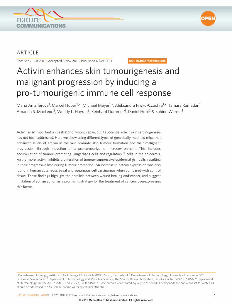
Act mice as well as sex- and age-matched wt littermates were subjected to a two-stage chemical skin carcinogenesis protocol, where tumours are induced by topical treatment of the back skin with 7,12-dimethylbenz[a]anthracene (DMBA), followed by weekly treatment with 12-O-tetradecanoylphorbol 13-acetate (TPA) 14,15 . A remarkable pro-tumourigenic eff ect of activin was observed in three independent experiments with mice of diff erent genetic background (experiments I – III, Table 1 ) as refl ected by the earlier appearance of papillomas and the strong increase in the percentage of mice with tumours (tumour incidence) and in the number of tumours per mouse (tumour multiplicity) ( Fig. 1a,b; Supplementary Fig. S2 ).
Table 1 | Summary of carcinogenesis experiments.
Experiment DMBA (1 × ) ( � g) TPA (20 × ) ( � g) Genotypes Genetic background N
I 25 5 Wt CD1 31 Act 29
II 25 7.5 Wt / wt CD1xC57BL / 6 17 Act / wt 17
III 25 7.5 Wt / wt CD1xB6D2F1 18 Act / wt 18 Wt / dnActRIB 18 Act / dnActRIB 18
Control I 25 — Act CD1 5 Wt 5
Control II — 5 Act CD1 6 Wt 6
0 2 4 6 8 10 12 14 16 18 200
20
40
60
80
100
0 2 4 6 8 10 12 14 16 18 200
20
40
60
80
100
0 2 4 6 8 10 12 14 16 18 20 2205
101520253035
##
0 2 4 6 8 10 12 14 16 18 20 220
5
10
15
20
**
% M
ice
with
tum
ours
wt
Act
Weeks after DMBA
Tum
ours
per
mou
seT
umou
rs p
er m
ouse
wt
Act
Weeks after DMBA
Weeks after DMBAWeeks after DMBA
% M
ice
with
tum
ours
wt/wtwt/dnActRIBAct/wtAct/dnActRIB
wt/wtwt/dnActRIBAct/wtAct/dnActRIB
###***
###***
###***
******
******
******
Figure 1 | Activin promotes chemically induced skin tumourigenesis in mice via action on stromal cells. ( a , b ) Kinetics of tumour incidence
( a ) or multiplicity ( b ) observed in experiment I ( Table 1 ). N = 31 wild-
type (wt) mice; N = 29 activin-transgenic (Act) mice. ( c , d ) Kinetics of
tumour incidence ( c ) and multiplicity ( d ) in experiment III. N = 18 mice per
genotype. Kaplan – Meier survival curves are shown in ( a ) and ( c ); mean ± s.
e.m. are shown in ( b ) and ( d ); * * P < 0.01, * * * P < 0.001 for the comparison
between wt / wt and Act / wt mice; # # P < 0.01, # # # P < 0.001 for the
comparison between Act / wt and Act / dnActRIB mice (two-way repeated
measures ANOVA and Bonferroni post-test).
ARTICLE
3
NATURE COMMUNICATIONS | DOI: 10.1038/ncomms1585
NATURE COMMUNICATIONS | 2:576 | DOI: 10.1038/ncomms1585 | www.nature.com/naturecommunications
© 2011 Macmillan Publishers Limited. All rights reserved.
Mice treated only with DMBA or TPA did not develop tumours, demonstrating that both a mutagenic insult and a proliferative stim-ulus are required. Th is is consistent with the fi nding that wounding alone did not induce tumourigenesis in control or Act mice (M.A. and S.W., unpublished results).
Activin induces skin carcinogenesis via the stroma . To determine if activin stimulates skin tumourigenesis through autocrine activa-tion of keratinocytes and / or through paracrine / endocrine activa-tion of stromal (mesenchymal or immune) cells, we used transgenic mice expressing a dominant-negative mutant of ActRIB (Alk4) in keratinocytes under control of the K14 promoter (dnActRIB mice) and mated them with Act mice. dnActRIB lacks the complete intra-cellular kinase domain, and through formation of non-functional heterodimers with type II activin receptors it effi ciently blocks activin but not TGF- β signalling 16 . Th is approach was chosen, as keratinocytes express multiple activin receptors 6 , which are all inhibited by dnActRIB. Th e dnActRIB mice have no obvious phe-notypic abnormalities under non-challenged conditions 16 , and the dnActRIB transgene neither aff ected expression of the activin β A transgene nor the bioavailability of the recombinant activin A (data not shown and Supplementary Fig. S1 ). Th erefore, in Act / dnActRIB mice, the highly diff usible, recombinant activin can activate all cells in the skin, but its eff ect on keratinocytes is antagonized by dnAc-tRIB expressed by these cells. Th e functionality of dnActRIB in the double-transgenic mice had previously been demonstrated 16 and was further verifi ed by analysis of S100A8 and S100A9 expression. Th us, we had previously shown a strongly increased expression of
these cytokines in the hyperthickened tail epidermis of Act mice 17 . We verifi ed this increase with new mice (250-fold) and found that it was almost completely suppressed ( > 90 % ) in Act / dnActRIB mice (data not shown).
We performed a two-stage chemical skin carcinogenesis study with the F1 progeny of dnActRIB and Act mice (experiment III, Table 1 ). Th e latency period and tumour incidence of the activin-overexpressing mice were not aff ected by dnActRIB ( Fig. 1c ), but the tumour multiplicity was signifi cantly increased in the double-transgenic mice as compared with Act single-transgenic animals ( Fig. 1d ). Th is fi nding points to the existence of a tumour-inhibitory eff ect of activin A, which is mediated via keratinocytes and there-fore aff ected by dnActRIB and which depends on the presence of high levels of activin. However, the potent pro-tumourigenic eff ect seems to be predominantly mediated via the stroma.
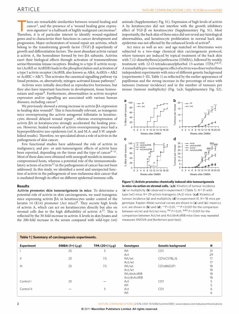
Activin promotes tumour malignancy . Th e vast majority of the tumours collected 20 weeks aft er initiation were papillomas ( Fig. 2a ). Th ere was no diff erence in tumour cell proliferation and apoptosis between genotypes ( Supplementary Fig. S2 and data not shown), but several tumours in activin-overexpressing mice already contained sites of high-grade dysplasia and foci of microinvasion ( Fig. 2a ; arrow). Twelve weeks aft er termination of TPA treatment, the percentage of papillomas that progressed to malignancy was slightly increased in activin-overexpressing compared with wt and dnActRIB mice. Most importantly, the percentage of mice with malignant tumours was signifi cantly higher in the groups of animals expressing the activin transgene ( Fig. 2b ).
K14/VimentinH/E
Actwt
H/EH/E K14
Genotype Percentage of papillomas thatprogress to malignancy a
Percentage of mice withmalignant tumours b
wt/wt 2.8 (1/36) 9.1 (1/11)
wt/dnActRIB 3.6 (2/55) 9.1(1/11)
Act/wt 7.1 (11/155) 63.6 (7/11)*
Act/dnActRIB 7.6 (19/251) 70.0 (7/10)**
Figure 2 | Activin increases malignancy and metastatic spread of skin tumours. ( a ) Hematoxylin / eosin (H / E)-stained sections from papillomas 20
weeks after initiation. Local micro-invasive foci (arrow) were often observed in papillomas of Act mice (lower panel). Bars: 1,000 μ m (top), 100 μ m
(bottom). Areas indicated by the box on the top pictures are shown at higher magnifi cation on the bottom pictures. ( b ) Malignant conversion rate of
tumours 32 weeks after initiation (experiment III). a () = number of carcinomas / total number of tumours. b () = number of mice with carcinomas / total
number of mice. N = 10 Act / dnActRIB mice, N = 11 wt / wt, Act / wt, wt / dnActRIB mice; * P = 0.0237 for Act / wt versus wt / wt, * * P = 0.0075 for Act /
dnActRIB versus wt / wt (Fisher ’ s exact test). ( c ) H / E staining (left) and double immunofl uorescence for keratin (K)14 and vimentin (right) of a spindle cell
variant of squamous cell carcinoma (SCC) in an Act mouse. Left bar: 1,000 μ m; right bar: 200 μ m. The area indicated by a box on the left picture is shown
at higher magnifi cation on the right picture. ( d ) Lung (top panel) and lymph node (bottom panel) metastases in carcinoma-bearing Act mice. Left and
middle: H / E staining; right: K14 immunohistochemistry. Left bar: 1,000 μ m; middle and right bars: 200 μ m.
ARTICLE
4
NATURE COMMUNICATIONS | DOI: 10.1038/ncomms1585
NATURE COMMUNICATIONS | 2:576 | DOI: 10.1038/ncomms1585 | www.nature.com/naturecommunications
© 2011 Macmillan Publishers Limited. All rights reserved.
Histopathological analysis and immunostaining for keratins 14, 6, 10 and 13 revealed that most of the malignant tumours were squa-mous cell carcinomas (SCCs) . Five out of 11 malignant tumours in Act / wt mice and 1 out of 19 in Act / dnActRIB mice had spindle-shaped tumour cells, showed vimentin immunoreactivity ( Fig. 2c ), and are likely to be a spindle cell variant of SCCs 18 .
Four out of 9 activin-overexpressing mice with malignant tumours developed lung metastases ( Fig. 2d ), whereas this was not observed in wt mice. In experiment II, we collected inguinal and axillary lymph nodes 32 weeks aft er DMBA initiation. K14-positive metastatic nod-ules were observed in 3 out of 3 carcinoma-bearing Act-transgenic mice ( Fig. 2d ) and in 1 out of 2 wt mice with carcinomas.
Th ese results suggest that activin A not only accelerates tumouri-genesis but also malignant progression of papillomas.
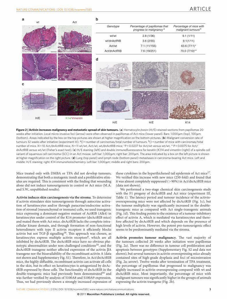
Activin does not aff ect DMBA or TPA responsiveness . To gain insight into the mechanisms underlying the enhanced tumourigen-esis in Act mice, we analysed the skin before and following short-term treatment with DMBA and TPA. Consistent with the lack of obvious histological abnormalities in the back skin 8 , proliferation of keratinocytes and epidermal thickness were not altered in untreated Act mice ( Fig. 3a,b ). Several pro-infl ammatory cytokines and chem-okines were normally expressed ( Supplementary Fig. S2 ), and there was no infl ammatory infi ltrate in the skin (data not shown).
We also could not detect alterations in the expression of several DMBA metabolizing or detoxifying enzymes, or in the proliferation rate of keratinocytes 24 h aft er DMBA treatment in Act versus wt mice ( Supplementary Fig. S2 ).
One day aft er the fi rst TPA treatment, we found a minor increase in keratinocyte proliferation and a slightly thicker epidermis in Act
and Act / dnActRIB mice compared with control mice ( Fig. 3a,b; Supplementary Fig. S2 ). Th e diff erence in keratinocyte proliferation remained non-signifi cant at later stages of TPA treatment ( Supple-mentary Fig. S2 ), but the diff erence in epidermal thickness increased continuously ( Fig. 3c ).
Th e TPA-induced infl ammatory response was not obviously altered by activin overexpression as determined by fl ow cytom-etry analysis of dermal single-cell suspensions (CD45 + leucocytes, Gr1 + CD11b + and Gr1 − CD11b + myeloid cells, CD3 + T cells and F4 / 80 + macrophages) ( Supplementary Fig. S3 ). Furthermore, sev-eral pro-infl ammatory cytokines and chemokines were expressed at similar levels in TPA-treated skin of wt and Act mice ( Fig. 3d ). Th e only diff erence was the increased expression of S100A8 and A9 in Act mice, which most likely refl ects an altered activation / diff erentiation state of the keratinocytes 17 . Several growth factors known to be induced by TPA and to contribute to TPA-induced tumour promotion 19 – 21 were expressed at similar levels in control and Act mice ( Fig. 3d ). In particular, expression of TGF- β 1, β 2 and β 3 was not altered, suggesting that the observed phenotype is not due to diff erences in TGF- β expression ( Fig. 3d ).
Finally, the number of blood vessels and the area of dermis cov-ered by blood vessels 1d aft er the fi rst TPA application as well as in papillomas collected aft er 20 TPA applications were similar in wt and Act mice ( Supplementary Fig. S3 ). Consistently, the expression levels of various angiogenesis-regulating factors were not altered by activin overexpression ( Supplementary Fig. S2 and S3 ).
Loss of epidermal γ δ T cells in TPA-treated Act mice . Diff er-ent subsets of T cells are either tumour-promotive or tumour-suppressive 22,23 . Dendritic epidermal T cells (DETCs) represent a
wt Act wt Act0
20
40
60****
0
5
10
15
20
25 ***
12345
406080
100
0
120
Untreated DMBA+1xTPA+1d
0.1
1
10
100
1,000
10,000 wt
****
Brd
U+ce
lls p
er m
m o
f BM
Epi
derm
al th
ickn
ess
(μm
)
DMBA+1xTPA+1dUntreated
DMBA+3xTPA+7d DMBA+8xTPA+3d
wt/wt
wt/dnA
ctRIB
Act/wt
Act/dn
ActRIB
wt/wt
wt/dnA
ctRIB
Act/wt
Act/dn
ActRIB
wt/wt
wt/dnA
ctRIB
Act/wt
Act/dn
ActRIB
wt/wt
wt/dnA
ctRIB
Act/wt
Act/dn
ActRIB E
pide
rmal
thic
knes
s (μ
m)
Rel
ativ
e to
Rps
29
Act
DMBA+1xTPA+1d
Il1b
Tnfa Il6
Tgfa
Csf2Tgf
b1
Tgfb2
Tgfb3
S100a
8
S100a
9Cxc
l1Ccl2 Ccl3 Ccl4
Figure 3 | Activin overexpression does not affect DMBA or TPA responsiveness. ( a ) Epidermal BrdU + cells per mm of basement membrane (BM) in
untreated back skin or 1 day after the fi rst TPA treatment. Scatter plot and mean values are shown; N = 5 – 8 mice per group; n = 6 – 23 microscopic fi elds
of skin sections per mouse. ( b ) Epidermal thickness in untreated mice or 1 day after the fi rst TPA treatment. N = 3 – 4; n = 6 – 13; * P ≤ 0.05, * * P ≤ 0.01 (one-
way ANOVA with Bonferroni post-test). ( c ) Epidermal thickness 7 days after the third TPA treatment or 3 days after the eighth TPA treatment. N = 6 – 7;
n = 3 – 30; * * P = 0.0047 for 3 × TPA, * * P = 0.0012 for 8 × TPA (Mann – Whitney test). ( d ) RNA samples from back skin 1 day after the fi rst TPA treatment
were analysed for expression of pro-infl ammatory cytokines and growth factors by quantitative reverse transcription PCR. N = 5 – 10 mice per genotype.
Rps29 was used as reference. Box-and-whisker plot is depicted, with boxes showing median and 25th and 75th percentile, whiskers showing minimal and
maximal values. * * P = 0.0047 for S100a8 , * * P = 0.0027 for S100a9 (Mann – Whitney test).
ARTICLE
5
NATURE COMMUNICATIONS | DOI: 10.1038/ncomms1585
NATURE COMMUNICATIONS | 2:576 | DOI: 10.1038/ncomms1585 | www.nature.com/naturecommunications
© 2011 Macmillan Publishers Limited. All rights reserved.
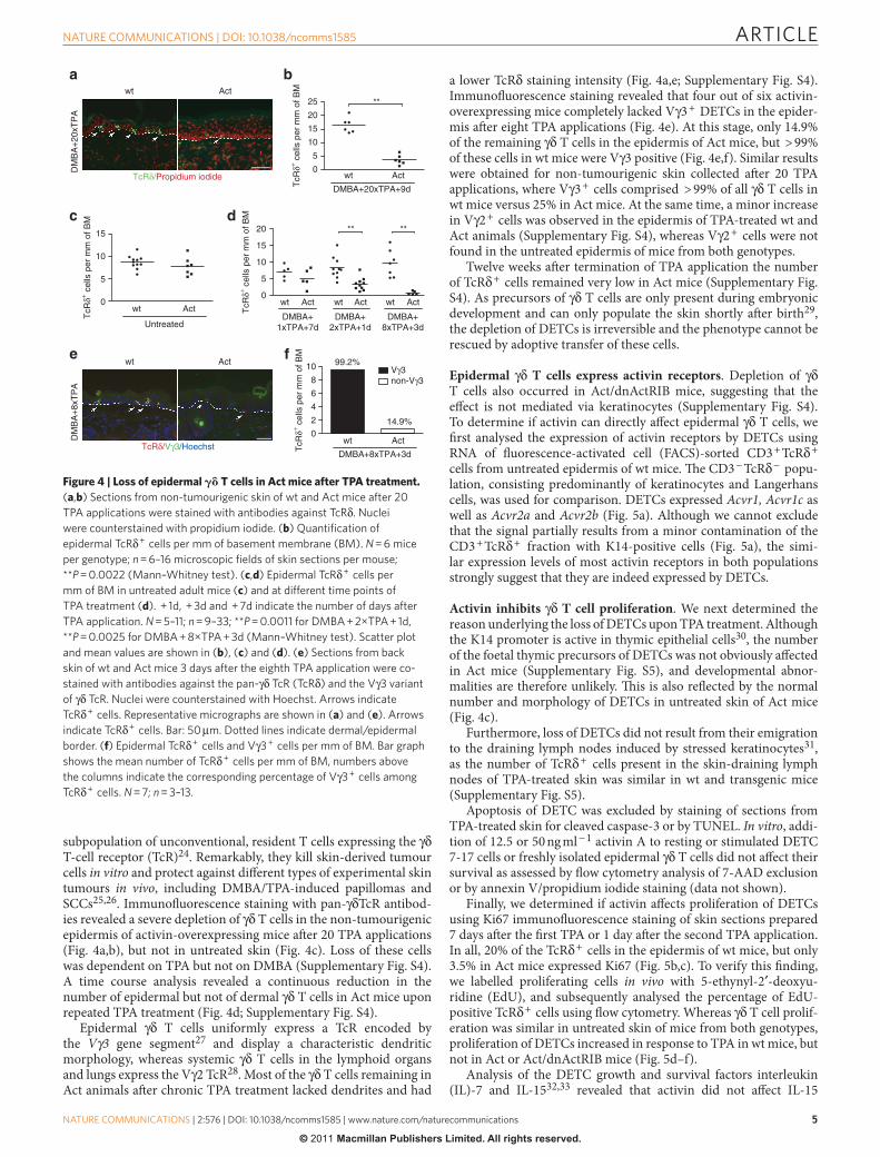
subpopulation of unconventional, resident T cells expressing the γ δ T-cell receptor (TcR) 24 . Remarkably, they kill skin-derived tumour cells in vitro and protect against diff erent types of experimental skin tumours in vivo , including DMBA / TPA-induced papillomas and SCCs 25,26 . Immunofl uorescence staining with pan- γ δ TcR antibod-ies revealed a severe depletion of γ δ T cells in the non-tumourigenic epidermis of activin-overexpressing mice aft er 20 TPA applications ( Fig. 4a,b ), but not in untreated skin ( Fig. 4c ). Loss of these cells was dependent on TPA but not on DMBA ( Supplementary Fig. S4 ). A time course analysis revealed a continuous reduction in the number of epidermal but not of dermal γ δ T cells in Act mice upon repeated TPA treatment ( Fig. 4d ; Supplementary Fig. S4 ).
Epidermal γ δ T cells uniformly express a TcR encoded by the V γ 3 gene segment 27 and display a characteristic dendritic morphology, whereas systemic γ δ T cells in the lymphoid organs and lungs express the V γ 2 TcR 28 . Most of the γ δ T cells remaining in Act animals aft er chronic TPA treatment lacked dendrites and had
a lower TcR δ staining intensity ( Fig. 4a,e; Supplementary Fig. S4 ). Immunofl uorescence staining revealed that four out of six activin-overexpressing mice completely lacked V γ 3 + DETCs in the epider-mis aft er eight TPA applications ( Fig. 4e ). At this stage, only 14.9 % of the remaining γ δ T cells in the epidermis of Act mice, but > 99 % of these cells in wt mice were V γ 3 positive ( Fig. 4e,f ). Similar results were obtained for non-tumourigenic skin collected aft er 20 TPA applications, where V γ 3 + cells comprised > 99 % of all γ δ T cells in wt mice versus 25 % in Act mice. At the same time, a minor increase in V γ 2 + cells was observed in the epidermis of TPA-treated wt and Act animals ( Supplementary Fig. S4 ), whereas V γ 2 + cells were not found in the untreated epidermis of mice from both genotypes.
Twelve weeks aft er termination of TPA application the number of TcR δ + cells remained very low in Act mice ( Supplementary Fig. S4 ). As precursors of γ δ T cells are only present during embryonic development and can only populate the skin shortly aft er birth 29 , the depletion of DETCs is irreversible and the phenotype cannot be rescued by adoptive transfer of these cells.
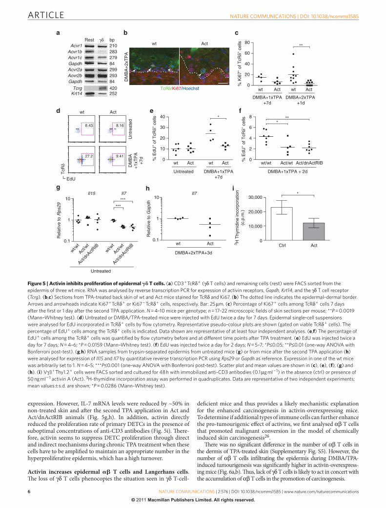
Epidermal γ δ T cells express activin receptors . Depletion of γ δ T cells also occurred in Act / dnActRIB mice, suggesting that the eff ect is not mediated via keratinocytes ( Supplementary Fig. S4 ). To determine if activin can directly aff ect epidermal γ δ T cells, we fi rst analysed the expression of activin receptors by DETCs using RNA of fl uorescence-activated cell (FACS)-sorted CD3 + TcR δ + cells from untreated epidermis of wt mice. Th e CD3 − TcR δ − popu-lation, consisting predominantly of keratinocytes and Langerhans cells, was used for comparison. DETCs expressed Acvr1, Acvr1c as well as Acvr2a and Acvr2b ( Fig. 5a ). Although we cannot exclude that the signal partially results from a minor contamination of the CD3 + TcR δ + fraction with K14-positive cells ( Fig. 5a ), the simi-lar expression levels of most activin receptors in both populations strongly suggest that they are indeed expressed by DETCs.
Activin inhibits γ δ T cell proliferation . We next determined the reason underlying the loss of DETCs upon TPA treatment. Although the K14 promoter is active in thymic epithelial cells 30 , the number of the foetal thymic precursors of DETCs was not obviously aff ected in Act mice ( Supplementary Fig. S5 ), and developmental abnor-malities are therefore unlikely. Th is is also refl ected by the normal number and morphology of DETCs in untreated skin of Act mice ( Fig. 4c ).
Furthermore, loss of DETCs did not result from their emigration to the draining lymph nodes induced by stressed keratinocytes 31 , as the number of TcR δ + cells present in the skin-draining lymph nodes of TPA-treated skin was similar in wt and transgenic mice ( Supplementary Fig. S5 ).
Apoptosis of DETC was excluded by staining of sections from TPA-treated skin for cleaved caspase-3 or by TUNEL. In vitro , addi-tion of 12.5 or 50 ng ml − 1 activin A to resting or stimulated DETC 7-17 cells or freshly isolated epidermal γ δ T cells did not aff ect their survival as assessed by fl ow cytometry analysis of 7-AAD exclusion or by annexin V / propidium iodide staining (data not shown).
Finally, we determined if activin aff ects proliferation of DETCs using Ki67 immunofl uorescence staining of skin sections prepared 7 days aft er the fi rst TPA or 1 day aft er the second TPA application. In all, 20 % of the TcR δ + cells in the epidermis of wt mice, but only 3.5 % in Act mice expressed Ki67 ( Fig. 5b,c ). To verify this fi nding, we labelled proliferating cells in vivo with 5-ethynyl-2 ′ -deoxyu-ridine (EdU), and subsequently analysed the percentage of EdU-positive TcR δ + cells using fl ow cytometry. Whereas γ δ T cell prolif-eration was similar in untreated skin of mice from both genotypes, proliferation of DETCs increased in response to TPA in wt mice, but not in Act or Act / dnActRIB mice ( Fig. 5d – f ).
Analysis of the DETC growth and survival factors interleukin (IL)-7 and IL-15 32,33 revealed that activin did not aff ect IL-15
wt Act
TcRδ/Propidium iodide
wt Act
DM
BA
+8x
TP
A
TcRδ/Vγ3/Hoechst
wt Act0
5
10
15
20
25 **
wt Act0
5
10
15
wt Act0
2
4
6
8
10 Vγ3non-Vγ3
wt Act wt Act wt Act0
5
10
15
20 ** **
TcR
δ+ cel
ls p
er m
m o
f BM
TcR
δ+ cel
ls p
er m
m o
f BM
TcR
δ+ cel
ls p
er m
m o
f BM
TcR
δ+ cel
ls p
er m
m o
f BM
99.2%
DM
BA
+20
xTP
A
14.9%
DMBA+8xTPA+3d
UntreatedDMBA+
8xTPA+3dDMBA+
2xTPA+1dDMBA+
1xTPA+7d
DMBA+20xTPA+9d
Figure 4 | Loss of epidermal � � T cells in Act mice after TPA treatment. ( a , b ) Sections from non-tumourigenic skin of wt and Act mice after 20
TPA applications were stained with antibodies against TcR δ . Nuclei
were counterstained with propidium iodide. ( b ) Quantifi cation of
epidermal TcR δ + cells per mm of basement membrane (BM). N = 6 mice
per genotype; n = 6 – 16 microscopic fi elds of skin sections per mouse;
* * P = 0.0022 (Mann – Whitney test). ( c , d ) Epidermal TcR δ + cells per
mm of BM in untreated adult mice ( c ) and at different time points of
TPA treatment ( d ). + 1d, + 3d and + 7d indicate the number of days after
TPA application. N = 5 – 11; n = 9 – 33; * * P = 0.0011 for DMBA + 2 × TPA + 1d,
* * P = 0.0025 for DMBA + 8 × TPA + 3d (Mann – Whitney test). Scatter plot
and mean values are shown in ( b ), ( c ) and ( d ). ( e ) Sections from back
skin of wt and Act mice 3 days after the eighth TPA application were co-
stained with antibodies against the pan- γ δ TcR (TcR δ ) and the V γ 3 variant
of γ δ TcR. Nuclei were counterstained with Hoechst. Arrows indicate
TcR δ + cells. Representative micrographs are shown in ( a ) and ( e ). Arrows
indicate TcR δ + cells. Bar: 50 μ m. Dotted lines indicate dermal / epidermal
border. ( f ) Epidermal TcR δ + cells and V γ 3 + cells per mm of BM. Bar graph
shows the mean number of TcR δ + cells per mm of BM, numbers above
the columns indicate the corresponding percentage of V γ 3 + cells among
TcR δ + cells. N = 7; n = 3 – 13.
ARTICLE
6
NATURE COMMUNICATIONS | DOI: 10.1038/ncomms1585
NATURE COMMUNICATIONS | 2:576 | DOI: 10.1038/ncomms1585 | www.nature.com/naturecommunications
© 2011 Macmillan Publishers Limited. All rights reserved.
expression. However, IL-7 mRNA levels were reduced by ~ 50 % in non-treated skin and aft er the second TPA application in Act and Act / dnActRIB animals ( Fig. 5g,h ). In addition, activin directly reduced the proliferation rate of primary DETCs in the presence of suboptimal concentrations of anti-CD3 antibodies ( Fig. 5i ). Th ere-fore, activin seems to suppress DETC proliferation through direct and indirect mechanisms during chronic TPA treatment when these cells have to be amplifi ed to maintain an appropriate number in the hyperproliferative epidermis, which has a high turnover.
Activin increases epidermal � � T cells and Langerhans cells . Th e loss of γ δ T cells phenocopies the situation seen in γ δ T-cell-
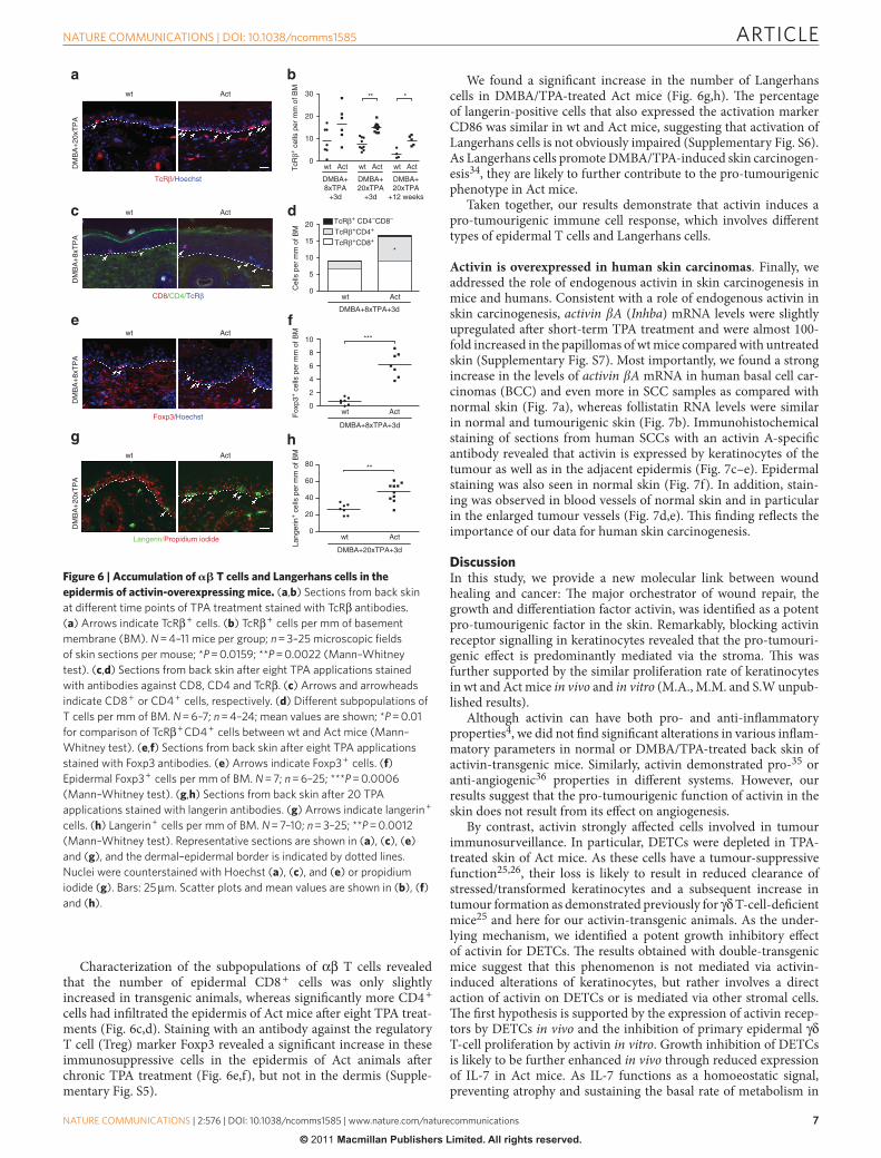
defi cient mice and thus provides a likely mechanistic explanation for the enhanced carcinogenesis in activin-overexpressing mice. To determine if additional types of immune cells can further enhance the pro-tumourigenic eff ect of activins, we fi rst analysed α β T cells that promoted malignant conversion in the model of chemically induced skin carcinogenesis 26 .
Th ere was no signifi cant diff erence in the number of α β T cells in the dermis of TPA-treated skin ( Supplementary Fig. S5 ). However, the number of α β T cells infi ltrating the epidermis during DMBA / TPA-induced tumourigenesis was signifi cantly higher in activin-overexpress-ing mice ( Fig. 6a,b ). Th us, lack of γ δ T cells is likely to act in concert with the accumulation of α β T cells in the promotion of carcinogenesis.
wt Act
TcRδ/Ki67/Hoechst
wt Act
DM
BA
+1x
TP
A+
7d
Unt
reat
ed
EdU
TcR
δ
9.4127.2
8.43 8.16
wt Act wt Act0
20
40
60
80**
wt Act wt Act0
10
20
30
40 *
wt/wt Act/wt Act/dnActRIB0
2
4
6
8*
**
0.1
1
10
wt Act0.1
1
10
Ctrl Act0
10,000
20,000
30,000*
Acvr1Acvr1b
Acvr2bAcvr2a
Gapdh
γδRest
Acvr1c
TcrgKrt14
Gapdh279
299293
252420
84
84
283210bp
% E
dU+ o
f TcR
δ+ cel
ls
% E
dU+ o
f TcR
δ+ cel
ls
Untreated DMBA+1xTPA+7d
DMBA+1xTPA + 2d
% K
i67+ o
f TcR
δ+ cel
ls
DMBA+2xTPA +1d
DMBA+1xTPA+7d
DM
BA
+2x
TP
A
Act/dn
ActRIB
Act/wt
wt/wt
Act/dn
ActRIB
Act/wt
wt/wt
Untreated
DMBA+2xTPA+3d
3 H T
hym
idin
e in
corp
orat
ion
(c.p
.m.)
******
Il15 Il7 Il7
Rel
ativ
e to
Rps
29
Rel
ativ
e to
Gap
dh
Figure 5 | Activin inhibits proliferation of epidermal � � T cells. ( a ) CD3 + TcR δ + ( γ δ T cells) and remaining cells (rest) were FACS sorted from the
epidermis of three wt mice. RNA was analysed by reverse transcription PCR for expression of activin receptors, Gapdh, Krt14 , and the γ δ T cell receptor
( Tcrg ). ( b , c ) Sections from TPA-treated back skin of wt and Act mice stained for TcR δ and Ki67. ( b ) The dotted line indicates the epidermal – dermal border.
Arrows and arrowheads indicate Ki67 + TcR δ + or Ki67 − TcR δ + cells, respectively. Bar: 25 μ m. ( c ) Percentage of Ki67 + cells among TcR δ + cells 7 days
after the fi rst or 1 day after the second TPA application. N = 4 – 10 mice per genotype; n = 17 – 22 microscopic fi elds of skin sections per mouse; * * P = 0.0019
(Mann – Whitney test). ( d ) Untreated or DMBA / TPA-treated mice were injected with EdU twice a day for 7 days. Epidermal single-cell suspensions
were analysed for EdU incorporated in TcR δ + cells by fl ow cytometry. Representative pseudo-colour plots are shown (gated on viable TcR δ + cells). The
percentage of EdU + cells among the TcR δ + cells is indicated. Data shown are representative of at least four independent analyses. ( e , f ) The percentage of
EdU + cells among the TcR δ + cells was quantifi ed by fl ow cytometry before and at different time points after TPA treatment. ( e ) EdU was injected twice a
day for 7 days; N = 4 – 6; * P = 0.0159 (Mann – Whitney test). ( f ) EdU was injected twice a day for 2 days; N = 5 – 7; * P ≤ 0.05; * * P ≤ 0.01 (one-way ANOVA with
Bonferroni post-test). ( g , h ) RNA samples from trypsin-separated epidermis from untreated mice ( g ) or from mice after the second TPA application ( h )
were analysed for expression of Il15 and Il7 by quantitative reverse transcription PCR using Rps29 or Gapdh as reference. Expression in one of the wt mice
was arbitrarily set to 1. N = 4 – 5; * * * P ≤ 0.001 (one-way ANOVA with Bonferroni post-test). Scatter plot and mean values are shown in ( c ), ( e ), ( f ), ( g ) and
( h ). ( i ) V γ 3 + Thy1.2 + cells were FACS sorted and cultured for 48 h with immobilized anti-CD3 antibodies (0.1 μ g ml − 1 ) in the absence (ctrl) or presence of
50 ng ml − 1 activin A (Act). 3 H-thymidine incorporation assay was performed in quadruplicates. Data are representative of two independent experiments;
mean values ± s.d. are shown; * P = 0.0286 (Mann – Whitney test).
ARTICLE
7
NATURE COMMUNICATIONS | DOI: 10.1038/ncomms1585
NATURE COMMUNICATIONS | 2:576 | DOI: 10.1038/ncomms1585 | www.nature.com/naturecommunications
© 2011 Macmillan Publishers Limited. All rights reserved.
Characterization of the subpopulations of α β T cells revealed that the number of epidermal CD8 + cells was only slightly increased in transgenic animals, whereas signifi cantly more CD4 + cells had infi ltrated the epidermis of Act mice aft er eight TPA treat-ments ( Fig. 6c,d ). Staining with an antibody against the regulatory T cell (Treg) marker Foxp3 revealed a signifi cant increase in these immunosuppressive cells in the epidermis of Act animals aft er chronic TPA treatment ( Fig. 6e,f ), but not in the dermis ( Supple-mentary Fig. S5 ).
We found a signifi cant increase in the number of Langerhans cells in DMBA / TPA-treated Act mice ( Fig. 6g,h ). Th e percentage of langerin-positive cells that also expressed the activation marker CD86 was similar in wt and Act mice, suggesting that activation of Langerhans cells is not obviously impaired ( Supplementary Fig. S6 ). As Langerhans cells promote DMBA / TPA-induced skin carcinogen-esis 34 , they are likely to further contribute to the pro-tumourigenic phenotype in Act mice.
Taken together, our results demonstrate that activin induces a pro-tumourigenic immune cell response, which involves diff erent types of epidermal T cells and Langerhans cells.
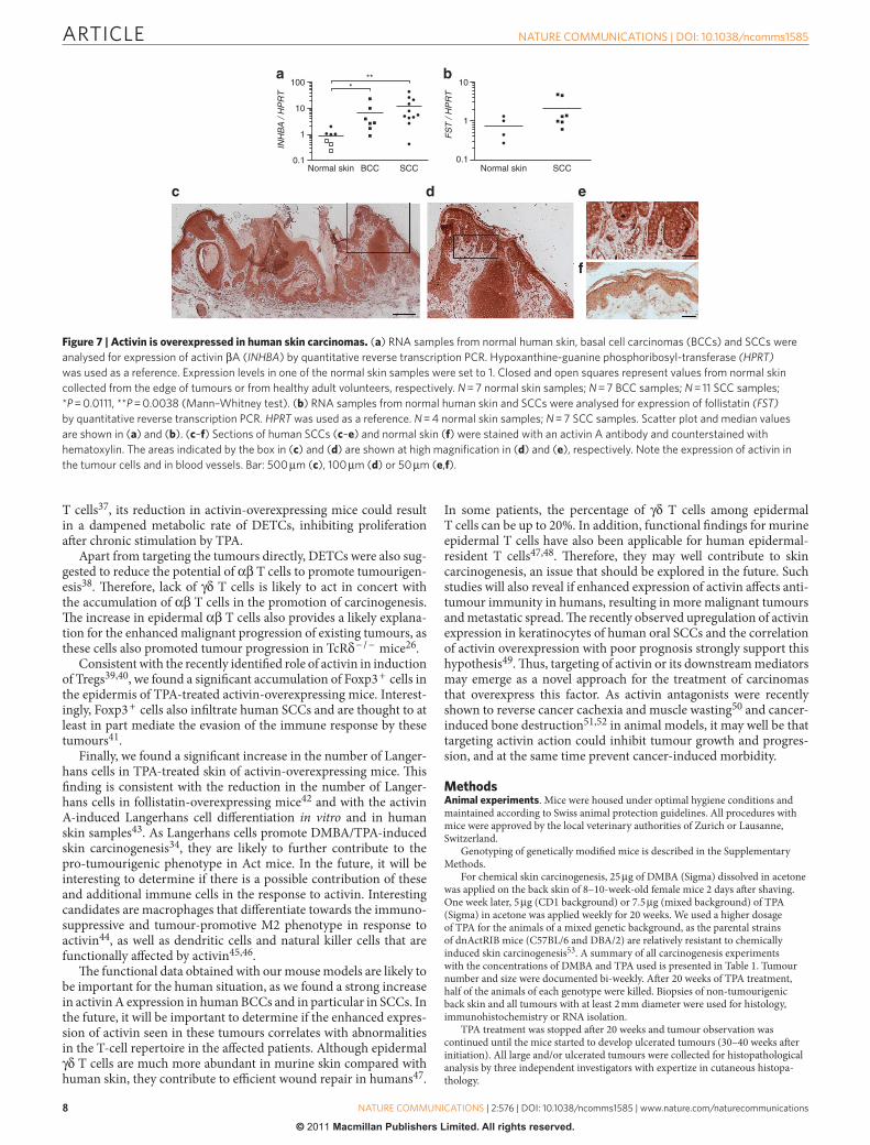
Activin is overexpressed in human skin carcinomas . Finally, we addressed the role of endogenous activin in skin carcinogenesis in mice and humans. Consistent with a role of endogenous activin in skin carcinogenesis, activin � A ( Inhba ) mRNA levels were slightly upregulated aft er short-term TPA treatment and were almost 100-fold increased in the papillomas of wt mice compared with untreated skin ( Supplementary Fig. S7 ). Most importantly, we found a strong increase in the levels of activin � A mRNA in human basal cell car-cinomas (BCC) and even more in SCC samples as compared with normal skin ( Fig. 7a ), whereas follistatin RNA levels were similar in normal and tumourigenic skin ( Fig. 7b ). Immunohistochemical staining of sections from human SCCs with an activin A-specifi c antibody revealed that activin is expressed by keratinocytes of the tumour as well as in the adjacent epidermis ( Fig. 7c – e ). Epidermal staining was also seen in normal skin ( Fig. 7f ). In addition, stain-ing was observed in blood vessels of normal skin and in particular in the enlarged tumour vessels ( Fig. 7d,e ). Th is fi nding refl ects the importance of our data for human skin carcinogenesis.
Discussion In this study, we provide a new molecular link between wound healing and cancer: Th e major orchestrator of wound repair, the growth and diff erentiation factor activin, was identifi ed as a potent pro-tumourigenic factor in the skin. Remarkably, blocking activin receptor signalling in keratinocytes revealed that the pro-tumouri-genic eff ect is predominantly mediated via the stroma. Th is was further supported by the similar proliferation rate of keratinocytes in wt and Act mice in vivo and in vitro (M.A., M.M. and S.W unpub-lished results).
Although activin can have both pro- and anti-infl ammatory properties 4 , we did not fi nd signifi cant alterations in various infl am-matory parameters in normal or DMBA / TPA-treated back skin of activin-transgenic mice. Similarly, activin demonstrated pro- 35 or anti-angiogenic 36 properties in diff erent systems. However, our results suggest that the pro-tumourigenic function of activin in the skin does not result from its eff ect on angiogenesis.
By contrast, activin strongly aff ected cells involved in tumour immunosurveillance. In particular, DETCs were depleted in TPA-treated skin of Act mice. As these cells have a tumour-suppressive function 25,26 , their loss is likely to result in reduced clearance of stressed / transformed keratinocytes and a subsequent increase in tumour formation as demonstrated previously for γ δ T-cell-defi cient mice 25 and here for our activin-transgenic animals. As the under-lying mechanism, we identifi ed a potent growth inhibitory eff ect of activin for DETCs. Th e results obtained with double-transgenic mice suggest that this phenomenon is not mediated via activin-induced alterations of keratinocytes, but rather involves a direct action of activin on DETCs or is mediated via other stromal cells. Th e fi rst hypothesis is supported by the expression of activin recep-tors by DETCs in vivo and the inhibition of primary epidermal γ δ T-cell proliferation by activin in vitro . Growth inhibition of DETCs is likely to be further enhanced in vivo through reduced expression of IL-7 in Act mice. As IL-7 functions as a homoeostatic signal, preventing atrophy and sustaining the basal rate of metabolism in
wt Act
TcRβ/Hoechst
wt Act
DM
BA
+20
xTP
A
Langerin/Propidium iodide
wt Act
DM
BA
+8x
TP
A
Foxp3/Hoechst
wt Act
DM
BA
+8x
TP
A
CD8/CD4/TcRβ
wt Act wt Act wt Act0
10
20
30 ** *
wt Act0
5
10
15
20
*
wt Act0
2
4
6
8
10
wt Act0
20
40
60
80 **
DM
BA
+20
xTP
A
TcR
β+ c
ells
per
mm
of B
M
DMBA+20xTPA
+12 weeks
DMBA+20xTPA
+3d
DMBA+8xTPA
+3d
TcRβ+ CD4–CD8–
TcRβ+CD4+
TcRβ+CD8+
DMBA+8xTPA+3d
***
Fox
p3+ c
ells
per
mm
of B
M
DMBA+8xTPA+3d
Cel
ls p
er m
m o
f BM
Lang
erin
+ c
ells
per
mm
of B
M
DMBA+20xTPA+3d
Figure 6 | Accumulation of � � T cells and Langerhans cells in the epidermis of activin-overexpressing mice. ( a , b ) Sections from back skin
at different time points of TPA treatment stained with TcR β antibodies.
( a ) Arrows indicate TcR β + cells. ( b ) TcR β + cells per mm of basement
membrane (BM). N = 4 – 11 mice per group; n = 3 – 25 microscopic fi elds
of skin sections per mouse; * P = 0.0159; * * P = 0.0022 (Mann – Whitney
test). ( c , d ) Sections from back skin after eight TPA applications stained
with antibodies against CD8, CD4 and TcR β . ( c ) Arrows and arrowheads
indicate CD8 + or CD4 + cells, respectively. ( d ) Different subpopulations of
T cells per mm of BM. N = 6 – 7; n = 4 – 24; mean values are shown; * P = 0.01
for comparison of TcR β + CD4 + cells between wt and Act mice (Mann –
Whitney test). ( e , f ) Sections from back skin after eight TPA applications
stained with Foxp3 antibodies. ( e ) Arrows indicate Foxp3 + cells. ( f )
Epidermal Foxp3 + cells per mm of BM. N = 7; n = 6 – 25; * * * P = 0.0006
(Mann – Whitney test). ( g , h ) Sections from back skin after 20 TPA
applications stained with langerin antibodies. ( g ) Arrows indicate langerin +
cells. ( h ) Langerin + cells per mm of BM. N = 7 – 10; n = 3 – 25; * * P = 0.0012
(Mann – Whitney test). Representative sections are shown in ( a ), ( c ), ( e )
and ( g ), and the dermal – epidermal border is indicated by dotted lines.
Nuclei were counterstained with Hoechst ( a ), ( c ), and ( e ) or propidium
iodide ( g ). Bars: 25 μ m. Scatter plots and mean values are shown in ( b ), ( f )
and ( h ).
ARTICLE
8
NATURE COMMUNICATIONS | DOI: 10.1038/ncomms1585
NATURE COMMUNICATIONS | 2:576 | DOI: 10.1038/ncomms1585 | www.nature.com/naturecommunications
© 2011 Macmillan Publishers Limited. All rights reserved.
T cells 37 , its reduction in activin-overexpressing mice could result in a dampened metabolic rate of DETCs, inhibiting proliferation aft er chronic stimulation by TPA.
Apart from targeting the tumours directly, DETCs were also sug-gested to reduce the potential of α β T cells to promote tumourigen-esis 38 . Th erefore, lack of γ δ T cells is likely to act in concert with the accumulation of α β T cells in the promotion of carcinogenesis. Th e increase in epidermal α β T cells also provides a likely explana-tion for the enhanced malignant progression of existing tumours, as these cells also promoted tumour progression in TcR δ − / − mice 26 .
Consistent with the recently identifi ed role of activin in induction of Tregs 39,40 , we found a signifi cant accumulation of Foxp3 + cells in the epidermis of TPA-treated activin-overexpressing mice. Interest-ingly, Foxp3 + cells also infi ltrate human SCCs and are thought to at least in part mediate the evasion of the immune response by these tumours 41 .
Finally, we found a signifi cant increase in the number of Langer-hans cells in TPA-treated skin of activin-overexpressing mice. Th is fi nding is consistent with the reduction in the number of Langer-hans cells in follistatin-overexpressing mice 42 and with the activin A-induced Langerhans cell diff erentiation in vitro and in human skin samples 43 . As Langerhans cells promote DMBA / TPA-induced skin carcinogenesis 34 , they are likely to further contribute to the pro-tumourigenic phenotype in Act mice. In the future, it will be interesting to determine if there is a possible contribution of these and additional immune cells in the response to activin. Interesting candidates are macrophages that diff erentiate towards the immuno-suppressive and tumour-promotive M2 phenotype in response to activin 44 , as well as dendritic cells and natural killer cells that are functionally aff ected by activin 45,46 .
Th e functional data obtained with our mouse models are likely to be important for the human situation, as we found a strong increase in activin A expression in human BCCs and in particular in SCCs. In the future, it will be important to determine if the enhanced expres-sion of activin seen in these tumours correlates with abnormalities in the T-cell repertoire in the aff ected patients. Although epidermal γ δ T cells are much more abundant in murine skin compared with human skin, they contribute to effi cient wound repair in humans 47 .
In some patients, the percentage of γ δ T cells among epidermal T cells can be up to 20 % . In addition, functional fi ndings for murine epidermal T cells have also been applicable for human epidermal-resident T cells 47,48 . Th erefore, they may well contribute to skin carcinogenesis, an issue that should be explored in the future. Such studies will also reveal if enhanced expression of activin aff ects anti-tumour immunity in humans, resulting in more malignant tumours and metastatic spread. Th e recently observed upregulation of activin expression in keratinocytes of human oral SCCs and the correlation of activin overexpression with poor prognosis strongly support this hypothesis 49 . Th us, targeting of activin or its downstream mediators may emerge as a novel approach for the treatment of carcinomas that overexpress this factor. As activin antagonists were recently shown to reverse cancer cachexia and muscle wasting 50 and cancer-induced bone destruction 51,52 in animal models, it may well be that targeting activin action could inhibit tumour growth and progres-sion, and at the same time prevent cancer-induced morbidity.
Methods Animal experiments . Mice were housed under optimal hygiene conditions and maintained according to Swiss animal protection guidelines. All procedures with mice were approved by the local veterinary authorities of Zurich or Lausanne, Switzerland.
Genotyping of genetically modifi ed mice is described in the Supplementary Methods .
For chemical skin carcinogenesis, 25 μ g of DMBA ( Sigma ) dissolved in acetone was applied on the back skin of 8 – 10-week-old female mice 2 days aft er shaving. One week later, 5 μ g (CD1 background) or 7.5 μ g (mixed background) of TPA ( Sigma ) in acetone was applied weekly for 20 weeks. We used a higher dosage of TPA for the animals of a mixed genetic background, as the parental strains of dnActRIB mice (C57BL / 6 and DBA / 2) are relatively resistant to chemically induced skin carcinogenesis 53 . A summary of all carcinogenesis experiments with the concentrations of DMBA and TPA used is presented in Table 1 . Tumour number and size were documented bi-weekly. Aft er 20 weeks of TPA treatment, half of the animals of each genotype were killed. Biopsies of non-tumourigenic back skin and all tumours with at least 2 mm diameter were used for histology, immunohistochemistry or RNA isolation.
TPA treatment was stopped aft er 20 weeks and tumour observation was continued until the mice started to develop ulcerated tumours (30 – 40 weeks aft er initiation). All large and / or ulcerated tumours were collected for histopathological analysis by three independent investigators with expertize in cutaneous histopa-thology.
Normal skin BCC SCC0.1
1
10
100 ***
Normal skin SCC0.1
1
10
FS
T /
HP
RT
INH
BA
/ H
PR
T
Figure 7 | Activin is overexpressed in human skin carcinomas. ( a ) RNA samples from normal human skin, basal cell carcinomas (BCCs) and SCCs were
analysed for expression of activin β A ( INHBA ) by quantitative reverse transcription PCR. Hypoxanthine-guanine phosphoribosyl-transferase (HPRT)
was used as a reference. Expression levels in one of the normal skin samples were set to 1. Closed and open squares represent values from normal skin
collected from the edge of tumours or from healthy adult volunteers, respectively. N = 7 normal skin samples; N = 7 BCC samples; N = 11 SCC samples;
* P = 0.0111, * * P = 0.0038 (Mann – Whitney test). ( b ) RNA samples from normal human skin and SCCs were analysed for expression of follistatin (FST)
by quantitative reverse transcription PCR. HPRT was used as a reference. N = 4 normal skin samples; N = 7 SCC samples. Scatter plot and median values
are shown in ( a ) and ( b ). ( c – f ) Sections of human SCCs ( c – e ) and normal skin ( f ) were stained with an activin A antibody and counterstained with
hematoxylin. The areas indicated by the box in ( c ) and ( d ) are shown at high magnifi cation in ( d ) and ( e ), respectively. Note the expression of activin in
the tumour cells and in blood vessels. Bar: 500 μ m ( c ), 100 μ m ( d ) or 50 μ m ( e , f ).
ARTICLE
9
NATURE COMMUNICATIONS | DOI: 10.1038/ncomms1585
NATURE COMMUNICATIONS | 2:576 | DOI: 10.1038/ncomms1585 | www.nature.com/naturecommunications
© 2011 Macmillan Publishers Limited. All rights reserved.
Lungs and lymph nodes were embedded in paraffi n, and every 7 – 10th section was stained with hematoxylin / eosin (H / E) and examined for the presence of metastatic nodules.
Immunofl uorescence and immunohistochemical analysis . For preparation of frozen sections, tissue samples were immediately frozen in tissue freezing medium ( Leica Microsystems ). For preparation of paraffi n sections, they were either fi xed in 95 % ethanol / 1 % acetic acid or in 4 % paraformaldehyde.
Cryosections (4 or 7 μ m) were fi xed with ice-cold methanol for 10 min. Paraffi n sections were deparaffi nized through incubation in xylene and subsequently in a graded series of ethanol. Sections were then blocked with 3 % bovine serum albu-min (BSA) and incubated with the primary antibody, followed by a fl uorochrome-labelled secondary antibody. For immunohistochemistry, sections were incubated with biotinylated secondary antibodies and developed using the Vectastain ABC kit and 3-amino-9-ethylcarbazole ( AEC ) or 3,3 ′ -diaminobenzidine ( DAB ) peroxidase substrate kits ( Vector Laboratories ). Th e antibodies used are listed in Supplemen-tary Table S1 .
For activin A immunohistochemistry, formalin-fi xed paraffi n sections were deparaffi nized and incubated for 45 min at 98 ° C in 0.5 M ethylenediaminetetraacetic acid pH 8 in a water bath for antigen retrieval. Endogenous peroxidase activity was blocked by incubation in 3 – 4 % H 2 O 2 , while unspecifi c binding sites were blocked by a 1-h incubation at room temperature in 12 % BSA, 0.1 % Tween 20, 0.1 % NP40 in phosphate-buff ered saline. Subsequently, the sections were incubated overnight at 4 ° C with the primary antibody, followed by a 1-h incubation at room temperature with a horse radish peroxidase (HRP)-conjugated secondary antibody. Th e anti-rabbit poly-HRP-IgG polymer from the Bond Polymer Refi ne Detec-tion System ( Leica Microsystems ) was used for enhancement of the signal. Th e substrate chromogen, DAB, was used for detection of the complex. Sections were counterstained with hematoxylin.
Stained sections were photographed using an Imager.A1 microscope equipped with an Axiocam Mrm camera and enhanced-contrast Plan-Neofl uor objectives (10 × / 0.3 NA, 20 × / 0.5 NA; all from Carl Zeiss, Inc .). Axiovision 4.6 soft ware ( Carl Zeiss, Inc ) was used for data acquisition.
BrdU incorporation assay . Mice were injected intraperitoneally with 250 mg kg − 1 BrdU 2 h before killing. Samples were fi xed in 95 % ethanol / 1 % acetic acid, and embedded in paraffi n. BrdU-positive cells were detected with a horseradish peroxi-dase-conjugated monoclonal antibody against BrdU ( Roche ), and visualized with 0.05 % DAB / 0.005 % H 2 O 2 . Counterstaining was performed with H / E.
TUNEL assay . Apoptosis on tissue sections was analysed using the In situ Cell Death Detection Kit ( Roche ) according to the manufacturer ’ s instructions.
RNA isolation and quantitative reverse transcription PCR . Total RNA was isolated using the RNeasy Fibrous Tissue Mini Kit or the RNeasy Midi Kit ( Qiagen ) according to the manufacturer ’ s instructions, including a DNase treatment step ( Promega ). cDNA was synthesized using the iScript kit ( Bio-Rad Laboratories ). Relative gene expression was determined using the Roche LightCycler 480 SYBR Green system ( Roche ). Th e gene-specifi c primers used are listed in Supplementary Table S2 .
Isolation of cells for fl ow cytometry . Single-cell suspensions of epidermal cells were prepared as described previously 54 . Disaggregated epidermal cells were fi ltered through a 70- μ m nylon cell strainer ( BD Pharmingen ), centrifuged at 1,200 rpm at 4 ° C for 10 min and resuspended in FACS buff er (phosphate-buff ered saline, 0.2 % BSA and 5 mM ethylenediaminetetraacetic acid).
Dermis was cut into small pieces, and incubated for 45 min at 37 ° C under continuous shaking in DMEM containing 2.5 mg ml − 1 collagenase II ( Worthing-ton ), 2.5 mg ml − 1 collagenase IV ( Invitrogen ), 0.5 mg ml − 1 DNase I and 1 mg ml − 1 hyaluronidase ( Worthington ). Th ymi were mechanically disaggregated and fi ltered through a 70- μ m nylon cell strainer. Lymph nodes were incubated for 30 min at 37 ° C under continuous shaking in DMEM containing 5 mg ml − 1 of collagenase IV ( Invitrogen ). Disaggregated dermal cells were treated as described for epidermal cells.
Flow cytometry . Cells were incubated for 10 min at 4 ° C with rat anti-mouse CD16 / CD32 monoclonal antibody ( clone 2.4G2 , BD Biosciences ) to prevent non-specifi c antibody binding, followed by a 30-min incubation at 4 ° C with directly labelled specifi c antibodies ( Supplementary Table S3 ). Aft er washing with FACS buff er, 7-AAD ( BD Biosciences ) was added to discriminate between viable and dead cells. Data acquisition was performed using a FACS Calibur equipped with CellQuestPro soft ware ( BD Biosciences ) and analysis was done using FlowJo soft ware ( Tree Star Inc. ).
FACS sorting and reverse transcription PCR . Epidermal cells were isolated, stained with antibodies as described above, and FACS sorted using a FACS Aria Cell Sorter ( BD Biosciences ). RNA was isolated from the sorted cells using the RNeasy Micro Kit ( Qiagen ), and cDNA was generated using the High-Capacity cDNA Reverse Transcription Kit ( Applied Biosystems ) according to the manufac-turers ’ instructions.
DETC culture . Th e 7 – 17 DETC line was grown in RPMI 1640 medium supple-mented with 10 % FCS, 25 mM HEPES, 100 U penicillin 100 μ g − 1 streptomycin, 2 mM glutamine, 100 μ M non-essential amino acids, 1 mM sodium pyruvate, 50 μ M 2-mercaptoethanol and 20 U ml − 1 of recombinant human IL-2. Cells were restimulated every 4 – 5 weeks overnight with 5 μ g ml − 1 of concanavalin A and were allowed to rest for 2 weeks before use in assays.
For isolation of primary DETCs, skin was incubated on 0.3 % trypsin for 2 – 2.5 h at 37 ° C, and the epidermis was separated from the dermis. Epidermis was incubated for 10 min in 0.3 % trypsin at 37 ° C. Th e epidermal preparation was enriched for live cells by centrifugation over lympholyte M, and cells recovered from the interface were cultured as described above, with the exception that the medium contained 10 U ml − 1 of IL-2. Survival and proliferation experiments were performed with recombinant activin A ( R & D Systems ).
Survival of DETCs was assessed by 7-AAD exclusion or annexin V / propidium iodide staining.
For proliferation analysis, V γ 3 + Th y1.2 + cells were FACS sorted and subse-quently cultured in the presence of immobilized anti-CD3 antibodies ( BD Bio-sciences , clone 500A2, 0.1 μ g ml − 1 ), with or without activin A. Th ey were pulsed with 0.5 μ Ci 3 H-thymidine, harvested 18 h later onto glass fi bre fi lters, and counts were detected in a scintillation counter.
EdU incorporation assay . EdU ( Invitrogen ) was injected intraperitoneally twice a day (100 μ g per mouse). Incorporated EdU was analysed in epidermal single-cell suspensions by fl ow cytometry using the Click-iT EdU Flow Cytometry Assay Kit ( Invitrogen ). Th e LIVE / DEAD Fixable Dead Cell Stain Kit ( Invitrogen ) was used to discriminate between live and dead cells.
Human skin biopsies . Normal human skin and skin cancer samples were obtained anonymously from the Departments of Dermatology, University Hospitals of Lausanne and Zurich (in the context of the biobank project), approved by the local and cantonal Research Ethics Committees. Normal skin was from healthy adult volunteers or from the edges of skin tumours. BCC or SCC was diagnosed by ex-perienced pathologists. Informed consent for research was obtained before routine diagnostic services. All samples included the dermis and the epidermis.
Statistical analysis . Statistical analysis was performed using the GraphPad Prism version 5.0 for Mac OS X ( GraphPad Soft ware ). For analysis of tumour incidence, comparison of the curves showing the mice with tumours was performed using log-rank χ 2 test. Tumour multiplicity was analysed using two-way repeated meas-ures analysis of variance (ANOVA). Malignant conversion rate and the percentage of mice with malignant tumours were analysed by Fisher ’ s exact test. Mann – Whitney test was used for comparing two groups of data; ANOVA test was used for comparing three or more groups. * P ≤ 0.05, * * P ≤ 0.01 and * * * P ≤ 0.001. Diff erences between groups that were not labelled with asterisks were non-signifi cant.
References 1 . Sch ä fer , M . & Werner , S . Cancer as an overhealing wound: an old hypothesis
revisited . Nat. Rev. Mol. Cell. Biol. 9 , 628 – 638 ( 2008 ). 2 . Chang , H . Y . et al. Gene expression signature of fi broblast serum response
predicts human cancer progression: similarities between tumors and wounds . PLoS Biol. 2 , E7 ( 2004 ).
3 . Chen , Y . G . et al. Activin signaling and its role in regulation of cell proliferation, apoptosis, and carcinogenesis . Exp. Biol. Med. (Maywood) 231 , 534 – 544 ( 2006 ).
4 . Werner , S . & Alzheimer , C . Roles of activin in tissue repair, fi brosis, and infl ammatory disease . Cytokine Growth Factor Rev. 17 , 157 – 171 ( 2006 ).
5 . Risbridger , G . P . , Schmitt , J . F . & Robertson , D . M . Activins and inhibins in endocrine and other tumors . Endocr. Rev. 22 , 836 – 858 ( 2001 ).
6 . Hubner , G . , Hu , Q . , Smola , H . & Werner , S . Strong induction of activin expression aft er injury suggests an important role of activin in wound repair . Dev. Biol. 173 , 490 – 498 ( 1996 ).
7 . Wankell , M . et al. Impaired wound healing in transgenic mice overexpressing the activin antagonist follistatin in the epidermis . EMBO J. 20 , 5361 – 5372 ( 2001 ).
8 . Munz , B . et al. Overexpression of activin A in the skin of transgenic mice reveals new activities of activin in epidermal morphogenesis, dermal fi brosis and wound repair . EMBO J. 18 , 5205 – 5215 ( 1999 ).
9 . Krneta , J . et al. Dissociation of angiogenesis and tumorigenesis in follistatin- and activin-expressing tumors . Cancer Res. 66 , 5686 – 5695 ( 2006 ).
10 . Panopoulou , E . et al. Activin A suppresses neuroblastoma xenograft tumor growth via antimitotic and antiangiogenic mechanisms . Cancer Res. 65 , 1877 – 1886 ( 2005 ).
11 . Yoshinaga , K . et al. Clinical signifi cance of the expression of activin A in esophageal carcinoma . Int. J. Oncol. 22 , 75 – 80 ( 2003 ).
12 . Hedger , M . P . , Winnall , W . R . , Phillips , D . J . & de Kretser , D . M . Th e regulation and functions of activin and follistatin in infl ammation and immunity . Vitam. Horm. 85 , 255 – 297 ( 2011 ).
13 . McDowell , N . , Zorn , A . M . , Crease , D . J . & Gurdon , J . B . Activin has direct long-range signalling activity and can form a concentration gradient by diff usion . Curr. Biol. 7 , 671 – 681 ( 1997 ).
ARTICLE
10
NATURE COMMUNICATIONS | DOI: 10.1038/ncomms1585
NATURE COMMUNICATIONS | 2:576 | DOI: 10.1038/ncomms1585 | www.nature.com/naturecommunications
© 2011 Macmillan Publishers Limited. All rights reserved.
14 . Yuspa , S . H . Th e pathogenesis of squamous cell cancer: lessons learned from studies of skin carcinogenesis – thirty-third G. H. A. Clowes Memorial Award Lecture . Cancer Res. 54 , 1178 – 1189 ( 1994 ).
15 . Abel , E . L . , Angel , J . M . , Kiguchi , K . & DiGiovanni , J . Multi-stage chemical carcinogenesis in mouse skin: fundamentals and applications . Nat. Protoc. 4 , 1350 – 1362 ( 2009 ).
16 . Bamberger , C . et al. Activin controls skin morphogenesis and wound repair predominantly via stromal cells and in a concentration-dependent manner via keratinocytes . Am. J. Pathol. 167 , 733 – 747 ( 2005 ).
17 . Th orey , I . S . et al. Th e Ca2+-binding proteins S100A8 and S100A9 are encoded by novel injury-regulated genes . J. Biol. Chem. 276 , 35818 – 35825 ( 2001 ).
18 . Klein-Szanto , A . J . , Larcher , F . , Bonfi l , R . D . & Conti , C . J . Multistage chemical carcinogenesis protocols produce spindle cell carcinomas of the mouse skin . Carcinogenesis 10 , 2169 – 2172 ( 1989 ).
19 . DiGiovanni , J . , Rho , O . , Xian , W . & Beltran , L . Role of the epidermal growth factor receptor and transforming growth factor alpha in mouse skin carcinogenesis . Prog. Clin. Biol. Res. 387 , 113 – 138 ( 1994 ).
20 . Vasunia , K . B . , Miller , M . L . , Puga , A . & Baxter , C . S . Granulocyte-macrophage colony-stimulating factor (GM-CSF) is expressed in mouse skin in response to tumor-promoting agents and modulates dermal infl ammation and epidermal dark cell numbers . Carcinogenesis 15 , 653 – 660 ( 1994 ).
21 . Akhurst , R . J . , Fee , F . & Balmain , A . Localized production of TGF-beta mRNA in tumour promoter-stimulated mouse epidermis . Nature 331 , 363 – 365 ( 1988 ).
22 . de Visser , K . E . , Eichten , A . & Coussens , L . M . Paradoxical roles of the immune system during cancer development . Nat. Rev. Cancer 6 , 24 – 37 ( 2006 ).
23 . Ostrand-Rosenberg , S . Immune surveillance: a balance between protumor and antitumor immunity . Curr. Opin. Genet. Dev. 18 , 11 – 18 ( 2008 ).
24 . Stingl , G . et al. Th y-1+ dendritic epidermal cells express T3 antigen and the T-cell receptor gamma chain . Proc. Natl Acad. Sci. USA 84 , 4586 – 4590 ( 1987 ).
25 . Girardi , M . et al. Regulation of cutaneous malignancy by gammadelta T cells . Science 294 , 605 – 609 ( 2001 ).
26 . Girardi , M . et al. Th e distinct contributions of murine T cell receptor (TCR)gammadelta+ and TCRalphabeta+ T cells to diff erent stages of chemically induced skin cancer . J. Exp. Med. 198 , 747 – 755 ( 2003 ).
27 . Havran , W . L . & Allison , J . P . Origin of Th y-1+ dendritic epidermal cells of adult mice from fetal thymic precursors . Nature 344 , 68 – 70 ( 1990 ).
28 . Cai , J . L . & Tucker , P . W . Gamma-delta T cells: immunoregulatory functions and immunoprotection . Chem. Immunol. 79 , 99 – 138 ( 2001 ).
29 . Allison , J . P . & Havran , W . L . Th e immunobiology of T cells with invariant gamma delta antigen receptors . Annu. Rev. Immunol. 9 , 679 – 705 ( 1991 ).
30 . Laufer , T . M . , DeKoning , J . , Markowitz , J . S . , Lo , D . & Glimcher , L . H . Unopposed positive selection and autoreactivity in mice expressing class II MHC only on thymic cortex . Nature 383 , 81 – 85 ( 1996 ).
31 . Shimura , E . et al. Epidermal gammadelta T cells sense precancerous cellular dysregulation and initiate immune responses . Int. Immunol. 22 , 329 – 340 ( 2010 ).
32 . Matsue , H . , Bergstresser , P . R . & Takashima , A . Keratinocyte-derived IL-7 serves as a growth factor for dendritic epidermal T cells in mice . J. Immunol. 151 , 6012 – 6019 ( 1993 ).
33 . Edelbaum , D . , Mohamadzadeh , M . , Bergstresser , P . R . , Sugamura , K . & Takashima , A . Interleukin (IL)-15 promotes the growth of murine epidermal gamma delta T cells by a mechanism involving the beta- and gamma c-chains of the IL-2 receptor . J. Invest. Dermatol. 105 , 837 – 843 ( 1995 ).
34 . Strid , J . et al. Acute upregulation of an NKG2D ligand promotes rapid reorganization of a local immune compartment with pleiotropic eff ects on carcinogenesis . Nat. Immunol. 9 , 146 – 154 ( 2008 ).
35 . Maeshima , K . , Maeshima , A . , Hayashi , Y . , Kishi , S . & Kojima , I . Crucial role of activin A in tubulogenesis of endothelial cells induced by vascular endothelial growth factor . Endocrinology 145 , 3739 – 3745 ( 2004 ).
36 . Breit , S . et al. Th e N-myc oncogene in human neuroblastoma cells: down-regulation of an angiogenesis inhibitor identifi ed as activin A . Cancer Res. 60 , 4596 – 4601 ( 2000 ).
37 . Jacobs , S . R . , Michalek , R . D . & Rathmell , J . C . IL-7 is essential for homeostatic control of T cell metabolism in vivo . J. Immunol. 184 , 3461 – 3469 ( 2010 ).
38 . Girardi , M . et al. Resident skin-specifi c gammadelta T cells provide local, nonredundant regulation of cutaneous infl ammation . J. Exp. Med. 195 , 855 – 867 ( 2002 ).
39 . Huber , S . et al. Activin A promotes the TGF-beta-induced conversion of CD4+CD25 − T cells into Foxp3+ induced regulatory T cells . J. Immunol. 182 , 4633 – 4640 ( 2009 ).
40 . Semitekolou , M . et al. Activin-A induces regulatory T cells that suppress T helper cell immune responses and protect from allergic airway disease . J. Exp. Med. 206 , 1769 – 1785 ( 2009 ).
41 . Clark , R . A . et al. Human squamous cell carcinomas evade the immune response by down-regulation of vascular E-selectin and recruitment of regulatory T cells . J. Exp. Med. 205 , 2221 – 2234 ( 2008 ).
42 . Stoitzner , P . et al. Langerhans cells are strongly reduced in the skin of transgenic mice overexpressing follistatin in the epidermis . Eur. J. Cell. Biol. 84 , 733 – 741 ( 2005 ).
43 . Musso , T . et al. Activin A induces Langerhans cell diff erentiation in vitro and in human skin explants . PLoS One 3 , e3271 ( 2008 ).
44 . Ogawa , K . , Funaba , M . , Chen , Y . & Tsujimoto , M . Activin A functions as a Th 2 cytokine in the promotion of the alternative activation of macrophages . J. Immunol. 177 , 6787 – 6794 ( 2006 ).
45 . Robson , N . C . et al. Activin-A: a novel dendritic cell derived cytokine which potently attenuates CD40 ligand-specifi c cytokine and chemokine production . Blood 111 , 2733 – 2743 ( 2008 ).
46 . Robson , N . C . et al. Activin-A attenuates several human natural killer cell functions . Blood 113 , 3218 – 3225 ( 2009 ).
47 . Toulon , A . et al. A role for human skin-resident T cells in wound healing . J. Exp. Med. 206 , 743 – 750 ( 2009 ).
48 . Macleod , A . S . & Havran , W . L . Functions of skin-resident gammadelta T cells . Cell. Mol. Life. Sci. 68 , 2399 – 2408 ( 2011 ).
49 . Chang , K . P . et al. Overexpression of activin A in oral squamous cell carcinoma: association with poor prognosis and tumor progression . Ann. Surg. Oncol. 17 , 1945 – 1956 ( 2010 ).
50 . Zhou , X . et al. Reversal of cancer cachexia and muscle wasting by ActRIIB antagonism leads to prolonged survival . Cell 142 , 531 – 543 ( 2010 ).
51 . Chantry , A . D . et al. Inhibiting activin-A signaling stimulates bone formation and prevents cancer-induced bone destruction in vivo . J. Bone Miner. Res. 25 , 2633 – 2646 ( 2010 ).
52 . Vallet , S . et al. Activin A promotes multiple myeloma-induced osteolysis and is a promising target for myeloma bone disease . Proc. Natl Acad. Sci. USA 107 , 5124 – 5129 ( 2010 ).
53 . Slaga , T . J . , Fischer , S . M . , Weeks , C . E . , Klein-Szanto , A . J . & Reiners , J . Studies on the mechanisms involved in multistage carcinogenesis in mouse skin . J. Cell. Biochem. 18 , 99 – 119 ( 1982 ).
54 . Sullivan , S . , Bergstresser , P . R . , Tigelaar , R . E . & Streilein , J . W . FACS purifi cation of bone marrow-derived epidermal populations in mice: Langerhans cells and Th y-1+ dendritic cells . J. Invest. Dermatol. 84 , 491 – 495
( 1985 ).
Acknowledgements We thank Christiane Born-Berclaz, Nicole Hallschmid, Olga Antsiferova and Andreia
Fernandez for excellent technical assistance, Dr Philippe Bugnon for help with the mouse
experiments, Drs Michael Detmar, Lisa Coussens, Simon Junankar and Petra Boukamp
for helpful suggestions, Dr Friederike B ö hm for help with the immunostainings, Drs
Nikolaus Romani and Patrizia Stoitzner for the langerin antibody and for helpful
suggestions, and Dr Lutz Langbein for the K13 antibody. Th is work was supported by
grants from the Swiss Cancer League (OCS-02017-02-2007 to S.W.), the Swiss National
Science Foundation (3100A0-109340 and 310030_132884 / 1 to. S.W., and 3100A0-118093
to M.H.), the Wilhelm Sander-Stift ung (to S.W.), the Gottfried and Julia Bangerter-
Rhyner Stift ung (to R.D.), the National Institute of Health (AI 36964 to W.L.H.), the
Deutsche Dermatologische Gesellschaft and the Arbeitsgemeinschaft Dermatologische
Forschung (to A.S.M), and the European Community (Marie-Curie Intraeuropean
fellowship PIEF-GA-2009-235322 to A.P.-C.).
Author contributions M.A. designed and performed experiments, analysed data and wrote the manuscript
together with S.W.; M.H., M.M., A.P.C., T.R. and A.S.M. performed and analysed
experiments; D.H. and R.D. performed histopathological analysis of mouse tumours, and
collected and characterized human tumour samples; W.L.H. designed and interpreted
experiments related to γ δ T cells; S.W. designed the study and wrote the manuscript
together with M.A.
Additional information Supplementary Information accompanies this paper at http://www.nature.com/
naturecommunications
Competing fi nancial interests: Th e authors declare no competing fi nancial interests.
Reprints and permission information is available online at http://npg.nature.com/
reprintsandpermissions/
How to cite this article: Antsiferova, M. et al. Activin enhances skin tumourigenesis and
malignant progression by inducing a pro-tumourigenic immune cell response.
Nat. Commun. 2:576 doi: 10.1038 / ncomms1585 (2011).
License: Th is work is licensed under a Creative Commons Attribution-NonCommercial-
Share Alike 3.0 Unported License. To view a copy of this license, visit http://
creativecommons.org/licenses/by-nc-sa/3.0/
Related Documents


